gastrointestinal physiology - cicm wrecks · 2019-03-18 · (a) to outline the autonomic and...
TRANSCRIPT

GASTROINTESTINAL PHYSIOLOGY

(a) To outline the autonomic and hormonal regulation of secretion from the gut. Enteric nervous system:
- “Intrinsic” innervation of GI tract → despite being connected to CNS via ANS fibres, it can function autonomously!
- Consists of two major networks of nerve fibres: o (a) Myenteric plexus (Auerbach’s plexus) → located between outer longitudinal and
middle circular muscle layers → innervates these muscle layers → controls GI motility
o (b) Submucous plexus (Meissner’s plexus) → located between middle circular layer and the mucosa → innervates glandular epithelium, intestinal endocrine cells and submucosal blood vessels → controls GI secretions
- Utilises various NTs (ACh, NAd, 5-hT, GABA, ATP, NO, CO) and many peptides Autonomic nervous system:
- “Extrinsic” innervation of GI tract via: o (a) PNS fibres (ACh) – Preganglionic fibres from sacrum synapse onto fibres of the
ENS → ↑ gut motility, ↑ sphincter relaxation, and ↑ GI secretions o (b) SNS fibres (NAd) – Postganglionic fibres generally synapse onto cholinergic
PNS fibres and inhibit them presynaptically; some terminate on GI smooth muscle (↓ GI motility) and blood vessels (vasoconstriction)
Enteroendocrine system:
- Biologically active peptides secreted by nerve cells and gland cells (“Enteroendocrine cells”) in the GI mucosa act in paracrine manner and/or enter the systemic circulation (hormone) → regulate GI secretion and motility
- There are two major families of peptides: o (a) Gastrin and CCK family o (b) Secretin family (includes secretin, enteroglucagon, GIP, VIP) o (c) Others (Eg. motilin, somatostatin, GRP, histamine, substance P, neurotensin)
Hormone Source Type Stimulus for release Effect Secretin S-cells –
Duodenal (and jejunal) epithelial mucosa
Peptide hormone → GPCR (Gs)
- (i) Acidic chyme (pH < 4.5) - (ii) FA in duodenal chyme - (iii) a.a./peptides in duodenal chyme
- (1) Produce alkaline intestinal environment: - (a) ↑ HCO3--rich watery secretion from
pancreatic and biliary ductal cells - (b) ↓ gastric acid secretion by ↓ “gastrin”
- (2) Augments action of CCK → ↑ secretion of pancreatic enzymes, and ↑ bile secretion - (3) ↓ gastric emptying and intestinal motility - (4) ↓ LOS tone - (5) ↑ pepsinogen release
CCK I-cells – Duodenal (and jejunal) epithelial mucosa
Peptide hormone → GPCR (Gq)
- (i) FA/MAG in duodenal chyme - (ii) a.a./peptides in duodenal chyme - Nb. +ve feedback → CCK promotes further fat/protein digestion → leads to more a.a./peptides and FA/MAG
- (1) ↑ biliary secretion (gallbladder contraction) - (2) ↑ pancreatic secretion of digestive enzymes - (3) Augments action of secretin → ↑ pancreatic secretion of HCO3--rich watery secretion - (4) ↓ gastric acid secretion by ↓ “gastrin” - (5) ↑ duodenal secretion of enterokinase - (6) ↓ gastric emptying but ↑ intestinal motility - (7) ↓ LOS tone - (8) ↑ glucagon release
Gastrin G-cells – antrum of stomach
Peptide hormone → GPCR (Gq)
- (i) gastric distension - (ii) peptides/a.a in stomach - (iii) GRP (↑ with vagal outflow)
- (1) ↑ gastric HCl secretion (1500x more potent vs. histamine) → stimulates parietal cells directly and indirectly (via histamine from ECL cells) - (2) ↑ pepsinogen secretion - (3) +ve trophic effect on small intestinal/colonic mucosa and parietal cell mass - (4) ↑ gastric and intestinal motility - (5) ↑ LOS contraction - (6) ↑ GB contractions and pancreatic secretions

Somatostatin D-cells – gastric gland
Peptide hormone → GPCR (Gi)
- (i) ↓ gastric pH - (ii) Various hormones (secretin, VIP, GIP, glucagon)
- (1) Inhibits secretion of most GI hormones (Eg. gastrin, VIP, GIP, secretin) - (2) Potent inhibitor of gastric acid secretion – acts directly on parietal cells and indirectly by inhibiting gastrin release - (3) Inhibits pancreatic secretion - (4) ↓ gastric emptying rate - (5) Inhibits gallbladder contraction - (6) Inhibits intestinal absorption of nutrients
GRP Vagal nerve endings
Peptide hormone
Vagal outflow ↑ gastric acid secretion due to ↑ gastrin release
GIP K-cells – Duodenal (and jejunal) epithelial mucosa
Peptide hormone
- (i) glucose in duodenal chyme - (ii) FAs in duodenal chyme - (iii) A.a./peptides in duodenal chyme
- (1) ↓ gastric acid secretion due to ↑ somatostatin - (2) ↓ gastric emptying - (3) ↓ LOS tone - (4) ↑ insulin secretion
VIP ANS and ENS nerves
Peptide hormone
- (1) ↑ intestinal water/electrolyte secretions - (2) ↑ biliary and pancreatic secretions - (3) ↓ gastric acid secretion - (4) ↓ gastric emptying but ↑ intestinal motility - (5) ↓ LOS tone - (6) Peripheral vasodilation
Motilin M-cells Peptide → GPCR
Regulate MMCs during interdigestive state
Histamine ECL cells – Body/fundus of stomach
- (i) Gastrin - (ii) Vagal outflow
↑ gastric acid secretion

(b) To outline the composition and volumes of secretions from the alimentary tract including saliva, gastric fluid, bile and intestinal fluid.
(I) Saliva: Salivary glands:
- There are 3 major pairs of salivary glands o (1) Parotid glands → serous secretion (watery) o (2) Sublingual glands → mucous secretions (viscous) o (3) Submandibular glands → mixed serous-mucous secretions
- Each gland consists of acini (3 types – serous, mucous, mixed) that open into intercalated and striated ducts → empty into excretory ducts
Functions of saliva:
(1) Lubricant and solvent
- Softens food → aids mastication and swallowing - Dissolves food → aids taste sensation - Moistens mouth → aids speech
(2) Chemical digestion Digests starch (salivary amylase) and fats (salivary liapse) (3) Oral hygiene - Saliva contains bactericidal/bacteriostatic substances (Eg. IgA, lysosome, Etc.)
- Saliva contains HCO3- → buffer bacterial acids - Mechanical washing of oral cavity
(4) Maintain teeth and oral mucosa
- HCO3- in saliva → buffers pH changes → maintains hydroxyapatite of teeth - Growth factors in saliva → maintain oral mucosa
Volume and composition of saliva:
- 0.5-1.5 L/day of saliva is secreted → basal rate of 0.5 mL/min that can ↑ to 5 mL/min with intense stimulation
- Saliva contains: o (1) H2O and electrolytes (99%)
Saliva is slightly hypotonic with ↑ K+ (15 mmol/L) and ↑ HCO3- (50
mmol/L), and ↓ Na+ (50 mmol/L) and ↓ Cl- (15 mmol/L) → cf. plasma
Saliva content, tonicity and pH vary with secretory flow rate: (i) ↑ salivary rate → saliva is more isotonic with ↑ Na+, ↑ Cl- and ↓
K+ (as there is less time for saliva to be modified) → and ↑ basic (pH 8) due to ↑ PNS-induced HCO3
- secretion (ii) ↓ salivary rate → saliva is more hypotonic with ↓ Na+, ↓ Cl- and ↑
K+ (as there is more time for saliva to be modified) → and ↑ acidic (pH 7) due to ↓ PNS-induced HCO3
- secretion
o (2) Proteins (1%) Saliva has ↓ protein content (cf. plasma) → due to ultrafiltration process in
acinar cell
Mechanism: - Acinar glands produce saliva by ultrafiltration of plasma → secretions are
isotonic with similar electrolyte content (but ↓ protein content) cf. plasma - Interlobular ducts modify saliva content by:
o (a) Extracting Na+ and Cl- → so LESS Na/Cl in saliva (cf. plasma) o (b) Adding K+ and HCO3- → so MORE K/HCO3 in saliva (cf.
plasma) o (c) Ductal epithelium is NOT permeable to H2O → saliva becomes
↑ hypotonic as more solute is reabsorbed than secreted in it
Note – Aldosterone → causes ↑ K+ excretion, and ↑ reabsorption of Na+ and Cl- from saliva

Includes – (i) Digestive proteins (salivary α-amylase, lingual lipase), (ii) Immunological proteins (IgA, lysozyme), (iii) Mucin and (iv) Growth factors
Control of salivary secretion:
- Regulated solely by ANS: o (i) PNS → causes release of LARGE volume of watery saliva (mainly serous) o (ii) SNS → causes release of SMALL amount of saliva with ↑↑↑ mucous content
- Salivary secretion induced by ANS occurs at many levels: o (i) Cephalic phase → salivation occurs in response to thought or smell of food o (ii) Oral phase → salivation occurs in response to food in the mouth o (iii) Oesophageal phase → salivation occurs in response food within oesophageus o (iv) Gastric and intestinal phases → “Irritant food” in the stomach and intestines
can induce salivation for the purpose of vomiting (II) Gastric secretion: Gastric juice: Overview Volume and composition of gastric juice:
- 2-2.5 L/day of gastric juice is produced - It contains:
o (1) H2O and electrolytes (> 99.5%) Gastric juice is slightly hyperosmotic (325 mOsm/L) with ↑ H+ (150-170
mmol/L), ↑ Cl- (190 mmol/L), and ↑ K+ (10 mmol/L), but ↓ Na+ (2-4 mmol/L) → cf. plasma
It is also more acidic (pH 1-1.5) → due to ↑ H+ content This varies depending on the flow rate:
(i) ↓ secretion rates – ↑ Na+/Cl- content, and ↓ H+/K+ content (ii) ↑ secretion rates – ↓ Na+/Cl- content, and ↑ H+/K+ content
o (2) Solid material (< 0.5%) (a) Digestive enzymes – Pepsin, Gastric lipase, Gastric amylase (b) Mucous in alkaline fluid (HCO3
--rich) (c) Intrinsic factor
Regulation of gastric juice secretion:
- (1) Cephalic phase → 50% of gastric juice secretions o Initiated by thought, sight, taste and smell of food o Mediated via vagal (ACh) outflow
- (2) Gastric phase → ~ 50% of gastric juice secretions (prolonged secretion at a slower rate) o Initiated by entry of food into stomach o Mediated by – (i) Local and vago-vagal reflexes → due to distension of body (of
stomach), and (ii) Gastrin release from G-cells → due to antral distension - (3) Intestinal phase → < 1% gastric juice secretion
o Initiated by chyme entering duodenum o Mediated by enterogastric neural (ANS/ENS) and hormonal (CCK, VIP, GIP, Etc.)
reflexes Gastric juice: Contents of secretion (1) Gastric acid (HCl):
- Source – Parietal (oxyntic) cells → in fundus and body of stomach - Functions – HCl produces an acidic gastric luminal environment (pH 1-1.5) → several roles:
o (i) Activation of pepsin (from pepsinogen I) → protein digestion o (ii) Facilitates protein digestion by ↓ pH alone

o (iii) Defence against micro-organisms o (iv) Facilitates iron absorption in duodenum o (v) Stimulates biliary/pancreatic juice secretion (via duodenal CCK/secretin) o (vi) –ve feedback on further HCl secretion
- Mechanism of secretion:
- Factors that ↑ HCl secretion:
Process: - (1) CO2 derived from IC metabolism and/or blood → then combined with H2O to forms
H2CO3 (using Carbonic anhydrase) → then dissociates into HCO3- and H+ - (2) Apical membrane H+/K+ ATPase actively pumps H+ into lumen in exchange for K+ →
Nb. ↑↑↑ [ ] gradient to pump against (pH 7.4 – pH 1.4 = 106 mmol/L gradient!) - (3) Basolateral membrane HCO3-/Cl- antiport exchanges HCO3- out into blood for Cl- into the
cell → Cl- then diffuses into lumen along electrochemical gradient → combines with H+ to form HCl!
- (4) –ve potential in lumen (caused by Cl- efflux) → causes ↑ extrusion of K+ into lumen across apical K+ channels → K+ later recycled back intracellularly via H+/K+ ATPase
Note – “Alkaline tide” occurs with ↑↑ gastric HCl secretion:- ↑ H+ secretion by ↑ H+/K+ ATPase activity → causes ↑ HCO3-/Cl- antiport
activity → produces ↑ luminal HCl secretion - BUT it also causes ↑ HCO3- secreted into blood → ↑ alkalinity of gastric venous
blood!!!
Note: Key ligands act via GPCR → via 2nd messenger systems → influence activity and energy provided to apical H+/K+ ATPase

- Factors that ↓ HCl secretion:
- Phases of HCl secretion: o (1) Cephalic phase (30%)
Mediated via vagal (CN X) outflow → ↑ parietal cell HCl secretion – (i) Directly, and (ii) Indirectly (via ↑ histamine, ↑ gastrin, and ↓ somatostatin)
o (2) Gastric phase (50%) Gastric distension and a.a./peptides present in stomach → stimulate ↑
parietal cell HCl secretion – (i) directly, and (ii) indirectly (via ↑ gastrin) ↑ gastric acidity → inhibits parietal cell HCl secretion – (i) directly, and (ii)
indirectly (via ↑ somatostatin) o (3) Intestinal phase (20%)
↑ secretion by → (i) a.a./peptides in blood and duodenum, and (ii) duodenal distension by chyme
↓ secretion by → (i) ↑ duodenal FA content and (ii) ↓ duodenal pH → causes secretin/CCK release (to ↓ gastrin), and GIP release (to ↑ somatostatin)
(2) Pepsinogen:
- Source – Chief (peptic) cells → at base of gastric glands in fundus and body of stomach
Main factors: - (1) ACh
o From CN X (vagal) outflow → acts on M1R (mAChR) → via Gq mechanism (PLC produces IP3 + DAG)
o ↑ parietal cell HCl secretion – (i) Directly (via M1R on BLM of parietal cell), and (ii) Indirectly (via ↑ histamine (ECL cell), ↑ gastrin (G-cell), and ↓ somatostatin (D-cell)
- (2) Gastrin o Secreted from G-cells in response to → (i) ACh, (ii) Gastric distension, and (iii)
a.a./peptides in stomach o POTENT stimulator of parietal cell HCl secretion:
(i) MOST secretion → due to indirect effects via histamine (ECL cells) (ii) Little secretion → due to direct effects on Gastrin receptors on BLM of
parietal cell (relatively absent) → acts via Gq mechanism (similar to ACh) - (3) Histamine
o Secreted from ECL cells in response to → (i) Gastrin and (ii) ACh o MAJOR stimulus for parietal cell HCl secretion → acts via H2R on BLM of parietal cell →
via Gs mechanism (AC produces cAMP)
Main factors: (1) Somatostatin (D-cells) → MAIN
inhibitor of HCl secretion via: - (i) Direct effects on parietal cell
(Gi mechanism) - (ii) ↓ gastrin release
(2) PGE-2 → direct effect (Gi) (3) Growth factors → direct effect (Gi) (4) Secretin, CCK, glucagon → indirect
effect via ↓ gastrin release (5) GIP → indirect effect via ↑
somatostatin release (6) Acidic gastric pH → direct effect (Gi)
and indirect effect (via ↑ somatostatin release)

- Function – Pepsinogen undergoes autocatalytic cleavage by acidic pH of stomach → forms pepsin → proteolytic enzyme that aids protein digestion
- Secretion – Stored as “proenzyme” in IC storage granules → secreted via exocytosis in response to → (i) ACh (via mAChR), (ii) β-adrenergic stimulation, and (iii) Secretin
- Regulation of secretion: o (i) Cephalic phase → mediated by vagal (ACh) outflow o (ii) Gastric phase → mediated by (a) ↓ gastric pH (local reflexes), (b) vagal (ACh)
outflow, and (c) Gastrin release o (iii) Intestinal phase → mediated by (a) Secretin release and (b) vagal (ACh) outflow
(3) Intrinsic factor (IF):
- Source – Parietal (oxyntic) cells → in fundus and body of stomach - Regulation of secretion – Secreted under same stimulatory conditions as HCl (BUT the
secretory response is NOT linked to acid suppression) → see above - Function – IF is a glycoprotein that facilitates Vitamin B12 (cobalamin) absorption
(4) Mucous in alkaline-rich fluid:
- Source – Mucous cells → within mucous glands - Function – Mucous (mucopolysaccharide-glycoprotein) and HCO3
—rich fluid → form viscous gel coat along gastric mucosa → roles include:
o (i) Forms “gastric mucosal barrier” → keeps H+ out of mucosa (prevents autodigestion by HCl → ulceration) and Na+ in it (maintains potential difference across mucosal surface)
o (ii) Lubricates food o (iii) Traps bacteria
- Regulation of secretion – Dependent on (i) adequate mucosal blood supply, and (ii) PGs (5) Gastric Lipase and Amylase:
- Source – Chief (peptic) cells → in body and fundus of stomach - Function – Aid digestion of fats and CHO (minor role only)
Non-gastric juice: Contents of secretion (1) Gastrin:
- Source – G-cells → in antrum of stomach - Peptide hormone → 2 types:
o (a) Little gastrin (G17) – 90% of gastrin produced → released after a large meal o (b) Big gastrin (G34) – 10% of gastrin produced → released at during interdigestive
state. Nb. it is equipotent as G17 but has a longer t ½ - Functions:
o (i) ↑ gastric HCl secretion (1500x more potent vs. histamine) → stimulates parietal cells directly and indirectly (via histamine from ECL cells)
o (ii) ↑ pepsinogen secretion from chief cells
Remember – Vitamin B12 absorption:- (i) Vitamin B12 is released from food by acid and pepsin in the stomach → then binds
to (and protected by) R-proteins that is present in saliva - (ii) In duodenum, pancreatic enzymes (Eg. trypsin) hydrolyse the R-proteins causing
release of Vitamin B12 → it then preferentially binds to “Intrinsic Factor” (a glycoprotein secreted by gastric parietal cells)
- (iii) The IF-Vitamin B12 complex binds to “Cubilin receptors” in the terminal ileum, where it is absorbed via “Receptor-mediated endocytosis”
o (iii) +ve trophic effect on small intestinal/colonic mucosa and parietal cell mass o (iv) ↑ gastric and intestinal motility o (v) ↑ LOS contraction (preventing reflux) o (vi) ↑ GB contractions and pancreatic secretions
- Regulation of secretion: o Cephalic phase – Vagal (CN X) outflow → (i) induces release of GRP (bombesin),
and (ii) inhibits somatostatin release from D-cells o Gastric phase – (i) ↑ secretion → peptides/a.a., EtOH, caffeine, gastric distension,
(ii) ↓ secretion → ↓ gastric pH, somatostatin o Intestinal phase – ↓ secretion → ↓ duodenal pH, CCK, secretin, glucagon
(2) Histamine:
- Source – Enterochromaffin (ECL) cells → in body and fundus of stomach - Function – MAJOR stimulus for gastric HCl secretion → acts via H2R (Gs) on parietal cells - Regulation of secretion – Histamine is released by ECL degranulation in response to → (i)
Gastrin and (ii) Vagal (ACh) outflow (3) Somatostatin:
- Source – D-cells (adjacent to G-cells and parietal cells in gastric glands) - Functions:
o (i) Potent inhibitor of gastric acid secretion (acts directly on parietal cells and indirectly by inhibiting gastrin release from G-cells)
o (ii) Inhibiting secretion of most GI hormones (Eg. gastrin, VIP, GIP, secretin) o (iii) Inhibits pancreatic exocrine secretion o (iv) Inhibits gastric motility (including ↓ gastric emptying rate) o (v) Inhibits gallbladder contraction o (vi) Inhibits intestinal absorption of nutrients
- Regulation of secretion – Released in response to (i) acidic luminal environment, and (ii) various GI hormones (Secretin, VIP, GIP, Enteroglucagon)
(III) Pancreatic secretion: Volume and composition of pancreatic juice:
- Exocrine secretions of pancreas (from acinar and ductal cells) → 1.5 L/day - Consists of:
o (1) Organic component (0.5%) → 1°ly digestive enzymes → by acinar cells (a) Active digestive enzymes – Pancreatic lipase and Cholesterol ester
hydrolase, Pancreatic α-amylase, Pancreatic RNase and DNase (b) Zymogens (precursor digestive enzymes that require activation in the
duodenum by Trypsin) – Endopeptidases (Trypsinogen, Chymotrypsinogen, and Proelastase), Exopeptidases (Procarboxypeptidase), Procollagenase, Propancreatic PLA-2
(c) Non-enzymatic proteins – Pancreatic Secretory Trypsin Inhibitor, Colipase
o (2) Inorganic component (99.5%) → 1°ly electrolytes and H2O → by ductal cells Isotonic secretion with ↑ HCO3
- (80-120 mmol/L) and ↓ Cl-, but similar Na+/K+ content cf. plasma → alkaline (pH 7.8-8.4) due to ↑ HCO3
- Tonicity, pH and composition will vary with flow rate:
↓ flow rate → ↓ HCO3- (80 mmol/L) and less basic (pH 7.8) → ↑ Cl-
(to maintain isotonicity) → thus 1°ly NaCl content ↑ flow rate → ↑ HCO3
- (120 mmol/L) and more basic (pH 8.4) → ↓ Cl- (to maintain isotonicity) → thus 1°ly NaHCO3
content
Functions of pancreatic juice:
- (1) Digestive function → pancreas is the MAJOR source of digestive enzymes that digest all components of food (fats, CHO, protein, nucleic acid)
- (2) Neutralises pH of duodenal contents (due to alkalinity)→ (a) Protect the duodenal mucosa from acidic damage, and (b) Provides optimal pH for pancreatic enzyme activity
- (3) Prevent autodigestion of pancreas → Pancreatic Secretory Trypsin Inhibitor is secreted to inhibit any prematurely released and activated trypsin (which is a very potent proteolytic enzyme), thus preventing pancreatic autodigestion
Secretion of pancreatic juice:
- (1) Acinar cells → secrete organic component o Acinar cells synthesise organic component in ribosomes lining rER → then
packages them in Golgi complex (in secretory granules) → secreted via exocytosis o Production and secretion of organic component stimulated by:
(i) ACh, Gastrin and CCK → act directly on acinar cell (via Gq mechanism) (ii) Secretin → acts indirectly by ↑ CCK (via Gs mechanism) → Nb. secretin
has NO direct effects on acinar cells! - (2) Ductal cells → secrete inorganic component
o Pancreatic ductal secretion is stimulated by: (i) Secretin → act directly on ductal cells (via Gs mechanism) (ii) CCK and ACh → act indirectly by ↑ secretin (via Gq mechanism) → Nb.
ACh/CCK cannot induce ductal secretions alone! Control of pancreatic secretions:
- (1) Cephalic phase (20%) → initiated by sight, smell, taste or though of food o Mediated by vagal (CN X) outflow → (i) Directly innervate acinar cells, and (b)
Indirectly innervate ductal cells by modulating peptidergic nerves that supply them - (2) Gastric phase (10%) → initiated by food in stomach
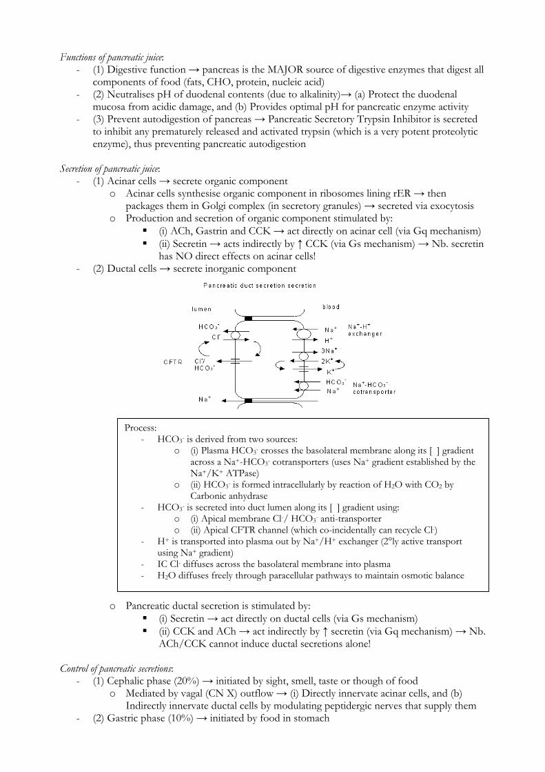
Process: - HCO3- is derived from two sources:
o (i) Plasma HCO3- crosses the basolateral membrane along its [ ] gradient across a Na+-HCO3- cotransporters (uses Na+ gradient established by the Na+/K+ ATPase)
o (ii) HCO3- is formed intracellularly by reaction of H2O with CO2 by Carbonic anhydrase
- HCO3- is secreted into duct lumen along its [ ] gradient using: o (i) Apical membrane Cl-/ HCO3- anti-transporter o (ii) Apical CFTR channel (which co-incidentally can recycle Cl-)
- H+ is transported into plasma out by Na+/H+ exchanger (2°ly active transport using Na+ gradient)
- IC Cl- diffuses across the basolateral membrane into plasma - H2O diffuses freely through paracellular pathways to maintain osmotic balance

o (i) Gastric distension – Vagovagal reflex → ↑ both acinar and ductal cell secretions o (ii) Peptides/a.a. in stomach – ↑ gastrin release (G-cells) → ↑ acinar cell secretions
- (3) Intestinal phase (70%) → initiated by chyme in duodenum o (i) Entry of acidic chyme in upper small intestines (MAIN stimulant for pancreatic
secretions) → ↑ secretin (from S-cells) stimulates ductal cell secretion, and ↑ CCK (from I-cells) stimulates acinar cell secretion
o (ii) A.a/FFA/MAG → stimulate CCK release → acinar cell secretion o (iii) Vagal input (via ACh) further potentiates these effects
(IV) Biliary secretion: Volume and composition of biliary secretion:
- Liver produces 1 L of bile/day → secreted into gallbladder (via bile canaliculi → common hepatic duct → cystic duct) where it is concentrated and stored
- Gallbladder contracts in response to a fatty meal (via neuroendocrine reflex) → secretes 100-200 mL of concentrated bile/day into duodenum
Function of bile:
- (1) Enhancing digestion and absorption of lipids (including fat-soluble vitamins) → by (i) emulsifying lipids and (ii) forming micelles using “bile salts”
- (2) Excretion of:
Bile consists of: - (1) “Bile-independent” fraction (97%)
o Produced by epithelial cells lining intra- and extrahepatic bile ducts o Consists of H2O and electrolytes → iso-osmotic to plasma (Na+ 145, Cl- 100,
K+ 5, Ca2+ 2), and alkaline with a pH ~ 8 (HCO3- 30) - (2) “Bile-dependent” fraction (3%)
o Produced by hepatocytes o Consists of → Primary and secondary bile salts (1%), Bilirubin (0.04%),
Cholesterol and phospholipids (0.1%), and Fatty acids (0.12%)
Gallbladder has 4 key functions: - (1) Concentrate bile (from 1L → 100-200 mL):
o It absorbs H2O, thereby ↑↑↑ [ ] of non-absorbable solutes (Eg. bile salts, bilirubin, Etc.) by 5-6 X → this ↑ potency of bile acids → intensifies digestion/absorption of lipids in small intestines
- (2) Storage of bile → gallbladder stores 50 mL of concentrated bile - (3) ↓ alkalinity of bile (pH 8 → 7.5) → due to HCO3- reabsorption during
concentration process - (4) Secretes mucus in bile → aids intestinal motility of chyme
Content Original [ ] Post-concentrate [ ] H2O 97g/100mL (97%) 92g/100mL (92%) Bile salts 1g/100mL (1%) 6g/100mL (6%) Bilirubin 0.04g/100mL (0.04%) 0.3g/100mL (0.3%) Cholesterol 0.1g/100mL (0.1%) 0.3g-0.9g/100mL (0.3-0.9%) Fatty acids 0.12g/100mL (0.12%) 0.3-1.2g/100mL (0.3-1.2%) Na+ 145 mmol/L 130 mmol/L Cl- 100 mmol/L 25 mmol/L Ca2+ 2 mmol/L 20 mmol/L K+ 5 mmol/L 12 mmol/L HCO3- 30 mmol/L 10 mmol/L
Note: Despite these functions, it is an organ that is NOT essential for life – with a cholecystectomy, bile flows directly into the intestine (hence, fat can still be digested and absorbed) → BUT avoidance of highly fatty foods is a must!

o (a) Cholesterol (Nb. This is its only route of excretion!) o (b) Bilirubin (breakdown of haemproteins) o (c) Xenobiotics and other endogenous compounds
- (3) Neutralises pH of acidic gastric chyme → allows intestinal digestive enzymes to function - (4) Endogenous intestinal lubricant & laxative → using mucus and bile salts - (5) Natural immunity → bile acts as a detergent - (6) Prevents formation of gallstones → using bile salts - (7) Choleretic action → bile salts in bile stimulate the hepatocytes to produce MORE bile
(via Bile-dependent biliary secretion) Regulation of biliary secretion:
- Regulation of bile secretion from the liver: o (a) Bile-independent fraction of biliary secretion is ↑ by:
(i) Secretin (from S-cells) → in response to FAs, a.a./peptides, bile and an acidic pH in the duodenum
(ii) Vagal reflex outflow → induced during “Intestinal phase” of digestion o (b) Bile-dependent fraction of biliary secretion → proportional to amount of bile
salts secreted by liver (which is proportional to the amount reabsorbed through the enterohepatic circulation) → thus, the rate is highest during the digestive phase
- Regulation of bile secretion from the gallbladder: o Gallbladder contraction and sphincter of Oddi relaxation causes release of
concentrated bile into duodenum → this occurs in response to: (i) CCK (from I-cells) → in response to presence of A.a’s/peptides and FAs
in the upper small intestines (ii) Vagal reflex outflow → induced during “Cephalic phase” and “Gastric
phase” of digestion (V) Intestinal secretion:
- (1) Small intestines → secrete 2 L/day o Crypts of Lieberkuhn (located between intestinal villi)
(i) Goblet cells → secrete alkaline (HCO3--rich) mucous → protects mucosal
epithelium and lubricate chyme (ii) Crypt enterocytes → secrete H2O and electrolytes (similar composition
to plasma but alkaline – pH 7.5-8) → acts as solvent for products of digestion to be absorbed into villus enterocytes
(iii) Enterocyte brush borders → secrete enzymes to finalise digestion of carbohydrates (Eg. isomaltase, maltase, sucrase, lactase), proteins (enterokinase, various endo- and exopeptidases) and fats prior to absorption
o Brunner’s glands (located in wall of duodenum) → secrete alkaline (HCO3--rich)
mucous → protects intestinal mucosa form acidic gastric juice o Regulation of secretion:
(i) Local intestinal stimuli – Local myenteric reflexes induce intestinal secretions following (a) Mechanical distension of mucosa by food, and/or (b) Chemical irritation from substances in chyme
(ii) Entero-endocrine control – (a) VIP (↑ secretions), (b) Secretin (↑ mucous secretions from Brunner’s glands)
(iii) Vagal (CN X) stimulation – ↑ mucous secretions from Brunner’s gland - (2) Large intestines → secrete 1 L/day
o Enterocytes within Crypts of Lieberkuhn → produce HCO3--rich mucous secretion
(alkaline – pH 8) o Functions – (i) Lubricate faecal matter, and (ii) Neutralise acids formed by bacterial
activity on faecal matter o Regulation – (i) Local tactile stimuli, (ii) Local enteric NS reflexes, (iii) PNS reflexes
(via pelvic nerves)

(c) To outline basic aspects of fat, protein and carbohydrate digestion and absorption. (I) Carbohydrate Digestion and Absorption: Digestion of carbohydrates:
- (1) Luminal digestion via → Salivary and Pancreatic Amylase o Initiate digestion of starch and glycogen → hydrolyse linear chains of glucose > 6
residues long linked by alpha-1,4 bond → produces: (a) Maltose (glucose-α-1,4-glucose) (b) Maltotriose (glucose-α-1,4-glucose-α-1,4-glucose) (c) Alpha-limit dextrins (glucose-α-1,6-glucose)
o Pancreatic amylase is more important than salivary amylase → b/c (i) salivary amylase is inactivated by low gastric pH and (ii) food remains in mouth very briefly
- (2) Intestinal brush border digestion via: o (a) Maltase – Cleaves (i) maltose → into two glucose molecules, and (ii) maltotriose
→ into three glucose molecules o (b) Isomaltase – Cleaves alpha-limit dextrins → into two glucose molecules o (c) Sucrase – Cleaves sucrose (glucose-α-1,2-fructose) → into fructose and glucose o (d) Lactase – Cleaves lactose (glucose-β-1,4-galactose) → into glucose and galactose
Absorption of carbohydrates:
- CHO are absorbed only as monosaccharides by mucosal cells in small intestinal brush borders (esp duodenum > jejunum > ileum):
o (1) Glucose (80% of CHO absorbed) (i) Passive diffusion (80%) → glucose is absorbed along its [ ] gradient via
transcellular or paracellular routes (ii) 2° active transport (20%) → Low IC [Na+] established by basolateral
Na+/K+ ATPase (consumes ATP) facilitates glucose absorption via co-transport with Na+ using apical SGLT-1 (Na+-Glucose linked transporter) → glucose is absorbed irrespective of its [ ] gradient across the membrane
o (2) Galactose (5% of CHO absorbed) → absorbed using SGLT-1 transporter (in similar manner to glucose)
o (3) Fructose (15% of CHO absorbed) → absorbed passively along its [ ] gradient via facilitated diffusion across apical GLUT-5 transporter
- All these monosaccharides are transported across the basolateral membrane passively down their [ ] gradients into blood by GLUT-2 transporter → travel to liver via the portal vein
Dietary carbohydrate
lactose maltose sucrosemaltotriose -dextrin
lactase maltase maltase(glucoamylase)
isomaltase(-dextrinase)
sucrase
glucose glucose glucose fructose
amylase
Brush-border surface
Intestinal lumen Starch, glycogen
Luminal digestion
Membrane digestion
Carbohydrates digested by GI tract include: - (1) Starch → branched poly-glucose with α-1,4 and α-1,6 linkages found in plants - (2) Glycogen → branched poly-glucose found in animals - (3) Oligosaccharides, which includes disaccharides (Eg. lactose, sucrose) - Note – Cellulose (plant polysaccharide) is NOT digested in the human GI tract
Nb. Amylase cannot act on α-1,6 bonds, terminal α-1,4 bonds or α-1,4 bonds next to a α-1,6 bond

(II) Protein Digestion and Absorption: Digestion of proteins:
- (1) Luminal digestion via:
o (a) Gastric digestion (minor): 10-15% of ingested proteins digested by Pepsin (endopeptidase) → produce
breakdown products that play a vital role in feedfoward secretion of pancreatic proteases (via entero-endocrine system)
Note – Pepsin is secreted inactive as Pepsinogen (by gastric chief cells) and activated by a gastric pH < 3
o (b) Pancreatic digestion (major): Pancreatic proteolytic enzymes are secreted as Zymogens (pro-enzymes) →
subsequently activated by Trypsin in duodenum Trypsin is activated when Trypsinogen is cleaved by → (i) Intestinal brush
border Enteropeptidase (or kinase) or by (ii) Autocatalysis by Trypsin The zymogens include:
(i) Exopeptidases (Carboxypeptidase) → produce free amino acids (ii) Endopeptidases (Trypsin, Chymotrypsin, Elastases) → produce
oligopeptides (including dipeptides and tripeptides) - (2) Intestinal brush border digestion:
o Intestinal brush border enzymes (Endopeptidases, Aminopeptidases, Carboxypeptidases, and Dipeptidases) digest oligopeptides formed by luminal digestion → into amino acids, di- and tripeptides
- (3) Cellular digestion: o Di- and tripeptides are transported into enterocytes → broken down into amino
acids by Dipeptidases and Tripeptidases
Na+
Na+
Fructose
Galactose
Glucose
K+
ATP
Lumen Interstitial
SGLT1
GLUT5GLUT2
GLUT2
H2O
Na+
Na+
Na+
Na+
Na+
Note – All these transporters are NOT insulin sensitive (cf. muscle and fat)
Proteins digested by GI tract include: - (1) Endogenous proteins (Eg. secretory proteins, desquamated cells) - (2) Exogenous proteins (Eg. dietary)

Absorption of proteins:
- Proteins are absorbed by mucosal cells in small intestinal brush borders (esp duodenum > jejunum > ileum) as follows:
o (1) Amino acids → via several apical membrane transporters (each differs in their specificity for an a.a. (Eg. acidic, dipolar, basic, imino a.a.’s) or whether they co-transport a.a’s with an ion (Eg. Na+))
o (2) Small peptides (Di- and Tri-peptides) → transported by a variety of 2ºly active transporters that cotransport them with H+ (namely PepT1) → H+ gradient is generated by a BLM Na+/K+ ATPase and an apical Na+/H+ coexchanger
o (3) Whole proteins → transported via phagocytosis → degraded intracellularly - Basolateral membrane contains > 5 transport systems that transport a.a. into portal vein:
o (1) Two systems are Na+-dependent → ensure a.a. are absorbed (via 2°ly active transport) during inter-digestive periods
o (2) Others are Na+-independent – A.a’s are transported passively across the basolateral membrane via facilitated diffusion along their [ ] gradients
(III) Fat Digestion and Absorption: Digestion of fats:
proteins
amino acids (AA) Dipeptides (AA)2
Tripeptides (AA)3
Oligopeptides (AA)4
AA (AA)2 (AA)3
Brush-border peptidases
Pepsin, Trypsin, Chymotrypsin, ElastaseCarboxypeptidase A & B
Brush-border surface
AA (AA)2
Cell
dipeptidases
AA AA
(AA)3
tripeptidases
Luminal digestion
Membrane digestion
Cellular digestion
Blood
peptide
Lumen Interstitial
H+
Na+
K+ATP
peptidases
H+
Na+
Na+
AA
Na+
H2O
Fats digested in GI tract include: - (i) TAGs (90%), (ii) CEs, (iii) Phospholipids, and (iv) Fat-soluble vitamins - These can be – (i) Exogenous (85%), or (ii) Endogenous (derived from fats within bile or
membranes of desquamated enterocytes)

- (1) Lingual and Gastric Lipases (minor): o Digest 15% of dietary TAGs to produce a single FA and DAG → these enzymes
are then inactivated in the small bowel by pancreatic proteases - (2) Intestinal digestion of lipids (MAJOR):
o Bile salts produced by the liver are important for lipid digestion as they – (i) Emulsify lipids (Ie. break them down), (ii) Solubilise lipids in “Micelles”
o “Micelles” are spherical aggregates of lipid molecules (FFA, 2-MAG, cholesterol and fat-soluble vitamins on inside) and bile salts (on outside) that enables – (i) Digestive enzymes to function, and (ii) Absorption of lipids
o Within these micelles: (a) Pancreatic Lipase – Breaks down TAGs into 2-MAG and 2 FAs.
Colipase cofactor is essential for this to occur (b) Pancreatic Cholesterol Esterase – Cleaves FAs from CEs (c) Phospholipase A-2 – Breaks down FAs form phospholipids
Absorption of fats: - Fats are absorbed by mucosal cells in small intestinal brush borders (esp duodenum >
jejunum > ileum) as follows: o (1) Short and medium-chain FAs (C6-C12) and glycerol → absorbed without
incorporation into micelle (due to their ↑ H2O solubility) → then transported across directly into portal venous circulation without incorporation into chylomicron
o (2) Long-chain FAs, cholesterol, MAGs, lysophospholipids and fat-soluble vitamins are absorbed within a “micelle” (due to their ↓ H2O solubility) → within enterocyte: MAGs, cholesterol and lysophospholipids are reesterified with FFAs (within
sER) to form TAGs, CEs and phospholipids → incorporated into “Chylomicron” (consists of a phospholipids monolayer with TAGs, CEs and fat-soluble vitamins in interior) → then diffuses across the basolateral membrane into “Lacteals”
Bile salts are recycled into portal vein (Nb. small amounts are reabsorbed by Na+-dependent active transporter in terminal ileum as part of “enterohepatic circulation”)
lipases
Surface emulsifiers(phospholipids and proteins)
Surface emulsifiers(phospholipids and proteins)
Bile saltsBile salts
TriglyceridesCholesteryl esters
colipases
Chlo
MG
Lyso
FFA (l)
Lumen
Bile salt
Bile salt
Bile
saltBile
sal
t
Micelle
Chlo + FFA
MG + FFA
Lyso + FFA
FFA
ChloE
TG
PL
Epithelial cell of small intestine
Chylomicron
ChloETG
PL
PL
PL
PL
PL
ApoB
Lymphexocytosis
FFA (s,m), glycerol blood

(IV) Water and Electrolyte Absorption:
- Absorption of H2O: o Most H2O is reabsorbed within the intestines → 7-8 L/day in small intestines and
1.4 L/day in large intestines → hence, only 100-150 mL is lost in faeces o H2O is reabsorbed passively via iso-osmotic movement 2° to active reabsorption of
electrolytes and nutrients → creates osmotic gradient favouring H2O reabsorption - Absorption of electrolytes:
o Small intestine – Electrolytes are absorbed via passive diffusion (Ie. [ ] gradient across enterocyte membrane), EXCEPT for: (1) Na+ → absorbed via 2° active transport using apical (i) Na+/Cl- co-
transporters (main), (ii) Na+ channels, (iii) glucose/a.a.-linked co-transporters → require Na+ gradient generated by basal Na+/K+ ATPase
(2) Cl- → absorbed via 2° active transport using apical Na+/Cl- co-transporters (require Na+ gradient generated by basal Na+/K+ ATPase)
(3) Ca2+ → absorbed in proximal small intestine via “carrier-mediated transport” (regulated by vitamin D)
o Large intestine Colonic epithelium actively absorb Na+ and Cl-, and passively absorb K+ It can also secrete K+ and HCO3
- (V) Vitamin Absorption:
- Fat-soluble vitamins (ADEK) → absorbed with dietary lipids in micelles (requires bile salts) - Water-soluble vitamins (EXCEPT vitamin B12) → absorbed via passive diffusion along [ ]
gradients (VI) Absorption of Iron:
Important to note → Absorption of vitamin B12: - (i) Vitamin B12 is released from food by acid and pepsin in the stomach → then
binds to (and protected by) R-proteins that is present in saliva - (ii) In duodenum, pancreatic enzymes (Eg. trypsin) hydrolyse the R-proteins causing
release of Vitamin B12 → it then preferentially binds to “Intrinsic Factor” (a glycoprotein secreted by gastric parietal cells)
- (iii) The IF-Vitamin B12 complex binds to “Cubilin receptors” in the terminal ileum, where it is absorbed via “Receptor-mediated endocytosis”
Each day, the GI tract handles 8 to 9.5 L of fluids. This includes: - (1) Intake of water (2-2.5 L/day) - (2) GI secretions (6-7 L/day) → saliva (0.5-1.5 L/day), stomach (2-2.5 L/day),
gallbladder (200 mL/day), pancreas (1.5 L/day) and intestines (3 L/day)
Note – Aldosterone regulates colonic epithelial absorption of Na+/Cl- and secretion of K+
Dietary Fe intake occurs in two pools: - (1) Haeme-Fe Pool
o Haem-proteins (Hb and Mb from meat) contains “ferrous” (Fe2+) iron o Have a “higher” iron content because Fe2+ is easily released from these ligands
and remains soluble at an alkaline duodenal pH → readily absorbed - (2) Nonhaeme-Fe Pool
o Ferric-protein and ferric hydroxide (Eg. vegetables, eggs, etc.) contain “ferric” (Fe3+) iron
o Have a “lower” iron content because Fe3+ precipitates in the alkaline duodenal pH → not readily absorbed

Absorption of iron: - Dietary iron is deconjugated from food by gastric enzymes and acid → then absorbed in
upper small intestines (mainly duodenum > jejunum) as follows: o (1) Iron is mainly absorbed in ferrous (Fe2+) state → via Divalent Metal Transporter
(DMT-1) on apical side of the enterocyte o (2) Iron in non-ferrous states are absorbed by the following means:
(a) Ferric iron (Fe3+) absorption (i) Ferrireductase (apical membrane enzyme) reduces Fe3+ to Fe2+ →
allows iron to be absorbed via DMT-1 (ii) Consuming reducing agents (ascorbic acid), chelators or ↓ gastric
pH enhances reduction of Fe3+ to Fe2+ → allows iron to be absorbed via DMT-1
(b) Haem absorption Hb and Mb are degraded to released haem → absorbed by
enterocyte via “receptor-mediated endocytosis” → haem is then broken down intracellularly to release ferrous iron (Fe2+)
- Iron within the enterocyte has two fates: o (a) Normally – Ferroportin (at basolateral membrane) transfers Fe2+ out of
enterocyte → then Ferrioxidase oxidises it to Fe3+ → then binds Transferrin within portal venous system
o (b) In event of excess iron load within enterocyte → ↑ Apoferritin production → causes ↑ iron-apoferritin complex formed (“Ferritin”) → stored within enterocyte
Aside: Regulation of iron absorption
- Body iron content is controlled SOLELY by regulating its absorption (Nb. its excretion cannot be regulated b/c it is heavily protein-bound)
- Each day, 10-15 mg of dietary iron is consumed BUT very little is absorbed in the GI tract (5-10%) → Absorption can increase by 30% during pregnancy or iron-deficient states
- Iron absorption is regulated via “Mucosal block” mechanism: o (i) When iron body stores are ↓ – Plasma transferrin levels are ↑ and their saturation is
↓ → causes transfer of iron from enterocyte ferritin stores into serum transferrin o (ii) When iron body stores are adequate or ↑ – Serum transferrin is relatively saturated
→ so minimal transfer of iron from the enterocyte to transferrin → but with continual iron absorption into enterocyte, the excess iron is then stored in enterocyte as ferritin, which is subsequently lost from the body when the cell dies

(d) To describe the control of gastric motility and emptying. (I) Functions of the Stomach:
(1) Storage of food Stomach can ↑ capacity from 1.5 L to 3 L via “Accommodation” (proximal stomach relaxes via vagovagal reflex triggered by food in fundus)
(2) Mixing and digestion of food into chyme
(i) Mechanical digestion → food broken down in distal stomach by back-and-forth processes of “Peristalsis” (antral contents slammed against pylorus) and “Retropulsion” (contents then pushed back into body) (ii) Chemical digestion (small amounts) → protein (by HCl/pepsin), CHO (by gastric amylase), fat (via gastric lipase)
(3) Emptying of chyme into intestines
Chyme is emptied from stomach into duodenum at a rate appropriate (as controlled via “Gastric emptying”) for intestinal digestion/absorption
(4) Antimicrobial protection Gastric HCl (pH 1.0) eliminates most organisms (5) Nutrient absorption (i) Intrinsic factor secreted → allows vitamin B12 to be absorbed in
terminal ileum (ii) Acidic gastric juice → allows Fe absorption in small intestine (iii) Small amounts of nutrients are absorbed in stomach (esp lipophilic substansces, such as EtOH)
(II) Gastric Motility:
- (1) Storage of food:
o Stomach can ↑ capacity from 1.5 L (normally) → ↑ to 3 L due to relaxation of proximal stomach (fundus)
o This process occurs in two phases: (a) Receptive accommodation – Food entering the oesophagus causes feed-
forward relaxation of the fundus (b) True accommodation – Food entering the fundus directly inhibits the
fundus, causing it to relax o Mechanism:
(i) Stretch receptors in fundus and oesophagus are stimulated by presence of food (causing luminal distension) → initiates vago-vagal reflex
(ii) Efferent vagus nerve innervates intrinsic enteric NS of fundus → VIP and NO released by enteric NS fibres → cause fundal relaxation
- (2) Mechanical digestion of food into chyme: o This is achieved by back-and-forth movements of gastric contents against the
pylorus generated by: (a) Peristalsis
Bands of peristaltic contractions travel from proximal stomach (fundus and body) to distal stomach (pylorus) → contractions are strongest distally where muscle layers are thickest → causes food to be “slammed” against pylorus
Aside: - Stomach has 3 functional parts – (i) Fundus, (ii) Body, and (iii) Antrum → forms thickened
circular SM at junction with duodenum (Pyloric sphincter) - Stomach has 3 muscle layer → each layer forms functional syncytium acting as a unit - Innervation:
o Extrinsic NS – (i) SNS (via coeliac plexus) → ↓ motility, (ii) PNS (via vagus) → ↑ motility
o Intrinsic NS – Submucosal (Meissner’s) and myenteric (Auerbach’s) plexus located between circular and longitudinal muscles of stomach → cause peristalsis and other gastric contractions

Generated by “pacemaker cells” within longitudinal muscle in greater curvature of stomach → produce ∆s in “slow wave” membrane potentials (consisting of upstroke and plateau phases) → causes 3 waves/min
Control – (i) PNS (Vagal) activity – ↑ force and frequency of contractions; (ii) Gastrin – ↑ force of contractions
(b) Retropulsion When food enters the pylorus, forceful antral contractions cause
retrograde movement of food particles through the antral ring back to the body of stomach for further mechanical digestion
(III) Gastric Emptying: Overview of gastric emptying:
- Gastric emptying (GE) involves coordinated emptying of chyme from the stomach into duodenum → this is driven by pressure generated by the antrum (via forceful waves of peristaltic contractions) against resistance of the pyloric sphincter
- GE rate is under neural and hormonal control by cephalic, gastric and duodenal factors → they mainly regulate antral pump activity (minimal regulation of pyloric resistance)
o (i) Neural control – Local enteric NS, vagal nerve, prevertebral SNS o (ii) Hormonal control – CCK, GIP, secretin, motilin
- Regulation of GE ensures → adequate chemical and mechanical digestion of gastric contents, before it is passed as chyme into duodenum for further digestion
- Normal GE is ~ 2-3 hrs → varies according to factors mentioned below Factors influencing gastric emptying:
- During “digestive period” → GE rate is determined by: o (1) Cephalic factors (thought, sight, smell of food) → variable effect on GE o (2) Gastric factors (presence of food in stomach) → generally ↑ GE
Content and volume of food entering stomach is sensed → +ve feedback on GE → cause ↑ antral pump activity and ↓ pyloric resistance
o (3) Duodenal factors (presence of chyme in small intestine) → generally ↓ GE (more potent control cf. others) Content and volume of chyme entering duodenum is sensed → –ve
feedback on GE via inhibitory enterogastric neurohormonal reflexes → cause ↓ antral pump activity and ↑ pyloric resistance
This ensures adequate digestion of gastric contents - During “interdigestive period” → GE rate is controlled by “Migrating motor complex”
(MMC) → food remnants in stomach are cleared by peristaltic waves the travel throughout the GIT every 60-90 mins
Factors Effect on GE rate Mechanism Cephalic factors Thought, sight, smell of food
↑ GE rate ↑ antral pump activity 2° to ↑ vagal activity
Pain, anxiety, fear ↓ GE rate ↓ antral pump activity 2° to ↓ vagal activity Gastric factors Consistency of chyme
Liquids → ↑ GE rate Solids → ↓ GE rate
Small particles and liquids → ↑ passage across resistance of pyloric sphincter
Protein content of ↑ content → ↑ GE rate Gastric mucosal chemoreceptors sense ↑ protein
Note → liquid chyme containing small particles are able to be pumped through pyloric sphincter → solid chyme as large particles cannot, and thus remain in stomach for further digestion

chyme content → ↑ gastrin release → ↑ antral pump activity Gastric volume ↑ volume → ↑ GE rate Gastric distension stimulates gastric mucosal stretch
receptors → ↑ gastrin release and excitatory vago-vagal reflex → ↑ antral pump activity
Duodenal factors Content of chyme CHO (↑ GE rate) >
protein > fat Duodenal chemoreceptors sense chyme content → duodenal CCK release (with ↑ fat/protein) and GIP release (with ↑ CHO) → inhibit effects of gastrin → ↓ antral pump activity
Acidity of chyme ↓ pH → ↓ GE rate Duodenal chemoreceptors sense chyme acidity → duodenal secretin release directly inhibits gastric SM → ↓ antral pump activity
Osmolarity of chyme
Isoosmolar → ↑ GE rate Hypo- or hyperosmolar → ↓ GE rate
Duodenal osmoreceptors sense chyme osmolarity → influence antral pump activity
Volume of chyme ↑ volume → ↓ GE rate Duodenal distension stimulates duodenal mucosal stretch receptors → vago-vagal reflex and GIP release → ↓ antral pump activity
Other factors MMC ↑ MMC → ↑ GE rate Motilin released by SB epithelium ↑ strength of MMC
→ ↑ antral pump activity

(e) To describe the physiology of swallowing and vomiting. (I) Mastication:
- “Mastication” (or chewing) is a process where food is broken down within the oral cavity into smaller particles and mixed with saliva
- Function – (i) Forms a softened food bolus to aid swallowing, and (ii) Initial phase of food digestion (as mechanical digestion)
- It is usually controlled subconsciously via a reflex mechanism involving the trigeminal mesencephalic nucleus → BUT can be influenced cortically also:
o (i) Food bolus in mouth causes reflex inhibition of masticatory muscles → causes lower jaw to drop
o (ii) This initiates a stretch reflex of jaw muscles → results in rebound contraction that raises lower jaw to compress teeth
o (ii) Tongue and cheek muscles keep food bolus between teeth as this process repeats itself
(II) Swallowing: Overview of swallowing:
- “Swallowing” is a complex reflex that transfers food from the oral cavity to the stomach Phases of swallowing:
(1) Oral phase Role (i) Initiate swallowing reflex
(ii) Pass food bolus from mouth into oropharaynx Control Voluntary (controlled by cerebral cortex) Process (i) Mastication of food in mouth
(ii) Food bolus then formed by tongue → passed into oropharynx by pushing up and against hard palate
(2) Pharyngeal phase Role Pass food bolus from oropharynx into upper oesophagus Control - Involuntary reflex (controlled by “swallowing centre” → medulla and pons)
- Triggered by food in pharynx → stimulates pharyngeal mechanoreceptors → afferent signal via CN V, IX and X to NTS/nucleus ambiguus → then to “swallowing centre” → efferent signals via CN V, VII, X and XII
Process (i) When food enters oropharynx → posterior faucal pillars approximate to shut oral cavity off from the pharynx → prevent regurgitation back into mouth
(ii) Soft palate then moves upwards to close off nasopharynx → prevent regurgitation into nasal cavities
(iii) To prevent tracheal aspiration of food bolus → (a) respiration is inhibited for 1-2 secs, (b) laryngeal inlet is closed by adduction of vocal cord and aryepiglottic muscle, and (c) larynx raised and epiglottis swings down to close off larynx
(iv) Pharyngeal contraction and UOS relaxation → pushes food bolus into upper oesophagus via peristalsis
(3) Oesophageal phase Role Pass food bolus from upper oesophagus into stomach Control - Involuntary reflex (controlled by “swallowing centre” → medulla and pons)
- Trigger same as pharyngeal phase → EXCEPT “2° peristalsis” phase is mediated via local enteric NS reflex (food within oesophagus causes distension → activates stretch receptors)
Process (i) Food enters oesophagus → causes (a) contraction of UOS (to prevent reflux back into pharynx) and (b) relaxation of LOS (to allow food to enter stomach)
(ii) Peristaltic contractions then propel food towards the stomach:
- (a) “1° peristalsis” → slow peristaltic waves continue on from pharynx (2-4 cm/s at
Note: Peristalsis → reflex response initiated when luminal wall stretched by its contents → initiates contraction behind food bolus, and relaxation in front of it → produces wave of contraction that propels food from oral to caudal direction
20-60 mmHg) → initiated by CN X motor activity (from swallowing centre) - (b) “2° peristalsis” → initiated by food within oesophagus (Ie. when primary
peristalsis fails to move all food into stomach) → oesophageal distension triggers stretch receptors → activates local enteric NS reflex to produce further peristalsis to clears remaining food within oesophagus
(iii) Gravity promotes movement of food bolus towards stomach (assuming erect position) → fluids flow at a more RAPID than solids (and also faster than via peristaltic wave)
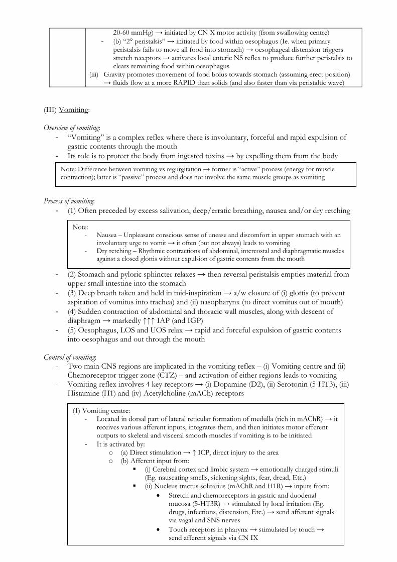
(III) Vomiting: Overview of vomiting:
- “Vomiting” is a complex reflex where there is involuntary, forceful and rapid expulsion of gastric contents through the mouth
- Its role is to protect the body from ingested toxins → by expelling them from the body Process of vomiting:
- (1) Often preceded by excess salivation, deep/erratic breathing, nausea and/or dry retching - (2) Stomach and pyloric sphincter relaxes → then reversal peristalsis empties material from
upper small intestine into the stomach - (3) Deep breath taken and held in mid-inspiration → a/w closure of (i) glottis (to prevent
aspiration of vomitus into trachea) and (ii) nasopharynx (to direct vomitus out of mouth) - (4) Sudden contraction of abdominal and thoracic wall muscles, along with descent of
diaphragm → markedly ↑↑↑ IAP (and IGP) - (5) Oesophagus, LOS and UOS relax → rapid and forceful expulsion of gastric contents
into oesophagus and out through the mouth Control of vomiting:
- Two main CNS regions are implicated in the vomiting reflex – (i) Vomiting centre and (ii) Chemoreceptor trigger zone (CTZ) – and activation of either regions leads to vomiting
- Vomiting reflex involves 4 key receptors → (i) Dopamine (D2), (ii) Serotonin (5-HT3), (iii) Histamine (H1) and (iv) Acetylcholine (mACh) receptors
Note: - Nausea – Unpleasant conscious sense of unease and discomfort in upper stomach with an
involuntary urge to vomit → it often (but not always) leads to vomiting - Dry retching – Rhythmic contractions of abdominal, intercostal and diaphragmatic muscles
against a closed glottis without expulsion of gastric contents from the mouth
Note: Difference between vomiting vs regurgitation → former is “active” process (energy for muscle contraction); latter is “passive” process and does not involve the same muscle groups as vomiting
(1) Vomiting centre: - Located in dorsal part of lateral reticular formation of medulla (rich in mAChR) → it
receives various afferent inputs, integrates them, and then initiates motor efferent outputs to skeletal and visceral smooth muscles if vomiting is to be initiated
- It is activated by: o (a) Direct stimulation → ↑ ICP, direct injury to the area o (b) Afferent input from:
(i) Cerebral cortex and limbic system → emotionally charged stimuli (Eg. nauseating smells, sickening sights, fear, dread, Etc.)
(ii) Nucleus tractus solitarius (mAChR and H1R) → inputs from: Stretch and chemoreceptors in gastric and duodenal
mucosa (5-HT3R) → stimulated by local irritation (Eg. drugs, infections, distension, Etc.) → send afferent signals via vagal and SNS nerves
Touch receptors in pharynx → stimulated by touch → send afferent signals via CN IX

(iii) Vestibular nucleus (H1R and mAChR) → from vestibular apparatus (via CN VIII) due to motion sickness/dizziness
(iv) CTZ– see below - In the event of vomiting, the “Vomiting centre” sends efferent outputs:
o (a) To the upper GI tract via CN V, VII, IX, X, and XII o (b) To muscles of the diaphragm and abdominal muscle via spinal nerves
(2) Chemoreceptor trigger zone (CTZ): - Located in area postrema → floor of 4th ventricle outside the BBB (as a
circumventricular organ) → rich in D2R and 5-HT3R - Activated by:
o (a) Circulating drugs or toxins in blood or CSF (Eg. cytotoxic agents, opioids, EtOH, uraemia)
o (b) Vestibular nucleus (H1R and mAChR), which receives input from vestibular apparatus (via CN VIII) due to motion sickness/dizziness
o (c) Stretch and chemoreceptors in gastric and duodenal mucosa (5-HT3R) → stimulated by local irritation (Eg. drugs, infections, distension, Etc.) → send afferent signals via vagal and SNS nerves

(f) To describe the consequences of prolonged vomiting, bowel obstruction and malabsorption syndromes.
(I) Consequences of Prolonged Vomiting:
- Gastric juice is an acidic fluid (pH 1-1.5) → rich in [H+] (150-170 mmol/L), Cl- (180 mmol/L), and K+ (10 mmol/L) → poor in Na+ (2-4 mmol/L)
- Prolonged vomiting of gastric juice (such as from pyloric stenosis) thus causes: o (1) Hypovolaemia → due to direct loss of fluid in vomitus o (2) Electrolyte imbalances (hyponatraemia, hypochloraemia and hypokalaemia) →
due to loss of gastric juice in vomitus o (3) Metabolic alkalosis → due to loss of gastric acid in vomitus o (4) Malnutrition → due to loss of nutrition
- Hypokalaemia and metabolic alkalosis are sustained inadvertently by compensatory mechanisms used to restore fluid and electrolyte balance:
o (1) RAAS is stimulated in response to hypovolaemia, thus causing net retention of Na+ (alleviating hyponatraemia) and H2O (alleviating hypovolaemia) → BUT aldosterone worsens hypokalaemia and metabolic alkalosis due to renal and GI wasting of H+ and K+
o (2) Renal reabsorption of Cl- to alleviate hypochloraemia leads to increased renal Na+ and HCO3
- reabsorption, thereby sustaining alkalosis o (3) Compensatory mechanisms to restore normokalaemia (transcellular K+ shifts,
renal and GI retention of K+) causes excretion or intracellular shift of H+, thus sustaining alkalosis
- Fluid and electrolyte therapy involves re-expansion of intravascular volume with IV NaCl supplemented with KCl → this treats hypovolaemia, corrects electrolyte imbalances, and circumvents compensatory mechanisms that cause persistent metabolic alkalosis
(II) Consequences of Bowel Obstruction:
- “Bowel obstruction” is the mechanical or functional obstruction of the intestines (either small or large) that prevents the normal transit of luminal contents → causes proximal loops of bowel to distend due to accumulation of air and fluid/secretions that cannot pass distally
- This results in severe dehydration, electrolyte imbalances, and acid-base disturbances: o (1) Bowel distension triggers stretch (mechanoreceptors) within intestinal luminal
wall, which induces nausea and vomiting (via the “vomiting reflex”) → causes: (a) Hypovolaemia (b) Hyponatraemia, hypochloraemia and hypokalaemia (c) Acid-base disturbance depends on the loss of alkaline small bowel
content and acidic gastric juice → there can be (i) no significant deviation in plasma pH (if loss of intestinal and gastric juices are balanced), (ii) metabolic alkalosis (if loss of gastric juice is excessive), or (iii) metabolic acidosis (if loss of alkaline intestinal contents is excessive)
o (2) Bowel distension triggers stretch receptors that increase intestinal secretions via a local reflex → causes further fluid and electrolyte loss (Na+, K+, Cl-) and metabolic acidosis (due to loss of HCO3
-) o (3) Bowel distension hampers the absorptive capacity of intestinal epithelium,
leading to reduced intestinal fluid, electrolyte and nutrient absorption → (i)
Note – Not all cases of prolonged vomiting cause alkalosis. In the absence of pyloric stenosis, most cases of persisting vomiting is associated with loss of alkaline small bowel content in addition to the loss of acidic gastric juice. Thus, there can be (i) no significant deviation in plasma pH (if loss of intestinal and gastric juices are balanced) or (ii) metabolic acidosis (if loss of alkaline intestinal contents exceeds gastric contents)

exacerbates dehydration, hyponatraemia, hypokalaemia, hypochloraemia and metabolic acidosis, and (ii) causes malnutrition
(III) Consequences of Malabsorption Syndromes:
- “Malabsorption” is a state arsing from abnormalities in absorbing nutrients across the GI tract. This can involve a single nutrient or multiple nutrients depending on the cause
- Malabsorption can be due to many pathological processes that involve: o (i) Digestion (Eg. pancreatic or biliary insufficiency due to fistulas) o (ii) Absorption (Eg. intestinal mucosal damage, acquired reduction in absorptive
surfaces, ion transport defects) o (iii) Transport of nutrients (Eg. impaired enterohepatic circulation)
- The physiological effects depend on the nutrient being affected: o (1) Diarrhoea
Generally an “osmotic” diarrhoea due to fat malabsorption (steatorrhoea), and also carbohydrate malabsorption (watery diarrhoea)
Exacerbated by malabsorption of fluids and electrolytes Fluid losses create dehydration and electrolyte disturbances
o (2) General weakness, weight loss, and failure to thrive Due to deficient absorption of amino acids, fats and carbohydrates and
reduced caloric intake o (3) Anaemia – Due to deficiencies in Vitamin B12 and folate (megaloblastic
anaemia) and iron (microcytic anaemia) o (4) Muscle cramps (due to hypocalcaemia) and pathological fractures (due to
osteomalacea/osteoporosis) from Vitamin D deficiency o (5) Coagulopathy due to Vitamin K deficiency and impaired CF production from
protein loss o (6) Generalised oedema due to loss of intravascular oncotic pressure from a protein-
deficient state o (7) Neurological disorders (ataxia, spinocerebellar disorders, dorsal column
disturbances) – Due to Vitamin B12 and Vitamin E deficiencies o (8) Poor night vision due to Vitamin A deficiencies o (9) Scurvy due to Vitamin C deficiencies o (10) Deficiencies in trace minerals (Eg. Zn, Cu)

(g) To explain the factors that prevent reflux of gastric contents into the oesophagus. Overview of lower oesophageal sphincter (LOS):
- A “physiological” sphincter that separates the distal oesophagus from the stomach → it consists of:
o (a) Internal sphincter → formed by: (i) Tonic contraction of circular smooth muscle fibres within lowest 2-4 cm
of oesophagus → controlled by excitatory vagal (cholinergic) fibres (ii) Oblique gastro-oesophageal angle (“flap-valve” mechanism) → gastric
mucosal folds are pushed up into (and close) the oesophagus with ↑ gastric tone (or pressure)
o (b) External sphincter → formed by constriction of distal oesophagus by diaphragmatic crura via coordinated breathing and coughing (“pinch-cock” mechanism) → controlled by the Phrenic nerve
- It functions to prevent reflux of gastric contents back into the oesophagus → this is b/c its resting pressure is usually 15-25 mmHg (20-30 cm H2O) ABOVE intragastric pressure
Factors determining gastro-oesophageal reflux:
- “Barrier pressure” of LOS determines whether gastric contents will reflux back into
oesophagus: o (1) Normally – LOS pressure is 35 cmH2O (20-30 mmHg) and IGP is 10 cmH2O
(7.5 mmHg) → so LOS barrier pressure is 25 cmH2O (or 15-25 mmHg), which is sufficient to prevent reflux
o (2) Reflux occurs when LOS barrier pressure is < 13 cmH2O (or < 10 mmHg) → this can occur due to either: (i) ↓ LOS pressure (Eg. hiatus hernia, bilateral vagotomy, progesterone in
pregnancy, DM neuropathy, phrenic nerve injury) (ii) ↑ intragastric pressure (Eg. obesity, pregnancy, post-prandially, head-
down position) Factors that influence LOS barrier pressure: Factor Effect on LOS barrier pressure
(i) Functional anatomy of LOS (internal and external sphincters)
- Intact functional anatomy → produce a resting LOS pressure of 35 cm H2O (20-30 mmHg) → maintains LOS barrier pressure at 25 cmH2O (15-25 mmHg) - Disrupted anatomy (Eg. hiatus hernia) → ↓ LOS tone and barrier pressure
Anatomical
(ii) Small portion of distal oesophagus is intra-abdominal
This portion is subject to ∆ in IAP (such that ↑ IAP → ↑ distal LOS pressure → ↑ LOS barrier pressure)
(i) Vagal (ACh) outflow (under the control of a ”medullary centre”)
- Contracts the ”internal sphincter” by ↑ circular SM tone → ↑ LOS pressure → ↑ LOS barrier pressure - Vagal nerve dysfunction (Eg. Bilateral vagotomy/DM neuropathy) → ↓ LOS tone and barrier pressure
Neural
(ii) Phrenic nerve stimulation - Contracts ”external sphincter” via ”pinch-cock” mechanism → ↑ LOS pressure → ↑ LOS barrier pressure - Phrenic nerve dysfunction (Eg. nerve injury, DM neuropathy) → ↓ LOS tone and barrier pressure
(i) Gastrin, CCK, motilin, α-adrenergic stimulation
↑ LOS pressure → ↑ LOS barrier pressure Hormonal
(ii) Secretin, VIP, GIP, glucagon, progesterone, and prostaglandins
↓ LOS pressure → ↓ LOS barrier pressure
IAP/IGP Obesity, pregnancy, post-prandially, ↑ IAP/IGP → ↓ LOS barrier pressure
“Barrier pressure” of LOS = (LOS pressure) – (Intragastric pressure)

head-down position Swallowing LOS undergoes a “relaxation-
contraction cycle” controlled by: - (a) Reflex mechanism
mediated by medulla → receive afferent input (gastric and oesophageal distenion by food) and coordinates efferent cholinergic vagal fibre output
- (b) Various hormonal factors
- (i) Upon swallowing, LOS begins to relax → when peristaltic waves arrive at LOS, it completely relaxes → permits passage of food bolus into stomach - (ii) After passage of food bolus → LOS actively contracts to 1-15 mmHg ABOVE resting tone for 10-15 secs before returning to resting level → prevents reflux of food back into oesophagus
(i) Metoclopramide, AChEi, α-adrenergic agents, histamine, SCh
↑ LOS pressure → ↑ LOS barrier pressure Drugs
(ii) Anti-muscarinic agents, ganglionic blockers, DA, EtOH, opioids, β-adrenergic agents
↓ LOS pressure → ↓ LOS barrier pressure
Aside: Upper oesophageal sphincter - Anatomical sphincter → consists of (i) cricopharyngeus and (ii) oesophageal circular SM - High resting UOS pressure of 50-100 mmHg → relaxes (and opens) on swallowing