gastrointestinal physiology - ju medicine€¦ · gastrointestinal physiology • the gi system is...
TRANSCRIPT


GASTROINTESTINAL PHYSIOLOGY
• The GI system is the largest system in the human body with multiple organs extending
from the oral cavity to the anus separated from each other by special muscles called
sphincters which normally stay tightly closed and which regulate the movement of food
and food residues from one part to another.
The system is like a tube, inside this tube is the interstitial fluid which is a part of external
environment not the internal environment.The four main physiological functions that
take place along the GI tract are:Motility, Secretion, Digestion,Absorption
specialized cells and tissues exist along the entire alimentary tract to enhance
performing these functions properly. For instance, there are special smooth muscles for
each organ to perform its distinctive type of motility. Also, interstitial cells, secretory
cells, etc. We will see how they are organized and so on.
The alimentary tube is composed of three main
layers: the most outer layer which is the muscular
one, the mucosal layer is the most inner one and
submucosa in between.
•although those are the general layers, there are
still variations in the anatomical and histological
structures for each organ.
•In addition, a very powerful neural control,
hormonal control and blood flow are found as
parts of the functional structure.
Two main systems are involved in the neural control:
1) The autonomic nervous system.
2) The enteric nervous system (ENS), which is found along the entire gut wall, starting from
the esophagus and extending all the way to the anus. (so, the GI tract has its own
nervous system).
-The ENS is not a part of the autonomic or the somatic nervous systems.

•with regard to the blood flow, its noteworthy that the blood flow in each area of the
gastrointestinal tract, as well as in each layer of the gut wall, is directly related to the level
of local activity. For example, the submucosa is highly vascularized along the entire tract,
while the vascularization of mucosa is specific to distinct parts (such as small intestine) due
to variations of levels functions as.
FIRSTLY, THE GASTROINTESTINAL SMOOTH MUSCLE CELLS:
Two parts of smooth muscle cells are distinguishable along the GI wall:
1) The longitudinal smooth muscles where the bundles extend, and those are the
outermost part.
2) The circular layer which extends around the gut (the fibers run around the circumflex of
the tube).
So, we can control the length and diameter of any part.
In addition, there is a very thin layer between the mucosa and the submucosa which is the
called muscularis mucosa.
• Gastrointestinal smooth muscles share some general structural characteristics with
others found in different places of human body like the presence of thick and thin
filaments attached to the dense bodies. But the GI smooth muscles function in a
different way from other smooth muscles in the body.
• One of the differences is the electrical control and the resting membrane potential.
Electrical activity of smooth muscle: Smooth muscle cells are characterized by the presence of slow waves (undulating changes in membrane potential known as basic electrical rhythm (BER)) and spike potentials
Most gastrointestinal contractions occur rhythmically, and this rhythm is determined mainly by the level of these “slow waves” of smooth muscle. Those are not action potentials. Instead, they are slow, fluctuating changes in the resting membrane potential.
True action potentials are formed automatically when the resting membrane potential of the gastrointestinal smooth muscle becomes more positive than about −40 millivolts . the normal resting membrane potential in the smooth muscle fibers of the gut is between −50 and −60 millivolts. These true APs are called the "spike potential that appear at the peak of slow waves.
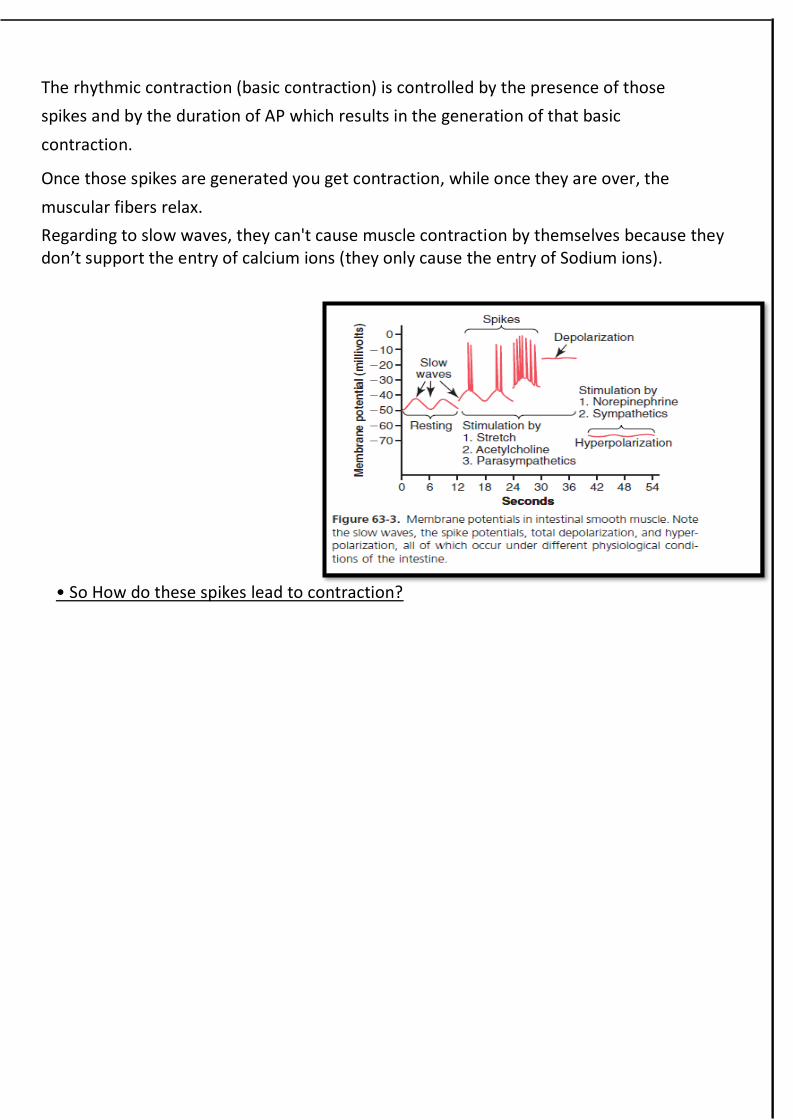
The rhythmic contraction (basic contraction) is controlled by the presence of those
spikes and by the duration of AP which results in the generation of that basic
contraction.
Once those spikes are generated you get contraction, while once they are over, the
muscular fibers relax.
Regarding to slow waves, they can't cause muscle contraction by themselves because they don’t support the entry of calcium ions (they only cause the entry of Sodium ions).
• So How do these spikes lead to contraction?

During the spike
potentials generated
at the peaks of the
slow waves,
significant quantities
of calcium ions enter
the smooth muscle
fibers which act
through a calmodulin
control mechanism,
which activates the
myosin filaments and actin filaments. thereby causing the muscle to contract. This is
called the" electrical control for smooth muscles activity ".
In additions, gap junctions between smooth muscles allow low-resistance movement of
ions from one muscle cell to the next. Therefore, electrical signals that initiate muscle
contractions can travel readily from one fiber to the next within each bundle gap
junctions also form syncytium; that is, when an action potential is elicited anywhere
within the muscle mass, it generally travels in all directions in the muscle.
In addition to "electric control" there is the chemical control, (follow the steps in the
graph).

But what type of control is achieved by chemical regulation? Importantly, there is no
"zero level of contraction", instead there is the tonic contraction which is continuous
and not associated with the basic electrical rhythm of the slow waves (it often lasts for
several minutes or even hours). Moreover, tonic contraction often increases or
decreases in intensity but it continues.
In anatomy, small intestine length is 6 meters while in physiology, they have a
difference opinion, they say it is 3 meters, why? Because of the tonic contraction which
make it shorter than in the relaxed state.
So, it’s the electrical control which regulates the rythmic contraction and it is the
chemical control which regulates the tonic contraction.
Summary of control for GI smooth muscle cells activity:
Smooth muscle cells activity is controlled by Electrical activity of smooth muscle cells: (slow waves and spike
potentials).
Neurochemical control: represented by the response of smooth muscle cells of the GI to a large number of transmitters that are released by many types of neurons in the ENS.
Another type of cells is interstitial cells of cajal.
They have lots of spikes joined together by gap junctions.
Slow waves appear to be caused by complex interactions between the smooth muscle
cells and interstitial cells of cajal.
These interstitial cells form a network with
each other and are interposed between the
smooth muscle layers (they are somehow
connected to smooth muscles by gap
junctions). For each 50-100 smooth muscles
there is one interstitial cell in between.
Those cells can generate action potential by
themselves.
Therefore, cajal cells are believed to act as electrical pacemakers for smooth muscle cells.
But functionally they differ from those found in the heart.
Some sources tell that these cells receive neural input, but this input has nothing to do
with its own ability to generate action potential. So how can they generate AP? Still not
clear, but one theory suggests that some metabolic changes inside the cells result in all of
a sudden generation of AP.

SECONDLY, SECRETORY CELLS:
As functional structures, we have different organizations for secretory cells starting from solitary cells (individual cells) up
to secretory organs located outside the tube. Examples:
Solitary cells dispersed in the mucosal epithelium.
Pits or simple glands in the mucosa too, a more complex organization is the compound gland in the submucosa, and
finally secretory organs outside the tube secreting in the lumen of the GI with the help of ducts. Examples are salivary
glands, pancreas and liver.
THIRDLY, CHARACTERISTICS OF ENTERIC NERVOUS SYSTEM:
A highly developed system to control gastrointestinal movements and secretions. The
number of neurons which are found along the GI tract is very huge, almost equal to the
number found in the spinal cord, so they call this system the brain of the gut.
The enteric nervous system is composed mainly of two plexuses:
(1) An outer plexus lying between the longitudinal and circular muscle layers, called the
myenteric plexus
(2) An inner plexus, called the submucosal plexus which lies within the submucosa.
The enteric nervous system can control smooth muscle cells, secretory cells, even some
endocrine cells and blood vessels.
The myenteric plexus controls mainly the gastrointestinal movements while the
submucosal plexus controls mainly gastrointestinal secretion and local blood flow.
It is noteworthy that extrinsic sympathetic and parasympathetic fibers connect to both
the myenteric and submucosal plexuses.
Neurotransmitters secreted by enteric neurons:
Enteric neurons could be inhibitory or excitatory.
Many different neurotransmitter substances are released by the nerve endings of
different types of enteric neurons, including: Ach, SP (Substance P), VIP (Vasoactive
intestinal peptide), CGRP (Calcitonin gene related peptide), GRP (Gastrin releasing
peptide)
*VIP relaxes smooth muscle cells in the walls of vessels so more blood flow.
Acetylcholine excites gastrointestinal activity.
Norepinephrine almost always inhibits gastrointestinal activity, as does epinephrine
The other aforementioned transmitter substances are a mixture of excitatory and
inhibitor
Although the enteric nervous system can function independently of extrinsic nervous
system, stimulation by the parasympathetic and sympathetic systems can greatly
enhance or inhibit gastrointestinal functions.

Note that the autonomic nervous system can affect the GI by affecting the enteric nervous
system or by acting directly over some parts only, for example, parasympathetic exerts
small effect on the blood vessel and indirectly, while the sympathetic does directly and to a
large extent.
HORMONAL CONTROL OF GASTROINTESTINAL MOTILITY:
Examples:
Gastrin
Cholecystokinin (CCK)
Secretin,
Glucose-dependent insulinotropic peptide (also called gastric inhibitory peptide
[GIP]) causes the release of insulin. So, when food start flowing to the GI, this
hormone get released to prepare the body for the glucose that will be soon in the
blood.
Other hormones are also secreted along GI tract, including: Glucagon-like
peptide-1(GLP-1), Motilin, Ghrelin, Amylin, Enterostatin, Neuropeptide Y (NPY),
and Pancreatic polypeptide which is closely related to polypeptide YY and NPY.
In addition, scattered endocrine cells releasing Somatostatin, Neurotensin.
*the doctor mentioned them very quickly by just reading them and pointed out that some of them still has an unknown
function and are still being under research. Also said that some of these have effects on blood flow, state of hunger...
Thyrotropin releasing hormone (TRH) released by the hypothalamus to cause the
release of TSH.
Adrenocorticotropic hormone (ACTH) which is involved in the ACTH axis for the
release of other hormone from the suprarenal gland, and also released by some cells
along the GI tract.
CONTROL OF BLOOD FLOW AS A FUNCTIONAL STRUCTURE:
The blood flow is very well controlled, sometimes we need an increase, sometime we
need a decrease in blood flow. How is it controlled?
1- Hormones like secretin, cholecystokinin.
2- The enteric nervous system; like VIP for example, substance P (constriction), CGRP
(constriction).
3- Vasodilators. These vasodilators are released from secretory cells in response to
parasympathetic stimulation for these secretory cells, so parasympathetic nervous
system indirectly affect blood flow.
4- Decreased oxygen concentration. When a tissue gets deprived or become on low
oxygen concentration, it releases adenosine which acts as a vasodilator.
5- Autonomic nervous system. Direct sympathetic effect decreases blood flow and
indirect parasympathetic increases blood flow.
Important note: the enteric nervous system is also called the intrinsic nervous system, while
the autonomic nervous system is also referred to as the extrinsic nervous system.
Summary of the control over the gastrointestinal system: There are 3 main effectors on the GI system: 1- The intrinsic nervous system.
2- The extrinsic nervous system.
3- Gastrointestinal endocrine system.
So now the question is how to change the activity of these 3 control systems, so by that we
control the GI system?
And the answer is either by external influences or local changes.
External influences: like smelling, seeing or hearing about food, then these external
influences affect the extrinsic nervous system.
Local changes: once you eat, distension of your stomach and the chemicals and the food
itself initiate some reflexes that activate any of the 3 effectors mentioned above.
Autonomic nervous system..
Parasympathetic nervous system: According to the location of neural cell bodies, it is
divided into:
Cranial division: provides innervations through vagus nerve to esophagus, stomach, pancreas,
small intestine and first half of large intestine.
Sacral division: Provides innervations through pelvic nerves to distal half of the colon,
sigmoidal, rectum and anal region. Fibers in this division have importance in executing
defecation reflex.
Generally, stimulation of parasympathetic system causes an increase in the activity of enteric
nervous system and consequently, enhances the activity of the gastrointestinal functions.
These include motility, secretion and blood flow.
Sympathetic nervous system: Sympathetic fibers that innervate gastro-intestinal tract
originate in the spinal cord (segments T5-L2). These fibers pass through paravertebral ganglia
and synapse with the second neuron in celiac, superior mesenteric or inferior mesenteric
ganglia. Generally, stimulation of sympathetic system causes a decrease in the activity of
enteric nervous system and GI smooth muscle cells.