gel electrophoresis by suresh b i
TRANSCRIPT

WELCOME

GEL ELECTROPHORESIS
Guided by: Ms. Ashwini D Dept. of Industrial chemistry
Presented by: Suresh B I

ELECTROPHORESIS
Electrophoresis is the migration of charged particle in an electric field towards the electrode bearing the opposite charge.
(OR) is a method where by charged molecules in solution, like
proteins and nucleic acids migrate in response to an
electric field
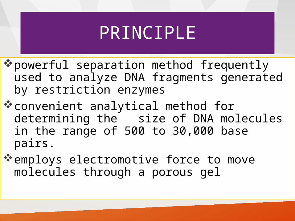
PRINCIPLE
powerful separation method frequently used to analyze DNA fragments generated by restriction enzymes
convenient analytical method for determining the size of DNA molecules in the range of 500 to 30,000 base pairs.
employs electromotive force to move molecules through a porous gel

separates molecules from each other on the basis of
>size and/or>charge and/or>shape
basis of separation depends on how the sample and gel are prepared


TYPES OF ELECTROPHORESIS
Based on supporting media (zone electrophoresis): Paper
electrophoresis gel electrophoresis Cellulose acetate
electrophoresis
Without supporting media (moving boundary method): Free electrophoresis
Supporting medias are : starch, Agar- Agarose, polyacrylamide ,

APPLICATIONS

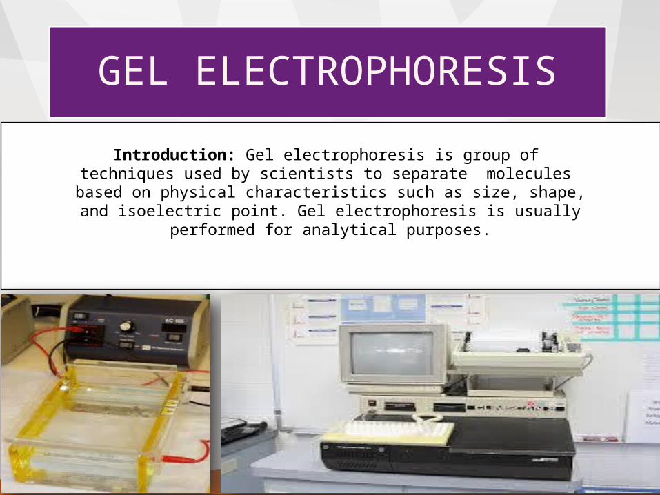
GEL ELECTROPHORESIS
Introduction: Gel electrophoresis is group of techniques used by scientists to separate molecules based on physical characteristics such as size, shape, and isoelectric point. Gel electrophoresis is usually
performed for analytical purposes.

APPICATION OF SAMPLE RUNNING OF SAMPLE VISUALISATION OF SAMPLE QUANTIFICATION
TECHNIQUES

REQUIREMENTS FOR GEL ELECTROPHORESIS
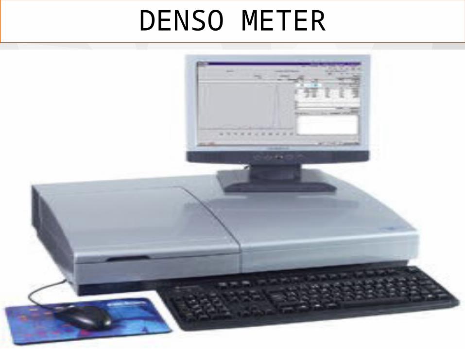
BufferFixativeStaining solutionDestaining solutionDensitometer – is essentially a double beam filter
photometer or spectrophotometer that scans the electrophoretic strip ( in the form of agarose , cellulose acetate or polyacrylamide) as it moves past the optical system.

What is Agarose ?A linear carbohydrate polymer extracted from seaweed , agarobiose
forms a porous matrix as it gelsshifts from random coil in solution to structure in which chains are bundled into double helices
DENSO METER

PROCEDURE1. Sample to be separated is applied to a supporting medium (paper, cellulose acetate, agar gel, polyacrylamide gel etc.) Electrophoresis is carried out at desired constant voltage or constant current in presence of specific pH.
3. After completion of electrophoresis the supporting medium is placed in a fixative to prevent diffusion of separated fractions.
4. Separated fraction is then visualized by using appropriate stains e.g. Bromophenol Blue & Amino Schwartz for plasma proteins and Sudan Black for lipoproteins.
5. Quantification of each fraction is done by either densitometer or elution followed by colorimeter or spectrophotometer of eluted fraction.
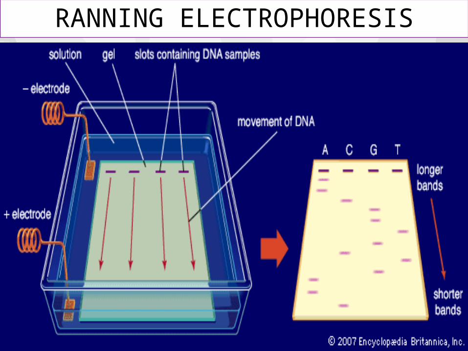
RANNING ELECTROPHORESIS

Gel electrophoresis is a process that separates fragments of DNA based on their sizes.

APPLICATION OF GEL ELECTROPHORESIS
Evidence in criminal cases To determine paternity To diagnose genetic diseases Help to determine kinship in animals Compare similarities and differences between
species Solve paternity casesDetermine genetic kinship among species
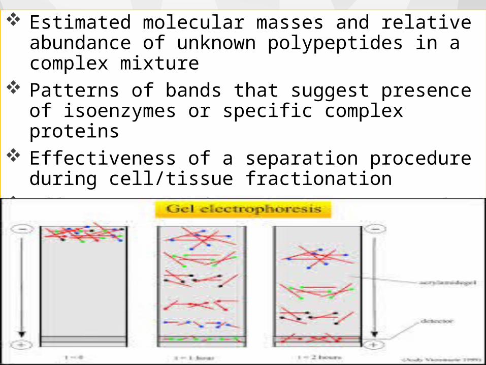
Estimated molecular masses and relative abundance of unknown polypeptides in a complex mixture
Patterns of bands that suggest presence of isoenzymes or specific complex proteins
Effectiveness of a separation procedure during cell/tissue fractionation
Effectiveness of a procedure to purify specific organelles, proteins, or polypeptides
CONCLUSION Gel electrophoresis is used in forensic, molecular
biology and bio chemistry The results can be analysed quantitatively by
visualizing the gel with Uv light and a gel imaging device
The image is recorded with a computer operated camera and the intensity of the band (or) spot of interest is measuring and compared against standard (or)markers loaded on the sample gel
The measurement and analysis are mostly done with specialised software's

REFERENCE
colloidal chemistry by G Whitmore published by prabhath Kumar Sharma or sarup and sons
colloidal chemistry by B K Sharma

THANK YOU