gemcitabinesynergizeswithimmunecheckpoint inhibitors and...
TRANSCRIPT

Translational Cancer Mechanisms and Therapy
Gemcitabine Synergizes with Immune CheckpointInhibitors and Overcomes Resistance in aPreclinical Model and Mesothelioma PatientsPaulino Tall�on de Lara1, Virginia Cecconi1, Stefanie Hiltbrunner1, Hideo Yagita2,Martina Friess3, Beata Bode4, Isabelle Opitz3, Bart Vrugt4,Walter Weder3,Paul Stolzmann5, Emanuela Felley-Bosco3, Rolf A. Stahel6, Verena Tischler4,Christian Britschgi6, Davide Soldini4, Maries van den Broek1, andAlessandra Curioni-Fontecedro6
Abstract
Purpose: Combination of immune checkpoint inhibitorswith chemotherapy is under investigation for cancertreatment.
Experimental Design: We studied the rationale of such acombination for treatingmesothelioma, a diseasewith limitedtreatment options.
Results: The combination of gemcitabine and immunecheckpoint inhibitors outperformed immunotherapy alonewith regard to tumor control and survival in a preclinicalmesothelioma model; however, the addition of dexametha-
sone to gemcitabine and immune checkpoint inhibitorsnullified the synergistic clinical response. Furthermore, treat-ment with gemcitabine plus anti-PD-1 resulted in an objectiveclinical response in two patients with mesothelioma, whowere resistant to gemcitabine or anti-PD-1 as monotherapy.
Conclusions: Thus, treatment of mesothelioma with acombination of gemcitabine with immune checkpoint inhi-bitors is feasible and results in synergistic clinical responsecompared with single treatment in the absence of steroids.Clin Cancer Res; 24(24); 6345–54. �2018 AACR.
IntroductionT cells can recognize and control transformed cells (1); how-
ever, protective immunity against cancer often is insufficient dueto a plethora of immune escape mechanisms, including immunecheckpoints such as programmed death receptor 1 (PD-1, CD279;ref. 2). PD-1 is expressed on the surface of activated T cells (3) andinteraction with its ligands PD-L1 (CD274, B7-H1) or PD-L2(CD273, B7-DC; ref. 4) results in T-cell unresponsiveness (5).PD-L1 canbe expressed by T andB cells,myeloid cells, endothelialcells, and by various tumors (6), whereas the expression of PD-L2is limited to dendritic cells (DC),macrophages, andmast cells (4).
Blockade of the interaction between PD-1 and its ligands hasshown remarkable clinical responses in a proportion of patients
withmelanoma or non–small cell lung cancer (NSCLC; refs. 7, 8).Also in mesothelioma, treatment with immune checkpoint inhi-bitors (ICI) is promising with responses in about 20% (9) anddisease control rate in 50% of patients (presented at ESMO 2017,Zalckman G., abstract LBA58_PR). Indeed, the efficacy of ICI inmesothelioma has been shown in different studies (9, 10). It isstill a matter of debate whether the expression of PD-L1 by thetumor cells might be a useful predictive marker for survival or forclinical responsiveness to treatment with anti-PD-L1 (11–13).These discordant data may be explained by the difficulty tofaithfully stain tumor samples for PD-L1, aswell as the availabilityof multiple staining protocols, which hampered conclusive inter-pretations regarding the prognostic relevance of PD-L1 expressionin many cancer types including mesothelioma (14, 15).
In this study, we show that the expression of PD-L1 by meso-thelioma cells has prognostic power in a large cohort of meso-thelioma patients. Furthermore, we demonstrate feasibility andefficacy of combining gemcitabine with ICI in a preclinical modelfor mesothelioma. We chose to combine gemcitabine with ICIbecause of its common use in mesothelioma and immunostimu-lating effects (16–18). Finally, we tested this combination in twopatients, who did not respond to pembrolizumab or gemcitabineas monotherapy, and observed clinical response in both cases.
Materials and MethodsClinicopathologic characteristics of human mesotheliomasamples
Large sections from 145 patients with mesothelioma werecollected between 1999 and 2009. Patients received either acombination treatment with cisplatin/gemcitabine (with cisplat-in on day 1 and gemcitabine on day 1 and 8, every three weeks) or
1Institute of Experimental Immunology, University of Zurich, Zurich, Switzerland.2Department of Immunology, Juntendo University School of Medicine, Tokyo,Japan 3Department of Thoracic Surgery, University Hospital Zurich, Zurich,Switzerland. 4Institute of Surgical Pathology, University Hospital Zurich, Zurich,Switzerland. 5Department of Nuclear Medicine, University Hospital Zurich,Zurich, Switzerland. 6Department of Hematology and Oncology, UniversityHospital Zurich, Zurich, Switzerland.
Note: Supplementary data for this article are available at Clinical CancerResearch Online (http://clincancerres.aacrjournals.org/).
Corresponding Authors: Alessandra Curioni-Fontecedro, Department of Hema-tology and Oncology, Division of Oncology, University Hospital Zurich, Raemis-trasse 100, Zurich 8091, Switzerland. Phone: 41-44-2558902; Fax: 414-4255-9780;E-mail: [email protected]; and Maries van den Broek,[email protected]
doi: 10.1158/1078-0432.CCR-18-1231
�2018 American Association for Cancer Research.
ClinicalCancerResearch
www.aacrjournals.org 6345
on April 1, 2019. © 2018 American Association for Cancer Research. clincancerres.aacrjournals.org Downloaded from
Published OnlineFirst August 28, 2018; DOI: 10.1158/1078-0432.CCR-18-1231

cisplatin/pemetrexed (both on day 1, every 3 weeks). Postche-motherapy tumor samples were collected after three cycles oftreatment. In total, we collected 251 samples ofwhich 147were ofthe epithelioid, 14 of the biphasic and 90 of the sarcomatoidsubtype. The 251 samples were collected as follows: 117 weresurgical biopsies, 132were surgical specimens, and 2were needle-core biopsies. From those 251 samples, 118 were collected beforeand 133 after chemotherapy; 160 of these samples were matchedsamples from 80 patients, taken before as well as after chemo-therapy. From this cohort of 145 patients, matching samples of80patients before andafter chemotherapywere available. Clinicaldata are summarized in Supplementary Table S1. All patientsgave informed, written consent and the study was conducted inaccordance with the declaration of Helsinki. The study wasapproved by the Institutional Ethical Review Board of the Uni-versity Hospital Zurich under reference number StV 29-2009 andEK-ZH 2012-0094.
Human mesothelioma tissue microarrayFormalin-fixed paraffin-embedded (FFPE) blocks of the above-
mentioned samples were retrieved from the archives of theInstitute of Surgical Pathology, University Hospital of Zurich(Zurich, Switzerland). All cases were reviewed by twopathologists(B. Vrugt andV. Tischler) on full sections of a representative tumorblock and classified following the definitions of the WHO clas-sification for each subtype of mesothelioma. In case of biphasicmesothelioma, areas were identified that clearly separated theepitheloid and sarcomatoid growth patterns and 2 cores weretaken from each area.
Tissue microarrays (TMA) were constructed with a custom-made, semiautomatic tissue array (Beecher Instruments) asdescribed previously (19, 20). The TMAs were built using atleast two tissue punches with cores of 0.6 mm diameter foreach patient using a Ventana ES instrument (Ventana MedicalSystems; ref. 21). TMA blocks were sectioned and stained withhematoxylin and eosin for morphologic assessment. Additionalcores of control tissues, including 8 tonsils, 8 normal lung tissues,8 normal pleura and 8 adenocarcinomas of the lung, wereincluded in duplicate. Technical factors leading to loss of coresduring TMAs processing (i.e., sectioning and staining) resulted ina reduction of the patient numbers available for subsequentstatistical analysis.
Human mesothelioma whole tumor sectionsIn addition, we analyzed 20 matching whole tumor sections
from 10 patients, taken before and after chemotherapy. Thesesamples are included in the TMA described above. Those patients
were treated with cisplatin [75 mg/m2, every three weeks (Q3W)]plus pemetrexed (500mg/m2Q3W) as part of a clinical trial (22).
Human mesothelioma cell lines and in vitro treatmentThe following human mesothelioma cell lines were used in
this study: ZL55 (epitheloid), SPC111 (sarcomatoid), andMSTO-211H (biphasic). ZL55 and SPC111 were generated inour laboratory (23) and MSTO-211H (24) was obtained fromATCC. Cells were cultured in humidified incubator at 37�Cand 5% CO2 in either DMEM (for ZL55 and SPC11) or RPMI(for MSTO-211H) supplemented with 10% heat-inactivated FBS,2 mmol/L L-glutamine and antibiotics (all from Gibco). Cellswere tested negative for Mycoplasma ssp. using the VenorGeMMycoplasma detection kit (Sigma).
Gemcitabine (Eli Lilly and Company), cisplatin (SigmaAldrich), and pemetrexed (Eli Lilly and Company) were obtainedfrom the local pharmacy and dissolved in PBS. Final concentra-tions in cell culture were as follows: gemcitabine, 0.1, 0.2, 0.5, 1,and 10 mmol/L; cisplatin, 0.01, 0.1, and 1 mmol/L; pemetrexed,0.1, 1, and 10 mmol/L. The drug was added at the start of cultureandoncemore after 24hours.Mediawere used as negative control(25). Analysis was performed 48 hours after the start of cultures.
Mouse experimentsC57BL/6 mice were obtained from Harlan Laboratories
(Envigo) and kept under specific pathogen-free conditions at theLaboratory Animal Services Center (LASC) of the University ofZurich (Zurich, Switzerland). Six- to 8-week-old female micewere used for all experiments. Experiments were performed inaccordance with the Swiss federal and cantonal regulations onanimal protection and were approved by the Cantonal VeterinaryOffice Zurich.
RN5 cells are crocidolite-induced mesothelioma cells ofC57BL/6 origin (26, 27). RN5 cells were tested negative forMycoplasma ssp. using the VenorGeM Mycoplasma detectionkit (Sigma). C57BL/6 mice were injected subcutaneously with106 RN5 cells in 100 mL PBS. Tumor growth was measured every3–4 days in two dimensions (length and width) with a caliper.When tumors reached a size of 40–50 mm2 (approximately 40days after tumor cell injection), mice were randomized intodifferent treatment groups and treatment was started. The deathevent was defined as tumor size reaching the legally acceptablelimit of 225 mm2.
Gemcitabine (Eli Lilly andCompany)was dissolved in PBS andgiven intraperitoneally (i.p.) once per week at a dose of 120mg/gbody weight (28). Anti–CTLA-4 (clone UC10-4F10-11) and anti-PD-1 (clone RMPI-14) were purified from culture supernatantusing protein G Sepharose 4 Fast Flow (GE Healthcare) columnsaccording to themanufacturer's protocol. Antibodieswere admin-istered intraperitoneally once perweek at 250mg/100mLPBS (29).Dexamethasone (Sigma-Aldrich) was administered orally onceper week at 0.3mg/kg bodyweight (30). All treatments were givenon the same day. This experiment was repeated two times with atleast 5 mice/group.
Flow cytometry and IHC on cell linesCultured cells were harvested by 0.5% Trypsin-EDTA treatment
and stained with the following fluorophore-labeled mAbs andappropriate isotype controls: PD-L1 (clone 29E.2A3), PD-L2(clone 24F.10C12), HLA-ABC (clone G46- 2.6), HLA-DR (cloneL243). Dead cells were stained using Zombie Violet Fixable
Translational Relevance
Mesothelioma is an aggressive cancer with few therapeuticoptions. Although immunotherapy might improve the courseof the diseases, we are often confronted with the challenge ofresistance. It is therefore of paramount importance to over-come such resistance and to improve patients' outcome. Wehave demonstrated here the synergy of a chemotherapy (gem-citabine) and immune checkpoint inhibitors in a preclinicalmodel and in patients. These findings are essential for thedesign of new therapeutic approaches.
Tall�on de Lara et al.
Clin Cancer Res; 24(24) December 15, 2018 Clinical Cancer Research6346
on April 1, 2019. © 2018 American Association for Cancer Research. clincancerres.aacrjournals.org Downloaded from
Published OnlineFirst August 28, 2018; DOI: 10.1158/1078-0432.CCR-18-1231

Viability Kit. Anti-HLA-ABC and anti-HLA-DR were obtainedfrom BD Biosciences and all other reagents from BioLegend. Cellswere incubated with appropriately diluted antibodies in PBS for25 minutes at 4�C. Subsequently, cells were washed and fixed for5 minutes with 2% paraformaldehyde in PBS. Samples weremeasured on a CyAn ADP9 machine (Beckman Coulter) andanalyzed using FlowJo v9.8.5 software (Tree Star).
For IHC, cell lines were pelleted after in vitro treatment, andformalin-fixed, paraffin-embedded, sectioned, and subjected toIHC for PD-L1 using clone E1L3N as described below under thesection "IHC."
Human patients treated with combination of gemcitabine withanti-PD-1
The use of gemcitabine plus pembrolizumab was justified bythe fact that both drugs are approved as monotherapy. Thecombination treatment was used off-label with written informedconsent of the patients according to the declaration of Helsinkiand was approved by the University Hospital Zurich (Zurich,Switzerland).
IHCHuman samples. For IHC, 4-mm thick paraffin sections were cutfrom the TMA block and mounted on silane-coated glass slides.The sections were stained using an automated IHC platform
(Ventana Benchmark). The following antibodies were used:anti-PD-L1 (E1L3N, Cell Signaling Technology), anti-FOXP3(236A/E7, Abcam), anti-CD3 (clone LN10, Leica Biosystems),anti-Perforin (clone 5B10,DBS). Anti-PD-L1 stainingwith E1L3Nantibody was performed by pretreatment of samples in H2O2 for30 minutes and then samples were incubated with the antibodydiluted as 1:1,000 at room temperature for 30 minutes and heat-induced epitope retrieval HIER2 solution with a Bond PolymerRefine Detection kit (Leica Biosystems). Samples with <50 evalu-able cells were excluded. Biopsies were considered PD-L1þ when>1% of tumor cells were stained positive with anti-PD-L1. Com-parison of PD-L1 staining using either the clone E1L3N from CellSignaling Technology or the clone SP142 from Ventana showed acorrespondence in 100% of cases either negative or positive(Supplementary Fig. S1). Each core was evaluated independentlyby twopathologists (D. Soldini andV. Tischler).Weused archivedFFPE samples. Although the use of archival FFPE samples todetermine PD-L1 expression in mesothelioma has not yet beenvalidated, it was shown that matching fresh and FFPE lung cancersamples gave similar results regarding PD-L1 expression deter-mined by IHC (31).
Murine samples. Resected tumors were processed for FFPE and4-mmthick sectionswere stained for IHCby Sophistolab AGusinga Leica BondMax instrument and RefineHRP-Kits (Leica DS9800,
A
B
Ove
rall
surv
ival
(%)
Time (months)
P = 0.04
PD-L1 positive (n = 14)
PD-L1 negative (n = 86)
0 12 24 36 48 60 72 84 96 108 120
100
80
60
40
20
0
CFigure 1.
PD-L1 is a negative prognostic marker for patients withmesothelioma. PD-L1 expression in mesothelioma tumorsamples. A, Representative example of negativeexpression. B, Representative example of positiveexpression (brown). The expression of PD-L1 washomogeneouswithin a core of the TMA.C,Overall survivaldata for patients with PD-L1-positive versus -negativemesothelioma. The PD-L1–negative patients had amediansurvival of 20.7 months (95% CI, 16.8–24.5 months), thePD-L1–positive patients of 8.8 months (95% CI, 0.9-16.6months). Continuous data were compared betweengroups using the Mann–Whitney U test.
Gemcitabine Synergizes with Immune Checkpoint Inhibitors
www.aacrjournals.org Clin Cancer Res; 24(24) December 15, 2018 6347
on April 1, 2019. © 2018 American Association for Cancer Research. clincancerres.aacrjournals.org Downloaded from
Published OnlineFirst August 28, 2018; DOI: 10.1158/1078-0432.CCR-18-1231

Leica Microsystems Newcastle) according to the manufacturer'sguidelines. The following antibodies were used: PD-L1 (LifespanLS-B9795), FoxP3 (Novus Biologicals, NBP1-18319), CD3(RMAB005, clone SP7, DBiosystem). Hematoxylin counterstain-ing was performed according to standard protocol. Wholeslides were scanned with a Zeiss Mirax Midi slide scanner (20�objective, NA0.8) equipped with a 3-CCD color camera (HitachiHV-F22) and analyzed using Pannoramic viewer 1.15.4(3DHISTECH).
Survival analysisThe Kaplan–Meiermethodwas used to estimate tumor-specific
overall survival (OS). Tumor-specific OS in humans was deter-mined from the date of histologic diagnosis of mesothelioma tothe date of death from the same cancer up to 5 years of follow-up.
Patients, who were alive at the time of last follow-up, werecensored for OS analysis. Continuous data were comparedbetween groups using the Mann–WhitneyU test. For comparisonof experimental groups, two-tailed Student t test with Welchcorrection was performed. Statistical significance P < 0.05.Statistical analyses were performed using IBM SPSS Statistics20.0 (SPSS Inc)
For mice, tumor-specific OS was determined from the date ofrandomization and the death event was defined as tumor sizereaching the legally acceptable limit of 225 mm2. For survivalanalysis, the Log-rank test (Mantel Cox test) was used. Forcomparison between each experimental groups ANOVA withBonferroni correction was performed. Statistical analyses wereperformed using PRISM (GraphPad Prism version 7 for Mac,GraphPad Software).
Gemcitabine Cisplatin/Pemetrexed
PD-L1
Num
ber o
f cel
ls/c
hann
el
0 101 102 1030
200
400
600
MSTO
ZTL55
SPC11
0
200
200
150
100
60
100
80
60
40
20
0
40
20
0
50
0
200
150
100
50
0
400
600
101100 102 103 104
101100 102 103 104
101100 102 103 104101100 102 103 104
101100 102 103 104
Figure 2.
Chemotherapy does not change theexpression of PD-L1 on humanmesothelioma cell lines in vitro. MSTO,ZL55, and SPC11 cells were treatedinvitrowith0.2mmol/Lgemcitabine or0.1 mmol/L cisplatin/1 mmol/Lpemetrexed for 48 hours. SurfacePD-L1 expression was determined byflow cytometry. Cells cultured inmedia served as control. PD-L1expression on untreated cells isdisplayed in light red, afterchemotherapy in dark red. Stainingwith isotype controls of untreated andchemotherapy-treated cells isdisplayed in light and dark gray,respectively.
Tall�on de Lara et al.
Clin Cancer Res; 24(24) December 15, 2018 Clinical Cancer Research6348
on April 1, 2019. © 2018 American Association for Cancer Research. clincancerres.aacrjournals.org Downloaded from
Published OnlineFirst August 28, 2018; DOI: 10.1158/1078-0432.CCR-18-1231
ResultsExpression of PD-L1 by human mesothelioma is a negativeprognostic biomarker for survival
We investigated the expression of PD-L1 in a cohort of 145patients with mesothelioma (Supplementary Table S1). Tumorsamples were available from 100 cases before chemotherapy and122 cases after chemotherapy; 80 of these cases were pairedsamples taken from the same patient before and after chemo-therapy. We detected expression of PD-L1 in 14% of samplesbefore chemotherapy (14 of 100) and in 18% of cases afterchemotherapy (22 of 122; Supplementary Table S2); expressionof PD-L1wasmembranous andhomogenous, and the intensity ofexpression varied from low (þ) to high (þþþ; Fig. 1A and B). Wefound that expression of PD-L1 on tumor cells was a negativeprognosticmarker (P¼ 0.04) for survival independent of both thehistologic subtype and other clinical features (Fig. 1C).
Using large sections, we detected CD3þ T cells throughout thetumor tissue, but none of them expressed perforin. Instead, mostof the CD3þ T cells expressed FOXP3þ and were preferentiallylocalized in PD-L1þ tumor areas (Supplementary Fig. S2).
Chemotherapy does not influence PD-L1 expression by humanmesothelioma
To investigate whether chemotherapy with cisplatin/peme-trexed or cisplatin/gemcitabine has an impact on the expressionof PD-L1 by mesothelioma cells, we compared the expression ofPD-L1 in paired samples taken before and after chemotherapyfrom 80 patients. Although we found that the PD-L1 expressiondiffered between matched samples, these changes seemed ran-dom (Supplementary Table S2), suggesting that the chemothera-pies investigated here had no straightforward impact on PD-L1expression. From the cohort of 80 paired samples taken beforeand after chemotherapy, we further analyzed 20 large sections
(i.e., paired samples from 10 patients) for expression of PD-L1 ontumor cells and confirmed the data described above (not shown).
To directly address whether chemotherapeutic drugs influ-ence the expression of PD-L1 on mesothelioma cells, weincubated two human PD-L1þ mesothelioma cell lines (MSTOand SPC11) and one human PD-L1� mesothelioma cell line(ZL55) with gemcitabine or cisplatin/pemetrexed. We did notobserve chemotherapy-induced changes with respect to PD-L1expression by FACS in any of the cell lines (Fig. 2). To furthervalidate our data, we made cell blocks of the previously men-tioned conditions and perform IHC for PD-L1 (SupplementaryFig. S3). There was a complete correlation between FACS andIHC for MSTO (positive) and ZTL55 (negative) cell lines (eitheruntreated, treated with gemcitabine or treated with cisplatin/pemetrexed). The SPC11 cell line (positive by FACS) wasweakly positive only in one of the three samples (untreated,treated with gemcitabine, or treated with cisplatin/peme-trexed). This is consistent with reports showing variationamong different PD-L1 antibodies (32) and that PD-L1 analysisby FACS and IHC is not fully overlapping (33).
Together, these data suggest that neither cisplatin/pemetrexed nor cisplatin/gemcitabine induce direct changes inPD-L1 expression by mesothelioma cells. Rather, we think thatthe random differences observed between paired samples maybe explained by intratumoral heterogeneity regarding PD-L1expression or by indirect effects of chemotherapy. For example,some chemotherapies including cisplatin anddoxorubicin induceimmunogenic cell death and result in local stimulation of tumor-specific immunity (34) and this will produce IFNg at the tumorsite. PD-L1 expression over time could be then the result of localIFNg production (35). Furthermore, our data suggest that stan-dard-of-care chemotherapy does not per se preclude subsequenttreatment with PD-1–blocking antibodies.
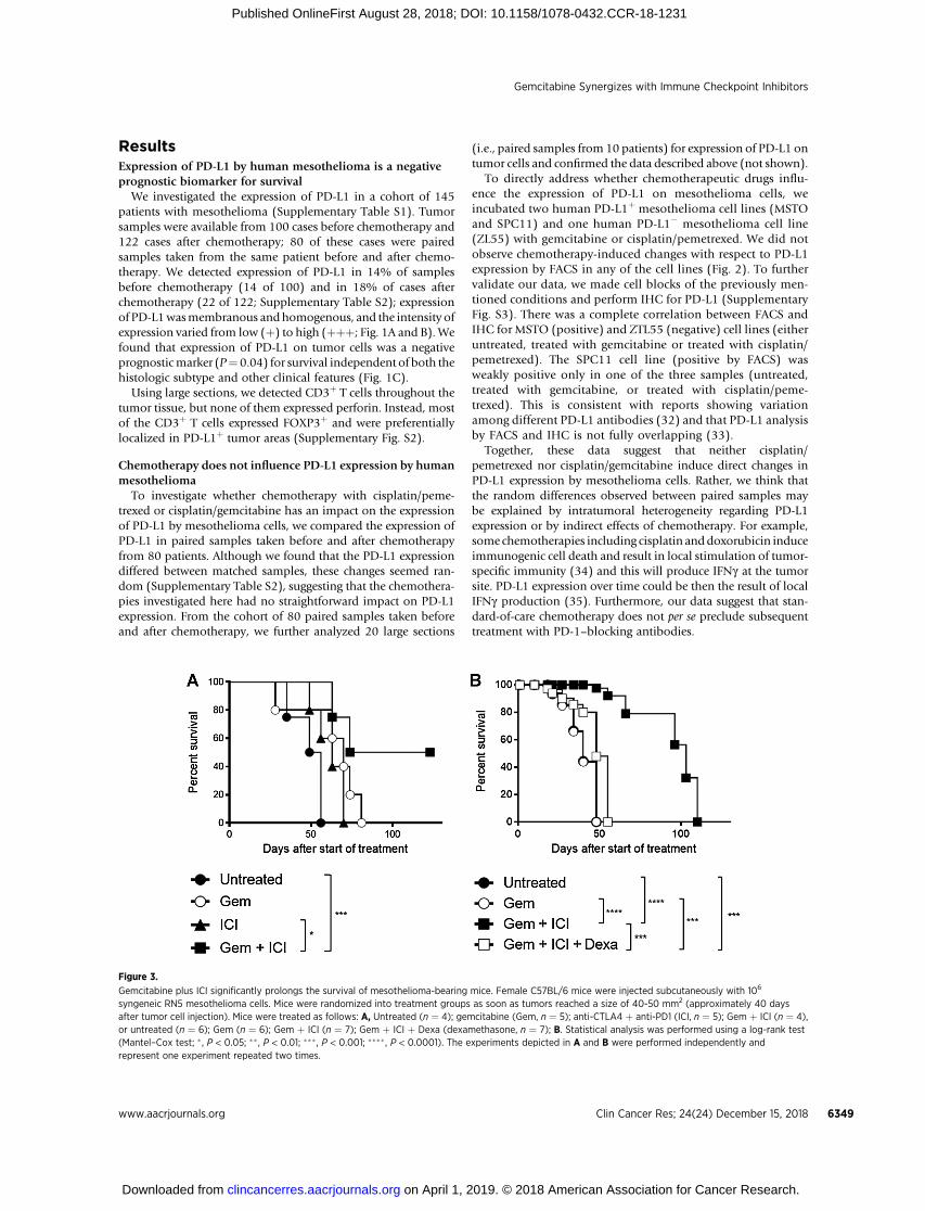
Figure 3.
Gemcitabine plus ICI significantly prolongs the survival of mesothelioma-bearing mice. Female C57BL/6 mice were injected subcutaneously with 106
syngeneic RN5 mesothelioma cells. Mice were randomized into treatment groups as soon as tumors reached a size of 40-50 mm2 (approximately 40 daysafter tumor cell injection). Mice were treated as follows: A, Untreated (n ¼ 4); gemcitabine (Gem, n ¼ 5); anti-CTLA4 þ anti-PD1 (ICI, n ¼ 5); Gem þ ICI (n ¼ 4),or untreated (n ¼ 6); Gem (n ¼ 6); Gem þ ICI (n ¼ 7); Gem þ ICI þ Dexa (dexamethasone, n ¼ 7); B. Statistical analysis was performed using a log-rank test(Mantel–Cox test; � , P < 0.05; �� , P < 0.01; ��� , P < 0.001; ���� , P < 0.0001). The experiments depicted in A and B were performed independently andrepresent one experiment repeated two times.
Gemcitabine Synergizes with Immune Checkpoint Inhibitors
www.aacrjournals.org Clin Cancer Res; 24(24) December 15, 2018 6349
on April 1, 2019. © 2018 American Association for Cancer Research. clincancerres.aacrjournals.org Downloaded from
Published OnlineFirst August 28, 2018; DOI: 10.1158/1078-0432.CCR-18-1231

Gemcitabine plus immune checkpoint has better efficacy thangemcitabine or immune checkpoint inhibitors asmonotherapyin mice
To investigate whether gemcitabine synergizes with ICI, weused amousemesotheliomamodel, subcutaneousRN5 tumors insyngeneic C57BL/6 mice. This model shares histopathologicfeatures with the aggressive human mesothelioma subtype(sarcomatoid). We found that treatment with gemcitabineimpeded tumor progression and promoted infiltration by CD3þ
T cells, whereas treatment with ICI had no significant impact ontumor progression or survival. The combination of gemcitabineand ICI, however, significantly prolonged survival and resulted intumor rejection in a proportion of the mice (Figs. 3A and B),suggesting a synergistic effect of ICI plus gemcitabine.
Corticosteroids nullify the synergy of chemo- andimmunotherapy in mice
Dexamethasone is an anti-inflammatory and immunosuppres-sive glucocorticoid that is commonly used together with chemo-therapy as an antiemetic drug. Specifically, dexamethasone isoften given with gemcitabine to prevent side effects includingchemotherapy-induced nausea and vomiting, fever, and chills. Incontrast to brief treatment schedules, long-term use of glucocor-ticoids is immunosuppressive (24). We observed a therapeuticsynergy when ICI was combined with gemcitabine, suggestingthat activation of immune defense supports the clinical responseto gemcitabine. Therefore, we wondered whether long-termtreatment with dexamethasone would negatively influence thistherapeutic synergy and treated mice with established RN5
tumors with gemcitabine plus ICI or gemcitabine plus ICI plusdexamethasone. Mice treated with gemcitabine alone or vehicleserved as controls. Again, we observed that gemcitabine plus ICIresulted in significantly better survival compared with gemcita-bine alone (Fig. 3B). However, addition of dexamethasone to thetreatment with gemcitabine plus ICI significantly reducedresponses in treated mice (Fig. 3B). Next, we investigated theimmune infiltrate in mouse mesothelioma by IHC for CD3 andFoxP3 at the endpoint. We observed increased fibrotic andnecrotic areas after treatment with gemcitabine plus ICI, as wellas increasednumber of infiltratingCD3þT cells comparedwith allother groups (Fig. 4). Furthermore, none of the CD3þ cellsexpressed FoxP3 (data not shown). In contrast, tumors of micetreated with gemcitabine plus ICI plus dexamethasone did notshow necrotic areas and very few infiltrating CD3þ T cells (Fig. 4),suggesting that corticosteroids negatively influence the clinicalresponse to gemcitabine plus ICI. We think that this may apply toany other therapy that involves immune stimulation includingstandard therapies (30, 34), suggesting that the use of steroids inthe context of cancer therapies should be avoided if possible. Itmay very well be of general applicability that clinical responses toICI are stronger in absence of steroids. The finding that concom-itant useof steroidswas associatedwithpoor outcomes of patientswith NSCLC treated with ICI (36) supports this idea.
The combination of gemcitabine with anti-PD-1 can overcomethe resistance to ICI in patients
Currently, the standard of treatment of mesothelioma is che-motherapy. However, blockade of the interaction between PD-1
Figure 4.
Gemcitabine plus immunecheckpoint inhibitors synergize toinduce tumor cell death and infiltrationby CD3þ cells. Representative IHCstaining of CD3þ cells in tumor samplesfrom mice described in Fig. 3B.
Tall�on de Lara et al.
Clin Cancer Res; 24(24) December 15, 2018 Clinical Cancer Research6350
on April 1, 2019. © 2018 American Association for Cancer Research. clincancerres.aacrjournals.org Downloaded from
Published OnlineFirst August 28, 2018; DOI: 10.1158/1078-0432.CCR-18-1231

and its ligands has shown promising clinical responses in aproportion of patients in clinical trials (9). We present heretwo patients with mesothelioma who were refractory tovarious therapies but responded to a combination of anti-PD-1and gemcitabine.
The first patient was 66 years old and diagnosed with epithe-lioid mesothelioma three years ago (in 2014). The patient under-went multimodality approaches including chemotherapy and
tumor resection. Because of relapse of disease, the patient wastreated with pembrolizumab. Upon progression, the patient wastreated with carboplatin/pemetrexed. Then he was switched tocarboplatin/gemcitabine and consecutively to gemcitabinemonotherapy. Because of further progression, the patient receiveda combination of gemcitabine (1,000 mg/m2 weekly) plus pem-brolizumab (200 mg every 3 weeks) without corticosteroids.Before starting this treatment, the patient complained about
i
Week
Timepoint
0 31
ii iii iv v
52 65 79
0
100
200
300
0
1
2
3
MTV
(mL)
EC
OG
Status
MTVECOG
i ii iviiiTimepoint
SUVmax
v0
10
20
30
SU
Vm
ax
Pembro Carbo+PMT Carbo+Gem Gem Pembro+GEM
i
Week
Timepoint
0 22
ii iii iv v
38 43 52
Pembro Carbo+PMT Radiotherapy Gem Pembro+GEM
0
50
100
150
200
0
1
2
3
Med
iast
inal
TV
(mL)
EC
OG
Status
Mediastinal TVECOG
i ii iviii
Lung TV
vTimepoint
0.0
0.2
0.4
0.6
0.8
1.0
i ii iii
iv v
* * *
* *
A
Patient 2
Patient 1
B
C
D
Figure 5.
Response to pembrolizumab andgemcitabinein two patients with mesothelioma. A and B,Patient 1; C andD, patient 2.A, Timeline of thetreatments. B, Quantification of the responseto the treatments by PET/CT scan at 5different timepoints (1–5), black arrow marksthe start of gemcitabine þ pembrolizumabtherapy. Performance status was evaluatedby Eastern Cooperative Oncology Group(ECOG) scale. MTV stands for metabolictumor volume. Bottom, representativepictures of whole-body PET/CT scan at thecorresponding 5 timepoints.C, Timeline of thetreatments. D, Quantification of the responseto the treatments by CT scan at 5 differenttime points (i–v), black arrow marks the startof gemcitabine þ pembrolizumab therapy.Performance status was evaluated by EasternCooperative Oncology Group (ECOG) scale.Bottom, representative pictures of the CTscan of themediastinum at the corresponding5 timepoints.Mediastinal lesion ismarkedwitha white asterisk. White arrow marks thepleural effusion. Lung metastasis in the leftupper lower that appeared after radiotherapy(time point iii) is shown in the inlet, markedwith a black circle.
Gemcitabine Synergizes with Immune Checkpoint Inhibitors
www.aacrjournals.org Clin Cancer Res; 24(24) December 15, 2018 6351
on April 1, 2019. © 2018 American Association for Cancer Research. clincancerres.aacrjournals.org Downloaded from
Published OnlineFirst August 28, 2018; DOI: 10.1158/1078-0432.CCR-18-1231

dysphagia and weight loss. However, two months after startingthis combination treatment, the patient could eat normally and aradiologic and metabolic response to treatment was detected(Fig. 5A and B). He is still under therapy, 20 weeks after the startof treatment.
The second case was a 57-year-old patient diagnosed withepithelioid mesothelioma 4 years ago (in 2013). The patientunderwent multimodality approaches including chemotherapyand tumor resection (Fig. 5C). Because of recurrence of disease in2016, the patient was treated with pembrolizumab (anti-PD-1),which resulted in progression of disease (Fig. 5D, time point i),and subsequently with carboplatin/pemetrexed with further pro-gression of disease (Fig. 5D, time point ii). Then one symptomaticlesion in the chest wall was irradiated with a total dose of 36 Gy(6 � 6 Gy). Restaging was performed (Fig. 5D, time point iii)before start of weekly chemotherapy with gemcitabine (withoutcorticosteroids). Within two months of gemcitabine monother-apy, there was evidence of progression of the mediastinal massand lung metastasis (Fig. 5D, time point iv) and the patient had areduced performance status with ECOG 3 due to cachexia anddyspnea. Because thepatient didnot showany limiting side effectson gemcitabine,we started a treatmentwithpembrolizumab (200mg every three weeks) and gemcitabine (1,000mg/m2weekly), inMarch 2017. After three cycles of this treatment, the patientclinically improved and went back to work using his bicycle asmeans of transport. Furthermore, radiologic images showed aclinical response of themediastinalmass and lungmetastasiswithincreased pleural effusion (Fig. 5D, time point v).We observed noside effects besides grade 2 anemia and pleural effusion that wasdrained after treatment. We analyzed the composition of thepleural effusion and observed that together with few mesotheli-oma cells therewas 80%of lymphocytes (Figs. 6A andB), of these,90% were CD3þ T cells, consisting of 20% CD4þ (Fig. 6C) and75% CD8þ cells (Fig. 6D). This suggests that the combination of
gemcitabine plus anti-PD-1 mobilized protective immunityresulting in an objective clinical response. Because of hisimproved general condition, the patient wished to stop thetreatment and undertake a journey of 5 weeks. Upon his return,treatment with gemcitabine plus anti-PD-1 was started with athree-weekly schedule for both medications as per the patient'srequest. After two cycles of this treatment, a progression of diseasewas detected and the treatment was discontinued.
DiscussionCombining different therapies for cancer is a promising
approach to overcome resistance and improve responses. Forexample, combination of nivolumab (anti-PD-1) withipilimumab (anti-CTLA-4) has shown improved clinicalresponses compared with monotherapy; however, alsoincreased side effects (37). Currently, combinations of differentchemotherapies with immunotherapy are under evaluation fordifferent tumor types (NCT02039674, NCT02366143). Weinvestigated whether the combination of gemcitabine with ICIis an effective and feasible treatment for mesothelioma thatoutperforms monotherapy and may even overcome resistanceto monotherapy.
Expression of PD-L1 by tumor cells is often used as inclu-sion criterion for treatment with antibodies that block theinteraction of PD-1 with its ligands (9). Therefore, we firstinvestigated whether gemcitabine influences the expression ofPD-L1 by mesothelioma cells in vivo and in vitro. In addition,we found that the expression of PD-L1 is a negative biomarkerfor survival in mesothelioma, which goes in line with previousstudies (14, 38) and with data obtained from other tumorentities (39–42).
We found that treatment with gemcitabine plus ICI significant-ly prolonged survival of mesothelioma-bearing mice compared
20% 75%
A B
C
CD
4
CD
8
CD3
D
Figure 6.
Cytologic analysis of the pleural fluid frompatient 2. Pleural fluid of patient 2 was collectedfrom the lesion shown in Fig. 5.Dv and thesediment was analyzed by cytology. A,Papanicolaou staining revealed the presence ofmesothelioma cells (black arrows), numerouslymphocytes (red arrows) and fewmacrophagesand neutrophilic granulocytes in the background(original magnification 400�). Mesotheliomacells were calretininþ, desmin�, BAP1� (data notshown). B, CD3 immunostaining revealed that90% of lymphocytes were CD3þ T cells (brownsignal; original magnification 400�). C and D,Flow cytometric analysis showed that 20% ofCD3þ T cells express CD4 (C) and 75% expressCD8 (D).
Tall�on de Lara et al.
Clin Cancer Res; 24(24) December 15, 2018 Clinical Cancer Research6352
on April 1, 2019. © 2018 American Association for Cancer Research. clincancerres.aacrjournals.org Downloaded from
Published OnlineFirst August 28, 2018; DOI: 10.1158/1078-0432.CCR-18-1231

with gemcitabine alone, whereas ICI as monotherapy had noimpact. Although ICI as monotherapy shows clinical efficacy inpatients with mesothelioma (43), this is not the case in ourpreclinical model. Absence of clinical efficacy of ICI as mono-therapy has been described in multiple mouse models (29) butthis discrepancy with humans is not understood.
Corticosteroids are commonly used to manage side effects ofchemotherapy. Because of their immunosuppressive properties,we evaluated the impact of dexamethasone on the clinical efficacyof gemcitabine plus ICI and observed an abrogation of therapeu-tic synergy. This suggests that side effects of chemotherapy shouldbemanaged by other medications than corticosteroids, wheneverpossible. Indeed, we have successfully applied this approach toboth patients presented here.
Our data show that PD-L1 expression is heterogeneous andprone to immune response–induced changes. In addition,various studies have shown a high degree of intratumoralheterogeneity in PD-L1 expression in different cancers(44, 45). Therefore, we think that using PD-L1 expression asan inclusion criterion for treatments targeting this pathway isnot sufficiently justified for patients with mesothelioma. Wedemonstrated the synergy of gemcitabine with ICI providing arationale for a clinical trial that can include patients withdifferent tumor types, where gemcitabine represents astandard of care and might be favorable in terms of tolerabilityand costs compared with combination of several immunomo-dulating agents. It has been shown that in patients withadvanced lung cancer that daily administration of steroidsreduces lymphocyte numbers (46) and may be associatedwith worse outcome (36). Obviously, highly emetogenictreatments must be combined with antiemetic drugs, however,corticosteroids should be avoided and established alternativesused instead. Here, we show in a preclinical model thatavoiding the use of dexamethasone improves the efficacy ofimmunotherapy for mesothelioma, a disease with very limitedeffective treatment options (47) and an unmet need for newtherapies (48).
Disclosure of Potential Conflicts of InterestC. Britschgi is a consultant/advisory board member for Roche, AstraZeneca,
and Pfizer. No potential conflicts of interest were disclosed by the other authors.
Authors' ContributionsConception and design: P. Tall�on de Lara, P. Stolzmann, M. van den Broek,A. Curioni-FontecedroDevelopment of methodology: E. Felley-Bosco, R.A. Stahel, V. Tischler,M. van den Broek, A. Curioni-FontecedroAcquisition of data (provided animals, acquired and managed patients,provided facilities, etc.): P. Tall�on de Lara, V. Cecconi, S. Hiltbrunner,B. Bode-Lesniewska, I. Opitz, P. Stolzmann, C. Britschgi, D. Soldini, M. vanden Broek, A. Curioni-FontecedroAnalysis and interpretation of data (e.g., statistical analysis, biostatistics, compu-tational analysis): P. Tall�on de Lara, V. Cecconi, M. Friess, B. Bode-Lesniewska,B.Vrugt,R.A. Stahel,V.Tischler,D.Soldini,M.vandenBroek,A.Curioni-FontecedroWriting, review, and/or revision of the manuscript: P. Tall�on de Lara,S. Hiltbrunner, H. Yagita, B. Bode-Lesniewska, I. Opitz, W. Weder, P. Stolzmann,E. Felley-Bosco, C. Britschgi, D. Soldini, M. van den Broek, A. Curioni-FontecedroAdministrative, technical, or material support (i.e., reporting or organizingdata, constructing databases): V. Cecconi, H. Yagita, M. Friess, I. Opitz,P. Stolzmann, M. van den Broek, A. Curioni-FontecedroStudy supervision: M. van den Broek, A. Curioni-Fontecedro
AcknowledgmentsThisworkwasfinancially supported by the Sophien StiftungZurich, the Swiss
National Science Foundation, the University of Zurich (University ResearchPriority Project "Translational Cancer Research"), the Forschungskredit of theUniversity of Zurich and the Science Foundation for Oncology (SFO).We thankAndr�e Fitsche, Susanne Dettwiler, and Martina Storz (Institute for SurgicalPathology, University Hospital Zurich, Zurich, Switzerland) for excellent tech-nical assistance with TMA construction and establishment of IHC staining. Wethank the personnel of the Laboratory Animal Services Center (LASC) at theUniversity of Zurich for expert animal care.
The costs of publication of this articlewere defrayed inpart by the payment ofpage charges. This article must therefore be hereby marked advertisement inaccordance with 18 U.S.C. Section 1734 solely to indicate this fact.
Received April 20, 2018; revised July 24, 2018; accepted August 21, 2018;published first August 28, 2018.
References1. Burnet FM. The concept of immunological surveillance. Prog Exp Tumor
Res 1970;13:1–27.2. Sharpe AH,Wherry EJ, AhmedR, FreemanGJ. The function of programmed
cell death 1 and its ligands in regulating autoimmunity and infection.Nat Immunol 2007;8:239–45.
3. Chen L. Co-inhibitory molecules of the B7-CD28 family in the control ofT-cell immunity. Nat Rev Immunol 2004;4:336–47.
4. Latchman Y, Wood CR, Chernova T, Chaudhary D, Borde M, Chernova I,et al. PD-L2 is a second ligand for PD-1 and inhibits T cell activation.Nat Immunol 2001;2:261–8.
5. Probst HC, McCoy K, Okazaki T, Honjo T, van den Broek M. Restingdendritic cells induce peripheral CD8þ T cell tolerance through PD-1 andCTLA-4. Nat Immunol 2005;6:280–6.
6. Freeman GJ, Long AJ, Iwai Y, Bourque K, Chernova T, Nishimura H, et al.Engagement of the PD-1 immunoinhibitory receptor by a novel B7 familymember leads to negative regulation of lymphocyte activation. J Exp Med2000;192:1027–34.
7. Brahmer JR, Tykodi SS, ChowLQ,HwuWJ, Topalian SL,HwuP, et al. Safetyand activity of anti-PD-L1 antibody in patients with advanced cancer.N Engl J Med 2012;366:2455–65.
8. Topalian SL,Hodi FS, Brahmer JR,Gettinger SN, SmithDC,McDermottDF,et al. Safety, activity, and immune correlates of anti-PD-1 antibody incancer. N Engl J Med 2012;366:2443–54.
9. Alley EW, Lopez J, Santoro A, Morosky A, Saraf S, Piperdi B, et al.Clinical safety and activity of pembrolizumab in patients with malig-nant pleural mesothelioma (KEYNOTE-028): preliminary results from anon-randomised, open-label, phase 1b trial. Lancet Oncol 2017;18:623–30.
10. Quispel-Janssen J, van der Noort V, de Vries JF, Zimmerman M, Lalezari F,Thunnissen E, et al. PD-1 blockade with nivolumab in patients withrecurrent malignant pleural mesothelioma. J Thorac Oncol 2018Jun 14[Epub ahead or print].
11. Herbst RS, Soria JC, Kowanetz M, Fine GD, Hamid O, Gordon MS, et al.Predictive correlates of response to the anti-PD-L1 antibody MPDL3280Ain cancer patients. Nature 2014;515:563–7.
12. Karim R, Jordanova ES, Piersma SJ, Kenter GG, Chen L, Boer JM, et al.Tumor-expressed B7-H1 and B7-DC in relation to PD-1þ T-cell infiltrationand survival of patients with cervical carcinoma. Clin Cancer Res2009;15:6341–7.
13. Thompson RH, Kuntz SM, Leibovich BC, Dong H, Lohse CM,Webster WS,et al. Tumor B7-H1 is associated with poor prognosis in renal cell carci-noma patients with long-term follow-up. Cancer Res 2006;66:3381–5.
14. Mansfield AS, Roden AC, Peikert T, Sheinin YM, Harrington SM, Krco CJ,et al. B7-H1 expression in malignant pleural mesothelioma is associatedwith sarcomatoid histology and poor prognosis. J Thorac Oncol 2014;9:1036–40.
Gemcitabine Synergizes with Immune Checkpoint Inhibitors
www.aacrjournals.org Clin Cancer Res; 24(24) December 15, 2018 6353
on April 1, 2019. © 2018 American Association for Cancer Research. clincancerres.aacrjournals.org Downloaded from
Published OnlineFirst August 28, 2018; DOI: 10.1158/1078-0432.CCR-18-1231

15. Cedres S, Ponce-Aix S, Zugazagoitia J, Sansano I, Enguita A, Navarro-Mendivil A, et al. Analysis of expression of programmed cell death 1 ligand1 (PD-L1) inmalignant pleuralmesothelioma (MPM). PLoSOne 2015;10:e0121071.
16. Homma Y, Taniguchi K, Nakazawa M, Matsuyama R, Mori R, Takeda K,et al. Changes in the immune cell population and cell proliferation inperipheral blood after gemcitabine-based chemotherapy for pancreaticcancer. Clin Translat Oncol 2014;16:330–5.
17. Duffy AG, Greten TF. Immunological off-target effects of standard treat-ments in gastrointestinal cancers. Ann Oncol 2014;25:24–32.
18. Rettig L, Seidenberg S, Parvanova I, Samaras P, Curioni A, Knuth A, et al.Gemcitabine depletes regulatory T-cells in human and mice and enhancestriggering of vaccine-specific cytotoxic T-cells (vol 129, pg 832, 2011). Int JCancer 2012;130:1714.
19. Kononen J, Bubendorf L, Kallioniemi A, BarlundM, Schraml P, Leighton S,et al. Tissue microarrays for high-throughput molecular profiling of tumorspecimens. Nat Med 1998;4:844–7.
20. HinterbergerM, Reineke T, StorzM,WederW, Vogt P,MochH. D2–40 andcalretinin - a tissue microarray analysis of 341 malignant mesotheliomaswith emphasis on sarcomatoid differentiation. Mod Pathol 2007;20:248–55.
21. Curioni-Fontecedro A, Nuber N, Mihic-Probst D, Seifert B, Soldini D,Dummer R, et al. Expression of MAGE-C1/CT7 and MAGE-C2/CT10predicts lymph node metastasis in melanoma patients. PLoS One2011;6:e21418.
22. Stahel RA, Riesterer O, Xyrafas A, Opitz I, Beyeler M, Ochsenbein A, et al.Neoadjuvant chemotherapy and extrapleural pneumonectomy of malig-nant pleural mesothelioma with or without hemithoracic radiotherapy(SAKK 17/04): a randomised, international, multicentre phase 2 trial.Lancet Oncol 2015;16:1651–8.
23. Schmitter D, Lauber B, Fagg B, Stahel RA. Hematopoietic growth factorssecreted by seven human pleural mesothelioma cell lines: interleukin-6production as a common feature. Int J Cancer 1992;51:296–301.
24. Phelps RM, Johnson BE, IhdeDC, Gazdar AF, CarboneDP,McClintock PR,et al. NCI-Navymedical oncology branch cell line data base. J Cell BiochemSuppl 1996;24:32–91.
25. Kitazono-Saitoh M, Takiguchi Y, Kitazono S, Ashinuma H, Kitamura A,Tada Y, et al. Interaction and cross-resistance of cisplatin andpemetrexed inmalignant pleural mesothelioma cell lines. Oncol Rep 2012;28:33–40.
26. BlumW, Pecze L, Felley-Bosco E, Worthmuller-Rodriguez J, Wu L, Vrugt B,et al. Establishment of immortalized murine mesothelial cells and a novelmesothelioma cell line. In Vitro Cell Dev Biol Anim 2015;51:714–21.
27. Rehrauer H, Wu L, Blum W, Pecze L, Henzi T, Serre-Beinier V, et al.Felley-Bosco. How asbestos drives the tissue towards tumors: YAP activa-tion, macrophage andmesothelial precursor recruitment, RNA editing andsomatic mutations. Oncogene 2018;37:2645–59.
28. Lesterhuis WJ, Salmons J, Nowak AK, Rozali EN, Khong A, Dick IM, et al.Synergistic effect of CTLA-4 blockade and cancer chemotherapy in theinduction of anti-tumor immunity. PLoS One 2013;8:e61895.
29. Bransi A, SalgadoOC, BeffingerM,Milo K, Silina K, Yagita H, et al. Rationalcombination of immunotherapies with clinical efficacy in mice withadvanced cancer. Cancer Immunol Res 2015;3:1279–88.
30. Surace L, Lysenko V, Fontana AO, Cecconi V, Janssen H, Bicvic A, et al.Complement is a central mediator of radiotherapy-induced tumor-specificimmunity and clinical response. Immunity 2015;42:767–77.
31. Herbst RS, Baas P, Kim DW, Felip E, Perez-Gracia JL, Han JY, et al.Pembrolizumab versus docetaxel for previously treated, PD-L1-positive,advanced non-small-cell lung cancer (KEYNOTE-010): a randomisedcontrolled trial. Lancet 2016;387:1540–50.
32. Hendry S, Byrne DJ, Wright GM, Young RJ, Sturrock S, Cooper WA, et al.Comparison of four PD-L1 immunohistochemical assays in lung cancer.J Thoracic Oncol 2018;13:367–76.
33. Chargin A, Morgan R, Sundram U, Shults K, Tsay EL, Ratti N, et al.Quantification of PD-L1 and PD-1 expression on tumor and immunecells in non-small cell lung cancer (NSCLC) using non-enzymatic tissuedissociation and flow cytometry. Cancer Immunol Immunother 2016;65:1317–23.
34. Zitvogel L, Apetoh L, Ghiringhelli F, Kroemer G. Immunological aspects ofcancer chemotherapy. Nat Rev Immunol 2008;8:59–73.
35. NomanMZ,DesantisG, Janji B,HasmimM,Karray S,Dessen P, et al. PD-L1is a novel direct target of HIF-1alpha, and its blockade under hypoxiaenhanced MDSC-mediated T cell activation. J Exp Med 2014;211:781–90.
36. Martinez-Bernal G, Mezquita L, Auclin E, Ferrara R, Planchard D, Remon J,et al. Baseline corticosteroids (CS) could be associated with absence ofbenefit to immune checkpoint inhibitors (ICI) in advanced non-small celllung cancer (NSCLC) patients. Ann Oncol 2017;28:1323P.
37. Hodi FS, Chesney J, Pavlick AC, Robert C, Grossmann KF, McDermott DF,et al. Combined nivolumab and ipilimumab versus ipilimumab alone inpatients with advanced melanoma: 2-year overall survival outcomes in amulticentre, randomised, controlled, phase 2 trial. Lancet Oncol 2016;17:1558–68.
38. Nguyen BH, Montgomery R, Fadia M, Wang J, Ali S. PD-L1 expressionassociated with worse survival outcome in malignant pleural mesotheli-oma. Asia-Pacific J Clin Oncol 2018;14:69–73.
39. Wu P, Wu D, Li L, Chai Y, Huang J. PD-L1 and survival in solid tumors: ameta-analysis. PLoS One 2015;10:e0131403.
40. Chowdhury S, Veyhl J, Jessa F, Polyakova O, Alenzi A, MacMillan C, et al.Programmed death-ligand 1 overexpression is a prognostic marker foraggressive papillary thyroid cancer and its variants. Oncotarget 2016;7:32318–28.
41. Cierna Z, Mego M, Miskovska V, Machalekova K, Chovanec M, SvetlovskaD, et al. Prognostic value of programmed-death-1 receptor (PD-1) and itsligand 1 (PD-L1) in testicular germ cell tumors. AnnOncol 2016;27:300–5.
42. Katsuya Y, Fujita Y, Horinouchi H, Ohe Y, Watanabe S, Tsuta K. Immu-nohistochemical status of PD-L1 in thymoma and thymic carcinoma.Lung Cancer 2015;88:154–9.
43. Scherpereel A, Mazieres J, Greillier L, Do P, Bylicki O, Monnet I, et al.Second- or third-line nivolumab (Nivo) versus nivo plus ipilimumab (Ipi)in malignant pleural mesothelioma (MPM) patients: Results of theIFCT-1501 MAPS2 randomized phase II trial. J Clin Oncol 2017;35:LBA8507–LBA.
44. Munari E, Zamboni G, Marconi M, Sommaggio M, Brunelli M, MartignoniG, et al. PD-L1 expression heterogeneity in non-small cell lung cancer:evaluation of small biopsies reliability. Oncotarget 2017;8:90123–31.
45. Nakamura S, Hayashi K, Imaoka Y, Kitamura Y, Akazawa Y, Tabata K, et al.Intratumoral heterogeneity of programmed cell death ligand-1 expressionis common in lung cancer. PLoS One 2017;12:e0186192.
46. Cook AM, McDonnell AM, Lake RA, Nowak AK. Dexamethasone co-medication in cancer patients undergoing chemotherapy causes substan-tial immunomodulatory effects with implications for chemo-immuno-therapy strategies. Oncoimmunology 2016;5:e1066062.
47. Vogelzang NJ, Rusthoven JJ, Symanowski J, Denham C, Kaukel E, Ruffie P,et al. Phase III study of pemetrexed in combination with cisplatin versuscisplatin alone in patients with malignant pleural mesothelioma. J ClinOncol 2003;21:2636–44.
48. Stahel RA, Weder W, Felley-Bosco E, Petrausch U, Curioni-Fontecedro A,Schmitt-Opitz I, et al. Searching for targets for the systemic therapy ofmesothelioma. Ann Oncol 2015;26:1649–60.
Clin Cancer Res; 24(24) December 15, 2018 Clinical Cancer Research6354
Tall�on de Lara et al.
on April 1, 2019. © 2018 American Association for Cancer Research. clincancerres.aacrjournals.org Downloaded from
Published OnlineFirst August 28, 2018; DOI: 10.1158/1078-0432.CCR-18-1231

2018;24:6345-6354. Published OnlineFirst August 28, 2018.Clin Cancer Res Paulino Tallón de Lara, Virginia Cecconi, Stefanie Hiltbrunner, et al. PatientsOvercomes Resistance in a Preclinical Model and Mesothelioma Gemcitabine Synergizes with Immune Checkpoint Inhibitors and
Updated version
10.1158/1078-0432.CCR-18-1231doi:
Access the most recent version of this article at:
Material
Supplementary
http://clincancerres.aacrjournals.org/content/suppl/2018/08/28/1078-0432.CCR-18-1231.DC1
Access the most recent supplemental material at:
Cited articles
http://clincancerres.aacrjournals.org/content/24/24/6345.full#ref-list-1
This article cites 47 articles, 6 of which you can access for free at:
E-mail alerts related to this article or journal.Sign up to receive free email-alerts
Subscriptions
Reprints and
To order reprints of this article or to subscribe to the journal, contact the AACR Publications Department at
Permissions
Rightslink site. Click on "Request Permissions" which will take you to the Copyright Clearance Center's (CCC)
.http://clincancerres.aacrjournals.org/content/24/24/6345To request permission to re-use all or part of this article, use this link
on April 1, 2019. © 2018 American Association for Cancer Research. clincancerres.aacrjournals.org Downloaded from
Published OnlineFirst August 28, 2018; DOI: 10.1158/1078-0432.CCR-18-1231