gene & chromosomal mutation chicken - suny- · pdf filemain types: 1. single-gene...
TRANSCRIPT
Gene & Chromosomal mutation
6
38
78
62
22
0 10 20 30 40 50 60 70 80 90
A's B's C's D F
Genetics Exam 1, F2009
Series1
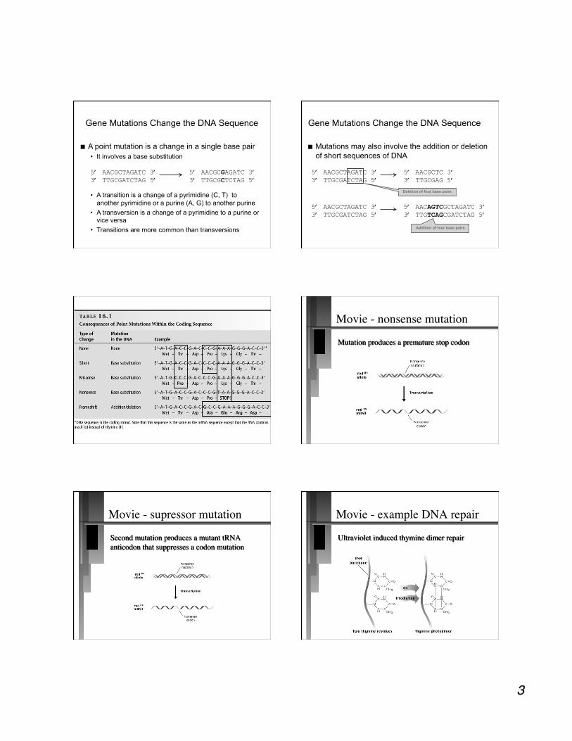
Chicken
Chicken??? New York State Fair Genetics Display
Scientific American, October 2005, Vol. 293 (4) pp.78-85

Mutations can be divided into three main types:
1. Single-gene mutations Relatively small changes in DNA structure that occur within a particular gene (promoter or transcriptional unit)
2. Chromosome mutations Changes in chromosome structure
3. Genome mutations Changes in chromosome number
Genetic variation
allele - One of the different forms of a gene that exist at a single locus. • locus - location of the gene on a
chromosome Where do different alleles come from? recombination of functional gene
domains and gene mutation (base deletions, additions, or substitutions)
mutate - to change
Gene mutation terms
wild-type (wt): designated standard (either in nature or lab)
forward mutation - any change away from wt
reverse mutation - any change back to the wt allele • also called reversion or back mutation
Think: Mutation and Nature
According to evolutionary theory, all genes are mutant genes, we arbitrarily name certain alleles as wt. Genes must mutate to evolve! • Today’s mutation is tomorrows allele
Mutation is the foundation of the diversity found in nature and agriculture.
Remember the chickens
IGF1 allele is the major determinate of small size in dogs What causes gene mutations?
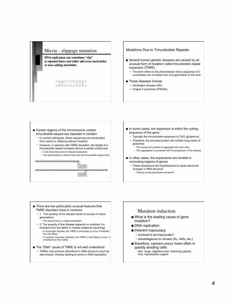
A point mutation is a change in a single base pair • It involves a base substitution
Gene Mutations Change the DNA Sequence
5’ AACGCTAGATC 3’ 3’ TTGCGATCTAG 5’
5’ AACGCGAGATC 3’ 3’ TTGCGCTCTAG 5’
• A transition is a change of a pyrimidine (C, T) to another pyrimidine or a purine (A, G) to another purine
• A transversion is a change of a pyrimidine to a purine or vice versa
• Transitions are more common than transversions
Mutations may also involve the addition or deletion of short sequences of DNA
Gene Mutations Change the DNA Sequence
5’ AACGCTAGATC 3’ 3’ TTGCGATCTAG 5’
5’ AACGCTC 3’ 3’ TTGCGAG 5’
5’ AACGCTAGATC 3’ 3’ TTGCGATCTAG 5’
5’ AACAGTCGCTAGATC 3’ 3’ TTGTCAGCGATCTAG 5’
Deletion of four base pairs
Addition of four base pairs
Movie - nonsense mutation
Movie - supressor mutation Movie - example DNA repair
Movie - slippage mutation
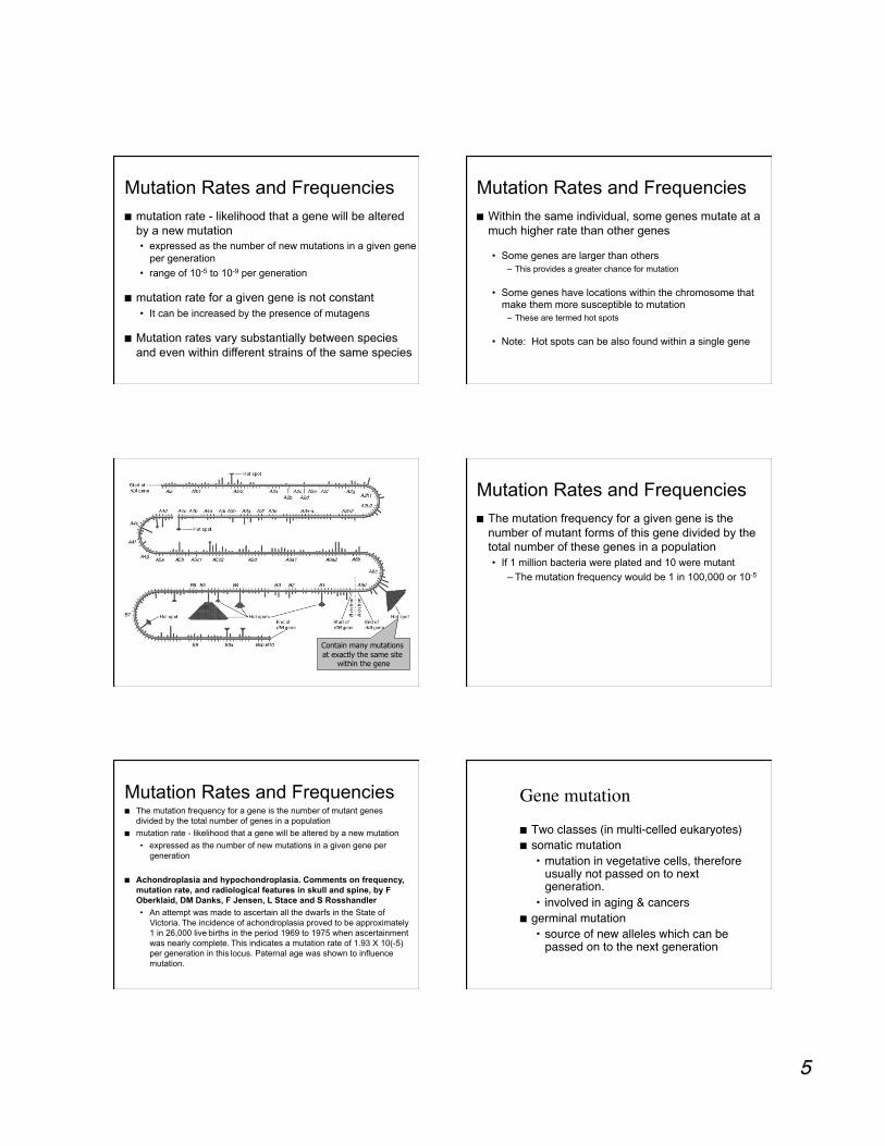
Several human genetic diseases are caused by an unusual form of mutation called trinucleotide repeat expansion (TNRE) • The term refers to the phenomenon that a sequence of 3
nucleotides can increase from one generation to the next
These diseases include • Huntington disease (HD) • Fragile X syndrome (FRAXA)
Mutations Due to Trinucleotide Repeats
Certain regions of the chromosome contain trinucleotide sequences repeated in tandem • In normal individuals, these sequences are transmitted
from parent to offspring without mutation • However, in persons with TNRE disorders, the length of a
trinucleotide repeat increases above a certain critical size – It also becomes prone to frequent expansion – This phenomenon is shown here with the trinucleotide repeat CAG
CAGCAGCAGCAGCAGCAGCAGCAGCAGCAGCAG
CAGCAGCAGCAGCAGCAGCAGCAGCAGCAGCAGCAGCAGCAGCAGCAGCAGCAG
n = 11
n = 18
In some cases, the expansion is within the coding sequence of the gene • Typically the trinucleotide expansion is CAG (glutamine) • Therefore, the encoded protein will contain long tracks of
glutamine – This causes the proteins to aggregate with each other – This aggregation is correlated with the progression of the disease
In other cases, the expansions are located in noncoding regions of genes • These expansions are hypothesized to cause abnormal
changes in RNA structure – Thereby producing disease symptoms
There are two particularly unusual features that TNRE disorders have in common • 1. The severity of the disease tends to worsen in future
generations – This phenomenon is called anticipation
• 2. The severity of the disease depends on whether it is inherited from the father or mother (paternal imprinting)
– In Huntington disease, the TNRE is more likely to occur if inherited from the father
– In myotonic muscular dystrophy, the TNRE is more likely to occur if inherited from the mother
The “DNA” cause of TNRE is not well understood • TNREs may produce alterations in DNA structure (such as
stem-loops), thereby leading to errors in DNA replication
Mutation induction What is the leading cause of gene
mutation? DNA replication Inherent inaccuracy
• evolved to be inaccurate? • advantageous to viruses (flu, Aids, etc.)
therefore, cancers occur more often in quickly dividing cells • skin, lungs, digestive tract, mammary glands,
liver, reproductive organs
mutation rate - likelihood that a gene will be altered by a new mutation • expressed as the number of new mutations in a given gene
per generation • range of 10-5 to 10-9 per generation
mutation rate for a given gene is not constant • It can be increased by the presence of mutagens
Mutation rates vary substantially between species and even within different strains of the same species
Mutation Rates and Frequencies Within the same individual, some genes mutate at a
much higher rate than other genes
• Some genes are larger than others – This provides a greater chance for mutation
• Some genes have locations within the chromosome that make them more susceptible to mutation
– These are termed hot spots
• Note: Hot spots can be also found within a single gene
Mutation Rates and Frequencies
Contain many mutations at exactly the same site
within the gene
The mutation frequency for a given gene is the number of mutant forms of this gene divided by the total number of these genes in a population
• If 1 million bacteria were plated and 10 were mutant – The mutation frequency would be 1 in 100,000 or 10-5
Mutation Rates and Frequencies
The mutation frequency for a gene is the number of mutant genes divided by the total number of genes in a population
mutation rate - likelihood that a gene will be altered by a new mutation • expressed as the number of new mutations in a given gene per
generation
Achondroplasia and hypochondroplasia. Comments on frequency, mutation rate, and radiological features in skull and spine, by F Oberklaid, DM Danks, F Jensen, L Stace and S Rosshandler • An attempt was made to ascertain all the dwarfs in the State of
Victoria. The incidence of achondroplasia proved to be approximately 1 in 26,000 live births in the period 1969 to 1975 when ascertainment was nearly complete. This indicates a mutation rate of 1.93 X 10(-5) per generation in this locus. Paternal age was shown to influence mutation.
Mutation Rates and Frequencies Gene mutation
Two classes (in multi-celled eukaryotes) somatic mutation
• mutation in vegetative cells, therefore usually not passed on to next generation.
• involved in aging & cancers germinal mutation
• source of new alleles which can be passed on to the next generation
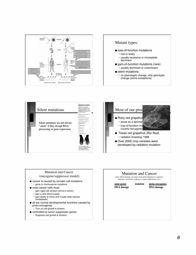
Therefore, the mutation can be
passed on to future generations
The size of the patch will depend on the timing of the mutation
The earlier the mutation, the larger the patch
An individual who has somatic regions that are genotypically different
from each other is called a genetic mosaic
Therefore, the mutation cannot be passed on to future generations
Mutant types:
loss-of-function mutations • null or leaky • usually recessive or incomplete
dominant gain-of-function mutations (rare)
• usually dominant or codominant silent mutations
• no phenotypic change, only genotypic change (some exceptions)
Silent mutations Most of our produce are mutants
Ruby red grapefruit • arose as a spontaneous mutant in 1926 • loss-of-function mutation - enzyme that
coverts red pigment to colorless pigment Texas red grapefruit (Rio Red)
• radiation breeding, 1988 Over 2000 crop varieties were
developed by radiation mutation
Mutation and Cancer �(oncogene/suppressor model)
cancer is caused by somatic cell mutations • gene or chromosome mutations
most cancer cells must: • gain rapid cell division (forms a tumor) • gain a new blood supply • gain ability to move and invade other tissues
(metastasis) all are normal developmental functions caused by
proto-oncogenes • Turn on cell growth & division
controlled by tumor suppressor genes • Suppress cell growth & division
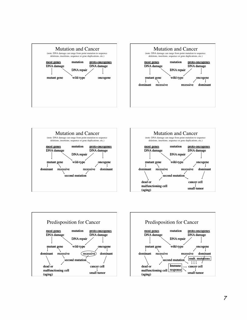
Mutation and Cancer�(note: DNA damage can range from point mutation to sequence
deletions, insertions, sequence or gene duplications, etc.)
Mutation and Cancer�(note: DNA damage can range from point mutation to sequence
deletions, insertions, sequence or gene duplications, etc.) Mutation and Cancer�
(note: DNA damage can range from point mutation to sequence deletions, insertions, sequence or gene duplications, etc.)
Mutation and Cancer�(note: DNA damage can range from point mutation to sequence
deletions, insertions, sequence or gene duplications, etc.) Mutation and Cancer�
(note: DNA damage can range from point mutation to sequence deletions, insertions, sequence or gene duplications, etc.)
Predisposition for Cancer Predisposition for Cancer
Gene mutations that help lead to cancer
Oncogenes Tumor suppressors Mutator genes
• Genes involved with DNA repair if mutated allow higher levels of mutation in general
Telomerase genes • If mutated to turn on when it should be turned
off, could lead to immortal cells (i.e. unlimited cell division)
Min. number of mutations
Retinoblastoma: 2 mutations • usually in children
colon cancer: 4-5 mutations • usually in adults
small-cell lung cancer: 10-15 mutations • usually in adults with high exposure to
mutagens, i.e. smokers
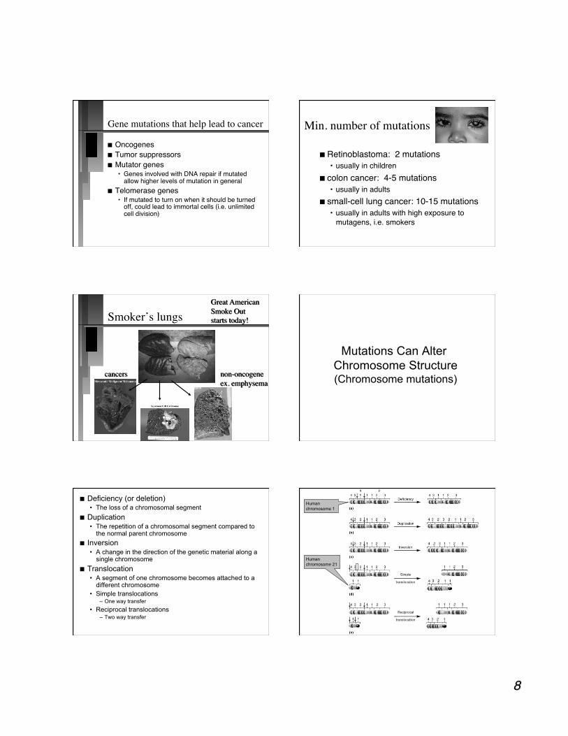
Smoker’s lungs
Mutations Can Alter Chromosome Structure (Chromosome mutations)
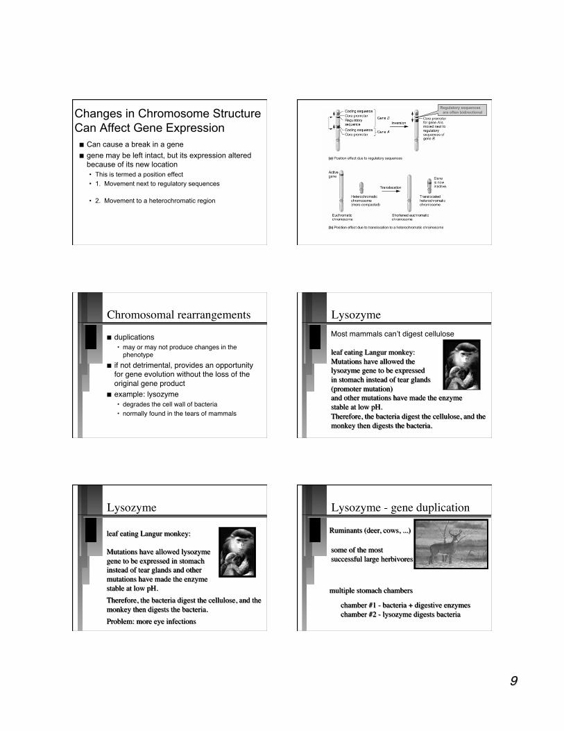
Deficiency (or deletion) • The loss of a chromosomal segment
Duplication • The repetition of a chromosomal segment compared to
the normal parent chromosome Inversion
• A change in the direction of the genetic material along a single chromosome
Translocation • A segment of one chromosome becomes attached to a
different chromosome • Simple translocations
– One way transfer • Reciprocal translocations
– Two way transfer
Human chromosome 1
Human chromosome 21
Can cause a break in a gene gene may be left intact, but its expression altered
because of its new location • This is termed a position effect • 1. Movement next to regulatory sequences
• 2. Movement to a heterochromatic region
Changes in Chromosome Structure Can Affect Gene Expression
Regulatory sequences are often bidirectional
Chromosomal rearrangements
duplications • may or may not produce changes in the
phenotype if not detrimental, provides an opportunity
for gene evolution without the loss of the original gene product
example: lysozyme • degrades the cell wall of bacteria • normally found in the tears of mammals
Lysozyme
Lysozyme Lysozyme - gene duplication
Lysozyme - gene duplication Lysozyme - gene duplication
Changes (mutation) in �chromosome number
Terms: monoploid number (X) - number of sets
of chromosomes (# genomes) • No duplicated or homologous
chromosomes in a set can be different than haploid number (n)
- the number found in gametes Examples:
• human: 46 chromosomes (2n = 2x) • wheat: 42 chromosomes (2n = 6x)
Euploids (multiples of X)
1x - monoploid • male bees, wasps, and ants • artificially derived plants
2x - diploid 3x - triploid 4x - tetraploid 5x - pentaploid 6x - hexaploid
polyploids
Polyploids (>2x) two types autopolyploids - multiple
chromosome sets from one species allopolyploids - chromosome sets
from different species • chromosome sets must be
homeologous – partially homologous which allows pairing
• example: Triticale (wheat x rye)
Genetic modification of wheat
Many Brassica species rutabaga oil rape mustards
(1n but 2X)
(2n but 4X)
(1n but 3X)
(2n but 6X)
Triploids
result from a cross of a tetraploid (4x) with a diploid (2x)
sterile due to problems with pairing during meiosis
example: bananas (no developed seeds!)
example 2: seedless watermelon other odd number of chromosome
sets will give similar results
Polyploidy in animals
much more rare than in plants examples: flatworms, leeches, brine
shrimp, some amphibians, and some fish
Salmonidae (Salmon and trout) thought to be polyploids because they contain twice as much DNA that closely related fish
Aneuploidy chromosome number differs from
wt by part of a chromosomal set (not euploid)
nomenclature: • 2n-1 = monosomic • 2n+1 = trisomic • 2n-2 = nullisomic • n+1 = disomic (in haploids)
generally deleterious
Aneuploidy
caused by nondisjunction during meioses or mitosis • disjunction is the normal separation of
chromosomes to opposite poles during nuclear division
• homologs in meiosis or sister chromatids in mitosis failing to separate
Aneuploidy - human disease Down syndrome - trisomic (2n+1)
• extra copy of autosome #21 • most common human aneuploid • 1:600-700 of all births • multiple phenotypes (vary):
– mental retardation – broad flat face – short stature – heart problems – etc.
Aneuploidy - human disease
chances of nondisjunction increases with age
Example: Down syndrome • mother age 20 - 1:2,300 • mother age 30 - 1:1,200 • mother age 40 - 1:100 • mother age 45 - 1:46
Downs syndrome
5% of cases linked to nondisjunction in father
Mother’s age more important • eggs resting - incomplete meiosis • meiosis completed only after conception
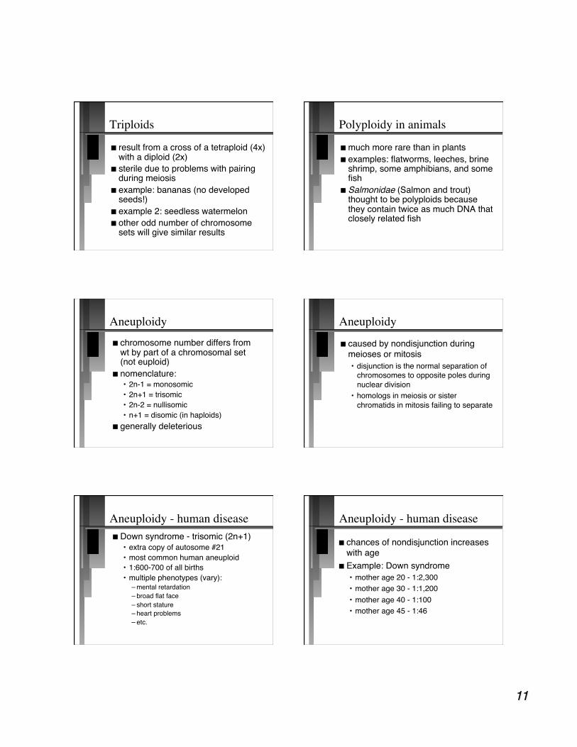
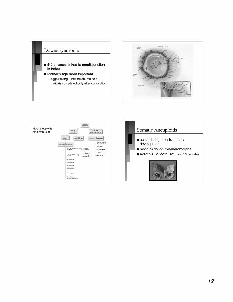
Most aneuploids die before birth Somatic Aneuploids
occur during mitosis in early development
mosaics called gynandromorphs example: Io Moth (1/2 male, 1/2 female)