gene expression in the genus deinococcus ian james …
TRANSCRIPT

GENE EXPRESSION IN THE GENUS
DEINOCOCCUS
IAN JAMES PURVIS
Presented for the degree of Doctor of Philosophy
University of Edinburgh
October 1984

Volume 11 Number 16 1983
Nucleic Acids Research
Isolation and characterisation of DraL, a type H restriction endonuclease recognising a sequence containing only A:T basepairs, and inhibition of its activity by uv irradiation of substrate DNA
I.J.Purvis and B.E.B.Moseley
Department of Microbiology, University of Edinburgh, School of Agriculture, Edinburgh EH9 3JG, UK
Received 11 May 1983; Reise&and-Accepted 29 July 1983
ABSTRACT A type II restriction endonuclease, Dral, isolated from Deinococcus
radiophilu5 ATCC 27603 recognises the palindromic hexanucleotide sequence
5' 3 '-A-A-AT-T-T-5'
and cleaves it, as indicated by the arrows, to produce blunt-ended frag-ments. The yield of enzyme is 100 to 1000 times that of the only other known type II restriction endonuclease that recognises a sequence composed solely of A:T basepairs, the isoschizomer AhaIll (1). Ultraviolet irradi-ation of the DNA substrate at relatively low doses inhibits the activity of Dral by "protecting' the recognition sequence and this may be exploited to give control of partial digestion of DNA by Dral.
INTRODUCTION
D. radiophilus belongs to the Family Deinococcaceae, the major char-
acteristic of members of this group being their extreme resistance to the
lethal effects of both ionising and ultraviolet radiations (2). The type
species of the group D. radiodurans ATCC 13939, has been shown to possess
a type II restriction endonuclease (3), MraI, recognising the sequence
5'-C-C-G-C-G-G-3'
3' -G-G-C-G-C-C-5'
We report here a description of the isolation and characterisation of the
site-specific endonuclease Dral, and a method by which partial digestion
of DNA molecules by Dral may be controlled.
METHODS
Assay for Restriction
Enzyme activity was estimated by incubating a sample (usually 1 p1)
with either 1 pg A-DNA or ColE1:Tn5 ccc DNA. One unit of enzyme represents
that activity which completely digests 1 pg of DNA during 1 hour's incub-
ation at 37 ° C in restriction buffer (10 mM Mg, 10 mM Tris-HC1, pH 8.0).
© I R L Press Limited, Oxford, England. 5467

Nucleic Acids Research
Protein estimation
Protein concentration was measured both by comparative absorption at
260 nm and 280 nm wavelength light and using a Bio-Rad protein assay kit.
Isolation of Dral
Three one-litre cultures of D. radiophilus in nutrient broth No 2
(Oxoid) were grown to early stationary phase by shaking at 37 °C. It should
be noted that protease activity increases greatly in late stationary and
decline phase. About 15 g wet weight of cells was harvested by centrifug-
ation at 10,000 g for 10 mm. The bacteria were resuspended in 30 ml 10 MM
Tris-HC1, 2 mM 2-mercaptoethanol (MSH), 0.1 mM phenylmethylsulphonylfluoride
(PMSF), pH 7.5 and broken open in a French pressure cell at 3,000 psi.
Following centrifugation at 10,000 g for 10 minutes to remove undamaged
cells and large fragments, the lysate was centrifuged at 100,000 g for
2 hours. Most of the DNA present in the supernate was removed using poly-
ethylene glycol 6,000 (PEG): dextran T500 phase partition (4). This pro-
cedure also removed significant amounts of non-specific exonuclease activity.
The enzyme preparation was dialysed (10-20 hours) against column buffer 1
(CB1 - 10 mM Tris-HC1, 2 mM MSH, 0.1 mM PMSF and 0.075M NaCl, pH 8.0) and
applied to a 20 x 2.6 cm DEAE-sephacel (Bio-Rad) column-, previously equili-
brated with CB1. After washing, the column was developed with a linear
NaCI gradieni
(M) sorbance 26OnM
Fig. 1. Protein Elution profile of D.radiophilus cell extraction DEAE-Sephacel
5468

Nucleic Acids Research
0.075-0.4 M NaCl gradient, Dral activity eluting in the 0.18-0.24 NaCl
region (Fig 1, shaded area represents region of 'unit' enzyme activity).
Active fractions were pooled, dialysed against column buffer 2 (CB2 -
0.01 M phosphate buffer, 2 mM MSH, 0.05 M NaCl, pH 7.5) and then applied to
a 10 x 2.6 cm hydroxylapatite (Bio-Rad) column previously equilibrated with
CB2. The column was washed with CB2 and then eluted with a linear gradient
of 0.01 to 0.4 M phosphate buffer. Dral activity eluted between 0.20 and
0.32 N phosphate. Each collecting tube contained enough restriction enzyme-
gradbovine-ser.um albumin (BRL) to ensure that the total protein concentra-
tion did not fall below 750 jig ml'; -- ----Fractions showing greater than 1,000
units ml' of enzyme activity were pooled and dialysedaiiIst -storagebuffer
(50% glycerol, 10 mM Tris-HC1, 2 mM MSH, 0.05 N NaCl, pH 8.0). Dral is
stable in this buffer for > 12 months at - 20 ° C.
RESULTS
The yield and specific activity of DraT during various stages of isol-
ation are shown in Table 1. The final preparation was considered to be free
of contaminating 5' and 3' exonucleases since there was no alteration of
cleavage patterns upon extensive incubation and because of the success of
sequencing techniques which are highly sensitive to such contamination.
Optimal Conditions for Dral Activity
Dral has an absolute requirement for Mg2+, a feature not uncommon in
type II restriction endonucleases (5). Maximum Dral cleavage was obtained
at 37 ° -39 ° C in a buffer containing 10 MM Mg 2+ and 10 mM NaCl at pH 8.0.
Concentrations of > 100 rnl1 NaCl, > 30 mM Mg2+, > 1 mM Mn 2 and > 0.1 mM Ca 2
were each inhibitory to enzyme activity. In general, conditions for maximum
activity were similar to those found for MraI (3).
Table 1. Stages of Dral Purification
Stage Total Total Specific Yield protein (mg) enzyme (units) activity (%)
(units mg' protein)
High speed centrifugation 1.86 x 10 8.0 x 10 5 4.32 x 10² 100
PEG:dextran phase partition 1.22 x 10 1 7.5 x 6.15 x 102 93.75
DEAE-sephacel 3.15 x 101 4.1 x 10 5 1.30 x 10 51.25
Hydroxylapatite 0.57 1.4 x 10 2.46 x iü 17.50
5469

Nucleic Acids Research
Table 2. Specificities of Type II Restriction Enzymes Dral and Ahalli
Enzyme Number of cleavage sites
pBR322 Ade 2 A SV40 0X174
AhalIl 3 16 13 12 2
Dral 2 (3a)
> 10 13 > 10 2
aFrom sequence data, see text.
Mapping of Dral cleavage sites
Cleavage of a variety of different DNA species, .ie A; Ade-2; 0X174 Rf;
SV40 and pBR322 (6-9) produced a pattern of bands, after polyacrilamide gel
electrophoresis (7-12% gradient), only previously seen in the case of Ahalil
(1) digestions (Table 2).
Double digestions of pBR322 using each of the commercially available
enzymes EcoRI, BamHI, PstI, Hindlil (all from Miles Laboratories) and Sail
(NBL Enzymes Ltd) with Dral indicated the presence of two Dral recognition
sites in this plasmid at the map positions 3250 and 3900 respectively.
These data suggested that a DNA sequencing procedure closely based upon
that described for AhaIII(1) should be used. The Sau3AI fragment 9 of
pBR322 was cloned into the M13mp7Rfl BamHI site (1, 10-12), sequenced using
the chain-terminator method (13) and the Dral cleavage sites determined 0
and 14). As was the case with Ahaill, during the course of sequending a
second Dral site was identified within the cloned fragment (3 in the whole
pBR322 molecule) producing upon cleavage a 19bp fragment.
From the above data it was concluded that D. radiophilus contains a
type II restriction endonuclease recognising the palindromic hexanucleotide
sequence
5' -T-T-T-A-A-A-3'
3' -A-A-A-T-T-T-5'.
Inhibition of Dral activity by uv irradiation of the DNA substrate
Cleaver at al (15) and Hall and Larcom (16) have shown that the activity
of type II sequence specific endonucleases is inhibited by thymine-thymine
dimer, and possibly cytosine.-thymine dimer, production within or adjacent
to the enzyme recognition sequence. In those studies the type II restriction
enzyme most sensitive to such inhibition was Hindlil, the recognition sequence
of which is 5'-A-A-G-C-T-T-3'
3' -T-T-C-G-A-A-5'
5470

Nucleic Acids Research
a b c d e f g h i j k I
Fig 4. Cleavage of uv irradiated 1-DNA by Dral, Hindlil or PstI. Lanes a-c show digestion of 1 pg 1-DNA by PstI after uv radiation
doses of 3600; 1800 and 0 Jm 2 respectively. Lanes d-g show cleavage of 1 ig A-DNA by Hindill after uv doses of 3600; 1800; 300 and 0 Jur° respec-tively. Lanes h-i show digestion of 1 ig A-DNA by Dral after uv doses of 3600; 1800; 300 and 0 Jm-2 . Fragment separation was by agarose gel electrophoresis (1%) and visualisation by EtBr staining.
distinctive separation of DNA molecules differing in size by only 19 base-
pairs. Initial alteration in the digestion pattern occurs at a uv dose of
60 Jar 2 while above a dose of 180 Jar 2 a fragment of 3567 basepairs appears
due to the inhibition of enzyme action at both the Dral recognition sequences
at positions 3250 and 3231 on the pBR322 molecule. Finally at doses
> 300 Jar 2 complete linear molecules of pBR322 (4362 basepairs) can be seen
due to the inactivation of all Dral recognition sites.
Comparative studies using Hindlil and Dral showed that inhibition of
Dral activity at individual recognition sites was three to four times more
sensitive to uv inhibition than that of Hindlil. This is illustrated by
comparative digestions of uv-damaged A-DNA with Dral, Hindlil and PstI
(Fig 4). In spite of differing numbers of restriction sites in the A-DNA,
(13 for DraI, 7 for Hindlil and 18 for PstI), it is still clear that Dral
activity is inhibited to a greater degree than that of Hindlil, whilst
irradiation of the DNA has little effect upon the action of PstI, the
recognition sequence of which contains no adjacent thymine residues.
DI S CUSSION
In contrast to the blue green alga Aphanothcoe haiophytica from which
Ahaill is isolated, Deinococcus rad'iophiiva is easily grown and gives a
5472

a h C d p f g h I I k I size (bp )
4362 3567
2850
1489
793 692
Nucleic Acids Research
32313250 3942
map position 375 436210 375
sit. (bp) 2850 690 692 793
yn,e Ban,HI Oro I(2) Orol BomHI
Fig 2. Schematic representation of the pBR322 molecule showing cleavage sites of Dral and Haml 1 I.
However Oral activity is much more sensitive to inhibition following
uv irradiation of the DNA substrate than Hindlil. By linearising cccpBR322
0 j.'g) with the type II restriction endonuclease BamHl, (map position 37 5 on
the pBR322 molecule), followed by uv irradiation at an incident dose rate
of 1.05 Jm 2 in a uv transparent buffer (10 mM Tris—HC1 pH 8.0), the effect
of different radiation doses upon Dral activity was observed (Figs 2 and
3). Unfortunately the presence in pBR322 of two Dral sites only 19 base—
pairs apart does complicate interpretation of the results. The appearance
of a 1489 basepair fragment allied to the simultaneous removal of both 692
and 793 basepair fragments indicates the inhibition of activity at the Oral
recognition site positioned at 3942 on the pBR322 molecule. Inhibition of
activity at either of the other two sites singly will not produce any new
bands as the fragment size resolution of a 1% agarose gel will not allow
U 11 LUll 1 U LI L I
Lanes ikillustrate1jig pBR322 linear ised with BamilI and c'iven uv radiation doses of 4200; 3600; 3000; 2400; 1800; 1200; 600; 300; 180; 60 and 0 Jm 2 respectively before 1 hr Dral digestion. Lane 1 shows BamilI linearised pBR322 plasmid DNA. Fragment separation was by agarose gel electrophoresis (1Z) and visualisation by EtBr staining.
5471

Nucleic Acids Research
relatively high yield of the Ahalli isoschizomer, Dral. D. radiophilus is
also extremely radiation resistant and this property provides an excellent
screen against contamination. These properties make it likely that Dral
will become a commercially important restriction endonuclease.
Since Dral recognises a sequence containing only A:T basepairs it
may be possible to expand studies on DNA-DNA and DNA-protein interactions
which were being delayed by a dearth of suitable, easily available enzymes.
Dral will also be useful in the study of eukaryotic DNA where, due to the
marked asyiiiinetry of A T distribution large-regions-of G C rich DNA may be
produced.
Difficulties in controlling partial digestion of DNA molecules during
restriction may, to some extent, be overcome by utilising the inhibition
of Dral activity by uv irradiation of the substrate DNA. Although Dral
digestion produces blunt-ended fragments, this disadvantage can be removed
by poly (dA) 'tailing" of the restricted DNA to be cloned, followed by
insertion into a poly (dT) "tailed' vector regenerating Dral sites either
side of the cloned fragment. To allow gene expression within the irradi-
ated insert, multiplication of the recombinant molecule in a uv repair-
proficient strain of bacterium, yeast or tissue culture line must occur.
However this process will 'reactivate' any Dral recognition sequences
present within the DNA insert.
ACKNOWLEDGEMENTS
We wish to thank Dr N Brown of the University of Bristol who provided
both instruction and assistance with the DNA sequencing. Special note
should be made of the help and support of other members of our research
group, M Mackay, D Evans and S Whyte.
REFERENCES Whitehead, P.R. and Brown, N.L. (1982) FEBS lett 143, 296-300. Moseley, B.E.B. (1983) Photochem. Photobiol. Revs. 7, 223-274. Wani, A.A., Stephens, R.E., D'Ambrosio, S.M. and Hart, R.W. (1982) Biochem. Biophys. Acta 697, 178-184. Schleif, R. (1980) Methods Enzymol. 65, 19-23. Modrich, P. (1982) CRC Crit. Rev. Biochem. 13 No. 3, 287. Sutcliffe, J.G. (1979) Cold Spring Harb. Symp. Quant. Biol. 43, 77-90. Sanger, F., Coulson, A.R., Hong, G.F., Hill, D.F. and Petersen, G.B. (1982) J. Mol. Biol. 162, 729-773. Sanger, F., Coulson, A.R., Friedmann, T., Air, G.M., Barrel, B.C., Brown, N.L., Fiddes, J.C., Hutchinson, C.A. III, Slocombe, P.M. and Smith, M. (1978) J. Mol. Biol. 125, 225-246. Fiers, W., Contreras, R., Haegeman, C., Rogiers, R., Van de Voorde, A.,
5473

Nucleic Acids Research
Van Heuverswyn, H., Van Herreweghe, J., Volckaert, G. and Ysebaert, M. (1978) Nature 273, 113-120. Messing, J., Gronenborn, B., Muller-Hill, B. and Hofschneider, P. (1977) Proc. Natl. Acad. Scj. USA 74, 3642-3646. Dagert, M. and Ehrlich, S.67(1979) Gene 6, 23-28. Sanger, F., Coulson, A.R., Barrell, B.G., Smith, A.J.H. and Rose, B.A. (1980) J. Mol. Biol. 143, 161-178. Sanger, F., Nicklen, S. and Coulson, A.R. (1977) Proc. Natl. Acad. Sci. USA 74, 5463-5467. Brown, N.L. and. Smith, M. (1980) Methods Enzymoi. 65, 391-404. Cleaver, J.E., Samson, L. and Thomas, G.H. (1982) Biochem. Biophys. Acta 697, 255-258. Hall, R.K. and Larcom, L.L. (1982) Photochem. Photobiol. 36, 429-432.
5474

DECL&RLTION
I declare that this thesis is composed of my own work and has
been compiled by myself.
Ian James PUrV±3

(i)
SUMMARY
The genus Deinococcus consists of four species, D.
radiodurans, D. radiophilus, D. radiopugnans and D. proteo-
lyticus. There are two strains of D. radiodurans, Ri
(Anderson) and Sark. All produce red-pigmented colonies
on agar and are characterised by their extreme resistance
to, and non-mutability by, both ionizing and ultraviolet
irradiation.
Attempts were made to phenotypically express, within
members of--the above genus, genes from a variety (> 10) of
common or constructed plasmid vectors introduced via trans-
formation or conjugation. All these plasmids failed to
functionally express their selectable traits and this led
to an investigation of possible parameters controlling
such expression.
The presence of type II site-specific endonucleases
and associated DNA modification systems was confirmed in
D. radiodurans Ri and demonstrated in D. radiophilus. Two
novel restriction enzymes, Dral and Drall, were isolated
from D. radiophilus, Dral recognising the DNA sequence
5 1 TTAAA 3' and now being available commercially. The ex-
treme sensitivity of this enzyme to inhibition by pre-
irradiation of the substrate DNA was demonstrated.
Gene banks of chromosomal and plasmid DNA derived
from D. radiophilus were created in the Escherichia coli
plasmid vector pAT153. Although the Dral restriction/
modification system did not express in E. coli 1 it was
found, on testing a wide range of mutants, that the leuB

(ii)
mutation of E. coli HB101 could be complemented by a 10.24
kilobase (kb) insert of D. radiophilus DNA in pAT153. The
size of the insert was subsequently reduced to a functional
unit of only 800-900 basepairs which appeared to code for
a protein of approximately 18.000 daltons. About two-
thirds of this DNA region was sequenced and showed many
open-reading frames but only one preceded.by an E. coli-
like promoter and without a terminator codon, in frame, close
to the translation initiation site. In conjunction with
further analysis of other D. radiodurans Ri cloned genes
this should give valuable information on the regulation
and organisation of coding regions within this odd group
of bacteria.

CONTENTS
Page
CHAPTER 1: INTRODUCTION
THE GENUS DEINOCOCCUS 1
REGULATION OF GENE EXPRESSION AT TRANSCRIPTION 5
GENE CLONING AND DNA SEQUENCING 9
SITE-SPECIFIC ENDONUCLEASES AND METHYLASES 16
5.- IDENTIFICATION OF PLASMID ENCODED --PROTEINS 19
CHAPTER 2: MATERIALS AND METHODS 21
CHAPTER 3: RESULTS
FOREIGN GENE EXPRESSION IN D. radiodurans - 60
NUCLEASE ACTIVITY IN THE NOCOCCACEAE 64
MODIFICATION METHYLASE ACTIVITY 81
DEVELOPMENT OF THE CLONING AND EXPRESSION OF D. radiophilus GENES 82
CHAPTER 4: DISCUSSION
1. FUNCTIONAL EXPRESSION BY GENES OF FOREIGN ORIGIN IN BACTERIA 94
ACKNOWLEDGEMENTS 114
REFERENCES 115

CHAPTER 1. INTRODUCTION

1.
1 THE GENUS DEINOCOCCUS
1.1 TAXONOMY AND GENERAL BIOLOGY
Interest in the small group of non-sporing, red-pig-
mented bacteria that comprise the genus Deinococcus has
been aroused largely because of the extreme resistance to
both the lethal and mutagenic effects of ionizing and
ultraviolet (UV) radiation which characterises the group.
The genus consists of four species; Deinococcus radiodurans
(strains Ri and Sark), D. radiophilus, D. proteolyticus and
D. radiopugnans. The initial isolation of D. radiodurans
(strain Ri) was made in 1956 while studying a sample from
a meat processing factory in Oregon, U.S.A. (Anderson et
al., 1956). This organism has become the type species of
the genus. The other species were found subsequently in a
range of irradiated materials, i.e. Bombay duck, llama
faeces and haddock. Further details can be found in the
review article of Moseley (1983). Originally these species
were classified in the genus Micrococcus but subsequent
investigation of cell wall composition (Sleytr et al., 1973),
fatty acid and phospholipid content (Thompson et al., 1980;
Jantzen et al., 1974) and cell ultrastructure (Lancy and
Murray, 1978) all detracted from this view. A comparative
study of 16s ribosomal RNA sequences (Brooks et al., 1980)
finally showed that a separate genus for the red-pigmented
radiation-resistant micrococci was required, the genus
Deinococcus. The genus is so distinct, in fact, that it
forms by itself one of the eight recognised groups of the
Eub.acteria (Fox et al., 1980).
Growth of Deinococcus spp. is normally achieved in
tp
tryptone-glucose-yeast extract (TGY) medium incubated at
30°C. Although D. radiophilus grows slowly under such
conditions there is a marked increase in its growth rate
if nutrient broth (Oxoid No. 2) is used. Generally,
doubling times of Deinococcus spp. in liquid culture are
approximately 80 to 100 minutes while cells on agar re-
quire two to three days incubation to become colonies
visible to the naked eye. The organisms are rarely seen
as single cells, diplo- and tetracoccal forms being pre-
valent. The sizes of cells varies depending upon the
species. Very little is known about the detailed metabolic/
biochemical infrastructure.' The cells grow only in the
presence of oxygen and they stain as Gram-variable.
Chemically-defined media have been described (Little and
Hanawalt, 1973; Shapiro et al., 1977) which allow for the
isolation of a variety of auxotrophic mutants.
As previously mentioned, interest has really been
focused upon the ability of members of the genus to not
only withstand very high levels of radiation without loss
of viability, 500 k rad or 500 Jm 2 (. Tempest, 1978),
but also to resist mutation induced by ultraviolet and
ionizing rays. Although all the species are pigmented,
this colouration would not appear to have any major role
in radiation tolerance (Moseley, 1967). The character-
istic resistance appears to reside in rapid and efficient
DNA repair. In D. radiodurans Rl enzymes involved in ex-
cision repair (Moseley and Evans, 1983) have been shown
to be of central importance in ultraviolet light-induced
lesion repair as well as in the repair of bulky adducts

caused by chemical agents such as mitomycin c. The pre-
sence of a recombination repair pathway system, but not an
error-prone mechanism, has also been inferred (Moseley and
Copland, 1975). Enzymatic photoreactivation is absent.
L2 GENETICS AND DNA CONTENT
The only known technique for inter- and intraspecies
gene transfer so far available within the Deinococcaceae
is via transformation (i.e. the uptake and integration of
DNA from the matrix surrounding the cell) (Moseley and
Setlow, 1968; Tirgari and Moseley, 1980). Despite in-
tensive efforts, no bacteriophages capable of plaquing on
Deinococcus spp. have been found (I. Masters, personal
communication). No conjugal transfer of DNA either within
or between the species has been observed (Tirgari, 1977).
Although protoplasting and protoplast regeneration tech-
niques have been successfully devised, no genetic evidence
was obtained for protoplast fusion (G. AlBakri, personal
communication). It has been shown that at least three
site-specific endonucleases exist within members of the
genus, MraI in D. radiodurans Rl (formerly Micrococcus
radiodurans, Wani et al., 1982) and Dral and Drall in D.
radiophilus (this thesis; Purvis and Moseley, 1983).
However1 close examination of the chromosomal DNA of these
two species indicates a lack of any methylation modifi-
cation normally associated with restriction/modification
systems (Mackay, 1983; Schein et al., 1972).
Genetic study of the type species D. radiodurans Rl
has been complicated by the apparent existence of multiple

4.
independently-segregating genome equivalents per viable
unit (Hansen, 1978; Tirgari and Moseley, 1980; Moseley
and Evans, 1981). This type of genome organisation is
thought to occur throughout the genus (Purvis and Duncan,
unpublished results) as well as in other bacterial species.
In Azotobacter vinelandii. actively dividing cells may con-
tain as many as 40 genome equivalents (Sadoff et al., 1979).
There is no positive evidence that this anomalous chromo-
somal organisation aids the repair to DNA damage. In fact,
vegetative cells of A. vinelandii are very sensitive to
ultra-violet light and D. radiodurans Ri shows no direct
correlation between genome copy number and increased rad-
iation resistance (Harsojo et al., 1981).
Further information on thesecharacteristics can be
seen in Table 1.1. Of particularinterest is the recent
discovery of plasmid molecules in all species of the genus
apart from D. radiodurans Ri (Mackay, 1984). Molecule
sizes ranged from 2.5kb (pt)E 30) to 139.1kb (tJE 21). It
was impossible to ascribe any characteristics of the
bacteria to the possession of the plasmids but the failure
to cure either D. radiophilus or D. radiodurans SARK of
any plasmid type may indicate the presence of vital in-
formation on these extrachromosomal replicons.
1.3 AIMS OF THE PROJECT
The original aim was to develop vector systems cap-
able of transferring genetic material both intra and inter-
genetically. The failure to do so led to an analysis of
the reasons for the failure of foreign genes to express

TABLE 1.1 DNA PARAMETERS OF SPECIES WITHIN THE GENUS DEINOCOCCUS
Species % G+C Genome size (x109d) (b)
Plasmid content Transformation
D. radioduransRl 68 1.8 - Yes
D. radiodurans Sark N.T. N.T. pUE 10 Yes pUE11
D. radiophilus 65 1.5 pUE 1
pUE2 No
pUE 3
D. proteolyticus 67 1.8 pUE 20 No pUE 21
D. radiopugnans 68 2.9 pUE 30 No pUE 31
C.M. Duncan, personal communication I. Purvis and B.E.B. Moseley, unpublished results M. Mackay (1984.). P. Tempest (1978)
N.T. - not attempted.

5.
in D. radiodurans Ri. Of particular interest was the
molecular structure of the Deinococcus genotype with
specific reference to the control of gene expression as
determined by the nucleotide sequence of the gene itself.
Nothing was known previously of the molecular mechanisms
of transcription and translation within the Deinococcaceae
and it was hoped that the use of E. coli, as a medium for
the investigation of gene structure in the genus
Deinococcus, would provide some insight.
2 REGULATION OF GENE EXPRESSION AT TRANSCRIPTION
2.1 INTRODUCTION
The functional expression of the information contained
within a gene is controlled by a myriad of interacting
mechanisms, many associated with the vital functions of
transcription and translation. Other control parameters
do come into play, particularly in the eukaryotic matrix.
This section, however, will be restricted mainly to the
relevant details of prokaryotic gene expression although
certain similarities do exist in the realm of eukaryotic
gene regulation.
The chromosome of prokaryotic organisms is organised
into transcriptional units, operons, containing either
single or multiple coding regions, cistrons. The operons
contain regulatory sequences of DNA that determine if,
when and at what level a particular gene is expressed.
The action of the regulatory sequences is via complicated
DNA/DNA, DNA/RNA, DNA/protein and RNA/protein interactions

1.90
and is dependent, to a large extent, upon the primary
nucleotide sequence.
2.2 CONTROL AT TRANSCRIPTION
Transcription is the first stage of gene expression
and is of prime importance in regulation, particularly in
the prokaryotic organism. Common prokaryotic regulatory
mechanisms involved, preceding, during and directly after
transcription, are shown in Figure 1.1 and excellent re-
views on promoters (Rosenberg and Court, 1979), attenuation
(Yanof sky, 1981) and termination (Adhya and Gottesman,
1978) are available.
Initially RNA polymerase must recognize the promoter
site of a gene before translocating 'downstream' to the
site of RNA strand synthesis. A promoter can be defined
as a segment of DNA containing signals within the nucleo-
tide sequence for the correct binding and subsequent acti-
vation of the RNA polymerase holoenzyme. Such interaction
between DNA sequence and RNA polymerase assembly is
strongly controlled by other proteins such as the a factors
and CAMP binding protein (CRP). Analysis of a large number
of mainly E. coli promoters has shown the presence, within
the. 40 to 50 basepair region comprising the promoter, of
conserved structural domains, illustrated in Figure 1.2.
This consensus sequence has been derived from averaged
observations (Rosenberg and Court, 1979) in a study of E.
coli promoters. Other sequence organisations can exist as
seen in the multiple a modifying factors present in
Bacillus subtilis (Losick and Pero, 1981). Although the major a

FIG. 1. 1. POSSIBLE CONTROL MECHANISMS FOR PROKARYOTIC TRANSCRIPTION (Stylised operon
promoter/ RNA polymerase binding and regulation
Attenuation
P 0 1 ATG A
internal promoters?
B C
Termination site
$
Protein/ DNA binding upstream of promoter e.g. cAMP Receptor protein (CRP)
Operator DNA/regulatory protein +/-control
Intracistronic termination or 'pausing ant it erm in at ion
The combined action of these possible regulatory mechanisms enable the transcriptional process to respond directly to external and internal events relevant to expression of the operon.

Promoter organisation (highlighted T appears to be invariable)
(a) Prokaryotic promoter (E.coli)
Conserved region in >40 promoters Initiation
* TTGACA 10-13bp TATArG 4-1bps 'CAT
-35 region Pribnow: box mRNA synthesis
() Ekay.icjrornoter(RNA polymerase:fli
Initiation 6GAATCT 35-40bp GTATAAOGOOG 9-llbp 4Py000PyAP
t- t
Goldherg-Hogness box mRNA sythesis

7.
factor of the vegetative cell causes initiation at the
consensus sequence (seen in Fig. 1.2a) , other a factors
exist within the cell that direct RNA polymerase binding
to other types of promoter site. This multiple promoter
organisation, modulated by different c factors, is thought
to play a major role in the temporal regulation of gene
expression required for the more complicated developmental
responses to fluctuating environmental conditions, e.g.
sporulation. Despite qualitative variations in nucleotide
sequence, a general pattern does emerge about the spatial
relationships between promoter regions and RNA polymerase.
In general, at least for bacterial promoters, the nucleo-
tide sequences of regions -35 and -10, upstream of actual
mRNA synthesis initiation, play a vital role.
After initiation of mRNA biosynthesis, transcription
is modulated by two processes; induction/repression via
protein binding at the operator site(s) or transcript
termination determined either purely by nucleotide
sequence or in association with a protein/ribosome. The
classic model of Jacob and Monod (1961) for induction and
repression of operon transcription has shown the, clear
effect that protein binding can have at a particular
regulatory region of DNA, the operator. The relationship
of regulatory proteins with operator regions of DNA is
comprehensively reviewed in various articles (Miller and
ReznickOff, 1978).
Recently,it has become obvious that termination of
transcription plays a central role in gene regulation.
Not only does termination occur at the end of an operon

EM
but in many cases there is the possibility for pre-
emptive termination or 'pausing' within both control and
'structural' regions of the operon. This intrinsic abil-
ity, largely residing in the primary nucleotide sequence,
leads to the observed processes of translational polarity
and attenuation. The signals for termination, like those
for promoters, reside in the secondary structure of both
DNA and primary transcript. In certain cases the p -
protein is essential for transcript termination although
pausing still occurs in the absence of the regulatory
protein. The most striking feature common to all DNA
sequenôes at which RNA elongation can be stopped is a
region of hyphenated dyad symmetry just proximal to the
termination point. For p independent termination in
prokaryotes the dyad symmetry is surrounded by a G/C rich
region allowing more stable loop-stem structures to be
formed than those produced by the A/T rich regions pre-
ceding the p-dependent and eukaryotic termination sites.
The actual point of termination would appear to occur
within a run of uridine residues on the transcript. The
identification and analysis of attenuator sites preceding
gene clusters involved in the biosynthesis of amino-acids
has demonstrated how the degree of gene transcription can
be controlled by the interplay between the transcript
secondary structure and translation mechanisms (Yanofsky,1981;
Gemmill et al., 1979), nucleotide sequence and regulatory
molecules.
Obviously the nucleotide sequence of DNA molecules
not only codes for the structural proteins and RNA molecules

of the cell but also plays a primary and varied role in
the degree of gene expression. This ranges from trans-
cription initiation through various termination controls
to the ancillary sequences present upon the mRNA.
necessary for successful translation (Kozak, 1984.). These
controls combined with the plethora of regulatory mechan-
isms not directly influenced by the DNA nucleotide sequence
e.g. pools of metabolites, protein stability, etc. enable
the bacterium to recognize and respond to the principal
external and internal events relevant to the expression
of the operon. With the aid of modern computer analytical
techniques it is hoped the sequencing of genes from members
of the genus Deinococcus will give vital information on
the organisation and regulation of gene expression.
3 GENE CLONING AND DNA SEQUENCING
3.1 GENERAL INTRODUCTION
Genetic manipulation can be described as the formation
of novel combinations of heritable material by the in-
sertion of nucleic acid molecules into any virus, bacterial
plasmid or other vector system so as to enable their in-
corporation into the genetic background of a host in which
they are capable of continued propagation. General aspects
of recombinant DNA technology are described by Old and
Primrose (1982). The essential requirements of DNA mani-
pulation are:-
i) A DNA vehicle or vector that can replicate in
living cells after foreign DNA has been incorporated
into it.

10.
A DNA molecule :to be cloned.
A means of joining vector to target DNA.
A means of introducing the recombinant DNA into
a host organism which will allow stable inheritance.
V) A direct or indirect method of screening for
host cells which now contain the recombinant DNA
molecule.
Only recently have all these elements been available
and some are still being modified. The important advances
have been in the isolation of type II restriction endo-
nucleases (Section 1.4), DNA-ligating enzymes (Higgins and
Cozzarelli, 1979), bacterial transformation (Polsinelli
and Mazza, 1980) and the introduction of agarose and poly_
acrylamide gel electrophoresis (Rickwood and Hames, 1982).
Four types of vector are generally available, i.e.
bacterial plasmid, single-strand DNA bacteriophage, A
bacteriophage and cosmid, the choice being determined by
the experimental aim. Only plasmid and M13 bacteriophage
vectors are of direct relevance to this thesis and the
reader is referred to review articles by Hendrix et al.
(1983) and Collins and Hohn (1979) for information on A
vectors and cosmids respectively.
DNA sequencing has become one of the most important
tools for the analysis of DNA regions isolated by the
various DNA cloning techniques. As reviewed in Section
2.2 the nucleotide sequence within and immediately
surrounding a gene plays a major role in the regulation
of expression. The development of two fast and reliable
11.
DNA sequencing systems by Maxam and Gilbert (1977) and Sanger
et al. (1977) has allowed accurate sequence determination
of DNA molecules of up to 400 nucleotides in length.
Longer regions may be sequenced by analysis of overlapping
short stretches.
3.2 PLASMIDS AS CLONING VEHICLES
Plasmids are DNA replicons that are stably inherited
in an extrachromosomal state (Novick et al., 1976). This implies
genetic homogeniety, constant monomeric size and ability
to replicate independently of the chromosome. Many bacter-
ial plasmids have been used as cloning vectors and all show
some of the following advantages:-
Bacterial plasmids, especially from E. coli, may be
easily isolated and purified in large quantitites.
A wide range of plasmid isolation techniques are
available many of which are capable of modification,
depending on whether preparative quantities of DNA
are required or for screening for recombinant
molecules (Birnboim and Doly, 1979; Gryczan et al.,
1978; Kado and Liu, 1981)
If not ubiquitous, plasmids do exist in a wide range
of bacterial species thus allowing for the possibil-
ity of 'shuttle vector' manufacture. The ability of
such a plasmid to replicate and express selectable
markers in two different host systems is of great
importance in the study of both chromosomal and
plasmid regulatory controls (Primrose and Ehrlich,
1981) .

12.
C) Many plasmids used as vectors are relatively small
in comparison with A or cosmids and therefore are
resistant to damage and have few restriction endo-
nuclease sites.
Plasmids can be found which replicate either in a
relaxed (multicopy) or stringent (single copy)
manner. These are particularly useful if large
quantities of cloned gene products are required or
multicopies of the gene would lead to cell death,
respectively (Hecker et al., 1983).
Advances are &llowing the construction of positive
selection vectors for many bacterial species.
Transformants containing recombinant molecules can
then be directly selected rather than having to
rely on the classical insertional inactivation
method (Hennecke et al., 1982; Gryczan and Dubnau,
1983)
In certain cases the number of plasmids per cell
can be greatly increased (amplified) by the inhib-
ition of protein synthesis by chioramphenicol or
even by raising the temperature.
Expression vectors, for those genes incapable of
normal function in the usual plasmid vehicles, and
promoter/terminator screening vectors are also
available (Rosenberg et al., 1983; Yanof sky,
1984)
E. coli has displayed a pleasing adaptability in terms
of genetic manipulation and this has led to the development
of a wide range of versatile host-vector systems for this

13.
bacterium (Bolivar and Backman, 1979). Perhaps the most
commonly used plasmid vectors are pBR322 (Fig. 1.3. .) and
its derivatives (Twigg and Sherratt, 1980). Developed
from a colicin determining plasmid, they illustrate
all the requirements of ideal plasmid cloning vehicles
(Sherratt, 1979; Peden, 1983). The most important pBR322
derivative in relation to this thesis is pAT153, isolated
by removing a HaeII-generated 755 basepair fragment from
pBR322. This plasmid has the advantage over pBR322 of
non-cotransmissbility (important for biological contain-
ment) and a slightly increased intracellular copy number.
Rapid progress is being made in the construction of
cloning vectors for a wide variety of organisms, both
prokaryotic and eukaryotic (Hofschneider and Goebel, 1982;
Bagdasarian et al., 1983). Of particular interest are
those plasmids capable of marker expression and maintenance
in both B. subtilis and Staphylococcus aureus (Gryczan and
Dubnau, 1978) and the complex plasmid/transposon organ-
isation of Streptococcus faecalis (Clewell, 1981; Jacob
and Hobbs, 197 It) war. 'e.c.
Possibly the major disadvantage of using plasmids as
cloning vehicles is the size limitation of inserted DNA.
An average insert of around 5 kilobases (kb) is normal and
greater than 20kb unusual. As a gene library consists of
a collection of cloned DNA fragments which statistically
comprise the entire genome of the organism, success ob-
viously depends upon the average size of insert and size
of the donor genome (Dahl et al., 1981) . It would be
impractical, in terms of individual clones required, to
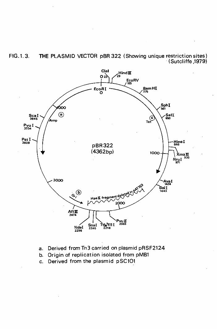
FIG.1.3. THE PLASMID VECTOR pBR 322 (Showing unique restriction sites) (Sutcliffe ,1979)
Sca I 3845
Pvu I 3734
Pst I 3608
Cial Hindlif 023 29
EcoRV 185
EcoRl BamHI 375 0
SphI
Sail C 650
let
Hinel pBR322 846
(4362bp) 1000 Xma]II Nru I
935
971
3000 1424
Hael ragmetet/
Bail
2000
Afilil 2474
> aITt111 2065 NdeI 2245 2218 2296
Derived from Tn3 carried on plasmid pRSF2124 Origin of replication isolated from pMB1 Derived from the plasmid pSC101

14.
construct gene libraries of organisms with large genome
complements (eukaryotes) in plasmid vectors. Cosmid and
A vectors are candidates for this kind of work allied to
subsequent sub-cloning into plasmid vehicles if necessary.
3.3 M13 - ITS ROLE IN CLONING AND SEQUENCING
The development of the single-stranded DNA bacterio-
phage (M13, Ff, etc.) has allowed the elegant combination
of gene cloning and ñucleotide sequencing in the same
vector molecule. Messing and his colleagues (Messing, 1983)
have constructed a range of modified M13 bacteriophages
that has found wide use in recombinant DNA technology.
The unique life cycle of filamentous bacteriophages, such
as M13, provides large amounts of packaged (+) single-
stranded DNA as well as double-stranded intracellular re-
plicating-form plasmid (RP). M13 infects E. coli via the
F-pilus resulting in chronic infection, the host releasing
infective virions through the cell wall. M13mp bacterio-
phages (Messing's modified M13 range) carry part of the
lac operon of E. coli inserted into a non-essential region
of the M13 genome (Fig. 1.4). The lac DNA codes for the
N-terminus of -galactosidase which complements a host lacZ
mutation (a-complementation) permitting the formation of
blue coloured plaques on medium containing the histo-
chemical stain, 5-Bromo-4-ch1oro-3-indolyl--D-ga1actoside
(X gal). The bacteriophage lacZ DNA contains multiple
unique restriction sites and insertion of foreign DNA into
this region nullifies the cL-complementation thus producing
clear rather than blue plaques upon growth and multiplication

15.
of the recombinant. Use of the M13mp constructs is
especially attractive for the rapid association of single-
stranded template necessary for DNA sequencing and hybrid-
isation. Frequently M13mp systems are closely associated
with the Sanger dideoxy terminator method of
sequencing using a primer which anneals to a homologous
region of N13 DNA just to the right of the cloning sites
(Fig. 1.4). This permits 5' 3' labelled extensions
which randomly terminate at the insertion of a dideoxy
molecule. The separate use of ddATP,ddGTP, ddTTP and
ddCTP allows the production of a 'ladder' when subjected
to electrophoresis and autoradiography. Such a ladder
allows the determination of a nucleotide sequence of over
400 basepairs. This length can be increased by sequencing
shorter, overlapping sequences either derived by random
or directed subcloning techniques. The data from such
experiments are assimilated by computer to produce longer
DNA sequences. Directed sub-cloning routines allow for
this nucleotide compilation whilst avoiding the asymptotic
nature of data accumulation associated with random
'shotgun' methods of fragment production, e.g. sonication
(Deininger, 1984; Hong, 1982; Guo et al., 1983). A
general view of cloning routines in Ml_-kap bacteriophages
is shown in Figure 1.5.

Ipal
M13 CLONING
Sail SmaI AccI XmaI
I I I
M13mp9 ATGACCATGATTACGCCAAGCTTGGCTGCAGGTCGACGGATCCCCGGGAATTCACT I I I I I I I
HindZ PstI BamHI EcoRl
1- -,
Ia z 1 DIRECTION OF CHAIN C ELONGATION
FIG. 1.4. MAP AND CLONING SITE NUCLEOTIDE SEQUENCE OF M13mp9.
(Messing &Wieira1982)
'bE 0 ". cI11
Co Q. EaEI ci'L 't. t'vLr

I Double-stranded DNA to be sequenced
ciease 10fl I enzyme I _4n
.1m digest
stiofl
F Double-stranded DNA fragments with ends compatible with
vector insertion site
I MI3RF I DNA
I suitable
$nuclea~se
I Cutwith
]gesho
restrictionenzyme at
digestion I unique cloning site
Ligation J Cut vector DNA
Double-stranded M13 DNA bearing double-stranded insert
£ coil Host cell
Transformation
Plating Out Blue plaques: I non recombinants
Colourless plaques: M13 recombinants
Select single plaques and grow up 1 .Sml cultures
Infected cells bearing double-stranded AF with double-stranded insert
Pure single- stranded DNA template containing single-stranded Insert
'Dideoxy sequencing
FIG. 1.5 CLONING WITH M13 TO GIVE SINGLE—STRANDED DNA TEMPLATE FOR SEQUENCING

16.
4 SITE-SPECIFIC ENDONUCLEASES AND METHYLASES
.4.1 GENERAL PROPERTIES
Sequence-specific endonucleases and their associated
methylase activities are largely responsible for the host
delineated barriers that regulate interstrain DNA transfer
in prokaryotic cells. It was the observation of such re-
striction and modification which eventually led to the
identification and utilization of the enzymes involved
(Luria, 1953). The investigation of enzyme properties,
activities and cofactor requirements has produced a class-
ification scheme consisting of three groups; types I,
II and III restriction endonucleases. Although of value
to the cell, types I and III are not generally used as
tools for genetic engineering and further information can
be gained from the review of Yuan (1981).
4.2 TYPE II RESTRICTION ENDONUCLEASES
Type II restriction endonucleaseá are DNases that
recognised specific oligonucleotide sequences, make double
strand breaks and generate unique, equimolar . fragments
of a DNA molecule substrate. Their use as analytical
and engineering tools resides in this predictable, controll-
able activity. Various review articles deal directly with
their sources (Roberts, 1983) , sequence specificity
(Modrich, 1982) and purification (Pirrotta nd Bickle,
1980). Type II classification covers a wide range of
enzymes showing the general characteristics listed in
Table 1.2. However,terminoiogy such as restriction endo-
nuclease may be misleading in so far as few of these

17.
enzymes have been shown to be involved in restriction/
modification systems in vivo and there may well be diverse
modes of DNA-protein interactions at work.
4.3 MODIFICATION TRANSMETHYLASES (TYPE II)
Such enzymes recognise a specific nucleotide sequence
and methylate bases within this sequence in the presence
of S-adenosyl-L-methionine (SAM). The activity can be
seen in conjunction with the complementary restriction
endonucleases but often species can be found having methylase
but no associated endonuclease activity. As in the case
of type II site-specific endonucleases, the methylases
tend to be simple proteins acting in monomeric or dimeric
form.
4.4 CLONING OF RESTRICTION/MODIFICATION SYSTEMS
It would be of obvious benefit to remove the genes
coding for enzymes of interest from their normal back-
ground and place them in plasmid vectors capable of allowing
expression in different hosts,. normally including the ubi-
quitous E. coli. There are two basic conditions that
must be fulfilled in order for a restriction-modification
system to be cloned and transferred successfully to a new
host. It is essential that the genes for the restriction
enzyme and inethylase be linked upon the chromosome or
plasmid of the original organism. Also the methylase gene
must be functionally expressed in advance of any endonuclease
production so as to protect both host and recombinant plasmid
from digestion. In contrast, the only major barriers to the

TABLE 1.2. GENERALISED PROPERTIES OF TYPE II RESTRICTION
ENDONUCLEASE S
Recognition of distinct oligonucleotide sequences,
frequently palindromic in nature.
Nuclease activity closely linked to the recognition
site.
Simple cofactor requirements, normally Mg2+.
Nuclease and associated methylase activities reside
in separate proteins, not usually multifunctional.
Variable dependence upon ionic strength, salt con-
centration and sulphydryl compounds for activity.
Mem
TABLE 1.3. SENSITIVITY OF RESTRICTION ENZYMES TO ULTRA-
VIOLET INDUCED DAMAGE TO THEIR SUBSTRATES
Enzyme Recognition Sequence and incision sites
+ Hind III 5' AAGCTT
3' TTCGAA 4, +
Eco RI 5' GAATTC
3' CTTAIjG
Barn HI 5' GGATCC
3' CCTAG$
Sal I 5' GCGAC
3' CAGCC
Hae III 5' GGC
3' GGfC
Hha I 5' GdbC
3' CG$G decreasing sensitivity

18.
cloning of the niethylase gene activity alone is the com-
patibility of gene expression pathways and suitability of
internal metabolic functions (Walder et al., 1983; Janulaitis
et al., 1982; Schoner et al., 1983; Walder et al., 1981).
4.5 ULTRAVIOLET LIGHT DAMAGE
It has been shown that ultraviolet light can induce
damage in DNA which leads to serious disruption of activity
of certain type II restriction endonucleases (Hall and Larcom,
1982; Cleaver et al., 1982). Inhibition seems closely
related to the presence of thymine-thymine cyclobutane
dimers either within the specified enzyme recognition site
or directly adjacent to it. Therefore,the thymine (and to
a lesser extent cytosine) content of a recognition sequence
determines the sensitivity of the associated restriction
enzyme to inhibition of activity by ultraviolet light
induced lesions (Table 1.3.).
4.6 BIOLOGICAL ROLE OF SITE SPECIFIC NUCLEASES AND
METHYLASES
There is no direct evidence for any single, common
function for either restriction endonucleases or methylases.
Sequence analysis of both genes and structural proteins
have shown no obvious commonality. Similar enzymatic
function seems to stem from convergent evolution rather
than shared ancestry. Restriction endonuclease/methylase
systems can function purely as barriers to foreign DNA
penetration (Szyf et. al., 1982). However double-stranded nucleases
are intimately linked with DNA recombination in both pro-
karyotes and eukaryotes ( Kostriken et al., 1983).

19.
DNA methylation has been implicated in the regulation of
biological processes including initiation of replication,
mutagenesis and gene expression (Ehrlich and Wang, 1981).
Therefore 1 any generalisations upon the use of these enzyme
systems must be carefully considered, particularly where
no extracellular/intracellular viral interlopers are seen
as in the case of the members of the genus Deinococcus.
5 IDENTIFICATION OF PLASMID ENCODED PROTEINS
As a result of the widespread study of cloned genes
and the biology of plasmids it has been necessary to de-
vise methods for identifying plasmid-coded proteins not
amenable to direct localisation (i.e. by sensitive radio-
immunoassays). Various convenient methods of labelling
such proteins with 35 S-methionine have been developed in-
cluding maxi-cells (Sancar et al., 1979), mini-cells
(Meagher et al., 1977) and in vitro translation (Yang and
Zubay, 1978)
5.1 MAXI-CELLS
It was observed that heavily irradiated E. coli cells
infected with A bacteriophage DNA preferentially incorpor-
ated added label into bacteriophage-coded proteins
(Ptashne, 1967). From this was developed an alternative
method of labelling plasmid-coded proteins avoiding the
somewhat time consuming routines of other procedures. If
E. coil recA uvrA cells are irradiated with 254nm light
chromosomal DNA synthesis stops and after several hours
the DNA is extensively degraded leaving only traces of

20.
chromosome remaining. However, if such a cell contains a
Col El-type multicopy plasmid, e.g. pAT153, which has a
much lower chance of receiving an ultraviolet light in-
duced lesion due to its relatively smaller size, the plas-
mid DNA will be amplified whilst the chromosome is being
degraded. When an E. coli CSR 603 strain, phenotype recA
uvrA and phr (photoreactivating enzyme inactive) is
used and irradiated then the resultant 'maxicells' contain
mostly plasmid DNA directing the synthesis ,almost exclus-
ively, of plasmid proteins which could thus be labelled with
35 S-methionine. Obviously such detection of proteins is
largely dependent upon the degree of gene expression.
Therefore, products of those genes that are expressed at
very low levels due to the presence of specific repressors,
inefficient promoters, attenuation sites or poorly trans-
lated mRNA's are not seen.

CHAPTER 2. MATERIALS AND METHODS

21.
BACTERIAL STRAINS
All the strains used are listed in Tables 2.1 - 2.4.
Plasmids used are listed in Table 2.2 although they may
have been transferred to hosts other than those in which
they were received.
MAINTENANCE OF CULTURES
All the strains of the genus Deinococcus with the
exception of D. radiophilus were grown in TGY broth or on
TGY agar. D. radiophilus was grown in nutrient broth No.
2 (Oxoid). E. coli and B. subtilis strains were grown on
L-broth or agar but chemically defined and supplemented
media were required for phenotypic characterisation. S.
faecalis strains were grown in Todd-Hewitt medium with a
supplement of 4% horse-blood when necessary. All
Deinococcus spp. were grown at 30 °C whilst E. coli, B.
subtilis and S. faecalis were grown at 37 °C.
MEDIA
The followin4 media were used: (All up to the litre
t. distilled water).
TGY medium (Anderson et al., 1956).
- gl 1
Bactotryptone 5
D-Glucose 1
Yeast extract 3
Nutrient broth No. 2 g 1 1
Nutrient broth No. 2tDxoid) 25

TABLE 2.1 Strains of Deinococcus used
SELECTION MARKER STRAIN
D..radiodurans RI.
D. radiodurans RifR
D.radiodurans TetR
D.radiodurans Rec30
D.radiodurans 302
D. radiodurans Nuc
D. radiodurans Sark
D. radiophilus
D. radiopugnans
D. proteolyticus
SOURCE
Dr. B.E.B. Moseleya I,
LI
II
it
Dr. D.M. Evans b
Prof.R0G0E0Murray C
Dr. B.E.B. Moseley
ID
rifampicin resistance 100 pg m1
tetracycline resistance 30 jg ml- 1
recombination deficient
mitomycin C sensitive
extracellular nuclease deficient
Department of Microbiology, University of Edinburgh
Department of Microbiology, University of Edinburgh
Department of Microbiology and Immunology, University of Western Ontario, Canada.

TABLE 2.2 Plasmids used
SELECTABLE
PLASM HOST MARKER SOURCE
pML2 E.coli HB101 Kn Dr. M. Mackay o'
pBR322 E.coli HB101 Tc,Ap it
pAT153 E.coli HB101 Tc,Ap It
pLV21 E.coli HB101 Kn(Su)
R68.45 E.coli J5-' r40..t,Tc,Ap,Kn Dr. B.E.B. Moseley
RP4 E.coli J53.4 r4Tc,Ap,Kn it
pUEllO B.subtilis HVS89 Kn Dr. M. Mackay
pC194 B.subtilisHVS62 Cm
- - pHV33 E.coli HVC181 ic,Ap,Cm_ "
Department oi Molecular Biology, University of Edinburgh
Abbreviations and selection concentration:
Kn - Kanamycin,20pg ml
Ap - Axnpicillin, 50ig ml-1
Tc - Tetracycline, 20pg m1 1
Cm - Chioramphenicol, 10 jig ml- 1
Su - Sulphonamide (not used)
Nmt - acJ... ,

TABLE 2.3 Strains of S.faecalis used
PLASMID STRAIN CONTENT GENETIC MARKERS 1 SOURCE
S.faecalis DS5 pAMal TcR Dr.D.B,C1ewel1 AMpl(a) EM AMYl(a) Hyl-Bac,UV1,PR
S.faecalis DS16 1(a) Hyl-Bac,UV1,PR
pAD2 EmR(Tn917) , mR,KnR
+ Tn916 TetR(a)
S.faecalis JH1 plasmid free FusR Dr.A.Jacob
S.faecalis JHkIÔ plasmid free RifR
Conjugative plasmids
Abbreviations and selective levels l
Tc - Tetracycline, lOpg ml- R (Tet represents Tn 916
• in chromosomal situation) -1 Em - Erythromycin, 50 g ml
-1 Sm - Streptomycin, 1000 pg ml
Km - Kanamycin, 20 p m1 1
Fus - Fusidic acid, 40pg m1
Rif - Rifampicin, 50jig ml-1
Hyl-Bac - Haemolysin-Bacteriocin (see plate 2.1)
UV R - Ultraviolet light resistance kt.%o%e. reSfPor%t..
Department of Medicine and Dentistry, University of
Michigan, USA.
Department of Medicine, University of Manchester

TABLE 2.4 Other bacterial strains
STRAIN PHENOTYPE SOURCE
E.coli HB101 RM,recA ,SupE,lacZ, Dr. B,E0B0Moseley leuB, proA,thi,SmR
E.coli NM522 hsd RMS, lac pro, Dr. N. Murray
SupE,thi,FtproAB laci lacZ,
M15
E.coli CSH42 thr,leu,lac,thyA,mal, Dr • I • Dawes (b)
E.coli CSH58 ara.thr.leu,proA j lac, qal En
,his,recA,thyA,Sm'
mtl,
E.coli 107
E.coli 128
E.coli 311
E.coli 312
B.subtilis 168
trpD 9778
trpB 9700
trpA 88
trpC am
plasmid free
Dr. B .E.B 0Moseley
It
II
It
It
Department of Molecular Biology, University of Edinburgh
Department of Microbiology, University of Edinburgh

1 Luria broth (L-broth) g
Bactotryptone 10
Yeast extract 5
NaCl 5
D-Glucose 1
M9 salts (x 10 concentrate) g 1
Na2HPO4 .H 20 60
KH2PO4 30
NaCl 5
NH4C1 10
(dissolved in order indicated before autoclaving)
M9 minimal medium
M9 salts (x 10) lOOmi
20% w/v D-Glucose 20m1
0.1M MgSO 4 lOmi
0.01M CaC1 2 lOmi
Sterile distilled water 860m1
(each solution sterile before mixing)
i Penassay broth g
Bacto-Antibiotic Medium No.3 (Difco) 17.5
SMMP medium
4 x strength Penassay broth
2 x strength SMN buffer
(autoclaved separately and equal volumes added)
22.
23.
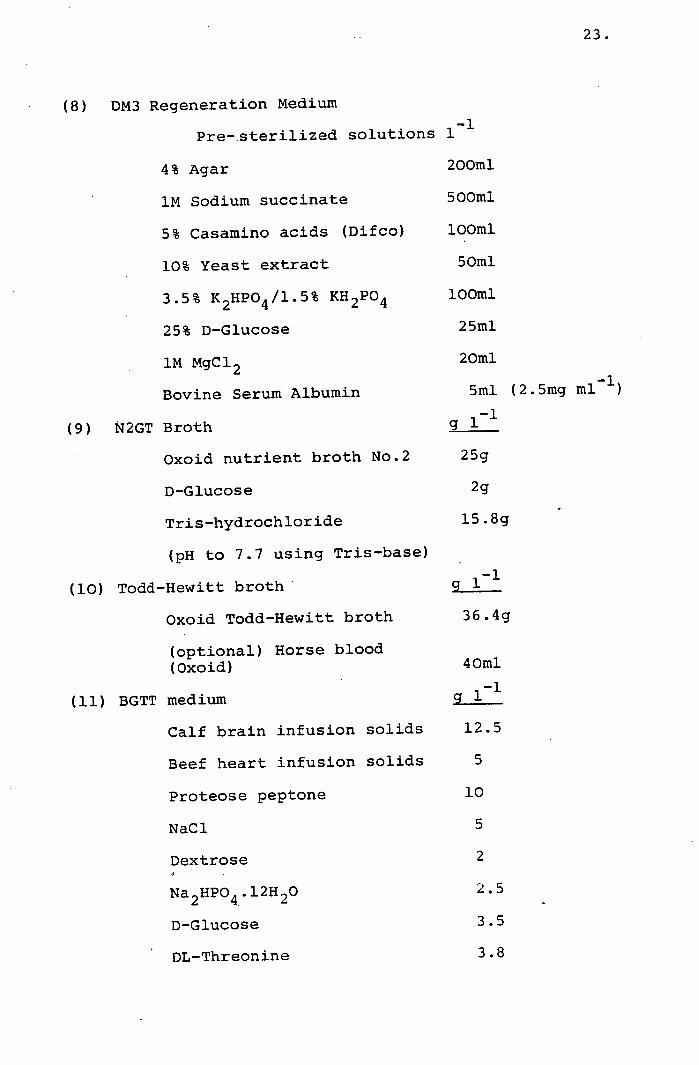
(8) DM3 Regeneration Medium
1_i Pre-.sterilized solutions
4% Agar 200m1
1M Sodium succinate 500m1
5% Casamino acids (Difco) lOOmi
10% Yeast extract 50m1
3.5% K 2HPO4 /l.5% KH 2PO4 lOOml
25% D-Glucose 25m1
1M MgC12 20ml
Bovine Serum Albumin 5m1 (2.5mg ml- 1
11 N2GT Broth g
Oxoid nutrient broth No.2 25g
D-Glucose 2g
Tris-hydrochloride 15.8g
(pH to 7.7 using Tris-base)
11 Todd-Hewitt broth' g
Oxoid Todd-Hewitt broth 36.4g
(optional) Horse blood (Oxoid) 40ml
11 g BGTT medium
Calf brain infusion solids 12.5
Beef heart infusion solids 5
Proteose peptone 10
NaCl 5
Dextrose 2
Na2HPO4.12H20 2.5
D-Glucose 3.5
DL-Threonifle 3.8

24.
2 x YT broth g
Bacto-tryptone 16
Yeast extract 10
NaCl 5
B broth g
Bacto-tryptone 10
NaCl 8
(supplemented with 1% vit B12 soin.)
K medium
M9 glucose minimal medium + 1% casamino acids
+ O.lpg m1 thiamine hydrochloride
1 Hershey salts g
NaCl 5.4
KC1 3.0
NH4C1 1.1
CaC1 2 .2H 20 0.015
MgC1 2 .6H 20 0.20
FeC1 3 .6H 20 0.0002
KH 2PO4 0.087
Tris base 12.1
Hershey medium
Hershey salts lOOmi
20% (w/v) glucose 2m1
2% (w/v) threonine 0.5ml
± 1% (w/v) leucine lml
2% (w/v) proline lml
2% (w/v) arginine imi
0.1% (w/v) thiamine hydrochloride imi

25.
All broths were converted to plating agars by the addition
of 15g agar 1- 1 (Oxoid No. 1) before autoclaving at 15 p.s.i.
for 20 mins. Sloppy agars were produced by using 0.5%
agar rather than 1.5%.
BUFFERS
Phosphate buffer (0.067M) pH 7.0
g (distilled water)
KH 2PO4 4.56
Na 2HPO4 .2H 20 4.73
Butanol-saturated phosphate/EDTA buffer
g1 1
KH 2PO4 4.56
Na 2HPO4 .2H 20 4.73
EDTA 0.34
n-butanol 6% (v/v)
Standard saline citrate (SSC) pH 7.0
1- 1
NaCl 8.7
Sodium citrate (dihydrate) 4.46
TE buffer, pH 7.4 g 11
Tris base 1.21
EDTA (disodium) 0.4
SMM buffer, pH 6.5 g 11
Sucrose 171
Sodium maleate 2.3
MgC1 2 4.0

Acetate electrophoresis gel buffer pH 8.2 (x 10)
g i 1
Tris base 48.4
Sodium acetate (trihydrate) 27.2
EDTA 3.72
Maxicell sample buffer pH 6.8 g 100ml 1
Sodium dodecyl sulphate(SDS) 2.3
2-mercaptoethanol (MSH) 5m1
Glycerol loml
Bromophenol blue (-BPB)
Tris-HC1 6.25
TM pH 8.0
100mM Tris HC1
100mM MgC1 2
10 x TBE (Tris-borate buffer) pH 8.0
g
Tris base 109
Boric acid 55
EDTA 9.3
Formamide dye mix g 100m1
Formamide lOOmi
Xylene Cyanol FF 0.1
BPB 0.1
EDTA 0.26
26.

27.
Deoxynucleoside triphosphate (dNTP) chase mix
Deoxythymidine 5 1 triphosphate (dTTP) 0.25mM
Deoxyadenosine 5 1 triphosphate (dATP) 0.25mM
Deoxycytosine 5 1 triphosphate (dCTP) 0.25mM
Deoxyguariosine 5t triphosphate (dTTP) 0. 25mM
all in TE buffer
40% Acrylamide stock solution
Acrylamide (electrophoretic grade) 38% (wlv)
Bisacrylamide 2% (w/v)
Solution -made up in deionised water followed by the addition
of 20g 1 of Amberlite MB1 resin (Hopkin and Williams).
Filtration removed the resin from the acrylamide stock
solution.
dNTP stock solutions
dTTP dCTP ) all 50mM in TE buffer (diluted 100 dGTP ) fold for use). dATP )
Dideoxynucleoside phosphate stock solutions (ddNTP)
ddTTP ddCTP all 10mM in TE buffer ddGTP ddATP
NTP ° mixes (ratio)
T° CO G ° A °
0.5mM dTTP 25 500 500 500 if dCTP 500 25 500 500 of dGTP 500 500 25 500
10mM ddTTP •50
ddCTP 8
ddGTP 16
ddATP 1 3 if [
32 P] M'P is used
TE buffer 1000 1000 1000 500

28.
ANTIBIOTICS AND NUTRITIONAL SUPPLEMENTS
All the antibiotics and nutritional, requirements
used in this study were obtained from Sigma Chemical Com-
pany, London.
RADIOACTIVELY-LABELLED COMPOUNDS
Labelled ( 32P) deoxyadenosine 5'-triphosphate, [ 35S]
deoxyadenosine 5'-triphosphate and [ 35 S] L-methionine were
gifts from either Dr N. Brown (University of Bristol) or
Dr M. Mackay (University of Edinburgh).
ENZYMES
T4 DNA ligase (EC.6.5.1.1.,), calf intestinal alkaline
phosphatase (EC.3 .1 .3.1.), 51 nuclease (EC.3. 1.30.1), Bal
31 nuclease and a variety of restriction endonucleases
were purchased from Boehringer Mannheim, Lewes. E. coil
DNA polymerase I (Kienow fragment) was supplied by both
NBL enzymes Ltd., Cramlington and PL Laboratories, Milton
Keynes. Pancreatic PNase (EC.3.1.4.22) and lysozyme came
from Sigma. Various restriction endonucleases were also
obtained from New England Biolabs, Bishops Stortford, NBL
enzymes, Miles Laboratories, Stoke Pages, and Bethesda
Research Laboratories (BRL), Cambridge.
CHEMICALS
Sodium dodecyl sulphate (SDS), ethylene diamine
tetra acetic acid: disodium salt (EDTA), hydroxymethyl
methylamine (Tris base), Tris-hydrochloride (Tris-HC1)
caesium chloride (Ana1a), polyethyleneglycol (AnaläR) 6000,

WE
bromophenol blue (BPB), ethidium bromide, xylene cyanol
FF, 3-(N-Morpholino) propanesuiphonic acid (MOPS), acryl-
amide and bisacrylamide (electrophoretic grade), S-
adenosyl L-methionine and spermidine were all obtained
from BDH Chemicals Ltd., England. Ficoll,agarose (type
II low EEO), deoxyadenosine 5-triphosphate (dATP), de-
oxyguanosine 5' triphosphate (dGTP), deoxythyinidine 5'
triphosphate (dTTP), deoxycytosine 5' triphosphate (dCTP),
dideoxyadenosine 5' triphosphate (ddATP), dideoxy
guanosine 5' triphosphate (ddGTP), dideoxythymidine 5'
triphosphate (ddTTP) dideoxycytosine 5' triphosphate
(ddCTP), and phenylmethylsuloflYlflUOride (PMSF) were
all supplied by Sigma. Dithiothreitol (DTT), ultrapure
phenol, isopropylthio--galactoside (IPTG), 5-bromo-4-
chloro-3 indolyl--D-galactoside (Xgal .)and (N,N,N'N')-
tetramethylethylenediamine (Temed) were products of BRL.
Hydroxylapatite-HTP and dextran T-500 were from Pharmacia
(GB) Ltd., Milton Keynes. Triton X-100 was obtained from
Koch-light Laboratories, Cotnbrook.
DYE-BUOYANT DENSITY GRADIENT CENTRIFUGATION
DNA was prepared from a variety of sources for puri-
fication on CsC1 2 gradients. These gradients were formed
by adding hg of CsC1 2 and lml of ethidium bromide (10g
ml 1 in TE buffer) to lOml of cleared lysate. The re-
fractive index was adjusted to 1.3925 (density 1.625g
cm 3 ) , using an Abbe 60 Refractometer (Bellingham &
Stanley Ltd., England), by adding further CsC1 2 crystals.

30.
The solution was then transferred to two lOmi polypro-
pylene tubes (MSE) and centrifuged at 130 000g for 60hr
at 18 °C in a 10 x lOmi fixed head rotor. When both
chromosomal and plasmid DNA were present, two bands
formed in the middle of the gradients. The lower co-
valently closed circular (ccc) band was intact plasmid
DNA, the upper band being chromosomal and 'nicked' piasmid
DNA. When required, the bands were extracted by carefully
puncturing the side of the tube with a 19 gauge needle
and drawing off the solution using a imi sterile syringe.
The ethidium bromide was extracted with salt saturated
iscpropanol 15M NaCl, 10mM Tris base, 1mM EDTA pH 7.5).
The DNA was collected by precipitation with 2vol. H 20 and
6vol. ethanol at -20 °C for 2 - 5 h. After centrifuga-
tion the pellet was resuspended in an appropriate volume
of TE buffer.
DNA ISOLATION
(a) CHROMOSOMAL DNA
Chromosomal DNA was prepared from D. radiophilus by
a modification of Marinur's method (1961). Two 1 of cell
culture, grown to stationary phase (approx. two days) , were
spun at 8000g for 10 mm. The pelleted cells were re-
suspended and washed in SOml of SSC buffer followed by re-
centrifugation. The cells were rendered sensitive to lyso-
zyrne activity by resuspending the pellet in 40ml butanol-
saturated phosphate/EDTA buffer and leaving the suspension
at room temperature for 45 minS. (Driedger and Grayston,

31.
1970). After collecting the cells and washing them again
in SSC, the total volume of the suspension was brought to
40m1 and lysozyme added to a final concentration of 2mg
the mixture being incubated at 37°C for 30 - 60 mini. Com-
plete cell lysis was achieved by the addition of 0.1 vol
of 20% SDS. After swirling to ensure adequate mixing,
13m1 of sodium perchiorate solution (70.25g NaC1O 4 .H 20
and 4.4g NaCl per lOOmi water) was added 1 followed immediately
by an equal volume of a 24:1 chioroform:iso-amyl alcohol
mixture. Deproteinisation occurred during 30 mm. of agit-
ation of the solution followed by centrifugation at 30000g
for 20 mins. in 'Corex' 25ml pyrex tubes. DNA and RNA
were present in the upper aqueous layer and were carefully
removed without disturbing the protein pellet present and
at the solvent interface. Careful pouring of the aqueous
component into 2 vol. of cold ethanol allowed the nucleic
acids to be wound out of solution on glass rods, dried and
then resuspended in a small volume (approx. 5m1) of SSC
buffer. Chromosomal DNA of very high purity was collected
after dye-buoyant density gradient centrifugation (Radloff
et al., 1967).
(b) PLASMID DNA
(i). E. coli
For large scale extraction of the majority of E. coli
plasmids used in this study (excluding the large R factors)
the basic alkaline extraction procedure of Birnboim. and
Doly (1979) was used. E. coli cells were grown in 11 amounts

32.
of L-broth at 37°C usually in the presence of a relevant
selective antibiotic. Chioramphenicol amplification was
possible for all Co1E1 derived plasmids but was not em-
ployed for the preparation of any recombinant plasmids.
For pBR322 and pAT153 extractions, the host cells were
grown to a turbidity of 60 - 100 (measured in a nephelo-
meter) before chioramphenicol was added to a final concent-
ration of lSOpg ml- 1. The culture was then shaken at
37°C for 16 h. One 1 of culture of plasmid-
bearing cells were spun down by centrifugation at 7000g
for 10 mm. allowed to drain, then resuspended in 30m1
of lysis mix (50mM glucose, 25mM Tris-HC1, 10mM EDTA, pH
8.0). To this, lOml of lysis mix containing 8mg ml -1 of
lysozyme was added and the mixture incubated for 10 mins
on ice. Cell lysis was achieved by the addition of 80m1
of 0.2M NaOH; 1% SDS solution w/v (fresh) and incubation on
ice for a further 5 min. Sixty ml of high salt solution
(3M sodium acetate adjusted to pH 4.8 using CH 3COOH) was
then added and the mixture kept on ice, with occasional
inversion, for 60 mins. Centrifugation at 12000g for 20
mins allowed a 'clear lysate' to be separated from the in-
soluble mass of cell debris/chromosomal material. Filt-
ration through a tea strainer removed any remaining lumps.
To this supernatant, 2 vol. of cold ethanol was added and
after complete mixing the solution was placed at -20°C for
at least 30 mins. The precipitate was collected by centri-
fugation at 5000g for 5 mins and resuspended in 50m1 of

33.
low salt solution (0.1M sodium acetate at pH 6.0).
Ethanol precipitation was repeated and the pellet resus.-
pended in <lOmi of TE buffer. Plasmid DNA was then separ-
ated from contaminating chromosomal DNA, proteins and RNA
by dye-buoyant density centrifugation.
For rapid screening of colonies for the presence of
plasmid DNA the boiling method of Holmes and Quigley (1981)
was used. 1.5m1 cultures of E. coli in L-broth were grown
overnight, poured into 1.5m1 Eppendorf tubes and spun for
15 s in a microfuge (Hettich Mikroliter). The pellet
was resuspended in 350pl of STET buffer (0.8% sucrose,
0.5% Triton X-l00, 50mM Tris, 1mM EDTA pH 8.0) and 25l
of lysozyme solution (10mg ml 1 ) added. Immediately upon
mixing, the tube was placed in a boiling-water bath for
40s and then centrifuged at 4 °C for 15 mins. The
gelatinous pellet was removed with a toothpick and the
supernatant precipitated by adding 40pl of 4M sodium ace-
tate and 400ul of cold isoproponol. After 5 mins at
ambient temperature the precipitate was collected by cen-
trifugation and the pellet washed with 70% ethanol before
being vacuum dried. The DNA was then resuspended in 10pl
of distilled water. This method was used for the isolation
of both recombinant plasmid and M13 PP DNA molecules, the
DNA being suitable for direct restriction enzyme digestion
and also for bacterial transformation.
(ii) B. subtilis
Plasmid isolation from B. subtilis was achieved using
Niaudet and Ehrlich's (1979) modification of the Gryczan
34.
et al. (1978) method.
250m1 of an overnight culture (L-broth) was centri-
fuged at 7000g for lOmins and the cells washed Q& tv
of buffer consisting of 0.1M NaCl, 0.05M Tris base and
1mM EDTA at pH 7.4. Lysozyme (0.25m1, 20mg m1 1 , freshly
made) was added and the mixture incubated at 37°C for 20
mins. 2.4m1 of 5M NaCl, 0.6m1 of 0.5M EDTA, pH 8.0 and
12.5ml of freshly prepared 2% SDS (w/v) - 0.7M NaC1were added
in that order and left overnight at 4°C. The lysed cells
were spun at 38000g for 30mins, the supernatant collected
and the salt concentration adjusted to 1M by adding SM
NaCl. One third volume of 40% polyethylenegiycol 6000
(PEG) was added and the DNA precipitated out during a lh
incubation at 0°C. The precipitate was collected by
centrifugation at 7000g for 5mins, and the pellet resus-
pended in 5m1 TE buffer. Further purification was achieved
by density gradient centrifugation.
(iii) S. faecalis
Essentially the plasmid preparation procedure for S.
faecalis was that described by Le Blanc and Lee (1979) but
with modifications in the lysis stages (Kado and Liu,
1981) and by Jacob (personal communication).
Cultures of S. faecalis were grown up in 20m1 of BGTT
broth at 37°C with aeration and harvested by centrifugation
at 8000g for 15mins at 4°C. The pellet was resuspended in
O.Sml of 0.01M Tris-HC1, 25% sucrose pH 8.0, followed by
the addition of 0.5m1 of lysozyme solution (40mg m1 1 in

35.
0.25M - Tris HC1 pH 8.0). These solutions were well mixed
and incubated at 37 °C for 30mins. The cells were lysed
by the addition of 0.25ml of 0.25M EDTA pH 8.0 and 2.5m1
of lysis mix (50mM Tris-HC1, 3% SDS (wlv) adjusted to pH
12.6 with 2M NaOH). The lysate was gently mixed and in-
cubated at 65 °C for 30mins. Proteins and cell debris were
separated by extraction with 2 vol of a 1:1 phenol:chloro-
form mixture (previously buffered by 1M Tris base, pH 8.0).
The layers were emulsified by gentle shaking, before phase
generation during a 15mir spin at 3000g. The upper aqueous
layer was carefully removed and nucleic acids precipitated
by the addition of 0.54vo1 cold isopropanol. The DNA was
recovered by centrifugation and resuspended in about'lOOyl
of TE buffer. The plasmid isolated in this manner was
used directly for transformation or as a check for plasmid
presence by agarose gel electrophoresis.
BACTERIAL TRANSFORMATION
(a) TRANSFORMATION OF E. coli HB101 WITH PLASMID DNA
E. coli HB101 was transformed with plasmid DNA using
the technique developed by Humphreys et al. (1978). An
overnight culture in L-broth (37 °C) was diluted 100 fold
and grown at 37 °C with vigorous aeration until the optical
density reached approximately 30 (2 - 3 h). For each
transformation 4x1. 5m1 Eppendorf tubes were filled with
the culture and spun for 20s in a microfuge. The pellets
were washed in 0.5vol of 10mM CaC1 2 and once again pelleted

36.
by spinning for 20s in the microfuge. The cells were resus-
pended in 5041 of 75mM CaC1 2 , 10mM MOPS, 0.5% glucose, pH
6.5 and all 4 tube contents pooled, i.e. from 6ml
culture 200pl of competent cell culture is produced. To
the 200pl cell suspension the transforming DNA was added
(normally dissolved in 100p1 of O.1M Tris buffer pH 7.0) followed
by 200pl of 75mM CaC1 2 , 10mM MOPS, 0.5% glucose, pH 6.5.
The mixture (0.5ml) was kept on ice for 45mins before a
heat-shock treatment of lOmins at 42 °C. The transformed
culture was then transferred to 1.5m1 of L-broth and shaken
for 30 to 60mins at 37 °C to allow for phenotypic lag.
(b) TRANSFORMATION OF E. coli NM522 WITH M13RF DNA
The re-introduction of manipulated M13 mp9 DNA into
the indicator strain E. coli NM522 was achieved using the
method of Messing (1983).
E. coli NM522 was grown up overnight in M9 minimal
medium + thiamine. The culture was diluted 100 fold into
2 x YT medium and grown at 37 °C with shaking until the
turbidity reached approximately 60.. The cells were col-
lected by centrifuging 1.5ml amounts in Eppendorf tubes for
20s (2 x 1.5m1 tubesper transformation). The pellets were
resuspended in ice-cold 50mM CaCl2 and left on ice for
20mins. The cells were again pelleted by centrifugation
for 20s and resuspended in 150pl of cold CaC1 2 , 2 tubes
being combined to give 300).i1 of competent cell mix. To
this the M13 DNA was added and after gentle mixing the

37.
tube was kept on ice for 40mins and then transferred to
37 ° C for 15mins to induce DNA uptake. The transformed
culture (0.3m1 or appropriate dilutions) was added to 3m1
of B sloppy agar containing 20u1 IPTG (25mg m1 1 ), 20jil
X-Gal (25mg ml - 1 ), 200p1 fresh indicator strain (NM522) and
20il thiamine (1mg m1 1 ). After vortexing for lOs the
mixture was poured onto dry B-agar plates and left to set
before incubating overnight at 37 °C.
(c) - TRANSFORMATION OF B. subtilis BY PLASMID DNA
B. subtilis 168 was transformed with plasmid DNA using
the polyethylene glycol (PEG) induced DNA uptake by proto-
plasted cells, a method developed by Chang and Cohen (1979). Twen
tymi of a mid-exponential culture of B. subtilis 168
growing in Penassay broth at 37 °C was harvested and resus-
pended in 2m1 of SMMP solution followed by the addition of
lysozyme, to give a final concentration of 2mg ml- 1. The
suspension was incubated for 2 h at 37 °C with gentle
aeration. The cells were centrifuged at 2500g for 15mins
and washed once in SMMP before being pelleted. The re-
sultant pellet of protoplasts .was resuspended in 2m1 SMMP
and 0.5m1 samples were mixed with O.lml of 2 x SMM, 5ag
plasmid DNA, 1.5ml 40% w/v PEG (40g PEG 6000, 50m1 2 x SMM
buffer in lOOmi) in order. After 2 mins,5m1 of SMMP was
added and the mixture centrifuged at 2500g for lOmins.
The pellet was resuspended in lml SMMP and incubated at
30 °C for 1.5h with gentle shaking to allow for phenotypic

38.
lag. The protoplasts were plated directly onto DM3 regen-
eration medium plus relevant antibiotics and incubated at
37 °C for 2-3 days.
(d) TRANSFORMATION OF D. radiodurans STRAINS
Transformation, with either chromosomal or plasmid DNA,
of D. radiodurans strains Ri, 302, Nuc 1, Rec 30 and Sark
followed the procedure developed by Tirgari and Moseley
(1980). An overnight culture of the required strain was
diluted 20 fold into fresh, warmed TGY broth and incubated
at 30 °C with shaking until an optical density of about 30
was reached 0.3 h). Ten ml of the culture was harvested
by centrifugation at 12000g for Smins, resuspended in 5m1
of prewarmed TGY broth and 2m1 of O.lM CaC1 2 solution
added. One ml samples of this culture were used for trans-
formation by adding l-lOpg of DNA and by chilling on ice for
lOmins. These samples were then transferred to a 30 °C
water bath for 90mins before 9ml of TGY broth was added and
the transformed cells allowed to express the integrated DNA
over a period of 4 to 10 h. The relevant selection
pressure was applied by plating onto selective TGY agar
and incubating for 3 - 4 days at 30 °C.
CONJUGATION
E. coli
The transfer of E. coli conjugative R factors RP4 and
R68.45 was followed by using a filter-mating technique.
Donor and recipient cells (Table 2.5) were grown separately

Table 2.5. Strains of E. coli and D. radiodurans used
in Conjugation Analysis.
Chromosomal Plasmid markers Markers
Donor Strains
E. coli J5- (RP4) Na1R TcR APR KnR
E. coli J53•41% ( R68-45) Na 1R of 11 of
Recipient Strains
E. coli HB101 smR
D. radiodurans RifR Rif'
(abbreviations: Nal = naladixic acid 1 50j.ig ml -1 ; Sm =
streptomycin, 200pg m1 1 ; Rif = rifampicin, 2Qz.ig
Tc = tetracycline, lOjig m1 1 ; Ap = ampicillin, 20jig ml;
Kn kanamycin, 20pg ml).

39.
overnight at 37 °C without shaking in L-broth (or for D.
radiodurans, TGY broth). In all cases both donor and recipient
strains had chromosomal markers allowing for differential
selection after mating. 0.lml of donor culture was added
to 0.9m1 of recipient cells and 9m1 of fresh broth, either
L-broth or TGY broth, was added. After thorough mixing,
the culture was put through a 0.45iU1l Millipore filter. The
filter was taken out of the support assembly, placed on a
dry L or TGY agar plate and incubated overnight at 37°C or
30°C. The filter was then removed, the cells washed into
fresh broth and donor, recipient and transconjugant cells
enumerated by the plating of dilutions onto the appropriate
selective medium. The frequency of conjugation was cal-
culated by dividing the number of transconjugants by the
number of donors.
S. faecalis
(a) FILTER MATING
Filter matings between streptococcal species were
carried out using the method outlined by Franke and Clewell
(1981). 0.05ml of an overnight donor culture, grown at
37°C with shaking in N2GT broth, was mixed with 0.5m1 of
overnight recipient culture in a total volume of 5ml N2GT
broth. The cells were collected on a 0.45um Millipore
filter which was then removed from its housing and placed
on horse-blood agar and incubated overnight at 37°C. The
cells were washed off in lml of N2GT broth and appropriate
dilutions spread onto selective plates. Total numbers of

40.
donors, recipients and transconjugants were determined,
the conjugation frequency being calculated by dividing
numbers of transconjugants (minus spontaneous mutants) by
the number of donors. If D. radiodurans T etR was used as
the recipient, the method remained the same except that
TGY broth and agar replaced the media noted above (this
seemed to have no effect on S. faecalis viability and little
effect on streptococcal conjugation frequency).
As well as the filter-mating protocol, a technique
developed by Smith and Guild (1980) was used. In this case
the procedure was essentially identical except that the
filter was overlayed with TGY agar (only used in S. faecalis
x D. radiodurans crosses) before incubating overnight.
This embedding in agar has been shown to increase the fre-
quency of conjugation in Streptococcus pneumoniae by bet-
ween 10 and 100 fold.
(b) BROTH MATING
S. faecalis x S. faecalis and S. faecalis x D. radio-
durans broth matings were undertaken using the procedure
described by Dunny and Clewell (1975). Overnight cultures
of the donor and recipient strains of S. faecalis (or D.
radiodurans) were grown at 37°C in either N2GT or TGY broth.
Then,to 4.5m1 of fresh broth ) O.Sml of recipient, and 0.05ml
of donor culture, were added. The mixture was incubated
at 37°C for 4h before vortexing followed by serial dilution
and plating on the appropriate selective medium for
enumeration of donors, recipients and transconjugants.

41.
MEASUREMENT OF BACTERIAL GROWTH
The phase of growth of a bacterial culture was
monitored by following changes in the optical density of
the medium using a nephelometer (Evans Electroselenium Ltd.,
Halstead) with an orange filter.
MEASUREMENT OF DNA AND PROTEIN CONCENTRATIONS
The concentrations of DNA preparations were determined
spectrophotometrically with a Pye Unicam 5P6-500 uv spectro-
photometer. The assumption was made that an absorbance
reading, at 260nm, of 1.0 corresponded to a DNA concentration
of 5Ojig m1 1 . The comparative absorbances of a solution
at 260nm and 280nm not only determined. DNA content more
accurately, but also allowed calculation of the solutions
protein content by using , a nomograph. Protein concentration
was also measured using a Bio-Rad protein assay kit.
AGAROSE GEL ELECTROPHORESIS
Horizontal agarose gel electrophoresis allowed the
identification of a wide range of intact and endonuclease
restricted DNA molecules. Concentrations of agarose, from
0.8 to 1.5% were used and in all cases gels were run in a
Tris-acetate buffer. Before loading into the wells of the
gel each sample had 0.1 vol. of STOP buffer added (0.05% Bromo-
phenol blue; 205vo w/v Ficoll; 30mM SDS, 0.5M EDTA) and was
heated to 65 °C for 10 mins. Electrophoresis conditions
were dependent upon the separation of DNA bands required
but were rarely above 10V cm or below 2.5V cm- 1. Band
visualization was by staining with the fluorescent dye

42.
ethidium bromide either by post-electrophoresis staining
in a 0.5).lg ml- 1 solution or pre-electrophoresis incorpor -
ation of the agent into the buffered agarose at 0.5)lg m1 1 .
Upon illumination with 260 - 300nm light DNA bands could
be seen down to a concentration of about 50ng. Photographs
were taken using a Polaroid MP-4 camera with Polaroid type
55 band film, and a Kodak 22A Wrattan filter.
Elution of particular restriction fragments (Yang,
Lis and Wu, 1979) was achieved by cutting a 'u' shaped well
directly in front of the required band (visualized in a
pre-stained agarose gel with incident ultraviolet light).
This well was then lined with dialysis tubing and filled
up with fresh electrophoresis buffer. The current was
then continued until all the relevant stained material in
the gel had moved into the well. •A short period of reversal
of electrical polarity released any closely bound DNA from
dialysis membrane and the well contents were collected.
The DNA was then phenol extracted twice (using lM Tris
base pH 8.0 buffered phenol), with phenol:chloroform (1:1
twice and ether (water saturated) washed once,before being
ethanol precipitated by the addition of O.lvol 4M sodium
acetate and 2vol cold ethanol. After 2 - 3 h at -20°C
the DNA precipitate was collected by centrifugation, dried
in a vacuum and resuspended in an appropriate volume of TE
buffer, usually about 50iil. Fragments of agarose gel were
removed,when necessary, from the preparation) bef ore the
phenol extractions,by filtering the well contents through
siliconised glass wool.

43.
The DNA fragments isolated in the above manner were
suitable for both restriction analysis and sub-cloning
using T4 DNA ligase.
POLYACRYLAMIDE GEL ELECTROPHORESIS FOR RESTRICTION MAPPING
Analysis of linear DNA fragments of <400bp is virtually
impossible on agarose gels 1 even of high concentration. In
order to accurately size such fragmentsgradient protein
denaturing, vertical polyacrylamide gels were used. Gel
dimensiorwere 23cm x 17cm x 0.05cm and the gradient was
produced by the controlled mixing of the solutions shown
in Table 2.6. When the 5 - 15% resolving gel was set, a 4.5%
polyacrylamide stacking gel, 3cm deep, was made above it
into which the comb was set.
Before application .)-the DNA samples (50j.il) had O.lvol
loading dye added (2% SDS w/v, 10% G1yine w/v, 0.05% bromo-
phenol blue w/v,pH8.0) and were heated to 65 °C for 15mins.
Electrophoresis proceeded at a voltage gradient of 10V cm- 1
using an electrophoresis buffer consisting of 1.5%w/vglycine
0.6% w/v Tris base and 0.1% w/v SDS at pH 8.4. DNA bands were
visualized by staining for 15mins in a 0.5pg ml 1 .solution
of ethidium bromide followed by illumination with 300nm
light. Photography was the same as for agarose gels.
SIZING OF DNA FRAGMENTS FROM AGAROSE AND POLYACRYLAMIDE GEL
ELECTROPHORESIS
Southern (1979) has described a simple equation repre-
senting the relationship between the molecular weight of a
DNA molecule or fragment and that fragment's mobility in a

Table 2.6. Relative Composition of Polyacrylamide Mixing
Solutions
Solution A Solution B Stacking (15%) ml (5%) ml gel ml
This l.SM, pH 8.8 3.25 3.25 -
This 0.5M, pH 6.8 - - 2.5
Acrylainide solution 6.5 2.2 1.5
(29.2% acrylainide, 0.8% bisacrylamide)
1% SDS 1.3 1.3 1
H20 0.5 6.2 5
Glycerol 1.3 - -
Tened 15jil 15pl
10% Aimonium Sulphate 2911 2911 2911

44.
gel during electrophoresis. This equation is more con-
venient than graphical methods, accurate over a wide range
of sizes and is outlined in Figure 2.1.
ENDONUCLEASE AND METHYLASE ASSAYS
Assay conditions for all commercially supplied re-
striction endonucleases were as stated in the manufacturers
instructions. The standard assay condition used for Dral
was the incubation of lig DNA sample with 2 units of Dral
for 60 mins at 37 °C in Dral restriction buffer (10mM Mg
Cl 2 , 1mM MSH, 25mM NaCl, 20mM Tris base pH 7.5).
The activity of Dral methylase was tested by incub-
ating 5p1.of protein sample, 2ig DNA (plasmid pPL3p%), 1 j4 S-adenosyl-L-methionine (0.25mM) and92ul 0.2M Tris-HC1
pH 8.0/8mM EDTA for lh at 37 °C. The mixture was then
phenol extracted twice, phenol-chloroform extracted twice,
ether washed and finally ethanol precipitated. The DNA was
collected by centrifugation and resuspended in 40jil of Dral
restriction buffer. One unit of Dral was added, incubated
at 37 °C and the activity of Dral methylase indicated by
the absence of Dral digest products upon analysisusing
agarose gel electrophoresis.
ENZYME PURIFICATION
(a) RESTRICTION ENZYME SCREENING
A variety of methods were used in attempts to identify
the presence of site-specific endonucleases within deinococcal
species. Small volumes of culture (about lOOmi) were spun
down at 7000g for lOmins and the pellet resuspended in lOmi

Fig. 2.1. Calculation of size of DNA molecules and fragments from mobility in agarose gels
Three points were chosen corresponding to size standards L 1 , L2 and L3 with
mobilities in 1 , in2 and En3 respectively. The value in0 that determines that these three points are jqined by a straight line is given by,
- ((L 1 -L2\(rn3 -m2 - \VJ.J 'm 2 -m
'no ((Li -Lj'm3 -m2 • \\L2 -L31\rn2 -m 1
The values k1 and k2 were then calculated from the following equations,
L,-L2 k1
m0)-(1n2 - m)
k, - MO
The equation given below was then used to calculate the size of a molecule or fragment 'L with a mobility in
M - m + k2

45.
10mM Tris-EC1, 0.1mM phenylmethylsuiphonyifluoride (PMSF)
pH 7.5 before the cells were broken open in a French
pressure cell at 3000p.s.i. (most species were refractory to
lysis by sonication). The presence of restriction enzymes
was investigated in this 'crude' extract after cell debris
had been removed by slow speed centrifugation (10000g for
lOmins). The crude extract was concentrated and fraction-
ated by using either ammonium sulphate precipitation or
polyethyleneglycol 6000:dextran T500 phase partition
(Schleif, 1980). In the former case Analã..R - (BDH) ammonium
sulphate (90% w/v solution) was added slowly to the crude ex-
tract to final concentrations of 35, 45, 55, 65 and >75%
sequentially. At each concentration stage the mixture was
left for lh at 0°C before the precipitate was collected
by centrifugation at 7000g for 15mins. Each pellet was re-
suspended in 10mM Tris-HC1, 0.1mM PMSF, pH 7.5 buffer and
extensively dialysed, with multiple buffer changes for 5h,
before being tested for enzyme activity. In the Schleif
(1980) method, to each imi of crude extract the following
solutions were added: 0.5m1 H 20, 0.6g polymer concentrate
(64g dextran T500, 256g polyethyineglycol 6000, 900m1 H 20)
and one of the following volumes of 4M NaCl: 0.002, 0.005,
0.01, 0.02, 0.04, 0.08, 0.17, 0.32, 0.64 and 1.2m1. After
the additionsthe mixtures were vortexed in 3ml Eppendorf
tubes and centrifuged for Smins in an Eppendorf microcentri-
fuge model 5413. The resulting clear supernatants were then
assayed for endonuclease activity.

46.
(b) PREPARATIVE ENZYME PURIFICATION OF Dral and DraII
Three 11 cultures of D. radiophilus were grown in
nutrient broth No. 2 (Oxoid) to stationary phase at 30 °C-
37 0C with shaking. About 15g wet weight cells were har -
vested from this volume by centrifugation at 10000g for
lOmins. The cells were resuspended in 30m1 10mM Tris-HC1,
2mM MSH and 0.1mM PMSF, pH 7.5, and broken open in a
French pressure cell at 3000-4000ps.i. The cell debris was
removed by a low speed spin at 10000g for lOmins at 0 °C,
the supernatant then being subjected to a high speed spin
at 100000g for 1 to 2h at 0 °C in a MSE Prepspin 65 ultra-
centrifuge. Polyethylene glycol -dextran phase partition
was carried out on the separated supernatant by adding per
ml of extract 0.5m1 H 20, 0.5g polymer concentrate and 0.32m1
4M NaC1 2 . After mixing, the phases were separated by cen-
trifugation at 6000g for lOmins, the upper solution being
kept and dialysed overnight against column buffer 1 (CB1
- 10mM Tris-HC1, 2mM MSH, 0.1mM PMSF, 0.075M NaCl, pH 8.0).
The dialysed extract was applied to a 20 x 2.6cm DEAE-
sephacel (Bio-Rad) column previously equilibrated with CB1.
The sample was applied at a rate of lOmi h -1, washed by
2x the column volume of CB1 at a rate of 10cm h -1 and
then the column was developed at Sml h.- 1 with a linear
0.0750.4M NaCl gradient of approximately 2x column volume.
Two ml samples of eluate were collected in acid-washed test
tubes and DraI/DraII activity tested. Fractions showing
greater than unit activity (complete digestion of lj.ig DNA
in lh at 37 °C) were pooled and dialysed overnight against
47.
column buffer 2 (CB2 - 0.01M phosphate buffer, 2mM MSH,
0.05M NaCl, pH 7.5). After dialysis the sample was
applied at a rate of 50m1 h. 1 to a 10 x 2.6cm column of
Bio-Gel HTP grade hydroxylapatite (Bio-Rad) previously
equilibrated with CB2. The column was washed with 2x
column volume of CB2 at 50m1 h -1 and then developed with
a linear 0.01M - 0.4M phosphate buffer gradient, of approx-
imately 2x column volume, at 40m1 h 1 .Two ml fractions
were collected in acid-washed tubes each containing lOO).11,
10mg ml 1of bovine serum albumin (BSA, restriction enzyme
grade, BCL) and tested for restriction enzyme activity.
All samples showing greater than 'unit' activity were
pooled and dialysed against CB3 (20mM Tris-HC1, pH 8.0)
overnight. This sample was applied to a 5 x 2.5cm heparin-
agarose column (BCL) at a rate of 5m1 h l, washed with
20ml of CB3 at the same rate and the column developed
using a 20ml 0 - 0.8M NaCl linear gradient. 0.7m1
fractions were collected in tubes containing lOpl, 50mg ml -1 ,
BSA, tested for the activity of restriction endonucleases,
and those fractions showing Dral activity (at least 1 unit
il) were pooled and dialysed against storage buffer (50% v/v
glycerol, 2mM MSH, 50mM NaCl, pH 8.0) overnight. The enzyme
preparation was stored in this form, at -20 °C.
ULTRA-VIOLET IRRADIATION
DNA with desired levels of ultra-violet light damage
was produced by resuspending the nucleic acid in an ultra-
violet transparent buffer (10mM Tris-HC1, pH 7.4) and

48.
irradiating lOul droplets for specified times at a dose rate
of l.O5J m 2 t
CONSTRUCTION OF GENE LIBRARIES OF D. radiophilus
Banks of D. radiophilus genomic DNA were constructed
in the E. coli vector plasmid pAT153 using three different
restriction endonuclease systems, i.e. cloning at the PstI
site (Qmp 1 inactivation), at the Hindlil site (tetR in-
activation) or using a MboI partial genomic digest inserted
at the BamHI site of pAT153 (tetR inactivation) (Fig. 2.2).
The genomic fragments for cloning were generated by digestion
of 2pg D. radiophilus chromosomal DNA and 0.2)ig D. radio-
philus plasntid DNA (a gift from G. A1-Bakri) with 5 units
of PstI or HindIll for 2h. In the case of MboI partial
digestion, the same DNA concentrations were divided into
three and separately digested with 0. 04, 0.08 and 0.15 units
of MboI for 2h and the digestion products-pooled (M. Mackay,
1983). All restriction reactions were terminated by heating
to 70 °C for lOmins. In paral1elthe vector plasmid DNA was
prepared by digesting 2pg pAT153 for 1 hour with 5 units
of the relevant restriction endonuclease, PstI, Hindlil or
BarnHI. The linearised plasmid was then ethanol precipitated
and resuspended in 40ul H 2 0 and 2jil of 20x alkaline phos-
phatase buffer added (1M Tris pH 9.5, 20mM EDTA; spermidine
to 1mM). The plasmid DNA had the 5' terminal phosphate
groups removed by the addition of 1 unit of calf intestinal
alkaline phosphatase (BCL) and incuba€ion for 30 mins at 37 °C.
This treatment prevented the recircularisation ot plasmid

FIG. 2.2. CONSTRUCTION OF D.radiophilus GENE AN( USING INSERTIONAL INACTIVATION OF pAT153
Hindlif
of HI
Pst __l_. ZZ/
I pAT153 3.65kb 1 D. radiodurans DNA
- Iinearisati;n - -
complete PstI or HindX digests,
partial MboI digest
rrR Amps
Psti V
1I
rw. Wa. :rni.i"
phosiphatase treatment
•
T4 DNA ligase
Transformation
H check of cloning efficiency
i.e. AmpRTetS ( Hind ffl;BamHI,MbàI)
Amp STetR(PstI)
Plasmid DNA preparation

49.
vector molecules unless D. radiophilus DNA was inserted.
The reaction was terminated by heating to 65 °C for 20mins.
All DNA molecules were deproteinised by two phenol and
two phenol:chloroform extractions followed by washing with
water saturated ether and ethanol precipitation at -20 °C.
De-phosphorylated vector molecules were then mixed with
the appropriate DNA fragments from D. radiophilus (i.e.
common PstI ends, Hindlil ends or BarnHI/MboI ends) in the
ratio of 4:1 so that the final DNA concentration in 20pl
of ligation buffer (10mM Tris-HC1, pH 7.8, 10mM MgCl 2 ,
20mM DTT, 0.6mM ATP) was no greater than 50pg rnl 1 . Lig-
ation was carried out by adding 0.1 units of T4 DNA ligase
(BCL) to the 20jil of DNA in ligase buffer and allowing in-
cubation for 16h at 14 °C. The DNA was then ethanol pre-
cipitated, resuspended in 100)11 0.1M Tris, pH 7.2 and used
to transform E. coli HB101. After transformationa sample
of cells was taken to test the level of insertional in-
activation and cloning efficiency whilst the rest of the
culture was used to prepare plasmid DNA by the Birnboim-
Doly (1979) method using the still-active antibiotic re-
sistance marker for pre-selection. The gene banks were
stored as DNA solutions at -4 °C rather than individual
colonies.

50.
DETERMINATION OF DNA SEQUENCE REPRESENTATION IN A GENOMIC
LIBRARY
The exact probability of having any DNA sequence
represented in the genomic library was calculated from the
formula:
N= : ln(l-P ) ln(1 - f) (1)
where P was the desired probability, f the fractional
proportion of the genome in a single recombinant and N
was the necessary number of recombinants (Clarke and
Carbon, 1976). For easy use the equation can be rearranged
as: -
P = 1 - (1 - .f)N (2)
In addition an assumption could be made that the length
(x) of a required DNA segment was small in comparison with
the length (L)of the inserts thus reducing the effects of
random breaks occurring within length (x). The equation
can thus be modified to read
(1 - (3)
with the f* value including the effect of random breaks,
these being particularly important if the size of the
average cloned fragment is small.
SCREENING OF THE GENOMIC LIBRARIES
As D.radiophilus is refractory to DNA uptake and
therefore cannot be transformed even by homologous DNAa1l
screening for genomic markers was carried out in E.coli
mutants. This procedure of course, has its own problems
which are covered in the Discussion. A wide variety of

FIG. 2.3 SCREENING OF RECOMBINANT GENE BANKS FOR PRESENCE
OF MODIFICATION METHYLASE ACTIVITY
0 00 0 Recombinant gene bank
inplasmid pAT153
Digestion with Dral
o 0
Linearization of all plasmids not protected by Drat methylase modification (plasmid borne)
I. Transformation of E.coli H 8101 FiVi (only ccc form transforms effectively)
Selection for Amp (Hind]IE or Barn HI/ MboI) or TetR (Pst I)
I Small scale rapid screening for presence of recombinant plasmids carrying Dral methylase gene

51.
auxotrophic, nutritional and other selectable phenotypic
traits were screened by attempted transformation of
appropriate E.col. mutants with 2 jig of each of the three
gene bank DNAs. Auxotroph to prototroph conversion
required plating onto defined medium lacking the appro-
priate growth factor. Sugar fermentation complementation
i.e. xylose to xyiose depended upon growth with that
compound as the sole carbon source. Other lesions could
be checked by complementation restoring at least partial
resistance to normally bacteriocidal doses of certain
agents e.g. ultra-violet light.
Of principal importance were the attempts to clone
the modification/restriction genes of D.radiophiiuS.
Screening for the expression of these genes in E.coli
HB101 was attempted in two ways. Firstly E.coli HB101
was transformed with 2 ui of each gene bank and allowed
2h for phenotypic expression. To one sample,T 4 bacteriophage
was added in L-broth to a multiplicity of infection of 10
(i.e. 10 phage per bacterium). Infection was allowed
60 minsto complete and then the cells were spun down in
Eppendorf microcentrifuge tubes and washed three times
with a 25 mg ml- 1 pronase solution. The cell pellet was
finally resuspended in a buffered 1% sucrose solution
(10mM Tris-HC1, 1mM EDTA, pH 7.4) and plated out onto
either ampicillin (40 jig m1) or tetracycline (20 jig ml- 1
selective medium. Thusse1ectiofl was for T 4 R ampRor
T 4 R tet cells. Secondly,the pro :edure for methylase
expression described by Walder et al. (1983) was followed

52.
(Fig. 2.3). Fifty pg WA from each gene bank was digested
for 3 h with 100 units of Dral, the DNA being.collected
by phenol extraction followed by ethanol precipitation.
The DNA was resuspended in 100 ul 0.1M Tris pH 7.2 and
used for the transformation of E.coli HB101. After
phenotypic lag,the transformed cells were plated onto
either ampicillin (40 pg ml- 1 ) or tetracycline (20 )ig ml)
L-agar depending on which gene bank was originally used.
In both cases all colonies growing on antibiotic
selective plates were screened for the presence of
recombinant plasmids (see earlier).
RECOMBINANT MOLECULE LIGATION
The religation of matching, 'sticky' ended DNA
fragments has already been described in the section on
the construction of the gene libraries. 23n-ended
ligation required modification of the reaction conditions
as this reaction exhibits bimolecular reaction kinetics
at low substrate concentration and is not linear with
respect to the DNA ligase concentration (Modrich and
Lehman, 1970). Thus for ligation involving blunt ends
higher concentrations of DNA were used, >200j.g ml,
with 1 unit of DNA ligase per 20 p1 of ligase buffer
(0.6mM ATP, 10mM DTT, 20mM Tris, 10mM MgCl 2 , pH 7.8).
Ml3mp8 and rnp9 BACTERIOPHAGE ASSAY
A single colony of indicator strain, E.coli NM522,was
taken from M9 minimal medium agar + thiamine and inoculated
into 2 ml of 2 x TY broth and grown at 37 °C for 7 h with
vigorous shaking. 0.2 ml of these host cells were added

53.
to 0.01 ml of bacteriophage dilution, 0.01 ml of 100mM IPTG,
0.05 ml of 297o Xgal and 3 ml of B sloppy agar and thoroughly
mixed by vortexing for 10 s before the agar was poured
onto dried B broth agar plates. The sloppy agar was
allowed to set and the plates incubated overnight at 37°C.
Blue plaques represent non-recombinant M13mP phage whilst
clear plaquesindicate the removal of a-complementation by
insertion of DNA into the Ml3mp phage constructed cloning
sites.
SIZE ASSAY
As stated previously all sizing of M13mpRF molecules
was done using the Holmes and Quigley boiling technique
(1981).
C-TESTING
Insert orientation and complementarity was checked
using the renaturation technique employed by Messing (1983).
Indicator cells were produced as above and when the optical
density reached around 10,2 ml samples were removed and
fresh plaques resuspended in them. The cultures were
vigorously rotated at 37°C for 6-7 h before being
transferred to 1.5 ml Eppendorf tubes. The tubes were
centrifuged for 5 minsat room temperature in a microfuge,
the supernatant being carefully removed into a different
tube. Twenty p1 of each supernatant was mixed with 20 p1 of a
control supernatant of a Ml3mp clone definitely carrying
inserted DNA of interest in the opposite orientation and
1 p1 of 2% SDS with 3 p1 loading buffer added. The
solutions were well mixed and overlayed with light mineral

54.
oil before being heated at 65°C for 1 h. The sample was
then electrophoresed through a 0.876 agarose gel. DNA
isolates with opposite orientations of the same insert
reannealed together, altering the position of the bands
within the agarose gel.
GROWTH AND ISOLATION OF M13mp9 AND M13mp8 RF DNA
A blue plaque was picked from a fresh overlay plate,
inoculated into 1 ml of 2 x TY broth and the culture
grown overnight with vigorous aeration at 37°C. One litre
of 2 x TY broth was inoculated with this starter culture
and 10 ml of a fresh NM522 culture. The flask was shaken
for a further 4 h before the RF DNA of the M13 'phage
was isolated by the Birnboim and Doly method (1979) as
described earlier. The purified M13mp9 and 8 DNA was
taken from the CsC1 2 gradient and tested for its suitability
to act as a substrate for restriction and ligation. One
0 ug of DNA was restricted with 4 units Sinai for 1 h at 30 C
in the relevant enzyme buffer. This sample was divided into
two and both had the DNA recollected by ethanol precipita-
tion. Both samples were resuspended in ligation buffer but
only one had the T4 ligase enzyme added. The DNAs were
then used to transform competent E.coli NM522 and suitable
dilutions of the transformed culture made to enable single
plaque identification. Comparison of plaque numbers and
morphology between a control of 0.5 ug uncut DNA, 0.5 ig
SmaI cut DNA and 0.5 jig Sinai cut then religated DNA gave
a strong indication of the susceptibility of the DNA to
restriction enzyme digestion and to the level of chromosomal

55.
DNA contamination of the RF DNA preparation.
PREPARATION OF TEMPLATE DNA
E.coli NM522 was inoculated into fresh 2 x TY broth,
from an overnight culture (40 x dilution) and grown to an
optical density of 30 by shaking at 37°C. 1 ml samples
of this culture were placed in bijoux. bottles and freshly
plated plaques were inoculated into each sample. The
bottles were shaken vigorously for 4½ h at 37 °C and the
cultures poured into 1.5 ml Eppendorf tubes and centrifuged
in a microfuge for 5 minsat room temperature. The super-
natants were transferred carefully to other Eppendorf
tubes (approx. 0.8 ml ) and 200 )il of a 205MNaC1, 2096 w/v
polyethylene glycol solution added. The tubes were
incubated for 30 min.s at room temperature before being
spun for another 5 mis period at room temperature. The
supernatants were discarded and all traces of liquid
around the pellet removed using a drawn out capillary.
The pellets were resuspended in TE buffer and 50 )al phenol
added to each. The tubes were vortexed for 10 s, left
standing for 10 mins,vortexed again and finally the phases
separated by centrifugation for one mm. The upper
aqueous layer was removed and to it was added 10 pl 4M
sodium acetate and 250 p1 ethanol. After incubation at
-700C for 5 mins the tubes were spun for 10 minSat 4 °C
and the pellets washed with 1 ml of ethanol. The procedure
of resuspension followed1 ethanol precipitation and washing
was repeated,thenthe pellets vacuum dried and resuspended in
50 ul TE buffer. The M13mp templates were ready for DNA
sequencing and were stored at -20 °C. Each template was checked
by agarose gel electrophoresis (5111 of SO)il) before sequencing
was begun.

56.
GENE PRODUCT ANALYSIS - THE MAXI-CELL TECHNIQUE
The E.coli strain CSR603 was transformed with the
recombinant plasmid as previously described in this chapter.
Individual clones were tested for the appropriate anti-
biotic resistance marker expression and for the presence
of plasmid DNA of the correct size using the Holmes and
Quigley boiling method (1981). Three ml of K medium was
inoculated with an E.coli CSR603 isolate containing the
recombinant plasmid and the sample was incubated, with
shaking, at 37°C overnight. 0.3 ml of the culture
was then diluted into fresh K medium (10 ml) and allowed
to reach an optical density of around 30. From this
exponential phase culture 2.5 ml was removed and placed WI
in an open petri dish where adose of 6 J m was
administered (with continuous stirring). After irradiation )
2 ml of the sample was transferred to a sterile bottle and
incubated for 1 h at 37°C. This was followed by the
addition of 15 ul of 15 mg ml cycloserine and incubation
for 16-20 hours at 37°C in a shaking water-bath. At the
end of the incubation period, a 1.5 ml sample was removed
and the cells harvested by centrifugation in a microfuge
for 40-50s at room temperature. The cell pellet was
washed twice with 2 x 1 ml of Hershey salts before it was
resuspended in 0.8 ml of Hershey medium. After incubation
for 1 h at 37°C, 0.2 ml of Hershey medium containing 5 )lCi
of [35S]-rrethionine was added with further incubation for
2 h at 37°C. The labelled cells were pelleted and washed

57.
twice with 2 x 1 ml 100mM NaCl. The cell pellet was
resuspended in 50 pl of sample buffer, vortexed thoroughly
and heated to 100°C for 3-5 mine. The sample was loaded
onto a 7-1557o gradient SDS_polyaCIYlaIflide gel, electrophoreSed
at by cm- 1 for 5 h and then the gel fixed by immersion
in 40916 methanol/10955 acetic acid (v/v) for 15 mins. The gel was
then given 2 washes with 10 176 ethanol/57o acetic acid (v/v) -before
soaking overnight in 30% methanol/3% glycerol (v/v). This
prevented cracking of the gel during the subsequent vacuum
drying. Radioactive labelling was detected by autoradio-
graphy, non-labelled size-control proteins were run on the
gel concurrently and visualized by kenacid blue staining..
These size controls consisted of thyroglobulin (330000
daltons), bovine serum albumin (68000 daltons), egg albumin
(43000 daltons) and trypsin inhibitor (20000 daltons).
DNA SEQUENCING (Sanger et al., 1977)
The single stranded templates were purified in the
manner previously described. Annealing of the template to
the synthetic primer (15-mer, New England Biolabs) was
accomplished by the incubation of 5 ul template ( 1 p DNA),
1 p1 TM buffer, 5 yl primer (0.2 p mol ul) and 3 pl H 2 0
at 60°C for 1.h. The, Eppendorf tube was centrifuged briefly
to collect any condensation. All subsequent sequencing
reactions were carried out in 1.5 ml uncapped Sarstedt tubes
capable of centrifugation in an Eppendorf Model 5413
centrifuge. Into 4 tubes (per clone) 2 p of one of the
respective tNTP 0 0 0 0
mixes was added (G , A , T and C )
using a Hamilton repetitive dispenser. This was followed

58.
by the addition of 2 pl of the template/primer annealed mix.
Finally, to each tube 2 ,pl of a ?Klenowl mix was added.
This solution consisted of 3.2 jil 100mM Tris pH 7.5,3.2 p1
100 mM DTT, 25 il H20 with 5 units of Kienow enzyme and
16 pCi of [35S ]ATP(note: these volumes are sufficient for
4 sets of clones). These solutions were mixed during a
short centrifugation and the tubes left at room temperature
for 25 mnins. Then 2 p1 of the chase solution was mixed into
each reaction tube and they were left for a further 20 mins at
room temperature. Prior to loading,2JJ.l of formamide dye
mix was added and the tubes placed in a boiling water bath
for 3 mins. 15s. Approximately half of each sample was
loaded onto the sequencing gel, a wattage of around 40W
( 1500 volts) was applied and the gel run for 2-2½ h. The
denatured sequenced strands separate by virtue of differing
chain length by polyacrylaluide gel electrophoresis. The
gel itself was 0.3 mm thick, 20 cm wide and 40 cm long, and
was produced by mixing 35 ml of 0.5 TEE gel mix with 70y.1
of ammonium persuiphate (0.25% w/v) and 70 )11 Temed. For
greater resolution a buffer-gradient gel was used,
requiring a second solution of 7 ml 2.5 TBE gel mix, 14p.l
ammonium persulphate and 14 pl TEMED to be made. In this
case 4 ml of the first solution was taken up in a 10 ml
pipette along with 6 ml of the latter. This mixture was
dispensed into the taped-up gel plates (notched plate
being siliconised) followed quickly by the rest of the 0.5
TBE gel mix solution. The comb was then inserted and the
gel allowed to set.

59.
After electrophoresis was completed the gel was fixed
in 1057o methanol, 10576 acetic acid and 80% H 2 0 for 15 mins.
The gel itself was then transferred to blotting paper,
covered in cellophane wrapping (clingfilm) and dried on a
Biorad Model SE1125B gel drier. The gel was. then placed
under X-ray film, the developing of the autoradiogram
depending upon the strength of banding required.

CHAPTER 3. RESULTS

60.
1. FOREIGN GENE EXPRESSION IN D. radiodurans
1.1 Construction and preparation of a range of S.faecalis
p1 asmids
S.faecalis DS-16 normally contains two plasmids, pAD1
and pAD2, and two transposons, Tn916 and Tn917 (Clewell,
1981). Table 2.3 shows the selectable markers carried by
the plasmids and transposons, Tn916 being inserted in the
chromosome and Tn917 in pAD2. From the original DS-16
strain it was possible to isolate novel cointegrate plasmid
molecules produced during the process of conjugation.
During broth mating experiments between DS-16 and the rifampi-
cm-resistant recipient strain JH2-16 1 the conjugative plasmid
pADl not only transferred a copy of itself but also in rare
cases stimulated the transfer of modified plasmid molecules.
By testing for transfer of either of the transposable elements,
tetracycline or erythromycin •resistance, as well as a zone of
haemolysis around the colonies grown on Todd-Hewitt agar + 47o
horse blood, a variety of modified S.f aecalis plasmid molecules
were isolated (Fig. 3.1 and Table 3.1). Alteration of the
degree of haemolysin production is indicative of the insertion
of DNA either into the control region of the gene (super-
haemolytic or non-haemolytic) or structural region of the
gene (non-haemolytic). The differences in the zone of
haemolysin production is illustrated in plate 1. The three
presumptive plasmid types GS-6, GS-28 and GS-32 were screened
for the presence of plasxnid DNA using the modified Kado and
Liu technique (1981). All three isolates showed the presence

TABLE 3.1 S.faecalis plasmids constructed for transformation/conjugation
into DeinococCuS
Phenotype (plasmid)
Plasmid/ Original rif/ Haemo- Plasmid Conjugate
Strain host Em Tc Kn Sm fus lytic :-._Lqe..per cell transfer
GS-6 DS-16 R S R R S S.Hyl plasmid 20kb All markers
GS-28 DS-16 R S S/R S S S.Hyl plasmid 20kb All markers
GS-32 DS-16 R R R R S Non-Hyl ' plasmid 20kb All markers
AMP DS-5 R S S S S Non-Hyl c plasmid 10-20kb EmR
aAbbrevi ations: Em - Erythromycin 50tg. m1 rif - r.ifampicin 50g mi 1
Tc - Tetracycline 1049 mi 1 fus - fusidic acid 40.tg Ml-1
Kn - Kanamycin 20g ml S.Hyl - super-haemolytic -1
Sm - Streptomycin 1000J.gflL Non-Hyl - non-haemolytic
R - cells grow in indicated level of antibiotic
S - cells do not grow in indicated level of. antibiotic.

-A M-5 K mRSmR E mR(Tn 917)
Superhaemolyt i c
FIG. 3.1. PLASMID CONTENT OF MODIFIED S. faecalis STRAINS
a) GS-6; pAD1/pAD2 cointigrate molecule
b) GS-28; Tn 917 insertion into pAD1
r n171
)erhaemolyt ic
C) GS32; Tn916/pAD2/pAD1 cointegrate molecule
Tn 916 or
pAD2.
6 or pAD2
mRTcRSmR K mR
Non - haemolytic

Zones Of haeo1ysis around 3t--eaks of StZeptOcOccus fRecali-3 gown
on Todd-Hewitt a&ar +4; horse blood.
2
Cl
1 3faecalis + haeio1sin 'z1asrid pA.D1
S 0 faeoalis + clerepressed haeiolysin gene due to 917 trensposition
S0faecalis with no haemolytic p1asid present

61.
of plasmid DNA of approximately the correct size to
reinforce the proposed structure, although accurate molecule
sizing was very difficult. If further broth mating
experiments were done using the S.faecalis strain JHL-
(fusidic acid resistant) as the recipient and JH2-16/pAD1
cointegrate plasmids, as the donor then all the plasmid
markers were transferred as a linked group at high frequency
(10 3 -10 per donor). All available evidence would there-
fore suggest the novel plasmid structures proposed in
Figure 3.1 are correct.
In addition to the construction of noveL plasmid/
transposon conjugative vectors from strain DS-16, the
multiple conjugative resistance factor pAM% was investigated..
The S.faecalis strain DS-5, which carries the pAMIp1asmid,
harbours two other plasmids p.AMaland pAM1(Clewell,
1981). The latter plasmid is capable of inducing conjuga-
tion but its presence is inhibitory to conjugation stimulated
by pAM3. To remove the inhibitory effect of the pAM
molecule DS-5 was grown in Todd-Hewitt broth containing
0.5 ug ethidium bromide at elevated temperature (42 °C).
Such treatment produced erythromycin resistant/non-haemolytic
colonies at a frequency of 3.2 x lO s . The screening of 20
of these colonies showed that 759 7o appeared to have lost
the pAM'lmo1ecu1e. When one of the'cured' strains was
tested for the ability to conjugate the pAMPImolecule into
strain JH2-16 it showed a 300 fold increase in erythromycin
resistance transfer over the original DS-5 strain.
One of the recipient JH2-16 colonies (Em R Rif R) was
used as the basis for further experiments as now both

62.
original DS-5 plasmids, pAMiand pAMoj, were absent leaving
pAM1 alone. The plasmid pAMP is a multiple resistance
factor (MLS_macrolide, lincomycifl and spectinomyCin) capable
of conjugal transfer not only into the original host species
but also into unrelated genera, e.g. Lactobacillus casei
(Clewell, 1981).
1.2 Transformation of D.radioduraflS strains with non-
endogenous plasmid DNAs
A variety of plasmid DNAs from E.coli, B.subtiliS and
S.faecaliS were purified using techniques described in
Chapter 2. In the case of plasmids isolated from E.coli
and B.subtilis the biological activity of the preparations
was tested by transformation and subsequent plasmid marker
selection within the appropriate plasmid-free recipient
(E.coli HB101 or B.subtiliS 168). The results of these
experiments are in Table 3.2. All the plasmid isolates )
except for the very large R68,45 plasinidshaw&1 significant transforming
activity. Unfortunately no transformation technique has
been developed for S.f aecalis and therefore p1asmid5
derived from this organism were tested purely by electro-
phoresis in a 0.6% agarose gel.
A variety of D.radioduran Rl mutants (302, Rec30 and
Nuc) as well as the wild type D.radioduranS Ri and Sark
strains were then transformed with approximately 5 ug
of each of the 11 different plasmid DNAs shown in Table 3.3.
After the normal D.radiodurafls transformation procedure
had been completed (Chapter 2) the cells were left growing
in TGY broth for 10 h before plating out serial dilutions
onto antibiotic media. Although a wide variety of

TABLE 3.2 Biological activity of plasmids assayed by transformation
Antibiotic' selection Frequency of Host Plasmid Host concentration ug m1 1 Transformation ug DNA
pML2 E.coli
pBR322 E.coli
pAT153 E.coli
pLV21 E.coli
R68.45 E.coli
pUB110 B.subtilis
pC194 B.subtilis
pHV33 .E.coli
Kn25 7.0 x l0
Tc20 or Ap50 7.0 x 10
Tc20 or Ap50 2.4 x lO
Kn25 8.0 x lO
Ap40 or Tc20 2.0 x
Kn25 1.2 x 10
10 Cm15 3.2 x
Cm15 or Tc20 3.1 x 10
a Abbreviations: Kn - Kanamycin
Cm - Chloramphenicol
Tc.- Tetracycline
Ap - Ampicillin

3LE 3.3 Attempted transformation of D.radiodurans strains using
foreign plasmid DNA.
rain/ D.radiodurans D..radiodurans D.radiodurans - asmid.............. RI, SARK 302 Nuc Rec30
- 1 - - - - - 12.5ugml
R322 1 plug ml- 1 1 ug ml
l53 i S plug ml- 1 - - - - - clugml
1/21 -1 - - - - n 12.5 ug ml.
845 -1 C 1 ug ml
V33 -1 plugml_1 - - clugmi ml0ugm
B110 n12.5ugml -
194
miOugmi
-6 1 m2Ougml 1 - - -
mlougml S -1 :n-125 ug ml
-28 1 m20ugml S
-32 1 ml m 20 ug
'C 1 ug m1 1 1 - - -
ml0ugml -1 in12.SugmI

63.
different antibiotic markers from different organisms
were used as well as S.faecalis transposable elements
there was no evidence for stable expression of any of
these 'foreign I genes. At no time were the spontaneous
mutation frequencies for the various antibiotic resistance
markers (Table 3.4) surpassed by the relevant transformation
frequencies (Table 3.3).
1.3 Conjugal transfer of foreign plasmids into D.radioduranS Ri.
A number of common or constructed resistance transfer
factors were tested for biological activity in their normal
donor-host systems. Conjugations between E.coii strains and
intra-species transfer in S.faecalis strains were achieved
as described in Chapter 2 and the results are shown in
Table 3.5. All the conjugative plasmids were capable of
transferring their selectable markers at high frequency.
However ) when D.radioduranS Ri (tetracyclineor rifampicin
resistant) was substituted as the recipient cell there was no
evidence for the subsequent conjugal transfer of any plasmid
as indicated by the expression of the antibiotic resistance
markers. Attempted conjugation using E.coli HB101 carrying
R68.45 or RP4 was by normal filter mating techniques. When
the constructed S.faecaiiS resistance factors GS-6, GS-28 and
GS-32 )were used both broth and filter matings were attempted,
the former with and without the presence of the recipient
S.faecaiiS strain JH2t. The D.radioduraflS Rl recipient cells
did appear to be inhibited slightly by the bacteriocin
produced from pADl expression in strains GS-6 and GS-28 but
not GS--32 probably indicating a linkage between haemolysifl

TABLE 3.4 Spontaneous mutation frequencies
Spontaneous Strain Marker & concentration mutation frequency
(ug m1 1 ) (sensitive to resistant)
E.coli HB101 Ampicillin 50 <'1.0 x io
Tetracycline 20 4.5 x 10"8
Kanamycin 12.5 6.9 x 10⁻⁷
Chioramphenicol 10 <1.0 x 10
Rifampicin 50 4.5x 10 8
S.faecalis JH2-16 Tetracycline 10 <1.0 x10 9
Erythromycin 50 1.6 x 10⁻⁸
Streptomycin 500 4.5 x 10
Kanamycin 20 7.2 x 10⁻⁸
Rifampicin 25 8.3 x 10⁻⁷
D.radiodurans Ri Ainpicillin 1 <6.2 x 10_8
Tetracycline 1 <5.8 x 10 8
Kanamycin 12.5 2.0 x io
Chioramphenicol 10 5.8 x 10 8
Erythromycin 20 8.4 x 10 8
Streptomycin 50 7.4 x 10_8

TABLE 3.5 Biological activity of plasmids assayed by
conjugation
Antibiotica Selection -1
Concentration ug ml P1 asmid
R68.45
RP4
GS-6
GS-28
GS-32
AM
Host
E.coli
B. coli
S. faecalis
S. faecalis
S. faecalis
S. faecalis
Ap40; SmlOO
Ap40; SmlOO
Em50; Rif 50
Em50; Rif 50
Em50; Rif 50
Em50; Rif 50
Conjugation b frequency
2.8 x l0
1.9 x 10
1.9 x 10
1.7 x 10
9.7 x 10
1.61 x 10
a Ap - Anipicillin
Sm - Streptomycin
Rif - Rif ampicin
Em - Erythromycin
b Filter mating method

64.
and bacteriOcin expression. A modified technique for filter
mating with S.faecalis strains containing the broad host
range plasmid pAM, developed by Smith and Guild (1980),
was used in an attempt to enhance any possible transfer of
the -pAMP plasmid into D.radiodurans but this had no obvious
affect.
2. NUCLEASE ACTIVITY IN THE DEINOCOCCACEAF-
2.1 Extracellular nucleases-
The production of activeextrace ular endo/exOflUcleaSes
could be a major influence on the ability of ccc (covalentlY
closed circular) DNA to transform competent cells. In order
to ascertain whether species of the genus DeinoCoccuS
produced such enzymes .) nutrient agar plates (TGY for
D.radiOdurans Ri wt, Nuc, Sark, D.proteOlYtiC1, D.radioPuQnP
and E.coI HE101 but NE2 agar for D.radioPhilus) were covered
with a thin layer of electrophoretic grade agaroSe (1% w/v)
containing either cccDNA (pML2) or salmon sperm DNA. A light
streak of each bacterial species was placed upon the set
agarOse and the plates incubated at 35 °C until the streak
had fully grown. The organisms were carefully wiped off
the agaroSe which was then separated from the supporting
agar and stained for 10 mins in a 0.5jig ml solution
of ethidiufli bromide. Zones of DNA degradation were viewed
by illuminating the thin agaroSe sheet with 300 nm wave-
length light. In comparison with the control organism
E.coiI HB101 it was obvious that D.radiOdUra11- Rl and
Sark produced large amounts of an extracellular
nuclease capable of diffusing through the agaroSe layer

65.
and digesting completely both CCC and linear DNA. The
mutated D.radioduraflS strain designated Nuc completely
lacked this nuclease activity. D.radioPhilus and -
D.radiopugnans produced only a slightly more detectable
zone of clearing than the non-nuclease producing E.coli
strains, whilst D.proteolvtiCUS seemed to be intermediate
between the two extremes. Alteration of the agarose
concentration in the overlay (0.6% w/v) appeared to have
no discernable effect upon the relative zones of DNA
degradation.
DNA being incubated with D.radiodurans Rl cells under
transforming conditions was reisolated by removing the cells,
and ethanol precipitation. The DNA, whether originally
homologous linear, heterologous linear or heterologous plasmid,
did not seem to show any degree of degradation in comparison
with control DNA upon agarose gel electrophoresis --(l% w/v
gel).
2.2 Non-specific nuclease activity associated with the cell
A range of non-specific DNA degrading enzymes probably exist, ±0
varying degrees, in most bacterial cells. To examine the
extent of such enzyme activity in deinococcal cells ) 100 ml
of each strain and E.coli HB101 was grown to late exponential
phase in the appropriate nutrient broth. The cells were
harvested at 10000 g and washed once in TE buffer. The
pellet was resuspended in 10 ml 50mM Tris HC1, 2mM EDTA,
0.2mM PMSF, pH 8.0 buffer and the cells broken open in the
French pressure cell (3000 p.s.i.). The lysed cells were
centrifuged at 24000 g for 15 minSat 0 °C and the supernatant
separated from the pellet. The latter was washed three times

66.
in resuspension buffer to remove any traces of supernatant
and 20 p1 of the resuspended mixture mixed with 20 p1
of 20 mJ4 Mg 2 , 20mM Mn2 , 20mM Ca 2+ or distilled water.
The same procedure of recentrifugation (three times) in a
1.5 ml Eppendorf tube followed by mixing with various ions
was used with the cell-extract supernatant. The 20pl
samples were placed as discrete drops onto a thin layer of
1% w/v agarose containing about 1 mg ml-1 of purified plasmid
prvlL2 DNA. The drops were allowed to dry by evaporation
before the agarose sheets were incubated at 30 °C overnight.
The sheet was then stained with a 0.5 pg ml- l
solution of
éthidium bromide and zones of DNA degradation viewed by
ultra-violet light illumination. In parallel,lOp.l of each
of the extracts + ion mixtures were incubated with 1 pg of
plasmid pML2 DNA or 1 pg ADNA for An hour at 300C and
then STOP buffer was added (0.1 vol.) and the effect upon the
DNA observed by agarose gel electrophoresis and ethidium
bromide staining.
In all cases a certain degree of DNA digestion was seen
both in the stained agarose layers and after gel electro-
phoresis. However D.radiodurans Rl and D.radiodurans Sark
both displayed by far the greatest amount of degradation.
This intracellular activity appeared to be largely located
in the cell supernate rather than membrane preparation
2+ and was stimulated by the presence of Mn ions. Also,
there was a Ca 2+ stimulated nuclease activity present in
the membrane preparations of these two strains only (Fig.3.2).
All other genus members displayed levels of intracellular
non-specific degradation of DNA intermediate between the
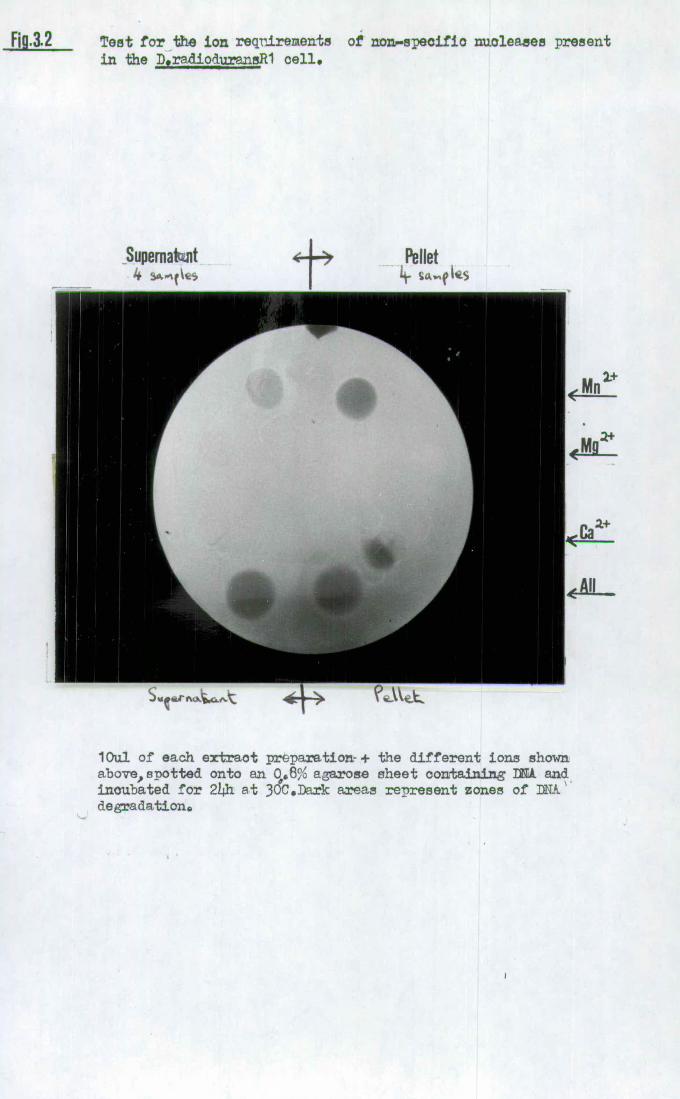
Fig.12 Test for the ion reqnirerien.1 of non.ecifio nuoleaaes present in the D,radio.umneR1 cello
Supernato.nt Pellet * I L_
lOul of each extract preparation- + the different ions shown above, spotted onto an c,.8% agaroe sheet containing MU and incubated for 214h at 30C.Dark areas represent zones of A' degradations

67.
aforementioned strains and the E.coli control organism.
The other major point of note was the presence in D.radio 2
-
philus of an enzymatic activity, stimulated by Mg only,
which altered banding patterns of both the A and pML..2
plasmid DNAs. This observation stimulated the screening
of all members of the group for the presence of such enzymes
tentatively identified as a restriction endonuclease.
2.3Sequence-specific endonuclease activity in the
beinococcaccae
All members of-the genus DeinococC14 were screened for
the presence of type ii restriction endonuclease activity
as described in chapter 2. Not only were crude cell extracts
tested for enzyme activity but also protein concentrates
obtained by ammonium sulphate precipitation and polyethylene
glycol_deXtlan T500 phase partition. For each method both
linear (XDNA) and ccc (pAT153) DNAs were used under an
extensive variety of restriction buffer conditions. These
were; high (100mM), medium (50mM) or low (0mM) salt
concentration; + 2_mercaPtoethanol (MSH); pH 7.0 or pH 8.0
medium salt Tris buffers; 30°C or 370C . iflCUbation for one
hour and finally, the substitution of KC1 for NaCl in the
medium salt buffer. All the buffers had constant magnesium
ion (10mM) and Tris (20mM) concentrations. A resume of the
screening results is shown in Table 3.6. Two species of
the genus were shown to contain site-specific endonucleases.
D.radiOdUrans Ri contains the enzyme MraI, this enzyme
being discovered by Wani et al (1982) and described in a
paper published just after my own initial observation.

TABLE 3.6 Presence of site specific endonuclease activity in the genus DeinococCJb
Polyethylene
Crude Ammonium sulphate glycol dextran
aptcies extracta precipitation phase separatiq
D.radioduranS Ri - +
D. radiodurans Sark - - -
D.radiophilUS + + +
D.proteolyticus - - -
D.radiopugnaflS - - -
aAll the techniques used both X and pAT153 DNA, high, medium and low salt buffers, + or - 2_mercaptOethanoi and also a KC1 restriction buffer rather than NaCl.

68.
Their characterisation of the enzyme reinforced my initial
observations that: -
the enzyme is essentially undetectable in crude cell
lysates due to the swamping activity of the non-specific'
nucleases previously described in section 2.2:
The enzyme has no site of action in the pAT153 molecule
but 2 or more sites in X DNA. In fact Wani et al. (1982) showed that the enzyme is
an isoschiZOmer of SstII and Sacli, the recognition sequence
being:-
5' C C G C G_ G 3'
3' G G C G C C 5'
The other member of the genus definitely shown to produce
sequence-specific endonucleases was D.radiophilUS. Altera-
tion of DNA banding patterns after supernatant incubation
and agarose gel electrophoresis was seen under all the
various conditions used for screening. The enzymes, for it
was later proven that there were in fact at least two, are
from now on called Dral and Drall. Magnus Hansen (Novo
Industries) has given Roberts (1983) personal communication
confirming my own findings on Drall but also proposing the
existence of a third enzyme Dralli. This enzyme appears
to cut DNA 13 times but in none of my experiments was
there any indication of this third enzyme.
From the results it would seem that D.radioduranS Sark,
D.proteOlvticus and D.radiopugflaflS do not contain active
restriction endonucleaseS, at least under the conditions
used. For the first of these the result is confirmed
by the observation that Sark DNA carrying selectable

69.
markers will transform Rl at a much lower frequency (at least
100 fold) than vice versa. Of course the clear-cut absence
of restriction enzymes from the remaining three DeinococCU
strains could not be totally confirmed due to the possible
deficiencies in isolation and assay procedure. Also there
appeared transient band alteration of ccc DNA on occasions,
in all cases tested. The production of multiple faint
bands intermediary between ccc and open circular .(oc) forms
may be due to topoisomeraSe activity or even inefficient
DNA digestion by a restriction endonucleaSe. This latter
explanation is unlikely as the linear DNA run concurrently
showed no pattern alteration.
2.4 Purification of site-specific endonucleaSeS from
D. radiophilUS
It was obvious from the screening procedures that
D.radioPhilUS produced sufficient quantities of restriction
endonuclease activity to make attempts at purification a
feasible proposition. The procedures outlined in Chapter 2
were therefore employed.
During the different phases of purification various
important observations were made. The growth state of the
D.radiOPhilUs cells .influenced dramatically the final
yield of enzyme. If the cells were left in a stationary
phase of growth for too long ( one day) subsequent lysis
appeared to free a very active protease which rapidly
removed most of the restriction enzyme activity present.
This destruction was not inhibited by the presence of PMSF
and seemed to be absent from cultures in late exponential
or early stationary phase. High-speed centrifugation did

70.
remove large amounts of cell debris and ribosomes but the
addition of magnesium ions to aid this operation stimulated
non-specific nuclease activity at this stage. At the
poly- ethylene glycol:dextran T500 phase separation ) I had already
shown that restriction enzyme activity remained bound to the
dextran phase at salt concentrations of <0.8 M NaCl. It
was possible to release this bound activity by washing the
dextran phase, after centrifugation, with high.salt buffer.
Although the enzyme preparation lost 99% of the protein
content it also lost 707o of the original enzyme activity.
This method could be of use to rapidly prepare relatively
small amounts of enzyme (without nuclease contamination).
For larger scale purification of both enzymes it was thought
preferable to resort to the standard preparatory use of the
phase partition technique. That was to use a high salt
concentration, the restriction enzyme separating into the
po1yethy1enlycol supernatant. Very little enzyme activity
was lost and almost all non-specific nuclease contamination
was removed. Also) a large proportion of the DNA and RNA
was excluded from the preparation, this being of vital
importance for successful column chromatography later.
During the overnight dialysis against column buffer 1 the
presence of PP1SF was essential to prevent proteolytic
degradation of the restriction enzymes by contaminating
serine proteases. The cell extract was then fractionated
during chromatography through a series of protein purifica-
tion columns as described in Chapter 2. In each case a
small scale experiment was performed to ensure that
enzyme activity was not adversely influenced by the column

71.
material and that binding/elution at differing buffer
concentrations could occur. Dral activity eluted from the
DEAE-sephacel column in the 0.18-0.24M NaCl buffer although
considerable 2 tailingV of enzyme activity did occur in
later fractions (Fig. 3.3). The vastly lower concentrations
of Drall enzyme eluted only slightly after the first Dral
fraction thus preventing any reasonable separation of the
two enzymes at this stage. However ) a hydroxylapatite column
allowed the removal from the Dral enzyme preparations of
contaminating Drall activity as well as any remaining non
specific exo/endonuclease. Dral eluted from this column
within the 0.20-0.32M phosphate buffer fractions whilst no
Drall activity could be detected (Fig.3.4). At this stage
it was shown to be essential that the protein concentration
of the collected fractions did not fall below 500 jag ml- 1.
If this occurred then rapid and irreversible loss of enzyme
activity occurred even with material stored at -20 0C.
Therefore, it was necessary to add 500 pg ml- 1 restriction
grade Bovine Serum Albumin (BSA) to each collecting tube
with the unfortunate effect of preventing accurate protein
and specific enzyme activity determination at the last
purification step. Although protracted enzymatic digestion
indicated that the Dral preparation was free of non-specific
exonuclease activity,the inability of T4 DNA ligase to work
on fragments produced by this preparation implied - the
presence of phosphatase contamination. This barrier was
overcome during the final phase of purification, heparin-
agarose column chromatography. The fractions containing

DD, AE-s ephacel
NciCI gradient
(M)
orbance 26OnM
3•3 Protein. elution profile of D.raiophilus cell extract on

FIG.3.4 PROTEIN ELUTION PROFILE OF D. radiôphilus CELL EXTRACT ON HYDROXYAPATITE
Dral activity
0.4- -
Phosphate : buffer gradient
02
0 ' 10 20 30 40
Sample number
Absorb lance at 1260nm

72.
peak Dral activity appeared in the 0.60-0.70 M NaCl eluate,
the fractions being pooled, dialysed against storage
buffer and kept at -20°C. This preparation was free of non-
specific nuclease and phosphatase activities, as shown
by the success of subsequent sequencing techniques which are
extremely sensitive to such contaminants. The Dral-cut
DNA did not religate efficiently, as indicated after agarose
gel electrophoresis, under the standard conditions used for
'sticky-ended' joining, i.e. <50 jig ml- 1 DNA concentration
and 0.1 units of T4 DNA ligase at 14°C for 24 h. However,,
by increasing the ligase concentration to one unit, the DNA
concentration to 100 jig DNA m1 1 and increasing the
incubation temperature to 20°C a much higher level of
religation was observed. This result strongly implies that
Dral action produces blunt-ended fragments of DNA. The
results of each stage of Dral purification are shown in
Table 3.7, the final preparation showing the presence of
four clear protein bands upon polyacrylamide gel electrophor-
esis (same as for DNA mapping) followed by a silver staining
protocol.
If the D-sephacei eluate containing peak Drall activity was loaded
directly onto the heparin-agaroSe column after dialysis then
partial separation from Dral could be achieved. The Drall
activity eluted in the 0.55-0.65M NaCl range and although
the pooled fractions contained elevated Drall levels they
still possessed a large amount of contaminating Dral.
Thus1 from an approximate ratio of DraI:DraII of S-.10000:1
units this preparation was in the 5:1 region. Unfortunately)
the enriched Drall preparation still had enough contaminating

TABLE 3.7
Stages of Dral Purification
Stage Total protein
(mg)
High speed 1.86 x 10 centrifugation
PEG-dextran 1.22 x 10 3 phase precipitation
DEAE-sephacel 3.15 x 101
Hydroxylapatite 0.57
Heparin-agarose N.D.
Total enzyme Specific activity Yield (units) (units mg- protein) (%)
8.0 x 10 5. 4.32 x 10 100
7.5 x 10 6.15 x 10 9375
4.1 x 10 1.30 x 1.0 51.25
1.4 x 10 2.46 x 10 17.50
3.0 x 10 3.75

73.
nuclease activity to prevent the subsequent analysis of its
active site using DNA sequencing.
2.5 Characteristics of Dral and Drall
Before detailed analysis of the recognition sequence
of either of these enzymes was made it was necessary to
investigate the parameters affecting enzymatic digestion of
substrate DNA. From section 1.4 of the Introduction it is
obvious that the major influences upon other type II
restriction endonucleaseS are those of divalent cation
concentration, pH, temperature and salt concentration. By
holding all factors except one constant and varying this
other parameter its effect upon DNA digestion by Dral was
shown. By doing this it was possible to produce reaction
conditions which give a reproducible ,maximised Dral
digestion of A DNA (Table 3.8). Of these factors the magnes-
ium concentration seemed to allow the least variation before
seriously affecting the Dral digestion rate, Dral activity
declining rapidly outside 5-25mM Mg 2+ limits. A wide
variation in salt (NaCl) concentration of between 0-75mM
was compatible with full activity. The enzyme had a
slight peak in activity in different pH buffers at pH 8.0
and it lost activity rapidly when the temperature was raised
above 44°C. Table 3.9 shows the influence of various other
treatments upon the activity of Dral. As well as being
inhibitory in the presence of Mg 2+ other divalent cations
• did not successfully replace Mg 2+ in the reaction buffer.
High ionic concentrations were inhibitory, i.e.'>lOOmM
NaCl or1O0mM Tris-HC1. KCl could replace NaCl in the
reaction buffer with no obvious detrimental effect.

TABLE 3.8 Optimum conditions for Dral activity
Mg2'ion NaCla
Concentration Concentration pH
Temperature
10mM
20mM
8.0
37°C
aTris buffer concentration 20mM throughout
TABLE 3.9 Inhibition of Dral activity
----------Compound- ------------------------- Concentration
Ammonium Sulphate 5% w/v
SDS 0.1% w/v
Cu2 ions 1mM
Zn2+ ions 0.1mM
Ca2 ions 0.1mM
ions 1mM
Tris-HC1 100mM
NaCl , 100mM
'All reactions carried out in 10mM Mg 2 , 20mM Tris-HC1, pH 8.0
buffer with 1 unit of Dral and 1 jig plasmid (pML2) DNA incubated
for 1 h at 37°C. Cocc&\ o3ry Co)i Sko)r frocl.Jc€s Coe
£L Lcc -r 'Trsct, Le.rQ. k.

74.
The addition of adenosine triphosphate (ATP), dithiothreitol
(DTT) or 2-mercaptoethanol (MSH) (all 10mM) had neither a
stimulatory or inhibitory influence. It may be possible
that the addition of weak reducing agents such as DTT or
MSH increases protein stability during long term storage.
The restriction enzyme reaction could be terminated by
addition of STOP buffer containing dye + 0.1% SDS or by
heating at 65°C for 5 minS. The enzyme activity was stable
in storage buffer at 4 0 C for >10 weeks and at -200C for2
years without major loss of activity.(sd'o"Ii CgtGL,
The inability to purify Drall free of Dral unfortunately
clouds the ionic requirements of the former enzyme. No
conditions were found that inhibited Dral activity whilst
leaving Drall unaffected. However, in very low salt buffers
the Drall activity began to wane. It would seem that the
enzymatic requirements of Drall closely match those of Dral
and both bear close similarity to MraI (Wani et al., 1982).
2.6 Mapping of Dral and Drall cleavage sites
The number and position of restriction enzyme sites
in a variety of different DNA species was investigated by
agarose and polyacrylamide gel electrophoresis. These
results are shown in Table 3.10 and it should be noted
that in all cases of Drall digestion the DNA was simultaneously
digested with contaminating Dral activity. The two observed
Dral sites in pBR322 were then mapped to within ±50 bp by
using double-enzyme digestions of the DNA (Table 3.11).
By using Dral in association with S single-cutting
enzymes, the Dral sites, were shown to be around map

Drc
Dm 11 1430
FIG. 3.5 APPROXIMATE POSITIONS OF Dral & Drall INCISION IN THE pBR322 MOLECULE (GEL ELECTROPHORESIS)
Drail 0
Iable3.10 DNA SPECIFICITIES OF TYPEIL RESTRICTIONS ENZYMES Dral &DraE
ENZYME NUMBER OF CLEAVAGE SITES
pBR322 Ade2 SV40 0x174
DRAI 3(2)a
13 >10 >10 2
DRAK 3 1 ? ? ?
(a) Only two fragments visible using agarose gel electrophoresis or non-radioactive polyacrjlamide gel techniques

TABLE 3.11 Mapping of Dral sites on pBR322 using double enzyme digestions
(composite agarose and polyacrylamide gel electrophoresis results)
Enzymes
Dral Dra-i-EcoRI Dra-i-BamHI DraI+HindIII DraI+PstI DraI+SalI
3435 3075 2625 3445 3300 2249 ' Fragment
Sizes 655 655 828 681 335 1046
452 655 505 305 655

75.
positions 3250 and SRW (Fig.3.5). All double digestions
were done in the restriction buffer of the second enzyme
as Dral showed unit activity in all these varying buffers.
The positioning of the Dral sites on the pBR322 molecule,
the number and approximate position of the sites in Øx174
(DraI/PstI double digest) and SV40 prompted a search within
the data produced by Fuchs et al, (1980) for a palindromic
sequence matching these findings. In fact 1 the only palindrome
which appeared to fit was the hexanucleotide,
52 T T T A A A 32
3 9 A A A T T T 5
Thus,DraI seemed to be an isoschizomer of the recently
isolated enzyme Ahalil (Whitehead and Brown, 1982), a product
of the blue-green alga Apanothece halophytica. This made
confirmation of the recognition sequence relatively simple in
that the enzymes Dral and Ahalil should produce exactly the
same fragement patterns from DNA digestions, which they did,
and the sequencing protocol recently perfected for Ahalil
could now be applied to Dral. The Sau3AI fragment 9 of
pBR322 was subcloned into M13mp7 in the manner described by
Whitehead and Brown (1982). Briefly, the fragment.was eluted
from an excised agarose block by soaking in 4 gel volumes
of 500 mM ammonium acetate, 1mM EDTA, 0.19 16 w/v SDS at 380C
for five hours. Gel fragments were removed by filtration
through glass wool (siliconized) and the DNA precipitated
with 2 vol. ethanol .i 0.1 vol 4M sodium acetate. The
purified fragment was subcloned into the BamNI site of
M13mp7. Clear plaques were isolated after transformation
into E.coli NM522 and the molecular size of the recombinant

76.
checked by the boiling isolation method of Holmes and
Quigley (1981). The cloned pBR322 fragment was sequenced by
the chain terminator method (Sanger et al., 1977) in
essentially the same manner as described in Chapter 2
except for the following variations:-
[32P ]ATPwas used to label the DNA rather than
[35S]ATPso that the ddATP in the reaction mixture for the
A2 lane was increased fourfold as [32P]ATP incorporates
r 35 , more efficiently than L SjATP.
Two lanes, denoted I and II were run simultaneously
either side of the GATC sequencing lanes. Both contained
the labelled Sau3AI fragment cut with DaI.. under conditions
which inhibit complete digestion. The difference between
the two lanes was the addition of the Kienow fragment of
DNA polymerase in lane II. This means that 3 possibilities
exist. The 3t_52 exonuclease function of this enzyme
removed a protruding 39 sticky-end (resulting in the band
in lane II being a number of basepairs shorter than lane I),
the enzyme had no effect (lane I and II showed bands of the
same size) or the 5 - 3 2 polymerase activity filled in a
3 1 recessed terminus (lane II showed a band larger than lane
I). Using partial digestion conditions ) it was possible to
locate the third Dral site in pBR322 only 18 basepairs
away from the primer proximal site of the Sau3AI fragment
9 molecule. A diagrammatic representation of the resulting
autoradiograph is shown in Figure 3.6,clearly reaffirming the
recognition site of the enzyme Dral as,

FIG. 3.6 DIAGRAMATIC REPRESENTATION OF AN AUTORADIOGRAM OF THE SEQUENCE OF THE pBR322Sau3AI FRAGMENT WHICH CONTAINS Dral CLEAVAGE SITES
I A G C T :ir
a LOCATION OF Dral CLEAVAGE IN SEQUENCED STRAND
b LOCATION OF Drat CLEAVAGE IN TEMPLATE STRAND

77.
52 T T T A A A 32
32 A A • A T T T 52
cleavage producing blunt-ended fragments.
By employing the double-digestion techniques used
initially in the case of Dral preliminary site localization,
it was possible to locate regions of pBR322 where Drall
cleaved the molecule. However, due to contamination of
the enriched Drall preparation with Dral enzyme these
digestions tended to involve three and not two restriction
endonucleases, Table 3.12. Further information was
gathered from digestion of the pBR322 molecule with a
tetranucleotide sequence-recognising. enzyme, 'Sau3AI(GATC),
FnuDII(CGCG) or HaeIII(GGCC) before incubation with either
Dral or the Drall/Dral preparation. Analysis of the
fragment sizes produced by polyacrylamide gel electrophoresis
(resolution of around 60 basepair fractions after ethidiuin
bromide staining) allowed the Drall sites in pBR322 to be
located with an accuracy of ± 25 basepairs. These sites are
illustrated in Fig.3.5 and occur at positions 550, 1476
and 4346. However when this information was fed into a
computer to analyse possible common recognition sites, no
known palindromic sequence capable of producing the
observed fragment configurations of Drall digestion of
pBR322 or X could be discovered. The definitive DNA sequencing protocol utilized for Dral site confirmation
could not be used for Drall as the very small quantities
of the enzyme could not be separated from a contaminating
nuclease. This latter enzyme was removed from Dral

TABLE 3.12 Mapping of DraII.sites using triple enzyme digestions of pBR322
Enzymes
Dral +DraI I +
DraI+DraI 1+ DraI+DraII+ DraI+DraII+
DraI+DraII
PstI
Hindill - BamHI Sail
1832 1832 1832 1832
968 968 968 811
575 710 710 710
445 518 445 575
376 445 407 445
334 (
57?) ( .168?)
( 157)
Fragment
Sizes
(bp)
1832
968
710
575
445

78.
during hydroxylapatite chromatography but as already noted
Drall was completely and irreversibly inactivated (or
bound) by this material, and also by phosphocellulose.
2.7 Inhibition of Dral activity by ultraviolet irradiation
of the substrate
As previously reported in section 4.5 of the
Introduction it has been shown that the activity of type
II sequence-specific endonucleases is inhibited by thymine-
thymine, or to a lesser degree cytosine-thyinine, dimer
damage within the substrate DNA molecule. This interference
with enzyme action only takes place however if the lesions
occur within, or directly adjacent to, the recognition
sequence. Obviously a recognition sequence containing a
high proportion of thymine residues ought to be
rendered particularly sensitive to ultraviolet light-induced
lesions in the DNA. To investigate whether this trend holds s'te. 4
for the enzyme DraI,)which contains a total of G.
thymine residues in comparison with I for Hindlil
(Chapter 2), the dose-dependent inhibition of Dral activity
by ultraviolet light irradiation of the pBR322 molecule
was studied. A total of 6 pg of pBR322 was linearised with
the restriction endonuclease BamHI, Figure 3.7, (6 units for
1 at 37°C), the DNA precipitated with ethanol and
resuspended in an ultraviolet transparent buffer (Chapter
2). The linear pBR322 was then irradiated for a series of
time periods corresponding to doses ranging from 0 to
3800Jm 2 , samples being removed at set time periods and
digested for 1 hour at 370C with 2 units of Dral.

31. Sites of action for the vario restriction enzyies used to study
the inhibition of activity on pBR322 after ultraviolet
irradiation (4cet Let).
3231 3250 3942
p position 375
375
ce(bp) 2850 $19t 692 793
i zym e
BamHt DraI(2) Dral BamHI

79.
The reactions were terminated with STOP buffer and the
DNA fragmentsanalysed using agarose gel electrophoresis.
In this manner the affect of increasing doses of irradiation
upon the action of Dral can be displayed (Fig. 3.8)..
Unfortunately, the presence of two Dral sites only
18 basepairs apart on the pBR322 molecule does complicate
interpretation of the results. The appearance of
a 1485 basepair fragment allied to the simultaneous
removal of both 692 and 793 basepair bands as the ultra-
violet dose increases does indicate the inhibition of Dral
enzyme action at position 3942 on the pBR322 molecule.
Inhibition of either of the two other Dral sites (3230 or
3250) would not resultin band alteration as fragment size
resolution on the 1% agarose gel used can not allow the
distinctive separation of bands differing in size by only
20 basepairs. The initial alteration in banding pattern
was seen after a dose of around 60 Jm 2 . Above a dose of
180 Jm 2 a fragment of 3567 basepairs appears due to the
inhibition of both Dral sites at positions. 3250 and 3230.
Finally, at doses of >300 Jm 2 , complete linear. molecules
of pBR322 (4362 basepairs) were seen due to the complete
inactivation of all Dral recognition sites by ultra-violet
induced lesions. This enzymatic inactivity could not be
overcome by increasing either the length of incubation or
the amount of enzyme s If Hindlil (recognition sequences)
was used as a comparative control for this experiment
(this being previously the most sensitive enzyme to ultra-
violet lesion inhibition tested), band alterations were

11i 3 .8 Dr.1 digestion of uv treatedBaBi lineazrised p322. Lanes A—K illustrate I ug pBR322 linearised. with BanHT and given itv doses of 14200,36OO 9 3OOO,2400 9 1800 0 1200,6OO 9 3OQ,18O,6O and 0 JW1 respectively before Ih Dray digestion 0Larte L shows BHt linea.rised pBR322 plaaid flM.& 0Franent separation was by 1% agarose gel electrrphresis0
4362 3561
2850
Size(bp)
1485
793 692 -
M 0 6 iii F Ci H I j K L
Lanes

3.9 Cleavage of uv irradiated )%-DNA by ])ral ,Hind= or Pst. Lanes A-C show digestion of 1 ug ),-DNA by Pstl after uv radiation doses of 3600,1800 and 0 Ji1'espeotiely.Lanes D-G show cleavage of 1 ug )-DNA by Hind W after doses of 3600,1800000 and 0 Jmrespective1y.Lanes H-K show digestion of 1 ug of A-DNA by DraT after doses of 3600 9 1800,300 and 0 Jri respectively.Lane L is I ug of undigested A-DNA.Praaent separatiai was by agarose gel ( 1%) electrophoresis.
A B C D £. F C H I L

80.
first observed after doses of between 180 and 300 Jm-2
In this case NruI was used to linearise the pBR322 molecule
(position 971) as a Hindlil/Bami-LI double digest would
normally produce a 327 basepair fragment which is on the
limit of resolution in a 1% agarose gel. The inhibition
of Dral activity resulting from ultraviolet irradiation
of the substrate DNA was therefore 2-4 times greater than
that of Hindlil. A direct comparison of the differing
sensitivities of restriction endonucleases to this kind of
damage is clearly illustrated in Figure 3.9. In this
case ADNA was irradiated with increasing doses of ultra-
violet.light and then digested for 1 hour with 4
units of either Dral, Hind III or PstI at 37 °C in the
appropriate restriction buffer. Fragment visualization
was by agarose gel electrophoresis (1% w/v),ethidium
bromide staining and ultraviolet light transillumination.
There was a striking difference in the banding pattern
of Dral digested DNA witi and without irradiation. After
a dose of 3600 Jm-2 almost all the Dral sites present in
the XDNA had been inactivated (13 altogether). To a
lesser degree the inhibition of Hindill action is also
obvious but PstI digestion was completely unaffected by
the radiation damage even at the highest dose.

81.
3. MODIFICATION METhYLASE ACTIVITY
Evidence for some manner of DNA modification
associated with Dral restriction enzyme activity is clear
cut. Both chromosomal and plasmid DNA from D.radiophilus
were isolated (Chapter 2) but proved to be completely
resistant to digestion by both purified Dral and enriched
DralI enzyme preparations. The plasmid pUEl from
D.radiophilus has been purified free of other plasmid
types and cloned into pAT153 (G. al Bakri, personal
communication). The plasmid itself, when isolated
directly from D.radiophilus, is not linearized by Dral
but the composite molecule pUE109 does show that a Dral
site exists within the pUEl region of DNA, this site only
becoming active after multiplication in E.coli.
Chromosomal DNA from A.halophytica (gift of N. Brown)
was resistant not only to Ahalli degradation but also
Dral action. The former enzyme also showed no activity
upon incubation with D.radiophilus DNA. Thus modification
seemingly occurred in such a manner as to render the
sequence 5-T T T A A A-3 1 resistant to both enzymes,
although not necessarily via the same alteration.
D.radiophilus late exponential phase cells were
harvested, washed in T.E. buffer (pH 7.4) and then lysed
in the French pressure cell as previously described for
restriction enzyme isolation. An assay for type II
modification methylase activity was undertaken as described
in Chapter 2. When the assay conditions included the

82.
presence of the methyl group donor S_adenosyl-L--methionine
then incubation of the DNA with crude cell extract in the
absence of divalent cations resulted in the protection
of the DNA present from subsequent degradtion by Dral.
However, if S_adenosyl-L-methiOnifle was excluded from
the reaction mix then no protection from Dral degradation
accrued to the DNA. These results strongly suggest the
existence of a classical 2 type II modification methylase
activity. A'previous 'investigation into DNA modification
in DeinococCUs spp. ( Mackay, 1984) indicated the absence
of any of the standard methylated bases in D.radiodurafls
(N6-methyl adenine and 5-methyl cytosine) even' though MraI
exists within the cell and the chromosomal DNA is modified
to resistance against the enzyme. Preliminary work did
indicate the absence of methylated bases in D.radiophilus
also and perhaps some novel modification system is common
to both of these organisms.
4. DEVELOPMENT OF THE CLONING AND 'EXPRESSION OF D...d1c)h1lis
GENES
4.1 Construction of D.radiophiluS gene libraries
The experimental details of the construction of
D.radioPhilUS genomic libraries in the E.coli plasmid
vector pAT153 have been described in Chapter 2. In the
case of partial MboI digestion,With insertion into the
Bami-LI site of pAT153, the degree of 'iboI action was
controlled by using different dilutions of the enzyme
ranging from 1 to 0.01 units per ug D.radiophilus
chromosomal DNA. The fragment patterns were visualized

83.
after agarose gel electrophoresis (Fig. 3.10). It was
decided to pool the three partial digestions resulting
from using 0.04, 0.08 and 0.15 units for use in the
recombination experiments.
The results from the three different plasmid cloning
experiments are shown in Table 3.13. The probability of
a DNA sequence being represented by the gene banks was
calculated from equation 3 in Chapter 2. The maximum
insert size observed in 25 randomly selected clones from
each gene bank was 20.6 kb, produced from a Hindlil digest.
The major drawback with using the plasmid vector system was
the relatively small insert size, particularly in the case
of the MboI/BamI-iI method.
4.2 Screening of recombinant DNA banks
Plasmid DNA was extracted from the cells of each gene
bank, using the Biinboim-Doly method (1979) and this was
used as the basis for testing,via transformation,for the
expression of D.radiophilus genes in E.coli mutant strains.
Each of the mutant E.coli strains (Table 3.14) was challenged
with DNA from all 3 gene banks and successful comple-
mentation of the chromosomal lesion detected as described
in Chapter 2. In the case of the attempted cloning of the
restriction/modification system of Dral, incubation of 50p.g
of gene bank DNA with 100 units of Dral for 2 h at 37 °C
caused a 10 fold decrease in the number of antibiotic
resistant transformants. In fact,plasmid containing
transforinants were recovered only from the PstI gene bank.
Eight out of 20 of these showed a pAT153 molecule
completely lacking the region containing all three Dral sites.

FIg.3.10 Partial digestion of Dradiohi.1uz DNA using the restriction endonuclea,e Mbol 0Eaoh lane consists of 2 ug chromosomal and 02 ug pla.81i. M& digested for 2h at 37 C with the appropriate amount of Mbol. Band separation was by agarose (176) gel eleotroph9esiA.
Units of MboI
,- C..J Co C) CD D ==

TABLE 3.13 Construction of genome libraries of D.radiophilus DNA using plasmid pAT153
Enzymes Number of
transformants % Iñsertional Inactivation
Average Insert size (kb)
3.2
2.9
1.2
a Probability of any DNA sequence being
represented
0.75
0.89
0.82
HindIll 1250 76
PstI 1849 92
BainHI/MboI 5300 96
allsing equation 3 from chapter 2.

TABLE 3.14 Selection of recombinant plasmids capable of
complementing E.coli chromosomal mutations.
No.transformants ug DNA Phenotypic
E.coli carrying complementary marker + trait
Strain appropriate antibiotic resistance
RM(DraI)
M(DraI)
uv rA
proA
leuB
thr
mal
liv
trp
his
ar(E)
t rpD
t rpB
t rpA
trpC
• HB1O1
HB1O1
159
HB1O1
1-lB 1 01
CSH42
CSH42
CSH42
CSH58
CSH58
CSH58
107
128
311
312
0
11
0
0
0
0
0
0
0
0
0
0

84.
Of the remaining clones, none were resistant to Dral
cleavage. Thus attempts to clone and express the Dral
methylase gene in E.coli were unsuccessful. From all the
transformations attempted with the derived gene banks )
successful complementation o the leucine B mutation was
the only one observed (Table 3.14)..
4.3 Mapping and subcloning of the leuB-complementing
DNA from D.radiophilus.
In the original experiment 11 E.coli HB101 colonies
were isolated with the ieu+ampR phenotype and these were
denoted as pleu 1 to 11. Of these only 8 showed stable
inheritance of the leucine prototrophic marker, although
all contained plasmid DNA. The recombinants pleul, 7 and
10 all appeared to have a high level of internal recombination
resulting in the production of l euamp+ daughter cells con-
taming deleted plasmid DNA. In the most severe case, pleul,
47 out of 50plasmid_containiflg cells had lost the ability
to synthesize leucine. The plasmid DNA from all 1eu
clones was extracted using the Holmes and Quigley boiling
technique (1981) and restricted with Hindlil, Pst.I or EcoRI
and the fragments analysed by agarose gel electrophoresis.
This showed that there were no EcoRI sites in any of the
inserts and that in all cases the insert DNA was of the
same size and orientation, except in leul, 7 and 10 where
complicated patterns due to deletion products were seen.
Further double digestions with the restriction enzymes
SstII/HindIII or PstI/SstII appeared to confirm that in all
cases the insert DNA was identical and inserted at the

FIG. 3. 11(a) RESTRICTION ENZYME MAP OF PLASM ID pleu5 INSERT
BssHfl NruI Sst]I PstI M lul
kb 0. 03 1.53 2.44 1.81 1.30 10.941
1.02 0.69 jO.48
Hind]I[ Hindifi
PstI PstI BssH]I
NruI
890bp region containing leuB gene
TOTAL SIZE = 10.24kb

FIG. 3.11 (b) PLASMID pLEU5 RESTRICTION MAP
SIZE = 13.89kb
PstI Hind]II
BamHI Nrut I I .SaII
NruI
M pAT 153
Sst I
Dra I
Pst I - Dra I
PstI
D.radiophilus DNA
EcoRI Hind in: BssH]t
NruI
PstI
Plasmid pleu 5

85.
Hindill site of pAT153. Large-scale plasmid preparations
were then made of pleu5 and pleu8 DNA in order for
accurate restriction mapping. Various single, double and
triple enzymatic digestions were attempted with a wide
range of 6 basepair-sequence-specific endonucleases. The
fragment patterns produced were analysed and sized using
agarose (fragments >500 basepairs) and polyacrylamide
(fragments 500-60 basepairs) gel electrophoresis.
The results were collated to produce the insert restriction
map shown in figure 3.11(a). The insert contained no
EcoRI, Bami-lI, Sail, Smal or Dral recognition sites. The
total length of the insert was 10.24. kilobases and the
orientation of insertion into the Hindlil site of pAT153
is shown in Figure 3.11 (b), the total plasmid pleu5 being
13.89 kilobases in length.
The three enzymes which had multiple sites within the
insert DNA, PstI(3), NruI(2) and BssHII(2) were then used
in attempts to subclone the DNA region coding for the leuB-
complementing activity. Both PstI and NruI digestions of
pleu5 (lpg DNA digested with 4 units of enzyme for 1 hour
at 37°C) were ligated into the appropriately digested pAT153
molecule. The conditions for ligation differed for the two
treatments as PstI produces sticky-ends and NruI blunt-
ends. After transformation of the ligation mixtures
into E.coli HB101 tet l eu+ or amp l eu+ transformants
were selected respectively. No ampRieu+ clones were
isolated, presumably indicating that one of the NruI sites
is within or very close to the region of DNA linked to
the relief of leucine auxotrophy. However about10% of

tetR transformantS were also l eu+ and plasmid screening of
these clones showed the presence of the 4.01 kilobase
fragment inserted into the PstI site of pAT153. One of
these tetRieu+ clones was purified and used for large
scale plasmid preparation by the Birnboim-D01Y method
(1979) and caesium, chloride density equilibrium centrifuga-
tion. This plasmid was termed pPL3. Attempts to use the
enzyme BssHII in such cloning experiments were unsuccessful.
This may have been due to enzyme impurities or to the BainHi
site used (as BssHII has no recognition sequence present in
the pAT153 molecule) and the BssHII produced fragments
requiring Si nuclease treatment to remove any extensions
before blunt-ended ligation could be applied.
4.4 Reorientation and stability of the 1euB-complementing
insert
A total of 24 tetleu transformantS produced by the
PstI digestion of pleu5 and re-insertion into the pAT153
were tested for insert orientation. This was done by
isolating the plasmid DNA by the Holmes and Quigley boiling
method (1981) followed by digestion with Hindl]I and SstII.
Band analysis was by agarose gel electrophoresis. In all
cases the NruI site was proximal to the 2 -iactamase
promoter sites (Stttber and Bujard, 198.1). To confirm this
result pPL3 was digested with PstI, treated with 1
unit of alkaline phosphatase at 37°C for 15 miris and then
56°C for 15 rniris. (both' steps repeated again) and finally,
after phenol extraction, ligated into the PstI site of
pAT153 again. Analysis of 10 subsequent transformafltS)once

87.
again, showed only the one type of insert orientation,
that with the NruI site not the SstII site close to the
Hindill site of pAT153.
The stability of the recombinant plasmid molecules
pleu5 and pPL3 was compared with that of. the vector molecule
pAT153 by growth of the E.coli strain harbouring the
respective plasmids in L-broth lacking any antibiotic
selection. The cultures were kept at 37 °C in exponential
phase for >100 generations and samples removed at
different times. Individual clones were isolated from
these by dilution plating on L-agar and the presence of
plasmid-borne phenotypic traits tested by replica plating.
The results are shown in Table 3.15. It is obvious that
for the period of investigation the recombinant plasmids
were as stable as the progenitor . pAT153 molecule.
Insertion of DNA may also influence the ability of
the DNA to transform the bacterial indicator strain. In
fact pAT153 gave an average of 3 x 10, pPL3 an average of
1.4 x 10 and pleu5 an average of 0.8 x 10 transformants
pg DNA. It would seem likely that this variation was
due to size differences rather than any inherent instability
caused by the insertion of foreign DNA.
4.5 Subclofling using nuclease Ba131
From the results above it is reasonable to assume that
the leuB_complerilenting activity produced by expression of
the insert DNA is localised in a region near to or surrounding
the NruI site within the 4.01 kilobase PstI fragment.
Further subcloning routines such as partial digestion with

TABLE 3.15 Plasinid stability in the absence of antibiotic
selection
pAT153 pleu5 pPL3 No. of
a % colonies % colonies % colonies
generations axnp&tet ampleu+ tetRleu+
0 100 100 100
5 99 88 96
15 99 100 100
30 100 100 100
50 96 100 100
68 100 100 90
80 100 99 100
104 100 100 99
a1 each case 100 colonies were tested for amp (30 pg ml-
tet )xg ml- 1 ) and leu (min.med + proline + thiamine)

88.
a restriction enzyme with a 4 basepair recognition site
eg. HaeIII, Sau3AI, might have been used. However the
presence of unique SstII and BamHI sites near the region
of interest prompted the use of Bal3l nuclease. As
mentioned in Chapter 2 Bal 31 nuclease has the ability to
sequentially digest DNA frombothends of a linear fragment
simultaneously. The rate of digestion is dependent upon
the relative concentrations of enzyme to DNA and also the
G:C ratio of the region being degraded. A major advantage
of Bal3l nuclease is that it requires Ca 2+ ions for activity
and therefore its activity can be inhibited by the addition
of ethylene glycoltetra acetic acid (EGTA) which chelates
2+- 2+ i i Ca but not Mg ions n any restriction buffer added. If
this Ca 2+ chelation is followedby -the dilution of the 200mM
NaCl concentration present in the Ba131 nuclease buffer ,by
the addition of a salt-free restriction buffer, digestion of
the Ba131 treated DNA with restriction enzymes can be
accomplished quickly. Before the sub-cloning regimen was
initiated a rough estimate of the rate of Ba131 nuclease
digestion was determined on plasmid pleu5 after digestion
with either SstII or BamHI. Using this information 2 p of
pleu5 was digested with 61. units of either BamHI or SstII
for 1 h - -at 4 °C. After ethanol precipitation 1 the
linear DNA was digested with Bal3l nuclease and samples
removed after periods during which approximately 300-500
basepairS were removed. In each sample the reaction was
terminated with EGTA (4 x concentration of Ca 2+ ions) and
buffer added to allow PstI digestion of the resultant
deletions (Fig ..t2c). Such treatment resulted in the

89.
formation of a series of PstI/blunt-ended fragments which
were then cloned into a modified pAT153 molecule (Fig.3.12 b ).
This molecule had the PstI-EcoRI 755 basepair fragment
removed and was therefore deficient in the normal (_lactamase
promoter region at position 4150. After ligation with T4
DNA ligase and transformation into E.coli HB101, selection
was for tetR ieu+ colonies. The insert sizes of 24
recombinant plasinids of each type (SstII or BainHI digestion)
were screened by using the rapid boiling method.
Of all the molecules screened the smallest plasmid
recombinant found still capable of producing a leu+
phenotype upon transforming E.coli HB101 was plasmid pTL27
(Fig. 3.13). This chaemer±c molecule was the product of
SstII/Bal31 nuclease digestion and consisted of a 1.38
kilobase insert as measured by agarose gel electrophoresis.
The molecule lacked both SstII (as expected) and MluI sites
but still retained the NruI site of the insert. The
smallest molecule produced upon digestion into the 4.01
kilobase region from the other direction, i.e. BamHI/Ba131
nuclease treatment was only 400 basepairs smaller than the
complete region (i.e. 3550 basepair insert). The plasmid
pTL27 was then purified on a large scale in the standard
manner and used as a basis for further localizing the
DNA region of interest. About lug of. pTh 27 was
linearised at the remaining PstI site and digested with
Bal3l nuclease, samples being taken at periods corresponding
to 200 basepair deletion. These samples were then allowed
to recircularise using T4 DNA ligase and were used to
transform E.coli HB101. Transformants with ieu Let R
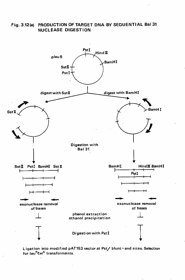
Sst K
Fig-3.12(a) PRODUCTION OF TARGET DNA BY SEQUENTIAL Bal 31 NUCLEASE DIGESTION
Pst I
pleu5
SstK
Pst I
JIL
BamHI
di with HI
1r Sstfl PstI BamHI SstII
I I
I -1
H II
amHI
1 BamHI Hind]IIBamHI
PstI I I I
I 11
Digestion with Bat 31
-
exonuc lease removal of bases
I phenol extraction ethanol precipitation
exonuclease removal of bases
-I-
Ii
Digestion with PstI
I Ligation into modified pAT153 vectorat Pst/ blunt-end sites-Selection
for leu+tet transformants.

FIG. 3.12(b) SUB-CLONING ROUTINES USED FOR LOCALIZING THE
leuB GENE OF D. radiophilus
PRODUCTION OF MODIFIED pAT153 VECTOR MOLECULE
EcoRI LiHindBI
digest with EcoRI
Ethanol precipitate
Pst I
Si nuclease - treatment
blunt-ended fragment
-H /,/Hind III
phenol extract & ethanol precipitate
P, \
Hind Ill
ligate With
recipitate target DNA
Pst I
Hind III
PstI pAT153

FIG. 3.13 PLASMID pTL27
Hind U!
BamHI
Nru Ij D . radiophilus
DNA
PstI-
- pTL27 (4300)
Dra I
•(b) pAT153 /
Derived from Sstfl/ Ba131/PstI treatment of pleu5
pAT153 pAT153-755bp region from EcoRI -PstI site
(fig. 3.12 ta

90.
phenotype were selected and screened for plasmid DNA.
It was found that the smallest recombinant molecule,
pTL54, had an insert of only 850 basepairs. The molecule
still retained the NruI site, the region of interest
lying between NruI and Miul sites of the original insert.
4.6 Sequencing the iso-propylmalate dehydrogenase gene
of D.radiophilus along with flanking DNA regions.
The advantages of using the M13 bacteriophage vector
systems for sequencing were discussed in Chapter 1. The
choice of the particular vector is dependent upon the
restriction site orientation required. For sequencing the
900 .basepair region delineated by the cloned D.radiophilus
fragment in plasmid pTL54 the vector used was M13mp9 (M13rnp8
for one particular clone set). Random techniques such as -
sonication are not particularly suited for fragmenting
such small regions successfully and accentuates the
asymptotic nature of information gathering produced normally.
The non-random method of choicwas developed from the use
of Bal3l nuclease by Guo et al. (1983) to produce sequential
deletions along the DNA region being sequenced. The
success of this method depends to a great extent upon the
presence of 2 unique restriction endonuclease sites
flanking the insert. This allows the use of'forced 2
cloning into the M13mp9 vector. It was for this reason
that pTL27 was constructed with unique Hindlil and FstI
sites on either side of the D.radiophilus DNA. Although
pTL54 has a smaller insert, the flanking enzyme sites,
Dral and Hindill, are not as easily used since -Dral produces

04 V
blunt-ended fragments no different functionally from those
produced by the Ba131 nuclease treatment. Therefore) it
was necessary to use pTL27 instead and sequence about 400
basepairs of flanking DNA. By digestion with either PstI
or Hindili followed by controlled Ba131 nuclease digestion
incremental deletions of 200 basepairs could be produced.
The alternate restriction enzyme was then used to create
fragments with one blunt and one sticky end. The deleted
molecules were then force-cloned into Sinai and PstI or
Hindili digested M13mp9. The complete inserts were cloned
by HindIII/PstI double digestion followed by insertion
into HindIII/PstI. digested M13mp8 or Ml3m39, thus giving
both orientations. The experimental details are shown in
Figures 3.14 and 3.15. The action of Bal3l nuclease on pTL27
was determined by digestion of 10 pg of the plasinid with 20
units of PstI for 1 hour at 37°C. After ethanol precipita-
tion the DNA was resuspended in 45 pl H 2 0 and 45 ul Bal3l
nuclease buffer (x2). A single unit of Bal3l nuclease was
added and after mixing ) the Eppendorf tube was incubated
at 30°C, 15 p1 samples being removed at 0, 2, 4, 6, 8 and 10
minS. The reaction in each sample was immediately terminated
by the addition of 15,u.1 of 100mM EGTA. The samples were
loaded onto a 1% (w/v) agarose gel along with size markers
and electrophoresed overnight. From the results, (Fig. 3.16)
it was possible to accurately gau.ge the length of incubation
time with Ba131 nuclease under these fixed conditions
which would give sequential deletions of about 200 basepairs..
To improve the percentage yield of correct inserts 1 the

Fig.3.16 Sequential digestion of Pstj linearised. pTL27 IA using the nuolease Ba131 ,
---Incubation time (mins.)
A 0 2 4 6 8 10 B,
The two oontrol lanes A and B consisted of iu,gA DNA digested for lii with 2units of Hj_ndX and EcoRI respectively. Ba13 1 digestion, (0.2 units) 01 2 ig of linearised. pTL27 IthL occurred for the time periods shown above at 3e C.Eleotro-phoresis was through a 1% agarose gel.

FIG. 3.14 STRATEGY FOR SUB-CLONING leuB REGION FROM pTL 27 INTO M13mp9 FOR COMPLETE SEQUENCE DE-
TERMINATION Hind 1ff
region to be sequenced ("-'1.3kb)
PstyI pTL27
digest with digest with
in d III Pst I
EtOH EtOH
Digest with Ba131 nuclease for time periods which allow the sequential removal of 200-300bp/end
I , $ phenol extract and
EtOH 4,
1 1 Digest with Digest with
PstI Hind III
I Run on 1 aga rose gel ( Et Br present), visualize bands and then elute 1300 to 300bp fragment containing leuB coding region.
I $ phenol extract and
EtOH 4r
$ I • ligateinto ligate into
PstI/SmaI digested Hind IlI/Smal digested
M13mp9 M13mp9

FIG. 3.15. SUB-CLONING INTO M13mp9 using Ba131 NUCLEASE
SEQUENTIAL DIGESTION (PONCZING ALONG)
DIGESTION WITH Pst/ BaI31 NUCLEASE/Hind ifi
PstI Hind pAT153*DNA pAT153 DNA
Bal 31 nuclease deletions
DIGESTION WITH Hind 111/ Ba131 NUCLEASE/ PstI
pAl5? Pstt
Hind DI
i4 • ______
(ARROWS INDICATE SEQUENCED REGION OF EACH DELETION)
Ligation into (a) Smal/Hindill
or (b) SmaI/PstI -digested M13mp9

92.
deleted regions of DNA of interest were purified, after
digestion with the second restriction enzyme (2 units
DNA) and agarose gel electrophoresis. The fragments were
eluted into wells cut in the agarose and then phenol
extracted before being inserted into the appropriately
treated M13mp9 vector usvt T4 NA ligase. The DNA
was transformed into the E.coli indicator strain NM522
and clear plaques picked, purified and tested for the
insert size. In this manner a series of 40 deletions were
produced covering the insert DNA and being in both
orientations. These recombinants were then used to produce
the M13mp9 templates required for sequencing (Chapter 2).
The combination of forced cloning and Ba131 nuclease
deletions is an elegant and rapid way of providing enough
template information to cover the DNA sequence of both
DNA strands. In this case, however) it was thought advisable
also to sequence the extra 500 basepairs of flanking
sequence in pTL27, subsequently shown not, to be involved
directly by the construction of pTL54.
4.7 Protein products of the leu+ subclones
The protein products of sub-cloned plasmids pPL3,
pTL27 and pTL54 were investigated using the maxicell
technique described in Materials and Methods. When
compared to the products of plasmid pAT153,or just the host
strain (E.coli CSR603) without plasmid 1 certain insert
11 specific proteins could be identified (Fig. 3.17).
The plasmid pPL3 coded for proteins of approximate molecular

18 000
14000
fig.311 Mail-cell analysis of the translational products of recombinant plasld
molecules with the leuB gene of D.radiophilus present.
Lane
1 2 3 4 5 6 7 8 9 10> Insert specific --
1
protein molecular weight (daltons)
50000
LkNE EQcoli strain Plaid Growth supplements
1 -
2 .• pPL3 + leucine
.3 pPL3 -
pTL27 + apicil1i!i
5 pTL27 + leucine
6 pTL27 -
7 pTL514 + leucine
8 pTh5L -
9 pAT153 + leucine
10 pAT153

93
weights 50000, 18000 and 14000 daltoriS in addition to those
bands represented in the control lanes. In the cases of
pTL27 and pTL54 the 50000 and 14000 dalton proteins
disappear leaving only the 18000 dalton band being insert
specifically coded. If leucine is removed from the
incubating medium just before labelling there appears to be
no effect upon the plasmid coded proteins. This was also
the case when a sub-lethal level of ampicillin (0.1)19 mr 1 )
was added to one of the pTL27 preparations. It would seem
that neither the absence of leucine or the presence of
ampicillin have a regulatory role in the expression of
any of the proteins produced from the insert DNA.
4.8 Nucleotide sequence in regions of the pTL27 insert
A total of 40 templates were tested for correct DNA
insertion using the reannealing procedure of Messing (1981).
Only 2 out of 40 failed this test. Thanks to the co-operation
of Dr. Scaife 2 S laboratory in the Department of Molecular
Biology in Edinburgh 10 of these templates were sequenced
allowing approximately 2/3 of the unique sequence of the
D.radiophilUs 1.4 kilobase insert to be distinguished using
the Sanger chain terminator method, (i977). The approximate
regions covered by the nucleotide sequence and the actual
sequence itself are displayed in Figs.3.18 and 3.19.

Re.cns of niasiid pTL27 sequenced usi the c..hin terminator method
Inessential region
PstI NruI Hind 690 710 I I
3 ) 140 252 299 144 J7
121
JTuc1eotide sequence of rena within the D0radiophi1u 1euB insert.
Aix CCAGAAGCCGCCCAGGCGGCQBkGAAGCAGCAGACCAGCCTGGCCCTGAGGGGGCCTGGACGGCA
GGACCGGGtCTCCCATCACCACACCCGACCTCCCGT
MR
CTA OC GOCCATAC TATC ACTTCC G.TCTT TGC CATAG C TAAGTAGXCXC CA G C GCTk&GCGC PCCATAAT
LTGATATCPGGTTATG TC'1'PCATTkTL'C CAGTTGC CTATTCAGTT C CA CC CÁO GCGCGGTACCT
4
ATCTCAACrFTGCLCT
çL CAGTCCCAAGCTTGGCTCCAGGPP1'GCCCGTGGTCAACTTGGACACCAGAPAGACTACC GTGTCATTGAC'19GAG
GGTCAC GGW XIL
.CCGTCACC!ACC .ACZCTATTTT?.GGJ TTCAGCTGGTGT TGTG GC'.L'G PCAGA !. GCCAGTGG'1'TGGPTC

CT
X-X WX XX
GTCA!AAGJ.CACTTTP
041-5 4W
ACLATAGcTL'AGTTGTATCTCGCCTTCaTkTCAC!TCGGACCCAC GATAC TAT CTTT ,••
34 X k )(X XXX X-X-X ) X W-X***44 -)4-- XX X -X-X X X XXX XXX XX X' XL XX X X XXX XXX XXX (
KA TCCCAACCCACCCCTCTCCGCCCAATyjTcccCcATcTAccccA ?fl?AGCTGCCrCGcCGGCfl?'jiTTaT
TTCG&CCWAGCCCCCG ACA
CGCTGCTGTGGCTCATGTF2GACAGCTTATCATCGLTAAGC 3' GCGACGACACQG>A.CAAACTGTC GUTk72AGCTATTCGAA 5'
pAT153
tosine A=adenine Xinnown base
Lanosine !c4byaine ?=secondary structure interference
possible AUG open reading frees are underlined and numbered

CHAPTER 4. DISCUSSION

94
1. FUNCTIONAL EXPRESSION BY GENES OF FOREIGN ORIGIN
IN BACTERIA
1.1 Barriers to Gene Transfer
The information embodied in the DNA primary sequence
of any organism basically determines the latter's develop-
mental and phenotypic traits. Due to the universal usage
of the DNA molecule to encode these data, it should theoret-
ically be possible for any gene to function within foreign
hosts of widely different phylogenetic backgrounds. That
this is not the case indicates the existence of factors
which modulate this simplistic view of DNA expression. The
studies of gene structure and function in bacteria have
largely been dependent upon the use of interspecies 'vectors'
capable of function in many different bacterial species
(Reanney,1976). The basic barriers to the successful in-
corporation of foreign DNA into a bacterial model system
are shown in Figure 4.1. Superimposed on these may be a
myriad of developmental processes particularly relevant
when considering eukaryotic/prokaryotic gene transfers,
although as mentioned previously (section 2.2) even the
'simple' bacterium may display a variety of differentiational
controls.
1.2 The Deinococcus gene organisation - an enigma?
The organisation of the genetic code and the levels
at which gene expression may be regulated in the genus
Deinococcus has never previously been studied. As trans-

FIG.4.1 FACTORS ANTAGONISTIC TO SUCCESSFUL FUNCTIONAL EXPRESSION OF FOREIGN GENES INTO A MODEL BACTERIAL CELL.
plasmid or linear DNA- heterologous
ATTACK BY C\
EXTRACEI NUCLEAS
FAILURE TO TKE-UP DNA
RECOGNITION OF
STAGE 1 UPTAKE
ALTERATION OF STRUCTURE DURING UPTAKE
DIGESTION
CHROMOSOME
INSUFF)tI ENT [FAILURE OF REPL ICON FUNCTION STAGE 2
STABILITY
ALTERATION OF
INAB I LI BILl TV OF TRANSCRIPTION/TRANSLATION APPARATUS TO RECOGNISE CONTROL REGIONS
INCOMPATIBLE CODON USAGE TRANSLATION PRODUCT BREAKDOWN
LACK OF CORRECT MATRIX CONDITIONS FOR PRODUCT FUNCTION
STAGE 3 EXPRESSION

95.
formation of D. radiodurans Ri and Sark cells with genetic-
ally 'marked' homologous DNA had already been demonstrated
at relatively high frequency (Tirgari and Moseley, 1980)
and endogenous plasmid molecules shown to exist in all
Deinococcus species except D. radiodurans Rl, it was hoped
that the development of plasmid vector systems for genetic
manipulation studies would be possible. It is obvious from
my results that conventional vector systems, whether via
conjugation or transformation, were ineffective, even though
a wide range of selectable markers and replicons derived
from both Gram-positive and Gram-negative bacterial sources
were used.
It is perhaps not surprising that conjugation between
such unlikely bedfellows as E. coli or S. faecalis and D.
radiodurans did not work. This process is a highly specific
mechanism, involving cell to cell contact, for the intro-
duction into the recipient of genetic information. Genet-
ically-distinct conjugation systems exist within Gram-
negative bacteria whilst transfer mechanisms in Gram-positive
types are distinct biochemically (Willetts and Wilkins,
1984; Clewell, 1981). The failure of both E. cbli and S.
faecalis conjugative systems to function with D. radiodurans
as a recipient could have been due to a breakdown in the
complicated DNA processing necessary, involving initiation
of DNA transfer, successful cell/cell contact and
binding, separation of the two plasmid strands, strand
transfer, conjugative DNA synthesis in both donor and re-
cipient cells and recircularisation of the transferred

plasmid. This situation would be further complicated by
the presence in D. radiodurans of an elaborate cell envelope
(Thornley et al., 1965) as well as to the enzymatic and
expressional barriers against the addition of foreign genes to
the cell phenotype. The obvious avenue of information
transfer into a member of the genus Deinococcus, i.e.
transformation, appeared refractory to all the standard or
- specially constructed plasmid vectors used. The failure
of such a wide variety of plasmids to express their
selectable markers could have been due to any one of the
inhibitory factors described in Figure 4.1. Attempts to
transform D. radiodurans Rl with hybrid recombinant plasmids
consisting of D. radiodurans marked chromosomal DNA allied
to either E. coli or D. radiophilus replicons have also
failed (Mackay, 1984; G. Al Bakri, personal communication).
Also, the plasmid pUE1, isolated from D. radiophilus, has
been successfully cloned into pAT153 and mapped using re-
striction enzymes (Al Bakri, Mackay and Moseley, personal
communication) yet did not successfully transform.
D. radiodurans Rl
Homologous DNA is taken up by D. radiodurans Rl and
Sark, recombination producing highly variable levels of
transformation depending upon the marker selected.
'Dominant' genes such as rifampicin resistance transform
at a high frequency, in the region of 10_ 2 , and express the
marker relatively quickly Ou 2 hours) whilst 'recessive'
markers such as streptomycin resistance may only reach a

97
frequency of 10 000 fold less after many more hours of
phenotypic lag. Whether such behaviour, thought to be
associated with the.multigenomic nature of the Deinococcus
cell (Tirgari and Moseley, 1980), would have an adverse
effect upon z'hetero1ogous DNA suppression associated
to an independent replicon is debatable. It is quite
reasonable to assume, however, that a recessive marker
inserted into a plasmid would never reach the homozygous
- state required for phenotypic selection. The cell always
carrying one or more copies of the 'sensitive' dominant
allele upon the chromosome. Such a recessive marker could
only succeed in altering the cell phenotype by integration
into the chromosomal allelic site followed by segregation
until the homozygous state was achieved.
Previous DNA labelling experiments (Tirgari, 1977)
have shown that E. coli plasmid DNA is taken into D. radio-
durans from the surrounding matrix. Thus ) there is no
selection of homologous DNA for uptake as is seen in
Haemophilus influenzae (Danner et al., 1980). Although it
has been shown that both transformable strains of D.
radiodurans secrete highly active non-specific nucleases
into the growth medium, such behaviour has little effect
upon the transformation process. No alteration to hetero-
logous plasmid structure could be demonstrated during the
short time period prior to DNA uptake, using the standard
transformation protocol, presumably because most enzyme
activity would be removed during resuspension of the bacteria

ME
into fresh medium. Nuc mutants lacking the extra cellular
nuclease activity showed no difference in response to trans-
formation compared with the Nuc wild type.
The structural conformation of the transforming DNA
in the extracellular matrix is unimportant.. Chimaeric DNA
constructed from E. coli plasmid and D. radiodurans Rl
chromosomal DNA will transform the cell at the same frequency
whether originally in covalently-closed-circular or linear
form (G. Al Bakri, personal communication).
The binding and uptake of foreign DNA itself appears
not to be a problem. However ,for successful plasmid marker
expression in a hóst having little DNA homology with the
plasmid, and therefore, no recombination, stable independ-
ently-replicating molecules would have to be produced.
Although any DNA can be taken up by the cell, major
structural alterations to plasmid integrity may occur during
the uptake process, perhaps in a manner analogous to the
B. subtilis system (Piechowska and Fox, 1971). Both trans-
formable strains of D. radiodurans were shown to have a
Ca2+ dependent nuclease associated with the cell membrane
fraction. As transformation in these organisms is vastly
stimulated by the presence of this divalent ion, but no
other, in the medium, it is not impossible that this enzyme
is closely linked to DNA uptake and processing. Thus,the
transformation frequency obtained using homologous DNA of
size <2kb containing a selectable marker can be greatly
increased by 1igtina the transforming DNA into a non-homologous

ME
E. coli plasmid DNA which is then linearised (G. Al Bakri,
personal communication).
Another major problem facing molecules attempting to
retain circular integrity during uptake by D. radiodurans
Ri is the presence, in the latter, of the restriction endo-
nuclease MraI (Wani et al., 1982). The host chromosomal DNA
is modified in such a manner that it is protected from the
enzyme. However, chromosomal DNA cloned into E. coli plas-
mid molecules and replicated within E. coli is not modified
and so no longer retains this intrinsic resistance. Such a
restriction/modification system may play a fringe role in
the inability of foreign plasmids to transform D. radiodurans
Rl successfully. The apparent absence of such enzymes in
D. radiodurans Sark and the lack of MraI sites in some in-
effective plasmid vectors, e.g. pBR322 suggests that other
factors are of more importance.
It is clear that at least two members of the Deinoco-
ccaceae, D. radiodurans Ri and D. radiophilus contain type
II site-specific endonucleases. The various theories for
the biological role for such enzymes in vivo have been aired
in the Introduction, section 4.6. Perhaps no simple em-
pirical raison d'tre exists. No bacteriophages have been
found capable of parasitising any member of this genus.
Conjugation does not seem to be a viable proposition and
transformation unlikely to be of biological importance in
the natural environment. Therefore,the persistence of such
enzymes purely as a defense mechanism against incoming DNA

100.
appears slightly perverse, particularly in the case of D.
radiophilus where at least two and possibly three separate
systems occur. The balance of methylation modification
and gene expression has clearly been demonstrated in eukar-
yotes and the novel method of DNA modifications seen in
these two organisms may have a vital role to play in gene
control. Also l the association of site-specific enzymes
Scel and Scell in yeast DNA recombination may be mirrored
in the highly complex genomic organisation found in the
Deinococcaceae. That comparable enzymes have not been
found in the other genus members may be a function of ex-
perimental ineptitude rather than non-existence.
Unlike D. radiophilus the blue-gree alga Apanothece
halophytica,the source of the Dral isoschizomer AhaIII,is
very difficult to grow and the yield of purified Ahaill is
very low (Whitehead and Brown, 1982). Therefore, although
Ahalil was commercially produced it was rapidly superseded
by Dral since D. radiophilus is easily grown in liquid
culture and produces large amounts ( 200 000 units g 1
wet weight cells) of enzyme which can rapidly be purified
into stable preparations. The enzyme recognises a sequence
containing only A:T bases and this property in itself boosts
the importance of its discovery. Very few enzymes behave
in this manner; previously only Ahaill, modified EcoRI
(Woodbury et al., 1980) and the tnpR gene product of the
transposon Tn3 did so (Reed and Grindley, 1981). The dearth
of available enzymes showing this property has hindered the

101
expansion of DNA/protein studies particularly into the
highly A:T-rich eukaryotic DNAs. Here, due to a marked
degree of asymmetry in A:T distribution large regions of
G:C rich DNA may be produced.
Dral, unlike the vast majority of restriction endo-
nucleases does show a high level of activity against
bacteriophage T 4 DNA. The DNA of ,T-even bacteriophages is
characterized by the presence of 5-hydroxymethylcytosine
glucosylated at either the a or a position. This gluco-sylation renders the DNA resistant to most restriction
endonucleases but has no effect, theoretically, upon such
enzymes acting only at T:A DNA. Digestion of T4 DNA by
Dral does occur but although the same pattern is produced
upon semi-glucosylated DNA ( position only) my inability
to obtain T 4 DNA free of glycosyl adducts prevented clear
proof of complete digestion of the original T 4 DNA by Dral.
It was shown that increasing the Dral enzyme concentration
from Ito S units or increasing the incubation period
to 5 hours had no effect upon the T4 DNA digestion
pattern.
The interesting behaviour of Dral upon ultraviolet
irradiation of the substrate DNA has been used to accurately
control the partial digestion of DNA5 required for cloning.
Assuming random lesion production within the DNA, varying
the ultraviolet dose can reduce the number of Dral sites
in a population of DNA molecules. Previously 1 partial di-
gestions had been achieved by lowering the amount of enzyme

102..
per pg DNA to well below one unit. However ,the easily-
controllable technique of ultraviolet lesion inhibition
affords some obvious advantages but previously the lengthy
period of irradiation necessary was a problem. This is no
longer true in the case of Dral. The partially digested
DNA can be directly cloned into a vector molecule at any
blunt-ended restriction site, directly or by using poly(dA)
and poly(dT) tailing. This latter method has the advantage
of recreating Dral sites at either end of the original
insert if correctly done, i.e. poly(dT) tailing of the
vector and poly(dA) for the target DNA. To allow gene ex-
pression within the insert DNA,the recombinant molecule
must be allowed to replicate in an ultraviolet damage
repair-proficient strain of bacterium, yeast or cell line.
This process would remove any translational/transcriptional
or replication block caused by the ultraviolet light-induced
lesion but would also reactivate any internal Dral sites.
Also of significance is that the three cleavage sites
in pBR322 are centred closely around the single PstI
cloning site. This enzyme is frequently used for cloning
restriction fragments or cDNA by theG/C tailing technique.
Thus, such fragments may be recovered from vector DNA with
little flanking ,pBR322 sequence via Dral digest even though
the original PstI insertion sites are no longer operative.
The mode of plasmid replication and partition within
the genus Deinococcus is unknown. Attempts to cure both D.
radiodurans Sark and D. radiophilus of any of their endo-
genous plasmids have failed (Mackay, 1984). Spatial

103.
compartmentation to protect plasmid and chromosomal DNA
from the many non-specific nuclease activities present
within the cells may occur. The absence of such separation
might explain the absence of plasmid DNA from D. radiodurans
Ri. Obviously,exposure of incoming plasmid molecules to
non-specific endonuclease activity would be disastrous in
terms of molecular integrity if not for the ability of
linear DNA to transform. The significant effects of super-
helicity of a DNA molecule upon its ability to be efficiently
transcribed have been detailed (West and Rodriguez, 1982),
but the possible secondary form of plasmid molecules
successfully surviving in the Deinococcus cells has never
been studied. In the case of vector molecules derived
purely from foreign sources, interference with the repli-
cating mechanisms encoded by the plasmid is not uncommon.
In B. subtilis, vector molecules not only require selectable
B. subtilis markers genes but also a B. subtilis replicon to
be present. The lack of transposition from 'suicide' plas-
mids unable to, replicate indicates that the breakdown of
plasmid replication machinery is not the simple answer to
why no transformants were found. Experiments aimed at
developing novel cloning vectors derived from D. radiodurans
Sark plasmid DNAs have failed, and the chimaeric pAT153/pUE1
recombinant molecule (pUE109) does not transfer effectively
between E. coli and D. radiodurans. Future develop-
ment of a successful shuttle vector will largely depend upon
a detailed understanding of the structure and function of

104.
Deinococcus genes and its replicational apparatus.
If a foreign gene product is successfully translated
from DNA it faces extreme difficulty in functioning effi-
ciently in a completely novel environment. Compatible
biochemical pathways or modes of anti-bacterial agent re-
sistance cannot be assumed to exist. In the extreme cases
where mammalian genes such as somatot or inteferon
are cloned into bacteria it would be folly to expect them
to function normally within these cells. Careful consider-
ation must be given, when analysing evidence for the in-
ability of genes to function, to the type of markers in
question. Furthermore, the presence of proteases capable
of recognising inessential, mistranslated or foreign proteins
may be a possibility.
1.3 Transcriptional Unit Organisation
Fundamental to the successful expression of genes bet-
ween bacterial species is the compatibility of their trans-
criptional and translational mechanisms. The basis for
these reside around and within the transcriptional units
themselves, whether operons or single cistrons, although
there may be a degree of developmental control for the
purposes of differentiation. The failure to. construct
shuttle vectors capable of gene isolation and transfer bet-
ween established bacterial systems such as E. coli or B.
subtilis and D. radiodurans precluded the study of the
latter's gene structure and control mechanisms directly.

105
The alternative course involved the construction of
Deinococcus genomic libraries in E. coli vectors, main-
taining and studying the recombinants mainly in this organism.
E. coli is amenable to a variety of cell manipulation tech-
niques and has the advantage that its gene expression
mechanisms function successfully upon a wide range of
foreign genes from such diverse organisms as B. subtilis
(Nagahari and Sakaguchi, 1978), Streptococcus mutans
(Jagusztyn-Krynicka et al., 1982), Achromobacter (Levesque
and Roy, 1982) , Clostridium thermocellum (Cornet et al.,
1983) and Thermus thermophilus (Nagahari et al., 1980).
Construction of D. radiophilus (this thesis) and D.
radiodurans Ri (M. Mackay, 1984) gene banks were success-
fully accomplished in parallel. In the case of D. radiophilus
the absence of a transformation system and a lack of mutants
required the search for structural genes to be limited to
those which successfully complemented E. coli lesions. The
success in finding a D. radiophilus gene capable of re-
moving leucine dependence in E. coli HB101 opened up the
possibility of studying the primary structure of a functional
Deinococcus gene. The complementing of the E. coli leuB
mutation was later paralleled using the D. radiodurans Rl
gene bank (G. Al Bakri, personal communication). Two
questions are of central importance:-
1. Is the product of the coding DNA a protein capable
of complementing the -isopropy1inalate dehydro-
genase activity absent in E. coli HB101 or is it

106.
a molecule capable of suppressing the actual E.
coli lesion itself?
2. Does the insert contain all or most of the regul-
atory sequences necessary for successful expression
both in E. coli and D. radiophilus or is trans-
cription and translation a product of inadvertant
read-through from E. coli regulatory regions?
1.4 The leuB gene of D. radiophilus
In E. coil the biosynthesis of leucine is largely
effected by the enzyme products of four genes, leuA, B, C,
and D. These code for c-isopropyamalate synthetase, s-iso-
propylmalate dehydrogenase and sub-units of c-isopropy1 -
malate isomerase respectively. The enzyme -isopropy1-
malate dehydrogenase catalyses the loss of protons from -
isopropylmalic acid associated to the removal of a carboxyl
group,
HO -CH -COO - NAD H - C
H - C
- COO
- CH I
CH NADH
-isopropylmalatè
O = C - COOH
H - - COOR
H - C - CH
CH
I a-keto- -carboxy
isocaproic acid
O =
CH H - C - CH
CH
c keto- isocaproate
107.
This activity corresponds to that of the leuC gene product
in B. subtilis.. Regions of DNA which produce proteins
complementing the leuB lesion in E. coli have been success-
fully cloned from an extremely wide variety of organisms
including Salmonella typhimurium (Geminill et al., 1983),
Azotobacter vinelandii (Medhor:a et al., 1983), B.subtilis
(Nagahari and Sakaguchi, 1978), Bacillus No. 221 (Honda et
al., 1984) and Thermus thermophilus (Nagahari et al., 1984).
Even if the operon organisation present within these various
species may vary, expression controls such as attenuation
regions (Wessler and Calvo, 1981; Genimill .et al., 1983) and co-
ordinate regulation (Travers, 1984) , common in amino-acid
biosynthetic gene clusters of E. coli and S. typhimurium,
could be present. Whether the D. radiophilus recombinant
plasmid pleu5 contains other leucine biosynthetic genes as
well as the leuB complementing gene and whether such genes
would be functional in E. coli can only be shown when leuA,
leuC and leuD mutants of E. coli become available (a set of
such mutants has been reported (Honda et al., 1984) and a
request made for them to be sent, but they have not arrived
from Japan). Why the enzyme -isopropylma1ate dehydrogenase
should be so amenable as to function in E. coil from such a
wide range of organisms can only be speculated upon. From
the sequence data from all these organisms allied to that
accruing from both D. radiophilus and eventually D. radio-
durans Ri, the complete primary, secondary and tertiary
structures of the proteins should become available. It would

108.
seem unlikely that a
biosynthetic pathway
servation. However,
with the NAD linked
occur.
The sub-cloning
protein involved in an amino acid
would show great evolutionary con-
certain analogous features associated
ehydrogenase/carboxylase activity may
rationale followed has allowed the
identification of the, coding region to an 850 basepair
region present in plasmid pTL54. All of the sequentially
produced smaller clones pPL3 pTL27 pTL54 produce plasmid
specific proteins as illustrated by the maxi-cell results
(Fig. 3.17) .A protein of about 18-000 daltons appears to be
the only protein common to all sub-clones but absent in all
control lanes. This fits in well with the sequence results.
Such a protein would not allow much DNA spare for further
proteins or even a tRNA molecule unless overlapping reading
frames or transcription of the opposite DNA strand occurred.
It would seem that D. radiophilus DNA, cloned originally
into pleu5 and finally modified to produce an 850 basepair
insert in pTL54, encodes an 18 000 dalton protein capable
of complementing the leuB gene mutation of E. coil.
The question of whether D. radiophilus control signals
are being recognised by the E. coli expression apparatus has
been complicated by the sub-cloning routines used. The
plasmid pAT153, a derivative of pBR322, has 5 major trans-
cription initiation sites (Fig. 4.2)(Stfiber and Bujard, 1981).
The original insertion of DNA into the Hindlil site should
have precluded any initiation from the tet promoter, P.

2065
3000
4000
1000
F'stL EcoRI Hind]IL Sail
pyuu BamHI
Pp P3 $
on
P1
bla tet
>
H indZ 1 ybla promoter P3 1 $
bla promoter P1
bla
4 tet promoter P2
FIG. 4.2 PROMOTER SITES OF pBR322 THE TRANSCRIPTIONAL UNITS ARE DEFINED BY ARROWS, FILLED- IN, (.vvPtM) -
CLEAR( Pru15) OR HATCHED (transcript which primes DNA synthesis). THE PRESENCE OF Pi ALLOWS TRANSCRIPTION OF RNA FROM DNA INSERTED INTO THE Hind 11E SITE. INSERTION OF DNA INTO THE 4000-0 REGION RESULTS IN SUPEREXPRESSION OF THE tet GENE INDICATING THE PRESENCE OF FURTHER CONTROL REGIONS IN THIS AREA UPSTREAM OF THE PROMOTER.
ff
tet

109.
However, it is still possible that the strong bla gene
promoter P situated to the right of P 2 was operating,
thus transcribing into the right side of the leu5 insertion.
As the leuB region is situated only 1600 basepairs away
from this promoter it is not impossible for read-through
to occur. When the 4.01kb PstI/PstI fragment of pleu5 was
sub-cloned into the PstI site of pAT153 it may have come
under the influence of both bla promoters although one
might have expected a fused protein product. The only
orientation successfully achieved matches the direction
of transcription that would result from P 1 into the leu5
insert. However orientation of inserts is strongly in-
fluenced by secondary structure and it is not uncommon
for one orientation to be favoured many-fold over its
opposite. The plasmid pPL3 directed the production of three
insert-specific proteins of molecular weights 50.000, 18,000
and 14 000 daltons. Therefore ) if transcription was ini-
tiating at the bla promoters at least the internal trans-
lational signals of D. radiophilus must have been directing
the production of three separate proteins only one of which
could have been a fusion product whilst another was a
functional protein. However,during the sub-cloning which
produced pTL27 the insert was forcibly re-orientated by
the technique used, the truncated insert being cloned
directly behind the P i promoter. The methodology of
isolation and selection would, of necessity, produce the
correct relative positioning between gene and E. coli

110
promoter. The inability to successfully remove the HindIII/
BamHI region (23-350) without destroying the leu phenotype
tends to indicate that the insert was dependent upon
for 'initiation of transcription, the Ba131 nuclease se-
quential removal of bases allowing the correct alignment
between promotion and translation. If this is correct then
a D. radiophilus promoter must be responsible for trans-
cription of leuB genes in both pPL3 and pleu5 as in both
cases E. coli promoters would have led to the transcription
of the non-coding strand. This problem may be resolved by
studying the sequence data of the 850 basepair essential
region. It should be noted that in clones pTL27 and pTL54
the deletion of the PstI (2857) - EcoRl (o) fragment of
pAT153 leads to hyper-expression of the tet gene indicating
that this region normally has a negative-controlling role.
1.5 The sequence of the leuB gene of D. radiophilus
Nearly 2/3 of the DNA region of interest has been
sequenced (Fig. 3.19).. Even this would not have been poss-
ible without the massive assistance of Dr Scaife's group
and in particular Dr M. Mackay. As DNA sequencing, parti-
cularly of such large stretches of DNA is very expensive
and the MRC has decided to terminate Dr Moseley's grant,
not only will sequencing of the remaining 3/4 of unused
templates for the leuB clone of D. radiophilus (almost
certainly covering the final 1/3 of sequence remaining) be
prevented but also that of the clones isolated by G. Al

111.
Bakri of the uvrC, uvrD, uvrE, leuB, trp and asp genes in
D. radiodurans.
obviously ,drawing clear conclusions from partial data
can be dangerous. In the sequenced regions of the leuB
insert there exist 19 possible sites for protein synthesis
initiation (if one assumes an AUG initiation codon). Of
these,6 are in an area deemed inessential for mutation
complementation. In the remainder only one appears to
have no stop codon (UAA, UAG or UGA) closely associated
in phase. This start codon has been denoted 13 in Figure
3.19 and resides round 150 - 250 basepairs to the left of
the Hindill insertion site. More detailed examination of
this region is shown in Figure 4.3.
As can be seen 21 basepairs upstream of this open
reading frame is a possible site of transcription initiation
* (CAT). Also preceding this site at -5 to -9 is the hepta-
nucleotide TATTATC which closely resembles the consensus
sequence of TATAATG for E. coli promoters at this region
(5 out of 7 bases in the same position). Unfortunately)
the -35 region lies in an unsequenced area of the. insert.
Following the AUG initiator codon is a region of 32 amino
acid codons in frame without a terminator codon,however
the DNA moves again into an unsequenced region. These
similarities to E. coli may be coincidence, without the
full sequence data it is impossible to tell as the correct
initiation site may lie in an unsequenced area. However )
if this is not the case then two points are obvious,

alqI&L serls3s erg rm wt Wwwar3 cjs ojj t (s em &IIe ;Le $ç hLe d
GCATTATACCCATLTCGTCL&LAGACACTT'pjA.
$n asf ser rk
GAVA 3' CTTTi
leader mRNA ' Promoter?
Possible dpen reading frame for the leu3 gene of D.radiophilus •The in—acid sequence is present above the appropriate nucleotide. triplet.
Initiation of mRNA synthesis i .indicated by the * below the sequence.

112..
firstly the E. coli promoter in the pAT153 molecule is
not essential and secondly no attenuation site occurs in
the leader mRNA sequence (reinforcing the maxi-cell in-
formation upon the constitutive protein production in the
presence and absence of leucine in the medium).
1.6 Where Are We Now?
It has been shown that at least one D.. radiophilus
gene, that coding for -isopropy1ma1átè dehydrogenase, can
functionally, express in the novel environment of the E.
coli cell. The D. radiophilus Ri gene also behaves in a
similar manner. No absolute conclusions can be drawn as
to the nature of any 'consensus' sequences associated
with Deinococcus genes until numerous other examples have
been sequenced and analysed. However ) the successful trans-
cription of these two genes does at least give a foothold
into the investigation. The absence of complementation of
other E. coli mutations is attributable either to the
failure of foreign protein products to harmonise into an
unusual biological matrix or to the existence, as-in B.
subtilis, of many types of promoter/translation systems for
developmental considerations.
From this study we can attribute the inability to
select for successful foreign gene expression in the
Deinococcaceae to no single factor. Undoubtedly, the unknown
aspects of DNA uptake and processing are important particularly
in view of plasmid integrity. The need for a known,

- 113
functional D. radiodurans replicon capable of insertion
into vectors for these transformable species is vital.
Although restriction of incoming DNA should be a consider-
ation it may only play an important role in future work
on D. radiodurans Ri and D. radiophilus. Possibly the
most important factors are the differences between DNA ex-
pressional apparatus and the choosing of a selectable
marker capable of function.
From this, and the closely-associated studies of M.
Mackay and G. Al Bakri, however, it may soon be possible to
overcome most if not all these difficulties. Markers have
been isolated, leuB, both from D. radiophilus and D.
radiodurans capable of effective selection in both the
Deinococcaceae and E. coli. A vector, pUE109 (pAT153/pUE1)
has been constructed that is capable of existing in E. coli
and carries a Deinococcus replicon. From the sequence data
it should be possible to localise the control mechanisms
associated with the leuB regions and manipulate them so
that any gene of choice .can receive a promoter capable of
directing transcription in E. coli and D. radiodurans or D.
radiophilus. The major obstacles remaining are:-
The isolation and use of a D. radiodurans plasmid
replication origin.
Ensuring transformation does not affect plasmid integrity.
Development of transformation and mutations in D.
radiophilus.
Purification of Dral and MraI modifying enzymes to ensure
the limitation of the effect of restriction.

114.
ACKNOWLEDGEMENTS
I would like to offer my deep thanks to Dr B.E.B.
Moseley for his advice and conversation both associated
with work and other aspects of life. Many other people
have made vital contributions to the completion of this
work and I take pleasure in thanking (in order of appear -
ance) Dr M. Mackay, Dr D. Evans, C. Duncan, S. Whyte, G.
Al Bakri, Dr N. Brown, all the technical staff of the
Microbiology Department, Dr D. Seale, Dr J. Scaife, I.C.
Masters and G. Kay. Particular recognition should be
given to Dr M. Mackay, Dr J. Scaife and Dr N. Brown without
who's help the DNA sequencing which has been done would
have been impossible.
I would like to extend my gratitude to Jackie Bogie,
Anne Brown and Mr G. Finnie for the expert aid in the
typing and construction of this thesis.
Finally I would like to thank my wife for supporting
my various vices over the last three years and all members
of the Kirk Brae Cricket Club for being even worse
cricketers than myself and for introducing me to the
dubious pleasures of real ale.

115.
REFERENCES
ADHYA, S. and GOTTESMAN, M. (1978). Control of trans-
cription termination. Annual Review of
Biochemistry, 47: 967-996.
ANDERSON, A.W., NORDAN, HOC., CAIN, R.F0, PARRISH, G.
and DUGGAN, D. (1956). Studies on a radio-
resistant micrococcus. I. Isolation, morphology,
cultural characteristics and resistance to gamma
radiation. Food Technology, 10: 575-578. II
BAGDASARIAN, M.M., AMANN, E., LURZ, E., RUCKERT, Be and
BAGDASARIAN, M. (1983). Activity of the hybrid
trp-lac (tac) promoter of Escherichia coli in
Pseudomonas putida. Constrction of broad-host-
range, controlled-expression vectors. Gene, 26:
273-282.
BAIRD-PARKER, .A.C. (1965). The classification of
staphylococci and micrococci from world-wide
sources. Journal of General Microbiology, 38:
363-387.
BIRNBOIM, H.C. and DOLY, J. (1979). A rapid alkaline
extraction procedure for screening plasmid DNA.
Nucleic Acids Research,7: 1513-1523.
BROOKS, B.W. and MURRAY, R.G.E. (1981). Nomenclature
for Micrococcus radiodurans and other radiation
resistant cocci: Deinococcaccae faine nov. and
Deinococcus gen.nov., including five species.
International Journal of Systematic Bacteriology,
31: 353-360.

116.
BOLIVAR, F. and BACKMAN, K. (1979). Plasmids of
Escherichia coli as cloning vectors. Methods
in Enzymology, : 245-268. Edited by R. Wu.
Academic Press, London.
CFIANG, A.C.Y. and COHEN, S.N. (1979). High frequency
transformation of Bacillus subtilis protoplasts
by plasmid DNA. Molecular and General Genetics,
168: 111-115.
CLARKE, L. and CARBON, J. (1976). A colony bank containing
synthetic ColEl hybrid plasmids representative of
the entire E.coli genome. Cell, 9: 91-99.
CLEAVER, J • E., SAMSON, L. and THOMAS, G .H. (1982).
Restriction enzyme cleavage of ultraviolet-
damaged DNA. Biochemica et Biophysica Acta, 697:
255-258.
CLEWELL, D.B. (1981). Plasmids, drug resistance, and gene
transfer in the genus Streptococcus. Microbiolo-
gical Reviews, 45: 409-436.
COLLINS, J. and HOHN, B. (1979). Cosmids: a type of
plasmid gene-cloning vector that is packageable
in vitro in bacteriophage heads. Proceedings of
the National Academy of Sciences, U.S.A., 75:
4242-4246.
CORNET, P., TRONIK, D., MILLET, J. and AUBERT, J-P.
(1983). Cloning and expression in Escherichia
coli of Clostridium thermocellum genes coding for
amino acid synthesis and cellulose hydrolysis.
FEMS Microbiology letters, 16: 137-141.

117.
DAHL, H.H., FLAVELL, R.A. and GROSVELD, F.G. (1981). The use
of genomic libraries for the isolation and study of eukary-
otic genes. In: Genetic Engineering, 2: 49-127. Edited
by R. Williamson, Academic Press, London, New York, Toronto,
Sydney, San Francisco.
DANNER, D.B., DEICH, R.A., SISRO, K.L. and SMITH, H.O. (1980). An
eleven-base-pair-sequence determines the specificity of DNA
uptake in Haemophilus' transformation. Gene, 11: 311-318.
DEININGER, P.L. (1983). Approaches to rapid DNA sequence
analysis. Analytical Biochemistry, 135: 247-263.
DRIEDGER, A.A. and GRAYSTON, M.J. (1970). Rapid lysis of cell walls
of Micrococcus radiodurans with lysozyme: effects of butanol
pretreatment on DNA. Canadian Journal of Microbiology, 16:
889-893.
DUNNY, G. and CLEWELL, D. (1975). Transmissible toxin (haemolysin)
plasmid in Streptococcus faecalis and its mobilization of a
noninfectious drug resistance plasmid. Journal of Bacteriology,
124: 784-790.
EHRLICH, M. and WANG, R. Y-H. (1981). 5-Methylcytosine in
eukaryotic DNA. Science, 212: 1350-1357.
FOX, G • E., STACKEBRANDT, B., HESPELL, R • B., GIBSON, J.,
MANILOFF, J., DYER, T., WOLFE, R.S., BALCH, W.,
TANNER, P., MAGRUM, L., ZABLEN, L • B., BLAKESMORE, R.,
GUPTA, R., LVERHRSEN, K • R., BONEN, L ., LEWIS, B .J.,
CHEN, K.N. and WOESE, C.R. (1980). The phylogeriy of
procaryotes. Science, 209: 457-463.
FRANKE, A.E. and CLEWELL, D.B. (1981). Evidence for a
chromosome-borne resistance transposon (Tn9l6) in
Streptococcus faecalis that is capable of
( 'F%3 c-%As et. QL~ . •? ('Qk pe

118.
'conjugal' transfer in the absence of a conjugative plasmid. Journal of Bacteriology,
145: 494-502.
GEMMILL, R.M., JONES, J.W., HAUGHN, G.W. and CALVO, J.M.
(1983). Transcription initiation sites of the
leucine operons of Salmonella typhimurium and
Escherichia coli. Journal of Molecular Biology,
170: 39-59.
GRYCZAN, T.J. and DTJBNAU, D. (1978). Construction and
properties of chimeric plasmids in Bacillus
subtilis. Proceedings of the National Academy
of Sciences U.S.A. 75: 1428-1432.
GRYCZAN, T • J •, CONTENTE, S • and DUBNALJ, D. (1978).
Characterisation of Staphylococcus aureus plasmids
introduced by transformation in Bacillus subtilis.
Journal of Bacteriology, 134: 318-329.
GRYCZAN, T.J. and DUBNAU, D. (1982). Direct selection of
recombination plasmids in Bacillus subtilis. Gene,
20: 459-469.
GUO, L.-H., YANG, R.C.A. and WU, R. (1983). An improved
strategy for rapid direct sequencing of both
strands of long DNA molecules cloned in a plasmid.
Nucleic Acids Research, 11: 5521-5540.
HALL, R.K. and LARCOM, L.L. (1982). Blockage of restriction
endonuclease cleavage by thymine dimers. Photo-
chemistry and Photobiology, 36: 429-432.
C.., OE o..'\O
o- ,, c.maC3
seck re%cw. okowJe
°S L.L rize.à evt q &or 't'tv a.s.ck tksi.A ifl\s 10.3SI - no.

119.
HANSEN, M.T. (1978). Multiplicity of genome equivalents
in the radiation-resistant bacterium Micrococcus
radiodurans. Journal of Bacteriology, 134: 71-75.
HARSOJO, KITAYAMA, S. and MATSUYAMA, A. (1981). Genome
multiplicity and radiation resistance in
Micrococcus radiodurans. Journal of Biochemistry,
90: 877-880.
HECKER, M., SCHROETER, A • and MACH, F. (1983). Replication
of pBR322 DNA in stringent and relaxed strains of
Escherichia coli. Molecular and General Genetics,
190: 355-357.
HENDRIX, R.W.., ROBERTS, J.W., STAHL, F.W. and WEISBERT, R.A.
(1983). Lambda II, Cold Spring Harbor Laboratory,
Cold Spring Harbor, New York.
HENNECKE, H., GUNTHER, I • and BINDER, F. (1982). A novel
cloning vector for the direct selection of
recombinant DNA in E.coli. Gene, 19: 231-239.
HIGGINS, N.P. and COZZARELLI, N. R. (1979). DNA-joining
enzymes: A review. Methods in Enzymology, 68:
50-75. Edited by R. wrJ, Academic Press, London.
HOLMES, D.S. and QUIGLEY, M. (1981). A rapid boiling
method for the preparation of bacterial plasmids.
Analytical Biochemistry, 114: 193-197.
HONDA, H., KATO, C., KUDO, T. and HORIKOSHI, K. (1984).
- Cloning of leucine genes of alkalophilic
Bacillus No.221 in E.coli and B..subtilis.
Journal of Biochemistry, 95: 1485-1490.

120..
HOFSCHNEIDER, P.H. and GOEBEL, W. (1982) editors.
Gene cloning in organisms other than Escherichia
coil. Current Topics in Microbiology and
Immunology, :
HONG, G.F. (1982). A systematic DNA sequencing strategy.
Journal of Molecular Biology, 158: 539-549.
HOTCHKISS, R.D. (1954). Cyclical behaviour in pneumococcal
growth and transformability occasioned by environ-
mental changes. Proceedings of the National
Academy of Sciences U.S.A.' Q: 49-55,
HLJMPHREYS, G.O., WESTON, A., BROWN, M.G.M. and SAUNDERS, J.R.
(1978). In: Transformation - 1978, Proceedings
of the Fourth European Meeting on Bacterial
Transformation and Transfection, York, England,
pp.287-312, Edited by S.W. Glover and L.O. Butler,
Cotswold Press Ltd., Oxford.
JACOB, A.E. and HOBBS, S.J. (1973). Conjugal transfer of
plasmid-borne multiple antibiotic resistance in
Streptococcus faec,alis var.zymogenesis. Journal
of Bacteriology, 117: 360-372.
JACOB, F. and MONOD, J. (1961). Genetic regulatory
mechanisms in the synttesis of proteins. Journal
of Molecular Biology, 3: 318-356.
JAGUSZTYN-KRYNICKA, E.K., SMORAWINSKA, M. and CURTISS, R • III
(1982). Expression of Streptococcus mutans
aspartate-semialdehyde dehydrogenase gene cloned
into plasmid pBR322. Journal of General Micro-
biology, 128: 1135-1145.
121.
JANTZEN, E., BERGAN, T. and BOURE, K. (1974). Gas
chromatography of bacterial whole cell methanolysates
VI. Fatty acid composition of strains within
Micrococcaceae. Acta Pathologica Microbiologica
Scandanavia, 82: 785-798.
JANTJLAITIS, A., POVILLIONIS, P. and SASNAUSKAS, K. (1982).
Cloning of the modification methylase gene of
Bacillus centrosporus in Escherichiacoli. Gene,
20: 197-204.
KADO, C.I. and LIU, S.-T. (1981). Rapid procedure for
detection and isolation of large and small
p1asiiic1s.. Journal of Bacteriology, 145: 1365-1373.
KOSTRIKEN, R., STRATHERN, J.N., KLAR, A.J.S., HICKS, J.B.
and HEFFRON, F. (1983). A site-specific endonuclease
essential for mating-type switching in Saccharomvces
cerevisiae. Cell, 35: 167-174.
KOZAK, M. (1984). Compilation and analysis of sequences
upstream from the translational start site in
eukaryotic mRNA5. Nucleic Acids Research, 12:
857-872.
LANCY, P., and MURRAY, R.G.E. (1978). The envelope of
Micrococcus radiodurans: isolation, purification
and preliminary analysis of the wall layers.
Canadian Journal of Microbiology, 24: 162-176.
LEBLANC, D. and LEE, L.N., (1979). Rapid screening procedure
for detection of plasmids in streptococci. Journal
of Bacteriology, 140: 1112-1115.
122.
LEVESQUE, R. and ROY, P.H. (1982). Mapping of a piasinid (pLQ3)
from Achromobacter and cloning of its cephalosporinase
gene in Escherichia coil. Gene, 18: 69-75.
LITTLE, J.G. and HWT P.C. (1973). Thymineless death and
ultraviolet sensitivity in Micrococcus radiodurans. Journal
of Bacteriology, 113: 233-240.
LOSICK, R. and PERO, J. (1981). Cascades of sigma factors. Cell,
: 582-584.
LURIA, S.E. (1953). Host-induced modifications of viruses. Cold
Spring Harbor Symposium of Quantitative Biology, 18: 237-244.
MACKAY, M.W. (1984). On the molecular genetics of members of the
genus Deinococcus. Ph.D. Thesis, University of Edinburgh.
MARMUR, J. (1961). A procedure for the isolation of deoxyribo-
nucleic acid from micro-organisms. Journal of Molecular
Biology, 3: 208-218.
MAXAM, A.M. and GILBERT, W. (1977). A new method of sequencing
DNA. Proceedings of the National Academy of Sciences U.S.A.
74: 560-564.
MEAGHER, R • E., TAIT, R • C., BETLAH, M • and BOYER, H .W. (1977).
Protein expression in E.coli minicells by recombinant
plasmids. Cell 12: 521-536.
MEDHORA, M., PHADNIS, S .H • and DAS, H .K. (1983).
Construction of a gene library from the nitrogen-fixing
aerobe Azotobacter vinelandii. Gene, 25: 355-360.
MESSING, J. (1983). New M13 vectors for cloning. Methods in
Enzymology 101: 20-78. Edited by R. WU, L. GROSSMAN and
K. MOLDAVE. Academic Press, London.
MESSING, 3. and VIEIRA, J. (1982). A new pair of 1-113 vectors for
selecting either DL strancl of double digest restriction franents.
Gene, 19 : 269-276.

123.
MILLER, J.H. and REZNIKOFF, W.S. (1978). In, The operon.
Cold Spring Harbor Laboratory, Cold Spring Harbor, U.S.A.
MODRICH, P. (1982). Studies on sequence recognition by type II
restriction and modification enzymes. Critical Reviews
in Biochemistry 13: 287-323.
MODRICH, P. and LEHMAN, I.R. (1970). Enzymatic joining of
polynucleotides. Journal of Biological Chemistry,
245: 3626-3631.
MOSELEY, B.E.B. (1967). The isolation and some properties of
radiation-sensitive mutants of Micrococcus radiodurans.
Journal of General Microbiology, 49: 293-300.
MSELEY, B.E.B. (1983). Photobiology and rádiobiology of
Micrococcus (Deinococcus) radiodurans. Photochemical
and Photobiological reviews, 7: 224-274.
MOSELEY, B.E.B. and COPLAND, H.J • R. (1975). Involvement of a
recombination repair function in disciplined cell div-
ision-of Micrococcus radiodurans. Journal of General
Microbiology, 86: 343-357.
MOSELEY, B.E.B. and EVANS, D.M. (1981). Use of transformation
to investigate the nuclear structure and segregation of
genomes in Micrococcus radiodurans. In: Transformation -
1980, Proceedings of the Fifth European Meeting on
Bacterial Transformation and Transfection, Florence,
Italy, pp. 371-379. Edited by M. Polsinelli and
G. Mazza, Cotswold Press Limited, Oxford.
MOSELEY, B.E.B. and EVANS, D.M. (1933). Isolation and properties
of strains of Micrococcus (Deinococcus) radiodurans
unable to excise ultraviolet light-induced pyrimidine
dimers from DNA: Evidence for two excision pathways.
Journal of General Microbiology, 129: 2437-2445.
MOSELEY, B.E.B. and SETLOW, J.K. (1968). Transformation in

124.
Micrococcus radiodurans and the ultraviolet sensi-
tivity of its transforming DNA. Proceedings of the
National. Academy of Sciences, U.S.A. 61: 176-183.
NAGAHARI, K. and SAKAGUCHI, K. (1978). Cloning of Bacillus
subtilis Leucine A, B and C genes with Escherichia
coli plasmids and expression of the leuC gene in E. coli. Molecular and General Genetics, 158: 263-270.
NAGAHARI, K., KOSHIKAWA, T. and SAKAGUCHI, K. (1980).
Cloning and expression of the leucine gene from
Thermus thermophilus in Escherichia coli. Gene, 10:
137-145.
NIAUDET, B. and EHRLICH, S.D. (1979). In vitro labelling
of Bacillus subtilis cryptic plasmid pHV400.
Plasmid, 2: 48-58.
NOVICK, R.P., CLOWES, R.C., COHEN, S.N., CURTISS, R., DATTA,
N. and FALKOW, S. (1976). Uniform nomenclature for
bacterial plasmids: a proposal. Bacteriological
Reviews, 40: 168-189.
OLD, R.W. and PRIMROSE, S.B. (1982). Principles of gene
manipulation. Studies in Microbiology 2. Blackwell
Scientific Publications, London.
PEDEN, K.W.C. (1983). Revised sequence of the tetracycline-
resistance gene of pBR322. Gene, 22: 277-280.
PIECHOWSKA, M. and FOX, M.S. (1971). Fate of transforming
deoxyribonucleate in Bacillus subtilis. Journal of
Bacteriology, 108: 680-689.
PIRROTTA, V. and BICKLE, T.A. (1980). General purification
schemes for restriction endonucleases. Methods In
Enzymology, 65: 89-96. Edited by L. GROSSMAN and
K. MOLDAVE, Academic Press, London.

125
POLSINELLI, M. and MAZZA, G. (1980). Transformation -
1980. Proceedings of the fifth european meeting
on bacterial transformation and transfection.
Florence, Italy, September 2nd - September 5th,
1980. Cotswold Press Limited,. Oxford.
PRIMROSE, S.B. and EHRL.ICH, S.D. (1981). Isolation of
plasmid deletion mutants and'a study of their in-
stability. Plasmid, 6: 193-201.
PTASHNE, M. (1967). Isolation of the X phage repressor.
Proceedings of the National Academy of Sciences,
U.S.A., 57: 306-313.
PURVIS, I.J. and MOSELEY, B.E.B. (1983). Isolation and
characterisation of Dral, a type II restriction
endonuclease recognising a sequence containing only
A:T basepairs, and inhibition of its activity by
u.v. irradiation of substrate DNA. Nucleic Acids
Research, 11: 5467-5474.
RADLOFF, R., BAUER, W. and VINOGRAD, J. (1967). A dye-
buoyant-density method for the detection and iso-
lation of closed circular duplex DNA: the closed
circular DNA in HeLa cells. Proceedings of the
National Academy of Sciences U.S.A., 57: 1514-1521.
REANNEY, D. (1976). Extrachromosomal elements as possible
agents of adaptation and development. Bacteria-
logical Reviews, 40: 552-590.
REED, R.R. and GRINDLg.Y, N.D.F. (1981). Tranposon-mediated
site-specific recombination in vitro: DNA cleavage
and protein-DNA linkage at the recombination site.
Cell, 25: 721-728.

126.
RICKWOOD, D. and HANES, B.D. (1982). Gel electrophoresis
of nucleic acids. A practical approach. IRL Press,
Oxford.
ROBERTS, R.J. (1983). Restriction and modification enzymes
and their recognition sequences. Nucleic Acids
Research, 11: r135-r167.
ROSENBERG, M. and COURT, D. (1979). Regulatory sequences
involved in the promotion and termination of RNA
transcription. Annual Review of Genetics, 13:
319-353.
ROSENBERG, M., CHEPELINSKY, A.B. and McKENNY, K. (1983).
Studying promoters and terminators by gene fusion.
Science, 222: 734-740.
SANCAR, A., HACK, A.M. and RUPP, W.D. (1979). Simple method
for identification of plasmid-coded proteins.
Journal of Bacteriology, 137: 692-693.
SADOFF, H.L., SHIMEI, B. and ELLIS, S. (1979). Character-
isation of Azotobacter vinelandii deoxyribonucleic
acid and folded chromosomes. Journal of Bacteriology,
138: 871-877.
SANGER, F., NICKLEN, S. and COULSON, A.R. (1977). DNA se-
quencing with chain-terminating inhibitors. Pro-
ceedings of the National Academy of Sciences U.S.A.
74: 5463-5467.
SCHEIN, A.H., BERDAHL, B.J., LOW, M. and BOREK, E. - (1972).
Deficiency of the DNA of Micrococcus radiodurans in
methyladenine and methylcyosine. Biochemica et
Biophysica Acta, 272: 481-485.

127
SCHLEIF, R. (1980). Assaying of organisms for the pre-
sence of. restriction endonucleases. Methods in
Enzymology 65: 19-23. Edited by L. GROSSMAN and K.
NOLDAVE, Academic Press, London.
SCHONER, B., KELLY, 5, and SMITH, H.O. (1983). The nucleo-
tide sequence of the HhaII restriction and modi-
fication genes from Haemophilus haemolyticus. Gene,
24: 227-236.
SHAPIRO, A., DILELLO, D., LOUDIS, M.C., KELLER, D.E. and
HUNTER, S.H. (1977). Minimal requirements in de-
fined media for improved growth of some radio-
resistant pink tetracocci. Applied and Environ-
mental Microbiology, 33: 1129-1133.
SHERRATT, D.J. (1979). Plasmid vectors for genetic mani-
pulation in vitro. In: Biochemistry of Genetic
Engineering, pp. 29-36. Edited by P.B. GARLAND and
R. WILLIAMSON. Biochemical Society Symposium No.44.
SLEYTR, U.B., KOCUR, M., GLAUERT, A.M. and THORNLEY, M.J.
(1973). A study of freeze-etching of the fine
structure of MicrococcuS radiodurans. Archives für
- Mikrobiologie, 94: 74-87.
SMITH, M.D. and GUILD, W.R. (1980). Improved method of
conjugative transfer by filter mating of Strepto-
coccus pneumoniae. Journal of Bacteriology, 144:
457-459.
SOUTHERN, E.M. (1979). Gel electrophoresis of restriction
fragments. In: Methods in Enzymology, 68: 152-175.
Edited by R. WU, Adadernic Press, London.

128.
SPENCER, H.T. and HERRIOT, R.M. (1965). Development of'
competence of Haexnophilus influenzae. Journal of
Bacteriology, 90: 911-920. it
STUBER, D. and BUJARI), H. (1981). Organisation and transcrip-
tional signals in plasmids pBR322 and pACYC184. Proceedings
of the National Academy of Sciences, 78: 167-171.
SUTCLIFFE, J.G. (1979). Complete nucleotide sequence of the
Escherichia coli plasmid pBR322. Cold Spring Harbor
Symposia on Quantitative Biology, 43: 79-90.
SZYF, M., GRtJ'ENBAUM, Y., URIELI-SHOVAL, S. and RAZIN, A. (1982).
Studies on the biological role of DNA methylation.: V. The
pattern of E.coli DNA methylation. Nucleic Acids Research,
10: 7247-7259.
TEMPEST, P.R. (1978). Mutagenesis and DNA repair in Micro-
coccus radiodurans. Ph.D. Thesis, University of Edinburgh.
THOMPSON, B.G., ANDERSON, R. and MURRAY, R.G.E. (1980).
Unusual polar lipids of Micrococcus radiodurans strain
SARK. Canadian Journal of Microbiology, 26: 1408-1411.
TIRGARI, S. (1977). Studies on the genetics of Micrococcus
radiodurans. Ph.D. Thesis, University of Edinburgh.
TIRGARI, S. and MOSELEY, B.E.B. (1980). Transformation in
Micrococcus radiodurans: measurement of various
parameters and evidence for multiple, independently
segregating genomes per cell. Journal of General
Microbiology, 119: 287-296.
TRAVERS, A.A. (1984). Conserved features of coordinately
regulated E.coli promoters. Nucleic Acids Research,
12: 2605-2618.

129,
TWIGG, A.J. and SHERRATT, D. (1980). Trans-complementable
copy-number mutants of plasmid Co1E1. Nature, 283:
216-218.
WALDER, R.Y., HARTLEY, T.L., DONELSON, J.E. and WALDER, J.A.
(1981). Cloning and expression of the PstI re-
striction-modification system in Escherichia coli.
Proceedings of the National Academy of Sciences
U.S.A., 78: 1503-1507.
WALDER, R.Y., LANGTIMM, C.J., CHATTERJEE, R. and WALDER, J.A.
(1983). Cloning of the MspI modification enzyme.
Journal of Biological. Chemistry, 258: 1235-1241.
WANI, A.A., STEPHENS, R.E., D'AMBROSIO, S.M. and HART, R.W.
(1982). A sequence specific endonuclease from
Micrococcus radiodurans. Biochemica et Biophysica
Acta, 697: 178-184.
WESSLER, S.R. and CALVO, J.M. (1981). Control of leu
operon expression in Escherichia coli by a trans-
criptioñ attenuation mechanism. Journal of Molecular
Biology, 149: 579-597.
WEST, R.W.Jr. and RODRIGUEZ, R.L. (1982). Construction and
characterization of E. coli promoter-probe plasmid
vectors. III. pBR322 derivatives with deletions in
the tetracycline resistance promoter region. Gene,
20: 291-304.
WHITEHEAD, P.R. and BROWN, N.L. (1982). AháIII: A restriction
endonuclease with a recognition sequence containing
only A:T basepairs. FEBS Letters, 143: 296-300.

130.
WILLETTS, N. and WILKINS, B. (1984). Processing of plasmid
DNA during bacterial conjugation. Microbiological
Reviews, 48: 24-41.
WQODBURY, C .P., HAGENB&HLE, 0. and VON HIPPELL, P .H. (1980).
DNA site recognition and reduced specificity of the
Ec0RI endonuclease. Journal of Biological Chemistry,
255: 11534-11546.
YANG, H-L. and ZUBAY, G. (1978). Expression of the cel
gene in ColEl and certain hybrid plasmids derived
from EcoRI-treated Co1E1. In Microbiology - 1978,
editor D. SCHLESSINGER: 154-155. American Society
for Microbiology, Washington, D.C.
YANOFSKY, C. (1981). Attenuation in the control of ex-
pression of bacterial operons. Nature, 289: 751-758.
YANOFSKY, C. (1984). Comparison of regulatory and struct-
ural regions of genes of tryptophan metabolism.
Molecular Biology and Evolution, 1: 143-162.
YUAN, R. (1981). Structure and mechanism of multifunctional
restriction endonucleases. Annual Review of Bio-
chemistry, 50: 285-315.
..c.-&, oojcye.. s 4L.r k041
6: Eàck Pte.s , rqea jock.