genetic and epigenetic changes … k. katiyar nicole c. riddle ... genetic and epigenetic changes...
TRANSCRIPT

GENETIC AND EPIGENETIC CHANGES REGULATED BY BIOACTIVE
MOLECULES IN CANCER THERAPEUTICS
by
SABITA NEETA SALDANHA
TRYGVE O. TOLLEFSBOL, COMMITTEE CHAIR
VITHAL K. GHANTA
KARYN S. GUNN
SANTOSH K. KATIYAR
NICOLE C. RIDDLE
A DISSERTATION
Submitted to the graduate faculty of The University of Alabama at Birmingham,
in partial fulfillment of the requirements for the degree of
Doctor of Philosophy
BIRMINGHAM, ALABAMA
2014

Copyright by
Sabita Neeta Saldanha
2014

iii
GENETIC AND EPIGENETIC CHANGES REGULATED BY BIOACTIVE
MOLECULES IN CANCER THERAPEUTICS
SABITA NEETA SALDANHA
BIOLOGY
ABSTRACT
Cancer biogenesis stems from genetic and epigenetic changes that regulate various
signaling pathways responsible for growth, division, and proliferation. Genetic changes
occur through mutations that contribute to loss of function, overexpression or repression
of proteins involved in metabolic pathways. The changes place individuals at risk to
diseases, including cancer. However, epigenetics is a mechanism of gene control that
occurs through changes in chromatin structure without alterations to the DNA sequence
and is mostly reversible. Epigenetic alterations have been implicated in colorectal cancers
(CRCs), transforming normal colonic epithelial cells. Aberrant DNA methylation and
chromatin modifications have been shown to contribute to CRCs. These mechanisms
alter genes that drive the initiation and progression of CRC. The novel approach of using
diet-derived bioactive molecules that reverse the nature of epigenetic events that control
gene expression important to apoptosis, differentiation or cell death pathways may serve
as a treatment option for CRC. The aim of this dissertation was to determine the
effectiveness of epigallocatechin-3-gallate (EGCG) and sodium butyrate (NaB) as
epigenetic modulators against CRCs. Our investigation showed that the combination of
EGCG and NaB was effective against CRCs by reducing cell viability and inhibiting
colony formation, inducing apoptosis and cell cycle arrest in G2/M phase for p53-wild
type (WT)-expressing RKO and HTC-116 and G1 phase for p53-mutant HT-29 CRC
cells, decreasing survivin, a highly upregulated anti-apoptotic protein for all CRC cell
iv
lines tested, inducing nuclear p21 in a p53-dependent manner in RKO CRC cells and
inducing phosphorylated γ-H2AX a marker of DNA damage in RKO CRC cells. The
EGCG and NaB combination also led to increased nuclear NF-κB-p65, a protein found to
be associated with DNA damage in order to enhance repair mechanisms, in RKO CRCs,
and a down-regulation of DNA methyltransferase1 (DNMT1) in all three CRC cell lines
as well as decreased DNNMT3A and 3B levels and percent CpG methylation in RKO
CRC cells. Further, HDAC activity was reduced in all cell lines tested. The levels of
acetylated H3 level an epigenetic marker for gene expression was also observed in
response to EGCG and NaB treatment in RKO CRC cells. Taken together, these findings
demonstrate that EGCG and NaB are promising and effective chemotherapeutic bioactive
molecules against colorectal cancers.
Keywords: Epigenetics, DNMTs, HDACs, EGCG, NaB, survivin

v
DEDICATION
I dedicate this dissertation to my dearest husband, Ivan. Your hours of single parenting
provided the much needed hours for research and writing. To our wonderful son Aidan,
thank you for being patient and understanding. You both have been my best cheerleaders.
To my loving mother Sophia and father Sylvester, a special feeling of gratitude. You
have been great role models and pillars of support throughout my life. To my sister and
my in-laws, your encouragement along the way is truly appreciated. Thank you for
understanding my absence at your family functions and get-togethers.

vi
ACKNOWLEDGEMENTS
My journey into the Doctoral program would not have been possible without the
help, support, and understanding of many people. I am eternally grateful to my advisor,
Dr. Trygve Tollefsbol, for giving me the opportunity to work under his guidance and
mentorship. He has been an unwavering source of support and has provided invaluable
advice not only in my research but also in day-to-day life situations. I would also like to
thank the Department of Biology at UAB for giving me the unique opportunity to work
towards a doctoral degree as a part-time student. To my Committee Members, thank you
so much for your time and valuable suggestions. I also appreciate you working around
your busy schedules to meet the dates and times for the various meetings. My special
thanks to Dr. Nicole Riddle for her willingness to participate as a committee member in
my final defense committee.
My deepest gratitude to Dr. Karyn Scissum Gunn for providing her laboratory
space at Alabama State University (ASU) to complete my research work and for being a
great co-mentor. My profound thanks to Dr. Sandra Walker, then Chair of the ASU Math
and Science Department at University College for working with my teaching schedules to
allow time for research. I am also grateful to Dr. Walker for recognizing my potential and
providing me an opportunity to teach at ASU. I would also like to thank the faculty of the
Department of Biological Sciences at ASU for allowing me the use of their laboratory
equipment and facilities. Thank you Ms. Tondalaire Ashford for helping me find my way
during my transition at ASU and the tremendous support as a friend.

vii
The unbelievable support I have received from all members of the Tollefsbol
laboratory is beyond my deepest gratitude. Thank you Dr. Yuanyuan Li for helping me
with my orders, you made it such a smooth process. Thank you Rishabh Kala for your
time and help with my experiments. I would also like to thank all my friends at work
who have supported me, provided scientific insights, and lent a shoulder when things did
not work right and have understood me through my journey.
I would like to thank my family, my husband Ivan and our son Aidan for their
personal support and patience at all times. You have been truly amazing. I would also
like to thank my parents, sister and my in-laws for the tremendous encouragement
received throughout these years. Words simply cannot express my gratitude to them.

viii
TABLE OF CONTENTS
Page
ABSTRACT ....................................................................................................................... iii
DEDICATION .....................................................................................................................v
ACKNOWLEDGMENTS ................................................................................................. vi
LIST OF TABLES ............................................................................................................. ix
LIST OF FIGURES .............................................................................................................x
GENERAL INTRODUCTION ............................................................................................1
MOLECULAR MECHANISMS FOR INHIBITION OF COLON
CANCER CELLS BY COMBINED EPIGENETIC-MODULATING
EPIGALLOCATECHIN GALLATE AND SODIUM BUTYRATE ..................................7
ALTERATIONS IN HISTONE ACETYLATION IN TUMORIGENESIS .....................48
DIETARY AND ENVIRONMENTAL INFLUENCES ON HISTONE
MODIFICATIONS IN CANCER......................................................................................71
EPIGENETIC APPROACHES TO CANCER THERAPY ............................................106
THE ROLE OF NUTRACEUTICALS IN CHEMOPREVENTION AND
CHEMOTHERAPY AND THEIR CLINICAL OUTCOMES........................................131
GENERAL DISCUSSION ..............................................................................................187
GENERAL LIST OF REFERENCES .............................................................................192
APPENDIX:
A ENVIRONMENTAL EFFECTS ON AGE-ASSOCIATED
EPIGENETICS ...................................................................................................197
B PATHWAY MODULATIONS AND EPIGENETIC
ALTERATIONS IN OVARIAN TUMORBIOGENESIS.................................218

ix
LIST OF TABLES
Tables Page
DIETARY AND ENVIRONMENTAL INFLUENCES ON HISTONE
MODIFICATIONS IN CANCER
1 Histone tail modifications and the corresponding effects on
chromatin complex.................................................................................................76
EPIGENETIC APPROACHES TO CANCER THERAPY
1 HDAC inhibitors ..................................................................................................111
2 Hisotne methylation marks in cancer development .............................................114
3 Nucleoside and Non-Nucleoside DNMT Inhibitors (DNMTi) ............................123
THE ROLE OF NUTRACEUTICALS IN CHEMOPREVENTION AND
CHEMOTHERAPY AND THEIR CLINICAL OUTCOMES
1 Classification of nutrients as phytochemicals and their major food
Source availability ...............................................................................................136
2 Pharmacokinetic studies evaluating the bioavailability of phytochemicals
at given doses .......................................................................................................140
3 Single-dose clinical studies evaluating the bioavailability of phytochemicals
or their conjugated or active metabolites .............................................................142
4 Assessment of the chemotherapeutic and chemopreventive effects of
nutraceuticals in combination studies ..................................................................144
5 Factors conducive to the anticarcinogenic efficacy of nutraceuticals .................150
6 Surface receptors expressed by breast cancer cells that alter their sensitivity
to treatment ..........................................................................................................156

x
LIST OF FIGURES
Figure Page
MOLECULAR MECHANISMS FOR INHIBITION OF COLON CANCER CELLS BY
COMBINED EPIGENETIC-MODULATING EPIGALLOCATECHIN GALLATE AND
SODIUM BUTYRATE.
1 EGCG and NaB inhibits the proliferation of RKO, HCT-116 and HT-29
CRC cells ...............................................................................................................19
2 EGCG and NaB combination can induce apoptosis of CRC cells.........................22
3 EGCG and NaB in combination can induce cell cycle arrest in CRC cells ...........24
4 Colony formation potential of RKO CRC cells .....................................................25
5 Effect of EGCG and NaB on the mRNA expression of specific epigenetic,
cell cycle and cell proliferation modulators in RKO CRC cells ............................28
6 Effect of the combinatorial compounds on the expression of HDAC1,
DNMT1 and survivin in RKO, HCT-116 and HT-29 CRC cells... .......................30
7 Effect of the combinatorial compounds on the expression of p21, p53 and
NF-κB-p65 from nuclear lysates; γ-H2AX, anti-acetylated H3, DNMT3A
and DNMT3B from total protein lysates, in RKO CRC cells... ............................33
8 p53-dependent induction of p21 by EGCG and NaB combination treatment
in RKO CRC cells.. ...............................................................................................35
9 Effect of EGCG and NaB on HDAC activity in all the three colon cancer
cells and percent CpG methylation in RKO CRC cells .........................................37
ALTERATIONS IN HISTONE ACETYLATION IN TUMORIGENESIS
1 Histone mediated gene expression .........................................................................50
2 Histone acetylation toward a neoplastic phenotype ...............................................52
3 Genes that are regulated by histone modulations across the cell cycle .................61

xi
DIETARY AND ENVIRONMENTAL INFLUENCES ON HISTONE
MODIFICATIONS IN CANCER
1 Dietary components and their influence on histone modification .........................75
EPIGENETIC APPROACHES TO CANCER THERAPY
1 Effects of acetylation and methylation on histone residues .................................109
2 Effects of acetylation on protein functions ..........................................................112
THE ROLE OF NUTRACEUTICALS IN CHEMOPREVENTION AND
CHEMOTHERAPY AND THEIR CLINICAL OUTCOMES
1 Cellular pathways affected by the activities of bioactive components in
dietary sources. ....................................................................................................134

1
INTRODUCTION
Diet influences both the etiology and treatment of diseases. A diet high in
saturated fats, cholesterol and sodium and low in fiber, protein, essential fat and vitamins
can trigger imbalances in the micronutrient environment of the cell affecting cellular
functions. Free radicals, the most common cell damaging molecules, stem from metabolic
activity within the cell. Dietary foods such as vegetables and fruits that are rich in
antioxidants quench free radicals and prevent DNA damage [1]. The chemical
constituents of foods that provide such health benefits are made available through the
digestive process and are called bioactive molecules and when used in disease treatments
are termed as nutraceuticals [1]. Bioactive molecules from various food origins are now
being used in preclinical and clinical trials against many diseases, including cancer [2] .
Some of these molecules have similar activities, trigger similar pathways or genes, and
some have different mechanistic actions. Because of their health promoting benefits,
relative safety and ease of availability, bioactive molecules are considered suitable for
treatment of cancers.
Initially, mutations in DNA were thought to be the primary cause of disease
outcomes. However, epigenetics, a new mechanism of gene control, was introduced to
the scientific world and with that the perception of gene regulation changed. It is now
accepted that changes to the chromatin by reversal of chemical modifications influence
gene expression. Of these, the most well-studied mechanisms are DNA methylation and
histone modifications [3]. These epigenetic modifications induce euchromatin or
heterochromatin states that determine gene expression. Enzymes that catalyze the

2
modifications control the dynamic equilibrium of the states through changes either on the
DNA or histones. DNA methylation is specific to methylation of cytosine residues in
eukaryotes [4] . However, histones can undergo several modifications such as acetylation,
methylation, ubiquitination, sumoylation and phosphorylation each of which, or as a
group, affects the outcome thereby adding another layer of complexity to epigenetic
control [5].
Epigenetics plays a major role both in aging and age-associated diseases,
including cancers of various origins [6]. Diet and the environment control these processes
[7]. Studies have shown that dietary constituents of a mother can have a major impact on
the health of her offspring and that of future generations contributed by a process known
as transgenerational epigenetics [7]. A closer examination of the link between diet and
cancer has shed light on the role of bioactive components in regulating epigenetic
mechanisms. A majority of these molecules target epigenetic enzymes. DNA
methyltransferases and histone acetylases/deacetylases target CpG dinucleotide residues
and histone moieties, respectively [8]. DNA methyltransferase 1 (DNMT1) has
maintenance methylation activities and requires a hemimethylated DNA template,
whereas DNMT3A and DNMT3B are considered de novo methyltransferase, are required
for new methylation patterns and act on nonmethylated substrates [8]. Other studies have
shown that epigenetic enzymes could have co-epigenetic enzymatic properties [9]. In
that, DNMT1 is a predominant methyltransferase but is also associated with deacetylase
activity [9].
When epigenomes are altered, induction of genes that promote tumorbiogenesis
can result. Synthetic drugs that target epigenetic enzymes for therapeutic purposes have

3
been tested in clinical trials [10]. With synthetic drugs, the potential of destroying normal
cells is relatively high resulting in various side effects. The novel approach of using
bioactive molecules with epigenetic enzyme inhibitory properties in treatment is therefore
pursued. Combination therapies have been more sought after than monotherapies as
synergistic or additive property of the molecules may enhance the observed
chemotherapeutic effect.
The commonly used and well-studied molecules with potent epigenetic effects
have been described elsewhere [2]. Of these the polyphenol EGCG has been associated
with DNMT1 inhibitory activity. Methylation activities of DNMT1 occur at cytosine
residues at the C5- position and require S-adenosylmethionine (SAM) as the methyl
donor [11]. Hypermethylation of CpG islands is usually associated with gene silencing
[11]. It is thought that oncogenes may become hypomethylated and tumor suppressors
become hypermethylated to enhance the tumorigenic phenotype [11]. EGCG functions by
docking with the catalytic site of DNMT1 thereby preventing the enzyme from
methylating the DNA [12]. Thus, treatment with EGCG can potentiate hypomethylated
states. EGCG is highly unstable and therefore its efficacies require higher doses [2].
However, combining EGCG with other bioactive molecules with similar or different
epigenetic potentials can enhance preventive or therapeutic effects [2] .
Chromatin consists in part of nucleosomes that are octamers of histone subunits
to which 146 bp of DNA are wrapped around [13]. The protruding tails of histones
undergo various reversible modifications that change the conformation of the chromatin
[13, 14] . The process, timing and cues for these modifications are still not well
understood and involve a complex set of enzymatic proteins. Acetylation and
4
deacetylation of histones are the most analyzed and understood of the reversible histone
modifications. Histone acetyltransferases (HATs) and histone deacetylases (HDACs)
govern these changes [13]. Molecules that target these enzymes alter the expression of
genes that are induced in cancer formation [2, 14] . There are 18 HDACs identified in
total in mammalian system and are divided into four major classes; Class I, II, class IV
and sirtuins [10]. The specificity of HDAC inhibitors to the HDACs varies and depends
on the HDAC and class to which it belongs. Drugs that target HDACs are classified into
four major categories; hydroxamates, cyclic peptides, aliphatic acids and benzamides
[15]. Of these categories, butyrate, an aliphatic HDAC inhibitor, is a dietary constituent
and is also produced through the microbial fermentation of fiber [16, 17]. HDAC1
protects from DNA damage, sustains DNA damage checkpoint, maintains DNA
replication, and regulates oxidative stress and non homologus end joining (NHEJ) [18].
A prodrug form of butyrate, tributyrin, has undergone clinical and pharmacological
testing in advanced solid tumors and has been shown to be effective and well tolerated
[19]. Combining the anti-neoplastic effects of individual bioactive molecules may thus
prove more effective in treatment of cancer than single therapies. Combination therapies
have been used against many cancers although combination therapies in colon cancer are
only a few. The choice of the combination depends on its concentration at the site of
treatment, the availability and source. EGCG and NaB reach the colon through the
digestive process and NaB from dietary sources such as milk-fat and the fermentation of
dietary fiber is present in the colon in millimolar concentrations. The effects of these
molecules in the colon may have a profound effect in terms of treatment.
5
Colon cancer is the third leading cause of death in the US among both men and
women and is commonly referred to as colorectal cancer (CRC). This includes the major
portion of the large intestine, the colon, followed by the rectum. Although genetic and
epigenetic mechanisms affect CRC development, diet and environmental factors also
contribute to the development of CRCs [20]. A high fat diet predisposes individuals to a
higher risk of CRCs [21, 22]. Among Asians, the rate of CRCs is much lowered, and a
diet rich in fruits and vegetables may account for the observed lowered rates [23].
Dietary molecules that have health-promoting and cancer-preventing benefits are
encouraged by many organizations including the American Cancer Society as a cancer
preventive strategy [24].
CRCs develop through a multi-stage process, starting as polyps that are benign
growths of the epithelial cells on the inner lining of the colon and rectum. Increased
mutations or chromatin changes in these polyps can lead to neoplastic transformation.
Although mutations play a role in CRCs, not all mutations present direct neoplastic
transformations. Only a few of them regulate neoplastic transformation of CRCs. Among
the genes implicated in CRCs, survivin, an anti-apoptotic gene, is highly upregulated and
is used as a prognostic tool for treatment outcome of CRCs [25]. Multiple class I HDACs
are upregulated in a subset of CRCs with 36.4% being HDAC1, and high HDAC
expression has been shown to be associated with reduced patient survival in CRCs [26].
DNA hypermethylation of tumor suppressor genes have also been shown to frequently
occur in CRCs [27, 28]. However, DNA hypermethylation and hypomethylation effects
are dependent on the gene in question. Hypermethylation of tumor suppressors and
hypomethylation of oncogenes induce tumor forming potentials. In CRCs, DNA

6
methylation and histone modifications are not considered as independent events but
rather function through crosstalk catalyzed by distinct enzymes that control gene
expression [29]. Dietary compounds with epigenetic properties are considered suitable
for the treatment of CRCs as diet influences the health of the normal colon and also
regulate the type of microbiome of the colon. A fiber-rich diet promotes butyrate-
producing bacteria [30, 31] . These dietary molecules are considered safe and are easily
obtained with minimal side effects. We therefore sought to investigate the combined
chemotherapeutic effects of EGCG and NaB in CRCs through the promotion or inhibition
of epigenetic functions that influence gene expression.

7
MOLECULAR MECHANISMS FOR INHIBITION OF COLON CANCER CELLS BY
COMBINED EPIGENETIC-MODULATING EPIGALLOCATECHIN GALLATE AND
SODIUM BUTYRATE.
by
SABITA N. SALDANHA, RISHABH KALA, AND TRYGVE O. TOLLEFSBOL
Experimental Cell Research
Copyright
2014
by
Elsevier
Used by permission
Format adapted for dissertation

8
Abstract
Bioactive compounds are considered safe and have been shown to alter genetic and
epigenetic profiles of tumor cells. However, many of these changes have been reported at
molecular concentrations higher than physiologically achievable levels. We investigated
the role of the combinatorial effects of epigallocatechin gallate (EGCG), a predominant
polyphenol in green tea, and sodium butyrate (NaB), a dietary microbial fermentation
product of fiber, in the regulation of survivin, which is an overexpressed anti-apoptotic
protein in colon cancer cells. For the first time, our study showed that the combination
treatment induced apoptosis and cell cycle arrest in RKO, HCT-116 and HT-29 colorectal
cancer cells. This was found to be regulated by the decrease in HDAC1, DNMT1,
survivin and HDAC activity in all three cell lines. A G2/M arrest was observed for RKO
and HCT-116 cells, and G1 arrest for HT-29 colorectal cancer cells for combinatorial
treatment. Further experimentation of the molecular mechanisms in RKO colorectal
cancer (CRC) cells revealed a p53-dependent induction of p21 and an increase in nuclear
factor kappa B (NF-κB)-p65. An increase in double strand breaks as determined by
gamma-H2A histone family member X (γ-H2AX) protein levels and induction of histone
H3 hyperacetylation was also observed with the combination treatment. Further, we
observed a decrease in global CpG methylation. Taken together, these findings suggest
that at low and physiologically achievable concentrations, combinatorial EGCG and NaB
are effective in promoting apoptosis, inducing cell cycle arrest and DNA-damage in
colorectal cancer cells.

9
Abbreviations: EGCG, epigallocatechin gallate; NaB, sodium butyrate; HDAC, histone
deacetylaseHAT, histone acetyltransferase; DMSO, dimethylsulfoxide, DNMT1, DNA
methyltransferase 1; DNMT3A, DNA methyltransferase 3A; γ-H2AX, gamma-H2A
histone family member X; NF-κB, nuclear factor kappa B; MTT, 3-(4,5-dimethylthiazol-
2-yl)-2,5-diphenyltetrazoliumbromide; PI, propidium iodide; PCR, polymerase chain
reaction; ChIP, chromatin immunoprecipitation; CRC, colorectal cancer; TBS-T, tris
buffered saline-tween 20
Keywords: Epigenetics; Colon cancer; Sodium butyrate; Epigallocatechine
gallate; Methylation; Histone deacetylases
Introduction
Survivin, a member of the inhibitor of apoptosis protein family, plays a
bifunctional role, as an anti-apoptotic protein and also as a regulator of cell cycle
progression [1,2]. It associates with the mitotic spindle during the cell cycle and serves as
a check point for correct association of the spindle with chromosomes in metaphase [2].
In colorectal cancers (CRCs), survivin is overexpressed but its expression in normal adult
tissue is undetectable [3]. The expression of survivin has been linked to poor survival,
recurrence rate and death due to CRCs [4]. Chemoregulatory expression of this protein is
therefore a promising target for cure.
The incidence rate of colon cancer is lower in Asian countries where the diet is
predominantly rich in vegetables and fruits [5]. The constituents of these dietary foods
provides for a healthy colonic environment. Dietary fiber, an important dietary

10
constituent, ensures that potential carcinogens are removed from the colon and the
microbiota within the colon converts the fiber into short chain fatty acids (SCFA) by the
process of fermentation [6]. These short chain fatty acids are a major source of energy for
the colon cells. Of the SCFAs, butyrate is the predominant energy providing
source [7] and is a natural epigenetic regulator functioning as an inhibitor of histone
deacetylases (HDACs) [8]. Sodium butyrate (NaB) can induce cell differentiation,
apoptosis and histone hyperacetylation [8–10] and these tumor inhibitory properties of
butyrate can be exploited as part of a treatment for CRCs.
Another dietary epigenetic molecule, epigallocatechin gallate (EGCG), is a
predominant constituent of green tea polyphenols, and regulates epigenetic changes by
altering methylation profiles of genes through its DNA methyltransferase 1 (DNMT1)
inhibitory activity [11]. Combination therapies incorporating EGCG with other bioactive
molecules may be very effective in numerous cancers [12]. However, many of these
studies employ high concentrations of the compound that may not be achievable in vivo.
Our rational is that when two effective compounds with potent epigenetic properties are
used the combined epigenetic effects may be more effective in reducing survivin
expression, an upregulated anti-apoptotic molecule in CRCs, and that this may allow
lower concentrations of the compounds for therapy. Studies in various other cancer cell
lines have shown that EGCG and NaB can effectively inhibit survivin independently,
albeit at higher concentrations [13,14]. However, the combination effects of these
compounds on colon cells, where the availability of the molecules are at the highest
physiological levels, are not known.

11
In our study, we treated RKO, HCT-116 and HT-29 CRC cells at physiologically
achievable concentrations of EGCG and NaB (10 µM and 5 mM, respectively) [15–
18] and the combined effects of these epigenetic regulators were observed in terms of
survivin down-regulation. RKO and HCT-116 are colorectal carcinoma cell lines and are
genetically similar. HT-29 is not genetically similar to RKO or HCT-116 cell lines and is
an adenocarcinoma cell line. We sought to determine if the compounds were effective
against cell lines that were genetically similar or different, and if p53 would govern the
molecular changes observed in the study. We also assessed p21, an important cell cycle
regulatory protein that has been reported to regulate survivin expression in other cancer
cell types [19,20]. We asked if the combined therapy of EGCG and NaB could have a
greater effect at inducing p21 expression with the concomitant down-regulation of
survivin in CRC cells, at lower molecular concentrations. NaB alone is potent enough to
induce DNA-damage, and when combined with EGCG this damage may be enhanced,
stimulating cell cycle arrest in parallel with p21 induction and down-regulation of
survivin. We found that the combination of EGCG and NaB arrested cells in the G2/M
phase for both the RKO and HCT-116 CRC cells and a G1 arrest was observed in HT-29
cells. All cells had a decreased S phase. p21 induction was observed in the RKO CRC
cell which was p53-dependent. Taken together this study provides a novel
chemotherapeutic approach in the treatment of CRCs at lower effective doses of natural
molecules.

12
Materials and methods
Cell culture
RKO (CRL-2577), HCT-116 (CCL-247) and HT-29 (HTB-38) CRC cells were
obtained from American Type Culture Collection (ATCC). RKO CRC cells were
cultured in DMEM 1X medium (Mediatech Inc., Manassas, VA, USA), HCT-116 and
HT-29 were cultured in DMEM-F12 (Mediatech Inc., Manassas, VA, USA), and all cell
cultures were supplemented with 10% fetal bovine serum (Atlanta Biologicals,
Lawrenceville, GA, USA) and 1% penicillin/streptomycin (Mediatech, Herndon, VA,
USA). The cells were cultured as per the manufacturer׳s protocol and were maintained in
a humidified 5% CO2 incubator at 37°C. RKO, HCT-116 and HT-29 CRC cells were
treated with 10 μM EGCG (Sigma, St. Louis, MO, USA) or 5 mM sodium butyrate
(NaB) (Sigma, St. Louis, MO, USA) for 48 h. EGCG was prepared in DMSO with a
stock concentration of 20 mg/ml and NaB was at a stock concentration of 100 mg/ml in
sterile water. The concentration of DMSO in medium was less than 0.1% (v/v). Cells
treated with DMSO served as a vehicle control. During treatments working solutions
were freshly prepared and the medium was changed every 24 h with the freshly prepared
compound solutions.
Cell viability assessment
Cell viability was determined by the 3-(4,5-dimethylthiazol-2-yl)-2,5-
diphenyltetrazolium bromide (MTT) assay after treatment with various concentrations of
EGCG and NaB and selected concentration of the combined drugs.
Approximately 1×10⁴ RKO, HCT-116 and HT-29 CRC cells were seeded in each well in
13
96-well plates. Cells were treated as indicated after 24 h. At the end of each treatment the
cells were washed twice with 100 μL PBS and 100 μL of media containing 10 μL
of 1 mg/mL MTT (Sigma, St. Louis, MO, USA) was added to each well before
incubation for 1 h at 37°C in a humidified 5% CO2 incubator. At the end of the
incubation period, the medium was aspirated and 200 μL DMSO was added to each well
to dissolve the formazan crystals. Dye absorbance in each well was measured at 490 nm
with a reference wavelength at 620 nm. Cells treated only with media served as negative
control and DMSO at a final concentration of 0.1%was used as experimental control.
Analysis of cell cycle progression
Propidium iodide (PI) staining-based flow cytometry cell cycle assay was used to
analyze cell cycle distribution. Approximately 5×10⁴ RKO, HCT-116 and HT-29
CRC cells were plated in each well of 6-well plates with a 2 mL volume of medium for
each well. Medium containing freshly dissolved EGCG or NaB was added 24 h later and
changed daily. Harvesting of the cells was carried out at the indicated time points from 6-
well plates by trypsinization. After washing with PBS, cells were fixed in 70% ethanol at
4°C overnight and centrifuged and washed with PBS the second day. Before analysis,
incubation of cells was carried out in the dark for about 30 min in PBS containing 0.1%
Triton X-100, 0.1% RNase, and 50 μg/mL PI. DNA contents in stained nuclei were then
analyzed with flow cytometry.
14
Apoptosis analysis
Annexin V and PI double-staining-based flow cytometry apoptosis assay was
used to determine the effect of EGCG and NaB treatment on apoptosis. Cells were
harvested with trypsinization followed by washing with PBS and staining with Annexin
V and PI (Invitrogen) in the binding buffer for 15 min in the dark. Subsequently, the cells
were analyzed through flow cytometry.
Protein expression analysis
Western blotting was employed to assess protein expression. After cells were
washed with PBS, protein lysates were obtained in RIPA lysis buffer containing protease
inhibitors (Upstate Biotechnology, Charlottesville, VA, USA). Ten micrograms of
protein sample was electrophoresed on 4–10% Tris glycine SDS-polyacrylamide gels
(Invitrogen) and transferred to nitrocellulose membranes. Nuclear lysates were assessed
for p21, p53 and NF-κB-p65. Total protein was used to assess HDAC1, DNMT1,
DNMT3A and DNMT3B, survivin, anti-acetylated H3 and γ-H2AX. Total protein
analysis instead of acid extraction was performed to determine anti-acetylated H3 and γ-
H2AX levels as they have been determined in the similar manner and has been previously
reported [21,22]. Antibodies against p21 and anti-acetylated H3 (Millipore, CA, USA),
HDAC1, DNMT1, NF-κB-p65 (Abcam), p53, survivin, DNMT3A, DNMT3B (Santacruz
Biotechnology) and γ-H2AX (Cell Signaling) were used to probe the corresponding
proteins. Briefly, blots were blocked in 5% Milk powder TBS–T solution for 30 min.
Primary antibody was added in 1% Milk powder TBS–T solution for 1 h at room
temperature with shaking; for survivin and γ-H2AX, incubation with primary antibody

15
was carried out at 4°C overnight, the rest were performed at room temperature for 1 h.
DNMT3A and DNMT3B and survivin were used at 1:100 dilution and the rest were used
at 1:1000 dilution. Blots were washed for 30 min at 10 min each wash. Secondary
antibody at 1:1000 dilution was added for 1 h at room temperature. Blots were washed
with TBS–T for 30 min at 10 min each wash and immunoreactive bands were visualized
with the enhanced chemiluminescence detection system (Millipore) using the BIORAD
chemidoc XRS image. Proteins were identified based on their molecular weights
compared to the standard that was run in parallel and transferred to the blot. The western
blots were conducted in triplicates and densitometric analysis of the bands were
performed using myImageAnalysis1.1 (Thermo Scientific).
Real-time quantitative PCR
Total cellular RNA was isolated using an RNeasy mini kit (Qiagen, Valencia,
CA, USA) according to the manufacturer׳s instructions. Total RNA (100 ng) was reverse-
transcribed into cDNA using the iScript cDNA synthesis kit (Bio-rad, Hercules, CA,
USA). The PCR primer sets that were used are the following: 5ʹ-ACCACAGTCCATG
CCATCAC-3ʹ (F), 5ʹ-TCCACCACC CTGTTGCTGTA-3ʹ (R) for GAPDH; 5ʹ-CACT
CCAAACGCCGGCTGATCTTC-3' (F), 5ʹ-TGTAGAGCGGGCCTT TGAGGCCCTC-
3ʹ (R) for p21; 5ʹ-ACCGCTTCTACTTCCTCGAGG CCTA-3ʹ(F) 5ʹ-GTTGCAGTCC
TCTGTGAACACTGTGG-3ʹ (R) for DNMT1; 5'-GACGGGGATGTT GGAAATTA-3'
(F), 5'-CATCTCCTCAGCATTGGCTT-3' (R) for HDAC1; 5ʹ-ATGGACGATCTGTTT
CCCCT-3' (F), 5ʹ-CGGTTTACTCGGCAGATCTT-3' (R) for NF-κB-p65; and 5ʹ-GCAT
GGGTGCCCCGACGTTG-3ʹ (F), 5ʹ-GCTCCGGCCAGAGGCCT CAA-3ʹ(R) for
16
survivin. GAPDH was used as an internal loading control. Real-time quantitative PCR
was conducted in a CFX96 Real-Time PCR System (Biorad) using SYBR Green
detection system (Bio-rad, Hercules, CA, USA). The delta–delta CT method was used to
determine the relative level of gene expression. Fold change in gene expression was
determined by the formula 2−ΔΔC
T whereΔΔCT=ΔCT(target)normalized −ΔCT(Control)
normalized; ΔCT(target)normalized=CTtarget (gene of interest)−CT target (GADPH)
and ΔCT(Control) normalized=CTcontrol (gene of interest)−CT control (GADPH).
Colonogenic assays
The cells were seeded at a density of 100 cells per well in a 24-well plate and
allowed to recover for a day prior to the experiment. EGCG and NaB were added to the
wells at the appropriate concentrations. After the 48 h time period, the media was
removed and fresh media was added and the plates were incubated at 37°C at 5% CO2 for
a week. The wells were washed with 1X PBS and the colonies were fixed using glacial
acetic acid and methanol at the ratio 1:7 for 5 min. Crystal violet (0.5%) in PBS was
added and kept for 30 min. The wells were gently washed and allowed to air dry.
Colonogenic assays were performed in two ways: (1) Cells were first treated to the
compounds and then colony formation potential was assessed and (2) the plated cells
were allowed to form colonies prior to treatment and then exposed to treatment to
determine colony reduction potential. Percent values were obtained by comparing to
control.

17
HDAC activity and CpG percent methylation
The epigenetic changes were assessed using the Epigenase HDAC and
Methylflash methylated DNA Epigentek kits as per the manufacturer׳s protocol. For
HDAC assays 20 μg/µl of nuclear protein was used and for percent methylation 100 ng of
genomic DNA was used. The absorbance was measured at 450 nm using the Bio rad 680
plate reader. Activities for each were calculated based on manufacturer׳s instructions.
Chromatin immunoprecipitation (ChIP) assay
RKO CRC cells were seeded in a 10 cm dish and were subjected to the respective
treatments for 48 h. After treatment the cells were fixed with 37% formaldehyde
solution and then subjected to enzymatic shearing as per the manufacturer’s protocol
(ChIP-IT express enzymatic kit, Active Motif). p53 antibody (3 µg, mouse monoclonal,
ab26 Abcam) was used to pull down the DNA protein complex. After reversing the
crosslinks, the p53-enriched DNA and input DNA was purified and then subjected to
PCR amplification using primers for p21 promoter sequences that amplify the region near
the p53-binding site: 5ʹ-GCACTCTTGTCCCCCAG-3ʹ and 3ʹ-TCTATGCCAGAGCTC
AACAT-5ʹ. The amplified product was electrophoresed on a 3% agarose gel and the
density of the bands was measured using the myImageAnalysis software
(ThermoScientific). The density of p53-enriched samples was normalized to the input
sample.
18
Statistical analyses
Statistical significance among treatments was evaluated using one-way ANOVA
followed by Tukey test. *P<0.05 and **P<0.01 were considered significant.
Results
EGCG and NaB suppresses RKO CRC cell proliferation
As an individual compound, EGCG was less effective than NaB at inhibiting the
cell proliferation of RKO CRC cells (10% at 10 μM) at lower concentrations (Fig. 1A).
The IC50 for EGCG was about 50 µM. However, the bioavailability of EGCG is
relatively poor at 0.1 μM plasma levels although it is delivered at 1–20 µM in the colon
based on the dose of green tea consumed [23,24]. NaB was more effective at inhibiting
the cells. At the 5 mM NaB dose, which is considered achievable in the colon [25], about
35% of the cells were inhibited. It is notable that the combined effect of 10 μM EGCG
and 5 mM NaB resulted in 50% cellular inhibition suggestive of an additive effect at this
IC50value for the combined treatment. The additive effect of the combined treatment on
HCT-116 and HT-29 CRC cells was significant at higher IC values than at IC50. For
consistency and given that at these doses had an additive effect, the same concentrations
were used for HCT-116 and HT-29 CRC cells (Fig. 1B). All three cell lines chosen are
positive for the CpG island methylator phenotype, indicative of a higher degree of
genomic methylation in these cells [26]. In addition, RKO and HCT-116 are p53 wild-
type (wt) and are colorectal carcinoma cell type. HT-29 cells carry a mutation in the
codon 273 in p53 and are adenocarcinoma cell types [26]. We wanted to test the
applicability and effectiveness of the combination in cell lines having similar and

19
B
A

20
Fig. 1 EGCG and NaB inhibits the proliferation of RKO, HCT-116 and HT-29 CRC
cells. The effective dose of the combination for EGCG and NaB was determined at
IC50 for the combined effect (A). EGCG and NaB independently inhibit the proliferation
of RKO CRC cells, with NaB having a greater effect at physiological concentrations. The
combination effect of EGCG and NaB at physiological available doses (10 μM and 5 mM
EGCG and NaB, respectively) produced a significant additive effect in terms
of inhibition of cell proliferation in RKO CRC cells (A). Significant additive effects were
also observed for HCT-116 and HT-29 CRC cells at the same doses, albeit at higher IC
values for the combination (B). Morphology of RKO, HCT-116 and HT-29 CRC cells
after various treatments is shown at 100× magnification (C). *P<0.05, **P<0.01
C

21
different genetic characteristics. Morphological changes associated with the respective
treatments were observed at 100× using a Leica DM 750 phase contrast microscope (Fig.
1C). As compared to non-treated control, the RKO CRC cells in the combination
appeared to gain a more spindle, flattened and circular characteristic. However, with
EGCG alone a very slight morphological change was observed which was consistent with
the efficacy to the cancer cells at the concentration of 10 μM. Prominent morphological
changes were visible with 5 mM NaB. The combination of EGCG and NaB resulted in
pronounced changes of a larger number of circular shaped cells from the normal
epithelial-like morphology, indicative of the enhanced effect of EGCG with NaB (Fig.
1C). More pronounced morphological changes were also observed for HCT-116 and HT-
29 CRC cells with NaB treatment only and the combination treatment as compared to the
control and EGCG treatment only.
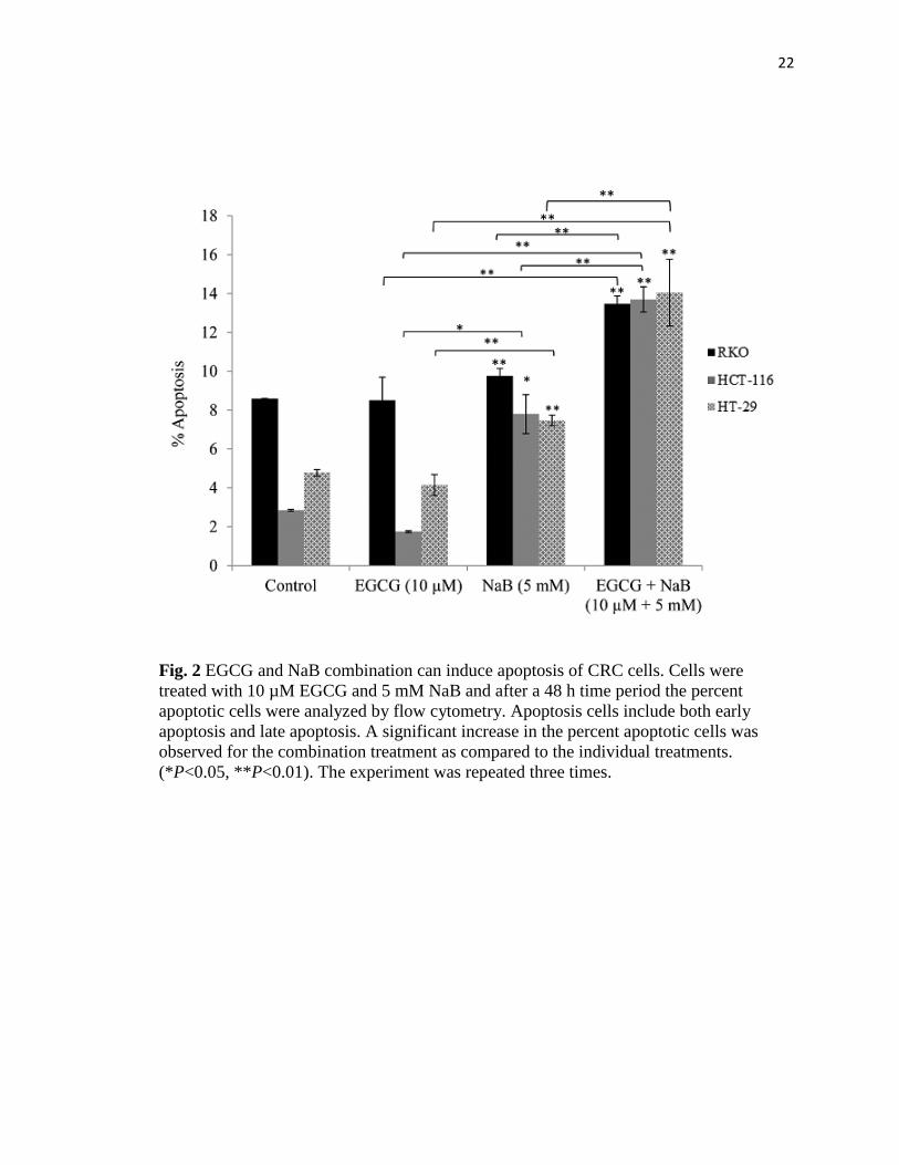
EGCG and NaB promotes apoptosis
Induction of apoptosis is one of the major pathways through which chemotherapeutic
agents inhibit tumor proliferation. We therefore examined the apoptotic potential of the
combination treatment of RKO, HCT-116 and HT-29 CRC cells. We found that the
combination of 10 μM EGCG and 5 mM NaB induced significant apoptosis (P<0.01) in
comparison to the individual treatments at 48 h (Fig. 2). However, the apoptotic induction
was not high enough to be considered the sole mechanism of inhibition of cell
proliferation. We therefore believe that a co-mechanism in addition to apoptosis may be
involved in the observed inhibition of cell proliferation.
22
Fig. 2 EGCG and NaB combination can induce apoptosis of CRC cells. Cells were
treated with 10 µM EGCG and 5 mM NaB and after a 48 h time period the percent
apoptotic cells were analyzed by flow cytometry. Apoptosis cells include both early
apoptosis and late apoptosis. A significant increase in the percent apoptotic cells was
observed for the combination treatment as compared to the individual treatments.
(*P<0.05, **P<0.01). The experiment was repeated three times.

23
EGCG and NaB arrest RKO, HCT-116 CRC cells predominantly in the G2/M phase
and HT-29 CRC cells in the G1 phase
Cell cycle progression analysis revealed a shift from S to the G2/M phase (Fig. 3). The
cells were predominantly in the S phase for EGCG at 10 μM concentration. However,
with NaB treatment at 5 mM a G2/M arrest was observed for RKO CRC cells
and in the combination treatment the cells were arrested in the G1 and G2/M phases. In
HCT-116 cells a significant G2/M arrest was observed and in HT-29 cells a significant
G1 arrest was observed (P<0.01) as compared to the controls (Fig. 3). In comparing all
three cell lines a reduction in S-phase was observed indicative of a possible mechanism
of preventing cell proliferation that may be associated with DNA damage
EGCG and NaB effectively inhibit colony formation in RKO CRC cells
From our apoptotic and cell cycle arrest results we next sought to determine if the
combination was effective in inhibiting the colony forming potential of RKO CRC cells.
Our results indicate that the combination was significantly more potent in terms of
reducing the number of colonies than individual treatments. Both types of colonogenic
assays showed that the combination was successful in inhibiting/reducing RKO cellular
tumor forming potential (Fig. 4A, B and C). The combination significantly inhibited
colony formation by 80%, while EGCG and NaB administered singly were about 20%
and 50% effective, respectively. These changes were found to be significant at P<0.01.
Thus, EGCG significantly enhances the effect of NaB in reducing colony formation.
Although slightly different efficacies were observed with respect to Fig. 4A, the reverse
colonogenic assay showed similar trends in colony reduction potential. EGCG reduced
the number of colonies formed by 10%, NaB by 40% and the combination by 70%. These

24
Fig. 3 EGCG and NaB in combination can induce cell cycle arrest in CRC cells. A shift
towards the G2/M phase was observed in the combination treatment (10 μM EGCG and
5 mM NaB) as compared to controls for both RKO and HCT-116 CRC cells and the
change was significant. A significant G1 arrest was observed for HT-29 CRC cells.
Therefore, depending on the cell line under study the combination treatment was able to
arrest cells in G1 and G2/M phases. *P<0.05, **P<0.01. The experiment was repeated
two times and each point indicates the mean±SEM.
B
A
C

25
B
A

26
Fig. 4 Colony formation potential of RKO CRC cells. Colony forming potential of RKO
CRC cells was assessed by treating the cells with 10 µM EGCG and 5 mM NaB for a
48 h time period and colonies formed in the absence of the compounds after a week of
incubation were counted. Values are represented as percent control ±SEM of two
independent experiments performed in triplicates. Representative photographs are shown
from the experiments. (A). Colonies that had greater than 50 cells were counted. A
significant decrease in colony formation was observed for the combination treatment as
compared to the individual treatments. Morphology and sizes of the colonies formed are
shown in (B). Reverse colonogenic assay was also performed to determine compound
effectiveness in reducing colonies once formed (C).
C

27
changes were significant with respect to the control (P<0.01). Also, the combination was
significantly different from the individual treatments alone (P<0.01). These data indicate
that the combination is potent in inhibiting as well as reducing colonies formed, activities
that are important to effective chemotherapeutic strategies.
EGCG and NaB increases p21, NF-κB-p65, HDAC1; and decreases DNMT1 and
survivin in RKO CRC cells
We examined the effect of the combination on DNMT1 and HDAC1 levels and
found that DNMT1 levels decreased for the combination treatment, whereas HDAC1
levels increased (Fig. 5A). Findings from other studies point to the existence of a
regulatory feedback mechanism that modulates HDAC levels, which was also observed
in this study [27]. Real time PCR analysis of RKO CRC cells revealed that p21 levels
increased for the individual treatments and was highly induced with the combination
treatment. EGCG was effective in enhancing NaB induction of p21 (Fig. 5B). We also
determined the effect of the combination treatment on survivin expression as it is
transcriptionally regulated by p21. Survivin mRNA levels decreased (Fig. 5B) in response
to the combination treatment and was significant (P<0.01) as compared to the control.
HDAC inhibitors are known to transactivate NF-κB and NF-κB is associated with p21
dependent G2/M arrest. We therefore determined the effect of the treatments on NF-κB-
p65. Ours is the first study to report that NF-κB-p65 levels were significantly up-
regulated for NaB alone and for the combination treatments of EGCG and NaB (Fig. 5C).
However, the combination was lower than NaB treatment but was significantly higher
than EGCG alone (P<0.01).

28
B
A

29
Fig. 5 Effects of EGCG and NaB on the mRNA expression of specific epigenetic, cell
cycle and cell proliferation modulators in RKO CRC cells. RKO CRC cells were treated
to 10 μM EGCG and 5 mM NaB and after a 48 h time period RNA was extracted. Real
time assessment of mRNA expression of the various genes revealed that DNMT1
and survivin expression decreased significantly for the combination treatment as
compared to EGCG and control (A, B). EGCG is a known DNMT1 inhibitor although its
effects are more pronounced at the protein than mRNA level. However, HDAC1and
p21 (A, B) increased several fold and the increase was significant when compared to the
control and individual treatments. A significant increase in NF-κB-p65 was observed for
both the NaB alone and the combination treatment (C). However, the combination
treatment was lower as compared to the NaB alone treatment, and significant and
relatively higher when compared to EGCG and the control. Data are in triplicates from
three independent experiments. *P<0.05, **P<0.01.
C

30
EGCG and NaB inhibits HDAC1, DNMT1 and survivin in all the three CRC cells
tested
EGCG and NaB are epigenetic modulators and therefore we assessed the levels of
DNMT1 and HDAC1 for the individual treatments and the combination treatment in all
three cell lines. Epigenetic proteins HDAC1 and DNMT1 levels were significantly
reduced in all three cell lines for the combination indicative of a combinatorial epigenetic
effect of the compounds under question (Fig. 6A and B, respectively). EGCG directly
inhibits the catalytic site of DNMT1 via its gallic acid moiety [11]. Sodium butyrate has
A

31
Fig. 6 Effect of the combinatorial compounds on the expression of HDAC1, DNMT1 and
survivin in RKO, HCT-116 and HT-29 CRC cells. The combination treatment was
significantly effective in down-regulating the expression of HDAC1, DNMT1 and
survivin in all the three CRC cells tested (A, B, C, respectively). The western blot was
performed in triplicate and densitometric analysis of the bands was performed. The data
represent values normalized to β-actin and then computed as relative to the control. A
representative western is shown as an inset in the graph for each protein type
tested. C=control; E=EGCG; N=NaB and E+N=EGCG+NaB. Survivin is highly
expressed in CRCs and we therefore determined the effect of the combination in
regulating survivin expression. Survivin expression was significantly inhibited with the
combination treatment as compared to the control (P<0.01) in all three cell lines (Fig.
6C). In RKO CRC cells the changes in survivin expression were significant in the
combination as compared to the individual treatments (Fig. 6C).
B
C
32
been shown to decrease DNMT1 levels in breast and prostate cancer cells via
proteasomal degradation and may be a possible mechanism of the inhibition seen in our
study [28]. The combination of EGCG and NaB in significantly reducing DNMT1 levels
may be due to the combination of inhibiting the catalytic site along with proteasomal
degradation.
EGCG and NaB activates nuclear p53, p21, NF-κB-p65; activates γ-H2AX and anti-
acetylated H3; inhibits DNMT3A and DNMT3B in total protein assessed in
RKO CRC cells
Densitometric analysis of p53, p21, and NF-κB-p65 from nuclear protein
revealed an upregulation of the proteins in the combination treatment and was significant
in comparison to the control (P<0.01) (Fig. 7A). NF-κB-p65 induction may be linked to
DNA damage and therefore we assessed the levels of γ-H2AX, the expression of which
indicates DNA damage. Our results showed an induction of the protein which was
significant as compared to the control (P<0.05) (Fig. 7B). Sodium butyrate is a HDAC
inhibitor and therefore we assessed the acetylation status of histone H3 and found a
significant increase in the combination treatment as compared to the control. A
significant (P<0.05) decrease in DNMT3A and DNMT3B levels was also observed in the
combination treatment as compared to the controls (Fig. 7C).

33
B
A

34
Fig. 7 Effect of the combinatorial compounds on the expression of p21, p53 and NF-κB-
p65 from nuclear lysates; γ-H2AX, anti-acetylated H3, DNMT3A and DNMT3B from
total protein lysates, in RKO CRC cells. Nuclear proteins were analyzed in triplicates for
p21, p53, and NF-κB-p65 in RKO CRC cells (Fig. 7A) and total protein in triplicates was
assessed for γ-H2AX and anti-acetylated H3 levels (Fig. 7B). At 10 μM EGCG and
5 mM NaB combination, p21 levels increased in RKO CRC cells. An induction in p53
expression was also observed in the combination treatment. Thus, EGCG may enhance
the effect of NaB by inducing p53 expression. NF-κB-p65 levels increased in the
combination and NaB treatment when compared to the control and EGCG treatment. This
increase may be associated with the double-strand breaks (DSBs) indicated by
increased γ-H2AX levels (7B) and G2/M arrest as was found in the study. De
novo methylation -associated epigenetic proteins DNMT3A and DNMT 3B levels also
decreased for the combination and this was tested only in RKO colon cancer cells (Fig.
7C). An increase in anti-acetylated H3 levels was also observed with the combination
(Fig. 7B), an epigenetic change probably associated by the decrease in HDAC1 levels
and HDAC activity. *P<0.05, **P<0.01.
C
35
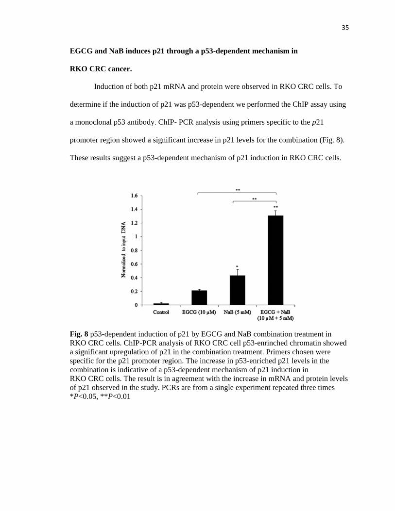
EGCG and NaB induces p21 through a p53-dependent mechanism in
RKO CRC cancer.
Induction of both p21 mRNA and protein were observed in RKO CRC cells. To
determine if the induction of p21 was p53-dependent we performed the ChIP assay using
a monoclonal p53 antibody. ChIP- PCR analysis using primers specific to the p21
promoter region showed a significant increase in p21 levels for the combination (Fig. 8).
These results suggest a p53-dependent mechanism of p21 induction in RKO CRC cells.
Fig. 8 p53-dependent induction of p21 by EGCG and NaB combination treatment in
RKO CRC cells. ChIP-PCR analysis of RKO CRC cell p53-enrinched chromatin showed
a significant upregulation of p21 in the combination treatment. Primers chosen were
specific for the p21 promoter region. The increase in p53-enriched p21 levels in the
combination is indicative of a p53-dependent mechanism of p21 induction in
RKO CRC cells. The result is in agreement with the increase in mRNA and protein levels
of p21 observed in the study. PCRs are from a single experiment repeated three times
*P<0.05, **P<0.01

36
EGCG and NaB affect global DNA methylation and chromatin structure
Epigenetic mechanisms control gene expression through changes predominantly
in DNA methylation and histone acetylation. The EGCG and NaB compounds that we
used in the study are potent DNMT1 and HDAC1 inhibitors, respectively, and therefore,
the epigenetic effects in the presence of two different types of epigenetic inhibitors were
assessed. HDAC activity was determined for all three cell lines and the activity
significantly decreased in the combination treatment as compared to the control for all of
the three cell lines (P<0.01) (Fig. 9A). No significant changes in HAT activity was
observed in the combined drug treatments (data not shown). Percent CpG methylation
was assessed only for the RKO cell line as the level of three DNMT proteins were
determined through western blots for this cell line only. Percent CpG methylation
significantly decreased for the combination treatment of EGCG and NaB for RKO cells
(P<0.01) (Fig. 9B). The increase in percent methylation for the NaB treatment may be
attributed to an increase in DNMT3B levels as was observed with the protein
densitometric analysis. The treatment of the cells that alter dominant epigenetic enzymes
may account for the observed changes in percent methylation and HDAC activity.

37
Fig. 9 Effect of EGCG and NaB on HDAC activity in all the three CRC cells and percent
CpG methylation in RKO CRC cells. The combination treatment (10 µM EGCG and
5 mM NaB) was significantly effective in decreasing HDAC activity (*P<0.05,
**P<0.01) in all three cell lines tested (A). Percent CpG methylation was analyzed in
RKO CRC cells and the decrease observed in the combination treatment was significant
as compared to the individual treatment (B). The increase in percent methylation of CpGs
with NaB may be due to the compensatory effects of de novo methylating enzymes such
as DNMT3B. Data are from three independent experiments and represent mean±SEM.
A
B

38
Discussion
Sodium butyrate (NaB) is non-toxic and is produced naturally within the colon through
microbial fermentation. The concentrations of NaB are effective in the millimolar range
and 1 mM is considered low, 5 mM intermediate and 10 mM high [29]. We considered
the intermediate concentration in terms of treatment as it was effective with 10 µM
EGCG in reaching an IC50 value for the combination within 48 h of treatment for
RKO CRC cells and these concentrations are achievable in vivo. NaB has shown promise
as a suitable chemotherapeutic agent due to its myriad anti-tumorigenic effects, including
cell proliferation inhibition, induction of cell cycle arrest and differentiation, promotion
of apoptosis, and even more significantly as a potent HDAC inhibitor inducing histone
hyperacetylation and altering gene expression [6,8,30,31]. Sodium butyrate has been
shown to induce p21 expression that is a prominent cell cycle regulatory protein [32,33]
In the current study we showed that the potent anti-cancer activities of EGCG and
NaB in RKO, HCT-116 and HT-29 CRC cells. On the contrary, a study showed that
EGCG interferes with butyrate-induced differentiation in CRC cells by preventing the
cellular uptake of NaB [34]. However, in the study conducted, the dose chosen was based
on the optimal values of the individual compounds and not on the combination of the
compounds. Two given compounds can act differently at different dose combinations.
From our MTT assays we showed that at 2 mM NaB and 10 µM EGCG there was no
additive or synergistic effect observed in HT-29 CRC cells which was the same cell line
chosen for the previous study [34]. Therefore, dose appears to be crucial in determining
chemotherapeutic effects. Based on the dose chosen we observed significant changes in
morphology, apoptosis, cell cycle arrest and epigenetic proteins for the HT-29 CRC cell

39
line. In addition, survivin, an antiapoptotic protein highly expressed in colon cancers, was
down-regulated in the HT-29 CRC cell line with the combination as compared to EGCG
alone. RKO and HCT-116 CRC cell lines also showed significant morphological changes
after the combination treatment. Epigenetically and genetically RKO and HCT-
116 CRC cells are more similar than HT-29. The presence of wt-p53 in RKO and HCT-
116 CRC cells may produce an improved response to the combination treatment than HT-
29 CRC cells that carry mutated p53. Reinduction of p53 perhaps may be important in
encouraging apoptosis or cell-replicative inhibition which may be p53-dependent. The
cell lines chosen were also CIMP positive, which is described in detail elsewhere [26].
Most of our analysis was performed in detail in the RKO wt-p53 CRC cell line and where
comparisons were necessary, we included the other cell lines as well.
From our in vitro colonogenic assays we were able to show that a combination of
EGCG and NaB was significantly effective in inhibiting colony formation in
RKO CRC cells by 80%, when many studies show that either EGCG or NaB acting
alone require higher concentrations and time periods for the same effective inhibition.
Clearly, the combination treatment at lower physiologically relevant doses is able to
generate a significant chemotherapeutic effect suggestive of strong activity against
colorectal cancer. Our study also showed that the combination treatment of EGCG and
NaB was successful in inducing apoptosis in all three cell lines we treated but was in the
range of 12–16% and therefore the apoptotic pathway may not be the only mode of
tumor-inhibition by the compounds we evaluated.
In addition to the induction of apoptosis, we found that the EGCG and NaB
combination induced cell cycle arrest predominantly in the G2/M phase for RKO and

40
HCT-116 CRC cells and G1 phase for HT-29 CRC cells. NaB has been shown to exert
cell cycle inhibitory effects in the G1 phase or G2/M phase based on the cell line under
study [35]. In addition, down-regulation of survivin via p21 induction and p53
upregulation provides an alternative strategy of cell replicative inhibition through DNA
damage. Our study for the first time showed that the combination therapy was successful
in down-regulating the expression of survivin both at the mRNA and protein levels with
p21 induction and G1 and G2/M arrests. We believe that this novel mechanism involved
in the inhibition of CRC cells could be applicable to tumors that are resistant to apoptosis.
Genetic and epigenetic changes are instrumental in promoting neoplastic
transformation in CRCs [36]. The reversibility of these changes presents a suitable
treatment strategy for colorectal cancers. Previous studies have shown that EGCC and
NaB are potent DNMT1 and HDAC1 inhibitors, respectively, both of which can
individually correct epigenetic observations in cancer cells [8,11]. In our study, NaB was
used and is a known HDAC1 inhibitor, and is the most extensively studied HDAC.
Contradictory to the norm of gene and protein regulation, our study showed that
the HDAC1 mRNA levels were significantly upregulated with the down-regulation of
HDAC1 level for the NaB treatment alone and the combination treatment of EGCG and
NaB. The upregulation of HDAC1 and the concomitant decrease in protein level may be
attributed to the autoregulation of HDAC1 by HDAC inhibitors and could be cell-type
specific [37]. HDAC1 has been found to be recruited to its own promoter thereby
mediating its own repression by negative feedback loop inhibition [37]. In addition,
HDACs are known to undergo several post-translational modifications, of which
acetylation, phosphorylation, sumoylation and ubiquitination have been reported to occur

41
with HDAC1 that can affect their activity and stability [38]. Taken together, the binding
of NaB to the catalytic site of HDAC1 and possible post-translational effects on the
protein may account for the decrease in HDAC1 levels despite the increase in HDAC1.
DNA methyltransferases and histone enzymes are no longer considered
independent epigenetic regulators but are thought to function in tandem or in cohesion to
alter the epigenome. Global CpG methylation response to NaB treatment has been shown
to vary on the basis of the cell-type under study [39]. HeLa cells treated with 3 mM NaB
for 48 h have shown an increase in global genomic methylation as compared to the
control and in glioblastoma multiforme cells a hypomethylated state has been observed at
2 mM NaB for 48 h [40]. The findings from our results showed that percent CpG
methylation for NaB increased in comparison to the control. A similar finding has been
observed in transformed lung fibroblast cell lines in the presence of 5 mM NaB [39]. At
this point we cannot determine the actual reasons for this observation but can infer from
the data that the compensatory effects by de novo enzymes may account for this.
A significant decrease in HDAC activity was observed that likely explains the
increase in global histone H3 acetylation levels in the combination treatment in
comparison to the individual compounds. Histone hyperacetylation has been associated
with inhibiting cell growth [41] and therefore we believe that the growth inhibition
observed in RKO CRC cells may be due in part to this epigenetic change. Our study
showed that in addition to its HDAC inhibitory activity, NaB was able to down-regulate
DNMT1 expression as well and the expression further decreased with the combination
treatment. Data from other studies support decreased DNMT1 expression, at both the
protein and mRNA levels, after HDAC inhibitor treatment [28,42].

42
The common expressed biomarker signaling DNA damage is γ-H2AX. Class I, II,
and III HDACs have been implicated in the DNA damage response, homologous
recombination (HR), and chromatin integrity [43]. HDAC1 have been shown to assist
with the DNA damage response by recruiting DNA repair proteins to the site of damage.
Butyrate and trichostatin A (TSA) have been show to exert defects in the repair process.
Studies with cells lacking HDAC1 have been shown to be hypersensitive to DNA
damaging agents and exhibit sustaining DNA damaging signaling reflective of defects in
double-strand break (DSB) repair [44]. The results from our study showed a significant
decrease in HDAC1 levels in the combination treatment for all three cell lines. Further,
an assessment of γ-H2AX levels in RKO CRC cell lines showed a significant increase in
the combination treatment significantly as compared to the control and individual
treatments. The increase in γ-H2AX levels with the concomitant decrease in HDAC1
levels may account for increased DNA damage with decreased cell-replicative capacity.
Colonogenic assays performed in RKO CRC cells showed that the combination
significantly reduced colony formation and a growth reduction potential was also
observed. These findings support the hypothesis that the combination treatment induces
DNA damage, arrests the cells in the cell cycle and prevents an increase in cell numbers
(data not shown).
HDAC1 has been shown to repress the transcriptional activity of NF-κB-p65.
Treatment of the RKO CRC cells to HDAC1 inhibitor NaB in our study increased NF-
κB-p65 mRNA and protein, and the expression was higher with NaB treatment and in the
combination treatment as compared to the control. Upregulation of NF-κB has been
observed in response to DNA damage. HDAC1 has been reported to directly interact with

43
the p65-subunit of NF-κB and affect its transactivation functions [45]. The inhibition of
HDAC1 by NaB in our studies may provide an explanation for the observed increase in
nuclear NF-κB-p65 levels where the protein is in its activated state and is associated with
DNA damage. Studies have reported that the increase in NF-κB in response to DNA
damage may allow for repair of the damaged DNA [46]; however, the repair process is
mediated through a host of various other proteins and includes HDAC1. We propose that
HDAC1 is a corepressor of NF-κB-p65 and that HDAC1 can directly associate with the
p65 subunit of NF-κB. Despite the increase in NF-κB-p65 levels, the decreased levels of
HDAC1 may hinder repair functions arresting cells in the cell cycle at the G2/M phase
through the induction of p21 as was specifically observed in RKO CRC cells. Thus,
EGCG and NaB through their epigenetic modulating effects, in addition to their potent
roles as regulators of chromatin structure, and through direct or indirect corepressor or
coactivator functions can mediate cellular responses by upregulating genes necessary for
cell cycle arrest favoring a therapeutic outcome.
Conclusions
In summary, we have shown that EGCG and NaB with different predominant
epigenetic functions are a potent combination with in vitro anti-colorectal cancer
properties by suppressing cell proliferation, inducing cell cycle arrest and promoting
apoptosis. These results can form the basis of suitable clinical therapies for CRC.
Acknowledgments
This work was supported in part by Grants from the NCI (CA 129415) and
the American Institute for Cancer Research. We would like to thank Dr. Karyn Scissum

44
Gunn for the use of her laboratory space and various facilities at Alabama State
University, Montgomery AL. We appreciate the efforts of Dr. Yuanyuan Li for
organizing and helping with experimental supplies when needed.
References
[1] U. Zangemeister-Wittke and H.U. Simon, An IAP in action: the multiple roles of survivin
in differentiation, immunity and malignancy, Cell Cycle 3, 2004, 1121–1123.
[2] M.J. Kallio, M. Nieminen and J.E. Eriksson, Human inhibitor of apoptosis protein (IAP)
survivin participates in regulation of chromosome segregation and mitotic exit, FASEB
J. 15, 2001, 2721–2723.
[3] M. Alper, S. Cukur, O. Belenli and M. Suna, Evaluation of the immunohistochemical stain
patterns of survivin, Bak and Bag-1 in colorectal cancers and comparison with polyps
situated in the colon,Hepatogastroenterology 55, 2008, 1269–1273.
[4] H. Kawasaki, D.C. Altieri, C.D. Lu, M. Toyoda, T. Tenjo and N. Tanigawa,Inhibition of
apoptosis by survivin predicts shorter survival rates in colorectal cancer, Cancer
Res. 58, 1998, 5071–5074.
[5] H. Yako-Suketomo and T. Marugame, Comparison of time trends in colon, rectum and
anus cancer incidence (1973-2002) in Asia, from ‘Cancer Incidence in Five Continents,
Vols IV–IX’, Jpn. J. Clin.Oncol. 39, 2009, 196–198.
[6] G. den Besten, K. van unen, A.K. Groen, K. Venema, D.J. Reijngoud and B.M.Bakker, The
role of short-chain fatty acids in the interplay between diet, gut microbiota and host
energy metabolism, J. Lipid Res. 2013.
[7] A. Hague, A.M. Manning, K.A. Hanlon, L.I. Huschtscha, D. Hart and C.Paraskeva, Sodium
butyrate induces apoptosis in human colonic tumour cell lines in a p53-independent
pathway: implications for the possible role of dietary fibre in the prevention of large-
bowel cancer, Int. J.Cancer 55, 1993, 498–505.
[8] J.R. Davie, Inhibition of histone deacetylase activity by butyrate, J.
Nutr. 133, 2003, 2485S–2493S.
[9] M. Domokos, J. Jakus, K. Szeker, R. Csizinszky, G. Csiko, Z. Neogrady, A.Csordas and P.
Galfi, Butyrate-induced cell death and differentiation are associated with distinct patterns
of ROS in HT29-derived human colon cancer cells, Dig. Dis. Sci. 55, 2010, 920–930.
[10] C. Nor, F.A. Sassi, C.B. de
Farias, G. Schwartsmann, A.L. Abujamra, G. Lenz,A.L. Brunetto and R. Roesler, The his
tone deacetylase inhibitor sodium butyrate promotes cell death and
differentiation and reduces neurosphere formation in human medulloblastoma cells, Mol.
Neurobiol. 2013.
[11] W.J. Lee, J.Y. Shim and B.T. Zhu, Mechanisms for the inhibition of DNA
methyltransferases by tea catechins and bioflavonoids, Mol. Pharmacol. 68, 2005, 1018–
1030.
[12] S.N. Saldanha and T.O. Tollefsbol, The role of nutraceuticals in chemoprevention and
chemotherapy and their clinical outcomes, J. Oncol. 2012, 2012, 192464.

45
[13] Y. Tang, D.Y. Zhao, S. Elliott, W. Zhao, T.J. Curiel, B.S. Beckman and M.E.Burow, Epiga
llocatechin-3 gallate induces growth inhibition and apoptosis in human breast cancer cells
through survivin suppression, Int. J. Oncol. 31, 2007, 705–711.
[14] E.H. Kim, H.S. Kim, S.U. Kim, E.J. Noh, J.S. Lee and K.S. Choi, Sodium butyrate
sensitizes human glioma cells to TRAIL-mediated apoptosis through inhibition of Cdc2
and the subsequent downregulation of survivin and XIAP, Oncogene 24, 2005, 6877–
6889.
[15] S. Kim, M.J. Lee, J. Hong, C. Li, T.J. Smith, G.Y. Yang, D.N. Seril and C.S.Yang, Plasma
and tissue levels of tea catechins in rats and mice during chronic consumption of green
tea polyphenols, Nutr. Cancer 37, 2000,41–48.
[16] L. Chen, M.J. Lee, H. Li and C.S. Yang, Absorption, distribution, elimination of tea
polyphenols in rats, Drug Metab. Dispos. 25, 1997, 1045–1050.
[17] P.B. Mortensen and M.R. Clausen, Short-chain fatty acids in the human colon: relation to
gastrointestinal health and disease, Scand. J.Gastroenterol. Suppl. 216, 1996, 132–148.
[18] J.A. Vogt and T.M. Wolever, Fecal acetate is inversely related to acetate absorption from
the human rectum and distal colon, J. Nutr. 133, 2003,3145–3148.
[19] B.M. Evers, T.C. Ko, J. Li and E.A. Thompson, Cell cycle protein suppression and p21
induction in differentiating Caco-2 cells, Am. J. Physiol. 271, 1996,G722–727.
[20] J. Xiong, Y.R. Li, Z.M. Tang, L.F. Dou, L. Wang and L.H. Hu, The effect of p21 on
transcription of survivin in hepatocellular carcinoma HepG2 cells and its regulation
mechanism, Zhonghua Zhong Liu Za Zhi 30, 2008, 583–587.
[21] S.J. Chiu, J.I. Chao, Y.J. Lee and T.S. Hsu, Regulation of gamma-H2AX and securin
contribute to apoptosis by oxaliplatin via a p38 mitogen-activated protein kinase-
dependent pathway in human colorectal cancer cells, Toxicol. Lett. 179, 2008, 63–70.
[22] B. Zhang, X. Wang and Y. Wang, Altered gene expression and miRNA expression
associated with cancerous IEC-6 cell transformed by MNNG, J. Exp. Clin. Cancer
Res. 28, 2009, 56.
[23] J.D. Lambert, M.J. Lee, L. Diamond, J. Ju, J. Hong, M. Bose, H.L. Newmarkand C.S. Yan
g, Dose-dependent levels of epigallocatechin-3-gallate in human colon cancer cells and
mouse plasma and tissues, Drug Metab. Dispos. 34, 2006, 8–11.
[24] J.D. Lambert, J. Hong, D.H. Kim, V.M. Mishin and C.S. Yang, Piperine enhances the
bioavailability of the tea polyphenol (-)-epigallocatechin-3-gallate in mice, J.
Nutr. 134, 2004, 1948–1952.
[25] V. De Preter, K.P. Geboes, V. Bulteel, G. Vandermeulen, P. Suenaert, P.Rutgeerts and
K. Verbeke, Kinetics of butyrate metabolism in the normal colon and in ulcerative
colitis: the effects of substrate concentration and carnitine on the beta-oxidation
pathway, Aliment. Pharmacol. Ther. 34, 2011, 526–532.
[26] D. Ahmed, P.W. Eide, I.A. Eilertsen, S.A. Danielsen, M. Eknaes, M. Hektoen,G.E. Lind
and R.A. Lothe, Epigenetic and genetic features of 24 colon cancer cell
lines, Oncogenesis. 2, 2013, e71.
[27] F. Dangond and S.R. Gullans, Differential expression of human histone deacetylase
mRNAs in response to immune cell apoptosis induction by trichostatin A and
butyrate, Biochem. Biophys. Res.Commun. 247, 1998, 833–837.
[28] S. Sarkar, A.L. Abujamra, J.E. Loew, L.W. Forman, S.P. Perrine and D.V.Faller, Histone
deacetylase inhibitors reverse CpG methylation by regulating DNMT1 through ERK
signaling, Anticancer Res. 31, 2011, 2723–2732.

46
[29] A.J. Wilson, A.C. Chueh, L. Togel, G.A. Corner, N. Ahmed, S. Goel, D.S. Byun,S. Nasser,
M.A. Houston, M. Jhawer, et al., Apoptotic sensitivity of colon cancer cells to histone
deacetylase inhibitors is mediated by an Sp1/Sp3-activated transcriptional program
involving immediate-early gene induction,Cancer Res. 70, 2010, 609–620.
[30] A. Hague, D.J. Elder, D.J. Hicks and C. Paraskeva, Apoptosis in colorectal tumour cells:
induction by the short chain fatty acids butyrate, propionate and acetate and by the bile
salt deoxycholate, Int. J. Cancer 60, 1995, 400–406.
[31] A. Hague, A.J. Butt and C. Paraskeva, The role of butyrate in human colonic epithelial
cells: an energy source or inducer of differentiation and apoptosis?,Proc.
Nutr. Soc. 55, 1996, 937–943.
[32] S.Y. Archer, J. Johnson, H.J. Kim, Q. Ma, H. Mou, V. Daesety, S. Meng andR.A. Hodin,
The histone deacetylase inhibitor butyrate downregulates cyclin B1 gene expression via a
p21/WAF-1-dependent mechanism in human colon cancer cells, Am. J. Physiol.
Gastrointest. Liver Physiol. 289, 2005, G696–703.
[33] S. Siavoshian, H.M. Blottiere, C. Cherbut and J.P. Galmiche, Butyrate stimulates cyclin D
and p21 and inhibits cyclin-dependent kinase 2 expression in HT-29 colonic epithelial
cells, Biochem. Biophys. Res. Commun. 232, 1997, 169–172.
[34] S. Sanchez-Tena, P. Vizan, P.K. Dudeja, J.J. Centelles and M. Cascante,Green tea
phenolics inhibit butyrate-induced differentiation of colon cancer cells by interacting with
monocarboxylate transporter 1, Biochim. Biophys. Acta 1832, 2013, 2264–2270.
[35] C.A. Afshari, P.J. Vojta, L.A. Annab, P.A. Futreal, T.B. Willard and J.C.Barrett,
Investigation of the role of G1/S cell cycle mediators in cellular senescence, Exp. Cell
Res. 209, 1993, 231–237.
[36] W.S. Samowitz, Genetic and epigenetic changes in colon cancer, Exp. Mol.
Pathol. 85, 2008, 64–67.
[37] F. Ajamian, A. Salminen and M. Reeben, Selective regulation of class I and class II
histone deacetylases expression by inhibitors of histone deacetylases in cultured mouse
neural cells, Neurosci. Lett. 365, 2004, 64–68.
[38] G. David, M.A. Neptune and R.A. DePinho, SUMO-1 modification of histone deacetylase
1 (HDAC1) modulates its biological activities, J. Biol. Chem. 277, 2002, 23658–23663.
[39] J.B. de Haan, W. Gevers and M.I. Parker, Effects of sodium butyrate on the synthesis and
methylation of DNA in normal cells and their transformed counterparts, Cancer
Res. 46, 1986, 713–716.
[40] D.E. Cosgrove and G.S. Cox, Effects of sodium butyrate and 5-azacytidine on DNA
methylation in human tumor cell lines: variable response to drug treatment and
withdrawal, Biochim. Biophys. Acta 1087, 1990, 80–86.
[41] J.T. Wu, S.Y. Archer, B. Hinnebusch, S. Meng and R.A. Hodin, Transient vs. prolonged
histone hyperacetylation: effects on colon cancer cell growth, differentiation, and
apoptosis, Am. J. Physiol. Gastrointest. Liver Physiol. 280, 2001, G482–490.
[42] Q. Zhou, A.T. Agoston, P. Atadja, W.G. Nelson and N.E. Davidson, Inhibition of histone
deacetylases promotes ubiquitin-dependent proteasomal degradation of DNA
methyltransferase 1 in human breast cancer cells,Mol. Cancer Res. 6, 2008, 873–883.
[43] P. Rajendran, E. Ho, D.E. Williams and R.H. Dashwood, Dietary phytochemicals, HDAC
inhibition, and DNA damage/repair defects in cancer cells, Clin. Epigenetics 3, 2011, 4.

47
[44] K.M. Miller, J.V. Tjeertes, J. Coates, G. Legube, S.E. Polo, S. Britton and S.P.Jackson, Hu
man HDAC1 and HDAC2 function in the DNA-damage response to promote DNA
nonhomologous end-joining, Nat. Struct. Mol. Biol. 17, 2010, 1144–1151.
[45] B.P. Ashburner, , S.D. Westerheide, and A.S. Baldwin, Jr., The p65 (RelA) subunit of NF-
kappaB interacts with the histone deacetylase (HDAC) corepressors HDAC1 and
HDAC2 to negatively regulate gene expression,Mol. Cell Biol. 21, 2001, 7065–7077.
[46] K.W. McCool and S. Miyamoto, DNA damage-dependent NF-kappaB activation: NEMO
turns nuclear signaling inside out, Immunol.Rev. 246, 2012, 311–326

48
ALTERATIONS IN HISTONE ACETYLATION IN TUMORIGENESIS
by
SABITA N SALDANHA AND TRYGVE TOLLEFSBOL
Cancer Epigenetics
Copyright
2008
by
Springer
Used by permission
Format adapted for dissertation

49
7.1 INTRODUCTION
Cellular functions, including but not limiting to the production of cell-specific
biomolecules, DNA replication and repair, apoptosis, and senescence are intricately and
inherently orchestrated by chromosomes [1]. These functions are specified by coding
messages in genes. The genetic code, however, is susceptible to alterations,
predominantly irreversible mutations, which can result in diseased states at the
organismal level. Epigenetic changes involve heritable alterations in gene expression
without mutations in the genetic code [2]. Such an outcome is possible as chromatin, the
structural component of chromosomes, can undergo changes affecting the condensation
states of the chromosome, which allow for or inhibit gene expression [3] (Figure 7.1).
These processes are catalyzed by reversible enzymatic modifications. The epigenetics of
gene control has gained considerable support and has since been a chosen target for
therapeutic intervention for several diseases, in particular cancer.
7.2 NUCLEOSOMES
At the molecular level, the nucleosome constitutes the core of chromatin and consists of
basic proteins called histones [2,4]. The 146 bp of DNA complexes with the nucleosome,
an octamer, and does so via its negatively charged phosphate backbone. In the octamer,
the dimeric H3 and H4 subunits interact in a tetramer formation, whereas H2A and H2B
histones remain as dimers [2,4]. The complex does not include the histone protein H1,
but this protein, a linker, appears to facilitate the condensation of the nucleosome units

50
FIGURE 7.1 Histone mediated gene expression. Histone-mediated chromatin fluxes are
depicted in the figure. These changes in histone dynamics are brought about by reversible
enzymatic reactions mediated by histone acetyltransferases and histone deacetylases.
Addition of an acetyl group changes the charge environment surrounding the DNA, as a
result the DNA is freed from the nucleosome core, allowing for the transcriptional
complexes to bind and induce transcription. Histone deacetylases on the other hand have
apposing effects as the neutral charge induced by the acetyl group is removed, allowing
the DNA to complex back with the histones, inhibiting expression and other
transactivating or repressible elements to the site. Within the same histone subunit,
differential alterations can have colossal effects on gene expression under its control. For
example, methylation and acetylation of the histone H3 lysine 9 (H3K9) subunit can have
apposing effects on gene expression in conjunction with other histone modifications

51
lysine residues are modified, in some cases serine or arginine residues are also altered [8–
10].
[2,3]. The amino terminal domains of histones protrude out of the complex and are
susceptible to enzymatic modification at lysine residues [5–8]. Although predominantly
the altered states largely depend on the enzyme catalyzing the modification and the
availability of the residues to the enzymes. The type and position of the modification on
the histone dictates a pattern, termed the histone code, which specifies the transcriptional
regulation of the complexed DNA [11]. This regulation is based upon the availability and
accessibility of transcription factors
7.3 EFFECTS OF NUCLEOSOMAL MODIFICATIONS
There are many modifications known to posttranslationally alter the residues, of which
acetylation is the most extensively studied [7,8]. The orientation and modulations of the
histones affects the fluidity of the chromatin, resulting in heterochromatin or euchromatin
states. In normal cells, the cell cycle is carefully regulated by cell cycle regulatory genes.
However, alterations in genes that control tumor suppressors or the cell cycle promote the
tumor phenotype. Therefore in abnormal cell growth and division there are three possible
outcomes (Figure 7.2): (1) Genes that control cell cycle division are turned off, (2) tumor
suppressor genes are turned off, or (3) oncogenes are switched on. This can arise by an
imbalance in the acetylation/deacetylation states of the histone residues in the

52
FIGURE 7.2 Histone acetylation toward a neoplastic phenotype. Involvement of the
histone acetylation/deacetylation balance in tumorigenesis. In cells that are normal, genes
involved in the cell cycle regulation are expressed and to a certain extent are controlled
by histone acetylation of the histones associated with the promoters. [ ] represents p21
and the [ ] represents p53; both are tumor suppressors. However, in response to local
hypoacetylation and in conjunction with defects in other cellular pathways, the tumor
suppressors are turned off and tumor formation may be induced as the cell cycle is de-
regulated. In some instances hyperacetylation of oncogenes [ ], as in the case with
HPVE6, occur that can induce a cancer phenotype.
53
nucleosome, in addition to other factors. This probably explains why most genes crucial
to maintaining the cell cycle balance are turned off or deregulated in neoplasms.
7.4 HISTONE ACETYLATION
Histone acetylases, commonly referred to as HATs, recognize the basic substrates and
add an acetyl group [8]. The addition of an acetyl group to the lysine residue neutralizes
the charge, which relaxes the bound DNA from the histone complex. However,
expression of the genes is dependent on the availability of transactivating factors and
proteins to the freed up DNA [8]. Moreover it is important which lysine residues are
acetylated because at any given time the position of the lysine and the histone subunit
involved in the acetylation largely dictates the downstream effects [8].
Histone molecules are maintained in a delicate balance between acetylated and
deacetylated states and work in perfect synchronization for optimal gene function.
However, deregulation of this posttranslational balance can essentially lead to the
upregulation of oncogenes that enable cells to acquire tumorigenic, metastatic, and
invasive phenotypes. Re-acetylation probably prevents such an outcome, as genes crucial
to cell cycle regulatory functions are upregulated. Attenuation of deacetylation, with an
increase in acetylation of histones, has positive consequences in terms of inhibiting
neoplastic growth. Increased acetylation tends to slow down cell growth and induce
differentiation and apoptosis, as evidenced by a number of studies utilizing compounds
that inhibit histone deacetylases in cancer cell lines. The effect is mediated by the
upregulation of proteins like p53 by acetylation of their lysine residues [12,13].

54
7.5 HISTONE ACETYLATION AND CANCER
HATs exhibit substrate preference toward the lysine moieties that they acetylate. For
instance, GCN4, a histone acetylase, preferentially acetylates H4 lysine residues at
positions 5, 8, 12, and 16 [14,15]. Acetylation of each of these residues has a different
effect. Acetylation of H4 K5 and H4 K12 are merely associated with directing the
histones to newly formed DNA in the S phase [14,15]. However, H4 K16 enrichment is
involved with actively transcribing genes [14,15]. Thus physiological conditions and
requirements of the cell most likely dictate the histone marks. Similarly in histone H3,
acetylation by GCN5 at positions 9, 14, 16, and 23 is observed. As in the case of H4, in
H3 the function of histone deposition to newly synthesized chromatin is mediated
by K9 acetylation [14,15]. The remaining acetylated positions are involved with gene
expression [14,15]. Therefore mutations in the enzyme that dictate this function can cause
mistargeting of acetylation, changes in the acetylation pattern, and deregulation of acetyl
enrichment. These events can disrupt the cell cycle balance and create an environment
conducive to the development of cellular transformations.
Acetylation events do not act alone in mediating the downstream effects but
require the collaborative effort of other pathways, influenced by other histone codes. In
some cases the effect of acetylation at a given residue can rescue the function or provide
for redundancy for mutated lysine residues. This phenomenon was clearly demonstrated
in a study where the substrates of GCN5 were mutated to determine the overall effects on
cell growth. Mutations in all the K residues of H4 resulted in accumulation of cells
predominantly in the G2 phase [14]. The observed mutated phenotype was rescued when
a lysine residue other than the ones mutated were acetylated, providing for the
55
redundancy of function. This clearly indicates that acetylation is required for cell
viability. Many neoplastic transformations arise from deregulation in genes that affect
particular target genes or several pathways. Some of the genes come under direct
epigenetic modifications whereas others are regulated indirectly. We have discussed
some of the cancers that have arisen due to changes in histone modifications and the
target genes they affect.
7.6 INVOLVEMENT OF HISTONE ACETYLATION IN BREAST CANCER
Aberrant histone acetylation can be one of the mechanisms of many different events that
result in cancer of breast tissue. Estradiol, an estrogen steroid, binds to estrogen
receptors. This complex associates with the estrogen response element and mediates its
downstream effects [16]. Interestingly this association also encourages the binding of
protein complexes that possess HAT activity. Breast cancers have been distinguished into
two categories, estrogen-dependent and estrogen-independent type [16]. In the estrogen-
dependent form, the breast tissue is constantly bathed with the steroid molecule, in part
due to the ovulation cycle. However, other mechanisms may also promote estrogen
synthesis. Over-expression of the hormone can induce changes in the histone acetylation
pattern although there is some debate regarding the type of acetylation patterning that
occurs. Some studies indicate that an increase in histone acetylation of H4 by estradiol in
breast cancer cells occurs [16] while other studies show a decrease in acetylation [16].
Acetylation of lysine residues can achieve different isoforms—mono, di, tri, and tetra.
Therefore, in theory, if the lysine is predominantly mono-acetylated, then histones can be
relatively easily deacetylated. However, the higher acetylated isoforms can decrease the
56
rate at which deacetylation occurs. Therefore, the increase in acetylation found in the
studies done on breast cancer by estradiol could be mediated by slower deacetylation
rates with no change in acetylation rates of higher acetylated isoforms.
All histone subunits are not acetylated/deacetylated at the same rates. Some
populations are acetylated faster than the others. Tetra-acetylation of H4 is mediated by
rapid acetylation whereas some other histones achieve higher acetylated isoforms much
more slowly. A study showed that 60%–70% of histones are acetylated in breast cancer
[16]. These histones are classified into three categories (1) those that are always
acetylated and remain in that state, (2) those that are acetylated/deacetylated quickly, and
(3) those that are acetylated/deacetylated relatively slow [16]. The majority of histones
fall into the last category (3). However, 10% of histones belong to category (2) [16]. In
addition to acetylated states, if the majority of the isoforms found are of the higher order,
then deacetylation of such molecules will be slowed down, culminating in continuous
gene expression.
The category (2) histones are the most affected by estradiol. Estradiol increases
the rate of histone deacetylation but does not alter the rate of histone acetylation.
Hyperacetylated histones H3 and H4 are found to be associated with coding regions of
E2-ER (required for HAT activity) and cMyc genes (expressed in proliferating cells)
[31]. cMyc is a well-studied oncogene and the upregulation of myc translates into tumor
phenotypes. Therefore, histone acetylation does not always correlate with a normal
phenotype. Localized hyperacetylation of genes involved in oncogenesis triggered by
hormones can precipitate a neoplastic phenotype.

57
7.7 LUNG AND NASAL CANCERS MEDIATED BY HISTONE
ACETYLATION
7.7.1 NICKEL EXPOSURE AND HISTONE ACETYLATION
Occupational exposure to soluble nickel is known to cause lung and nasal cancers [17].
Besides DNA damage and slow repair response, nickel affects chromatin configuration
[17]. Nickel has been shown to decrease the acetylation of histone H3 and H4. In fact,
inhibition of gene expression has been associated with hypoacetylation of histones H3
and H4 as a result of nickle-mediated inhibition of HAT activity [17]. Nickel induces an
overall decrease in acetylation of all histones. In the group of histones, H2b is the most
sensitive to deacetylation and H3 is the least sensitive. Lysines at position 5, 12, 15, and
20 are affected in H2b [17]. It has been observed though that the sensitivity to
deacetylation of H2b is related to the exposure periods and shorter exposures have no
profound effect on the acetylated states of H2b K5 and K15 residues. K12 and K20
however undergo a different fate, and directly correlate to the time and dose of nickel
exposure [17]. Therefore genes containing residues that are primarily acetylated at these
positions will be affected and would induce a cascade of downstream effects. In most
instances hyperacetylation is associated with gene expression, however, some studies
have shown that acetylation of histones can inhibit expression. This inhibition is
primarily caused by a blend of hyper/hypoacetylated patterns at specific lysine residues
on different histone subunits at promoter and coding locations. These patterns are more
gene-specific and do not associate with the overall global hypo-or hyperacetylation states.

58
7.7.2 LUNG CANCERS ASSOCIATED WITH ACETYLATION OF THE RARβ
GENE
In most lung cancers, the RARβ gene is defective and therefore is not expressed in the
presence of retinoic acid (RA) imparting a retinoic acid refractory phenotype to the cells
[18]. Analysis of lung cancer cell lines have shown that cell lines unresponsive to RA
have deacetylation of histone H3 with hyperacetylation of histone H4 [18]. However, in
RA-responsive cells, both H3 and H4 are acetylated at the promoter of RARb receptor,
irrespective of the methylation status of the promoter [18]. Therefore, a reduction in H3
acetylation has been shown to correlate with the RA refractoriness in lung cancer cells
[18]. In addition to reduced histone acetylation at specific subunits, methylation of a
promoter may be a secondary essential mechanism to gene expression. Therefore, some
lung cancers are associated with hypoacetylation and hypermethylation of specific
moieties that control the expression of RARβ, which is crucial for downstream signaling
pathways.
7.8 MODULATION OF HISTONE H4 IN GASTRIC CANCERS
In a majority of gastric and colorectal cancers H4 acetylation is markedly reduced which
precipitates invasive and metastatic events [19]. In colon cancers, global hypoacetylation
enhances tumor invasiveness and metastasis allowing for a means of possible cancer
therapy. Gastric carcinomas are of two types, intestinal and diffuse [19]. In both these
types of cancers deacetylation of histone H4 is observed. This reduction is gradual and
begins from the early stage where precancerous lesions predominate and progress into the
late stages of invasiveness and metastasis [19, 20]. However, one should be careful in

59
interpreting the observed results as acetylation alone cannot dictate a cancerous
phenotype but relies on a multitude of factors and mechanisms, all of which target genes
that control normal cell phenotype.
7.9 CANCERS OF THE THYROID AND HISTONE ACETYLATION
Essentially the process of acetylation or deacetylation of histones are not directly
involved in neoplastic transformations. It is a domino effect induced by modulations of
transcription factors, which control cell-specific functions that influence oncogenic
events. For instance in many thyroid cancers the sodium iodide symporter (NIS)
expression is downregulated [21]. The expression of this protein, NIS, is important as it is
involved in iodine uptake. In primary solid thyroid tumors the cells are unable to absorb
iodine in part due to low or no expression of NIS [22]. Reduced acetylation in
conjunction with methylation at the NIS promoter is probably responsible for the
decreased NIS expression [21]. Studies have shown that histone reacetylation by
inhibition of the deacetylation process stimulates NIS expression [21, 22]. Thus, targeting
enzymes that inhibit histone deacetylation may have potential as a therapeutic measure
against thyroid cancers. This process helps not only in target-specific treatments but also
in the induction of differentiation and apoptosis by increasing the expression of cell cycle
regulatory genes.
RA treatment is another approach to restore radioactive iodine uptake in a small
subset of metastatic thyroid cancers [22]. The promoter region of RARb, a receptor for
RA, appears to be unmethylated and deacetylated at H3 and H4, but one must be cautious
to interpretations, as these observations are cell-type specific. In the presence of the

60
HDACs alone or in combined treatments with RA, the acetylation levels can be restored.
Therefore alterations in histone patterns can contribute to the refractoriness in
differentiated thyroid cancers and may be a suitable tool for therapy. This study
highlights that acetylation is not responsible for the direct conversion of a normal cell to
neoplastic forms but controls a host of other genes crucial for downstream effects.
7.10 GENES AFFECTED BY ACETYLATION THAT PLAY A ROLE IN
CARCINOGENESIS
A myriad of genes are associated with cancers. In this section, we discuss a few genes
that are affected by modulations of histone molecules. These modulations may affect
gene expression in several ways: (1) directly affect the promoter region of the genes, (2)
affect coactivators or transcription factors that bind to promoter regions, and (3) affect the
expression and stability of proteins that bind to other coactivators. We have discussed
those genes whose expression is crucial to various stages of the cell cycle (Figure 7.3)
and the expression of which is completely deregulated in a majority of cancers.
Deregulation in any of these phases by changes in regulation of genes controlling these
phases can lead to a cancer phenotype.
7.11 p53 AND p21
Cylins and cyclin-dependent kinase genes control the transition of cells in each of the G1,
S, and G2 phases of the cell cycle and are essential as they direct cell division in a
regulated manner. The cell cycle is constantly monitored and a close surveillance is kept

61
FIGURE 7.3 Genes that are regulated by histone modulations across the cell cycle.
Acetylation can affect any of the genes or pathways depicted above. Hypoacetylation of
genes that induce apoptosis can induce the cell to enter the S phase for continued growth.
Similarly, hyperacetylation of genes required for the S and G phases of the cell cycle can
result in uncontrolled growth resulting in transformations and sustained tumor growth.
62
on damage to DNA. When DNA damage is detected, proteins involved in rectifying the
damage are induced. These proteins force the cell to exit the cell cycle and undergo
apoptosis. p53 and p21 are two very important proteins required for this function. p21
comes under the direct control of p53; however, p21 expression can be induced by p53-
independent pathways as well. The induction of p53 stimulates the production of
p21 that is a cyclin-dependent kinase. This protein inhibits cdk4/cyclinD and the
downstream pathways associated with it resulting in cell cycle arrest.
Transactivators not only have the ability to induce gene expression but some of
them, such as PCAF and p300 posses intrinsic HAT-like activity. [23]. In most cases
histones associated with promoter regions are susceptible to HAT activity, which alters
the residues to modulate expression. In some instances HATs associate with proteins
directly and modulate downstream effects. Acetylation of lysine residues in proteins can
enhance their binding ability to DNA domains. The affinity to the binding site, which
could be a DNA binding domain or a protein domain, is important, because this affinity
dictates the exponential downstream effects responsible in maintaining a structurally and
functionally stable cell. Such is the case with the protein p53 in that its transcriptional
regulation is not directly mediated by acetylation, but the protein itself is acetylated to
enhance its stability and binding affinity to its consensus DNA binding regions [23].
p21 can be induced by p53 dependent and independent pathways. Studies have
shown that acetylation of p53 specifically at residues K373/382 is important to induce
p21 expression [12]. This induction is independent of the phosphorylation of Ser residues
at 15, 20, and 392. In the majority of cancers p53 expression is reduced or completely
inhibited. p53 regulates its expression via a feed back loop mechanism. Thus, in the

63
absence of p53 or in the presence of deacetylated p53, the tumor suppressor is unable to
bind to the p21 promoter thereby attenuating the expression of p21. Acetylation of
specific residues is necessary to bring about this effect. Acetylation of K 373/382 may
bring about a conformation change that increases the binding affinity and specificity of
the p53 molecule; however, other modulations may reverse this affinity. If p21 is no
longer expressed then the apoptotic pathways controlled by this protein are affected and
the cells escape this check and proceed toward a proliferating phenotype. In some
malignancies histones conjoining at the promoter of p21 are hypoacetylated, inactivating
expression [24]. Therefore, inhibition of p21 expression mediated by hypoacetylation of
its promoter or hypoacetylation of the p53 protein can be one of the early mechanisms
directing cells toward a more cancer-like phenotype [24–26].
7.12 hTERT
hTERT, the catalytic subunit of telomerase, is upregulated in the majority of cancers and
p53 and p21 (a p53-induced protein) are downregulated. Many studies have focused on
the promoter region of hTERT to determine factors that govern its expression since
hTERT is the rate-limiting message for telomerase activity. DNA methylation and
histone modulations, primarily acetylations, have been linked to regulation of hTERT
both in normal as well as malignant cells [27-31].
In most normal cells, with the exception of the germ cells and adult stem cells as
well as a few other cell types such as those of the intestinal lining, hTERT is absent. In
normal cells, hTERT repression is mediated by negative modulators, which when bound
to the hTERT promoter can induce deacetylation of the promoter, primarily at histones

64
H3 and H4 [30]. The hTERT promoter has binding sites for Mad1, a repressor protein by
function. It is believed that Mad1 modulates the expression of hTERT via the
deacetylation of the histones that complex to the promoter [27,30]. Mad1 binds to the
mSin3 corepressor protein that has deacetylase activity. Thus, when these repressor
complexes bind to their DNA binding regions at the hTERT promoter, the deacetylation
switch is turned on and the histones surrounding the promoter are deacetylated [30]. With
deacetylation, changes in histone and chromatin dynamics of the hTERT promoter ensue,
which prevent oncogenic activators of hTERT from binding to their DNA-binding
domains. This change in histone dynamics may not entirely be brought about by histone
acetylation but also by other modulating processes like methylation of CpG islands and
specific lysine residues of histone protein with the hTERT promoter.
In some cases, normal cells tend to escape the stringent cell cycle checks and
instead of following the senescent or apoptotic pathways, proliferate by the induction of
the repressed hTERT and reactivation of the telomerase molecule. The reactivation of
hTERT has been linked to many mechanisms [30]. Recently though, epigenetic
mechanisms are found to be the hallmark in hTERT control [30]. In certain malignancies
reactivation of hETRT has been linked to the phosphorylation of Ser 10 of the histone H3
subunit via the p38 mitogen-activated kinase pathway [32]. However, this transactivation
is dependent on the acetylation of the H3 K14 residue [32]. Histone modulations
sometimes function using various histone marks and this is one such example. Ser 10
phosphorylation alone weakly induces hTERT expression and telomerase activity.
However, only when the Ser 10 moiety of H3 is phosphorylated with the combined
acetylation of H3 K14 at the hTERT promoter is the hTERT specifically transduced. This

65
occurrence may be cell-specific but is proof to the epigenetic mechanisms of hTERT
control.
Interpretations of the effects of histone acetylation on gene expression should be
made carefully as histone modulations may be global but in most tumorigenic cases local
hyperacetylation of oncogenes, such as in the case of HPVE6 is observed. The HPVE6
protein is responsible for a majority of virally transduced cervical cancers. Interestingly
this protein is also know to be a modulator of hTERT expression and has a DNA-binding
domain on the promoter. The E6 protein modulates the expression of hTERT, the rate-
limiting molecule of telomerase, by the acetylation of histone H3 of the hTERT promoter
in association with E6-associated protein [33].
7.13 cMYc TRANSCRIPTION ACTIVATOR
cMyc is an oncogene and is notably overexpressed in many tumors including breast,
colon, and prostate. About 5% of the total of genes studied so far are affected by cMyc
[34]. The mechanism of cMyc-mediated target gene expression is not yet fully
understood. Like many other transcription factors, Myc has the ability of affecting target
gene expression directly by binding to its promoter residing domains. Indirect pathways
can include sequential or combinatorial protein interactions [35]. Myc has the ability to
acetylate histones, especially H4 and H3 [35,36] and cMyc interacts with transactivators
like TRRAP, a protein that tethers to the GCN5 and Tip60 proteins that have HAT
activity [35,36]. Acetylation of cMyc target genes is modulated by this large protein
complex; however, actual transactivation of the promoters of its target genes is dependent
on other mediators as well [34,35]. Histone acetylation mediated gene expression by

66
cMyc, however, has been the subject of some debate [34]. Some studies have
demonstrated that the transcriptional expression of cMyc target genes may be
independent of histone acetylation but could be a very essential step in further
downstream gene expressions [34,37]. Therefore when cMyc is overexpressed it can
essentially cause the hyperacetylation of histones of its target genes which otherwise in
normal cells are maintained at optimal acetylation/deacetylation balance. This modulation
of histone dynamics can dramatically affect the expression of cMyc-mediated mitogen
activated target genes.
7.14 MAD1 REPRESSOR
Mad1 is an antagonist of Myc and is involved in repressing gene expression. Both these
transcription factors compete for the same binding site at the E-box as a complex with
Max. This binding is seen in the transcriptional control of promoters of genes like cyclin
D2, Cad, and hTERT [38]. For these genes, in quiescent cells or differentiated cells,
Mad1 attenuates and represses expression by deacetylation of their promoters. This
inactivation is brought about by HDACs (histone deacetylases) recruited to the promoter
regions by Mad1 in tandem with corepressor Sin3 [30,38]. Deacetylation recoils the
chromatin at these important promoter regions terminating expression of cancer
promoting genes in conjunction with reactivation of cell cycle control genes.
7.15 Mnt COTRANSACTIVATOR OR REPRESSOR
Mnt, a transcription repressor, interacts with coactivators such as cMyc and Max and
forms a regulatory network that controls downstream genes, important in fine tuning the

67
proliferation, differentiation, and quiescent phases of a cell. Myc and Mnt are
coexpressed in proliferating cells and Mnt competes with Myc to dimerize with Max and
bind to the E-box elements [39,40]. However Mnt initiates the induction of Myc target
genes such as cyclin D2 important for the S phase of the cell cycle [39]. It has been
documented that the functions of Mnt are mediated by the association with corepressors
Sin3, which recruits HDAC to deacetylate histones [39]. Mnt functions in repressing
Myc-mediated tumor formation as well as the activation of Myc-targeted promoters that
contain E-boxes. Therefore Mnt repressive or activating function is perhaps regulated
by many mechanisms but fluctuations in histone dynamics is a plausible explanation.
Thus, when the functions of this transcription factor is de-regulated, either by mutations
or other such changes, its downstream histone modulations are affected inducing the
overexpression of Myc or other such targets resulting in the development of neoplasms.
7.16 Sp1 COACTIVATOR
Sp1 modulates gene activation or repression. This action is solely dependent on the target
transcription factor to which it binds. For example Sp1 influences the regulation of MCP1
(monocyte chemoattractant protein 1). The MCP1 protein is a chemokine that is involved
in recruiting monocytes/macrophages to the areas of inflammation. However, this protein
is also linked to prostate neoplasia and prostate adenocarcinomas. Tumor necrosis factor
(TNF) modulates the expression of MCP1 and does so by recruiting acetylases CBP/p300
to the promoter regions. This chromatin-modulated induction of MCP1 requires the
activity of p65 and Sp1. TNF action acetylates both the proximal and distal regions of
MCP1 promoter in conjunction with p65 activity, but Sp1 acetylates only the distal

68
regions of MCP1. p65 and Sp1 perhaps only aid in the regulation of gene activity but
may not be involved with direct induction of the gene. Such an observation is interesting
as binding sites for Sp1 are present on other transcription factor promoters and genes
associated with cancers such as hTERT. Any disturbance in the genes involved in
regulating gene activity by acetylation may generate imbalances in histone acetylation
patterns. This may lead to the induction of continuous oncogenic expression in
conjunction with the reduction of cell cycle regulatory proteins.
7.17 FUTURE PROSPECTS
Many genes are important in orchestrating the fine balance between cell proliferation
versus tumorigenesis. Even if it is not the primary mechanism of gene control, this
balance is mediated by chromatin fold changes via histone modulation. For a given gene
expression, several pathways may function in tandem to bring about the expression.
Failure in any step along the pathway can have detrimental effects, which is seen in many
cancers. This failure occurs when the nucleosome units undergo aberrations, resulting in
changes in the histone code and affecting the fluidity of DNA regions blocking access to
binding elements. Therapeutics may have potential in utilizing the compounds that can
reverse the abnormal histone code bringing the functions to normal.
REFERENCES
1. Bernstein, E. and Hake, S.B., The nucleosome: a little variation goes a long way,
Biochem. Cell Biol., 84, 505, 2006.
2. Ducasse, M. and Brown, M.A., Epigenetic aberrations and cancer, Mol. Cancer,
5, 60, 2006.
3. Kornberg, R.D. and Lorch, Y., Chromatin-modifying and -remodeling complexes,
Curr. Opin. Genet. Dev., 9, 148, 1999.

69
4. Kornberg, R.D. and Lorch, Y., Twenty-five years of the nucleosome, fundamental
particle of the eukaryote chromosome, Cell, 98, 285, 1999.
5. Khorasanizadeh, S., The nucleosome: from genomic organization to genomic
regulation, Cell, 116, 259, 2004.
6. Eberharter, A. and Becker, P.B., Histone acetylation: A switch between repressive
and permissive chromatin. Second in review series on chromatin dynamics,
EMBO. Rep., 3, 224, 2002.
7. Marushige, K., Activation of chromatin by acetylation of histone side chains,
Proc. Natl. Acad. Sci. U S A, 73, 3937, 1976.
8. Santos-Rosa, H. and Caldas, C., Chromatin modifier enzymes, the histone code
and cancer, Eur. J. Cancer., 41, 2381, 2005.
9. Akbarian, S. et al., Chromatin alterations associated with down-regulated
metabolic gene expression in the prefrontal cortex of subjects with schizophrenia,
Arch. Gen. Psychiatry, 62, 829, 2005.
10. Davie, J.K. and Dent, S.Y., Transcriptional control: an activating role for arginine
methylation, Curr. Biol., 12, R59, 2002.
11. Wang, Y. et al, Beyond the double helix: Writing and reading the histone code,
Novartis Found. Symp., 259, 3, 2004.
12. Zhao, Y. et al., Acetylation of p53 at lysine 373/382 by the histone deacetylase
inhibitor depsipeptide induces expression of p21(Waf1=Cip1), Mol.Cell. Biol.,
26, 2782,
2006.
13. Sykes, S.M. et al., Acetylation of the p53 DNA-binding domain regulates
apoptosis induction, Mol. Cell, 24, 841, 2006.
14. Verdone, L., Caserta, M., and Di Mauro, E., Role of histone acetylation in the
control of gene expression, Biochem. Cell Biol., 83, 344, 2005.
15. Verdone, L. et al., Histone acetylation in gene regulation, Brief Funct. Genomic.
Proteomic., 5, 209, 2006.
16. Sun, J.M., Chen, H.Y., and Davie, J.R., Effect of estradiol on histone acetylation
dynamics in human breast cancer cells, J. Biol. Chem., 276, 49435, 2001.
17. Golebiowski, F. and Kasprzak, K.S., Inhibition of core histones acetylation by
carcinogenic nickel(II), Mol. Cell. Biochem., 279, 133, 2005.
18. Suh, Y.A. et al., Loss of retinoic acid receptor beta gene expression is linked to
aberrant histone H3 acetylation in lung cancer cell lines, Cancer Res., 62, 3945,
2002.
19. Yasui, W. et al., Histone acetylation and gastrointestinal carcinogenesis, Ann.
N.Y. Acad. Sci., 980, 220, 2003.
20. Tahara, E., Histone acetylation and retinoic acid receptor beta DNA methylation
as novel targets for gastric cancer therapy, Drug News Perspect., 15, 581, 2002.
21. Puppin, C. et al., Effects of histone acetylation on sodium iodide symporter
promoter and expression of thyroid-specific transcription factors, Endocrinology,
146, 3967, 2005.
22. Haugen, B.R., Redifferentiation therapy in advanced thyroid cancer, Curr. Drug
Targets Immune Endocr. Metabol. Disord., 4, 175, 2004.
23. Liu, L. et al., p53 sites acetylated in vitro by PCAF and p300 are acetylated in
vivo in response to DNA damage, Mol. Cell Biol., 19, 1202, 1999.
70
24. Mitani, Y. et al., Histone H3 acetylation is associated with reduced
p21(WAF1/CIP1) expression by gastric carcinoma, J. Pathol., 205, 65, 2005.
25. Archer, S.Y. and Hodin, R.A., Histone acetylation and cancer, Curr. Opin. Genet.
Dev., 9, 171, 1999.
26. Fang, J.Y. et al., Epigenetic modification regulates both expression of tumor-
associated genes and cell cycle progressing in human colon cancer cell lines:
Colo-320 and SW1116, Cell Res., 14, 217, 2004.
27. Cong, Y.S. and Bacchetti, S., Histone deacetylation is involved in the
transcriptional repression of hTERT in normal human cells, J. Biol. Chem., 275,
35665, 2000.
28. Lv, J. et al., Molecular cloning of a novel human gene encoding histone
acetyltransferase-like protein involved in transcriptional activation of hTERT,
Biochem. Biophys. Res. Commun., 311, 506, 2003.
29. Hou, M. et al., The histone deacetylase inhibitor trichostatin A derepresses the
telomerase reverse transcriptase (hTERT) gene in human cells, Exp. Cell Res.,
274, 25, 2002.
30. Xu, D., Switch from Myc=Max to Mad1/Max binding and decrease in histone
acetylation at the telomerase reverse transcriptase promoter during differentiation
of HL60 cells, Proc. Natl. Acad. Sci. U S A, 98, 3826, 2001.
31. Takakura, M. et al., Telomerase activation by histone deacetylase inhibitor in
normal cells, Nucleic Acids Res., 29, 3006, 2001.
32. Ge, Z. et al., Mitogen-activated protein kinase cascade-mediated histone H3
phosphorylation is critical for telomerase reverse transcriptase
expression/telomerase activation induced by proliferation, Mol. Cell Biol., 26,
230, 2006.
33. James, M.A., Lee, J.H., and Klingelhutz, A.J., HPV16-E6 associated hTERT
promoter acetylation is E6AP dependent, increased in later passage cells and
enhanced by loss of p300, Int. J. Cancer., 119, 1878, 2006.
34. Eberhardy, S.R., D’Cunha, C.A. and Farnham, P.J., Direct examination of
histone acetylation on Myc target genes using chromatin immunoprecipitation, J.
Biol. Chem., 275, 33798, 2000.
35. Frank, S.R. et al., Binding of c-Myc to chromatin mediates mitogen-induced
acetylation of histone H4 and gene activation, Genes Dev., 15, 2069, 2001.
36. Vervoorts, J. et al., Stimulation of c-MYC transcriptional activity and acetylation
by recruitment of the cofactor CBP, EMBO Rep., 4, 484, 2003.
37. Faiola, F. et al., Dual regulation of c-Myc by p300 via acetylation-dependent
control of Myc protein turnover and coactivation of Myc-induced transcription,
Mol. Cell Biol., 25, 10220, 2005.
38. Bouchard, C. et al., Regulation of cyclin D2 gene expression by the
Myc/Max/Mad network: Mycdependent TRRAP recruitment and histone
acetylation at the cyclin D2 promoter, Genes Dev., 15, 2042, 2001.
39. Popov, N. et al., Mnt transcriptional repressor is functionally regulated during
cell cycle progression, Oncogene, 24, 8326, 2005.
40. Smith, A.G. et al., Expression and DNA-binding activity of MYCN/Max and
Mnt/Max during induced differentiation of human neuroblastoma cells, J. Cell.
Biochem., 92, 1282–1295, 2004.

71
DIETARY AND ENVIRONMENTAL INFLUENCES ON HISTONE
MODIFICATIONS IN CANCER
by
SABITA N SALDANHA, VIJAYALAKSHMI NANDAKUMAR, ADA ELGAVISH
AND TRYGVE TOLLEFSBOL
Cancer Epigenetics
Copyright
2008
by
Springer Science
Used by permission
Format adapted for dissertation

72
11.1 INTRODUCTION
Many factors intricately regulate the expression of genes responsible for development
and growth. Aberrations in their expression can result in growth and developmental
abnormalities, including cancer. Initially, the understanding was that mutations in the
genome, i.e., variations in the DNA sequence, were the primary reason [1–3]. However,
recent advances have proven, otherwise. Epigenetic mechanisms modifying gene
expression without affecting the DNA sequence are heritable and can perpetuate altered
phenotypes as well.
11.1.1 CHROMATIN STRUCTURE: THE BASIS OF EPIGENETIC MECHANISMS
Chromatin is the basic organizational form of DNA in the eukaryotic nucleus. The repeat
unit of chromatin is the core nucleosome in which 145 base pairs of DNA are wrapped
around a histone octamer consisting of two molecules each of the core histones H2A,
H2B, H3, and H4. Core histones are susceptible to enzymatic modifications of their tails.
The tails that protrude out of the complex are primarily lysine, arginine, and serine
residues which can be acetylated, methylated, ubiquitinated, biotinylated,
phosphorylated, and sumoylated to influence many different functions pertaining to gene
expression and transcriptome stability [4,5].
Nucleosomal arrays along the DNA are believed to fold into a 30 nm fiber upon
incorporation of the linker histone H1 [5]. Given its folded structure, chromatin does not
generally allow extraneous access. Ionic interactions between the charged DNA and
histones have to be overcome to unfold the chromatin structure and allow access of the
transcription machinery to the DNA and, finally, gene expression. Moreover, the original
73
chromatin structure has to be reinstated after the tasks have been completed. These
chromatin-expression states can be maintained through multiple rounds of cell division
that occur during development [6–8]. Given their nature, these chromatin states are prone
to intrinsic, intracellular, as well as external cues such as diet, environmental carcinogens
and pollutants, resulting in cells with identical DNA sequences but phenotypic
differences between them.
11.1.2 TYPES OF EPIGENETIC MODIFICATIONS
The most widely investigated and understood epigenetic changes that contribute to
altered gene expression are genomic methylation of CpG islands, histone modifications,
and RNA-associated silencing [5–9]. These modifications seem to be interpreted by
proteins that recognize specific modifications and facilitate the appropriate downstream
effects. Such modifications which influence normal development, growth, and aging can,
under certain conditions, initiate diseased states.
Epigenetic imprinting, established during gametogenesis, is faithfully passed on
from one generation to the next [10]. A wide variety of dietary components including
grains, legumes, fruits, vegetables, tea, and wine which consist mostly of isoflavones
possess anticarcinogenic activities [11]. Although the antioxidant and antiproliferative
activities of these compounds have been attributed to the action of specific
phytochemicals, evidence suggests that the optimal anticarcinogenic effect is obtained by
consuming the complex food containing them rather than the isolated putative active
ingredients. Section 11.3 discusses epigenetic mechanisms and the influence of diet on
each of them.

74
11.2 GENOMIC METHYLATION OF CPG ISLANDS AND THE DIET
The activity of DNA methyltransferase 1 (DNMT1), an enzyme which methylates CpG
residues, is affected by the level of S-adenosylmethionine (SAM). SAM function requires
the availability of the methyl moiety, the absence or presence of which can affect
DNMT1 activity. The source of the methyl moiety is in methyl-rich diets. Thus, the
methyl-content of the diet may affect the activity of DNMT1, methylation of CpG islands
and, indirectly, the epigenomic code.
The reversible histone-tail modifications discussed in Section 2.1 can result in
higher or lower affinity interactions between DNA and core histones, inducing silenced
or activated states of gene expression, respectively. Moreover, they can also affect the
modification of other histones to co-mediate effects on gene expression (Table 11.1) [5].
Bioactive molecules and nutrients derived from dietary sources, such as dairy products,
soyabeans, vegetables, and green tea [10–13] are known to mediate such effects (Figure
11.1).
11.3 HISTONE MODIFICATIONS
11.3.1 HISTONE ACETYLATION/DEACETYLATION AND THE EFFECT OF DIET
Acetylation of the ε amino group of the lysine residues of histones neutralizes their
positive charge and reduces the strength of the interaction between DNA and the
acetylated histones thereby leading to chromatin unfolding and euchromatin formation.

75
FIGURE 11.1 Dietary components and their influence on histone modification.
Acetylation, phosphorylation, and glycosylation of histones are mostly associated with
euchromatin formation whereas methylation, sumoylation, ADP-ribosylation, and
biotinylation of histones are mostly associated with heterochromatin formation.
Diallyl sulfide and butyrate act as histone deacetylase (HDAC) inhibitors in part by
decreasing c-myc levels and increasing p21 levels. Flavones and sulforanes are also
HDAC inhibitors, while curcumin inhibits histone methyltransferases (HMT).
Polyphenols such as EGCG and genistein inhibit HDACs by downregulating
DNMT1 associated with increased levels of S-adenosyl methionine. Methyl deficient-
diets result in hypomethylation by inhibiting histone methyltransferases (HMT). Nickel at
very low concentrations causes mono-ubiquitination of histones which in turn inhibits
HMT. Vinblastine, zearalenone, and genistein (at >30 mM/L) increase kinase activity and
promotes histone phosphorylation while genistein (at 2–20 mM/L) causes
hypophosphorylation of phosphorylated histones (p histones) associated with
angiogenesis.
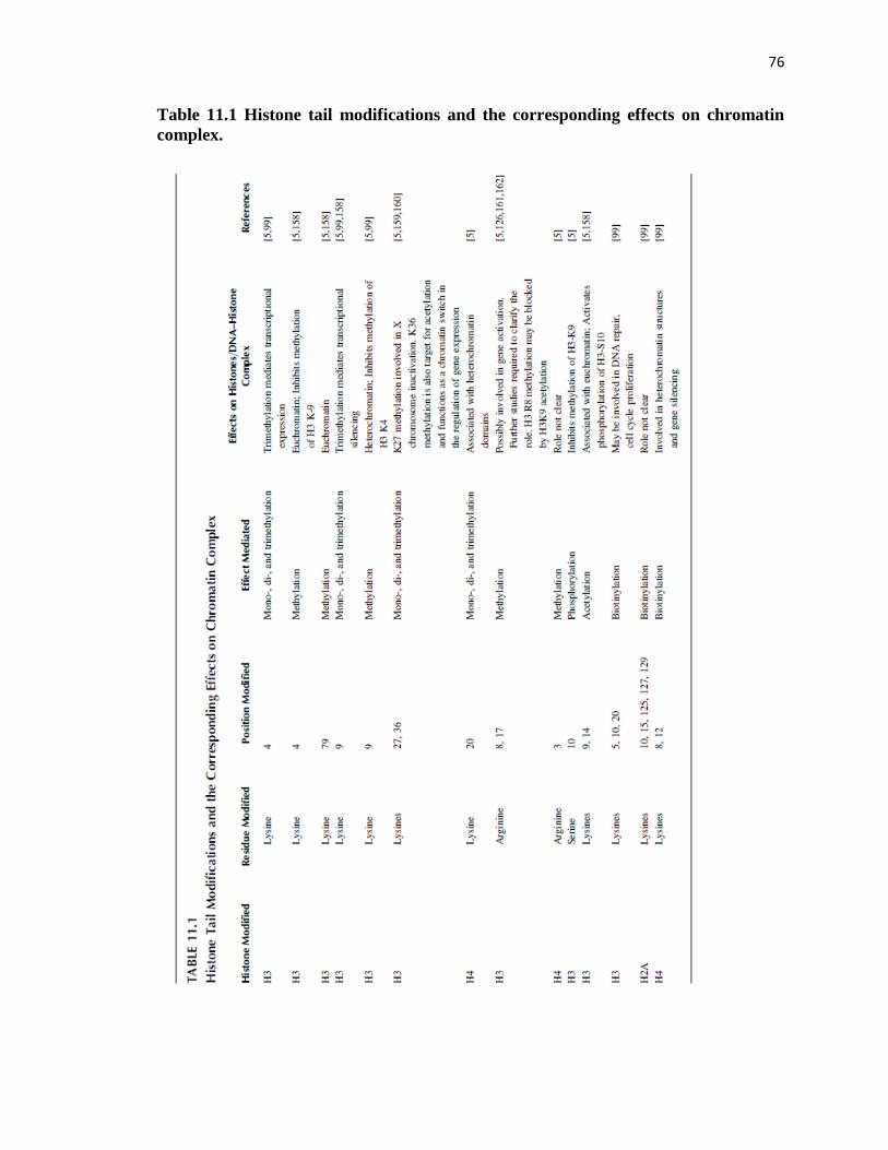
76
Table 11.1 Histone tail modifications and the corresponding effects on chromatin
complex.

77
As a result, histone acetylation usually causes transcriptional activation [5]. Histone
acetylation is regulated by the action of two antagonistic enzymes: histone
acetyltransferases (HATs) and histone deacetylases (HDACs). Interestingly,
inhibitors of these enzymatic activities interfere with the cell cycle, at least in part by
triggering changes in the acetylation/deacetylation balance of histones. This induces
growth arrest, differentiation, and apoptosis [14].
Several dietary ingredients known to possess anticarcinogenic activities [12, 15]
have the ability to inhibit HDAC and HAT enzymes. For example, butyrate (Figure 11.1),
generated in the colon by the fermentation of dietary fiber, is a histone deacetylase
inhibitor. Butyrate promotes acetylation of histones, leading to expression of genes
involved in cellular differentiation and apoptosis of cancer cells [16]. Further supporting
the link between histone acetylation and the activation of gene expression, butyrate
increases the expression of the CDKNIA gene in human colon adenocarcinoma (Colo-
320) and human colon cancer (SW116) cell lines, inducing expression of the p21 protein.
Interestingly, the latter is due to the hyperacetylation of H3 and H4 histones associated
with the promoter of the p21 gene [14].
Flavones are potent cell-specific triggers of apoptosis in a variety of cells [17–45].
Flavopiridol, a flavonoid derived from an indigenous plant from India, induces apoptosis
in human leukemia cells by the disruption of cell cycle progression. The result is more
pronounced when flavopiridol is used in combination with sodium butyrate as
demonstrated in U937 leukemia cells [46]. In combination, these compounds inhibit
leukemic cell differentiation and promote mitochondrial damage and cell death by the
induction of multiple perturbations in cell cycle and apoptosis regulatory proteins, further

78
supporting the possibility that butyrate effects on histone acetylation may be necessary
for an optimal anticarcinogenic effect [47].
The Coxsackie and adenovirus receptor (CAR) is downregulated in several types
of cancer [48–55]. Phytoestrogens in combination with FK228, a depsipeptide HDAC,
increase the expression of CAR. Pong et al. [54] have reported the potency of
phytoestrogen in combination with FK228 to be: genistein>biochanin A>ipriflavone>
daidzein. Interestingly, this order correlates with the degree of acetylated H4 levels
associated with the CAR promoter, supporting the possibility that the combined effect of
phytoestrogens and FK228 on the expression of CAR is regulated by histone acetylation
[47].
Diallyl disulfide (DADS), present in garlic and other allium vegetables, has been
shown to induce G2/M cell cycle arrest in HT-29 and Caco-2 human colon cancer cell
lines and this effect also seems to be mediated by the state of histone acetylation. Thus,
treating these cells with 200 mM DADS increases CDKNIA mRNA expression and p21
protein levels, accompanied by an increase in H4 or H3 acetylation within the CDKNIA
gene promoter region. Increase in H4 or H3 acetylation patterns are also observed in vitro
with other organosulfur compounds found in garlic such as S-allyl-mercaptocysteine [56–
60].
Sulforaphane, a compound found in cruciferous vegetables, acts in vitro as a
HDAC inhibitor in the range of 3–15 mM, producing an increase in acetylation of
histones in human embryonic kidney 293 cells, HCT116 human colorectal cancer cells,
and prostate epithelial cells lines (e.g., BPH-1, LnCaP, and PC-3). Cells treated with
histone deacetylase inhibitors have been shown to have increased acetylation of histones

79
that specifically regulate genes involved in differentiation and apoptotic pathways [61–
65]. HDAC inhibitors work by inactivating the deacetylases thereby changing the
acetylation/deacetylation balance and increasing the expression of genes that they
control.
Although the molecular pathways that are affected by HDACs are poorly
understood, in most cancer phenotypes the use of HDAC inhibitors are associated with
the induction of cell differentiation, apoptosis, and death of the tumor cells [66]. For
example, induction of G2/M cell cycle arrest and apoptosis by HDAC inhibitors, e.g.,
sulforaphane, correlates with elevated expression of p21, a protein known to be involved
in cell cycle arrest and apoptosis, [16,61,63–64]. Probably the acetylation levels of
histones, in this case histone H4 associated with the p21 promoter, increases [63–64].
Furthermore, increased acetylated levels of histones H3 or H4 were found in intestinal
polyps of Apcmin mice treated with sulforaphane, which also displayed increased p21
expression and decreased multiplicity of the polyps when compared to the Apcmin mice
fed with a control diet without sulforaphane [58,63].
Curcumin (diferuloylmethane) (Figure 11.1), an active ingredient derived from
the rhizome of the plant Curcuma longa, has also been shown to have anticancer activity
both in vitro and in vivo. At 20 mM, curcumin induces histone hypoacetylation by
inhibiting HAT activity in brain cancer cells in vitro [67]. The inhibitory effect of
curcumin on cell proliferation was associated in a time- and concentration-dependent
manner with histone acetylation [67]. Interestingly, curcumin upregulates or
downregulates histone acetylation, depending on the cell type. For example, in brain
cancer cells treated with curcumin, H4 histones are hypoacetylated [67]. In contrast, H3

80
and H4 histone subunits are hyperacetylated in androgen-dependent and -independent
prostrate cancer cells treated with curcumin [68]. This cell-specific effect may be
attributed to different genes affected downstream of the histone modification in each of
these cell types. Alternatively, the opposite findings may be due to the different
concentrations of curcumin used in the two studies.
Recently, a small polyisoprenylated benzophenone molecule, garcinol from the
Garcinia indica (coccum) fruit rind, has been found to be a naturally occurring inhibitor
of HAT [68]. Therefore, it is possible that treatment of cells with garcinol may induce
hypoacetylation by inactivating HAT. Further studies will be necessary to determine the
efficacy of using compounds that target HAT activity.
11.3.2 HISTONE METHYLATION/DEMETHYLATION AND THE EFFECT OF DIET
11.3.2.1 Histone Methylation
Evidence exists that histone methylation regulates fundamental processes such as
heterochromatin formation, X chromosome inactivation, genomic imprinting,
transcriptional regulation, and DNA repair [69]. The most heavily methylated histone is
H3, followed by H4. Histone methylation is a covalent modification that occurs on the
side chain nitrogen atoms of key histone lysines and arginines. Arginine can be either
mono- or dimethylated and lysine can be mono-, di-, or trimethylated. The multiple
possible states of methylation of a particular histone residue, determined by different
histone methyltransferases [5,70–71], leads to different biological outcomes; thus, histone
methylation has greater combinatorial potential as compared to other histone
modifications.

81
Trimethylation of the histone H4 lysine 20 residue (H4-Lys20), the only lysine
that undergoes methylation in H4, has been associated with constitutive heterochromatin,
gene silencing, and aging, and serves as a marker for them [69,72]. Fraga et al. observed
that cancer cells have a loss of trimethylated forms of histone H4 [72]. This loss occurs
early and accumulates during the carcinogenic process in the skin cancer model with
which Fraga worked. The resulting overall hypomethylation may be responsible for gene
silencing observed at various stages of carcinogenesis [72].
Further evidence for the involvement of histone methylation in cancer was
provided by studies in mice, in which knockout of the enzymes that govern H3-Lys9
methylation resulted in genomic instability and formation of B-cell lymphomas in 28% of
the mice between 9 and 15 months of age [73].
11.3.2.2 Histone Demethylation
The removal of a methyl group from histones is achieved by arginine and lysine
demethylases, e.g., by deimination and amino-oxidase reactions [69,74]. In contrast to
acetylation or phosphorylation, which has fast turnover rates and fit the expected features
of a regulatory modification, histone methylation has been demonstrated to have a slow
turnover rate. Modifications such as methylation are more stable and are responsible for
long-term expression status of certain regions of the genome. Active histone
demethylation is involved in both either transcriptional expression or repression and
depend on the histone residues that are demethylated.

82
11.3.2.3 Dietary Influence on Histone Methylation and Demethylation
11.3.2.3.1 Effect of a Methyl-Deficient Diet
Histone methyltransferases require methyl-rich sources to catalyze the transfer of methyl
groups to histone lysines or arginines. In the normal cell, SAM, a product of methionine
metabolism, serves as a methyl-donor, resulting in methylated CpG islands. Diet is a
major source of methyl groups [75–77]. A diet low in methyl-donors may lead to changes
in trimethylation and acetylation patterns of H4-Lys 20 and H3-Lys 9, respectively,
resulting in the formation of relaxed DNA–histone complexes. Such changes have been
observed during hepatocarcinogenesis [77].
Folate is an important dietary methyl-donor, which has dual effects in cancer
inhibition and progression [78–79]. Folate is required for 1-C carbon metabolism as well
as nucleotide synthesis, all of which are essential for cell growth. Folate is also
metabolized to 5 methyltetrahydrofolate, which is a source of methyl groups for
methionine and SAM synthesis.
Carcinogenesis in several organs such as colon, prostrate, and lung [78,80], have
been associated with folate-deficient diets. In contrast, the incidence of breast cancer has
been associated with diets high in folate. The key factors that tip the balance towards
tumor progression or regression are the timing and dosage of folate administered. This
may be a reason that a folate-deficient diet or a diet high in folate may increase the risk of
tumor incidence. DNA methylation has been associated with carcinogenesis in these
organs. Determining the genes silenced by this mechanism could provide much needed
markers for the diagnosis of these cancers [79]. Understanding the molecular

83
mechanism of folate action in different tissues is, therefore, crucial to the understanding
of its chemopreventive potential.
11.3.2.3.2 Dietary Polyphenols
The catechol polyphenols inhibit DNA methyltransferases 1 (DNMT1) activity in part by
increasing intracellular SAH (S-adenosyl-L-homocysteine), a potent inhibitor of DNA
methyltransferases [81] (Figure 11.1). The effective in vitro apoptotic and cell
proliferation inhibitory concentrations of epigallocatechin gallate (EGCG) (10–50 mM), a
key component of green tea, are ~50 times higher than the plasma and tissue levels of
EGCG generally observed after ingestion of tea [82–83]. The effective high in vitro
concentrations may be attributed to the inactivation of bioactive metabolites of EGCG by
the culture media used in studies in vitro. Therefore more of the compound is required to
bring about an effective response. However, in vivo, a host of factors could be involved in
stabilizing the EGCG molecule, allowing low plasma doses to be more effective.
Discrepancies have also been found between the in vitro and in vivo effective
concentrations for genistein and may have similar causes. For example, in vitro, genistein
concentrations (5–20 mM) that trigger DNA demethylation are orders of magnitude
higher than plasma levels of genistein (~270 nM) [84–85].
11.3.3 HISTONE PHOSPHORYLATION AND THE EFFECT OF DIET
The core histones and histone H1 undergo phosphorylation on specific serine (Ser) and
threonine (Thr) residues by H1 and H3 kinases [86–87]. H1 is phosphorylated on the N-
terminal and C-terminal domains of Ser/Thr residues, whereas H3 is phosphorylated only

84
on the N-terminal domains of Ser/Thr residues. Both phosphorylation processes are cell
cycle dependent [88]. H1 phosphorylation weakens H1 binding to DNA, promoting free
access of transcriptional replication factors to the DNA, which facilitates gene
expression. H3 phosphorylation at Ser-10, a more frequent process, has been associated
with the transcriptional activation of the early response genes c-fos and c-jun. Thus,
chromatin decondensation is possibly coordinated by H3 phosphorylation [89]. These
findings are consistent with the possibility that H3 phosphorylation may play a role in
cell cycle progression [89].
The activation of checkpoints in response to DNA damage leads to cell cycle
arrest but when the damage is very severe, it results in apoptotic cell death [90–91].
Sulforaphane, found in mustard seeds, although not an antioxidant itself, is an effective
inducer of enzymes that enhance the activity of the crucial intracellular antioxidant,
glutathione. In vitro, 20 mM sulforaphane increases phosphorylation at Ser-139 of
H2A.X., an isoprotein of histone 2A (H2A) and is a sensitive marker for the presence of
DNA double-strand breaks. The ensuing accumulation of cell cycle damage may be the
mechanism that triggers apoptosis [89].
Histone phosphorylation has been associated with the anticarcinogenic activity of
several flavones [92]. For example, H2A.X phosphorylation known to be induced at
double-strand break sites have been observed in MCF-7 cells treated with 50 nM
aminoflavones. Moreover, the aminoflavone had a dose-dependent effect on histone
phosphorylation [92–94]. The ability of aminoflavone to generate phosphorylated H2A.X
selectively in replicating cells makes it a suitable candidate for the treatment of cancers.

85
The chemopreventive effect of genistein has been associated with increased H1
histone phosphorylation in cancer cells [95]. Genistein-treated breast cancer cell lines
such as MDA-MB-231 and BT20 display an increase in kinase activity. This study
primarily focused on the expression of cyclin and cyclin-dependent kinases. The increase
in kinase activity in genistein-treated cancer cell lines is accompanied by histone
phosphorylation, transcriptional activation, and finally G2M arrest [95]. In contrast to this
observation genistein also blocks the recruitment of histone H3 kinase MSK1, thereby
inhibiting H3 phosphorylation. This observation is gene specific, however. Interleukin-6,
an inflammatory response gene, and a growth factor for many tumors [96], is
transcriptionally regulated by nuclear factor-kB (NF-kB) and requires the activation of
the mitogen-activated protein kinase (MAPK)/MSK kinase pathway, which
phosphorylates NF-kB p65 and histone H3. In the presence of genistein, as seen in the
breast cancer cell line MDA-MB-231, histone H3 kinase MSK1 recruitment to the IL-6
promoter is blocked leading to the loss of H3 phosphorylation and acetylation and
reduced IL-6 expression. However, the activity of MSK1 is reduced but not completely
inhibited. Although genistein plays a significant role in H3 phosphorylation, the
contradictory results obtained by the two studies was primarily due to the different
pathways analyzed and the concentrations of genistein used were different (1–30 mM and
200 mM, respectively).
Zearalenone, a fungal metabolite found in dairy sources, meat, and grains, is a
nonsteroidal estrogen molecule [97]. Ingestion of zearalenone increases the endogenous
estrogen load thereby stimulating estrogen receptor-positive cells to undergo mitosis by
86
activating cyclin-dependent kinases, [97]. It is, therefore, not surprising that
phosphorylation of histone H3, a marker of mitotic activity, is also increased [97].
Another molecule that influences the phosphorylation status of histones H3 is
vinblastine, an alkaloid derived from the Madagascar Periwinkle plant. This compound
has been used in the treatment of cancers [98]. In vitro treatment of cancer cell lines with
this alkaloid shows that the proportion of phosphorylated versus nonphosphorylated H3
molecules progressively increases during the cell cycle arrest in the metaphase; but in the
interphase chromatin, the fraction of phosphorylated H3 histones is several-fold lower
compared to that in mitotic cells [98].
11.3.4 HISTONE BIOTINYLATION AND DIET
Recent studies have shown that histone modulations involving the covalent attachment of
biotin to specific lysine residues, catalyzed by the enzymes biotinidase and
holocarboxylase synthetase, occur on histones H2A, H3, and H4 [99]. Biotinylation of
histone H4 at lysines 8 and 12 has been associated with heterochromatin structures, gene
silencing, mitotic condensation of chromatin, and DNA repair. Histone biotinylation is a
reversible process, although debiotinylases have not been characterized [99].
Biotinylation appears to be important in DNA repair and chromatin structure and
is more predominant in proliferating than in quiescent cells [99,100]. Biotinylation of
histones is known to occur in the presence of DNA double-strand breaks. In some cases,
this occurs at specific lysine residues of H4, probably associated with genes involved in
the DNA repair machinery [99]. Dietary supplementation of biotin is required for
biotinylation and biotin deficiency may have profound effects on chromatin structures
87
[99,100]. Further studies will be necessary to test the possibility that dietary biotin may
have epigenetic effects via histone biotinylation.
11.3.5 HISTONE UBIQUITINATION
Recent evidence has implicated histone ubiquitination in gene transcription control [101].
Ubiquitination occurs at lysine residues of histones H2A and H2B at positions 119 and
120, respectively; H1 and H3 are not yet mapped. Mono-ubiquitination of histone H2B is
required for methylation of histone H3 at K4 and K79. Histone ubiquitination seems to
promote methylation by recruiting proteosomal ATPases by ubiquitin-modified H2B
[102]. Nickel is an essential nutrient. However, a nickel overdose can be deleterious, as
insoluble nickel compounds, and soluble nickel compounds to a lesser extent, are
carcinogenic [103]. The exact mechanism by which nickel induces carcinogenesis is not
clear. An interesting find is that, once in the cell, nickel compounds can exert epigenetic
effects and deregulate gene expression. This is a possible mechanism underlying the
etiology of nickel-induced cancers.
Nickel compounds affect three types of histone modifications (1) deacetylation of
histones H2A, H2B, H3, and H4; (2) dimethylation of histone H3 at lysine 9; and (3)
ubiquitination of histone H2A and H2B. At lower concentration, Ni(II) stimulates mono-
ubiquitination of histones H2B and H2A while at higher concentrations ubiquitination is
suppressed, apparently because of the presence of a truncated H2N which lacks the K120
ubiquitination site. This modification affects gene expression and DNA repair leading to
cell transformation [104].

88
11.3.6 OTHER DIET-MEDIATED HISTONE MODIFICATIONS
Histone ADP-ribosylation occurs mostly on the glutamate residues of histone H2A
during DNA double-strand breaks. In the thyroid, ADP-ribosylation is mainly associated
with transcriptional inactivation of chromatin. Reduction of histone ADP-ribosylation is
associated with various forms of cancer such as lymphoma [105]. Phosphorylation of S14
of histone H2B and poly (ADPribosylation) of glutamate residues on histone H2A are
known markers of double-strand DNA breaks, which are mainly associated with
transcriptional inactivation [106].
The ε-amino group of histone lysine residues is also subject to modification by
ubiquitin-like proteins such as small ubiquitin-related modifier (SUMO). In contrast to
histone ubiquitination, histone modifications by SUMO are generally associated with
decreased gene expression, and are reversible. Sumoylation occurs on histones H2A,
H2B, H3, and H4 and blocks histone acetylation and ubiquitination [107].
Histones have been reported to be glycosylated. O-linked N-acetylglucosamine
(O-GlcNAc) is thought to act in a manner analogous to protein phosphorylation. O-
GlcNAc additions on nuclear and cytosolic proteins possess intrinsic histone
acetyltransferase (HAT) activity in vitro [108–110]. Further studies are necessary to
elucidate the mechanisms and the relevance of diet-related glycosylation of histones in
cancers.
In vivo histone carbonylation occurs to variable extents on all histones except
histone H4. In contrast, in vitro, carbonylation has been observed on all histones,
including histone H4. Histones H1 and H2A/H2B are predominantly more carbonylated
than histone H3. Carbonylation is irreversible and addition of this moiety to histones rich

89
in basic amino acids, like lysine and arginine residues, could mask the positive charge of
the nucleosome-DNA complex. This may allow the chromatin to relax, a phenomenon
that normally allows gene expression. Interestingly, carbonylation of histones decreases
with age, but caloric restriction has been shown to increase the levels of
carbonylation [111].
11.4 EFFECT OF ENVIRONMENTAL FACTORS ON EPIGENETIC
MODIFICATIONS
As shown above, diet, which may be considered an ‘‘environmental factor,’’ is a major
trigger of epigenetic modifications. Plants and animals are the major source of
nourishment to humans. Therefore, the conditions in which plants and animals are reared
affect the human diet. For example, if the soil in which crops are grown has a high metal
content or is sprayed with pesticides that are not easily degradable, the latter make their
way into the foodchain, resulting in detrimental changes to humans, including effects on
the epigenome. Below, we analyze a few environment-mediated mechanisms that are
well studied and are proven to contribute to epigenetic changes in cancer.
11.4.1 POLLUTANTS: SMOKING AND PARTICULATE MATTER
11.4.1.1 Mechanisms Underlying the Effect of Cigarette Smoke and Other Particulate Matter
Lung and throat cancers, as well as other respiratory problems such as chronic obstructive
pulmonary disease (COPD), ensue due to chronic exposure to cigarette smoke [112–113].
The molecular pathways underlying inflammation of these tissues by cigarette smoke is

90
well studied. [113–114].One of the most influential factors mediating this response is
histone modification of proinflammatory genes [113–114].
Particulate matter in the air of <10 mM (PM10) diameter have also been
associated with chronic lung and cardiovascular disease, including severe asthma attacks
[115–121]. Just like cigarette smoke, the mechanism underlying their deleterious effect
involves acetylation of H4 associated with the promoter region of inflammatory genes,
e.g., IL-8 [112].
Cigarette smoke induces oxidative stress, recruits proinflammatory cells [113],
and causes the overexpression of proinflammatory genes such as NF-κB and AP-1 by
affecting the balance of histone-tail acetylation/deacetylation [113]. Inflammatory
molecules such as interleukins and cytokines produced by the inflammatory cells induce
and maintain the chronic inflamed condition [112–113]. Prolonged inflammation of the
lungs increases the risk of developing COPD, which can give rise to lung cancers [122].
The mechanistic action of cigarette smoke in the induction of COPD has been
investigated and chromatin changes have been implicated in its etiology. Acrolein, a
reactive aldehyde, is a component of cigarette smoke which has also been shown to have
epigenetic effects by interfering with the function of enzymes such as histone
deacetylases (HDACs) [114].
Of the HDAC class of histone deacetylases, HDAC2 appears to be an important
deacetylase involved in the pathogenesis of COPD. Studies have shown that levels of
HDAC1 remain unaffected but HDAC2 levels are restored to normal after prolonged
exposure to smoke [112]. Although HDAC2 levels are restored, hyperphospho-
acetylation of H4 and acetylation of H3 is observed. This may be possible as the HDAC2

91
levels, though at optimal levels, may be inactive due to the effect of the reactive
aldehydes present in the smoke component. These aldehydes have affinities for histidine
and lysines, and react with the histidine groups at the HDAC2 active site. Therefore,
drugs that are anti-inflammatory may reverse the acetylation status of the genes by
increasing HDAC levels and activity [113–114].
Another example of histone acetylation/deacetylation imbalance induced by
cigarette smoke, leading to altered gene expression, has been demonstrated for the
inflammatory genes TNF-α and IL-8 mediates [112–113]. Cigarette smoke leads to
hyperacetylation of the histone H4 associated with the promoters of TNF- α and IL-8
[121,123]. Thus, under normal conditions, NF-κB is complexed with HDACs. A
component of NF-κB, p65, has HAT-like activity. When complexed with HDAC, the p65
component of NF-κB is rendered inactive and hypoacetylation of histones ensues
[113,121]. In contrast, under conditions of oxidative stress, NF-κB is released from the
HDAC complex. NF-κB becomes phosphorylated at serine residues, which transactivates
p65 and leads to the hyperacetylation of histone H4. These events trigger the expression
of the proinflammatory genes IL-8 and IL-6 [113,114,121].
11.4.1.2 Factors Modulating the Magnitude of Epigenetic Effects of Cigarette Smoke and Other
Particulate Matter
It is presumed that individuals who smoke are at a greater risk of developing various
pulmonary diseases as discussed above. However, not all smokers develop lung cancers
or other respiratory problems. The reason for the susceptibility of some but not all
smokers is not well understood. This question was addressed in a Korean population of

92
smokers versus nonsmokers [124]. This study provided strong evidence for a role of
epigenetics in lung cancer development in susceptible populations.
Single nucleotide polymorphisms (SNPs) could be an alternative mechanism that
modulates the epigenetic effects of cigarette smoke. The histone methyltransferase,
SMYD3, methylates histone H3 at the K4 residue [124]. Single nucleotide
polymorphisms of the gene SMYD3 are associated with greater risk for developing
colorectal cancers, hepatocarcinomas, and breast cancers [124]. It is possible that
polymorphisms may cause structure and activity modification of the catalytic domain of
this enzyme, affecting the methylation status of histone residues. This would be likely to
affect heterochromatin states and gene repression.
Yoon et al. [124] showed that SUV39H2, a mammalian histone
methyltransferase, exhibits eight different SNPs. The polymorphism G to C at 1624 in the
3’ UTR is critical to the development of lung cancers. This enzyme methylates histone
H3 at K9 and is involved in transcriptional repression. The addition of the methyl moiety
to the K9 residue creates a binding site for heterochromatin protein 1 (HP1) [124].
SUV39H2 and HP1 both mediate the repressive functions of Rb protein that is important
in controlling the cell cycle through cyclin E [124]. Therefore, histone modification and
the enzymes that modulate the covalent modification may act in tandem to influence gene
expression and the susceptibility to cancer.

93
11.4.2 OCCUPATIONAL EXPOSURE TO POLLUTANTS, HISTONE MODIFICATION, AND
CANCER
In mines and refineries, workers become exposed to harmful particulate or soluble metal
ions [125]. This occupational hazard has been associated with increased susceptibility to
cancer. For example, occupational exposure to insoluble nickel compounds plays a
significant role in the development of lung and nasal cancers [125]. When insoluble
nickel enters cells it is converted to Ni2+ ion, which induces the production of reactive
oxygen species (ROS), and may repress tumor suppressor gene expression [125]. One
pathway underlying Ni2+ effects on gene expression is epigenetic. Ni2
+ inhibits the
activity of histone acetyltransferases (HATs), causing histone hypoacetylation primarily
of the H4 subunit. Ni2+ binds to the histidine residue at position 18 of the amino terminal
domain of H4, which is in close proximity to the acetylated lysine residue [125]. This
triggers the generation of ROS which associate with HATs and may interfere with their
catalytic activity, inducing hypoacetylation of associated genes. In addition to
inactivating HATs, Ni2+ bound to the histidine residue may prevent HAT binding to the
H4 subunit and may consequently induce H4 hypoacetylation, which silences gene
expression due to the induction of heterochromatin states [125]. ROS have a strong
affinity for proteins rich in cysteine and histine residues [125]. The catalytic domains of
certain proteins, which posses HAT activity, are rich in these residues. When these HATs
undergo oxidative modifications, mediated by ROS, the proteins are rendered inactive
and thereby reduce histone acetylation leading to transcriptional suppression of genes
associated with tumor suppression.
11.4.3 EXPOSURE TO DRUGS AND CHEMICALS, HISTONE MODIFICATION, AND

94
CANCER
In the late 1940s to the early 1970s expectant mothers, especially in their first trimester,
were given diethylstilbestrol (DES). This synthetic estrogen was given to prevent
miscarriages. Moreover, animal livestock and cattle were fed with DES to increase
growth rate, providing the human population with another source of this estrogenic
compound. Since then, DES has been shown to induce developmental defects of the
cervix, uterus, and vagina, as well as rare cancers. The observed effect is trans-
generational in nature, i.e., it skips the mothers and affects the daughters and
granddaughters. This outcome is speculated to have a basis in epigenetic processes [126].
Some DES-mediated effects are mediated by DNA methylation and in certain
instances methylation of CpG residues affects the modification of histones surrounding
the genes. DES is a synthetic estrogen molecule. In the absence of the DES ligand or
estrogen, the estrogen receptor (ER) is complexed to heat-shock protein 90 (Hsp90), and
is inactive because Hsp90 inhibits ER-chromatin complex formation [126]. However, in
the presence of DES, ER dimerizes with DES and enters the cell nucleus to mediate its
downstream effects [126].
Steroid hormones effect the expression of WNT genes, which are involved in the
development and regulation of the female reproductive system. Therefore, it is not
surprising that administration of synthetic estrogen molecules like DES to pregnant
women alters the expression of Wnt [126]. The association of Hsp90 with estrogen has
opposite effects on WNT-associated gene expressions. In the presence of the chaperon
Hsp90, optimal activity of histone 3 lysine 4 methyltransferases, SYMD3 is observed
which is required for the activation of WNT genes [126]. However, DES may deregulate

95
this activity by quenching Hsp90, thereby disrupting the association of Hsp90 with WNT
genes [126].
The proposed model by Ruden et al. [126] attempts to explain the trans-
generational effects of DES-induced uterine abnormalities. Under normal conditions, it is
proposed that during development, specific methylation of CpG residues in confined
regions of the chromatin in germline stem cells may occur and possibly enhancers of
genes associated with uterine cancers such as c-fos are turned off [126]. However,
exposure to DES may trigger hypomethylation of the enhancers of these genes allowing
regulatory elements to bind to the transcriptome and induce activation of uterine cancer
promoting genes [126].
Wnt signaling is also affected by DES and the expression of Wnt is crucial to
maintain the methylation pattern and to replenish the uterine stem cells [126]. Since DES
has opposing effects on ER-regulated genes and Wnt-mediated gene expression, the
presence of this molecule can induce epigenetic effects on the genes that promote cancer
phenotypes by modulating methylation patterns and histone modifications.
11.5 CONCLUSIONS
Evidence exists that dietary components affect epigenetic processes such as histone
modifications [10,14–17,22,26,45,47,61,66,67,75,99,127–133]. Evidence also exists that
histone modifications play a role in the development of cancer [14,74,77,134–157].
Therefore, epigenetic modulation of histone modifications by dietary components may be
a useful preventive and therapeutic approach against cancer. To optimize these cancer
preventive effects, it will be necessary to explore agents derived from natural sources, to

96
determine the conditions for their optimal effects, e.g., optimal concentration, timing,
administration of the natural complex or its putative active ingredients. Mechanisms
underlying the effect of dietary ingredients will also need to be elucidated. Studies
discussed above suggest that epigenetic mechanisms are likely to be a major mechanism
underlying the chemopreventive action of some naturally occurring agents.
Studies discussed in this review also reveal that the negative effects of some
environmental pollutants may be mediated by epigenetic mechanisms. Moreover, some
pollutants and the diet act synergistically via mechanisms affecting the epigenetic code
[10]. Under normal conditions, epigenetic marks maintain proper gene function.
However, diet and pollutants-mediated modifications of epigenetic marks affect gene
expression, resulting in aberrant cellular division and triggering the neoplastic phenotype.
Elucidating the epigenetic code and mechanisms that underlie its modification by
the diet and the environment may serve as a tool to predict the susceptibility of an
individual to cancer and the necessary precautions or treatment required to reverse
epigenetic changes. Given the diversity of histone modifications, this may not be an easy
task.
REFERENCES
1. Vastag, B., Genome analysis yields mutations linked to hereditary prostate cancer,
JAMA, 287, 827, 2002.
2. Wang, C.Y. et al., Somatic mutations of mitochondrial genome in early stage
breast cancer, Int. J. Cancer, 121, 1253, 2007.
3. Zhao, H. et al., Genome-wide characterization of gene expression variations and
DNA copy number changes in prostate cancer cell lines, Prostate, 63, 187, 2005.
4. Peterson, C.L. and Laniel, M.A., Histones and histone modifications, Curr. Biol.,
14, R546, 2004.
5. Lusser, A., Acetylated, methylated, remodeled: Chromatin states for gene
regulation, Curr. Opin. Plant Biol., 5, 437, 2002.

97
6. Wolffe, A.P., Inheritance of chromatin states, Dev. Genet., 15, 463, 1994.
7. Vermaak, D., Ahmad, K., and Henikoff, S., Maintenance of chromatin states: An
open-and-shut case, Curr. Opin. Cell Biol., 15, 266, 2003.
8. Santoro, R. and De Lucia, F., Many players, one goal: How chromatin states are
inherited during cell division, Biochem. Cell Biol., 83, 332, 2005.
9. Henikoff, S., McKittrick, E., and Ahmad, K., Epigenetics, histone H3 variants,
and the inheritance of chromatin states, Cold Spring Harb. Symp. Quant. Biol.,
69, 235, 2004.
10. Feil, R., Environmental and nutritional effects on the epigenetic regulation of
genes, Mutat. Res., 600, 46, 2006.
11. Milner, J.A., Molecular targets for bioactive food components, J. Nutr., 134,
2492S, 2004.
12. Milner, J.A. et al., Molecular targets for nutrients involved with cancer
prevention, Nutr. Cancer, 41, 1, 2001.
13. Junien, C., Impact of diets and nutrients=drugs on early epigenetic programming,
J. Inherit. Metab. Dis., 29, 359, 2006.
14. Davis, C.D. and Ross, S.A., Dietary components impact histone modifications
and cancer risk, Nutr. Rev.,65, 88, 2007.
15. Dashwood, R.H. and Ho, E., Dietary histone deacetylase inhibitors: From cells to
mice to man, Semin. Cancer Biol., May 5, (PMID 17555985) [epub ahead of
print], 2007.
16. Myzak, M.C., Ho, E., and Dashwood, R.H., Dietary agents as histone deacetylase
inhibitors, Mol. Carcinog., 45, 443, 2006.
17. Hirano, T. et al., Citrus flavone tangeretin inhibits leukaemic HL-60 cell growth
partially through induction of apoptosis with less cytotoxicity on normal
lymphocytes, Br. J. Cancer, 72, 1380, 1995.
18. Habtemariam, S., Flavonoids as inhibitors or enhancers of the cytotoxicity of
tumor necrosis factor-alpha in L-929 tumor cells, J. Nat. Prod., 60, 775, 1997.
19. Kuntz, S., Wenzel, U., and Daniel, H., Comparative analysis of the effects of
flavonoids on proliferation, cytotoxicity, and apoptosis in human colon cancer cell
lines, Eur. J. Nutr., 38, 133, 1999.
20. Wang, I.K., Lin-Shiau, S.Y., and Lin, J.K., Induction of apoptosis by apigenin and
related flavonoids through cytochrome c release and activation of caspase-9 and
caspase-3 in leukaemia HL-60 cells, Eur. J. Cancer, 35, 1517, 1999.
21. Iwashita, K. et al., Flavonoids inhibit cell growth and induce apoptosis in B16
melanoma 4A5 cells, Biosci. Biotechnol. Biochem., 64, 1813, 2000.
22. Wenzel, U. et al., Dietary flavone is a potent apoptosis inducer in human colon
carcinoma cells, Cancer Res., 60, 3823, 2000.
23. Sakagami, H. et al., Induction of apoptosis by flavones, flavonols (3-
hydroxyflavones) and isoprenoidsubstituted flavonoids in human oral tumor cell
lines, Anticancer Res., 20, 271, 2000.
24. Mittra, B. et al., Luteolin, an abundant dietary component is a potent anti-
leishmanial agent that acts by inducing topoisomerase II-mediated kinetoplast
DNA cleavage leading to apoptosis, Mol. Med., 6, 527, 2000.

98
25. Gupta, S., Afaq, F., and Mukhtar, H., Selective growth-inhibitory, cell-cycle
deregulatory and apoptotic response of apigenin in normal versus human prostate
carcinoma cells, Biochem. Biophys. Res. Commun., 287, 914, 2001.
26. Birt, D.F., Hendrich, S., and Wang, W., Dietary agents in cancer prevention:
Flavonoids and isoflavonoids, Pharmacol. Ther., 90, 157, 2001.
27. Choi, J. et al., Flavones from Scutellaria baicalensis Georgi attenuate apoptosis
and protein oxidation in neuronal cell lines, Biochem. Biophys. Acta, 1571, 201,
2002.
28. Ko, W.G. et al., Effects of luteolin on the inhibition of proliferation and induction
of apoptosis in human myeloid leukaemia cells, Phytother. Res., 16, 295, 2002.
29. Chen, Y.C. et al., Flavone inhibition of tumor growth via apoptosis in vitro and in
vivo, Int. J. Oncol., 25, 661, 2004.
30. Monasterio, A. et al., Flavonoids induce apoptosis in human leukemia U937 cells
through caspase- and caspase-calpain-dependent pathways, Nutr.Cancer, 50, 90,
2004.
31. Quiney, C. et al., Flavones and polyphenols inhibit the NO pathway during
apoptosis of leukemia B-cells, Leuk. Res., 28, 851, 2004.
32. Way, T.D., Kao, M.C., and Lin, J.K., Apigenin induces apoptosis through
proteasomal degradation of HER2/neu in HER2/neu-overexpressing breast cancer
cells via the phosphatidylinositol 3-kinase/Akt-dependent pathway, J. Biol.
Chem., 279, 4479, 2004.
33. Brachmann, S.M. et al., Phosphoinositide 3-kinase catalytic subunit deletion and
regulatory subunit deletion have opposite effects on insulin sensitivity in mice,
Mol. Cell. Biol., 25, 1596, 2005.
34. Brusselmans, K. et al., Induction of cancer cell apoptosis by flavonoids is
associated with their ability to inhibit fatty acid synthase activity, J. Biol. Chem.,
280, 5636, 2005.
35. Chen, D. et al., Dietary flavonoids as proteasome inhibitors and apoptosis
inducers in human leukemia cells, Biochem. Pharmacol., 69, 1421, 2005.
36. Cheng, A.C. et al., Induction of apoptosis by luteolin through cleavage of Bcl-2
family in human leukemia HL-60 cells, Eur. J. Pharmacol., 509, 1, 2005.
37. Daskiewicz, J.B. et al., Effects of flavonoids on cell proliferation and caspase
activation in a human colonic cell line HT29: An SAR study, J. Med. Chem., 48,
2790, 2005.
38. Duraj, J. et al., Flavonoid quercetin, but not apigenin or luteolin, induced
apoptosis in human myeloid leukemia cells and their resistant variants,
Neoplasma, 52, 273, 2005.
39. Elsisi, N.S. et al., Ibuprofen and apigenin induce apoptosis and cell cycle arrest in
activated microglia, Neurosci. Lett., 375, 91, 2005.
40. Zheng, P.W., Chaing, L.C., and Lin, C.C., Apigenin induced apoptosis through
p53-dependent pathway in human cervical carcinoma cells, Life Sci., 76, 1367,
2005.
41. Wu, K., Yuan, L.H., and Xia, W., Inhibitory effects of apigenin on the growth of
gastric carcinoma SGC-7901 cells, World J. Gastroenterol., 11, 4461, 2005.
42. Chiang, L.C. et al., Anti-proliferative effect of apigenin and its apoptotic
induction in human Hep G2 cells, Cancer Lett., 237, 207, 2006.

99
43. Horinaka, M. et al., The dietary flavonoid apigenin sensitizes malignant tumor
cells to tumor necrosis factor-related apoptosis-inducing ligand, Mol. Cancer
Ther., 5, 945, 2006.
44. Khan, T.H. and Sultana, S., Apigenin induces apoptosis in Hep G2 cells: Possible
role of TNF-alpha and IFN-gamma, Toxicology, 217, 206, 2006.
45. Lim do, Y. et al., Induction of cell cycle arrest and apoptosis in HT-29 human
colon cancer cells by the dietary compound luteolin, Am. J. Physiol. Gastrointest.
Liver Physiol., 292, G66, 2007.
46. Dasmahapatra, G., Almenara, J.A., and Grant, S., Flavopiridol and histone
deacetylase inhibitors promote mitochondrial injury and cell death in human
leukemia cells that overexpress Bcl-2, Mol. Pharmacol., 69, 288, 2006.
47. Rosato, R.R. et al., The cyclin-dependent kinase inhibitor flavopiridol disrupts
sodium butyrate-induced p21WAF1/CIP1 expression and maturation while
reciprocally potentiating apoptosis in human leukemia cells, Mol. Cancer Ther., 1,
253, 2002.
48. Hemmi, S. et al., The presence of human coxsackievirus and adenovirus receptor
is associated with efficient adenovirus-mediated transgene expression in human
melanoma cell cultures, Hum. Gene Ther., 9, 2363, 1998.
49. Li, Y. et al., Loss of adenoviral receptor expression in human bladder cancer
cells: A potential impact on the efficacy of gene therapy, Cancer Res., 59, 325,
1999.
50. Li, D. et al., Variability of adenovirus receptor density influences gene transfer
efficiency and therapeutic response in head and neck cancer, Clin. Cancer Res., 5,
4175, 1999.
51. Matsumoto, K. et al., Loss of coxsackie and adenovirus receptor expression is
associated with features of aggressive bladder cancer, Urology, 66, 441, 2005.
52. Okegawa, T. et al., The dual impact of coxsackie and adenovirus receptor
expression on human prostate cancer gene therapy, Cancer Res., 60, 5031, 2000.
53. Okegawa, T. et al., The mechanism of the growth-inhibitory effect of coxsackie
and adenovirus receptor (CAR) on human bladder cancer:Afunctional analysis of
car protein structure, Cancer Res., 61, 6592, 2001.
54. Pong, R.C. et al., Mechanism of increased coxsackie and adenovirus receptor
gene expression and adenovirus uptake by phytoestrogen and histone deacetylase
inhibitor in human bladder cancer cells and the potential clinical application,
Cancer Res., 66, 8822, 2006.
55. Sachs, M.D. et al., Integrin alpha(v) and coxsackie adenovirus receptor expression
in clinical bladder cancer, Urology, 60, 531, 2002.
56. Lea, M.A. et al., Induction of histone acetylation and inhibition of growth of
mouse erythroleukemia cells by S-allylmercaptocysteine, Nutr. Cancer, 43, 90,
2002.
57. Lea, M.A., Randolph, V.M., and Hodge, S.K., Induction of histone acetylation
and growth regulation in erythroleukemia cells by 4-phenylbutyrate and structural
analogs, Anticancer Res., 19, 1971, 1999.
58. Lea, M.A. and Randolph, V.M., Induction of histone acetylation in rat liver and
hepatoma by organosulfur compounds including diallyl disulfide, Anticancer
Res., 21, 2841, 2001.

100
59. Lea, M.A. et al., Induction of histone acetylation in mouse erythroleukemia cells
by some organosulfur compounds including allyl isothiocyanate, Int. J. Cancer,
92, 784, 2001.
60. Lea, M.A., Randolph, V.M., and Patel, M., Increased acetylation of histones
induced by diallyl disulfide and structurally related molecules, Int. J. Oncol., 15,
347, 1999.
61. Myzak, M.C. and Dashwood, R.H., Histone deacetylases as targets for dietary
cancer preventive agents: Lessons learned with butyrate, diallyl disulfide, and
sulforaphane, Curr. Drug Targets, 7, 443, 2006.
62. Myzak, M.C. and Dashwood, R.H., Chemoprotection by sulforaphane: Keep one
eye beyond Keap1, Cancer Lett., 233, 208, 2006.
63. Myzak, M.C. et al., Sulforaphane inhibits histone deacetylase in vivo and
suppresses tumorigenesis in Apc-minus mice, FASEB. J., 20, 506, 2006.
64. Myzak, M.C. et al., Sulforaphane inhibits histone deacetylase activity in BPH-1,
LnCaP and PC-3 prostate epithelial cells, Carcinogenesis, 27, 811, 2006.
65. Myzak, M.C. et al., Sulforaphane retards the growth of human PC-3 xenografts
and inhibits HDAC activity in human subjects, Exp. Biol. Med. (Maywood), 232,
227, 2007.
66. Dashwood, R.H., Myzak, M.C., and Ho, E., Dietary HDAC inhibitors: Time to
rethink weak ligands in cancer chemoprevention? Carcinogenesis, 27, 344, 2006.
67. Kang, S.K., Cha, S.H., and Jeon, H.G., Curcumin-induced histone
hypoacetylation enhances caspase-3-dependent glioma cell death and
neurogenesis of neural progenitor cells, Stem Cells Dev., 15, 165, 2006.
68. Shankar, S. and Srivastava, R.K., Involvement of Bcl-2 family members,
phosphatidylinositol 3′-kinase/AKT and mitochondrial p53 in curcumin
(diferulolylmethane)-induced apoptosis in prostate cancer, Int. J. Oncol., 30, 905,
2007.
69. Tian, X. and Fang, J., Current perspectives on histone demethylases, Acta.
Biochem. Biophys. Sin (Shanghai), 39, 81, 2007.
70. van Dijk, K. et al., Monomethyl histone H3 lysine 4 as an epigenetic mark for
silenced euchromatin in Chlamydomonas, Plant Cell, 17, 2439, 2005.
71. Rice, J.C. et al., Histone methyltransferases direct different degrees of
methylation to define distinct chromatin domains, Mol. Cell, 12, 1591, 2003.
72. Fraga, M.F. et al., Loss of acetylation at Lys16 and trimethylation at Lys20 of
histone H4 is a common hallmark of human cancer, Nat. Genet., 37, 391, 2005.
73. Peters, A.H. et al., Loss of the Suv39h histone methyltransferases impairs
mammalian heterochromatin and genome stability, Cell, 107, 323, 2001.
74. Santos-Rosa, H. and Caldas, C., Chromatin modifier enzymes, the histone code
and cancer, Eur. J. Cancer, 41, 2381, 2005.
75. Brunaud, L. et al., Effects of vitamin B12 and folate deficiencies on DNA
methylation and carcinogenesis in rat liver, Clin. Chem. Lab. Med., 41, 1012,
2003.
76. Niculescu, M.D. and Zeisel, S.H., Diet, methyl donors and DNA methylation:
Interactions between dietary folate, methionine and choline, J. Nutr., 132(Suppl.
8), 2333S, 2002.
101
77. Pogribny, I.P. et al., Methyl deficiency, alterations in global histone
modifications, and carcinogenesis, J. Nutr., 137(Suppl. 1), 216S, 2007.
78. Kim, Y.I., Folate, colorectal carcinogenesis, and DNA methylation: Lessons from
animal studies, Environ. Mol. Mutagen., 44, 10, 2004.
79. Kim, Y.I., Folate and DNA methylation: A mechanistic link between folate
deficiency and colorectalcancer? Cancer Epidemiol. Biomarkers Prev., 13, 511,
2004.
80. Jang, H., Mason, J.B., and Choi, S.W., Genetic and epigenetic interactions
between folate and aging in carcinogenesis, J. Nutr., 135(Suppl. 12), 2967S, 2005.
81. Lee, W.J. and Zhu, B.T., Inhibition of DNA methylation by caffeic acid and
chlorogenic acid, two common catechol-containing coffee polyphenols,
Carcinogenesis, 27, 269, 2006.
82. Lee, L.T. et al., Blockade of the epidermal growth factor receptor tyrosine kinase
activity by quercetin and luteolin leads to growth inhibition and apoptosis of
pancreatic tumor cells, Anticancer Res., 22, 1615, 2002.
83. Lambert, J.D. et al., Dose-dependent levels of epigallocatechin-3-gallate in human
colon cancer cells and mouse plasma and tissues, Drug Metab. Dispos., 34, 8,
2006.
84. Adlercreutz, C.H. et al., Soybean phytoestrogen intake and cancer risk, J. Nutr.,
125(Suppl. 3), 757S, 1995.
85. Xu, X. et al., Bioavailability of soybean isoflavones depends upon gut microflora
in women, J. Nutr., 125, 2307, 1995.
86. Deshpande, A., Sicinski, P., and Hinds, P.W., Cyclins and Cdks in development
and cancer: A perspective, Oncogene, 24, 2909, 2005.
87. Nowak, S.J. and Corces, V.G., Phosphorylation of histone H3: A balancing act
between chromosome condensation and transcriptional activation, Trends Genet.,
20, 214, 2004.
88. Huang, X., et al., Sequential phosphorylation of Ser-10 on histone H3 and ser-139
on histone H2AX and ATM activation during premature chromosome
condensation: Relationship to cell-cycle phase and apoptosis, Cytometry A, 69,
222, 2006.
89. Davie, J.R. and Chadee, D.N., Regulation and regulatory parameters of histone
modifications, J. Cell Biochem. Suppl., 30, 203, 1998.
90. Tang, D. et al., ERK activation mediates cell cycle arrest and apoptosis after DNA
damage independently of p53, J Biol Chem, 277, 12710, 2002.
91. Zhou, B.B. and Elledge, S.J., The DNA damage response: Putting checkpoints in
perspective, Nature, 408, 433, 2000.
92. Meng, L.H. et al., DNA-protein cross-links and replication-dependent histone
H2AX phosphorylation induced by aminoflavone (NSC 686288), a novel
anticancer agent active against human breast cancer cells, Cancer Res., 65, 5337,
2005.
93. Yang, C.S. et al., Molecular targets for the cancer preventive activity of tea
polyphenols, Mol. Carcinog., 45, 431, 2006.
94. Yang, C.S. et al., Tea and cancer prevention: Molecular mechanisms and human
relevance, Toxicol. Appl. Pharmacol., November 29, (PMID: 17234229) [epub
ahead of print], 2006.
102
95. Cappelletti, V. et al., Genistein blocks breast cancer cells in the G(2)M phase of
the cell cycle, J. Cell Biochem., 79, 594, 2000.
96. Vanden Berghe, W. et al., Attenuation of mitogen- and stress-activated protein
kinase-1-driven nuclear factor-kappaB gene expression by soy isoflavones does
not require estrogenic activity, Cancer Res., 66, 4852, 2006.
97. Ahamed, S. et al., Signal transduction through the Ras=Erk pathway is essential
for the mycoestrogen zearalenone-induced cell-cycle progression in MCF-7 cells,
Mol. Carcinog., 30, 88, 2001.
98. Juan, G. et al., Histone H3 phosphorylation and expression of cyclins A and B1
measured in individual cells during their progression through G2 and mitosis,
Cytometry, 32, 71, 1998.
99. Hassan, Y.I. and Zempleni, J., Epigenetic regulation of chromatin structure and
gene function by biotin, J. Nutr., 136, 1763, 2006.
100. Crisp, S.E. et al., Biotin supply affects rates of cell proliferation, biotinylation of
carboxylases and histones, and expression of the gene encoding the sodium-
dependent multivitamin transporter in Jar choriocarcinoma cells, Eur. J. Nutr., 43,
23, 2004.
101. Conaway, R.C., Brower, C.S., and Conaway, J.W., Emerging roles of ubiquitin in
transcription regulation, Science, 296, 1254, 2002.
102. Ezhkova, E. and Tansey, W.P., Proteasomal ATPases link ubiquitylation of
histone H2B to methylation of histone H3, Mol. Cell, 13, 435, 2004.
103. Ke, Q. et al., Alterations of histone modifications and transgene silencing by
nickel chloride, Carcinogenesis, 27, 1481, 2006.
104. Kacian, D.l., and Fultz, T.J., Nucleic acid sequence amplification methods, U.S.
Patent, 5, 399, 1995.
105. Jaylata Devi, B. and Sharan, R.N., Progressive reduction in poly-ADP-
ribosylation of histone proteins during Dalton’s lymphoma induced ascites
tumorigenesis in mice, Cancer Lett., 238, 135, 2006.
106. Althaus, F.R., Poly ADP-ribosylation: A histone shuttle mechanism in DNA
excision repair, J. Cell Sci., 102, 663, 1992.
107. Gill, G., SUMO and ubiquitin in the nucleus: Different functions, similar
mechanisms? Genes Dev., 18, 2046, 2004.
108. Toleman, C. et al., Characterization of the histone acetyltransferase (HAT)
domain of a bifunctional protein with activable O-GlcNAcase and HAT activities,
J. Biol. Chem., 279, 53665, 2004.
109. Yang, X., Zhang, F., and Kudlow, J.E., Recruitment of O-GlcNAc transferase to
promoters by corepressor mSin3A: Coupling protein O-GlcNAcylation to
transcriptional repression, Cell, 110, 69, 2002.
110. Yao, D. et al., Methylglyoxal modification of mSin3A links glycolysis to
angiopoietin-2 transcription, Cell, 124, 275, 2006.
111. Goto, S. et al., Beneficial biochemical outcomes of late-onset dietary restriction in
rodents, Ann. N.Y. Acad. Sci., 1100, 431, 2007.
112. Marwick, J.A. et al., Cigarette smoke-induced oxidative stress and TGF-beta1
increase p21waf1/cip1 expression in alveolar epithelial cells, Ann. N.Y. Acad.
Sci., 973, 278, 2002.

103
113. Marwick, J.A. et al., Cigarette smoke alters chromatin remodeling and induces
proinflammatory genes in rat lungs, Am. J. Respir. Cell Mol. Biol., 31, 633, 2004.
114. Moodie, F.M. et al., Oxidative stress and cigarette smoke alter chromatin
remodeling but differentially regulate NF-kappaB activation and proinflammatory
cytokine release in alveolar epithelial cells, FASEB. J., 18, 1897, 2004.
115. Donaldson, K., Gilmour, M.I., and MacNee, W., Asthma and PM10, Respir. Res.,
1, 12, 2000.
116. Donaldson, K. et al., Oxidative stress and calcium signaling in the adverse effects
of environmental particles (PM10), Free Radic. Biol. Med., 34, 1369, 2003.
117. Li, X.Y. et al., Free radical activity and pro-inflammatory effects of particulate air
pollution (PM10) in vivo and in vitro, Thorax, 51, 1216, 1996.
118. Gilmour, P.S. et al., Adverse health effects of PM10 particles: Involvement of
iron in generation of hydroxyl radical, Occup. Environ. Med., 53, 817, 1996.
119. Peden, D.B., Air pollution in asthma: Effect of pollutants on airway inflammation,
Ann. Allergy Asthma Immunol., 87, 12, 2001.
120. Rahman, I. et al., Oxidative stress and TNF-alpha induce histone acetylation and
NF-kappaB/AP-1 activation in alveolar epithelial cells: Potential mechanism in
gene transcription in lung inflammation, Mol. Cell Biochem., 234, 239, 2002.
121. Gilmour, P.S. et al., Histone acetylation regulates epithelial IL-8 release mediated
by oxidative stress fromenvironmental particles, Am. J. Physiol. Lung Cell Mol.
Physiol., 284, L533, 2003.
122. Brody, J.S. and Spira, A., State of the art chronic obstructive pulmonary disease,
inflammation, and lung cancer, Proc. Am. Thorac. Soc., 3, 535, 2006.
123. Barnes, P.J., Adcock, I.M. and Ito, K., Histone acetylation and deacetylation:
Importance in inflammatory lung diseases, Eur. Respir. J., 25, 552, 2005.
124. Yoon, K.A. et al., Novel polymorphisms in the SUV39H2 histone
methyltransferase and the risk of lung cancer, Carcinogenesis, 27, 217, 2006.
125. Kang, J. et al., Nickel-induced histone hypoacetylation: The role of reactive
oxygen species, Toxicol. Sci., 74, 279, 2003.
126. Ruden, D.M. et al., Hsp90 and environmental impacts on epigenetic states: A
model for the transgenerational effects of diethylstibesterol on uterine
development and cancer, Hum. Mol. Genet., 14, R149, 2005.
127. Galvez, A.F. et al., Chemopreventive property of a soybean peptide (lunasin) that
binds to deacetylated histones and inhibits acetylation, Cancer Res., 61, 7473,
2001.
128. Gozzini, A. and Santini, V., Butyrates and decitabine cooperate to induce histone
acetylation and granulocytic maturation of t(8;21) acute myeloid leukemia blasts,
Ann. Hematol., 84, 54, 2005.
129. Hinnebusch, B.F. et al., The effects of short-chain fatty acids on human colon
cancer cell phenotype are associated with histone hyperacetylation, J. Nutr., 132,
1012, 2002.
130. Jung, M., Inhibitors of histone deacetylase as new anticancer agents, Curr. Med.
Chem., 8, 1505, 2001.
131. Moyers, S.B. and Kumar, N.B., Green tea polyphenols and cancer
chemoprevention: Multiple mechanisms and endpoints for phase II trials, Nutr.
Rev., 62, 204, 2004.

104
132. Chen, J. et al., Valproic acid and butyrate induce apoptosis in human cancer cells
through inhibition of gene expression of Akt/protein kinase B, Mol. Cancer, 5, 71,
2006.
133. Ohnishi, H. et al., Inhibition of cell proliferation by nobiletin, a dietary
phytochemical, associated with apoptosis and characteristic gene expression, but
lack of effect on early rat hepatocarcinogenesis in vivo, Cancer Sci., 95, 936,
2004.
134. Altucci, L. et al., Acute myeloid leukemia: Therapeutic impact of epigenetic
drugs, Int. J. Biochem. Cell Biol., 37, 1752, 2005.
135. Kobayashi, H., Tan, E.M. and Fleming, S.E., Acetylation of histones associated
with the p21WAF1/CIP1 gene by butyrate is not sufficient for p21WAF1/CIP1
gene transcription in human colorectal adenocarcinoma cells, Int. J. Cancer, 109,
207, 2004.
136. Toyota, M. and Issa, J.P., Epigenetic changes in solid and hematopoietic tumors,
Semin. Oncol., 32, 521, 2005.
137. Kothapalli, N., Sarath, G., and Zempleni, J., Biotinylation of K12 in histone H4
decreases in response to DNA double-strand breaks in human JAr
choriocarcinoma cells, J. Nutr., 135, 2337, 2005.
138. Perez-Plasencia, C. and Duenas-Gonzalez, A., Can the state of cancer
chemotherapy resistance be reverted by epigenetic therapy? Mol. Cancer, 5, 27,
2006.
139. Oligny, L.L., Cancer and epigenesis: A developmental perspective, Adv. Pediatr.,
50, 59, 2003.
140. Momparler, R.L., Cancer epigenetics, Oncogene, 22, 6479, 2003.
141. Plass, C., Cancer epigenomics, Hum. Mol. Genet., 11, 2479, 2002.
142. Ting, A.H., McGarvey, K.M. and Baylin, S.B., The cancer epigenome—
components and functional correlates, Genes Dev., 20, 3215, 2006.
143. Esteller, M., Cancer epigenomics: DNA methylomes and histone-modification
maps, Nat. Rev. Genet., 8, 286, 2007.
144. Esteller, M., CpG island methylation and histone modifications: Biology and
clinical significance, Ernst Schering Res. Found. Workshop, 57, 115, 2006.
145. Schafer, S. and Jung, M., Chromatin modifications as targets for new anticancer
drugs, Arch. Pharm (Weinheim), 338, 347, 2005.
146. Zhang, K. and Dent, S.Y., Histone modifying enzymes and cancer: Going beyond
histones, J. Cell Biochem., 96, 1137, 2005.
147. Herranz, M. and Esteller, M., DNA methylation and histone modifications in
patients with cancer: Potential prognostic and therapeutic targets, Methods Mol.
Biol., 361, 25, 2007.
148. Ducasse, M. and Brown, M.A., Epigenetic aberrations and cancer, Mol. Cancer,
5, 60, 2006.
149. Kondo, Y. and Issa, J.P., Epigenetic changes in colorectal cancer, Cancer
Metastasis Rev., 23, 29,2004.
150. Li, L.C., Carroll, P.R., and Dahiya, R., Epigenetic changes in prostate cancer:
Implication for diagnosis and treatment, J. Natl. Cancer. Inst., 97, 103, 2005.
151. Sugimura, T. and Ushijima, T., Genetic and epigenetic alterations in
carcinogenesis, Mutat. Res., 462, 235, 2000.

105
152. Verma, M. and Srivastava, S., Epigenetics in cancer: Implications for early
detection and prevention, Lancet Oncol., 3, 755, 2002.
153. Mielnicki, L.M., Asch, H.L., and Asch, B.B., Genes, chromatin, and breast
cancer: An epigenetic tale, J. Mammary Gland Biol. Neoplasia, 6, 169, 2001.
154. Gallinari, P. et al., HDACs, histone deacetylation and gene transcription: From
molecular biology to cancer therapeutics, Cell Res., 17, 195, 2007.
155. Gray, S.G. and Teh, B.T., Histone acetylation=deacetylation and cancer: An
‘‘open’’ and ‘‘shut’’ case? Curr. Mol. Med., 1, 401, 2001.
156. Gui, C.Y. et al., Histone deacetylase (HDAC) inhibitor activation of p21WAF1
involves changes in promoter-associated proteins, including HDAC1, Proc. Natl.
Acad. Sci. U S A, 101, 1241, 2004.
157. Orr, J.A. and Hamilton, P.W., Histone acetylation and chromatin pattern in
cancer, a review, Anal. Quant. Cytol. Histol., 29, 17, 2007.
158. Espino, P.S. et al., Histone modifications as a platform for cancer therapy, J. Cell
Biochem., 94, 1088, 2005.
159. Morris, S.A. et al., Identification of histone H3 lysine 36 acetylation as a highly
conserved histone modification, J. Biol. Chem., 282, 7632, 2007.
160. Plath, K. et al., Role of histone H3 lysine 27 methylation in X inactivation,
Science, 300, 131, 2003.
161. Bauer, U.M. et al., Methylation at arginine 17 of histone H3 is linked to gene
activation, EMBO. Rep., 3, 39, 2002.
162. Pal, S. et al., Human SWI/SNF-associated PRMT5 methylates histone H3
arginine 8 and negatively regulates expression of ST7 and NM23 tumor
suppressor genes, Mol. Cell Biol., 24, 9630, 2004.

106
EPIGENETIC APPROACHES TO CANCER THERAPY
by
SABITA N. SALDANHA AND TRYGVE O. TOLLEFSBOL
Epigenetics in Human Diseases
Copyright
2012
by
Elsevier
Used by permission
Format adapted dissertation

107
6.1 INTRODUCTION
Condensation of DNA is achieved by the interaction of basic proteins called histones that
encircle 147 bp of DNA forming a structure called the nucleosome [1,2]. The histones are
arranged as dimers of each subunit; H2A, H2B, H3, and H4 in the octet [2]. Histone H1
is independent of the octet but helps tether the nucleosome complex [2]. The octet
complex with the DNA is so arranged that certain amino acid residues of the histones
extend out serving as regulatory substrates for nucleosomal stability [1]. These substrates
establish the condensed and decondensed states of the chromatin [1]. Condensation of the
chromatin prevents the transcriptome machinery from binding and consequently inhibits
gene expression. However, when these projected tails are modified through enzymatic
transformations such as acetylation, methylation, phosphorylation, sumoylation, and
ubiquitination, the accessibility of DNA changes based on the residue modified [3].
Interestingly, current research has emphasized the roles of these modifications in the
transformation process of a normal cell to a tumorigenic phenotype by creating
imbalances in net expression of tumor suppressor versus oncogenes or overall genomic
imbalances [4]. These covalent modifications are reversible and therefore can have
profound impacts on the cellular phenotype when the activities of the enzymes that
mediate these modifications are altered. Intense interest has been directed toward the
mechanistic pathways of these modifications in carcinogenesis. However, substrate
specificity and residue-specific alterations still need to be ascertained.
In addition to histone modifications, CpG dinucleotides can be subjected to epigenetic
changes by the methylation of cytosine residues [5,6]. These methylation patterns are

108
heritable and are governed by four isoforms of DNA methyltransferases; DNMT1,
DNMT3a, DNMT3b, and DNMT3L [6]. Another area of epigenetics that still requires
further exploration and can potentially compound the effects of chromatin epigenomics in
a neoplastic cell is the epigenetic regulation of non-histone proteins. Epigenetic
regulations of non-histone proteins can drastically affect pathways within the cell, the cell
cyclical controls, and cellular phenotypes. For example, acetylation of key residues of
p53 stabilizes the protein and thus the cell cyclical function with which it is associated
[7,8]. This chapter discusses the current treatments that are designed to target epigenetic
enzymes with the hope of reversing the epigenome of cancerous cells. Non-histone
protein modifications are also important in cancerous cells and therefore the current
approaches to therapy aimed at targeting non-histone proteins will also be discussed.
6.2 HISTONE ACETYLATION
Positively charged amino acids such as lysine (K) and arginine (R) located at amino (-
NH2) terminal ends of histones are variously modified (Figure 6.1) [3]. Histones are
preferentially methylated or phosphorylated at arginine residues and acetylated at lysine
residues [3,9]. Acetylation of lysines initiates active gene expression. Acetylation of
histone residues not only establishes euchromatin states but has crucial roles in
nucleosome assembly and maintenance of chromatin states that affect various phases of
the cell, including DNA repair [9-11]. Currently, no mathematical models are available
that can determine the exact pattern of epigenetic marks which alter sets of genes in

109
FIGURE 6.1
Effects of acetylation and methylation on histone residues. The red circle represents
acetyl groups, the yellow circle symbolizes methylation and the green symbolizes
methylation of arginine residue. Acetylation of lysine residues is associated with gene
expression whereas methylation-mediated expression is dependent on the residue
methylated and the position. These reversible processes exerted by epigenetic enzymes
HATs, HDACs, and HMTs largely affect genomic stabilities, local gene expression, and
factors governing cell fate or phenotype. This figure is reproduced in the color plate
section.

110
cancer tissues. Another hindrance in determining these marks is that these chemical
transformations are dynamic and affect the genome globally rather than at specific
targets. However, certain histone-lysine residues are specifically acetylated or
deacetylated at key positions. Histone acetyltransferases (HATs) are enzymes that
orchestrate the acetylation of histones and are placed in three superfamilies based on
homologies to the yeast class of HATs; GNAT (Gcn5-related N-acetyltransferase),
MYST (MOZ, Ybf2/Sas3, Sas2 and TIP60), and p300/CBP145 [4]. In a few cancers,
dysregulation of HAT activities by mutations in the HAT genes or the dysfunction of the
gene by translocations account for tumor promotion [4]. In breast cancer cells, NCOA3
(AIB1) is overexpressed and in certain leukemias, p300, CBP, and MYST3 (MOZ)
translocations are observed [4]. Targeting HAT activity can affect the acetylation patterns
and possibly control the expression of oncogenes that are overexpressed.
6.3 HISTONE DEACETYLASES
The dynamic equilibrium of chromatin architecture is finely regulated by the activity of
HATs and HDACs. In most cancers, HATs are mutated and include chromosomal
translocations of the respective HAT, but HDACs are frequently overexpressed [12].
Certain cofactors exhibit intrinsic HAT or HDAC activity and in most instances the
effects are conglomerative with other complexes. Therefore aberrant recruitment of
HDACs to transcription factors that affect genes such as oncogenes or tumor suppressor
genes or their expression may facilitate a switch from normal to abnormal phenotype. A
total of 18 HDACs have been identified and are classified into four major classes [4]. The
classification of HDACs is based on the homology of the catalytic site [13]. Class IV

111
HDACs exhibit homologies similar to class I and II HDACs [4]. The Sir2 HDACs have
prominent roles in DNA repair and different Sir2s have varied nuclear functional roles
[14,15]. Chromatin organization is a well-studied area; however, the actual roles of
HDACs at the gene level and their roles in specific cancers still require further
elucidation. In vitro analysis of the effects of HDAC inhibitors has demonstrated their
profound effects on inhibiting cell proliferation, and inducing cell differentiation and
apoptosis [4]. HDAC inhibitors are considered important tools in cancer therapeutics and
are currently being evaluated for their therapeutic efficacies in vitro and in clinical trials.
HDAC inhibitors tested to date fall under four categories, short-chain fatty acids (SCFA),
hydroxamic acid derivatives, benzamindes, and cyclic tertrapeptides [4] (Table 6.1).
Inhibition of HDACs increases the acetylation levels of specific histone residues and in
some instances increases stabilities of nonhistone proteins, both of which are essential to
gene regulatory functions (Figure 6.2). These observations have been supported by gene
expression profiling studies [16]. Of about 1750 proteins that have been identified to be
acetylated at lysine residues, 200 of these become modified in the presence of HDAC
inhibitors and represent a significant number that may contribute to changes in gene
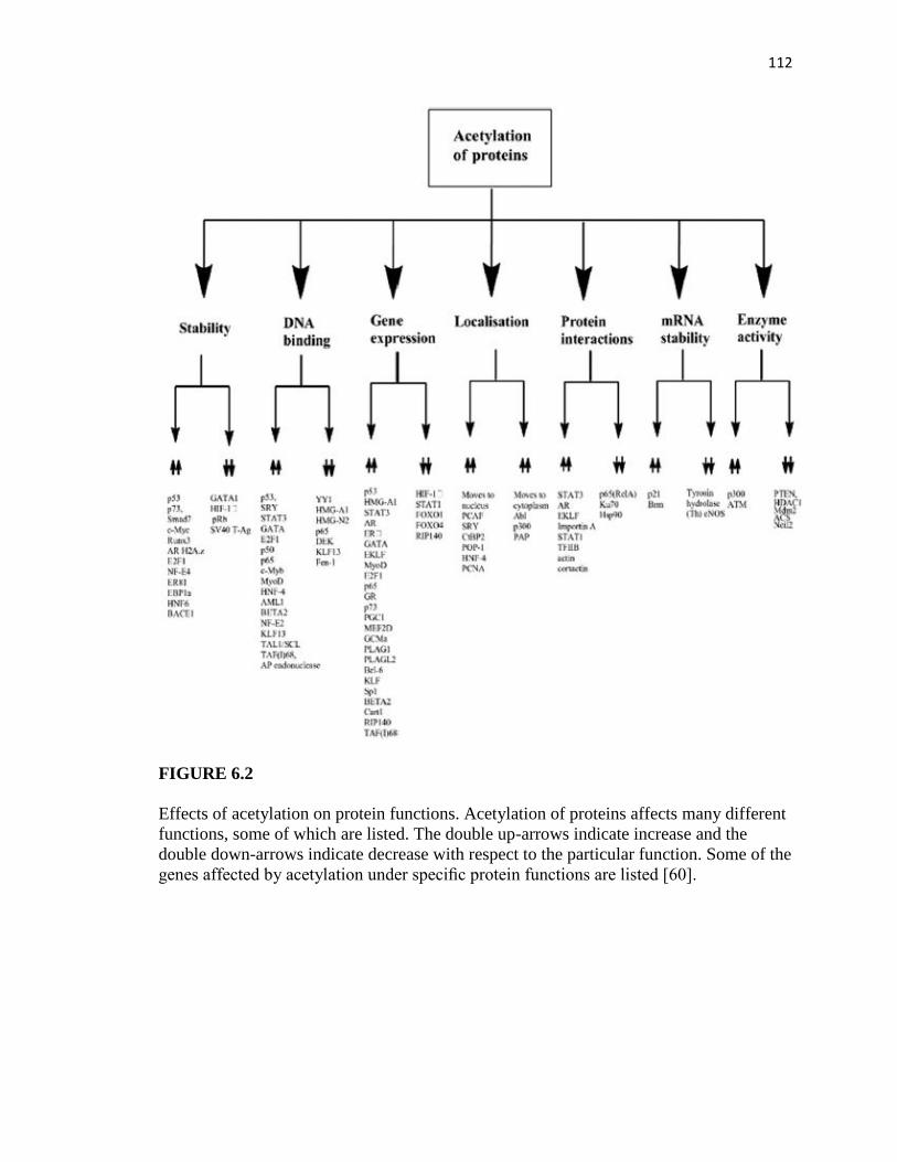
112
FIGURE 6.2
Effects of acetylation on protein functions. Acetylation of proteins affects many different
functions, some of which are listed. The double up-arrows indicate increase and the
double down-arrows indicate decrease with respect to the particular function. Some of the
genes affected by acetylation under specific protein functions are listed [60].

113
expression and probably initiate antitumor activity [16]. What needs to be determined
however is the substrate specificities for each HDAC and what drugs are specific for each
HDAC.
6.4 HISTONE METHYLATION AND DEMETHYLATION
Histone acetylation is key to promoting gene expression. Acetylated lysines are always
associated with gene expression but their methylation status contributes to varied gene
expression and primarily depends on the position and form of the methylated lysine
residue (Figure 6.1). Lysines exists in mono-, di-, and tri-methylated forms and in some
instances the same lysine can be acetylated or methylated, for example, K4 and K9
residues of histone H3 [17]. However, in this instance the acetylated lysine status will not
govern the methylated status of the same lysine in the histone [17]. In other cases, either
acetylation or methylation will influence the covalent modified status of the neighboring
lysine residues and the summation of these effects will determine the outcome. A
commonly found histone pattern in many cancers is the loss of H4K16 acetylation and
H4K20 tri-methylation [18].
Patterns affect the histone residues globally or histones of gene-specific loci can
independently influence cancer outcomes (Table 6.2). This implies that when tumor
suppressor genes are down-regulated by hypermethylation, oncogenes may be stimulated
by acetylation or hypomethylation. For example, hypermethylation of H3K79 promotes
leukemogenesis [24]. Tumor-specific epigenetic abnormalities can stem from altered
modifications of the histone residues, and/or altered expression of the enzymes that

114
catalyze the modifications. These changes are driven by mutations or chromosomal
rearrangement of genes that code for epigenetic enzymes regardless of their epigenetic
modification. As shown in Table 6.2, medulloblastoma arises from the hypomethylation
of H3K9, and the loss of H3K9 methyltransferase; amplification of demethylases or
acetyltransferases could trigger the outcome observed. Hyperacetylation of H3K9 could
inhibit the methylation of its residue.
Like DNA, histone lysine residues are methylated by the activity of methyltransferases
and utilize S-adenosyl methionine (SAM) in catalyzing the transfer of the methyl group
to specific histone residues [6]. The methyltransferases are specific based on the residues
they target. Protein lysine methyltransferases (PKMTs) and arginine methyltransferases
(PRMTs) are specific for lysine and arginine residues respectively, and mediate mono-,
di-, and trimethylation. PRMTs primarily catalyze mono- and di-methylation of histone
arginine residues 2, 8, 17, and 26 of H3 and arginine residue 3 of H4 [25]. PKMTs have a
conserved SET domain that is required for the methyltransferase activity and several of
these have been implicated in cancers [25]. H3K27 methylation is mediated by a PKMT
called EZH2 [26,27], but this enzyme is over-expressed in many tumors and appears to
have major roles in cancer aggressiveness as seen in breast and prostate tissues [26,27].

115
In another case, leukemogenesis is promoted by the aberrant recruitment of H3K79 non-
SET domain DOT1L [28]. Most of the tumor-related effects of HMTs are associated with
the over-expression, amplification, and translocation of the genes coding the enzymes.
Some of these include SMYD3 [29], CARM1 [30], and PRMT1 [31]. Drugs that can
inhibit the activities of these enzymes are currently being investigated and a certain few
are showing great promise in clinical trials. 3-Deazaneplanocin (DZNep) is a compound
that targets HMTs, including EZH2, and is instrumental in inducing global
hypomethylation at several lysine residues and includes H3K27 and H4K20 [32]. As seen
with HDACs, in vitro analysis of DZNep treatment of tumor cell lines induced apoptosis
through the activation of key target genes [32]. A combination of a HMT and HDAC
inhibitors may profoundly affect the synergistic induction of apoptosis and has been
demonstrated in colon cancer cells [33,34].
Like with most chemical compounds, non-specific and indirect mechanisms of action
may limit their clinical applications. Certain generic compounds may inhibit HMT
activity directly with low to no specificity (generic analogs of SAM such as S-adenosyl
homocysteine (SAH) and sinefungin) [35]. However, more selective compounds have
been identified through current screening methods. A fungal mycotoxin, chaetocin, is a
potent inhibitor of H3K9 HMTases SUV39H1 and G9a (EHMT2) at IC50 concentrations
of 0.8 mM and 2.5 mM, respectively [36,37]. This compound specifically depletes H3K9
di- and tri-methylation levels. However the compound exhibits cytotoxic effects
independent of its inhibitory activity. Another non-SAM competitive inhibitor of HMT
G9a, BIX-01294 has been effective at 1.7 mM and the inhibition is selective toward

116
HMTs with SET domains such as SETDB1 (ESET), and SETD7 (SET7/9) [38,39]. BIX-
01294 was found after screening 12 500 compounds and is specific in action towards
H3K9me2, reducing the di-methylated levels in mammalian cells. Structural analysis
of the SET domain reveals that the compound binds to the H3 substrate-binding groove
[38]. Both natural and chemical analogs with similar affinities to SET domains can serve
as suitable inhibitors in cancer treatments. Once the mechanistic action is determined, the
compounds can be modified to improve concentration efficacies and minimize non-
specific or cytotoxic effects. Similar inhibitors towards PRMTs, have been found [40].
However, more robust inhibitors are yet to be discovered as the current PRMT inhibitors
are non-specific and have low selectivity and activity. Pyrazole-containing CARM1
inhibitors with lower but highly potent IC50 concentrations have been reported [41]. It is
encouraging that such molecules targeting essential epigenetic enzymes can potentially
reverse epigenetic-mediated cancerous phenotypes and that further optimizations and
discoveries of effective yet non-cytotoxic drugs need to be identified for clinical testing.
Histone demethylases (HDMs) promote hypomethylation of their target residues and the
gene output is determined by the residue, or position demethylated and/or gene-specific
target that is demethylated. Therapeutically, targeting HDMs can be of significant
importance as oncogenes that are normally expressed through histone hypomethylation of
their promoters can be switched off by hypermethylation. Certainly, smaller molecules
with effective catalytic inhibitory activity toward HDMs are valuable and screening for
such compounds is crucial. HDMs catalyze the removal of methyl groups from lysine
residues in two ways. First by the amine oxidation reaction which is specific for mono-

117
and di-methylated residues and second by the hydroxylation of methylated residues
creating an unstable intermediate that degrades to release formaldehyde [42]. The second
process is specific towards mono-, di-, and trimethylated residues. The first process
requires FAD and the second uses alpha-ketoglutarate and iron as cofactors [42]. The
only known HDM to date is lysine-specific demethylase 1 (LSD1) which mediates its
demethylating action through the amine oxidation process [43]. The family of Jumonji
(JmjC) domain-containing proteins demethylate by the hydroxylation of the methyl
groups and includes JARID1B (PLU-1) and Jumonji C (JMJC) 4 domaincontaining
protein (JMJD2C) [42]. These HDMs have been implicated in tumor progression as well.
LSD1 has varied roles in terms of the residues it catalyzes. H3K4 mono- or di-methylated
residues are demethylated by LSD1 in conjunction with corepressor RE1-silencing
transcription factor (CoREST) [44]. However, H3K9 mono- and di- methylated marks are
demethylated by LSD1, in which it interacts with androgen receptor (AR) as a coactivator
to enhance the demethylating function [45]. This enzyme is over-expressed in certain
cancers and has been reported to be associated with aggressive prostate cancer and poorly
differentiated neuroblastomas.
Since LSD1 is homologous to monoamine oxidases (MAO) [46], molecules that are
effective against MAO can inhibit the activity of LSD1. One such inhibitor is the
tranylcypromine. When used, this inhibitor increases the di-methylated levels of H3K4
both in vitro and in vivo and inhibits the neuroblastoma tumor growth [47]. LSD1 has
been found to be inhibited by polyamine compounds, a few of which have shown
remarkable positive outcomes in colon cancer cells [47]. These compounds increase the

118
mono- and di-methylated H3K4 levels and reexpresse many silenced genes important in
colon cancer development. In breast and prostate cancer, the HMD, JARID1B, is
overexpressed and demethylates H3K4me3 that induces cell proliferation, inhibits tumor
suppressor functions, and results in AR coactivation [48]. JMJD2C, a H3K9me2/3
demethylase, is amplified and overexpressed in esophageal squamous carcinoma and
targeting this enzyme has been proven effective in inhibiting esophageal squamous cell
growth in vitro [49]. JMJD2C in conjunction with LSD1 enhances ARdependent gene
expression in prostate cancer where this enzyme is overexpressed [50]. Targeting the
enzyme or cofactor can effectively inhibit the activity of HDMs such as JmjC. For
example, derivatives of noggin (NOG), analog of alpha-ketoglutarate, are effective
against members of the Jumonji-C family of HDMs and a dimethylester of NOG
(DMOG) showing cellular activity has been reported [51]. However, these molecules that
are being used as HDM inhibitors are effective at much higher concentrations. Further
screening of potential drugs with higher efficacies and potencies at lower concentrations
is still required and the knowledge of the structural configurations of LSD1 and JMJD2C
can assist in the find.
6.5 DNA METHYLATION
The architectural configuration of the nucleosome is strictly governed by histones and
their covalent modifications. However, the DNA encompassed by the histone octet
dictates cellular functions and stability. Apart from the normally associated transcription
factors with promoters, methylation of CpG residues is another important mechanism
regulating gene expression. Aberrant expression of this predominant epigenetic

119
modification has been reported to play significant roles in a variety of diseases, including
cancers. Fortunately, CpG methylation can be reversed and therefore this heritable
change when exposed to demethylating compounds or compounds that inhibit the
catalytic function of the enzyme itself (DNMTs), presents as potential cancer therapeutic
tools. DNMTs are required for CpG methylation and small molecules that can target
these enzymes are being tested in vitro as well as in clinical trials. The very first DNMT
inhibitor was Vidaza (5-azacytidine) and was approved for use by the US Food and Drug
Administration (FDA) as treatment against myelodysplastic syndrome (MDS) [52,53].
Another molecule, Dacogen (5-aza-20-deoxycytidine, or decitabine) developed by MGI
Pharma Inc. (Bloomington, MN, USA) has also been used to treat MDS [52,53]. These
compounds facilitate their action through both methylation-dependent and -independent
pathways and in some cases direct proteasomal degradation of the enzyme has been
reported [53]. In theory, the use of these inhibitors is aimed at reversing the expression of
methylation-silenced critical gene expression. Clinical trials using these molecules have
shown great potential as therapeutic agents against leukemia, including MDS, acute
myeloid leukemia, chronic myelogenous leukemia, and chronic myelomonocytic
leukemia [53]. So far, the therapeutic improvements are seen against leukemias, although
this is not the case with solid tumors since it is likely that a multitude of factors govern
the growth of the mass. Cellular toxicity is also a major concern and the use of
these molecules triggers cell cycle arrest by their integration into the DNA molecule
itself. Therefore, it is imperative to develop or screen for drugs that have less cytotoxicity
and more efficacy at lower concentrations. Another concern is the relatively low stability
of these compounds in vivo and therefore modifications that enhance their stability are to

120
be considered when selecting the drug as a chemotherapeutic agent. It is imperative to
design and develop drugs that are relatively stable, mediate the degradations of DNMTs
without incorporation into DNA, and facilitate gene expression crucial to cell
differentiation and apoptotic pathways.
In mammalian cells, DNMTs exist as four active forms, DNMT1, DNMT3a, DNMT3b,
and DNMT3L and either singly or in combination catalyze the methylome patterning
crucial to gametogenesis, embryogenesis, development, and carcinogenesis [54].
Structural analysis of these enzymes shows that the catalytic domain resides in the C-
terminal region of the protein with the N-terminal essential for DNA recognition states,
hemimethylated versus unmethylated. DNMT3a is ubiquitously expressed and DNMT3b
is present at very low levels with the exception of a few tissues [55]. These levels change
in tumor cells and global hypomethylation and regional hypermethylation of specific
genes becomes an apparent pattern and has been reported to be the case in cervical,
prostate, and metastatic hepatocellular carcinoma [55]. Most preclinical studies focus on
the hypermethylation of key genes, in some cases tumor suppressor genes and others
oncogenes and the correlation of DNMT levels, methylation patterns of the promoters,
and gene expression. There appears to be some level of correlation between DNMT
levels and hypermethylation; however, regression analysis does not seem to support this
one-to-one correlation indicative of a much more complex regulatory mechanism in vivo
[55]. Some of the commonly reported hypermethylated genes include RAR, RASSF1A,
CDNK2A, CHD13, APC, p15, and p16 [56]. p15 is used as a marker to determine

121
leukemia transformation, and in some other tumors the levels of hypermethylation of
tumor suppressor genes determine the stage of the tumor [57]. Inhibitors of DNMTs are
grouped under two categories, those that interfere with the methylation of cytosine
residues by chelating into the DNA complex and second, the non-nucleosides that target
DNMT activity or stability (Table 6.3).
Results from both non-nucleoside and nucleoside DNMT inhibitors (DNMTi) in
preclinical and clinical settings are encouraging but many of these have drawbacks that
need to be revamped. Certainly the need to screen for and design small molecules with
potent DNMTi activity, less cytotoxicity, and improved specificities is very evident and
significant progress has been made in this direction. Combination treatments of two
nucleosides and two nonnucleosides are in progress and the data generated from in vitro
studies have shown remarkable synergistic DNA-hypermethylating activity. These have
been well described in a review by Jiang S-W [55].
6.6 ACETYLATION OF NON-HISTONE PROTEINS
Histones are the likely targets for reversible modifications and much focus has been on
understanding the roles of these modifications in cellular processes and the enzymes that
catalyze these chemistries. However, gene products, primarily proteins, are in many ways
subjected to similar regulations, of which acetylation and phosphorylation are key
modifications. Many cellular processes are governed by the activity of proteins and are
involved in cell signaling, transcription, and even protein degradation (Figure 6.2).
Reviews on the acetylation of non-histone proteins are limited, yet this protein

122
modification in conjunction with histone modifications is very relevant to cancer
epigenomics. Acetylation of proteins can affect many aspects of protein function as
shown in Figure 6.2. Especially pertinent are the effects of acetylation on p53 (tumor
suppressor), nuclear factor-kB (NF-kB ) (metastatic gene), and myelocytomatosis
oncogene (c-Myc) (oncogene) and the plausible roles of indirect effects of HAT/HDAC
inhibitors on these proteins in restoring normal cellular phenotypes.
Protein p53 is essential to many cell regulatory functions, and in particular, is important
to rescue a cell from DNA damage and maintain normal cell division [58]. As seen in
many cancers, p53 is mutated and the protein is dysfunctional, contributing to a
tumorigenic phenotype. Activation of p53 in cells requires the phosphorylation of the
protein that promotes the acetylation of key lysine residues 120, 164, 320, 370, 372, 373,
381, 382, and 386 mediated by different acetyltransferases [59,60]. Although
controversial, findings stronglysupport that acetylation of p53 at the C-terminus end
favorably enhances the DNA binding ability of the protein to its target genes [60]. K120
and K164 along with the C-terminus are required for p53 activity, and single-site
mutational losses can be rescued by the acetylation of key functional residues [60].
Acetylation of p53 K120 by specific HATs, such as Tip60 and hMOF, induces genes of
the apoptotic pathway [60]. When K382 is acetylated, p53 recruits CREB binding protein
(CBP) that further enhances the transcription of genes, suggestive of coactivator
functions [60]. In all, acetylation of p53 improves the stability and binding ability
of p53, allowing for recruitment of coactivators to the transcriptional binding sites in the

123
promoters of genes crucial to cell cycle regulatory functions, such as for p21. Therefore,
HDAC inhibitors that increase acetylated levels can contribute to a much more stable p53
even when it is mutated and can enhance its DNA-binding abilities, which otherwise
would be ineffective. Further studies are necessary to validate the roles of key acetylated
residues that improve the DNA-binding ability of p53 in the mutated form.
NF-κB is an important gene of the immune system and is observed to be important in the
inflammatory process. This gene is also essential to cell survival, differentiation, and
proliferation and is reported to be overexpressed in many tumors that are aggressive and
metastatic [60]. Nuclear activation of NF-κB target genes occur only when the protein is
acetylated but otherwise exists as an inactive complex tethered by Ik-B inhibitor [60].
The ubiquitination mediated degradation of Ik-B initiated by its phosphorylation, frees
the NF-κB that translocates to the nucleus and binds to its target sequence. The NF-κB is

124
a complex of p50, p52, p65(RelA), c-Rel, and RelB [60]. In mammals, the p50/p65
heterodimer is the most commonly found complex [60]. Modifications of these dimers
are essential to many of its downstream functions. Phosphorylation of p65 initiates its
acetylation at multiple sites mediated by the recruitment of p300/CBP. NF-κB target gene
expression is enhanced greatly when its K221 and K310 residues are acetylated and the
full activity of the protein is dependent on this modification [61]. This observation is

125
supported by the fact that SIRT1 deacetylation of p65 K310, HDAC1, and HDAC3
deacetylation of K221 or K310 inhibits transcription of its target genes [61]. Acetylation
of NF-κB supports many functions and the acetylation of K122 and K123 enhances the
export of the protein and re-association with IkB forming an inactive complex [62].
When p50 subunit is acetylated at positions K431, K440, and K441 the protein molecules
bind with a higher affinity to its target gene sequences [63,64]. Thus the role of
acetylation in NF-κB is many-fold. Since this gene is deregulated in many diseases
including cancers a further investigation into the application of HDACi or HATi in
regulating NF-κB functions is warranted.
Overexpression of c-Myc has been documented in many cancers. c-Myc binds and
activates target genes as a complex with Max [64]. However, regulatory functions of the
complex are soley through the transcription activation domain (TAD) of c-Myc located at
the N-terminus [64]. This domain interacts with HATs, such as GCN5 and Tip60,
forming coactivator complexes [64]. The c-Myc interacts with p300 via its TAD region
that acetylates lysines at several positions between the TAD and DNA-binding regions of
the protein, enhancing Myc turnover [64]. Thus, HAT-specific interactions with c-Myc
dictate its stability and turnover in mammalian cells, and molecules that target these
interactions by the induction of deacetylated levels are promising strategies in cancer
therapy.
126
6.7 FUTURE DIRECTIONS
Epigenetic phenomena affect histone and non-histone proteins and molecular compounds
that target enzymes influencing these roles are important to further develop. Drawbacks
of compounds such as those with high cytotoxicity, low specificity, and low stability all
have to be considered when selecting an antiepigenetic compound promoting antitumor
activity. One way of improving drug design or compound efficacy is by a dual approach
which has been proven to be much more efficient as seen with 5 azacytidine and other
chemical compounds. Further work in these areas is therefore warranted. In addition to
drug-based compounds, natural plant-based products with similar characteristics need to
be screened and tested.
References
[1] Bernstein BE, Humphrey EL, Erlich RL, Schneider R, Bouman P, Liu JS, et al.
Methylation of histone H3 Lys 4 in coding regions of active genes. Proc Natl Acad
Sci USA 2002;99:8695-700.
[2] Wong CA, Recktenwald AJ, Jones ML, Waterman BM, Bollini ML, Dunagan WC.
The cost of serious fall-related injuries at three Midwestern hospitals. Jt Comm J
Qual Patient Saf 2011;37:81-7.
[3] Cohen I, Poreba E, Kamieniarz K, Schneider R. Histone modifiers in cancer: friends
or foes? Genes Cancer 2011;2:631-47.
[4] Ma X, Ezzeldin HH, Diasio RB. Histone deacetylase inhibitors: current status and
overview of recent clinical trials. Drugs 2009;69:1911-34.
[5] Deaton AM, Bird A. CpG islands and the regulation of transcription. Genes Dev
2011;25:1010-22.
[6] Li Y, Tollefsbol TO. Impact on DNA methylation in cancer prevention and therapy
by bioactive dietary components. Curr Med Chem 2010;17:2141-51.
[7] Kim JY, Lee KS, Seol JE, Yu K, Chakravarti D, Seo SB. Inhibition of p53
acetylation by INHAT subunit SET/TAF-I {beta} represses p53 activity. Nucleic
Acids Res 2011.
[8] Seo SK, Jin HO, Woo SH, Kim YS, An S, Lee JH, et al. Histone deacetylase
inhibitors sensitize human non-small cell lung cancer cells to ionizing radiation
through acetyl p53-mediated c-myc down-regulation. J Thorac Oncol 2011;6:1313-
9.
[9] Torres-Padilla ME, Parfitt DE, Kouzarides T, Zernicka-Goetz M. Histone arginine

127
methylation regulates pluripotency in the early mouse embryo. Nature
2007;445:214-8.
[10] Kouzarides T. SnapShot: Histone-modifying enzymes. Cell 2007;128:802.
[11] Kouzarides T. Chromatin modifications and their function. Cell 2007;128:693-705.
[12] Marks PA, Richon VM, Miller T, Kelly WK. Histone deacetylase inhibitors. Adv
Cancer Res 2004;91:137-68.
[13] Yang XJ, Seto E. The Rpd3/Hda1 family of lysine deacetylases: from bacteria and
yeast to mice and men. Nat Rev Mol Cell Biol 2008;9:206-18.
[14] Horio Y, Hayashi T, Kuno A, Kunimoto R. Cellular and molecular effects of
Sirtuins in health and disease. Clin Sci (Lond) 2011;121:191-203.
[15] McGuinness D, McGuinness DH, McCaul JA, Shiels PG. Sirtuins, bioageing, and
cancer. J Aging Res 2011:235754.
[16] Choudhary C, Kumar C, Gnad F, Nielsen ML, Rehman M, Walther TC, et al.
Lysine acetylation targets protein complexes and co-regulates major cellular
functions. Science 2009;325:834-40.
[17] Rice JC, Allis CD. Histone methylation versus histone acetylation: new insights
into epigenetic regulation. Curr Opin Cell Biol 2001;13:263-73.
[18] Varier RA, Timmers HT. Histone lysine methylation and demethylation pathways
in cancer. Biochim Biophys Acta 2011;1815:75-89.
[19] Lindsey JC, Lusher ME, Anderton JA, Bailey S, Gilbertson RJ, Pearson AD, et al.
Identification of tumourspecific epigenetic events in medulloblastoma development
by hypermethylation profiling. Carcinogenesis 2004;25:661-8.
[20] Seligson DB, Horvath S, McBrian MA, Mah V, Yu H, Tze S, et al. Global levels of
histone modifications predict prognosis in different cancers. Am J Pathol
2009;174:1619-28.
[21] Seligson DB, Horvath S, Shi T, Yu H, Tze S, Grunstein M, et al. Global histone
modification patterns predict risk of prostate cancer recurrence. Nature
2005;435:1262-6.
[22] Kondo Y, Shen L, Cheng AS, Ahmed S, Boumber Y, Charo C, et al. Gene
silencing in cancer by histone H3 lysine 27 trimethylation independent of promoter
DNA methylation. Nat Genet 2008;40:741-50.
[23] Kondo Y, Shen L, Issa JP. Critical role of histone methylation in tumor suppressor
gene silencing in colorectal cancer. Mol Cell Biol 2003;23:206-15.
[24] Muller CI, Ruter B, Koeffler HP, Lubbert M. DNA hypermethylation of myeloid
cells, a novel therapeutic target in MDS and AML. Curr Pharm Biotechnol
2006;7:315-21.
[25] Yost JM, Korboukh I, Liu F, Gao C, Jin J. Targets in epigenetics: inhibiting the
methyl writers of the histone code. Curr Chem Genomics 2011;5:72-84.
[26] Chase A, Cross NC. Aberrations of EZH2 in cancer. Clin Cancer Res
2011;17:2613-8.
[27] Chang CJ, Yang JY, Xia W, Chen CT, Xie X, Chao CH, et al. EZH2 promotes
expansion of breast tumor initiating cells through activation of RAF1-beta-catenin
signaling. Cancer Cell 2011;19:86-100.
[28] Okada Y, Feng Q, Lin Y, Jiang Q, Li Y, Coffield VM, et al. hDOT1L links histone
methylation to leukemogenesis. Cell 2005;121:167-78.
[29] Hamamoto R, Furukawa Y, Morita M, Iimura Y, Silva FP, Li M, et al. SMYD3

128
encodes a histone methyltransferase involved in the proliferation of cancer cells.
Nat Cell Biol 2004;6:731-40.
[30] Majumder S, Liu Y, Ford 3rd OH, Mohler JL, Whang YE. Involvement of arginine
methyltransferase CARM1 in androgen receptor function and prostate cancer cell
viability. Prostate 2006;66:1292-301.
[31] Cheung N, Chan LC, Thompson A, Cleary ML, So CW. Protein arginine-
methyltransferase-dependent oncogenesis. Nat Cell Biol 2007;9:1208-15.
[32] Tan J, Yang X, Zhuang L, Jiang X, Chen W, Lee PL, et al. Pharmacologic
disruption of Polycomb-repressive complex 2-mediated gene repression selectively
induces apoptosis in cancer cells. Genes Dev 2007;21:1050-63.
[33] Yadav S, Singhal J, Singhal SS, Awasthi S. hSET1: a novel approach for colon
cancer therapy. Biochem Pharmacol 2009;77:1635-41.
[34] Lea MA. Recently identified and potential targets for colon cancer treatment.
Future Oncol 2010;6:993-1002.
[35] Saavedra OM, Isakovic L, Llewellyn DB, Zhan L, Bernstein N, Claridge S, et al.
SAR around (l)-S-adenosyl-lhomocysteine, an inhibitor of human DNA
methyltransferase (DNMT) enzymes. Bioorg Med Chem Lett 2009;19:2747-51.
[36] Iwasa E, Hamashima Y, Fujishiro S, Higuchi E, Ito A, Yoshida M, et al. Total
synthesis of (þ)-chaetocin and its analogues: their histone methyltransferase G9a
inhibitory activity. J Am Chem Soc 2010;132:4078-9.
[37] Greiner D, Bonaldi T, Eskeland R, Roemer E, Imhof A. Identification of a specific
inhibitor of the histone methyltransferase SU(VAR)3-9. Nat Chem Biol
2005;1:143-5.
[38] Chang Y, Zhang X, Horton JR, Upadhyay AK, Spannhoff A, Liu J, et al. Structural
basis for G9a-like protein lysine methyltransferase inhibition by BIX-01294. Nat
Struct Mol Biol 2009;16:312-7.
[39] Kubicek S, O’Sullivan RJ, August EM, Hickey ER, Zhang Q, Teodoro ML, et al.
Reversal of H3K9me2 by a smallmolecule inhibitor for the G9a histone
methyltransferase. Mol Cell 2007;25:473-81.
[40] Lakowski TM, Hart P, Ahern CA, Martin NI, Frankel A. Neta-substituted arginyl
peptide inhibitors of protein arginine N-methyltransferases. ACS Chem Biol
2010;5:1053-63.
[41] Sack JS, Thieffine S, Bandiera T, Fasolini M, Duke GJ, Jayaraman L, et al.
Structural basis for CARM1 inhibition by indole and pyrazole inhibitors. Biochem J
2011;436:331-9.
[42] Schneider J, Shilatifard A. Histone demethylation by hydroxylation: chemistry in
action. ACS Chem Biol 2006;1:75-81.
[43] Shi Y, Lan F, Matson C, Mulligan P, Whetstine JR, Cole PA, et al. Histone
demethylation mediated by the nuclear amine oxidase homolog LSD1. Cell
2004;119:941-53.
[44] Yang M, Gocke CB, Luo X, Borek D, Tomchick DR, Machius M, et al. Structural
basis for CoREST-dependentdemethylation of nucleosomes by the human LSD1
histone demethylase. Mol Cell 2006;23:377-87.
[45] Yu V, Fisch T, Long AM, Tang J, Lee JH, Hierl M, et al. High-Throughput TR-
FRET Assays for Identifying Inhibitors of LSD1 and JMJD2C Histone Lysine

129
Demethylases. J Biomol Screen 2011.
[46] Mao B, Zhao G, Lv X, Chen HZ, Xue Z, Yang B, et al. Sirt1 deacetylates c-Myc
and promotes c-Myc/Max association. Int J Biochem Cell Biol 2011;43:1573-81.
[47] Singh MM, Manton CA, Bhat KP, Tsai WW, Aldape K, Barton MC, et al.
Inhibition of LSD1 sensitizes glioblastoma cells to histone deacetylase inhibitors.
Neuro Oncol 2011;13:894-903.
[48] Xiang Y, Zhu Z, Han G, Ye X, Xu B, Peng Z, et al. JARID1B is a histone H3 lysine
4 demethylase up-regulated in prostate cancer. Proc Natl Acad Sci USA
2007;104:19226-31.
[49] Hamada S, Suzuki T, Mino K, Koseki K, Oehme F, Flamme I, et al. Design,
synthesis, enzyme-inhibitory activity, and effect on human cancer cells of a novel
series of jumonji domain-containing protein 2 histone demethylase inhibitors. J
Med Chem 2010;53:5629-38.
[50] Wissmann M, Yin N, Muller JM, Greschik H, Fodor BD, Jenuwein T, et al.
Cooperative demethylation by JMJD2C and LSD1 promotes androgen receptor-
dependent gene expression. Nat Cell Biol 2007;9:347-53.
[51] Spannhoff A, Hauser AT, Heinke R, Sippl W, Jung M. The emerging therapeutic
potential of histone methyltransferase and demethylase inhibitors. ChemMedChem
2009;4:1568-82.
[52] Vigna E, Recchia AG, Madeo A, Gentile M, Bossio S, Mazzone C, et al. Epigenetic
regulation in myelodysplastic syndromes: implications for therapy. Expert Opin
Investig
Drugs 2011;20:465-93.
[53] Ghoshal K, Bai S. DNA methyltransferases as targets for cancer therapy. Drugs
Today (Barc) 2007;43:395-422.
[54] Liu L, Li Y, Tollefsbol TO. Gene-environment interactions and epigenetic basis of
human diseases. Curr Issues Mol Biol 2008;10:25-36.
[55] Ren J, Singh BN, Huang Q, Li Z, Gao Y, Mishra P, et al. DNA hypermethylation as
a chemotherapy target. Cell Signal 2011;23:1082-93.
[56] Tsou JA, Hagen JA, Carpenter CL, Laird-Offringa IA. DNA methylation analysis: a
powerful new tool for lung cancer diagnosis. Oncogene 2002;21:5450-61.
[57] Abd El-Hamid TM, Mossallam GI, Sherisher MA. The Clinical Implications of
Methylated p15 and p73 Genes in Adult Acute Lymphoblastic Leukemia. J Egypt
atl
Canc Inst 2010;22:175-84.
[58] Benkirane M, Sardet C, Coux O. Lessons from interconnected ubiquitylation and
acetylation of p53: think metastable networks. Biochem Soc Trans 2010;38:98-103.
[59] Das C, Kundu TK. Transcriptional regulation by the acetylation of nonhistone
proteins in humans - a new target for therapeutics. IUBMB Life 2005;57:137-49.
[60] Spange S, Wagner T, Heinzel T, Kramer OH. Acetylation of non-histone proteins
modulates cellular signaling at multiple levels. Int J Biochem Cell Biol
2009;41:185-98.
[61] Yeung F, Hoberg JE, Ramsey CS, Keller MD, Jones DR, Frye RA, et al.
Modulation of NF-kappaB-dependent transcription and cell survival by the SIRT1
deacetylase. EMBO J 2004;23:2369-80.
[62] Kiernan R, Bres V, Ng RW, Coudart MP, El Messaoudi S, Sardet C, et al. Post-

130
activation turn-off of NF-kappa Bdependent transcription is regulated by acetylation
of p65. J Biol Chem 2003;278:2758-66.
[63] Deng WG, Wu KK. Regulation of inducible nitric oxide synthase expression by
p300 and p50 acetylation. J Immunol 2003;171:6581-8.
[64] Deng WG, Zhu Y, Wu KK. Up-regulation of p300 binding and p50 acetylation in
tumor necrosis factor-alphainduced cyclooxygenase-2 promoter activation. J Biol
Chem 2003;278:4770-7.

131
THE ROLE OF NUTRACEUTICALS IN CHEMOPREVENTION AND
CHEMOTHERAPY AND THEIR CLINICAL OUTCOMES
by
SABITA N. SALDANHA AND TRYGVE O. TOLLEFSBOL
Journal of oncology
Copyright
2012
by
Sabita Saldanha and Trygve Tollefsbol
Used by permission
Format adapted for dissertation

132
Abstract The genesis of cancer is often a slow process and the risk of developing cancer
increases with age. Altering a diet that includes consumption of beneficial
phytochemicals can influence the balance and availability of dietary chemopreventive
agents. In chemopreventive approaches, foods containing chemicals that have anticancer
properties can be supplemented in diets to prevent precancerous lesions from occurring.
This necessitates further understanding of how phytochemicals can potently maintain
healthy cells. Fortunately there is a plethora of plant-based phytochemicals although few
of them are well studied in terms of their application as cancer chemopreventive and
therapeutic agents. In this analysis we will examine phytochemicals that have strong
chemopreventive and therapeutic properties in vitro as well as the design and
modification of these bioactive compounds for preclinical and clinical applications. The
increasing potential of combinational approaches using more than one bioactive dietary
compound in chemoprevention or cancer therapy will also be evaluated. Many novel
approaches to cancer prevention are on the horizon, several of which are showing great
promise in saving lives in a cost-effective manner.
1. Introduction
The transformation of a normal cell into a cancerous phenotype requires stages of
initiation, progression, and promotion by altering specific genes [1–3]. Although
predisposition to cancer cannot be signaled out by a single factor, a group of factors place
some individuals at a higher risk of acquiring the disease. Most of the high-risk cases
may have a genetic background, but in some instances dietary choices can dictate the
outcome of health. As determined by population and epidemiological studies, the
predominant forms of cancer and cancer-related deaths are those of the lung and

133
bronchus, breast, colorectal, and prostate [4, 5]. These cancers are also more prevalent in
the western parts of the world and are much lower in Asian countries. A well-balanced
diet that includes more of vegetables and fruits with less fat/meat intake is in most cases a
staple of many Asian countries [4, 5]. Many hypotheses have supported that diet and
environment greatly influence cellular function and health [6].
Phytochemicals are plant-based chemicals that mediate their positive health
benefits directly, by affecting specific molecular targets such as genes, or indirectly as
stabilized conjugates affecting metabolic pathways [7]. Many genes play significant roles
in the cell cycle pathway, and some of these are altered in cancer cells [1, 2]. The aim of
most studies is to understand and formulate mechanistic pathways by which these
naturally derived chemicals can alter the fate of a cell. For a cancerous cell to survive, it
should be able to proliferate, obtain energy, and establish angiogenic pathways, in a
tumor mass. Altering genes that affect these pathways can serve as suitable tools to
decrease tumor mass and also allow for tumor regression. In this paper, the key focus will
be on mechanistic pathways that are regulated by nutraceuticals to bring about changes in
the tumor environment and serve as alternative approaches for cancer prevention and
therapy (Figure 1).
The study of phytochemicals and the classification of these compounds have been
previously reviewed [8]. However, in this paper only some of the most potent and
promising chemopreventive and therapeutic molecules will be analyzed, with emphasis
on combination therapy of these with other nutramolecules. Most phytochemicals derived

134
Figure 1: Cellular pathways affected by the activities of bioactive components in dietary
sources. Of the natural compounds present in dietary sources, some are more involved in
regulating chemopreventive pathways and some are more effective in influencing
chemotherapeutic pathways. However, a few of the bioactive molecules found to date can
impart both chemopreventive and therapeutic effects, such as EGCG and genistein.
Compound combinations as discussed in the paper that can affect different pathways are
shown and can have profound effects on tumor growth and inhibition.
135
from dietary sources are classified under an umbrella of specific chemical compounds as
value but are germane to the function of a cell. Various studies have shown that these
molecules can induce apoptosis, inhibit cellular proliferation, affect angiogenesis, and
affect cancer metabolism in various cancers, all of which are hindrances to tumor growth
(Figure 1) [7].
Several of the phytochemicals listed in Table 1 have been investigated in terms of
their curative properties. However, one must carefully interpret the observed results in
vitro and in vivo before testing the same in a clinical setting. The reasons for this are
manyfold. Tests in culture are pure, in that there is only one cell type in the culture plate
and all conditions are controlled, including the bioactive compound. In vivo, however, the
scenario changes as there are a host of other factors that need to be taken into account,
including age, weight, diet, and metabolism of the compound. A bioactive molecule in
culture may be subjected to less metabolic changes and may be presented to the cell in its
native form. However, in vivo the same compound may be presented differently, perhaps
as a conjugate, and its mode of action may change amongst the multitude of other
molecules in the host’s microenvironment. Many in vivo experiments also control for the
type of diet being administered to the organism, where the concentrations or plasma
availability can be adjusted. Therefore, what may work well in vitro, may have no
agonistic effects or even antagonistic effects in vivo, and such discrepancies are often
seen when comparing population and epidemiological studies in terms of chemical
efficacy.

136
An effective nutraceutical is one that will have a low nontoxic dose while creating
a magnitude of change in tumor dynamics. This means that at a low dose the compound
should act fast on the tumor load. However, if the time taken to be effective is slow, the
Table 1: Classification of nutrients as phytochemicals and their major food source
availability.
problems faced would be maintaining a tolerable dose and increasing bioavailability and
stability. A solution to such a problem would be to use a combinatorial approach to
therapy, a bioactive molecule with an effective synthetic drug or double-nutratherapy
(e.g., curcumin and resveratrol). Once tumor regression sets in, dietary composition of
the molecule can be adjusted.

137
2. Nutraceuticals and Their Preventive and Therapeutic Roles
2.1. Genistein: A Potent Isoflavone. Many phytochemicals are currently being
investigated for their promising anticarcinogenic properties. In vitro investigations have
shown that some compounds exert their antitumor functions at much higher
concentrations and that dietary consumption is insufficient to achieve such effective
concentrations at the tumor site. Therefore, the mode of delivery is a very important
factor that needs to be considered at clinical trials and during in vivo studies. The
nontoxic properties of natural compounds are essential to the design of a formulated
therapy. However, evidence along several lines of treatment has shown that some
compounds are preferentially more potent in activity when administered early in life [9,
10]. For instance, soy-based prevention of breast cancer is thought to be more successful
when soy products and their derivatives are consumed in early development [9].
Isoflavones are a group of phytochemicals that are predominant constituents of a
soy-based diet [9, 10]. Among isoflavones, the three major constituents that have been
shown to have remarkable influences in cancer prevention and therapy are genistein,
diadzein, and glycitin [11]. They are collectively grouped as phytoestrogens for their
weak estrogen-like activity and bind preferentially to ER-β receptors [12–15]. Evidence
of antiproliferative activity of genistein in vitro stems from its ability to inhibit the
tyrosine kinase enzyme that is most often upregulated in cancer cells [16, 17]. As a
chemopreventive agent, genistein is thought to influence the differentiation process of
mammary tissue. It is believed that early differentiation of mammary tissue into terminal
buds, as seen in rats, serves as a chemopreventive strategy as it reduces the susceptibility
of the epithelial cells in the ducts to carcinogens or estrogen and the ontogeny process

138
[9]. Many aggressive cancers have altered epidermal growth factor (EGF) receptors on
their cell surface allowing for a continuous downstream signaling pathway for cell
division [18, 19]. This is interesting, as genistein can serve as a two-fold approach
molecule for prevention and treatment. When EGF binds to its receptors, tyrosine kinase
activation results in the phoshorylation of tyrosine residues of proteins involved in
downstream cell signaling pathways that trigger cell division. Though studies have shown
that genistein increases the EGF transcript early in development of mammary tissue, this
perhaps is essential for differentiation and faster development of the breast tissue. In the
long run this is a positive preventive strategy of breast lesion formation in ducts [9].
However, as seen in older rats [9], EGF mRNA decreases. Therefore, a decrease in EGF
mRNA coupled with inhibition of tyrosine kinase by genistein would profoundly
decrease tumor growth as cell signaling pathways are crucial to tumor maintenance.
Numerous studies have highlighted the antiproliferative role of genistein in
various cancers; however, there are some studies indicating that genistein may increase
cell proliferation [19, 47]. A key point to note is that nutraceuticals can be effective based
on the form of genistein or its dose given at the time of the study (Tables 2 and 3),
especially with respect to in vitro and in vivo models. Importantly, the downstream
targets of bioactive molecules under investigation need to be ascertained for each specific
tissue, if overall health applications are an issue. The nutraceutical may not affect a
specific common pathway for tumors of different origins. For example, in breast tissue,
EGF may be highly expressed, but, in colon cells or pancreatic cells, genes that regulate
cell division other than EGF may be affected [48]. Cell culture experiments using plant-
based nutrients depend on the sensitivity of the cells that are being investigated. When

139
cell lines are established, they are derived from cancerous tissues of specific organs and
are, therefore, cell-type specific. This is drastically different in a clinical setting where the
molecule has to mediate its activity amongst a host of various molecules and cell types.
Therefore, the concentration of the phytonutrient in the supplemented diet will be crucial
to its efficacy in the tumor environment. This can help explain the discrepancies seen in
clinical trials of genistein for different tissues [47, 49, 50]. Outcomes of some in vitro
studies suggest that, like other bioactive compounds, genistein appears to have a specific
cut-off concentration at which this isoflavone can exhibit anticarcinogenic activity
(10 μM or even higher) [48, 51], and it is, therefore, imperative to achieve such
concentrations in vivo.
Isoflavones, in particular, genistein, have been extensively studied as prospective
antitumor molecules in the treatment of prostate cancer [19, 52, 53]. There has been a
well established line of evidence that genistein works against prostate cancer, but a
majority of studies are in vitro in cultured cells [19, 52–56]. Limited clinical trials have
tested the therapeutic efficacy of genistein in prostate cancer and those that have revealed
inconsistencies in cell proliferation and tumor growth [57–60]. Given the inconsistencies
in some of the outcomes, emphasis should be on the dose of the supplement and the form
of the nutrient in the supplement at the time of administration to the patient in clinical
trials. The highest achievable plasma concentration of isoflavones is 1 μM through orally
administered food sources. From previous studies, this concentration is not sufficiently
significant to bring about anticarcinogenic effects on the tissue. However, there is ample
evidence that genistein and other isoflavones do exhibit anticancer properties and inhibit

140
Table 2: Pharmacokinetic studies evaluating the bioavailability of phytochemicals at
given doses.

141
cell proliferation and tumor growth. A clinical study by Gardner et al. [61] showed that
treatment of patients with dietary supplements (82 mg/day aglycone equivalents) of
isoflavone yielded a higher concentration of total isoflavones in the prostatic tissues than
in serum. Therefore, there is a possibility of increasing the concentration of isoflavones to
anticarcinogenic levels in tissue. An orally administered dose of isoflavones must
withstand the rigors of the alimentary canal and become metabolized before they can be
made available to tissues. Most isoflavones exist as conjugates rather than in their free
state. This conjugation is perhaps the best way to present the molecule to the cell in
tissues, and the hydrolysis of the conjugates in the tissue allows available free genistein
delivery to the cells, as presented or tested in vitro. For pharmaceutical companies, it is
required to formulate supplements with precise ratios of individual constituents of the
compound. Unless a very pure form, a capsule or supplement may contain a mixture of
genistein, diadzein, and glycetin (Tables 1, 2, 3, and 4). The percentage of each nutrient
in the mixture will have a profound effect on the bioavailability of the compound after
metabolism (Tables 2 and 3). To design such a product is certainly not easy and is
dependent on many factors, but the two essential factors are the grade/stage of the tumor
and the site or origin of the tissue. Of the two isoflavones, diadzein has been shown to
have a lesser apoptotic effect on prostate cancer cells but can inhibit neoplastic
transformation [61]. Therefore, it would be advantageous to use supplements containing
the two bioactive nutrients as chemopreventive agents.
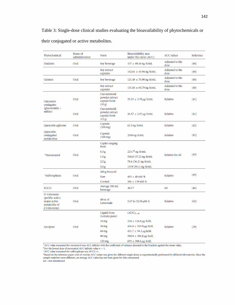
142
Table 3: Single-dose clinical studies evaluating the bioavailability of phytochemicals or
their conjugated or active metabolites.

143
Of the predominant high-risk cancers, genistein appears to have a greater affect
on prostate cancers [52–54]. Genistein mediates the apoptosis of cancer cells by
activating and/or inhibiting genes and/or enzymes germane to tumor maintenance (Figure
1, Table 4). Some of these important mechanisms are the inhibition of the activity
of tyrosine kinase, nuclear factor kappa B (NF-κB), and vitamin D 24-hydroxylase [86],
activation of tumor suppressor genes, and modulation of androgen-responsive gene
expression, prostate-specific antigen (PSA), and the androgen receptor (Table 4).
Of the prominent isoflavonones in soy, diadzein is less effective in its action on prostate
cancer, but, unlike genistein, it is metabolized to equol, an isoflavandiol which has a
longer half-life than genistein [87]. The longer half- life of equol creates the possibility of
using this chemical in combination with other available nutraceuticals, where the net
effect may be synergistic. However, prior preclinical tests are required to investigate this
possibility.
Other dietary compounds are also of great interest in this regard. In vitro, vitamin
D (Vit D) has potent tumor prevention ability and can induce differentiation and
apoptosis in some of the most predominant cancers [48]. The use of nutrients as a
possible treatment approach is based on the fact that chemicals occurring naturally will
minimize side effects when applied to a biosystem. However, the in vitro dose at which
Vit D induces its antitumor properties causes hypercalcemic conditions that can preclude
treatment in patients [49]. In prostate cancer, a leading cause of cancer deaths in the
western parts of the world, androgen ablation therapy is the choice of treatment.

144
Table 4: Assessment of the chemotherapeutic and chemopreventive effects of
nutraceuticals in combination studies.

145
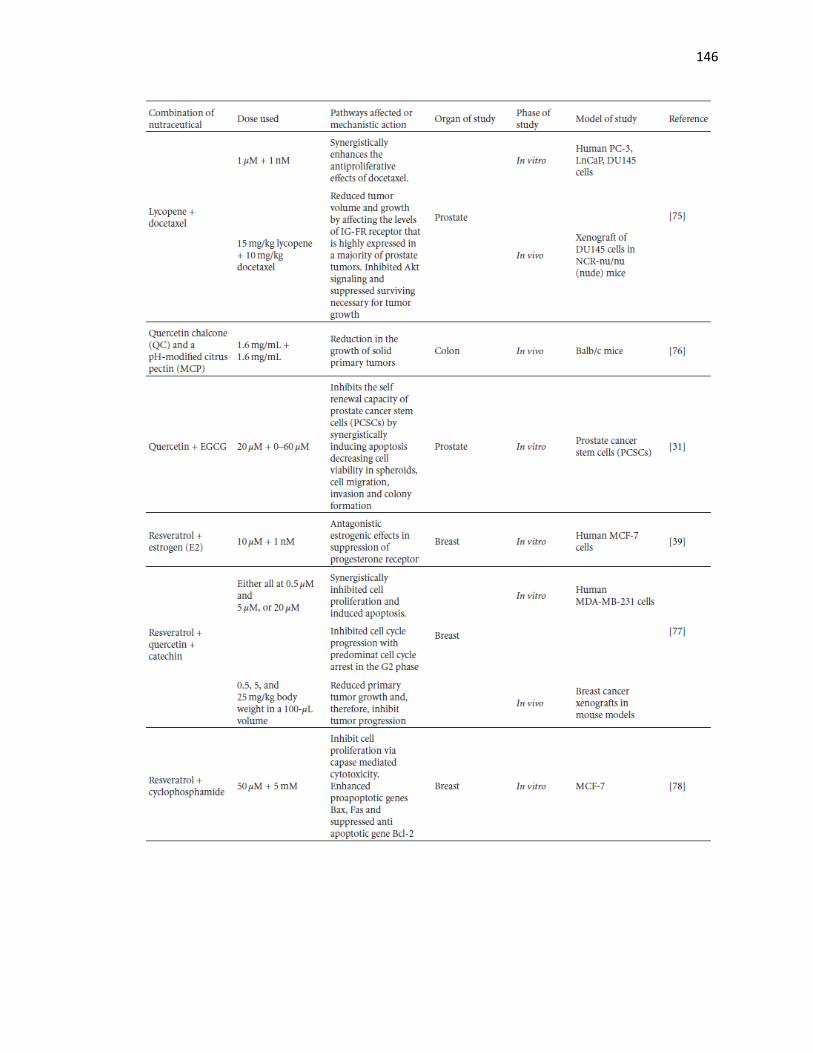
146

147

148
However, as the cancer becomes aggressive, hormone ablation therapy fails, and
progression ensues via androgen-independent pathways. Therefore, alternate therapies are
very much in demand. Vitamin D is an alternate form of treatment in prostate cancer
(PCA) and is shown to induce apoptosis in PCA cells in vitro. However, all PCA cell
lines in vitro are not equally receptive to the vitamin D treatment or genistein [88]. Cell
lines such as DU145 prostate cancer cells are especially more resistant as they express
high levels of CYP24, an enzyme that catabolizes Vit D3 into less active metabolites
[88]. To circumvent this problem, a recent study showed that a dual combination therapy,
of DU145 to genistein and Vit D3, increased the sensitivity of the cells to Vit D3 by
decreasing CYP24 expression. What is interesting to note is that the combination
approach not only lowered the effective dose, but was able to abrogate cell proliferation
as well. This lowered concentration of genistein at 100 nM is achieveable in vivo through
dietary sources, and clinical studies would be required to determine the localization of
genistein and Vit D3 in prostatic tissues.
An in vivo study for colorectal cancer has demonstrated a similar effect [89], but
in this case the mice were given a single gavage of 250 μg of genistein. This mode of
nutrient administration is useful for a preclinical test and probably has applications as a
chemopreventive supplement. However, in terms of a clinical setting, patients are often

149
exposed to a host of other nutrients or isoflavones in their diet, and; therefore, an in vivo
model replicating such an environment with various percentages of isoflavones will allow
for a better understanding of concentration and bioavailability of genistein that can
mediate an apoptotic effect and reduce CYP24 expression in colonic tissues in the
presence of vitamin D.
The antimetastatic properties of genistein are mediated by altering the expression
of NF-κB, and inhibiting the tyrosine kinase enzyme [17, 90]. Non-small-cell lung cancer
(NSCLC) is a highly aggressive form of lung cancer with a poor prognosis. Therefore,
alternate approaches that drastically reduce tumor growth are of utmost importance.
Activation of epidermal growth factor receptor tyrosine kinase (EGFR-TK) enhances the
cell signaling pathways allowing tumor growth. The use of drugs that inhibit EGFR-TK
and affect NF-κB, a gene whose transcribed products are essential for invasion and
metastasis, can induce a more aggressive approach of reducing tumor size and the spread
of the disease. A clinical therapy should be aimed at reducing tumor growth and spread
by inhibiting mechanisms that contribute to the activation of metastasis. In NSCLC,
genistein remarkably enhances the effects of EGFR-TK inhibitors, such as erlotinib and
gefitinib, when used in combination with each of them, respectively. This effect was seen
to be mediated by a marked reduction in NF-κB and others, such as EGFR, pAkt, COX-2,
and PGE(2), essential for regulating genes that control division, proliferation and
metastasis [90]. A few studies have shown how a combined approach can lower the
effective dose concentration even of chemotherapeutic drugs, minimizing potential side
effects. A study conducted on breast and pancreatic cells showed that, when the cells
were primed with genistein, lower concentrations of the chemotherapeutic drugs were

150
needed to significantly bring about growth inhibition and apoptosis than with the drugs
alone. In addition, NF-κB was transcriptionally inhibited in the combined treatment [90].
From a number of investigations, a common thread of evidence seems to emerge
that considerable variation in the efficacy of bionutrients in cancer treatment exists and
differs even among the same cell lines tested. The reasons for this are manyfold (Table
5). Cell lines derived from the same tissue hypothetically should be sensitive to the same
dose or chemical class of the phytonutrients, but such is not always the case. Alternate
medicinal approaches have an important task to identify crucial factors that change the
sensitivity of the chemical and determine chemical modifications that would be necessary
to modulate more synchronized results across several cell lines expressing similar
genotypic and phenotypic signatures.
Table 5: Factors conducive to the anticarcinogenic efficacy of nutraceuticals
.
151
2.2. Epigallocatechin-3-gallate (EGCG): A Potent Flavanol. Of the major food-derived
phytochemical constituents that are extensively studied for their chemopreventive and
chemotherapeutic use, EGCG and genistein are by far the most investigated. EGCG has
been shown to have numerous anticancer properties which include antiangiogenic activity
by affecting the transcriptional expression of vascular endothelial growth factor (VEGF)
[91], inhibiting tumor initiation and promotion by inhibiting signal transduction pathways
via [phosphatidyloinositol 3-kinase-Akt kinase- NF-κB] [92–94], inhibiting EGFR [95],
inhibiting Her-2 receptor phophorylation in breast carcinoma cells that constitutively
expresses Her-2/neu receptor [95], inducing apoptosis in estrogen receptor-(ER-)
independent breast cancer cells [96], causing antimetastatic activity [97], inhibiting
proteasome formation [98], inhibiting glucose-regulated protein (GRP78) activity [99];
inhibiting insulin-like growth factor-I receptor (IGF-IR) [100], and preventing invasion
of tumors by inducing HMG-box transcription factor 1 (HBP1) transcriptional repressor,
an inhibitor of the Wnt signaling pathway crucial for tumor-invasive property [101].
The serum level concentrations of EGCG are important to ensure that an effective
response is seen without adverse or even tumor-promoting functions. Studies have shown
that high doses of catechins that include a higher concentration of two prominent
compounds, epicatechin gallate (ECG) and EGCG, induce hypoxia-inducible factor 1
which is responsible for activating genes related to hypoxia conditions. This allows tumor
cell proliferation through alternate survival pathway mechanisms [102]. Most breast
cancers are ER dependent; however, for breast cancers and others that are ER
independent, EGCG inhibits the growth of tumor cells through the process of apoptosis
[96, 103]. As seen in MDA-MB-468 ER-negative cells, cellular apoptosis is mediated by

152
inducing p53 and Bax proteins that enhance apoptotic functions in cells [96]. Such
observations have been corroborated by in vivo studies using animal models [97].
Most studies have shown that anticancer properties of EGCG are mediated at
higher doses. However, such doses may be irrelevant to clinical applications as they may
be physiologically unachievable through dietary consumption. Therefore, clinical trials
should be aimed at achieving desired anticancer preventive or tumor functions at much
lowered doses. Such outcomes are possible with a dual-drug approach. One study [95]
demonstrated that combining EGCG with the drug taxol, which is commonly used to treat
breast carcinomas, lowered the effective dose of EGCG, ranging from 0.1–1.0 μg/mL
which is a serum obtainable level through metabolism. This same group showed that
higher doses (30–40 μg) of EGCG were required to mediate a similar effect when used
alone [95].
EGCG can be exploited as a chemopreventive agent if it prevents cancerous
lesions from occurring at lower dose concentrations and for prolonged periods of time.
Most in vitro studies have used relatively high doses of EGCG and such doses may prove
to be more tumor promotive than preventive in longer exposure time periods. In a study
designed by Pianetti et al. [92], contradictory results on the effects of EGCG on Her-
2/neu overexpressed receptor in NF639 breast cancer cells was observed. At short
exposure times, EGCG was very effective in reducing cell proliferation, but at prolonged
exposure cells became resistant to EGCG with increased levels of NF-κB. This observed
change in drug-induced resistance was related to the activation of mitogen-activated
protein kinase. It appears that single doses or one specific chemical constituent is mostly
insufficient to induce tumor suppression or regression. Such in vitro data outcomes
153
emphasize that a dual-drug treatment approach is necessary to treat the disease. This also
signals that the timing of the nutradrug that is administered is important. Perhaps EGCG
should be administered early in treatment, but later other phytochemicals or drugs, in
conjunction with EGCG, may need to be administered in the treatment regimen. In their
dual-drug treatment of NF639 Her-2/neu breast cancer cells, Yang et al. found that
treating the cells initially with EGCG lowered cell proliferation and the later introduction
of the MAPK inhibitor, U0126, reduced invasive phenotype [93].
Most studies determining the anticancer drug properties of EGCG are preclinical.
For better understanding of specific EGCG effects, clinical trials should be carefully
designed to include parameters that influence EGCG effectiveness. EGCG has different
roles in ER-dependent versus ER-independent receptors, and, therefore, the type of diet
needed to emulate in vitro doses need to be clearly understood through clinical trials and
careful pharmacokinetic studies of these doses in healthy individuals, ER-positive breast
cancer patients, and ER-independent tumors.
In testing phytochemicals of the same or different class it is rather uncertain
which markers are necessary to determine comparable dosage values for in vitro versus in
vivo efficacies. Formulation of a diet is one of the major deciding factors in the functional
efficacy of a chemical constituent. It defines the concentration of the dose that will be
available in vivo, after metabolism, and determines the diet that needs to be given to
achieve such an outcome. Even though single-dose individual or mixed phytochemical
treatments are currently available to cancer patients, they are relatively new and much
more research in this direction is warranted. One such therapy that is rapidly gaining

154
importance and holds promise for future cancer treatments is combination therapies using
plant-based chemical compounds known as nutraceuticals.
3. Combinatorial Therapy: A Promise of the Future (See Table 3)
In prevention or treatment, combinatorial approaches can be of the following types: a
phytonutrient and an effective drug, two or more phytonutrients, a synthetic phytonutrient
and an effective drug, or a synthetic phytonutrient and a natural nutrient. Studies in the
last few decades have focused attention on unraveling the protective properties and
mechanistic actions of many phytochemicals. Still the pharmacokinetics of quite a few of
these phytochemicals are not known, and, for a few that are known, there is much
variability based on mode and form of delivery, dose, and the model organism of study
(Tables 2, 3, and 4). Another interesting approach to enhancing curative and preventive
properties of these nutrients is combination therapies. The therapy is based on the factual
information available at hand and using the potent properties of one with that of another
to enhance synergistic or additive actions (Figure 1). In this paper, groups that have
worked with different phytomolecules belonging to a different or the same chemical class
of compounds have been analyzed for their antitumorigenic activities, and the overall
results of the experiments for each group are described in Table 4.
3.1. Curcumin and Taxol (See [63]). Primary breast cancer cells are commonly treated
with the drug taxol. Sustained chemotherapeutic treatment with this drug has often
resulted in drug resistance and tumor progression. Many chemotherapeutic drugs induce
the expression of the metastatic gene NF-κB which encourages tumor progression.

155
Interestingly, natural-based compounds that are pharmacologically safe have been shown
to inactivate NF-κB expression. Taxol is a powerful drug in the treatment of cancer
therefore, in order to prevent metastasis, a combination of Taxol with curcumin has been
shown to downregulate the expression of NF-κB and induce apoptosis.
3.2. Curcumin and Xanthorrhizol (See [64]). A study conducted on an invasive breast
tumor cell line, MDA-MB-231, has shown how and when compounds added to the cells
determine the overall efficacy of the treatment. A sequential addition of curcumin and
xanthorrhizol (a rhizomal sesquiterpenoid of Curcuma xanthorrhiza) in culture resulted in
additive and antagonistic effects depending on which compound was added first to the
culture. However, simultaneous addition of the compounds resulted in synergistic effects
at lower concentrations and agonistic effects at higher concentrations. Such experiments
provide evidence that the efficacy of a drug is dependent on dose, time, and how it is
presented to the cells. Therefore, results obtained might be contradictory if doses used are
simply antagonist or additive. For a successful combination therapy or prevention,
synergistic doses are more relevant to mediate downstream effects, as lower
concentrations of the test biomolecules will be required.
3.3. Curcumin and Docosahexaenoic Acid (DHA) (See [65]). DHA is a dietary
compound present in fish oil that has been shown to have potent chemopreventive affects
against cancer. Chemotherapeutic effects of compounds are often analyzed using in vitro
models. However, what is most often observed is that all cells from the same tissue
sample do not react the same way to the test compound. It is essential to have a

156
chemopreventive or therapeutic agent that can induce its effects on a wide range of
cancerous cells arising from the same tissue. In this study, the authors analyzed five cell
lines expressing different cell surface receptors (Table 6) which make them susceptible to
chemotherapeutic compounds but in different ways and to different degrees. The
combinatorial synergistic doses for each cell line were different, as shown in the Table 4.
In particular, one breast cancer cell line, SK-BR-3, which is ER-negative exhibited a
higher uptake of curcumin in the presence of DHA. DHA does not directly contribute to
cell inhibition, but the combination of this compound with curcumin greatly enhances the
uptake of curcumin by the cells. This compound, DHA, can reach a plasma concentration
level of 200 μM. Although the focus of this study was entirely based on the SK-BR-3 cell
line, the effects of reduced synergy on other cell lines in terms of transcriptome effects
need to be investigated. Mammary tumors may contain a heterogenous population of
cells exhibiting different surface receptors. Using combination therapy should be aimed
at reducing the populations of all these cell types within the tumor site to truly exhibit
antitumor potency with minimal side effects.
Table 6: Surface receptors expressed by breast cancer cells that alter their sensitivity to
treatment.

157
3.4. Curcumin and Genistein (See [66]): A Preventive Strategy.
The aim to use natural compounds in diets is to render the chemopreventive properties of
the compounds to the tissues. Numerous studies have shown that single dosage of
compounds used alone is effective for chemoprevention. The problem faced is the
inability to achieve high serum concentrations in vivo. Although combination studies are
just beginning to surface as more prominent approaches in clinical treatment, studies,
though limited, have shown that synergistic effects of the compounds are able to be
achieved at much lower doses than when compounds are used alone. Especially in
cancers that are hormonally regulated, the tissues are often exposed to external or internal
hormonal stimulation, like estrogen, as in the case of breast tissue. Environmental agents
that mimic estrogen-like activity can often stimulate or initiate the carcinogenic process.
Curcumin, a curcuminoid, and genistein, an isoflavone, are derived from two different
chemical classes, yet they have been known to inhibit a variety of tumor types in vitro
and in vivo. Clinical trials of these compounds individually have been tested [19, 60, 104,
105]. The mechanistic action of the individual compounds in many different cancers has
been investigated as well. However, using these compounds in combination drastically
affects the development of tumors by mediating changes in shape and growth inhibition.
Such changes were observed both in ER-positive and ER-negative cells, indicative of the
dual use of such a combination in prevention and therapy.
3.5. Curcumin and Sulfinosine (SF) (See [67]). The ineffectiveness of certain drugs in
prolonged chemotherapy stems from the resistance that some cancers develop with time.
This is one of the major obstacles in cancer therapy, especially in cancers that are

158
multidrug resistant (MDR). The problem in finding a suitable cure for non-small-cell
lung cancers is the MDR phenotype it exhibits. Treating MDR cells such as non-small-
cell lung carcinoma NCI-H460/R cells with a commonly employed drug, SF (obtained by
the amination and subsequent oxidation of 6-thioguanosine), in cell cultures has been
shown to inhibit cell growth. This observed cytotoxicity is enhanced several folds when
low doses of the natural compound, curcumin, are used in combination, which are
otherwise ineffective unless very high concentrations are used. These compounds
mediate a synergistic effect in regulating the cell cycle phases and downregulate MDR
genes, thereby, enhancing tumor regression phenotypes even in the presence of mutated
p53 molecules.
3.6. Curcumin and Celecoxib (See [68]) Cyclooxygenase-2 (COX-2) expression is central
to the carcinogenesis of colorectal cancers. Compounds that regulate the expression or
activity of COX-2 in cells may be instrumental in mediating chemotherapeutic effects on
the tissue or cells. Celecoxib is a potent inhibitor of COX-2 and is presumed to target its
active site. However, prolonged exposure to celecoxib results in cardiovascular problems.
It appears that monotherapy regimes are very effective in inhibiting cancer growth,
proliferation, metastasis, and invasion, as seen in numerous in vitro and in vivo models.
However, prolonged exposures at concentrations relatively higher than what can be
achieved with combination doses may result in unwanted side effects. Testing the
efficacy of celecoxib with cucumin showed that at lower doses of celecoxib it was
possible to enforce synergistic inhibitory growth effects on colon cells which expressed
various levels of COX-2. Like many other in vitro investigations, this study emphasizes

159
the fact that combining powerful drugs with naturally available potent compounds can
reduce the dose needed to mediate potent anticarcinogenic effects with minimal side
effects. Clinically, such studies are relevant as the doses used or needed are within the
physiologically dose range. With colon cancer having such a high incidence rate in the
western populations, such therapies can be taken as advantage, and biomolecules having
preventive potential against the formation of precancerous lesions need to be
supplemented in diets of patients at high-risk.
3.7. Coltect and 5-Aminosalicylic Acid (5-ASA) (See [69]). Coltect is a novel
chemotherapeutic dietary drug with a formulation of curcumin, a turmeric extract (95%
curcuminoids) mixed with turmeric powder 1 : 1, green tea (60% polyphenols and 25%
EGCG) in a 2 : 1 ratio, and 0.1 mg/mL of L-selenomethionine. 5-ASA is an anti-
inflammatory drug, which has been shown to have a preventive role in polyp formation
that is thought to occur via the inflammation process in conditions like inflammatory
bowl disorder. Coltect has been effective against HT-29 human colon adenocarcinoma
grade II cells in vitro, and this nutraceutical complex in combination with 5-ASA has
been shown to inhibit the formation or growth of chemically induced aberrant crypt foci
(ACF) in rat models. The molecular mechanism by which this inhibition is mediated is
via the inhibition of COX-2 pathways in HT-29 cells, which has been supported by in
vitro studies of other groups [106, 107]. However, growth inhibition can be affected via
COX-2-independent pathways possibly through mechanisms that are regulated by the
functional polyphenol complex in coltect. Such complex mixtures are of clinical
significance as many different control mechanisms can be regulated by the presence of

160
individual constituents of the polyphenols which are a part of the formulated mixture of
coltect.
3.8. Phenylethylisothiocynate (PEITC) and Curcumin (See [27]). Most prostate cancers
begin as a hormone-dependent tumor, and the hormone is primarily androgen. However,
the more aggressive forms of prostate cancer are androgen-independent and hormonal
therapies fail to be effective. Alternate therapies are, therefore, necessary to treat such
aggressive forms. Most cancerous cells express various surface receptors that propagate
cellular growth. Targeting such receptors can be an effective chemotherapeutic approach.
Curcumin, obtained from Curcumin longa, has been shown to inhibit the phosphorylation
of EGFR, inhibit the Akt signaling pathway, and negatively regulate NF-κB. It is an
effective molecule against prostate cancer. Phenylethylisothiocyanate, a phytochemical in
cruciferous vegetables, has been shown to inhibit prostate cancer cell growth in vitro and
this observation has been supported by epidemiological studies showing that
consumption of cruciferous vegetables has an inverse effect on prostate cancer risk.
When two bioactive molecules with similar effects are used in treating hormonally
independent tumors in affecting receptor mediated signaling, the effects are more
pronounced than when used as individual compounds. With PEITC and curcumin, the
observed effect was more additive than synergistic, but cell growth inhibition was
profoundly affected by the inhibition of NF-κB pathways and Akt signaling pathways.
Such responses were seen at lower physiological achievable doses. These results were
corroborated by in vivo studies in mice using human PC-3 prostate cancer cells [70].

161
Since EGCG has similar effects on prostate cancer cells, EGCG could also possibly serve
as a substitute in place of curcumin for such a treatment strategy.
4. D-Limonene and Its Combination Therapies (See Table 3)
Although a few studies have shown that D-limonene, an abundant monoterpene in citrus
oils, exhibits antimitogenic activity, its alcohol-derivated perillyl alchohol (PA) has a
greater inhibitory effect on cell migration in cancerous cells [108]. A study by Reddy et
al. [108] used subtoxic doses of PA to determine this effect. Further preclinical studies
are necessary to determine the effective yet nontoxic serum/tissue concentration that can
be achieved from a diet rich in citrus intake, in conjunction with phytonutrients of the
same class or a different class. Not much is known about the percentage composition of
D-limonene and its metabolized constituents that are required to achieve an effective
monterpene anticarcinogenic activity. In comparison to its oxygenated derivatives,
limonene has the least cytotoxic effect on both noncancerous and cancerous breast cell
lines and, therefore, can be applicable in chemoprevention [109]. D-limonene appears to
be more effective against chemically induced colonic crypt foci [110]. These foci are
preneoplastic lesions and are biomarkers for the progression into colon cancer. In colonic
crypts that are chemically induced, limonene asserts its effect by inhibiting the activity of
ornithine decarboxylase, an enzyme essential for the polyamine biosynthesis pathway.
This pathway regulates the cell cycle, and D-limonene-dependent inhibition of ornithine
decarboxylase (ODC) encourages an antiproliferative activity. If aberrant crypt foci are
the initial markers for colon carcinogenesis, and D-Limonene and its derivatives assert
their roles against initiation and promotion phases of cancer, then a diet rich in citrus

162
foods can prevent crypt formation. Therefore, D-limonene appears to have potential as a
chemopreventive agent in colon carcinogenesis. However, in vivo studies often do not
correlate with results in vitro for many of the reasons discussed earlier. Once the intake of
a compound is deemed safe for human consumption, it is imperative to analyze and study
the mechanistic and metabolic functions in human subjects to determine the efficacy of
the nutrient in question. As in the case of understanding limonene protection against
colonic carcinogenesis, the studies were performed on rats and for shorter exposure time
to the compound or its derivatives. Therefore, further in vivo models are required to
determine the toxicity of the treatment for longer periods of time, as D-limonene is
nontoxic but its alcohol derivatives could be toxic.
4.1. D-Limonene and Docetaxcel (See [72]). Many combination studies are underway to
determine an effective approach in treating advanced and aggressive prostate cancers.
Docetaxel, a synthetic derivative of taxol, is primarily used to inhibit the microtubular
structures in cancerous cells that support cell division. In addition to its role as a
microtubule disruptive molecule, it has a host of inhibitory actions on genes which
regulate cell proliferation, mitotic spindle formation, transcription factors, and
oncogenesis. It also upregulates genes involved in apoptosis and cell cycle progression in
prostate cancer. D-Limonene, discussed earlier has been shown to have anti-prostate
carcinogenic effects at low dose concentrations. Logically; therefore, combining the two
compounds may have a plethora of positive antitumor functionalities. In a study by Rabi
and Bishayee [72], the combined treatment enhanced the sensitivity of DU145 prostate
cancer cells that are known to be apoptotic resistant. This enhanced sensitivity was
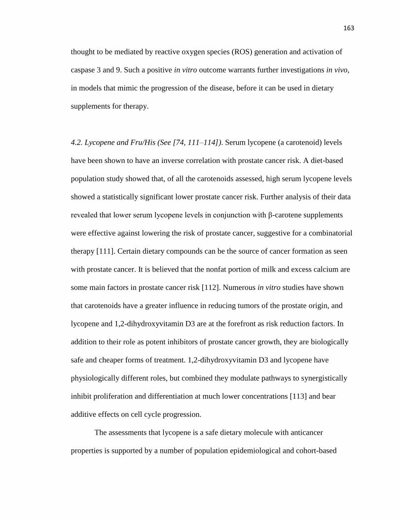
163
thought to be mediated by reactive oxygen species (ROS) generation and activation of
caspase 3 and 9. Such a positive in vitro outcome warrants further investigations in vivo,
in models that mimic the progression of the disease, before it can be used in dietary
supplements for therapy.
4.2. Lycopene and Fru/His (See [74, 111–114]). Serum lycopene (a carotenoid) levels
have been shown to have an inverse correlation with prostate cancer risk. A diet-based
population study showed that, of all the carotenoids assessed, high serum lycopene levels
showed a statistically significant lower prostate cancer risk. Further analysis of their data
revealed that lower serum lycopene levels in conjunction with β-carotene supplements
were effective against lowering the risk of prostate cancer, suggestive for a combinatorial
therapy [111]. Certain dietary compounds can be the source of cancer formation as seen
with prostate cancer. It is believed that the nonfat portion of milk and excess calcium are
some main factors in prostate cancer risk [112]. Numerous in vitro studies have shown
that carotenoids have a greater influence in reducing tumors of the prostate origin, and
lycopene and 1,2-dihydroxyvitamin D3 are at the forefront as risk reduction factors. In
addition to their role as potent inhibitors of prostate cancer growth, they are biologically
safe and cheaper forms of treatment. 1,2-dihydroxyvitamin D3 and lycopene have
physiologically different roles, but combined they modulate pathways to synergistically
inhibit proliferation and differentiation at much lower concentrations [113] and bear
additive effects on cell cycle progression.
The assessments that lycopene is a safe dietary molecule with anticancer
properties is supported by a number of population epidemiological and cohort-based

164
studies [112]. However, it is important to ensure that the statistical models used are able
to adjust for many parameters for a true significant outcome. Regardless of the statistical
model employed in these assessment studies, lycopene has emerged as a potent risk-
reducing factor of prostate cancer and has been even supported by a study that was
carried out across 28 countries. Intervention combination studies have not yet been
performed. However, in vivo-based studies in mice models have shown that lycopene
administered in the form of tomato powder and broccoli powder in a 10 : 10 ratio,
increases its serum concentration to about 538 nM/g with about 0.4 nM/g concentrated in
the prostate tissue itself. Diet-based intervention studies are required to determine the
formulated diet required to improve the availability in the serum of patients and enhance
the localized concentration of the molecule in the tissue. Such a diet-based treatment may
serve as a suitable chemopreventive approach against prostate cancer or with patients at
high-risk of the disease. Even though bioactive molecules successfully work in
administering their protective functions in vitro, it appears through in vivo studies that
diet and availability crucially dictate outcomes. A critical question to be asked is what
factors constitute a perfect blend of bioactive mixtures. With the current research thus far,
it is hard to address what the cut-off ratios are that need to be used in a diet that contain
mixtures of potent nutraceuticals to coordinate similar effects clinically. Possibly a slight
change in concentration of even one of the effective biomolecules may render the mixture
ineffective in its function. It is rather an important task for pharmaceutical chemists and
nutritionists to determine the ratios of effective biomolecules in a mixture and determine
the pharmacokinetics and dynamics of that mixture.

165
Fru/His, a ketosamine, is also a derived product from tomatoes obtained by the
reaction of a carbohydrate with an amino acid. This particular ketosamine has been found
to assert chemopreventive effects by synergistically enhancing the activity of lycopene,
by neutralizing ROS species and inhibiting DNA damage. Therefore, the complex of
these two molecules may have a pivotal role in prostate cancer prevention. Although a rat
model was used to determine the results of the treatment and pharmacokinetics of the
compound are still not known, the combination of the two seemed to preferentially
localize in the prostate more than in other tissues that were tested [74].
Occasionally, a combination may fail to incite anticarcinogenic effects as was
seen by Mossine et al. [114]. Their experiments were conducted on the prostate
adenocarcinoma rat model that was used by other groups, and their data had contradictory
results to the effective action of lycopene itself and in conjunction with other
micronutrients. Their study revealed that lycopene was not able to inhibit or reduce tumor
load alone or in combination and that selenium alone in the mixture was able to induce
antitumorigenic effects. Such outcomes are important as they open up more questions as
to why a molecule that affects a given pathway behaves differently when tested within
the same experimental model. Is it always dose or concentration or does molecule
preparation and delivery impart effects on the efficacy of a drug?
4.3. Lycopene and Docetaxel (See [75]). Docetaxel is a potent chemotherapeutic drug
that is clinically used to treat patients with advanced metastatic prostate cancers.
Although the drug extends survival, it is for a very limited time period and with a poor
prognosis. Lycopene, a natural compound, has been shown to have strong cancer
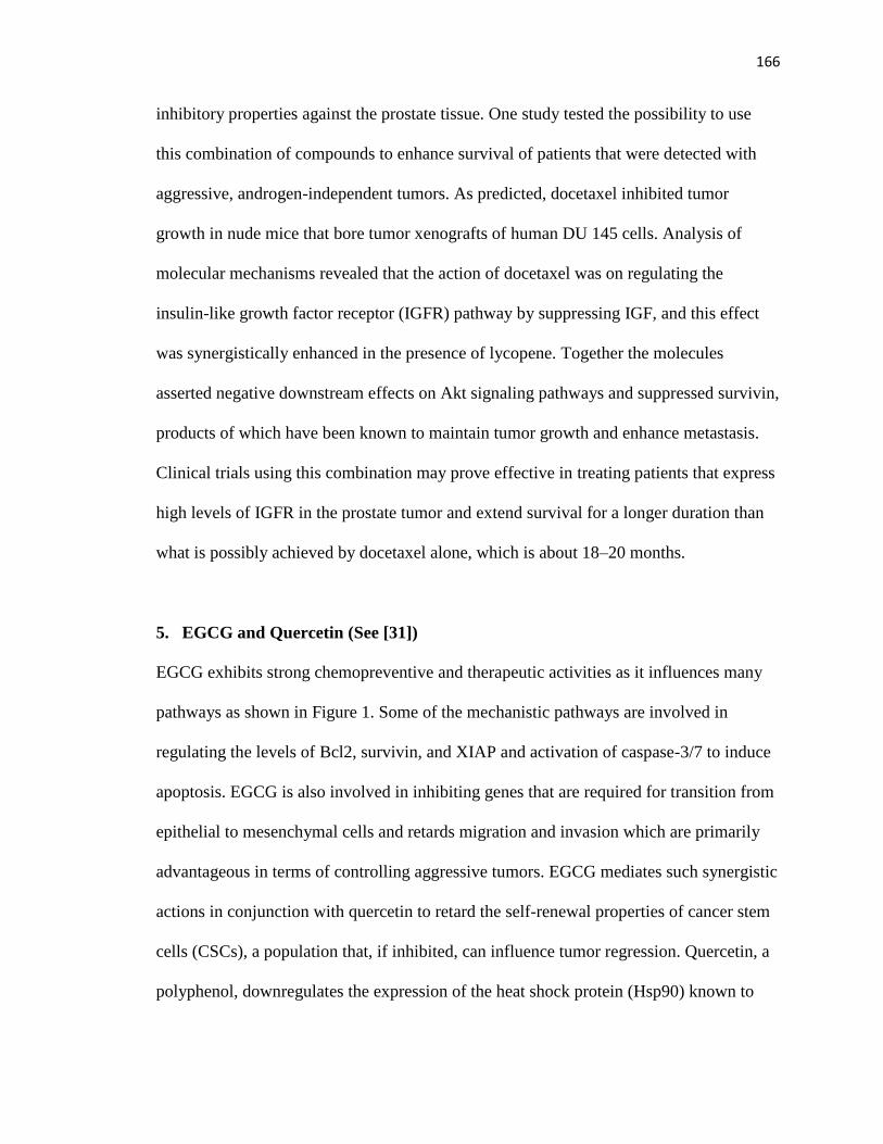
166
inhibitory properties against the prostate tissue. One study tested the possibility to use
this combination of compounds to enhance survival of patients that were detected with
aggressive, androgen-independent tumors. As predicted, docetaxel inhibited tumor
growth in nude mice that bore tumor xenografts of human DU 145 cells. Analysis of
molecular mechanisms revealed that the action of docetaxel was on regulating the
insulin-like growth factor receptor (IGFR) pathway by suppressing IGF, and this effect
was synergistically enhanced in the presence of lycopene. Together the molecules
asserted negative downstream effects on Akt signaling pathways and suppressed survivin,
products of which have been known to maintain tumor growth and enhance metastasis.
Clinical trials using this combination may prove effective in treating patients that express
high levels of IGFR in the prostate tumor and extend survival for a longer duration than
what is possibly achieved by docetaxel alone, which is about 18–20 months.
5. EGCG and Quercetin (See [31])
EGCG exhibits strong chemopreventive and therapeutic activities as it influences many
pathways as shown in Figure 1. Some of the mechanistic pathways are involved in
regulating the levels of Bcl2, survivin, and XIAP and activation of caspase-3/7 to induce
apoptosis. EGCG is also involved in inhibiting genes that are required for transition from
epithelial to mesenchymal cells and retards migration and invasion which are primarily
advantageous in terms of controlling aggressive tumors. EGCG mediates such synergistic
actions in conjunction with quercetin to retard the self-renewal properties of cancer stem
cells (CSCs), a population that, if inhibited, can influence tumor regression. Quercetin, a
polyphenol, downregulates the expression of the heat shock protein (Hsp90) known to

167
influence apoptosis and growth inhibition of prostate tumors. Therefore, the combination
of these molecules modulates their respective therapeutic effects to mediate synergistic
growth retardation of CSCs. The study by Tang et al. [31] used relatively higher
concentrations of EGCG (60 μM) in the presence of 20 μM quercetin. Probably
concentrations of EGCG that can mediate similar synergistic levels, albeit at lower doses,
need to be investigated, and the therapeutic potential across cancer stems cells of other
origins need to be assessed if clinical applications are to be considered.
6. Resveratrol and Estrogen (See [39])
Selective estrogen receptor modulators that are used in the clinical treatment of breast
cancers display dual agonist/antagonist effects in the tissues, especially in cancer
initiation and progression. Drugs like tamoxifen emulate antagonistic effects on estrogen
to contain the tumor. Agonistic-estrogen-like activity can in some instances enhance
tumor progression which is not desired in most clinical treatments. Resveratrol, a
polyphenolic compound abundant in grape skin and grape products including wine, is
known to have chemopreventive properties as supported by numerous in vitro and in vivo
studies. However, based on the experimental cell type, resveratrol induces either
agonistic or antagonistic effects that can be weak or very pronounced. Resveratrol
agonistic effects are totally reversed in the presence of estrogen, possibly mediated
through estrogen receptor β. This reversal of effects is pertinent to prevention of breast
cancer lesions in ducts that could become long-term neoplastic and cancerous. Of its
many cancer protective functions, resveratrol in combination with glucan are potent
immunomodulators by upregulating Cdc42 expression [115]. When natural compounds

168
exhibit multi-chemopreventive properties, conjugation therapies are advantageous over
monotherapies. Albeit not clinically tested, harnessing cancer preventive and immune
modulating functions of nutraceuticals seems to be a plausible approach to targeting
hormonally independent aggressive tumors.
6.1. Resveratrol, Quercetin and Catechin (See Table 4 and [77]). The protective
functions of polyphenols are manyfold. Numerous studies have analyzed their protective
and therapeutic functions in vitro on tumor initiation that was chemically induced or in
vivo via cellular implanted tumor formation. Few studies have established the functions
of combined polyphenols on established tumors, as the majority of investigations have
focused on individual mechanistic effects of the compounds. Dietary serum
concentrations are influenced by the individual percentage of biomolecules present in the
diet. Therefore, individual protective assessments of a compound show higher dose
requirements, whilst mixtures may require lower doses to achieve the same effects.
Additive and synergistic effects of compounds occur if their individual functions are
enhanced in the presence of other molecules, perhaps by reinforcing the serum stability
and availability of the various compounds in the mixture. Such observations were seen in
both in vitro and in vivo testing of a mixture of three polyphenols, resveratrol, quercetin,
and catechin, albeit pharmacokinetics studies are warranted.
6.2. Resveratrol and Cyclophosphamide (See [78]). Cyclophosphamide, a neoplastic
drug, has a broad spectrum of activity on a variety of cancers, including breast cancers.
The shortfall of the drug is its myriad of toxic effects on other systems. Dose reduction of

169
the compound would be a means of reducing its toxicity without compensating its
anticarcinogenic activity. Resveratrol has been shown to successfully lower the effective
dose of cyclophosphamide without altering its anticarcinogenic activity. Both of the
compounds together synergistically enhance caspase-mediated cytotoxic activity, as
demonstrated in MCF-7 cells, an aggressive breast cancer cell line (Table 4). The
combination therapy resulted in the upregulation of p53, proapoptotic genes, Bax and
Fas, and downregulation of antiapoptotic gene Bcl-2, suggestive of an apoptotic
mechanism involved in cell death.
6.3. Resveratrol and n-Butyrate (See [79]). n-Butyrate is a short chain fatty acid
produced by bacterial fermentation of fiber in the colon. The compound is a known
differentiating agent and induces an epithelial phenotype in certain cultured cells. n-
butyrate is a potent histone deacetylase (HDAC) inhibitor as well and one of its
differentiation-inducing properties stems from its ability to inhibit HDACs. Resveratrol,
discussed above, induces apoptosis through other mechanistic pathways. The
combination of two bioactive molecules influencing apoptosis via different mechanistic
pathways may associate to render an apoptotic phenotype in cancerous cells and inhibit
tumor formation and progression. The 2 mM dose of n-butyrate used in the Wolter and
Stein study [79] is probably much higher than what can be physiologically achieved. This
dose is probably suitable for treatment of colon cancers where higher molar doses of n-
butyrate are possible. However, n-butyrate is highly unstable, and its serum
concentrations are lower than 2 mM. Since this molecule is a differentiating agent, its
clinical use in treatment of other cancers is relevant. However, such therapies require

170
absolute lower effective doses and can probably be achieved by combining with
molecules other than resveratrol or modifying the compound to specific conjugates to
reach serum concentration levels.
6.4. Resveratrol and 5-Fluorouracil (5-FU) (See [80]). 5-fluorouracil inhibits
thymidylate synthase, prevents DNA proliferation, and induces DNA damage-related
apoptosis in colon cancer cells. Phase I clinical trials using a combination of resveratrol
and grape powder have shown that resveratrol at low doses inhibit Wnt, a gene that is
upregulated in colon cancers. Taking advantage of therapeutic effects of nutraceuticals,
combined therapy of aforementioned resveratrol with 5-FU surfaces as a principal
strategy in treating colon cancers. When used in combination, the presumption is that
either additive or synergistic effects of the two could mediate tumor inhibition by
modulating their individual apoptotic effects. The concern in using resveratrol is that
higher concentrations of the doses are required in the treatment which clinically may not
be reached through dietary consumption.
6.5. Genistein and Resveratrol (See [81]). Genistein and resveratrol as individual
phytochemicals are very effective in the treatment and prevention of prostate cancer
progression in rodent studies. Poorly differentiated prostate cancers often fail to respond
to androgen-dependent treatments, and alternate treatments are required. Androgen
receptors likewise have two functional roles, one as a tumor suppressor in normal
prostate tissue and the other as an oncogene in neoplastic transformation, where it is
altered either by mutations or DNA damage. Genistein and resveratrol used in an in vivo

171
rat-based study, modeled to understand the mechanistic action of combined treatments in
the progression of prostate cancer, showed that they had more pronounced effects, albeit
not synergistic. The statistically significant additive functions of reducing cell
proliferation through mechanisms that regulate the androgen receptor levels and IGF-1, a
biomarker found in patient serums with progressive and aggressive prostate cancers were
achieved in combined therapies over the monotherapy regimes. Interestingly, the
combination of genistein and resveratrol increased serum availability of both, but higher
concentrations of resveratrol were achievable as compared to the single-dose regimen.
Perhaps, absorption and stability of resveratrol were profoundly affected in a combined
environment, which is clinically a clear advantage. The doses used in the study are
physiologically safe and achievable in vivo by consumption of a soy-based diet high in
the percentage of genistein. However, resveratrol is found in low levels in grape-based
dietary products, and, therefore; a pure supplement of the compound is necessary in case
that higher doses are required.
7. Genistein and Sulforaphane (SFN) (See [82]). Previous studies have shown that
EGCG, a major polyphenol in green tea, can inhibit tumor growth through mechanisms
that alter DNA methylation activity, reversing the expression of silenced genes involved
in tumor inhibition in cancer cells. Hypomethylation of the promoters that are CpG-rich
is more likely to be transcribed, with an exception of few like hTERT, the regulatory
gene of telomerase [82, 116–119]. Epigenetics is a mechanism that has been studied for
decades, and factors that regulate epigenetics are now believed to be very important as
treatment possibilities in controlling tumors. DNA methylation and histone deacetylation

172
are well known epigenetic mechanisms that regulate many of the genes involved in
cancers of various origins. Genistein combined with SFN, an histone deacetylase
inhibitor (HDACI) has been successful in inducing the transcription of genes involved in
regulating cell cycle by reversing the hypermethylated states of their promoters. This
change was observed at low doses and was enhanced in the presence of sulforaphane
more than that when genistein was used alone. However, in vivo studies of the same are
warranted to determine epigenetic behavior of the dietary compounds before applications
to human treatments are considered.
7.1. Benzylisothiocynate (BITC) and SFN (See [83]). BITC and sulforaphane are ITCs
derived from cruciferous vegetables like broccoli. Individually both these molecules exert
potent chemotherapeutic properties strongly supported by numerous studies. Oddly, even
though both are isothiocyanates, they exert their therapeutic effects by controlling
different pathways involved in tumorigenic inhibition. STAT3, a member of the STAT
group of transcriptional factors, is required for early development and is dispensable in
adult tissues. However, there appears to be a correlation between the constitutive
expression of STAT3 and tumor development, indicative of its role as an oncogene. This
gene appears to have important roles in cell proliferation, angiogenesis, and metastsis, a
crucial requirement of tumor survival. Both BITC and sulforaphane have cancer
inhibitory effects, affecting independent cell signaling pathways. However, the sequential
combination of the two has been shown to regulate the STAT3 gene and others (Table 4),
thereby, inducing apoptosis. How dietary molecules are presented to the cells in vitro is
important to its cellular mechanistic actions. In the study by Hutzen et al. [83], sequential
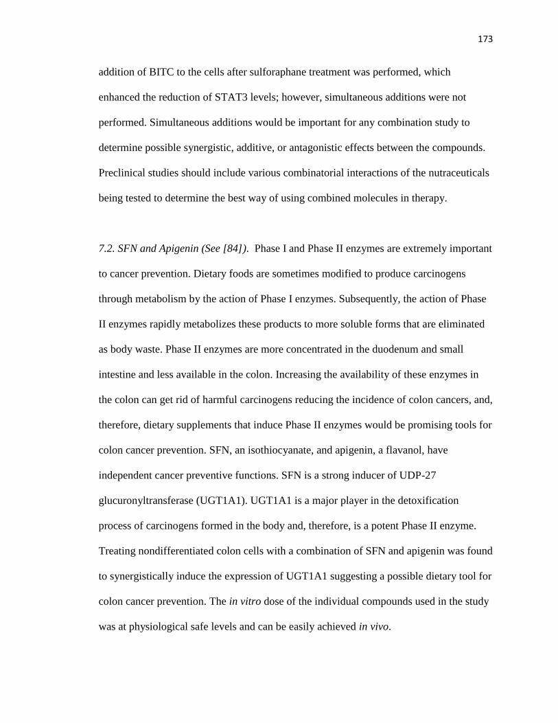
173
addition of BITC to the cells after sulforaphane treatment was performed, which
enhanced the reduction of STAT3 levels; however, simultaneous additions were not
performed. Simultaneous additions would be important for any combination study to
determine possible synergistic, additive, or antagonistic effects between the compounds.
Preclinical studies should include various combinatorial interactions of the nutraceuticals
being tested to determine the best way of using combined molecules in therapy.
7.2. SFN and Apigenin (See [84]). Phase I and Phase II enzymes are extremely important
to cancer prevention. Dietary foods are sometimes modified to produce carcinogens
through metabolism by the action of Phase I enzymes. Subsequently, the action of Phase
II enzymes rapidly metabolizes these products to more soluble forms that are eliminated
as body waste. Phase II enzymes are more concentrated in the duodenum and small
intestine and less available in the colon. Increasing the availability of these enzymes in
the colon can get rid of harmful carcinogens reducing the incidence of colon cancers, and,
therefore, dietary supplements that induce Phase II enzymes would be promising tools for
colon cancer prevention. SFN, an isothiocyanate, and apigenin, a flavanol, have
independent cancer preventive functions. SFN is a strong inducer of UDP-27
glucuronyltransferase (UGT1A1). UGT1A1 is a major player in the detoxification
process of carcinogens formed in the body and, therefore, is a potent Phase II enzyme.
Treating nondifferentiated colon cells with a combination of SFN and apigenin was found
to synergistically induce the expression of UGT1A1 suggesting a possible dietary tool for
colon cancer prevention. The in vitro dose of the individual compounds used in the study
was at physiological safe levels and can be easily achieved in vivo.

174
7.3. SFN and 3,3′-Diindolylmethane (DIM) (See [85]). The importance of investigating
the roles of dose combinations on chemopreventive or therapeutic functions has been
well dissected in a study by Pappa et al. [85]. Lower doses of SFN demonstrate
antagonistic effects on cell proliferation and higher doses of both compounds had
synergistic effects. Synergism of compounds is preferred if the outcome is tumor
regression, but in clinical treatments synergistic actions should be mediated at safe lower
concentrations rather than at cytotoxic levels. Presumably, the choice of compounds used,
based on the genetic or cellular function required, is imperative to the success of the
treatment. Possibly, SFN can synergistically inhibit the proliferation of cancer cells with
compounds other than DIM at much lower doses, which has been investigated in studies
using SFN with flavanols. This clearly highlights the problems of using combined
therapies, especially since dosage is of critical importance for the success of clinical
trials.
7.4. SFN and Dibenzoylmethane (DMB) (See [28]). When seeking for dietary molecules
with potential chemoprotective and therapeutic properties, it is essential to understand
how they mediate their combined action. Based on mechanistic studies, only compounds
that are able to achieve synergistic or additive inhibitory or inductive actions on cellular
genes, pathways, and/or phenotypes can then be chosen for treatment, even though their
individual actions may be more pronounced. DMB is antimutagenic. Patients with
aberrant polyp crypt (Apc) mutations are prone to spontaneously form aberrant polyps in
their intestinal tissue, which later can transform to colorectal cancers. Treatment with
DMB found in licorice can effectively inhibit such mutations in Apc, thereby protecting

175
individuals from aberrant polyp formations. This molecule, therefore, has potential in
terms of colon cancer prevention.
SFN has a myriad of chemopreventive functions as seen before in other studies
and in various tissues. A combination of these two chemopreventive agents will have a
profound impact on individuals that are at high-risk or reduce the incidence of colon
cancers through dietary supplementation. The study by Shen et al. [28] showed that
dietary intake of SFN and DMB negatively influenced the incidence and number of
tumors formed in the Apc mice. The combined doses used were half that of the individual
doses. However, the observed effects were still synergistic at these doses. Interestingly,
the serum and plasma levels of SFN and DBM were lower in the combined doses than
when the compounds were used individually. Regardless of the low serum availability,
the combination was able to mediate synergistic tumor inhibitory effects. This has
important clinical significance as it is possible to achieve greater tumor toxic effects at
low plasma concentrations. Mechanisms that influence such actions at low serum
availability need to be further investigated.
8. Future Directions
Chemopreventive agents are much sought after as an early interventional approach to
prevent tumor development or to lower the incidence risk of cancers. Given that the
current available methods of treatment are chemotherapy, radiation, and surgery, all of
which can induce significant side effects, an urgent need for alternate or adjuvant
therapies has arisen. Phytochemicals are relatively safe and abundantly available from
dietary sources. Therefore, alternate medicine aims at harnessing the protective properties

176
of these nonessential nutrients toward cancer prevention and treatment. A large database
of studies supports the use of biomolecules in cancer treatment, albeit a majority of those
are in vitro studies. Regardless of limited in vivo studies and clinical trials,
phytochemicals show great promise in cancer treatment considering their safe use.
Caution must be taken when addressing the efficacy of these molecules in clinical trials
as many factors modulate their effects on cellular functions as detailed in Table 5.
Combinatorial studies also show great promise, especially when lower nontoxic doses are
required for prolonged periods to mediate potent chemotherapeutic functions with
minimal side effects. Two of the major problems currently faced are dosage and delivery.
To maintain a constant physiological serum dose availability, it is imperative that the
agent is concentrated and stable in the tissue of concern. Combination technologies may
be a solution to this problem. Nanotechnology is fast catching pace as the next level of
technology in all spheres of science. Limited in vitro studies have shown that
encapsulating dietary supplements in nanoparticles can effectively deliver the supplement
and increase its stability and availability. Perhaps research needs to focus on such
possibilities as avenues of using combination therapies.
Conflicts of Interest
The authors declare that they have no conflicts of interest.

177
Acknowledgments
This work was supported in part by grants from the National Cancer Institute (RO1
CA129415), the American Institute for Cancer Research, and the Norma Livingston
Foundation.References
References
[1] W. C.Hahn, S. K. Dessain, M.W. Brooks et al., “Enumeration of the simian virus
40 early region elements necessary for human cell transformation,” Molecular
and Cellular Biology, vol. 22, no. 7, pp. 2111–2123, 2002.
[2] W. C. Hahn, C. M. Counter, A. S. Lundberg, R. L. Beijersbergen, M.W. Brooks,
and R. A.Weinberg, “Creation of human tumour cells with defined genetic
elements,” Nature, vol. 400, no. 6743, pp. 464–468, 1999.
[3] B. Elenbaas, L. Spirio, F. Koerner et al., “Human breast cancer cells generated by
oncogenic transformation of primary mammary epithelial cells,” Genes and
Development, vol. 15, no. 1, pp. 50–65, 2001.
[4] A. Jemal,M.M. Center,C.DeSantis, and E.M.Ward, “Global patterns of cancer
incidence and mortality rates and trends,” Cancer Epidemiology Biomarkers and
Prevention, vol. 19, no. 8, pp. 1893–1907, 2010.
[5] A. Jemal, F. Bray, M. M. Center, J. Ferlay, E. Ward, and D. Forman, “Global
cancer statistics,” CA Cancer Journal for Clinicians, vol. 61, no. 2, pp. 69–90,
2011.
[6] G. Fernandes, “The influence of diet and environment,” Current Opinion in
Immunology, vol. 2, no. 2, pp. 275–281, 1989.
[7] G. J. Kelloff, J. A. Crowell, V. E. Steele et al., “Progress in cancer
chemoprevention: development of diet-derived chemopreventive agents,” Journal
of Nutrition, vol. 130, no. 2, supplement, pp. 467S–471S, 2000.
[8] J. Krzyzanowska, A. Czubacka, andW. Oleszek, “Dietary phytochemicals and
human health,” Advances in Experimental Medicine and Biology, vol. 698, pp.
74–98, 2010.
[9] C. A. Lamartiniere, M. S. Cotroneo, W. A. Fritz, J. Wang, R. Mentor-Marcel, and
A. Elgavish, “Genistein chemoprevention: timing and mechanisms of action in
murine mammary and prostate,” Journal of Nutrition, vol. 132, no. 3, pp. 552S–
558S, 2002.
[10] C. A. Lamartiniere, “Protection against breast cancer with genistein: a component
of soy,” American Journal of Clinical Nutrition, vol. 71, no. 6, supplement, pp.
1705S–1709S, 2000.
[11] A. Sayeed, S. D. Konduri, W. Liu, S. Bansal, F. Li, and G. M. Das, “Estrogen
receptor α inhibits p53-mediated transcriptional repression: implications for the
regulation of apoptosis,” Cancer Research, vol. 67, no. 16, pp. 7746–7755, 2007.
[12] M. Stettner, S. Kaulfuß, P. Burfeind et al., “The relevance of estrogen receptor-β
expression to the antiproliferative effects observed with histone deacetylase

178
inhibitors and phytoestrogens in prostate cancer treatment,” Molecular Cancer
Therapeutics, vol. 6, no. 10, pp. 2626–2633, 2007.
[13] X. Tang, X. Zhu, S. Liu, R. C. Nicholson, and X. Ni, “Phytoestrogens induce
differential estrogen receptor β-mediated responses in transfected MG-63 cells,”
Endocrine, vol. 34, no. 1-3, pp. 29–35, 2008.
[14] A. Breithofer, K. Graumann, M. S. Scicchitano, S. K.Karathanasis, T. R. Butt,
and A. Jungbauer, “Regulation of human estrogen receptor by phytoestrogens in
yeast and human cells,” Journal of Steroid Biochemistry and Molecular Biology,
vol. 67, no. 5-6, pp. 421–429, 1998.
[15] S. Barnes, “Phytoestrogens and breast cancer,” Bailliere’s Clinical Endocrinology
and Metabolism, vol. 12, no. 4, pp. 559–579, 1998.
[16] T. Akimoto, T. Nonaka, and H. Ishikawa, “Genistein, a tyrosine kinase inhibitor,
enhanced radiosensitivity in human esophageal cancer cell lines in vitro: possible
involvement of inhibition of survival signal transduction pathways,” International
Journal of Radiation Oncology Biology Physics, vol. 50, no. 1, pp. 195–201,
2001.
[17] S. M. Gadgeel, S. Ali, P. A. Philip, A. Wozniak, and F. H. Sarkar, “Genistein
enhances the effect of epidermal growth factor receptor tyrosine kinase inhibitors
and inhibits nuclear factor kappa B in nonsmall cell lung cancer cell lines,”
Cancer, vol. 115, no. 10, pp. 2165–2176, 2009.
[18] H. Song, C. W. Li, A. M. Labaff et al., “Acetylation of EGF receptor contributes
to tumor cell resistance to histone deacetylase inhibitors,” Biochemical and
Biophysical Research Communications, vol. 404, no. 1, pp. 68–73, 2011.
[19] H. Nakamura, Y. Wang, T. Kurita, H. Adomat, G. R. Cunha, and Y. Wang,
“Genistein increases epidermal growth factor receptor signaling and promotes
tumor progression in advanced human prostate cancer,” PLoS One, vol. 6, no. 5,
Article ID e20034, 2011.
[20] M. Li, J. Zhou, X. Gu, Y. Wang, X. J. Huang, and C. Yan, “Quantitative capillary
electrophoresis and its application in analysis of alkaloids in tea, coffee, coca
cola, and theophylline tablets,” Journal of Separation Science, vol. 32, no. 2, pp.
267–274, 2009.
[21] J. Sun, “D-limonene: safety and clinical applications,” Alternative Medicine
Review, vol. 12, no. 3, pp. 259–264, 2007.
[22] S. V. Singh, “Impact of garlic organosulfides on p21H-ras processing1,2,”
Journal of Nutrition, vol. 131, no. 3, pp. 1046S–1048S, 2001.
[23] J. A. Milner, “Mechanisms by which garlic and allyl sulfur compounds suppress
carcinogen bioactivation: garlic and carcinogenesis,” Advances in Experimental
Medicine and Biology, vol. 492, pp. 69–81, 2001.
[24] J. A. Milner, “A historical perspective on garlic and cancer,” Journal of Nutrition,
vol. 131, no. 3, pp. 1027S–1031S, 2001.
[25] J. A.Milner, S. S. McDonald, D. E. Anderson, and P. Greenwald, “Molecular
targets for nutrients involved with cancer prevention,” Nutrition and Cancer, vol.
41, no. 1-2, pp. 1–16, 2001.
[26] B. B. Aggarwal and H. Ichikawa, “Molecular targets and anticancer potential of
indole-3-carbinol and its derivatives,” Cell Cycle, vol. 4, no. 9, pp. 1201–1215,
2005.

179
[27] J. H. Kim, C. Xu, Y. S. Keum, B. Reddy, A. Conney, and A. N. T. Kong,
“Inhibition of EGFR signaling in human prostate cancer PC-3 cells by
combination treatment with β-phenylethyl isothiocyanate and curcumin,”
Carcinogenesis, vol. 27, no. 3, pp. 475–482, 2006.
[28] G. Shen,O. K. Tin, R.Hu et al., “Chemoprevention of familial adenomatous
polyposis by natural dietary compounds sulforaphane and dibenzoylmethane
alone and in combination in Apc Min/+ mouse,” Cancer Research, vol. 67, no.
20, pp. 9937–9944, 2007.
[29] D. M. Gustin, K. A. Rodvold, J. A. Sosman et al., “Singledose pharmacokinetic
study of lycopene delivered in a well-defined food-based lycopene delivery
system (tomato paste-oil mixture) in healthy adult male subjects,” Cancer
Epidemiology Biomarkers and Prevention, vol. 13, no. 5, pp. 850–860, 2004.
[30] Y. H. Kao, R. A. Hiipakka, and S. Liao, “Modulation of endocrine systems and
food intake by green tea epigallocatechin gallate,” Endocrinology, vol. 141, no. 3,
pp. 980–987, 2000.
[31] S. N. Tang, C. Singh, D. Nall, D. Meeker, S. Shankar, and R. K. Srivastava, “The
dietary bioflavonoid quercetin synergizes with epigallocathechin gallate (EGCG)
to inhibit prostate cancer stem cell characteristics, invasion, migration and
epithelial-mesenchymal transition,” Journal of Molecular Signaling, vol. 5, no.
14, 2010.
[32] S. K. Vareed, M. Kakarala, M. T. Ruffin et al., “Pharmacokinetics of curcumin
conjugate metabolites in healthy human subjects,” Cancer Epidemiology
Biomarkers and Prevention, vol. 17, no. 6, pp. 1411–1417, 2008.
[33] X. J. Luo, J. Peng, and Y. J. Li, “Recent advances in the study on capsaicinoids
and capsinoids,” European Journal of Pharmacology, vol. 650, no. 1, pp. 1–7,
2011.
[34] H. S. Aiyer and R. C. Gupta, “Berries and ellagic acid prevent estrogen-induced
mammary tumorigenesis by modulating enzymes of estrogen metabolism,”
Cancer Prevention Research, vol. 3, no. 6, pp. 727–737, 2010.
[35] H. S. Aiyer, C. Srinivasan, and R. C. Gupta, “Dietary berries and ellagic acid
diminish estrogen-mediated mammary tumorigenesis in ACI rats,” Nutrition and
Cancer, vol. 60, no. 2, pp. 227–234, 2008.
[36] C. Y. O. Chen and J. B. Blumberg, “Phytochemical composition of nuts,” Asia
Pacific Journal of Clinical Nutrition, vol. 17, no. 1, pp. 329–332, 2008. Journal of
Oncology 21
[37] K. Y. Wang, “Study on the qualitative and quantitative methods of gallic acid in
pomegranate rind,” Zhongguo Zhongyao Zazhi, vol. 30, no. 15, pp. 1171–1172,
2005.
[38] M. H. Pan, Y. H. Chang, V. Badmaev, K. Nagabhushanam, and C. T. Ho,
“Pterostilbene induces apoptosis and cell cycle arrest in human gastric carcinoma
cells,” Journal of Agricultural and Food Chemistry, vol. 55, no. 19, pp. 7777–
7785, 2007.
[39] K. P. L. Bhat and J. M. Pezzuto, “Resveratrol exhibits cytostatic and
antiestrogenic properties with human endometrial adenocarcinoma (Ishikawa)
cells,” Cancer Research, vol. 61, no. 16, pp. 6137–6144, 2001.

180
[40] E. Anupongsanugool, S. Teekachunhatean, N. Rojanasthien, S. Pongsatha, and C.
Sangdee, “Pharmacokinetics of isoflavones, daidzein and genistein, after
ingestion of soy beverage compared with soy extract capsules in postmenopausal
Thai women,” BMC Clinical Pharmacology, vol. 5, article no. 2, 2005.
[41] S. K. Vareed, M. Kakarala, M. T. Ruffin et al., “Pharmacokinetics of curcumin
conjugate metabolites in healthy human subjects,” Cancer Epidemiology
Biomarkers and Prevention, vol. 17, no. 6, pp. 1411–1417, 2008.
[42] Y. J. Moon, L. Wang, R. DiCenzo, and M. E. Morris, “Quercetin
pharmacokinetics in humans,” Biopharmaceutics and Drug Disposition, vol. 29,
no. 4, pp. 205–217, 2008.
[43] D. J. Boocock, G. E. S. Faust, K. R. Patel et al., “Phase I dose escalation
pharmacokinetic study in healthy volunteers of resveratrol, a potential cancer
chemopreventive agent,”Cancer Epidemiology Biomarkers and Prevention, vol.
16, no. 6, pp. 1246–1252, 2007.
[44] D. J. Boocock, K. R. Patel, G. E.S. Faust et al., “Quantitation of trans-resveratrol
and detection of its metabolites in human plasma and urine by high performance
liquid chromatography,” Journal of Chromatography B, vol. 848, no. 2, pp. 182–
187, 2007.
[45] M. Vermeulen, I. W.A.A. Kl¨opping-Ketelaars, R. Van Den Berg, and W. H.J.
Vaes, “Bioavailability and kinetics of sulforaphane in humans after consumption
of cooked versus raw broccoli,” Journal of Agricultural and Food Chemistry, vol.
56, no. 22, pp. 10505–10509, 2008.
[46] G. Williamson, F. Dionisi, and M. Renouf, “Flavanols from green tea and
phenolic acids from coffee: critical quantitative evaluation of the pharmacokinetic
data in humans after consumption of single doses of beverages,” Molecular
Nutrition and Food Research, vol. 55, no. 6, pp. 864–873, 2011.
[47] D. F. Hargreaves, C. S. Potten, C. Harding et al., “Two-week dietary soy
supplementation has an estrogenic effect on normal premenopausal breast,”
Journal of Clinical Endocrinology and Metabolism, vol. 84, no. 11, pp. 4017–
4024, 1999.
[48] H. S. Cross, E. K´allay, D. Lechner, W. Gerdenitsch, H. Adlercreutz, and H. J.
Armbrecht, “Phytoestrogens and vitamin D metabolism: a new concept for the
prevention and therapy of colorectal, prostate, and mammary carcinomas,”
Journal of Nutrition, vol. 134, no. 5, pp. 1207S–1212S, 2004.
[49] P. G. Brooks, “Epidemiology and risk factors in breast cancer. Can we change
the odds?” Journal of Reproductive Medicine for the Obstetrician and
Gynecologist, vol. 27, no. 11, pp. 670–674, 1982.
[50] K. B. Bouker and L. Hilakivi-Clarke, “Genistein: does it prevent or promote
breast cancer?” Environmental Health Perspectives, vol. 108, no. 8, pp. 701–708,
2000.
[51] P. J. Magee, H. McGlynn, and I. R. Rowland, “Differential effects of isoflavones
and lignans on invasiveness of MDAMB-231 breast cancer cells in vitro,” Cancer
Letters, vol. 208, no. 1, pp. 35–41, 2004.
[52] S. Smith, D. Sepkovic, H. L. Bradlow, and K. J. Auborn, “3,3’-Diindolylmethane
and genistein decrease the adverse effects of estrogen in LNCaP and PC-3

181
prostate cancer cells,” Journal of Nutrition, vol. 138, no. 12, pp. 2379–2385,
2008.
[53] R. Kumar, V. Verma, A. Sarswat et al., “Selective estrogen receptor modulators
regulate stromal proliferation in human benign prostatic hyperplasia by multiple
beneficial mechanisms-action of two new agents,” Investigational New Drugs, pp.
1–12, 2010.
[54] A.Hsu, T.M. Bray, W. G. Helferich, D. R. Doerge, and E. Ho, “Differential
effects of whole soy extract and soy isoflavones on apoptosis in prostate cancer
cells,” Experimental Biology and Medicine, vol. 235, no. 1, pp. 90–97, 2010.
[55] C. S. Craft, L. Xu, D. Romero, C. P. H. Vary, and R. C. Bergan, “Genistein
induces phenotypic reversion of endoglin deficiency in human prostate cancer
cells,” Molecular Pharmacology, vol. 73, no. 1, pp. 235–242, 2008.
[56] G. Zhu, W. Yan, H. C. He et al., “Inhibition of proliferation, invasion, and
migration of prostate cancer cells by downregulating elongation factor-1α
expression, ”MolecularMedicine, vol. 15, no. 11-12, pp. 363–370, 2009.
[57] R. A. Jarred, M. Keikha, C. Dowling et al., “Induction of apoptosis in low to
moderate-grade human prostate carcinoma by red clover-derived dietary
isoflavones,” Cancer Epidemiology Biomarkers and Prevention, vol. 11, no. 12,
pp. 1689–1696, 2002.
[58] Y.Nagata, T. Sonoda, M.Mori et al., “Dietary isoflavonesmay protect against
prostate cancer in Japanese men,” Journal of Nutrition, vol. 137, no. 8, pp. 1974–
1979, 2007.
[59] C. J. Rosser, “Prostate cancer—to screen, or not to screen, is that the question?”
BMC Urology, vol. 8, no. 1, article 20, pp. 1–3, 2008.
[60] R.W. deVereWhite, A. Tsodikov, E. C. Stapp, S. E. Soares,H. Fujii, and R. M.
Hackman, “Effects of a high dose, aglyconerich soy extract on prostate-specific
antigen and serum isoflavone concentrations in men with localized prostate
cancer,” Nutrition and Cancer, vol. 62, no. 8, pp. 1036–1043, 2010.
[61] C. D. Gardner, B. Oelrich, J. P. Liu, D. Feldman, A. A. Franke, and J. D. Brooks,
“Prostatic soy isoflavone concentrations exceed serum levels after dietary
supplementation,” Prostate, vol. 69, no. 7, pp. 719–726, 2009.
[62] H. H. S. Chow, D. Salazar, and I. A. Hakim, “Pharmacokinetics of perillic acid in
humans after a single dose administration of a citrus preparation rich in d-
Limonene content,” Cancer Epidemiology Biomarkers and Prevention, vol. 11,
no. 11, pp. 1472–1476, 2002.
[63] B. B. Aggarwal, S. Shishodia, Y. Takada et al., “Curcumin suppresses the
paclitaxel-induced nuclear factor-κB pathway in breast cancer cells and inhibits
lung metastasis of human breast cancer in nudemice,” Clinical Cancer Research,
vol. 11, no. 20, pp. 7490–7498, 2005.
[64] Y. H. Cheah, F. J. Nordin, R. Sarip et al., “Combined xanthorrhizol-curcumin
exhibits synergistic growth inhibitory activity via apoptosis induction in human
breast cancer cells MDA-MB-231,” Cancer Cell International, vol. 9, no. 1,
article 1, pp. 1–12, 2009.
[65] J. D. Altenburg, A. A. Bieberich, C. Terry et al., “A synergistic antiproliferation
effect of curcumin and docosahexaenoic 22 Journal of Oncology acid in SK-BR-3

182
breast cancer cells: unique signaling not explained by the effects of either
compound alone,” BMC Cancer, vol. 11, no. 149, pp. 1–16, 2011.
[66] S. P. Verma, B. R. Goldin, and P. S. Lin, “The inhibition of the estrogenic effects
of pesticides and environmental chemicals by curcumin and isoflavonoids,”
Environmental Health Perspectives, vol. 106, no. 12, pp. 807–812, 1998.
[67] T. Andjelkovic, M. Pesic, J. Bankovic, N. Tanic, I. D. Markovic, and S. Ruzdijic,
“Synergistic effects of the purine analog sulfinosine and curcumin on the
multidrug resistant human non-small cell lung carcinoma cell line (NCI-H460/R),
Cancer Biology and Therapy, vol. 7, no. 7, pp. 1024–1032, 2008.
[68] S. Lev-Ari, L. Strier, D. Kazanov et al., “Celecoxib and curcumin synergistically
inhibit the growth of colorectal cancer cells,” Clinical Cancer Research, vol. 11,
no. 18, pp. 6738–6744, 2005.
[69] I. Aroch, S. Kraus, I.Naumov et al., “Chemopreventive effects of Coltect, a novel
dietary supplement, alone and in combination with 5-aminosalicylic acid in 1,2-
dimethylhydrazine induced colon cancer in rats,” Therapeutic Advances in
Gastroenterology, vol. 3, no. 5, pp. 281–289, 2010.
[70] T. O. Khor, Y. S. Keum, W. Lin et al., “Combined inhibitory effects of curcumin
and phenethyl isothiocyanate on the growth of human PC-3 prostate xenografts in
immunodeficient mice,” Cancer Research, vol. 66, no. 2, pp. 613–621, 2006.
[71] A. ´ Slusarz, N. S. Shenouda, M. S. Sakla et al., “Common botanical compounds
inhibit the hedgehog signaling pathway in prostate cancer,” Cancer Research, vol.
70, no. 8, pp. 3382–3390, 2010.
[72] T. Rabi and A. Bishayee, “d-Limonene sensitizes docetaxel induced cytotoxicity
in human prostate cancer cells: generation of reactive oxygen species and
induction of apoptosis,” Journal of Carcinogenesis, vol. 8, no. 9, 2009.
[73] K. Canene-Adams, B. L. Lindshield, S. Wang, E. H. Jeffery, S. K. Clinton, and J.
W. Erdman, “Combinations of tomato and broccoli enhance antitumor activity in
dunning R3327-H prostate adenocarcinomas,” Cancer Research, vol. 67, no. 2,
pp. 836–843, 2007.
[74] J. K. Campbell, K. Canene-Adams, B. L. Lindshield, T.W. M. Boileau, S. K.
Clinton, and J. W. Erdman, “Tomato phytochemicals and prostate cancer risk,”
Journal of Nutrition, vol.134, no. 12, supplement, pp. 3486S–3492S, 2004.
[75] L. Tang, T. Jin, X. Zeng, and J. S. Wang, “Lycopene inhibits the growth of human
androgen-independent prostate cancer cells in vitro and in BALB/c nude mice,”
Journal of Nutrition, vol. 135, no. 2, pp. 287–290, 2005.
[76] A. Hayashi, A. C. Gillen, and J. R. Lott, “Effects of daily oral administration of
quercetin chalcone and modified citrus pectin on implanted colon-25 tumor
growth in balb-c mice,” Alternative Medicine Review, vol. 5, no. 6, pp. 546–552,
2000.
[77] A. Schlachterman, F. Valle, K. M. Wall et al., “Combined resveratrol, quercetin,
and catechin treatment reduces breast tumor growth in a nude mouse model,”
Translational Oncology, vol. 1, no. 1, pp. 19–27, 2008.
[78] N. Singh, M. Nigam, V. Ranjan, R. Sharma, A. K. Balapure, and S. K. Rath,
“Caspase mediated enhanced apoptotic action of cyclophosphamideand
resveratrol-treated cf-7 cells,” Journal of Pharmacological Sciences, vol. 109, no.
4, pp. 473–485, 2009.

183
[79] F. Wolter and J. Stein, “Resveratrol enhances the differentiation induced by
butyrate in Caco-2 colon cancer cells,” Journal of Nutrition, vol. 132, no. 7, pp.
2082–2086, 2002.
[80] J. Y. Chan, S. P. Meng, M. V. Clement, S. Pervaiz, and C. L. Shao, “Resveratrol
displays converse dose-related effects on 5-fluorouracilevoked colon cancer cell
apoptosis: the roles of caspase-6 and p53,” Cancer Biology and Therapy, vol. 7,
no. 8, pp. 1305–1312, 2008.
[81] C. E. Harper, L. M. Cook, B. B. Patel et al., “Genistein and resveratrol, alone and
in combination, suppress prostate cancer in SV-40 tag rats,” Prostate, vol. 69, no.
15, pp. 1668–1682, 2009.
[82] M. Z. Fang, D. Chen, Y. Sun, Z. Jin, J. K. Christman, and C. S. Yang, “Reversal
of hypermethylation and reactivation of p16INK4a, RARβ, and MGMT genes by
genistein and other isoflavones from soy,” Clinical Cancer Research, vol. 11, no.
19 I, pp. 7033–7041, 2005.
[83] B. Hutzen, W. Willis, S. Jones et al., “Dietary agent, benzyl isothiocyanate
inhibits signal transducer and activator of transcription 3 phosphorylation and
collaborates with sulforaphane in the growth suppression of PANC-1 cancer
cells,” Cancer Cell International, vol. 9, no. 24, pp. 1–7, 2009.
[84] V. Svehlikova, S. Wang, J. Jakub´ıkova, G. Williamson, R. Mithen, and Y. Bao,
“Interactions between sulforaphane and apigenin in the induction of UGT1A1 and
GSTA1 in CaCo-2 cells,” Carcinogenesis, vol. 25, no. 9, pp. 1629–1637, 2004.
[85] G. Pappa, J. Strathmann, M. L¨owinger, H. Bartsch, and C. Gerh¨auser,
“Quantitative combination effects between sulforaphane and 3,3’-
diindolylmethane on proliferation of human colon cancer cells in vitro,”
Carcinogenesis, vol. 28, no. 7, pp. 1471–1477, 2007.
[86] Y. Li, S. Upadhyay, M. Bhuiyan, and F. H. Sarkar, “Induction of apoptosis in
breast cancer cells MDA-MB-231 by genistein,” Oncogene, vol. 18, no. 20, pp.
3166–3172, 1999.
[87] Y. C. Chang, M. G. Nair, and J. L. Nitiss, “Metabolites of daidzein and genistein
and their biological activities,” Journal of Natural Products, vol. 58, no. 12, pp.
1901–1905, 1995.
[88] S. Swami, A. V. Krishnan, D. M. Peehl, and D. Feldman, “Genistein potentiates
the growth inhibitory effects of 1,25-dihydroxyvitamin D3 in DU145 human
prostate cancer cells: role of the direct inhibition of CYP24 enzyme activity,”
Molecular and Cellular Endocrinology, vol. 241, no. 1-2, pp. 49–61, 2005.
[89] E. K´allay, H. Adlercreutz, H. Farhan et al., “Phytoestrogens regulate vitamin D
metabolism in themouse colon: relevance for colon tumor prevention and
therapy,” Journal of Nutrition, vol. 132, no. 11, supplement, pp. 3490S–3493S,
2002.
[90] Z. Wang, Y. Zhang, S. Banerjee, Y. Li, and F. H. Sarkar, “Inhibition of nuclear
factor κB activity by genistein is mediated via Notch-1 signaling pathway in
pancreatic cancer cells,” International Journal of Cancer, vol. 118, no. 8, pp.
1930–1936, 2006.
[91] M. R. Sartippour, Z. M. Shao, D. Heber et al., “Green tea inhibits vascular
endothelial growth factor (VEGF) induction in human breast cancer cells,”
Journal of Nutrition, vol. 132, no. 8, pp. 2307–2311, 2002.

184
[92] S. Pianetti, S. Guo, K. T. Kavanagh, and G. E. Sonenshein, “Green tea polyphenol
epigallocatechin-3 gallate inhibits Her-2/neu signaling, proliferation, and
transformed phenotype of breast cancer cells,” Cancer Research, vol. 62, no. 3,
pp. 652–655, 2002.
[93] F. Yang, H. S. Oz, S. Barve, W. J. S. De Villiers, C. J. McClain, and G. W.
Varilek, “The green tea polyphenol (-)-epigallocatechin-3-gallate blocks nuclear
factor-κB activation by inhibiting IκB kinase activity in the intestinal epithelial
cell line IEC-6,” Molecular Pharmacology, vol. 60, no. 3, pp. 528–533, 2001.
Journal of Oncology 23
[94] I. A. Siddiqui, V. M. Adhami, F. Afaq, N. Ahmad, and H. Mukhtar, “Modulation
of phosphatidylinositol-3-kinase/protein kinase B- and mitogen-activated protein
kinase -pathways by tea polyphenols in human prostate cancer cells,” Journal of
Cellular Biochemistry, vol. 91, no. 2, pp. 232–242, 2004.
[95] M. Masuda, M. Suzui, J. T. E. Lim, and I. B. Weinstein, “Epigallocatechin-3-
gallate inhibits activation of HER-2/neu and downstream signaling pathways in
human head and neck and breast carcinoma cells,” Clinical Cancer Research, vol.
9, no. 9, pp. 3486–3491, 2003.
[96] A. M. Roy, M. S. Baliga, and S. K. Katiyar, “Epigallocatechin-3-gallate induces
apoptosis in estrogen receptor-negative human breast carcinoma cells via
modulation in protein expression of p53 and Bax and caspase-3
activation,”Molecular Cancer Therapeutics, vol. 4, no. 1, pp. 81–90, 2005.
[97] M. S. Baliga, S.Meleth, and S. K. Katiyar, “Growth inhibitory and antimetastatic
effect of green tea polyphenols on metastasis-specific mouse mammary
carcinoma 4T1 cells in vitro and in vivo systems,” Clinical Cancer Research, vol.
11, no. 5, pp. 1918–1927, 2005.
[98] K. R. Landis-Piwowar, C. Huo, D. Chen et al., “A novel prodrug of the green tea
polyphenol (-)-epigallocatechin-3-gallate as a potential anticancer agent,” Cancer
Research, vol. 67, no. 9, pp. 4303–4310, 2007.
[99] S. P. Ermakova, B. S. Kang, B. Y. Choi et al., “(-)-Epigallocatechin gallate
overcomes resistance to etoposide induced cell death by targeting the molecular
chaperone glucose-regulated protein 78,” Cancer Research, vol. 66, no. 18, pp.
9260–9269, 2006.
[100] M. Li, Z. He, S. Ermakova et al., “Direct inhibition of insulin-like growth factor-I
receptor kinase activity by (-)-epigallocatechin-3-gallate regulates cell
transformation,” Cancer Epidemiology Biomarkers and Prevention, vol. 16, no. 3,
pp. 598–605, 2007.
[101] X. Zhang, J. Kim, R. Ruthazer et al., “The HBP1 transcriptional repressor
participates in RAS-induced premature senescence,” Molecular and Cellular
Biology, vol. 26, no. 22, pp. 8252–8266, 2006.
[102] Y. D. Zhou, Y. P. Kim, X. C. Li et al., “Hypoxia-Inducible factor-1 activation by
(-)-epicatechin gallate: potential adverse effects of cancer chemoprevention with
high-dose green tea extracts,” Journal of Natural Products, vol. 67, no. 12, pp.
2063–2069, 2004.
[103] E. P. Moiseeva, L. H. Fox, L.M. Howells, L. A. F. Temple, and M. M. Manson,
“Indole-3-carbinol-induced death in cancer cells involves EGFR downregulation

185
and is exacerbated in a 3D environment,” Apoptosis, vol. 11, no. 5, pp. 799–812,
2006.
[104] M. Evans, J. G. Elliott, P. Sharma, R. Berman, and N. Guthrie, “The effect of
synthetic genistein on menopause symptom management in healthy
postmenopausal women: a multicenter, randomized, placebo-controlled study,”
Maturitas, vol. 68, no. 2, pp. 189–196, 2010.
[105] H. Ide, S. Tokiwa, K. Sakamaki et al., “Combined inhibitory effects of soy
isoflavones and curcumin on the production of prostate-specific antigen,”
Prostate, vol. 70, no. 10, pp. 1127–1133, 2010.
[106] F. Rodr´ıguez-Moranta and A. Castells, “Mechanisms of colon cancer prevention
with and beyond COX-2 inhibition,” Current Topics in Medicinal Chemistry, vol.
5, no. 5, pp. 505–516, 2005.
[107] M. V. Swamy, C. R. Herzog, and C. V. Rao, “Inhibition of COX-2 in colon
cancer cell lines by celecoxib increases the nuclear localization of active p53,”
Cancer Research, vol. 63, no. 17, pp. 5239–5242, 2003.
[108] B. S. Reddy, C. X.Wang,H. Samaha et al., “Chemoprevention of colon
carcinogenesis by dietary perillyl alcohol,” Cancer Research, vol. 57, no. 3, pp.
420–425, 1997.
[109] P. L. Crowell, A. S. Ayoubi, and Y. D. Burke, “Antitumorigenic effects of
limonene and perillyl alcohol against pancreatic and breast cancer,” Advances in
Experimental Medicine and Biology, vol. 401, pp. 131–136, 1996.
[110] T. Kawamori, T. Tanaka, Y. Hirose,M. Ohnishi, and H.Mori, “Inhibitory effects
of d-limonene on the development of colonic aberrant crypt foci induced by
azoxymethane in F344 rats,” Carcinogenesis, vol. 17, no. 2, pp. 369–372, 1996.
[111] G. M. Lowe, L. A. Booth, A. J. Young, and R. F. Bilton, “Lycopene and β-
carotene protect against oxidative damage in HT29 cells at low concentrations but
rapidly lose this capacity at higher doses,” Free Radical Research, vol. 30, no. 2,
pp. 141–151, 1999.
[112] W. B. Grant, “An ecologic study of dietary links to prostate cancer,” Alternative
Medicine Review, vol. 4, no. 3, pp. 162–169, 1999.
[113] D. Heber and Q. Y. Lu, “Overview of mechanisms of action of lycopene,”
Experimental Biology andMedicine, vol. 227, no. 10, pp. 920–923, 2002.
[114] V. V. Mossine, P. Chopra, and T. P. Mawhinney, “Interaction of tomato lycopene
and ketosamine against rat prostate tumorigenesis,” Cancer Research, vol. 68, no.
11, pp. 4384–4391, 2008.
[115] V. Vetvicka, T. Volny, S. Saraswat-Ohri, A. Vashishta, Z. Vancikova, and J.
Vetvickova, “Glucan and resveratrol complex–possible synergistic effects on
immune system,” Biomedical papers of the Medical Faculty of the University
Palack´y, Olomouc, Czech Republic, vol. 151, no. 1, pp. 41–46, 2007.
[116] K. Jabbari and G. Bernardi, “Cytosine methylation and CpG, TpG (CpA) and
TpA frequencies,” Gene, vol. 333, pp. 143–149, 2004.
[117] J. B. Berletch, C. Liu,W. K. Love, L. G. Andrews, S. K. Katiyar, and T. O.
Tollefsbol, “Epigenetic and genetic mechanisms contribute to telomerase
inhibition by EGCG,” Journal of Cellular Biochemistry, vol. 103, no. 2, pp. 509–
519, 2008.

186
[118] D. Iliopoulos, P. Oikonomou, I. Messinis, and A. Tsezou, “Correlation of
promoter hypermethylation in hTERT, DAPK and MGMT genes with cervical
oncogenesis progression,” Oncology Reports, vol. 22, no. 1, pp. 199–204, 2009.
[119] D. Iliopoulos, M. Satra, A. Drakaki, G. A. Poultsides, and A. Tsezou, “Epigenetic
regulation of hTERT promoter in hepatocellular carcinomas,” International
Journal of Oncology, vol. 34, no. 2, pp. 391–399, 2009.

187
GENERAL DISCUSSION
Diet-based combination therapies present a promising approach of treating
cancers than monotherapies. Combination therapies are considered a more effective
strategy by targeting the genes of interest of the same or different pathways [2]. The
outcome, however, could be a synergistic or additive effect. In some instances, the
combination may prove to be counter effective. Monotherapies against neoplasms have
been successful in preclinical studies, yet in clinical trials either reduced to no effects are
observed. The interaction of the molecule of interest with molecules in the host system
may contribute to the reduced efficiency. Thus, the choice of the bioactive molecules and
their combinatorial optimal dosage is crucial for an effective outcome. Combination
epigenetic treatments are chosen such that each molecule in the combination has a
specific epigenetic effect on the gene or genes of interest. Nonetheless, it can be difficult
to separate epigenetic mechanisms as individual events since they now appear to function
as cohesive mechanisms with cross-talk involved [32]. Combination treatment of cells
can also provide insights into to how different dietary molecules interact in an in vivo
environment.
We investigated the effects of the combination therapy of EGCG and NaB in
colorectal cancers (CRCs). As epigenetic modulators, we postulate that EGCG and NaB
combination therapy mediate demethylation and induce acetylation to re-express silenced
genes in CRCs. Currently, surgery is the only form of treatment for advanced CRCs and
therefore, inhibition of CRC cell growth should be considered as an added approach of

188
improving colon cancer treatment. Survivin, a member of the inhibitor of apoptosis
(IAP), is highly expressed in most of the human tumors, including CRCs [33]. The
differential expression of survivin in cancer versus normal tissues makes it an ideal
diagnostic tool and a promising therapeutic target [34]. The mechanisms governing the
upregulation of survivin are not fully understood, but wild-type (wt) p53 has been
implicated in its regulation [35-39]. Disrupting the survivin induction pathway by
bioactive molecules may become strategically important to promoting apoptosis and
inhibiting tumor growth in CRCs. In our study, we evaluated the effectiveness of EGCG
and NaB as a combination treatment in reducing survivin levels in CRCs, where the
protein is highly expressed. Since survivin is considered to be a p53 responsive gene we
investigated the effects of the combination treatment in p53-wt and p53-mutated cell
lines. In our study, re-induction of nuclear p53 was observed in response to the
combination treatment of EGCG and NaB. Induction of nuclear p21 was also observed,
which in RKO CRCs was p53-dependent. However, since the combination treatment was
equally effective in inhibiting HT-29 CRC cells, we believe that the combination is
effective against CRC cells irrespective of p53 status.
Apart from its anti-apoptotic functions, survivin is also associated with the cell
cycle in the G2/M phase [40]. The induction of p53 and p21 with the decrease in survivin
resulted in cell cycle arrest in the G2/M phase for RKO and HCT-116 CRC cells and G1
phase in HT-29 CRC cells. Therefore, the anti-cancer effects of EGCG in RKO, HCT-
116 and HT-29 are most likely to occur through a pathway that inhibits survivin and
induces p21 in conjunction with NaB. The significant decrease in survivin as observed in
our study of all three colorectal cancer cell lines is encouraging of the chemotherapeutic

189
effects of the combination treatment. This is an important finding as the combination
treatment of EGCG and NaB induces a dual effect targeting apoptosis and cell cycle
arrest through the reduction of survivin expression.
We believe that EGCG and NaB are effective in inducing genes that regulate cell
division and apoptosis through changes of the epigenome of CRCs through their inherent
epigenetic modulation abilities. Examination of DNMT1 and HDAC1 levels after
treatment showed significant reduction in the combination treatment as compared to the
individual treatment. Although EGCG and NaB are potent DNMT1 and HDAC1
inhibitors, respectively, our data suggest that there is a possibility of crosstalk involved in
inducing additive effects. Studies have also shown that HDAC1 is highly expressed in a
subset of colorectal carcinomas and that high levels of HDAC indicate reduced patient
survival in colon cancers [26]. Inhibition of HDAC1 has been shown to promote reduced
cell growth in colon cancer cells and such an observation was supported by our
colonogenic assay studies in RKO CRC cells [41]. Therefore, inhibiting HDAC1 and
survivin together by the combination treatment of epigenetic inhibitors can greatly
improve the prognosis and survival of patients with CRC.
From our investigation, the combination treatment of EGCG and NaB appears to
regulate more than one signaling pathway to induce CRC apoptosis and inhibit cell
growth. In addition to regulating the survivin/p53 pathway, the increase in the mRNA
and nuclear protein levels of NF-κB-p65 as observed in our study is suggestive of the
involvement of the NF-κB pathway in mediating chemotherapeutic effects of the
combination treatment. NF-κB-p65 is also activated in the presence of DNA damage and
requires HDAC1 for the DNA repair process [42]. Our results showed that EGCG and

190
NaB as a combination reduced the levels of HDAC1 and increased NF-κB-p65 nuclear
levels in RKO CRC cells. We postulate that the reduced levels of HDAC1 by the
combination treatment followed by DNA damage as evident by the increase in gamma-
H2AX levels further enhanced the inhibition of RKO CRC growth via the NF-κB-p65
pathway. Taken together, increase in DNA damage, induction of cell cycle arrest and
apoptosis, inhibits CRC cell growth and is mediated by the survivin/p53 pathway and
NF-κB-p65 pathway.
EGCG and NaB as a combination have tremendous potential in the treatment of
CRCs. Though EGCG has important anti-cancer properties, the major limitation of this
polyphenol is its bioavailability and stability [2, 43]. EGCG undergoes rapid
biotransformation in the digestive system by methylation, glucuronidation, and sulfation
[2]. However, despite low plasma concentrations, a large amount of conjugated and free
EGCG makes it way to the colon [44]. In the colon, EGCG conjugates are deconjugated
by microbial activity increasing the availability in the colon. EGCG has also been shown
to have prebiotic-like functions modulating the colonic microbial landscape and
enhancing the production of short chain fatty acids (SCFAs) [44, 45]. Of the two
bioactive molecules, NaB is more readily available to the colon through dietary
consumption or fermentation of dietary fiber by microbes in the colon. Nevertheless, as a
combination, strategies for further improving EGCG and NaB availability and stability at
concentrations that promote chemotherapeutic effects need to be considered. Previous
studies have shown that the prodrug form of EGCG not only enhances its stability but
improves its health promoting benefits [46-48]. Similarly, butyrate delivered in the form
of tributyrin shows remarkable and improved chemotherapeutic effects [49-57].
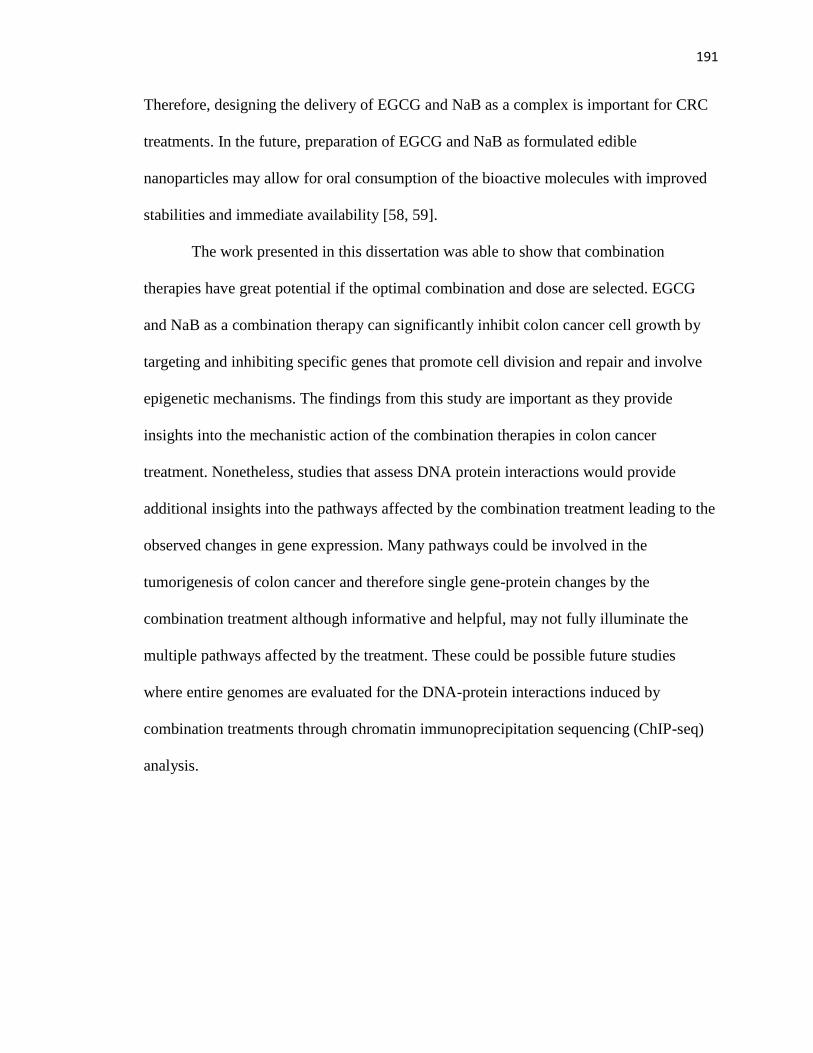
191
Therefore, designing the delivery of EGCG and NaB as a complex is important for CRC
treatments. In the future, preparation of EGCG and NaB as formulated edible
nanoparticles may allow for oral consumption of the bioactive molecules with improved
stabilities and immediate availability [58, 59].
The work presented in this dissertation was able to show that combination
therapies have great potential if the optimal combination and dose are selected. EGCG
and NaB as a combination therapy can significantly inhibit colon cancer cell growth by
targeting and inhibiting specific genes that promote cell division and repair and involve
epigenetic mechanisms. The findings from this study are important as they provide
insights into the mechanistic action of the combination therapies in colon cancer
treatment. Nonetheless, studies that assess DNA protein interactions would provide
additional insights into the pathways affected by the combination treatment leading to the
observed changes in gene expression. Many pathways could be involved in the
tumorigenesis of colon cancer and therefore single gene-protein changes by the
combination treatment although informative and helpful, may not fully illuminate the
multiple pathways affected by the treatment. These could be possible future studies
where entire genomes are evaluated for the DNA-protein interactions induced by
combination treatments through chromatin immunoprecipitation sequencing (ChIP-seq)
analysis.

192
GENERAL LIST OF REFERENCES
[1] Lobo, V.; Patil, A.; Phatak, A.; Chandra, N. Free radicals, antioxidants and
functional foods: Impact on human health. Pharmacogn Rev. (2010), 4, 118-126.
[2] Saldanha, S.N.; Tollefsbol, T.O. The role of nutraceuticals in chemoprevention
and chemotherapy and their clinical outcomes. J Oncol. (2012), 2012, 192464.
[3] Xu, Z.; Taylor, J.A. Genome-wide age-related DNA methylation changes in blood
and other tissues relate to histone modification, expression and cancer.
Carcinogenesis. (2014),
[4] Dugast-Darzacq, C.; Grange, T. MethylQuant: a real-time PCR-based method to
quantify DNA methylation at single specific cytosines. Methods Mol Biol. (2009),
507, 281-303.
[5] Leroy, G.; Dimaggio, P.A.; Chan, E.Y.; Zee, B.M.; Blanco, M.A.; Bryant, B.;
Flaniken, I.Z.; Liu, S.; Kang, Y.; Trojer, P.; et al. A quantitative atlas of histone
modification signatures from human cancer cells. Epigenetics Chromatin. (2013),
6, 20.
[6] Calvanese, V.; Lara, E.; Kahn, A.; Fraga, M.F. The role of epigenetics in aging
and age-related diseases. Ageing Res Rev. (2009), 8, 268-276.
[7] Li, Y.; Saldanha, S.N.; Tollefsbol, T.O. Impact of epigenetic dietary compounds
on transgenerational prevention of human diseases. AAPS J. (2014), 16, 27-36.
[8] Handy, D.E.; Castro, R.; Loscalzo, J. Epigenetic modifications: basic mechanisms
and role in cardiovascular disease. Circulation. (2011), 123, 2145-2156.
[9] Fuks, F.; Burgers, W.A.; Brehm, A.; Hughes-Davies, L.; Kouzarides, T. DNA
methyltransferase Dnmt1 associates with histone deacetylase activity. Nat Genet.
(2000), 24, 88-91.
[10] Delcuve, G.P.; Khan, D.H.; Davie, J.R. Roles of histone deacetylases in
epigenetic regulation: emerging paradigms from studies with inhibitors. Clin
Epigenetics. (2012), 4, 5.
[11] Momparler, R.L.; Bovenzi, V. DNA methylation and cancer. J Cell Physiol.
(2000), 183, 145-154.
[12] Lee, W.J.; Shim, J.Y.; Zhu, B.T. Mechanisms for the inhibition of DNA
methyltransferases by tea catechins and bioflavonoids. Mol Pharmacol. (2005),
68, 1018-1030.
[13] Mersfelder, E.L.; Parthun, M.R. The tale beyond the tail: histone core domain
modifications and the regulation of chromatin structure. Nucleic Acids Res.
(2006), 34, 2653-2662.
[14] Esteller, M. CpG island methylation and histone modifications: biology and
clinical significance. Ernst Schering Res Found Workshop. (2006), 115-126.
[15] Pan, L.N.; Lu, J.; Huang, B. HDAC inhibitors: a potential new category of anti-
tumor agents. Cell Mol Immunol. (2007), 4, 337-343.
[16] Parodi, P.W. Cows' milk fat components as potential anticarcinogenic agents. J
Nutr. (1997), 127, 1055-1060.

193
[17] Hassig, C.A.; Tong, J.K.; Schreiber, S.L. Fiber-derived butyrate and the
prevention of colon cancer. Chem Biol. (1997), 4, 783-789.
[18] Miller, K.M.; Tjeertes, J.V.; Coates, J.; Legube, G.; Polo, S.E.; Britton, S.;
Jackson, S.P. Human HDAC1 and HDAC2 function in the DNA-damage
response to promote DNA nonhomologous end-joining. Nat Struct Mol Biol.
(2010), 17, 1144-1151.
[19] Edelman, M.J.; Bauer, K.; Khanwani, S.; Tait, N.; Trepel, J.; Karp, J.;
Nemieboka, N.; Chung, E.J.; Van Echo, D. Clinical and pharmacologic study of
tributyrin: an oral butyrate prodrug. Cancer Chemother Pharmacol. (2003), 51,
439-444.
[20] Young, G.P.; Hu, Y.; Le Leu, R.K.; Nyskohus, L. Dietary fibre and colorectal
cancer: a model for environment--gene interactions. Mol Nutr Food Res. (2005),
49, 571-584.
[21] Tang, F.Y.; Pai, M.H.; Chiang, E.P. Consumption of high-fat diet induces tumor
progression and epithelial-mesenchymal transition of colorectal cancer in a mouse
xenograft model. J Nutr Biochem. (2012), 23, 1302-1313.
[22] Trichopoulou, A.; Tzonou, A.; Hsieh, C.C.; Toupadaki, N.; Manousos, O.;
Trichopoulos, D. High protein, saturated fat and cholesterol diet, and low levels of
serum lipids in colorectal cancer. Int J Cancer. (1992), 51, 386-389.
[23] Riboli, E.; Norat, T. Epidemiologic evidence of the protective effect of fruit and
vegetables on cancer risk. Am J Clin Nutr. (2003), 78, 559S-569S.
[24] Kushi, L.H.; Doyle, C.; McCullough, M.; Rock, C.L.; Demark-Wahnefried, W.;
Bandera, E.V.; Gapstur, S.; Patel, A.V.; Andrews, K.; Gansler, T.; et al. American
Cancer Society Guidelines on nutrition and physical activity for cancer
prevention: reducing the risk of cancer with healthy food choices and physical
activity. CA Cancer J Clin. (2012), 62, 30-67.
[25] Krieg, A.; Werner, T.A.; Verde, P.E.; Stoecklein, N.H.; Knoefel, W.T. Prognostic
and clinicopathological significance of survivin in colorectal cancer: a meta-
analysis. PLoS One. (2013), 8, e65338.
[26] Weichert, W.; Roske, A.; Niesporek, S.; Noske, A.; Buckendahl, A.C.; Dietel, M.;
Gekeler, V.; Boehm, M.; Beckers, T.; Denkert, C. Class I histone deacetylase
expression has independent prognostic impact in human colorectal cancer:
specific role of class I histone deacetylases in vitro and in vivo. Clin Cancer Res.
(2008), 14, 1669-1677.
[27] Bae, J.M.; Kim, J.H.; Kang, G.H. Epigenetic alterations in colorectal cancer: the
CpG island methylator phenotype. Histol Histopathol. (2013), 28, 585-595.
[28] Lin, S.Y.; Yeh, K.T.; Chen, W.T.; Chen, H.C.; Chen, S.T.; Chiou, H.Y.; Chang,
J.G. Promoter CpG methylation of tumor suppressor genes in colorectal cancer
and its relationship to clinical features. Oncol Rep. (2004), 11, 341-348.
[29] Bardhan, K.; Liu, K. Epigenetics and colorectal cancer pathogenesis. Cancers
(Basel). (2013), 5, 676-713.
[30] Nordgaard, I.; Hove, H.; Clausen, M.R.; Mortensen, P.B. Colonic production of
butyrate in patients with previous colonic cancer during long-term treatment with
dietary fibre (Plantago ovata seeds). Scand J Gastroenterol. (1996), 31, 1011-
1020.

194
[31] McIntyre, A.; Gibson, P.R.; Young, G.P. Butyrate production from dietary fibre
and protection against large bowel cancer in a rat model. Gut. (1993), 34, 386-
391.
[32] Saldanha, S.N.; Tollefsbol, T.O. Pathway modulations and epigenetic alterations
in ovarian tumorbiogenesis. J Cell Physiol. (2014), 229, 393-406.
[33] Xiaoyuan, C.; Longbang, C.; Jinghua, W.; Xiaoxiang, G.; Huaicheng, G.; Qun,
Z.; Haizhu, S. Survivin: a potential prognostic marker and chemoradiotherapeutic
target for colorectal cancer. Ir J Med Sci. (2010), 179, 327-335.
[34] Xi, R.C.; Biao, W.S.; Gang, Z.Z. Significant elevation of survivin and livin
expression in human colorectal cancer: inverse correlation between expression
and overall survival. Onkologie. (2011), 34, 428-432.
[35] Chang, H.L.; Chen, C.Y.; Hsu, Y.F.; Kuo, W.S.; Ou, G.; Chiu, P.T.; Huang, Y.H.;
Hsu, M.J. Simvastatin induced HCT116 colorectal cancer cell apoptosis through
p38MAPK-p53-survivin signaling cascade. Biochim Biophys Acta. (2013), 1830,
4053-4064.
[36] Jin, Y.; Wei, Y.; Xiong, L.; Yang, Y.; Wu, J.R. Differential regulation of survivin
by p53 contributes to cell cycle dependent apoptosis. Cell Res. (2005), 15, 361-
370.
[37] Zhou, M.; Gu, L.; Li, F.; Zhu, Y.; Woods, W.G.; Findley, H.W. DNA damage
induces a novel p53-survivin signaling pathway regulating cell cycle and
apoptosis in acute lymphoblastic leukemia cells. J Pharmacol Exp Ther. (2002),
303, 124-131.
[38] Nabilsi, N.H.; Broaddus, R.R.; Loose, D.S. DNA methylation inhibits p53-
mediated survivin repression. Oncogene. (2009), 28, 2046-2050.
[39] Wang, Z.; Fukuda, S.; Pelus, L.M. Survivin regulates the p53 tumor suppressor
gene family. Oncogene. (2004), 23, 8146-8153.
[40] Zhao, J.; Tenev, T.; Martins, L.M.; Downward, J.; Lemoine, N.R. The ubiquitin-
proteasome pathway regulates survivin degradation in a cell cycle-dependent
manner. J Cell Sci. (2000), 113 Pt 23, 4363-4371.
[41] Mariadason, J.M. HDACs and HDAC inhibitors in colon cancer. Epigenetics.
(2008), 3, 28-37.
[42] Rajendran, P.; Ho, E.; Williams, D.E.; Dashwood, R.H. Dietary phytochemicals,
HDAC inhibition, and DNA damage/repair defects in cancer cells. Clin
Epigenetics. (2011), 3, 4.
[43] Liu, L.; Saldanha, S.N.; Pate, M.S.; Andrews, L.G.; Tollefsbol, T.O. Epigenetic
regulation of human telomerase reverse transcriptase promoter activity during
cellular differentiation. Genes Chromosomes Cancer. (2004), 41, 26-37.
[44] Jacobs, D.M.; Gaudier, E.; van Duynhoven, J.; Vaughan, E.E. Non-digestible
food ingredients, colonic microbiota and the impact on gut health and immunity: a
role for metabolomics. Curr Drug Metab. (2009), 10, 41-54.
[45] Xin Zhang, X.Z., Yongkang Sun, Bing Hu, Yi Sun, Saqib Jabbar, Xiaoxiong
Zeng. Fermentation in vitro of EGCG, GCG and EGCG3"Me isolated from
Oolong tea by human intestinal microbiota. Food Research International. (2013),
54 1589–1595.
[46] Wang, C.C.; Xu, H.; Man, G.C.; Zhang, T.; Chu, K.O.; Chu, C.Y.; Cheng, J.T.;
Li, G.; He, Y.X.; Qin, L.; et al. Prodrug of green tea epigallocatechin-3-gallate

195
(Pro-EGCG) as a potent anti-angiogenesis agent for endometriosis in mice.
Angiogenesis. (2013), 16, 59-69.
[47] Kok, S.H.; Wong, R.S.; Gambari, R.; Cheung, F.; Lam, W.S.; Lau, F.Y.; Cheng,
G.Y.; Cheng, C.H.; Lam, K.H.; Chan, S.H.; et al. In vitro cytotoxicity of (-)-
EGCG octaacetate on MDAMB-231 and SKHep-1 human carcinoma cells: a
pharmacological consideration on prodrug design. Int J Mol Med. (2008), 22,
841-845.
[48] Lam, W.H.; Kazi, A.; Kuhn, D.J.; Chow, L.M.; Chan, A.S.; Dou, Q.P.; Chan,
T.H. A potential prodrug for a green tea polyphenol proteasome inhibitor:
evaluation of the peracetate ester of (-)-epigallocatechin gallate [(-)-EGCG].
Bioorg Med Chem. (2004), 12, 5587-5593.
[49] Miyoshi, M.; Sakaki, H.; Usami, M.; Iizuka, N.; Shuno, K.; Aoyama, M.; Usami,
Y. Oral administration of tributyrin increases concentration of butyrate in the
portal vein and prevents lipopolysaccharide-induced liver injury in rats. Clin Nutr.
(2011), 30, 252-258.
[50] Egorin, M.J.; Yuan, Z.M.; Sentz, D.L.; Plaisance, K.; Eiseman, J.L. Plasma
pharmacokinetics of butyrate after intravenous administration of sodium butyrate
or oral administration of tributyrin or sodium butyrate to mice and rats. Cancer
Chemother Pharmacol. (1999), 43, 445-453.
[51] Kang, S.N.; Lee, E.; Lee, M.K.; Lim, S.J. Preparation and evaluation of tributyrin
emulsion as a potent anti-cancer agent against melanoma. Drug Deliv. (2011), 18,
143-149.
[52] Kuefer, R.; Hofer, M.D.; Altug, V.; Zorn, C.; Genze, F.; Kunzi-Rapp, K.;
Hautmann, R.E.; Gschwend, J.E. Sodium butyrate and tributyrin induce in vivo
growth inhibition and apoptosis in human prostate cancer. Br J Cancer. (2004),
90, 535-541.
[53] Gaschott, T.; Maassen, C.U.; Stein, J. Tributyrin, a butyrate precursor, impairs
growth and induces apoptosis and differentiation in pancreatic cancer cells.
Anticancer Res. (2001), 21, 2815-2819.
[54] Li, Y.; Le Maux, S.; Xiao, H.; McClements, D.J. Emulsion-based delivery
systems for tributyrin, a potential colon cancer preventative agent. J Agric Food
Chem. (2009), 57, 9243-9249.
[55] Schroder, C.P.; Maurer, H.R. Tributyrin-induced differentiation promotes
apoptosis of LS 174T colon cancer cells in vitro. Int J Oncol. (2002), 20, 195-200.
[56] Schroder, C.P.; Maurer, H.R. Tributyrin enhances the cytotoxic activity of
interleukin-2/interleukin-12 stimulated human natural killer cells against LS 174T
colon cancer cells in vitro. Cancer Immunol Immunother. (2001), 50, 69-76.
[57] Gaschott, T.; Steinhilber, D.; Milovic, V.; Stein, J. Tributyrin, a stable and rapidly
absorbed prodrug of butyric acid, enhances antiproliferative effects of
dihydroxycholecalciferol in human colon cancer cells. J Nutr. (2001), 131, 1839-
1843.
[58] Li, Z.; Gu, L. Fabrication of Self-Assembled (-)-Epigallocatechin Gallate (EGCG)
Ovalbumin-Dextran Conjugate Nanoparticles and Their Transport across
Monolayers of Human Intestinal Epithelial Caco-2 Cells. J Agric Food Chem.
(2014),

196
[59] Minelli, R.; Serpe, L.; Pettazzoni, P.; Minero, V.; Barrera, G.; Gigliotti, C.;
Mesturini, R.; Rosa, A.C.; Gasco, P.; Vivenza, N.; et al. Cholesteryl butyrate solid
lipid nanoparticles inhibit the adhesion and migration of colon cancer cells. Br J
Pharmacol. (2012), 166, 587-601.

197
APPENDIX A
ENVIRONMENTAL EFFECTS ON AGE-ASSOCIATED EPIGENETICS
by
SABITA N SALDANHA, ASHLEY MCCOLLUM AND TRYGVE TOLLEFSBOL
Epigenetics of Aging
Copyright
2010
by
Springer Science
Used by permission
Format adapted for dissertation

198
Abstract Aging of an organism appears to be a precursor for most, if not all disease
outcomes. However, some diseases associated with aging are due to genetic mutations,
while others occur even without altering the genetic sequence, through a phenomenon
known as epigenetics. DNA methylation and histone modifications are classic examples
of epigenetic modifications. These modifications appear to be in a constant flux and,
therefore, can be altered by exposure to environmental insults over the life time of the
organism. This chapter focuses on how environmental conditions such as heat stress,
exposure to metals, and nurturing can affect the dynamic epigenetic modifications in
aging. In addition, how these modifications can either directly or indirectly result in age-
associated pathologies are also addressed.
Keywords: Epigenetics · Aging · Environment and Epigenetic modifications.
Introduction
Organismal systems are comprised of three types of cell populations. The stem cell
populations that primarily give rise to new cells, the differentiated cell populations that
are tissue and function specific, and apoptotic populations composed of dead cells.
Differentiated cell populations are of particular interest as these populations are
responsible for maintaining the functional integrity of the organs. Differentiated somatic
cells, however, have a finite life span and follow Hayflick’s limit (Hayflick 1979, 1980,
1985), that is, after a programmed number of cell divisions, the cells are unable to divide
and thereby enter the senescent phase. Senescent cell populations are the hallmark of the
aging process and may induce the aging phenotype and in some cases age-associated
199
pathologies (Price et al. 2002; Wiemann et al. 2002; Minamino and Komuro 2007) (Fig.
1). Several other determinants contribute to the aging process which are discussed in
other reviews (Sedivy et al. 2008). It is interesting to note that although several molecular
changes within a cell contribute to the aging phenomenon, of particular interest is the
change in gene expression as these fundamental units control almost all cellular
processes. The previously held belief that mutations solely affect gene expression has
been refuted. Recent investigations have shown otherwise (Sedivy et al. 2008). Altered
gene expression can be mediated by an epigenetic process where the sequence of the gene
remains unaltered, and the patterns of these changes are stably inherited through cellular
divisions. Stable inheritance of these patterns is controlled by mechanisms such as DNA
methylation and post-translational modifications of key histone residues (Fraga and
Esteller 2007). The resultant accumulation of certain epigenetic marks and the interplay
of these events may influence how an organism ages (Fraga and Esteller 2007). Breaking
this code can help to understand what marks trigger a cell to age or proliferate. The most
predominant factors known to influence such epigenetic events are the environment and
nutrition (Fraga et al. 2005; Feil 2006; Fraga and Esteller 2007).
DNA Methylation
DNA methylation is a predominant epigenetic mechanism that controls gene expression.
Methylation occurs at CpG dinucleotide residues controlled by the DNA
methyltransferase enzymes (Feil 2006). The methylation patterns that are inherited are
substrate specific, and maintenance methylation by DNA methyltransferase 1 (DNMT1)
will occur only if a premethylated template exists. This allows for a faithful reproduction

200
Fig. 1 Cellular and tissue changes occurring with the onset of aging. The circles (light
gray) symbolize mitotically active and proliferating cells as seen in young tissues.
However, an internal genetic clock causes the cells to switch from mitotically active to
slow proliferating cells as seen in adult tissue, with predominant silent but metabolically
active senescent cells (middle gray, spindle shaped). This phase deteriorates further, and
genes responsible for the healthy sustenance of cells may be silenced (e.g., housekeeping
genes). Eventually cell death (dark gray, star shaped) ensues which may trigger many
age-related diseases
Aft
er a
fin
ite
nu
mb
er o
f
div
isio
ns
P
rog
ress
ive
yea
rs
Cel
ls i
n y
ou
ng
tis
sue
Cel
ls i
n a
du
lt t
issu
e p
red
om
inan
tly
sen
sen
cen
t w
ith
so
me
mit
oti
call
y
acti
ve
Pre
do
min
ant
apo
pto
tic
and
sen
esce
nt
cell
s
Tis
sue
and
org
an f
un
ctio
n
op
tim
al
Tis
sue
and
org
an f
un
ctio
n
sub
op
tim
al
Tis
sue
and
org
an f
un
ctio
n
det
erio
riat
es.
Deg
ener
atio
n o
f
the
ner
vo
us
and
im
mu
ne
syst
ems
Tu
mo
r fo
rmat
ion
an
d t
he
on
set
of
oth
er a
ge-
rela
ted
bio
pat
ho
log
ies
Der
egu
lati
on
of
ho
use
kee
pin
g g
enes
Rea
ctiv
atio
n o
f ce
rtai
n g
enes
;
telo
mer
ase,
on
cog
enes
,
inac
tiv
atio
n o
f tu
mo
r
sup
pre
sso
r g
enes
Dea
th o
f th
e o
rgan
ism
Fig
ure
1.
201
of the previous methylation pattern, without altering the epigenetic mark. Methylation of
CpG dinucleotides is primarily associated with gene repression. Observations of global
hypomethylation have been documented in some studies (Wilson and Jones 1983; Mays-
Hoopes 1989; Richardson 2003; Liu et al. 2007). However, hypermethylation of locus-
specific genes or specific promoter elements can induce gene expression (Feil 2006).
Epigenesis is not a simple process but has some degree of complexity, as seen in higher
order organisms. The epigenetic process of DNA methylation in gene control has been
well established in X-chromosome inactivation and genomic imprinting (Feil 2006).
X-Chromosome Inactivation
Although very few phenotypic genes are present on the X chromosome, the genes on this
chromosome control many developmental processes, and thus, maintaining the dosage is
of utmost importance. X-chromosome inactivation occurs at the X inactivating center
(XIC) (Avner and Heard 2001; Boumil and Lee 2001; Heard et al. 2001; Sado et al.
2001). Inactivation of X (Xi) chromosome is mediated through epigenetic intervention
(Wang et al. 2001). The inactivation of X takes place randomly in the somatic cells, and
only in the germ line of the females are both X chromosomes active. Inactivation of the X
chromosome is initiated at the X-chromosome inactivating center and is mediated by a
sole gene Xist (X inactive signal transcript) (Avner and Heard 2001; Salstrom 2007;
Vincent-Salomon et al. 2007). An RNA molecule transcribed by this gene covers the X
chromosomes and initiates the wave of inactivation across the whole chromosome with
the exception of a few regions. In addition to the specific RNA transcript that is not
translated, heterochromatization of the X chromosome is mediated by DNA methylation
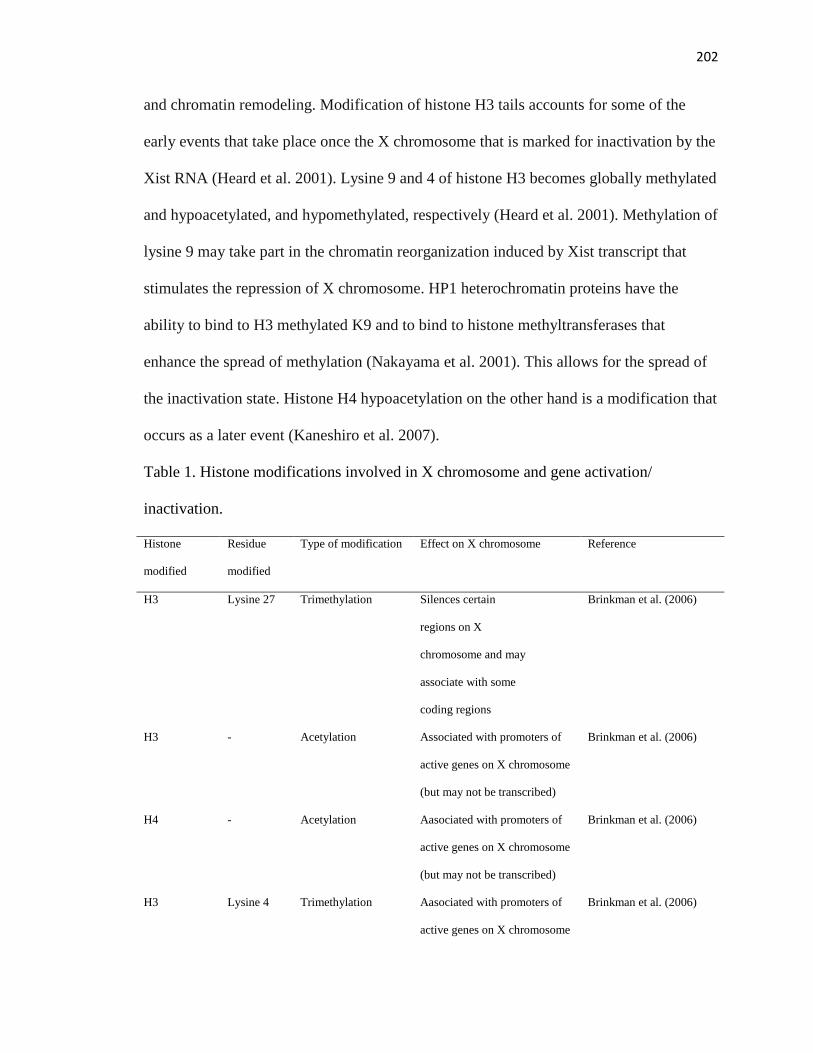
202
and chromatin remodeling. Modification of histone H3 tails accounts for some of the
early events that take place once the X chromosome that is marked for inactivation by the
Xist RNA (Heard et al. 2001). Lysine 9 and 4 of histone H3 becomes globally methylated
and hypoacetylated, and hypomethylated, respectively (Heard et al. 2001). Methylation of
lysine 9 may take part in the chromatin reorganization induced by Xist transcript that
stimulates the repression of X chromosome. HP1 heterochromatin proteins have the
ability to bind to H3 methylated K9 and to bind to histone methyltransferases that
enhance the spread of methylation (Nakayama et al. 2001). This allows for the spread of
the inactivation state. Histone H4 hypoacetylation on the other hand is a modification that
occurs as a later event (Kaneshiro et al. 2007).
Table 1. Histone modifications involved in X chromosome and gene activation/
inactivation.
Histone
modified
Residue
modified
Type of modification Effect on X chromosome Reference
H3 Lysine 27 Trimethylation Silences certain
regions on X
chromosome and may
associate with some
coding regions
Brinkman et al. (2006)
H3 - Acetylation Associated with promoters of
active genes on X chromosome
(but may not be transcribed)
Brinkman et al. (2006)
H4 - Acetylation Aasociated with promoters of
active genes on X chromosome
(but may not be transcribed)
Brinkman et al. (2006)
H3 Lysine 4 Trimethylation Aasociated with promoters of
active genes on X chromosome
Brinkman et al. (2006)

203
(but may not be transcribed)
H3 Lysine 9 Trimethylation Associated with transcription Brinkman et al. (2006)
H3
H3
H4
Lysine 27
Lysine 9
Lysine 20
Trimethylation
Dimethylation
Facultative X chromatin states Heard, et al. (2001);
Kohlmaier et al. (2004);
Plath et al. (2002); Silva et
al. (2008)
H3
H3
H4
Lysine 27
Lysine 9
Lysine 20
Monomethylation
Trimethylation
Trimethylation
Constitutive X chromatin states Ebert et al. (2004);
Kohlmaier et al. (2004);
Schotta et al. (2004a, b)
This later event suggests that early H3 histone modifications are primarily responsible
for chromosome-wide inactivation (Heard et al. 2001; Jenuwein and Allis 2001). Studies
have shown that the Xi chromosome has interspersed regions of active and inactive
regions. This indicates that the inactivation is not signaled by individual histone marks
but rather the cumulative effects of various histone modifications acting together (Table
1). What has been observed is that there are several genes on the inactive X that escape
inactivation (Kaneshiro et al. 2007). A few that are silenced are housekeeping genes such
as Hypoxanthine phosphoribosyltransferase 1 (HPRT1) and phosphoglycerate kinase1
(PGK1). The genes are silenced through epigenetic mechanisms of DNA methylation as
the promoters of these genes are CpG rich. Many of these genes also control various
cellular processes. The inactivation and silencing of genes occur early in embryonic
development (Kaneshiro et al. 2007). However, reprogramming of these genes can be
achieved through environmental influences as the epigenome is quite dynamic. The exact
relationship between environmental factors and X reactivation via epigenetic mechanisms
yet needs to be ascertained. The speculation is that with age, probably this stability can be

204
reprogrammed or erased resulting in reactivation that could possibly trigger or amplify
phenotypic defects.
Histone Modifications
Histones are basic proteins that are evolutionary well conserved and are essential to the
proper packaging of DNA in the nucleus. About ∼146 bp of DNA are wrapped around
the histone octamer molecule which is comprised of two subunits each of H2A, H2B, H3,
and H4 (Luger et al. 1997; Jenuwein and Allis 2001). This structure is called the
nucleosome and forms the repeating unit of chromatin. The activation and inactivation of
gene function has been established by the reversible chemical modification of histone
amino ‘tails’ that initiate euchromatic or heterochromatic states. Commonly, acetylation,
phosphorylation, methylation, ubiquitination, and sumoylation are involved in modifying
the exposed histone sequences (Jenuwein and Allis 2001; Ke et al. 2006; Iniguez-Lluhi
2006; Sims et al. 2006). Amino acid residues such as lysine and arginine are prone to
such modifications. However, the degree of chemical modification, the number of
residues modified, and the type of modification greatly influence the control on gene
expression. For example, two processes such as acetylation and methylation of histone
residues can act simultaneously either on identical or different subunits, but the overall
pattern or code dictates if a gene is turned on or off. The modified histone states are
initiated by enzymes such as histone methyltransferases (add methyl groups), histone
acetylases [HATs] (add acetyl groups), or histone deacetylases [HDACs] (remove acetyl
groups). Investigations have demonstrated that a cross talk can exist between epigenetics
states (Vaissiere et al. 2008). Histone acetylation allows the unwinding of DNA

205
associated with the nucleosome exposing it to regulatory elements. Thus, acetylation not
only alters the structure of chromatin but also can dictate whether a gene is expressed or
repressed based on the regulatory factors that can bind to exposed promoter elements.
Although histone acetylation and its role in carcinogenesis is intensively studied,
relatively little is known about the molecular mechanisms by which this epigenetic
process influences changes in the histone code in aging. One can speculate that in young
healthy cells, loss of methylation of CpG islands with acetylation maintains the
expression of crucial genes. However, with age and exposure to environmental insults, a
shift in epigenetic profile may occur whereby the spread of methylation to CpG residues
with deacetylation of local histones can induce progressive loss of gene expression
(Vaissiere et al. 2008). This shift in profiles may enhance age-related etiologies (Cairns
2001; Rowley 1998; Wolffe 2001; Vaissiere et al. 2008).
Environmental Effects and Epigenetics
Organisms are constantly exposed to environmental insults throughout their lifetime that
can inadvertently change the landscape of molecular processes. Environmentally induced
stresses contributed by an individual’s occupation, lifestyle, and external surroundings
are considered to be extremely crucial to these changes. Exposure to metals such as
nickel, lead, cadmium, arsenic are known to initiate tumorigenesis (Herceg 2007;
Vaissiere et al. 2008). These factors therefore have a potential effect on chromatin
organization, mediated by DNA methylation and histone alterations that code specific
epigenetic information. These metals therefore can significantly affect changes in normal
epigenetic patterns, initiate aberrant epigenetic signaling, and induce neoplasms. There is

206
a well-established link between ionizing radiations and chromosomal instability. This
instability most probably arises from abnormal methylation of unmethylated regions.
Chronic exposure to UV radiation has been shown to induce global hypomethylation as
seen in studies carried out in mice (Herceg 2007). The end result of these abnormalities
early on in life culminates in age-associated malignancies.
Nickel
Epigenetic damage by nickel particles occurs in heterochromatic regions of
chromosomes. Studies have shown that nickel tends to increase DNA methylation near
heterochromatin (Herceg 2007). Interestingly, tumor suppressor genes and senescence
genes may be located in these regions. Therefore, critical cellular genes can be turned off
by de novo methylation of the promoters. Sometimes more than a single epigenetic event
can transcriptionally silence important genes. This has been seen in a gene such as
guanine-hypoxanthine phosphoribosyltransferase (gpt). This gene is transcriptionally
silenced not only by DNA methylation but also by a decrease in global acetylation of
histones H3 and H4 and increased histone H3K9 dimethylation (Herceg 2007). Studies
have shown in the presence of nickel that specific lysine residues are hypoacetylated,
primarily at position 12 and 16 of histone H4 (Broday et al. 1999, 2000; Zoroddu et al.
2000. In addition, nickel preferentially binds to a site at histidine 18 (Broday et al. 2000).
This binding of nickel to histidine at position 18 prevents or hinders the accessibility of
neighboring lysine residues to the HAT complex. Thus histone code patterns are
significantly modified or changed by chronic exposure to the nickel molecule (Table 2).

207
Table 2. Changes in histone modification by exposure to nickel
Histone Subunit Epigenetic Modification Reference
H2A Cleaves C-terminal tail; increases ubiquitination Bal et al. (2000a, b); Karaczyn et al. (2003); Ke et
al. (2006)
H2B Truncates, deaminates and oxidizes; increases
ubiquitination
Kaneshiro et al. (2007)
H3 Increase in lysine 9 dimethylation, decrease in
acetylation
Chen et al. (2006)
H4 Decrease in acetylation of lysines 12 and 16
respectively.
Broday et al. (2000)
Arsenic and Cadmium
Drinking water and food contain low but permissible levels of arsenic. However, chronic
exposure of this element can exert perturbations in epigenetic patterns. Arsenic-related
hypomethylation is due to the depletion of the cofactor S-adenosylmethionine, required
for methylation as well as arsenic metabolism. The sodium form of arsenic has been
shown to increase genomic hypomethylation (Zhao et al. 1997) of certain crucial genes
such as Harvey rat sarcoma viral oncogene (Ha-Ras), and these perturbations can allow
for neoplastic development later on in an aged individual. Cadmium inhibits the
epigenetic modifying DNA methyltransferases leading to hypomethylation. However,
prolonged exposure to cadmium stimulates regional hypermethylation and global
hypomethylation which is a commonly observed phenomenon in aged tissues.

208
Lead (Pb)
The brain undergoes numerous changes throughout life in humans. The set of genes that
are passed along from each generation has much to do with the direction of aging in the
brain, but environmental influences can contribute to the process as well. For example,
the structure of chromatin can be influenced by the environment which can cause a
lifetime of phenotypical changes and possibly be passed on to future generations.
Diseases such as Alzheimer’s and Parkinsons are prime examples of environmental
effects on phenotype (Miller and O’Callaghan 2008). Since some of the diseases have a
later onset of the phenotype, one can speculate that the genes are initially silenced until
affected by an environmental factor, which may reverse the inactivation through
epigenetic mechanisms. Methylation is one such epigenetic element that partakes in
altering the epigenome. DNA methylation can be affected by environmental factors, such
as drugs, maternal care, and social situations, thus confirming the association of DNA
methylation to the long-term changes of gene expressions in the brain (Champagne and
Meaney 2007; Champagne 2008; Champagne and Curley 2008). Exposure to lead early
in development is known to have effects on brain development (Zawia and Basha 2005,
Wu et al. 2008). Brain development normally occurs during early pre- and postnatal
periods followed by further development until early adulthood. Lead-mediated changes in
brain development have been associated with specific genes such as amyloid precursor
proteins (Wu et al. 2008). In normal aging individuals, the accumulation of amyloid
plaques and associated proteins has been observed, and this process if accelerated can
induce neurodegenerative diseases such as Alzheimer’s disease. Exposure to lead
decreases methylation of a crucial few genes involved in brain development such as the
209
amyloid beta (A4) precursor protein (APP) gene (Wu et al. 2008). Hypomethylation of
the APP promoter leads to decrease in the methylation burden, thereby inducing APP
mRNA production. Consequently the increase in APP production is associated with an
increase in the amyloidgenic β cleavage product during senescence. This cleavage
product forms aggregates and generates free radicals that attack biomolecules such as
DNA. The presence of a methylated cytosine impairs the repair of adjacent oxidized
guanine bases, thereby increasing the susceptibility of neurons to further damage. Events
such as these may collectively enhance the neurodegenerative process. Maternal behavior
appears to play a significant role in altering behavior and physiology. One study (Meaney
and Szyf 2005) demonstrated that maternal nurture influenced epigenomic patterns,
especially of the brain. This group analyzed the epigenetic patterns of a specific gene, the
glucocorticoid receptor. Differential methylation patterns of the promoter of the
glucocorticoid receptor were observed in the hippocampus region, dependent on the type
of nurture provided (grooming, licking, arched back feeding) (Meaney and Szyf 2005). In
addition, the expression of nerve growth factor-induced clone A (NGFIA) that binds to
the promoter region of the glucocorticoid receptor is altered by histone acetylation
(Weaver et al. 2004a, b). Nevertheless, these marks and consequently the effect of the
maternal influence can be reversed through pharmacological interventions indicating that
the marks are dynamic rather than stable.
Heat Stress
A few studies have shown that heat stress may induce changes in methylation imprinting
patterns (Zhu et al. 2008. However, these changes may be specific to only a few genes

210
involved in embryo implantation and development. Aberrant imprinting patterns have
been shown to cause drastic developmental failures in embryos exposed to heat stress
(Ealy et al. 1995, Ozawa et al. 2002). Imprinting of genes is primarily controlled by a
well-known epigenetic mechanism, DNA methylation. Genomic demethylation and
remethylation occur in germ cell development and after fertilization (Reik et al. 1990,
Howlett and Reik 1991). However, the methylation patterns acquired are maintained after
fertilization and preimplantation development. Thus, these patterns which are otherwise
maintained in normal methylation imprinting may be lost due to heat stress. To study the
effect of heat stress and its effects on methylation patterns, some groups have assessed
four imprinted genes, two paternal and two maternal genes (Lau et al. 1994; Lefebvre et
al. 1998; Zhu et al. 2008). These studies showed that the paternal imprinted genes H19
and imprinted maternally expressed transcript (non protein coding) (H19) insulin-like
growth factor 2 receptor (Igf2r) and the maternal imprinted genes parternally expressed
gene 1 (Peg1) and paternally expressed gene 2 (Peg2) reveal a possible link between heat
stress and methylation imprinting (Lau et al. 1994; Lefebvre et al. 1998; Zhu et al. 2008).
Surprisingly, heat stress-related abnormal methylation imprinting has been shown to
affect paternal imprinting rather than maternal (Zhu et al. 2008). But this evidence is
limited to only a few genes. For a more concrete conclusion several imprinted genes need
to be analyzed. One can speculate that under heat stress, changes may occur in the
maternal body. These changes may induce intracellular oxidative damage that can affect
sensitive biomolecules such as DNA (Zhu et al. 2008). However, a clear-cut relationship
between heat stress and epigenetic changes in the embryo is still unclear. The paternally
imprinted genes H19 and Igf2r tend to be hypomethylated under heat stress, which has

211
been supported by several studies (Doherty et al. 2000; Khosla et al. 2001a, b; Zhu et al.
2008). Abnormal methylation imprinting may affect phenotypic outcomes of the genes
resulting in developmental errors. Maternally imprinted genes, at least those that have
been studied, have been shown to be unaffected by heat stress (Zhu et al. 2008).
However, a large body of evidence is required to support these observations. Therefore,
extensive investigations need to be carried out to discern the exact changes in epigenetic
patterns induced by heat stress. These patterns may differ from what are seen in vitro
versus in vivo.
Environment and Epigenetic Drift in Populations
In the approach of understanding how environment affects the epigenetic mechanisms of
populations as a whole, identical genomes are analyzed for any subtle or drastic changes
based on geographical location, lifestyle, or environment. This assessment has been
investigated in monozygotic twin population studies (Petronis 2006). Thus any changes
in the phenotype of a given trait in identical genomes can be attributed as an
environmental contribution. Although biases may exist, in large-scale epigenetic studies
of twin populations, these studies can provide a means of understanding the impact of
environment on gene expression, the genome, and organism as a whole (Petronis 2006).
Studies and observations in different organisms have shown the effect of environment on
chromatin modifications. For example, plants exposed to cold temperatures fail to flower
and this phenomenon is mediated by histone modifications to genes that encode
repressors of flowering (Amasino 2004; Sung and Amasino 2006). Environmental stress
can bring about expression of previously silenced genes, which has been observed in

212
Drosophila studies. During stress, a specific heat shock chaperon protein Hsp90 increases
the activity of histone H3 lysine 4 methyltransferase, inducing the expression of specific
target genes (Ruden et al. 2005). Extensive grooming which includes licking and arched
back nursing have shown to alter the epigenetic patterns in the promoter of the
glucocorticoid receptor in the hippocampus of the pups (Weaver et al. 2004a, b). All
these studies indicate that epigenetic modifications serve as a substrate for the
environmental insults. Thus, through these studies, assess ment of molecular epigenetic
changes mediated by environmental factors can be assessed. Comparison of identical
twins for testing environmental epigenetics is highly suitable as their DNA sequences are
identical and any change or discordance seen in a given phenotype may be attributed to
the environmental factor. Biases and multi-environmental factors can change the overall
scenario; however, initial studies using these cohorts can provide a wealth of information.
For example, a study which analyzed the Beckwith–Wiedemann syndrome demonstrated
that imperfect imprinting of a gene at KCNQ1OT1 (KCNQ1 overlapping transcript 1)
due to DNA methylation differences in monozygotic twins (MZ) affected by the
syndrome was the cause for this disorder (Weksberg et al. 2002). Therefore, lack of
maintenance methylation at a key stage of embryogenesis is a factor for the observed
phenomenon. Similar studies that used MZ twins to assess global and locus-specific
epigenetic differences showed that in a cohort ranging from 3 to 74 years of age, an age-
specific epigenetic drift was observed (Fraga et al. 2005). However, the discordance in
the onset of disease does not always correlate with the age of onset (e.g., Alzheimer’s
versus diabetes mellitus type 1). Twin studies revealed that the concordance of
Alzheimer’s disease was 83% but that of diabetes type 1 was as low as 23% (Petronis

213
2006). Further studies are required to analyze the implication of DNA sequence
differences as compared to epigenetic differences in the contribution to the
synchronous/asynchronous nature of the onset of the disease.
Conclusion
Nurture and nature tend to influence the physiological and behavioral outcomes of
individuals. The observation may be attributed to the effects of environment, both
extrinsic and intrinsic, on differential developmental patterns. Epigenomic marks are
relatively stable and occur early in the developmental program. These stably inherited
patterns, however, can be changed or reversed by environmental influences such as
stress, toxic metal exposure, or even maternal nurture. Understanding these epigenetic
marks is crucial to knowing the patterns associated with aging cells or if particular codes
are associated with diseased phenotypes. Since aging epigenetics is a newly explored
field, well-structured studies are required to understand the interplay of individual or
coordinated codes on gene expression mediated by environmental cues.
References
Amasino, R. 2004. Vernalization, competence, and the epigenetic memory of winter.
Plant Cell.16: 2553–2559.
Avner, P., and Heard, E. 2001. X-chromosome inactivation: counting, choice and
initiation. Nat Rev Genet. 2: 59–67.
Bal, W. et al. 2000a. Ni(II) specifically cleaves the C-terminal tail of the major variant of
histone H2A and forms an oxidative damage-mediating complex with the
cleaved-off octapeptide. Chem Res Toxicol. 13: 616–624.
Bal, W., Kozlowski, H., and Kasprzak, K. S. 2000b. Molecular models in nickel
carcinogenesis. J Inorg Biochem. 79: 213–218.
Boumil, R. M., and Lee, J. T. 2001. Forty years of decoding the silence in X-
chromosome inactivation.Hum Mol Genet. 10: 2225–2232.
Broday, L., Cai, J., and Costa, M. 1999. Nickel enhances telomeric silencing in

214
Saccharomyces cerevisiae. Mutat Res. 440: 121–130.
Broday, L. et al. 2000. Nickel compounds are novel inhibitors of histone H4 acetylation.
Cancer Res. 60: 238–241.
Brinkman, A. B. et al. 2006. Histone modification patterns associated with the human X
chromosome. EMBO Rep. 7: 628–634.
Cairns, B. R. 2001. Emerging roles for chromatin remodeling in cancer biology. Trends
Cell Biol. 11: S15–S21.
Champagne, F. A. 2008. Epigenetic mechanisms and the transgenerational effects of
maternal care. Front Neuroendocrinol. 29: 386–397.
Champagne, F. A., and Meaney, M. J. 2007. Transgenerational effects of social
environment on variations in maternal care and behavioral response to novelty.
Behav Neurosci. 121:1353–1363.
Champagne, F. A., and Curley, J. P. 2008, Jan 18. Epigenetic mechanisms mediating the
long-term effects of maternal care on development. Neurosci Biobehav Rev.
[Epub ahead of print] PMID:18430469
Chen, H. et al. 2006. Nickel ions increase histone H3 lysine 9 dimethylation and induce
transgenesilencing. Mol Cell Biol. 26: 3728–3737.
Doherty, A. S. et al. 2000. Differential effects of culture on imprinted H19 expression in
the preimplantation mouse embryo. Biol Reprod. 62: 1526–1535.
Ealy, A. D. et al. 1995. Developmental changes in sensitivity of bovine embryos to heat
shock anduse of antioxidants as thermoprotectants. J Anim Sci. 73: 1401–1407.
Ebert, A. et al. 2004. Su(var) genes regulate the balance between euchromatin and
heterochromatin in Drosophila. Genes Dev. 18: 2973–2983.
Feil, R. 2006. Environmental and nutritional effects on the epigenetic regulation of genes.
Mutat Res. 600: 46–57.
Fraga, M. F. and Esteller, M. 2007. Epigenetics and aging: the targets and the marks.
Trends Genet. 23: 413–418.
Fraga, M. F. et al. 2005. Epigenetic differences arise during the lifetime of monozygotic
twins. Proc Natl Acad Sci USA. 102: 10604–10609.
Hayflick, L. 1979. Cell biology of aging. Fed Proc. 38: 1847–1850.
Hayflick, L. 1980. The cell biology of human aging. Sci Am. 242: 58–65.
Hayflick, L. 1985. The cell biology of aging. Clin Geriatr Med. 1: 15–27.
Heard, E. et al. 2001. Methylation of histone H3 at Lys-9 is an early mark on the X
chromosome during X inactivation. Cell. 107: 727–378.
Herceg, Z. 2007. Epigenetics and cancer: towards an evaluation of the impact of
environmental and dietary factors. Mutagenesis. 22: 91–103.
Howlett, S. K., and Reik, W. 1991. Methylation levels of maternal and paternal genomes
during preimplantation development. Development. 113: 119–127.
Iniguez-Lluhi, J. A. 2006. For a healthy histone code, a little SUMO in the tail keeps the
acetyl away. ACS Chem Biol. 1: 204–206.
Jenuwein, T., and Allis, C. D. 2001. Translating the histone code. Science. 293: 1074–
1080.
Kaneshiro, K. et al. 2007. An integrated map of p53-binding sites and histone
modification in the human ENCODE regions. Genomics. 89: 178–188.
Karaczyn, A. A. et al. 2003. The octapeptidic end of the C-terminal tail of histone H2A is
cleaved off in cells exposed to carcinogenic nickel (II). Chem Res Toxicol. 16:

215
1555–1559.
Ke, Q. et al. 2006. Alterations of histone modifications and transgene silencing by nickel
chloride. Carcinogenesis. 27: 1481–1488.
Khosla, S. et al. 2001a. Culture of preimplantation embryos and its long-term effects on
gene expression and phenotype. Hum Reprod Update. 7: 419–427.
Khosla, S. et al. 2001b. Culture of preimplantation mouse embryos affects fetal
development and the expression of imprinted genes. Biol Reprod. 64: 918–926.
Kohlmaier, A. et al. 2004. A chromosomal memory triggered by Xist regulates histone
methylation in X inactivation. PLoS Biol. 2: 0991–1003.
Lau, M. M. et al. 1994. Loss of the imprinted IGF2/cation-independent mannose 6-
phosphate receptor results in fetal overgrowth and perinatal lethality. Genes Dev.
8: 2953–2963.
Lefebvre, L. et al. 1998. Abnormal maternal behaviour and growth retardation associated
with loss of the imprinted gene Mest. Nat Genet. 20: 163–169.
Luger, K. et al. 1997. Crystal structure of the nucleosome core particle at 2.8 A
resolution. Nature. 389: 251–260.
Liu, L. et al. 2007, Sep 10. DNA methylation impacts on learning and memory in aging.
Neurobiol Aging. [Epub ahead of print] PMID: 17850924
Mays-Hoopes, L. L. 1989. DNA methylation in aging and cancer. J Gerontol. 44: 35–36.
Meaney, M. J., and Szyf, M. 2005. Environmental programming of stress responses
through DNA methylation: life at the interface between a dynamic environment
and a fixed genome. Dialogues Clin Neurosci. 7: 103–123.
Miller, D. B., and O’Callaghan, J. P. 2008. Do early-life insults contribute to the late-life
development of Parkinson and Alzheimer diseases? Metabolism. 57 Suppl 2:
S44–49.
Minamino, T., and Komuro, I. 2007. Vascular cell senescence: contribution to
atherosclerosis. Circ Res. 100: 15–26.
Nakayama, J. et al. 2001. Role of histone H3 lysine 9 methylation in epigenetic control of
heterochromatin assembly. Science. 292: 110–113.
Ozawa, M., Hirabayashi, M., and Kanai, Y. 2002. Developmental competence and
oxidative state of mouse zygotes heat-stressed maternally or in vitro.
Reproduction. 124: 683–689.
Petronis, A. 2006. Epigenetics and twins: three variations on the theme. Trends Genet.
22:347–350.
Plath, K. et al. 2002. Xist RNA and the mechanism of X chromosome inactivation. Annu
Rev Genet. 36: 233–278.
Price, J. S. et al. 2002. The role of chondrocyte senescence in osteoarthritis. Aging Cell.
1: 57–65.
Reik,W., Howlett, S. K., and Surani,M. A. 1990. Imprinting by DNA methylation: from
transgenes to endogenous gene sequences. Dev Suppl. 99–106.
Richardson, B. 2003. Impact of aging on DNA methylation. Ageing Res Rev. 2: 245–61.
Rowley, J. D. 1998. The critical role of chromosome translocations in human leukemias.
Annu Rev Genet. 32: 495–519.
Ruden, D. M. et al. 2005. Hsp90 and environmental impacts on epigenetic states: a model
for the trans-generational effects of diethylstibesterol on uterine development and
cancer. Hum Mol Genet. 14 Spec No 1: R149–15.

216
Sado, T. et al. 2001. Regulation of imprinted X-chromosome inactivation in mice by
Tsix. Development. 128: 1275–1286.
Salstrom, J. L. 2007. X-inactivation and the dynamic maintenance of gene silencing. Mol
Genet Metab. 92: 56–62.
Schotta, G. et al. 2004a. A silencing pathway to induce H3-K9 and H4-K20
trimethylation at constitutive heterochromatin. Genes Dev. 18: 1251–1262.
Schotta, G. et al. 2004b. The indexing potential of histone lysine methylation. Novartis
Found Symp. 259: 22–37; discussion 37–47, 163–169.
Sedivy, J. M., Banumathy, G., and Adams, P. D. 2008. Aging by epigenetics – a
consequence of chromatin damage? Exp Cell Res. 314: 1909–1917.
Silva, S. S. et al. 2008. X-chromosome inactivation and epigenetic fluidity in human
embryonic stem cells. Proc Natl Acad Sci USA. 105: 4820–4825.
Sims, J. K. et al. 2006. A trans-tail histone code defined by monomethylated H4 Lys-20
and H3Lys-9 demarcates distinct regions of silent chromatin. J Biol Chem. 281:
12760–12766.
Sung, S., and Amasino, R. M. 2006. Molecular genetic studies of the memory of winter. J
Exp Bot. 57: 3369–3377.
Vaissiere, T., Sawan, C., and Herceg, Z. 2008. Epigenetic interplay between histone
modifications and DNA methylation in gene silencing. Mutat Res. 659: 40–48.
van Doorn, R. et al. 2005. Aberrant DNA methylation in cutaneous malignancies.
Semin Oncol. 32: 479–487.
Vincent-Salomon, A. et al. 2007. X inactive-specific transcript RNA coating and genetic
instability of the X chromosome in BRCA1 breast tumors. Cancer Res. 67: 5134–
5140.
Wang, J. et al. 2001. Imprinted X inactivation maintained by a mouse Polycomb group
gene. Nat Genet. 28: 371–375.
Weaver, I. C. et al. 2004a. Early environmental regulation of hippocampal glucocorticoid
receptor gene expression: characterization of intracellular mediators and potential
genomic target sites. Ann NY Acad Sci. 1024: 182–212.
Weaver, I. C. et al. 2004b. Epigenetic programming by maternal behavior. Nat Neurosci.
7:847–854.
Weksberg, R. et al. 2002. Discordant KCNQ1OT1 imprinting in sets of monozygotic
twins discordant for Beckwith-Wiedemann syndrome. Hum Mol Genet. 11:
1317–1325.
Wiemann, S. U. et al. 2002. Hepatocyte telomere shortening and senescence are general
markers of human liver cirrhosis. FASEB J. 16: 935–942.
Wilson, V. L. and Jones, P. A. 1983. DNA methylation decreases in aging but not in
immortal cells. Science. 220: 1055–1057.
Wolffe, A. P. 2001. Chromatin remodeling: why it is important in cancer. Oncogene. 20:
2988–2990.
Wu, J., Basha, M. R., and Zawia, N. H. 2008. The environment, epigenetics and
amyloidogenesis. J Mol Neurosci. 34: 1–7.
Zawia, N. H., and Basha, M. R. 2005. Environmental risk factors and the developmental
basis for Alzheimer’s disease. Rev Neurosci. 16: 325–337.
Zhao, C. Q. et al. 1997. Association of arsenic-induced malignant transformation with

217
DNA hypomethylation and aberrant gene expression. Proc Natl Acad Sci USA.
94: 10907–10912.
Zoroddu, M. A. et al. 2000. Interaction of Ni(II) and Cu(II) with a metal binding
sequence of histone H4: AKRHRK, a model of the H4 tail. Biochim Biophys
Acta. 1475: 163–168.
Zhu, J. Q. et al. 2008. Heat stress causes aberrant DNA methylation of H19 and Igf–2r in
mouse blastocysts. Mol Cells. 25: 211–215.

218
APPENDIX B
PATHWAY MODULATIONS AND EPIGENETIC ALTERATIONS IN OVARIAN
TUMORBIOGENESIS
by
SABITA SALDANHA AND TRYGVE TOLLEFSBOL
Journal of Cellular Physiology
Copyright
2014
by
Wiley Online Library
Used by permission
Format adapted for dissertation

219
Abstract
Cellular pathways are numerous and are highly integrated in function in the control of
cellular systems. They collectively regulate cell division, proliferation, survival and
apoptosis of cells and mutagenesis of key genes that control these pathways can initiate
neoplastic transformations. Understanding these pathways is crucial to future therapeutic
and preventive strategies of the disease. Ovarian cancers are of three major types;
epithelial, germ-cell, and stromal. However, ovarian cancers of epithelial origin, arising
from the mesothelium, are the predominant form. Of the subtypes of ovarian cancer, the
high-grade serous tumors are fatal, with low survival rate due to late detection and poor
response to treatments. Close examination of preserved ovarian tissues and in vitro
studies have provided insights into the mechanistic changes occurring in cells mediated
by a few key genes. This review will focus on pathways and key genes of the pathways
that are mutated or have aberrant functions in the pathology of ovarian cancer. Non-
genetic mechanisms that are gaining prominence in the pathology of ovarian cancer,
miRNAs and epigenetics, will also be discussed in the review.
Abbreviations: CDK, cyclin dependent kinases; p27kip1, cyclindependent
kinase inhibitor1B; p16INK4a, inhibitor of kinases 4a; KRAS, Kirsten rat sarcoma
oncogene; NF-kB, nuclear factor kappa B; PI3K, phosphatidylinositide-3 kinase; PTEN,
phosphatase and tensin homolog; E2F, transcription factor.
Introduction
Ovarian neoplasms are challenging to detect and their genesis is often considered as de
novo or sporadic (Scully, 1995; Liu and Ganesan, 2002; Zikan et al., 2007; Weberpals et
220
al., 2008, 2011; Sarojini et al., 2012; Wysham et al., 2012). When detected the cancer is
often advanced and is at stage III/IV category (Kurman and Shih Ie, 2010). The absence
of or poor detection methods of precancerous lesions of the tissue account for the late
detection of ovarian cancer (Fleischer et al., 2012; Sarojini et al., 2012). Thus, ovarian
cancer is considered to be a very lethal disease of gynecological origin.
In contrast to previous reports, ovarian cancer is not considered a single disease
of epithelial origin, but instead covers a group of tumors that are morphologically and
genetically distinct (Kurman and Shih Ie, 2010, 2011). The hypothesis however is still
highly debated. Some of the reasons include late detection, inability to detect
precancerous lesions and therefore insufficient evidence of neoplastic origination. This
obscurity in the pathogenesis of the disease requires the understanding of the molecular
aspects that influence the cellular pathways of the ovarian tissue. Based on morphology,
genetics, and site of origination, ovarian cancers of epithelial-cell origin have been
categorized into two groups. The type I group are those that are strictly confined to the
ovary and are low- grade serous, endometrial, mucinous, and clear-cell type (Fig. 1;
Kurman and Shih Ie, 2010, 2011; Le Page, 2010). These tumors are genetically more
stable, have few to rare p53 mutations, are easily diagnosed and have a good prognosis.
However, only 25% of the ovarian cancers detected are of this type. The Type 2 group
contains tumors that are aggressive and comprise highgrade serous carcinomas,
undifferentiated carcinomas and carcinosarcomas (Fig. 1; Le Page, 2010). These tumors

221
Fig. 1. Proposed theories of origination of epithelial-cell ovarian cancers (EOCs). The
primary ovarian cancers reside in the ovary tissue itself. However, cells from the
endometrium, fallopian tube, stroma can give rise to the histological types of tumors of
epithelial-cell origin. As shown in the diagrams, 1a, 1b, and 1c denote the commonly
seen ovarian tumors epithelia, stroma, and germ-cell tumors, respectively. However,
EOCs are the predominant form and closer examination reveals that the origin could stem
from other reproductive tissues such as the endometrium lining the uterus, cervical tissue
or from the epithelia of the fallopian tube. Retrograde menstruation problems can be a
pathway for the transfer of endometrial cells to that of the ovary. Ovulation and
encystment of cells can be a mode of travel for cells from the fallopian tube. Therefore,
the serous type EOCs arise from the fallopian tube (2a); the endometroid EOCs arise
from the cervical or uterine tissue (2a and 2b); the mucinous and clear-cell tumors could
arise from cells of 1b, 1c, (internal environment of the ovary) and 2a (cervical tissue).
Hence, ovarian epithelial tumors are of the multi-histological type. Symbols used: grey
filled cloud-like structure, tumors; small open oval circles, follicles in the ovary.

222
constitute 75% of the ovarian cancers with a 90% death rate and the site of origination
stems from tissues other than the ovary. These tumors exhibit genetic instabilities with a
higher percentage of p53 mutations (Kurman and Shih Ie, 2011).
Since the last 30 years or so, many preclinical and clinical trials have been
conducted to determine the therapeutic efficacies of drugs against ovarian tumors.
However, the success rates against ovarian cancers have been few. A majority of tumors
that are removed through cytoreductive surgery resurface and become aggressive and
have been proposed to be mediated in part through the treatment itself (Steffensen et al.,
2011a; Modugno and Edwards, 2012; Stathopoulos et al., 2012). Elucidating the
underlying factors that govern the genesis, progression and metastasis of this disease
would provide valuable information for the development of diagnostic, prognostic and
therapeutic approaches of the disease. Further understanding the genes and pathways that
become dysregulated either due to loss of heterozygosity, loss of function, amplification
or mutation would facilitate progress in managing ovarian cancer. This review will focus
on the pathways deregulated and instrumental in the pathogenesis of ovarian tumors and
key genes that are involved in these pathways. Genetic and epigenetic factors that
contribute to these changes will also be analyzed.
Genes Involved in the Biogenesis of Ovarian Cancers
p53
Mutations in cell-cycle regulatory genes, primarily tumor suppressor genes, favor an
uninhibited growth of cells and serve as precursors of immortality. Combined mutations
in these genes and oncogenes encourage a neoplastic phenotype. How these tight

223
regulations become deregulated through mutations and how these mutations affect
important regulatory pathways is important to a diagnostic and prognostic approach for
all cancers. Unlike cancers of other origins, a majority of ovarian cancers are of
epithelial-cell origin, commonly designated as epithelial ovarian carcinomas (EOCs).
Nonetheless this single layer epithelia morphs to give rise to histologically different types
(Kurman and Shih Ie, 2011). The transition is thought to occur through the cell cycle and
genes that control metabolic and molecular pathways within the ovarian cell or via
changes of the epithelia of surrounding reproductive tissue that find their way into the
ovary through the inflammation and repair process of ovulation (Figs. 1 and 2).
Fig. 2. Cell cycle pathways involved in ovarian carcinogenesis. The cell cycle traverses
through the G1-S-G2-M phases tightly controlled by check points between each phase
through a set of regulatory proteins, primarily cyclins and kinases. The levels and
expression of these regulators ensure the fate of the preceding and proceeding phases of
the cycle. Cellular destinations, division, differentiation, senescence and apoptosis are

224
controlled by a certain restriction in the late G1 phase termed the restriction (R) point. It
is when cells successfully cross this threshold point, that cellular destinations are met.
Various genes involved in tightly regulating the cell cycle in ovarian cancer. A:
Deregulation of the key genes (black circle with a star) initiates aberrant proliferation
triggering the development of neoplasms. Single or multiple pathways may be involved
in the process. In early stage development there are chances of single gene mutations or
epigenetic mechanisms that transform the cell into an immortal phenotype. However,
when multiple pathways are hit or crucial genes such as NF-kB are mutated, far more
aggressive tumors can develop. Symbols used: open circle, normal cell; open oval, cell
towards senescence or apoptosis; black jagged star, cancerous cell; black circle with
white star, mutated gene/pathway; black circle with P, phosphorylated. B: Gradual stages
of early to late ovarian tumor development and pathway abnormalities have been
highlighted in miniature figures below the main one. Symbol used: gray cloud-like
structure, tumor.
Of the many regulators, tumor suppressor proteins play a pivotal role in the cell cycle
process and these proteins are highly deregulated in ovarian cancers (Table 1).
Tumor suppressor genes, proto-oncogenes and cell-cycle proteins cohesively
regulate cell growth and division. The primary cyclins and kinase inhibitors that have a
major role in ovarian cancer are cyclin D1, p16INK4a, p27, and p21 (Table 1, Fig. 2;
Vikhanskaya et al., 1996; Kusume et al., 1999; Shigemasa et al., 1999; Bali et al., 2004;
Barbieri et al., 2004; Hashimoto et al., 2011). The regulation and function of cyclin D1,
p16INK4a, p27, and p21 have been shown to be controlled in part by the tumor
suppressor gene p53, which is predominantly mutated in high grade ovarian cancers
(Shigemasa et al., 1999; Bali et al., 2004). The changes in p53 as seen in ovarian cancers
could be both at the gene and protein level. Point mutations, missense mutations and
truncations have been observed, and overexpression of the p53 protein has been detected
in many of the immunohistochemical studies that have been performed (Table 1; Marks
et al., 1991; Liu et al., 1994; Havrilesky et al., 2003). The accumulation of non-functional
p53 has been shown to affect the expression and interaction of proapoptotic genes such as

225
BCL2-associated X protein (BAX) and B-cell lymphoma 2 (BCL-2; Tai et al., 1998;
Schuyer et al., 2001; Ziolkowska-Seta et al., 2009). The aberrations in p53 result in the
accumulation of the altered protein within the cell that has a negative effect on BAX, a
transcriptional target of p53 (Ozer et al., 2012). It seems likely that p53 alterations in high
grade tumors reduce BAX expression allowing the progression of solid tumors. However,
immunohistological studies have not provided a definitive conclusion of a positive
correlation with p53 expression and tumor prognosis and survival (Reles et al., 2001;

226
Rose et al., 2003; Leffers et al., 2008). Contradictions in observations are possible due to
the type of ovarian tissue analyzed, the method used and statistical programs employed to
determine the correlation (Table 1).
The BCL family of apoptotic proteins along with p53 can serve as tools for
histotyping (Skirnisdottir et al., 2002). The expression of p53-BCL-2 and BCL-2-BAX
have been shown to have a strong association with tumor grade and histopathological
subtyping, factors that could be vital for identifying the specific EOC for adjuvant or
combined therapies (Skirnisdottir et al., 2002). What has been observed is that ovarian
tumors are initially very responsive to treatment but later become chemoresistant.
Possibly the treatment itself may cause a few cells within the tumor mass to harbor
mutations in p53 that, in addition to epigenetic silencing of promoter regions of
apoptotic favorable genes such as p16 or Rb, may account for the relapse and progression
of the tumor. Ovarian cancers are categorized as low or high-grade tumors of various
histological subtypes (Le Page, 2010). These range from well-differentiated, to
moderately differentiated to non-differentiated tumors, respectively, and morphologically
are identified as benign, borderline or malignant ovarian tumors (Fig. 2; Le Page, 2010).
Studies have shown that in low-grade ovarian tumors, p53 mutations are absent, but
transitioning into the aggressive type, a significant increase in mutations of the gene are
observed, with a majority of them being of the serous epithelial type (Marks et al., 1991;
Kohler et al., 1993). The factors that contribute to this observed increase in p53 mutations
are still very unclear. p53 is a quintessential player in the cell cycle and its role in
maintaining healthy cell populations is very apparent. Since ovarian cancers are of multi-
histological phenotype the alterations of p53 in tumors of this type may be varied. An

227
altered state of p53 is observed both at the gene and at the protein level in type 2 ovarian
cancers (Yemelyanova et al., 2011; Jones et al., 2012; Kuhn et al., 2012). Most mutations
observed in p53 gene are point mutations that cause the accumulation of the non-
functional protein in the cell due to increased stability. In some instances, the binding
domains of p53 could be altered such that p53 cannot bind to the DNA binding elements
of its target genes hindering subsequent pathways that encourage apoptosis. In other
scenarios, the functional role of the non-functional p53 is based on p16INK4a, a
cyclin kinase inhibitor within the cell (Leong et al., 2009).
p21 and p27
Cyclin dependent kinase (CDK) inhibitors are major coregulatory proteins in the cell
cycle along with the p16, p53 and retinoblastoma (Rb) pathways (Sherr, 1996; Bartkova
et al., 2003; Jayasurya et al., 2004). p21 is a direct transcriptional target of p53 (Gallagher
et al., 2012). In the presence of wildtype (WT) p53, p21 induction ensues followed by the
inhibition of cyclin E/CDK2 preventing the G1-S transition, encouraging the apoptotic
phenotype of cells (Hindley and Philpott, 2012). CDK inhibitors p21 and p27 control
various phases of the cell cycle based on the cyclin with which they associate. p21
association with cyclin D-CDK4/6 inhibitsG1-S transition; with cyclin E-CDK2 the late
G1-S transition; with cyclin A-CDK2 preventing the S-G2 transition; and with cyclinA-
cdc2 the G2-M transition (Cariou et al., 2000; Sandhu et al., 2000). p27 exhibits the same
level of control with cyclins D, E and A from the G1 to the G2 phase (Cariou et al.,
2000). Although the roles of p21 and p27 have been discussed in many cancers, they

228
appear to have secondary roles in the etiology of ovarian cancer rather than in the genesis
(Milde-Langosch et al., 2003; Plisiecka-Halasa et al., 2003; Oudit et al., 2004; Lee et al.,
2007). The role of p27 in ovarian cancer is somewhat contradictory. On examination of
the protein level and localization of p27 in 150 advanced epithelium ovarian cancers
(EOCs), lower nuclear p27 expression was associated with an improved prognosis (Psyrri
et al., 2005). However, another study found that negative p27 expression was associated
with poor prognosis (Shigemasa et al., 2001). Possibly, different grading scales,
techniques and samples assessed might account for these inconsistencies. Loss of
function or accumulation of these proteins in ovarian cancers has not been reported. In
some cases, the absence of or low expression of p21 with p53 expression appears to place
patients at a higher risk for disease recurrence (Anttila et al., 1999b). Studies have also
shown that high p21 expression correlates with early stage ovarian tumors and that only
high p27 expression was necessary for disease free survival (Schmider-Ross et al., 2006).
Overall, there appears to be no correlation between p21 and p27 expression and in terms
of ovarian cancers, as yet these proteins are not especially useful tools for the prognosis
of the disease (Baekelandt et al., 1999).
Pathways Implicated in the Biogenesis of Ovarian Cancer
p16INK4a and Rb pathway
In normal cells, p16INK4a regulates cell proliferation by promoting genes of the
apoptotic pathways (Leong et al., 2009). However, in ovarian cancer cells, it has been
observed that p16INK4a is mutated or its promoter region is hypermethylated switching
off its expression. Transfection studies using ovarian cancer cell lines have shown that in

229
the absence of WT p53, p16INK4a is upregulated to rescue the apoptotic pathway
(Modesitt et al., 2001; Ramirez et al., 2001). In cells that contain double defects of both
p53 and p16INK4a, cell proliferation is rampant and the tumors are more aggressive
(Modesitt et al., 2001; Ramirez et al., 2001). Far advanced tumors have been shown to
have low expression of p16INK4a but higher expressions of retinoblastoma (Rb; Todd
et al., 2000). p16INK4a is considered to be a direct target of Rb expression (Todd et al.,
2000). However, some studies have contradicted this finding. It appears that p16INK4a
and Rb mutually regulate the expression of one another. While trying to understand the
mechanistic pathways involved in ovarian cancers, the Rb-p16INK4a pathway in
conjunction with p53 has been implicated in the genesis of a majority of cancers and has
a pivotal role in the ovarian epithelial specific type (Konecny et al., 2011). The
p16INK4a inhibitor of kinase prevents the association of the cyclin D1-CDK4/6 complex
necessary for the transition of cells from the G1-S phase, in conjunction with
hyperphosphorylated Rb. Studies performed to determine the expression patterns of these
keys genes have shown that loss of p16INK4a function through promoter methylation is
more frequent than mutations and deletions, in comparison to Rb, where hemizygous
deletions are more pronounced with few mutations (Todd et al., 2000; Hashiguchi et al.,
2001). The level of cyclin D1 necessary for cyclin-CDK complex formation is
overexpressed and has been found to be associated with aggressive phenotype and poor
prognosis in about 19% of ovarian cancers. (Todd et al., 2000; Konecny et al., 2011). As
discussed, although alterations/loss of function appear to be the highlighted changes
associated in the Rb-p16INK4a pathway, most ovarian cancers appear to co-express Rb
and p16 proteins, with overexpression of Rb found in advanced ovarian tumors (Todd et

230
al., 2000). Post-translational modifications that affect protein function are also an
important factor to consider in the analysis of tumor biogenesis. The Rb protein release
from the Rb-E2F complex occurs by the hyperphosphorylation of its serine/threonine
residues. A further examination of the phosphorylation levels of Rb protein therefore
needs to be examined. Post-translational modifications have been found to affect protein
function based on the residues phosphorylated as seen in p53 (Smeenk et al., 2011). In
addition, epigenetic alterations as seen in the p16INK4a gene, may also regulate Rb and
p53 function through CCCTC binding factor (CTCF; De La Rosa-Velazquez et al., 2007;
Soto-Reyes and Recillas-Targa, 2010).
KRAS/MAPK/ERK pathway
Kirsten rat sarcoma oncogene (KRAS) activation triggers a sequence of events through
the RAF/MEK and mitogen activated protein kinases (MAPK) pathways, and in
conjunction with mammalian target of rapamycin (mTOR), a target of the protein AK
strain thymoma (AKT) pathway, control cell proliferation (Table 2; Janku et al., 2012).
Point mutations in KRAS provide an advantage for the survival and progression of
tumors (Mane et al., 1990; Edkins et al., 2006; Harris and McCormick, 2010; Li et al.,
2011; Oliveira-Cunha et al., 2012). KRAS mutations have been implicated in the genesis
of lowgrade ovarian tumors, inducing an overactive proliferative phenotype (Vereczkey
et al., 2011; Stewart et al., 2012). In addition to breast cancer associated protein 1
(BRCA1) and 2 (BRCA2) that have familial roles in sporadic germline-based
breast and ovarian cancers, KRAS is now considered the third player (Kundu et al., 2012;
Pilarski et al., 2012). KRAS expression levels differ based on the histopathological type

231
of ovarian tissue and the expression levels may help determine the various type of
ovarian epithelial cancers (Auner et al., 2009; Steffensen et al., 2011b; Nowak-Markwitz
and Spaczynski, 2012). These signatures have a specific pattern in tandem with the
expression of RAF/MAPK components, phosphatase and tensin homolog (PTEN) levels
and AKT (Table 2) and may help differentiate normal from borderline to early-stage
cancers. Determining the signatorial patterns is relatively difficult due to heterogeneity of
the tumors.
A mutant form of KRAS, called KRAS-variant, carries a mutation in its sequence
that has the binding site of a micro RNA (miRNA) termed lethal-7 (Let-7), limiting the
association of Let-7 with KRAS (Ratner et al., 2010). Consequently the levels of KRAS
rises followed by the induction of the nuclear transcription factor (NF-kB) that promotes
the induction of anti-apoptotic genes. It is quite probable that in late stage ovarian
cancers, the earlier mutations in KRAS can stimulate and induce the over-activation of
NF-kB in cancer stem cells (CSCs) that survive chemotherapy. However,
immunohistochemical-based correlation assessments between the KRAS expression and
alterations in NF-kB components and PTEN have not yet been able to confirm a direct
association (Laudanski et al., 2011). KRAS-variants have also been shown to target NF-
kB expression independent of the phosphatidylinositide-3 kinase PI3K-AKT pathway

232
(Keane and Ratner, 2010; Ratner et al., 2010, 2012; Kinross et al., 2011; Mizumoto et al.,
2011; Pharoah et al., 2011; Weidhaas and Slack, 2011; Pilarski et al., 2012). The
disparities in KRAS expression and association patterns with downstream targets
may stem from the type of tissues analyzed. In a study conducted on 489 high grade
ovarian adenocarcinomas, Spellman et al. (2011) showed that in 45% cases that contained
altered PI3K/RAS signaling, less than 1% was due to mutations and 11% were
amplifications. Also, the downstream target, serine/threonine-protein kinase B-Raf
(BRAF) was mutated (0.5%). In addition to PI3K/RAS signaling, KRAS induces
mitogen activated protein kinase/extracellular signal-regulated kinases (MAPK/ERK)
through BRAF. Stimulation of KRAS via GTPase activity activates BRAF. The
stimulation can be mediated by cytokines, growth factors or proto-oncogenes.
The downstream target of BRAF, MEK stimulates ERK when activated. ERK targets
transcription factors that are involved in cell proliferation such as Myc (Smolle et al.,
2013). In ovarian carcinomas, activation of the ERK pathway stems from hormonal
activation of G-protein coupled receptors (Smolle et al., 2013). MAPKs, part of the
MAPK/ERK pathway, are involved in the transduction of signals through hormones.
MAPKs are of two types, tyrosine protein kinase and G-protein coupled receptor. In
ovarian cancers, hormones such as follicle stimulating hormone (FSH) and luteinizing
hormone (LH) may participate in signal transduction via MAPKs, and are thought to
be responsible for ovarian cancer cell growth in carcinomas that express the receptors
(Hilliard et al., 2013).
233
PI3K/PTEN/AKT pathway
Phosphatidylinositide-3 kinase (PI3K) and PTEN proteins influence multiple pathways
within the cell that have many effects on the cellular phenotype through protein AK strain
thymoma (AKT), also known as protein kinase B (PKB). These genes are crucially
required to transcend the input from external stimuli (growth factors) and convey them to
AKT. Phosphorylation of key amino acid residues in these proteins triggers the
downstream activation processes. PTEN is a tumor suppressor gene and either mutations
in the gene or loss of function of the protein is observed in many cancers, including
ovarian cancers (Table 2; Saga et al., 2002; Meng et al., 2006; Yan et al., 2006; Paige and
Brown, 2008). Loss of function in this gene is mediated through epigenetic silencing and
its role in ovarian cancer needs to be further assessed (Kurose et al., 2001). AKT is an
oncogene, and its function and activation depends on the function of PTEN (Meng et al.,
2006; Blanco-Aparicio et al., 2007). However, when PTEN becomes inactivated, or loss
of function is observed, AKT is overexpressed which serves as an impetus for tumor
formation by changes in sub-pathways such as NF-kB, mammalian target of
rapamycin (mTOR), and p53, that in turn regulates proliferation, cell division, and cell
apoptosis (Tables 1 and 2; Blanco-Aparicio et al., 2007; Hussain et al., 2012). PTEN and
AKT are genes of a pathway controlled by PI3K phosphorylation of its internal amino
acid residues, the signals of which depend on either growth receptors or tyrosine kinase
receptors (Xu et al., 2004; Lee et al., 2005; Meng et al., 2006).
PI3K is a heterodimeric protein comprised of a regulatory subunit, p85, and a
catalytic subunit, p110 (Berenjeno and Vanhaesebroeck, 2009; He et al., 2010; Folgiero
et al., 2012; Hofmann and Jucker, 2012; Li et al., 2012; Takayama et al., 2012). The p110

234
catalytic subunit exits as isoforms a, b, g, and d (Li et al., 2012). This enzyme
phosphorylates the hydroxyl group of inositol of phosphatidylinositol triggering
endocellular pathways from external stimuli. For the activation of PI3K,
autophosphorylation of tyrosine kinase receptors (RTKs) is essential (Smith et al., 2002).
The p85 subunit of PI3K tethers to the phosphorylated residues of RTKs. Once the
docking of its regulatory subunit occurs, p110 begins its phosphorylation of
phosphatidylinositol-4,5 biphosphate (PIP)2 to phosphatidylinositol-3,4,5-triphosphate
(PIP)3. (PIP)3 is essential for the activation of AKT (Carnero, 2010). However, AKT
activation requires phosphorylation activities of (PIP)3 and mTOR (Carnero, 2010).
PTEN, an intermediate factor of the PI3K/PTEN/AKT pathway, plays an important role
in ensuring that the activity of AKT is held in check by dephosphorylating (PIP)3
(Stocker et al., 2002). While assessing the roles of PI3K/PTEN/AKT in ovarian
carcinogenesis, amplifications of PI3K, p110 a subunit, and loss of PTEN function are
found (Blanco-Aparicio et al., 2007). When events such as these occur, AKT levels
cannot be controlled triggering pathways of survival, progression and invasion through
subpathways controlled by AKT (Blanco-Aparicio et al., 2007). It has been observed that
inhibition of p110 a subunit or AKT, can induce cell cycle regulatory proteins, p21 and
p27, inhibit phosphorylation of Rb and reduce the levels of cyclins D1 and CDK4 that
essentially serve the apoptotic pathway (Meng et al., 2006). Some of the resistance
offered by ovarian tumors to treatment by certain drugs stem from the amplification of
PIK3Ca, a gene that encodes PI3K p110 a subunit, and AKT expression with reduced
PTEN levels (Janku et al., 2011; Yamamoto et al., 2011; Abe et al., 2013). In such
instances, cell cycle regulatory functions are impaired, and pro-apoptotic proteins such as

235
Bax translocation is inhibited (Lee et al., 2005). PI3K mutations in ovarian tumors are
rare, but an increase in gene copy number has been observed. The amplifications are
more pronounced in high grade ovarian tumors than low grade tumors along with AKT
phosphorylation contributing to survival, and progression of the disease (Huang et al.,
2011; Abe et al., 2013). There does not appear to be a collective association of the three
proteins together in ovarian carcinogenesis and are thought to act independently (Carden
et al., 2012). However, the alterations in the expression of these proteins have been
shown to have a relationship between the resistance offered against therapies and the
recurrence of the disease (Dent et al., 2009).
mTOR promotes cell growth and proliferation inassociation with the PI3K/AKT
pathway (Dobbin and Landen, 2013). The mTOR has the ability to phosphorylate AKT at
the serine residue 473 through its mammalian target of rapamycin complex 2 (mTORC2)
complex. The mammalian target of rapamycin complex 1 (mTORC1), raptor, and
mTORC2, rictor, differ in composition by a few elements that make up the respective
complexes, in addition to difference in resistance to rapamycin (Mabuchi et al., 2011).
mTORC1 promotes cell growth and cell mass by the activation of molecules involved in
protein synthesis (No et al., 2011). mTORC2 promotes cell survival and proliferation
through AKT activation. Under normal physiological conditions in the ovary, the
PI3K/AKT/mTOR pathway protects the primordial follicles from destruction. However,
in the presence of environmental toxins, overstimulation of the pathway results in
follicular proliferation, depletion of primordial follicles and an induction of a condition
called premature ovarian failure (Borman et al., 2000; Sobinoff et al., 2011). Together,
PI3K/ AKT/mTOR contributes to cell survival, and proliferation. The PI3K/AKT/mTOR

236
pathway is involved in type I and type II ovarian cancers. Single mutations of a member
of the pathway coupled with a mutation with members of another pathway promote
ovarian hyperplasia, and double mutations within the same pathway are necessary for
ovarian tumorigenesis. Mutations in parallel pathways that are involved in cross-talk
are found to be mutated in ovarian carcinomas. The integrated genomic analysis study of
ovarian carcinomas showed that at least 45% of the cases contained mutations in the
PI3K/RAS signaling pathway, where PTEN deletions (7%); mutations (<1%), PIK3CA
amplifications (18%); mutations (<1%), AKT isoform amplifications AKT 1 and AKT 2
(3 and 6%, respectively), were observed in conjunction with KRAS amplification (11%;
Spellman et al., 2011). KRAS independently controls survival through BRAF but can
also activate PIK3CA that induces AKT and cell cycle progression (Spellman et al.,
2011). Similarly, in the PI3K/AKT/mTOR signaling, a study by Kinross et al. (2011)
using a mouse model demonstrated that PTEN double deletions with PIK3CA activation
was necessary for ovarian serous adenocarcinomas and granulosa cell tumors (Dobbin
and Landen, 2013). Therefore, a single event does not appear to influence ovarian
tumorigenesis but multiple hits in the pathways that regulate growth, proliferation, and
survival are required for tumorigenesis and progression. Thus, a one cure all for ovarian
tumors cannot exist. Rather, ovarian tumor type-pathway specific form of treatment may
be more appropriate.
Hedgehog pathway
Hedgehog is a signaling pathway that controls development and is expressed during
embryonal development (Chen et al., 2007). Adult tissues have significantly reduced

237
expression of the protein and alterations, either by mutations of mediators of the pathway
or overexpression of the ligand receptor, appear to promote ovarian tumorigenesis (Chen
et al., 2013a). Sonic hedgehog (SHh), the Indian hedgehog (IHh), and the dessert
hedgehog (DHh) have distinct biological roles and induce effects through 10-pass
transmembrane patch (Ptch; Chen et al., 2013a). The canonical stimulation of Hh
pathway induces the 7-pass transmembrane smoothend (Smo)-triggered Gli activation of
Hh targeted genes through Ptch inhibition (Chen et al., 2013a). The hedgehog signaling
pathway is important to organ development at early embryonal stages and is selectively
expressed during tissue maintenance and repair and in stem cells (Song et al., 2011;
Coni et al., 2013). The pathway can be activated by ligand dependent or independent
mechanisms (Ehteshamet al., 2007; Christiansen et al., 2012). Overexpression of the
ligand receptor or mutations of the members in the pathway can contribute to cancer
progression. Activation through epidermal growth factor receptor (EGFR) constitute the
noncanonical mode of Hh signaling (Mangelberger et al., 2012). The expression of Hh in
adult ovarian tissue has not been observed. However, the signaling mechanism is likely to
be activated within the stem cell population in the ovarian tissue that is necessary for the
repair of the ovarian surface epithelium (OSE). Cancers arising from OSE have an
epithelial phenotype. They are thought to arise through mutated Hh signaling and
produce spheroid like structures with cancer stem cell-like properties (Ray et al., 2011).
Suppressor of fused (Su(fu)), a tumor suppressor, is a repressor of the Hh pathway
and loss of Su(fu) accounts for increased Hh signaling (Cheng and Yue, 2008). Reports
suggest that Hh helps with clonal growth, and that the expression of members of the
pathway, Gli1 and patched correlate with poor outcome. The three ligands of Hh

238
signaling have distinct biological functions and yet, it appears that of the three ligands,
SHh is the prominent ligand-overexpressed. It remains to be ascertained if the ligand-type
overexpressed is ovarian-tumor-type specific or if the overexpression of any one ligand
suffices to induce an ovarian tumor phenotype. Studies have shown that using SHh and
IHh agonists, the number of spheroid formations increase (Ray et al., 2011). SHh
expression has also been found to be expressed in ovarian dermoid and requires the
induction of Gli (Sabol et al., 2012). DHh has been shown to correlate with poor
prognosis (Chen et al., 2013a). Finally, the role of Hh pathway dysfunction in ovarian
carcinogenesis is still not completely understood as its association with other signaling
pathways in addition to its non-canonical pathway adds to the complexity.
The Wnt pathway is important to ovarian folliculardevelopment (Sarkar et al.,
2010). The Wnt pathway is activated downstream of Hh pathway and Gli transcription
factors, terminal activators of Hh, induce Wnt ligands. Also, glycogen synthase kinase-
3B (GSK-3B) of Wnt pathway regulates molecules of Hh signaling (Sarkar et al., 2010).
The Wnt/B-catenin, the canonical pathway of Wnt signaling influences oncogenic Hh
signaling. Thus cross-talk activity between the two pathways is apparent. An analysis of
26 and 20 genes of the Hh and Wnt pathway, respectively, of matched fresh frozen and
paraffin embedded ovarian endometrial carcinomas showed that the expression of tumor-
specific genes associated with Hh and Wnt pathway were not consistent and those that
showed statistical significance had varied genetic profiles (Steg et al., 2006).
Overexpression of genes SMO, GLI, GLI2, GLI3, and Wnt7A, Frizzled homolog
(FRZD1), low-density lipoprotein receptor-related protein 6 (LRP6), and Frizzled related
239
protein (FRZB) that was detected in both freshly frozen and FFPE tissue, were not
statistically significant. Similar observations was seen with genes DHH, IHH, SHH,
PTCH, PTCH2, GLI, GLI3, and SMO specific to the Hh pathway (Steg et al., 2006). For
suitable anti-pathway therapies, further studies that determine specific change from inter-
individual variations are warranted. From a treatment perspective, an ovarian tumor type-
pathway specific form of treatment is therefore more appropriate.
Multiple drug resistant (MDR) pathway
ATP-binding cassette (ABC) transporters are implicated in multidrug resistance (MDR)
of many tumors. Ovarian cancers by far appear to build up resistance to many treatments
and ABC upregulation is thought to play a major role in this process (Januchowski et al.,
2013). A comparative study analyzing benign versus recurrent ovarian tumors showed
that of the 9 ABC transporters, 4 (ABCC1, ABCC2, ABCC3, and ABCB3) of them are
significantly elevated in recurrent ovarian tumors (Auner et al., 2010). Analysis of these
four transporters in recurrent versus primary ovarian lesions transporters showed
significant differences but were minor and insignificant with respect to benign tumors.
Changes in ABC transporters appear to stem from treatment rather than in situ tumor
development. Abnormal expression of the transcription factor Gli1, a downstream target
of Hh signaling, has been shown to induce MDR resistance in a subset of ovarian cancers
and that the promoter regions of ABCB1 and ABCG2 genes contain Gli1 binding specific
consensus sequence (Sims-Mourtada et al., 2007; Chen et al., 2013b).

240
Notch pathway
Activation of Notch signaling pathway is important to cell fate determination and
organogenesis in embryogenesis (Reynaud-Deonauth et al., 2002; Petersen et al., 2006).
Downstream targets of Notch signaling such as hairy and enhancer of split-1 (HES1) are
expressed in many ovarian cancers indicative of the role of Notch (Schreck et al., 2010;
Wang et al., 2010a). Notch pathway is regulated by ligands such as Jagged1, 2 and Delta-
like 1, 3, 4 (Fleming et al., 1997; Lendahl, 1998; Gray et al., 1999; Yamaguchi et al.,
2002). When Notch receptors are activated by ligand binding, the transmembrane portion
of the receptor undergoes postranslational modifications and a complex cleavage process
(Pratt et al., 2011). The cleaved C- terminal domain then translocates to the nucleus and
induces downstream targets such as HES1, a transcriptional factor (Schreck et al., 2010).
Elements of the Notch pathway are expressed in EOCs (Hopfer et al., 2005). Notch
pathway signaling appears to be fundamentally important to cell survival, motility and
development of vasculature (Shin et al., 2008; Li et al., 2010). The chemoresistance
observed to platinum-based therapy of ovarian cancers stems from the expansion of
cancer stem cells that have Notch activated (Notch 3) (McAuliffe et al., 2012). A study
analyzing pathway deregulations in ovarian tissue specimens showed that of the tumors
analyzed, 22% had altered Notch signaling, mostly as amplifications in the ligand
Jagged 1 and 2 and Notch 3 (Spellman et al., 2011). Overactivation of Notch ligands
(Jagged 1 and 2) has been shown to be mediated through the stimulation of p73, a
member of p53 (Sasaki et al., 2002).

241
Forkhead box M1pathway (FOXM1)
FOXM1 is a transcription factor that regulates genes that control the cell cycle and thus
proliferation and tumor progression (Mencalha et al., 2012; Yang et al., 2013) and its
role in angiogenesis has also been observed (Li et al., 2009). Serous ovarian cancers
express high levels of the protein that correlates with tumor progression (Lok et al.,
2011). In the study by Spellman et al. (2011), 85% of the cases studied showed
alterations in FOXM1 pathway. Of the important elements of the FOXM1 pathway,
baculoviral inhibitor of apoptosis repeat-containing 5 (BIRC5/survivin) and cell cyclin
B1 (CCNB1) were found to be highly up-regulated when compared to aurora kinase-B
(AURKB), cell division cycle 25 homolog B (CDC25B) and polo-like kinase 1 (PLK1)
(Spellman et al., 2011). Collectively, the proteins are required for regulated cell cycle
progression and aberrations lead to deregulated tumor progression. FOXM1 is also
involved in the induction of breast cancer type 2 susceptibility protein (BRCA2) a
downstream target that regulates DNA repair (Kwok et al., 2010; Millour et al., 2011).
FOXM1 is involved in cell migration/invasion in ovarian cancers via ERK that acts
upstream of FOXM1 (Lok et al., 2011) and regulates cell proliferation through a number
of elements in the pathway.
Breast Cancer Type 1/2 Susceptibility Protein (BRCA1)/(BRCA2) and Homologous
Repair and Nucleotide Excision Repair Pathway
BRCA1 and BRCA 2 are tumor suppressors that readily associate with p53 to serve
apoptotic functions (Zhang et al., 1998; Abramovitch and Werner, 2003; Navaraj

242
et al., 2009). Most of the mutations seen in ovarian tumors are somatic mutations
(Zweemer et al., 1999; Amikura et al., 2006; Usha et al., 2011; Szabova et al., 2012).
However, mutations in BRCA1 and BRCA2 are heritable germline mutations. Only 10%
of the ovarian tumors that arise are hereditable, which involve the BRCA1/2 mutations
(Zhou et al., 2013). The remaining 90% arise through somatic mutations affecting
proteins such as BRCA1/2 or p53 (Zikan et al., 2008). A study conducted by
Zweemer et al. (1999) determined the relationship between p53 accumulation and the
presence of BRCA1, BRCA2 or both mutations, respectively. When compared alone to
the specific mutation itself, it was found that p53 accumulation was more pronounced
with BRCA1 than BRCA2 mutation (Zweemer et al., 1999). However, similar
observations are seen in the absence of BRCA mutations (Cramer, 2012). Thus the
correlation between p53 and BRCA mutated genes need further clarification. BRCA1
expression is favored over BRCA2 expression in terms of staging the cancer. p53
accumulation is more apparent with BRCA1 mutations in late stage or stage III ovarian
cancers (Johannsson et al., 1997; Kaern et al., 2005; Wysham et al., 2012).
BRCA proteins are associated with DNA repair and exist as a complex with other
repair proteins (Brugarolas and Jacks, 1997; Jasin, 2002; Tutt and Ashworth, 2002).
Hereditable ovarian cancer involving BRCA requires two-hits for tumor formation. A
single BRCA1 affected allele may not be sufficient to promote tumor formation.
However, DNA stability is affected as DNA repair is affected that can encourage tumor
formation through the loss of function of the second allele or mutations in genes
governing the repair pathway. The paradoxical role of BRCA in cancers is apparent
(Powell and Kachnic, 2003). Hereditable and somatic mutations in BRCA are responsible

243
for breast and ovarian cancer formation, and yet, cells that carry WT BRCA with other
pathway anomalies are less sensitive to treatment or develop chemoresistance. The
observations are controversial as some studies point out that the BRCA status (proficient
vs. absent) of the cells do not have a significant correlation to treatment outcome,
whereas others have shown that the absence of BRCA1 enhances the sensitivity to
treatment by agents that induce DNA damage, including ionizing radiations (Wiltshire et
al., 2007; Johnson et al., 2011). Spellman et al. (2011) showed that the presence of
BRCA1 mutations was associated with better survival outcomes as compared to those
patients that carried WT BRCA1.
Current therapies against ovarian cancer involve cisplatin treatment (Laios et al.,
2013; Song et al., 2013). Patients that are initially responsive become resistant to the
treatment. Platinum-based cisplatin therapy involves the induction of DNA damage
through inter and intrastrand crosslinking between purines (Basu and Krishnamurthy,
2010). The 1, 3 and the 1, 2-intrastrand crosslinks are excised and removed through
nucleotide excision repair (NER) with the former lesions being easier to remove due to
less distortion of the helix (Enoiu et al., 2012). The 1, 3-interstrand leision are complex
and require homologous recombination where double stranded breaks (DSB) are involved
(Hinz, 2010). The NER system is robust in terms of lesion recognition and requires a host
of various NER components. The up-regulation of these elements by DNA-induced
damage could account for the gain of resistance to treatment. In terms of DSBs, Rad51
recombinase is required and acts in conjunction with BRCA2 (Davies et al., 2001).
Therefore, loss of function of BRCA2 or mutations that silence the expression of the
protein favors sensitivity to treatment and thus women with BRCA1/2 mutations have a

244
better diagnosis and are more responsive to platinum-based therapy. The direct
correlation between the expression of NER genes or its members and resistance to
therapy does not always hold true. The data obtained from a study analyzing NER
efficiency and cisplatin resistance showed that altering the HRR pathway, but not NER
member expression, could enhance the sensitivity of cisplatin-resistant tumors to
platinum-based agents (Wang et al., 2011). These observations have been corroborated
by the Spellman et al. study that showed that of the samples tested 23% carried
mutations/epigenetic altered states in BRCA1, while 11% carried mutations in BRCA2
(Spellman et al., 2011) and that 51% of the cases had altered HRR pathway that involved
BRCA2 and Rad51C (Spellman et al., 2011).
Epigenetics and ovarian cancer
Histones and DNA are primary targets of epigenetic regulation (Seeber and van Diest,
2012). Genes that harbor CpG islands are susceptible to epigenetic modifications,
consisting primarily of DNA methylation (Maradeo and Cairns, 2011). Both types,
histone-based and DNA-based methylation marks, are largely known to inhibit gene
expression through transcriptional inhibition (Seeber and van Diest, 2012). Of the amino
acid residues, lysines are more prone to epigenetic modifications via methylation,
acetylation, phosphorylation sumoylation and unbiquitination (Saldanha and Tollefsbol,
2012). However, the well-studied acetylation modifications primarily affect the exposure
of theDNAto the transcriptome machinery. In some cases, non-histone proteins like p53
when acetylated affect its cellular functions through its DNA binding interactions and

245
stability (Ito et al., 2002; Scoumanne and Chen, 2008; Pirola et al., 2012; Kim et al.,
2013). Alterations to the epigenome have been shown to be instrumental in the genesis of
diseased phenotypes. The current status of epigenomic research shows that DNA
methylation, histone modifications, miRNA collectively alter epigenetic profiles of
tissues that promote tumorigenesis. Epigenetic modifications are promising targets
for treatment as the mechanisms can be reversed through epigenetic enzyme targeted
therapy.
The role of methylation in cancer formation can be gene or loci specific. In
general, hypermethylation of tumor suppressor genes and hypomethylation of oncogenes
contribute to pathway deregulations that promote tumor formation. In the genesis of
ovarian cancers, overexpression or silencing of members of the pathways that control
proliferation and growth promote tumor initiation and progression. In mammalian germ
cell-derived ovarian tumor, promotion is associated with the deregulation of pRb pathway
where p16 reduction is observed. The reduction is found to be associated with the
hypermethylation of p16 (INK4A) promoter region that correlates with cell promotion
(Kawauchi et al., 2004). Increased methylation of tumor suppressor genes have also
been observed in ovarian tumors (Chmelarova et al., 2012; Ozdemir et al., 2012).
Increase in promoter methylation of O6- methylguanine DNA methyltransferase
(MGMT), paired box 5 (PAX5), Cadherin 13, H-Cadherin (Rose et al.) (CDH13),
Wilms tumor 1 (WT1), Thrombospondin 1 (THBS1), and GATA5 have been observed in
endometroid ovarian cancer as compared to serous ovarian cancer (Chmelarova et al.,
2012). In the same study, surprisingly, commonly deregulated genes in ovarian cancer
ataxia telangiectasia mutated (ATM), TP53, PTEN, Von Hippel–Lindau tumor
246
suppressor (VLH), glutathione S-transferase pi (GSTP1), RB1a, MGMTb, and PYCARD
that encodes apoptosis-associated speck-like protein containing a CARD did not show
significant methylation above the cut off value of 15% (Chmelarova et al., 2012).
Another study showed that tumor suppressors cyclin-dependent kinase inhibitor 2B
(CDKN2B), CDH13, and RASSF1, a gene that encodes Ras association domain-
containing protein 1 have significant hypermethylation and that CDKN2B promoter
hypermethylation was observed in clear cell carcinomas as compared to other histological
types (Ozdemir et al., 2012). Hypermethylation of BRCA1 has been shown to be frequent
in spontaneous breast and ovarian cancers (Wang et al., 2010b). Demethylation of
BRCA1 appears to decrease chemosensitivity of platinum-sensitive cells associated with
partial increase of BRCA1. Thus BRCA1 hypermethylation favors treatment sensitivity
and has been shown to function independently of PI3K-Akt pathway (Wang et al.,
2010b). Methylation analysis of ovarian tumors of genes involved in the Wnt pathway
demonstrated that the naked cuticle homolog 1 (NKD1) and disheveled homolog (DVL1)
methylation increased risk of disease progression (Dai et al., 2011). Hypermethylation of
members of SHh pathway, zinc finger protein 1 (ZIC1), results in poor progression free
survival (PFS; Huang et al., 2013). In addition to the role in PFS, silencing of SHh
members, ZIC1 and zinc finger protein 4 (ZIC4) by methylation correlate with
increased proliferation, migration, and invasion (Huang et al., 2013). Therefore,
methylation shows promise as a marker for PFS. However, studies using larger sample
size may be required to support the observation. The relationship between DNA
hypermethylation generally favors reduced gene expression. The examination of 1,505
CpG sites between ovarian cancer cell lines and primary ovarian tumors showed

247
that ovarian cancer cell lines seem to exhibit distinct methylation profiles as compared to
the primary ovarian tumors (Houshdaran et al., 2010). Ovarian cancer cell lines
tend to have higher methylation patterns as compared to primary tumors. An explanation
may be that ovarian derived cancer cell lines are pure cultures that represent one type of
cell population. However, primary tumors are a heterogeneous mass of cells with a
mixture of other cells as well. Therefore, careful interpretation is essential as to the use of
methylation as a marker for PFS and also in terms of therapeutic treatments with various
compounds that target methylation. These studies make it clear that preclinical findings
cannot be directly applicable to in situ tumors. This may also account for the reason why
so many treatments that work successfully in vitro fail to show promise in vivo.
Methylation of lysine residues takes many different forms; mono, bi and tri
valencies (Zhang et al., 2012). However, the position and number of lysines methylated
determine the methylation-based gene activity status. Bi- and tri-methylation of histone
H3 lysine 27 (H3K27(me3)) contributes to gene silencing and tri-methylation of histone
H3 lysine 4 residue (H3K4(me3)) activates gene expression (Payne and Braun, 2006;
Lilja et al., 2013). In ovarian tumors, especially in the case of a subset of cells that escape
chemotherapy, these epigenetic alterations are observed (Lotem and Sachs, 2006;
Balch and Nephew, 2010; Min et al., 2012). What have been analyzed in studies thus far
are comparisons of gene sets at various stages of ovarian cancer to normal tissue. The
bivalent marks assessed in these studies are based on comparison of ovarian cancer stem
cells to patterns in human embryonic stem cells (hES; Chapman-Rothe et al., 2012).
These findings are important as chemoresistance and recurrence of more aggressive
248
tumors could stem from the population of cancer stem cells that have epigenetic
plasticity. The process of ovulation and the stress exerted upon the organ due to
ovulation requires the need of continual cell replacement. Continual change and plasticity
of tissues are maintained by stem cells and studies have established the presence of such
in the endometrium of uterine tissue (Teixeira et al., 2008). In all likelihood a similar
scenario could exist in the ovary. Recent findings of the presence of adult stem cells in
the ovaries is quite exciting and may provide a key link into how tumors arise in the
ovary (Djordjevic et al., 2012; Foster et al., 2012). Linking the methylation patterns to
these adult stem cells, normal epigenetic marks may undergo a change mediated by
environmental cues (external/internal), for example, parity, inflammation, toward a more
tumorigenic phenotype by the suppression or loss of tumor suppressor genes. Also, tumor
suppressor genes that harbor these specific bi-and tri-methyl marks are more pronounced
to gene silencing through epigenetics affecting pathways such as the PI3K pathway (Min
et al., 2012; Seeber and van Diest, 2012).
In normal tissues, DNA hypomethylation of tumor suppressor genes is observed,
which is reversed in tumorigenic tissues, where hypermethylation of CpG-promoter rich
genes and global hypomethylation is predominant (Bammidi et al., 2012; Ozdemir et al.,
2012). The patterns can be readily reversed by targeting enzymes that regulate these
transient modifications. DNA methyltransferases (DNMTs), histone acetyl transferases
(HATs) and histone deacetylases (HDACs) serve as targets for therapies against many
cancers (Cherblanc et al., 2012) However, single therapies have been found to be less
effective against solid tumors as compared to combined therapy of conventional drugs
with epigenetic therapies or combined epigenetic therapies.

249
There is a possible cross talk betweenDNA methylation and histone modification
that dictates the dynamic states of chromatin and the genes associated with tumor
biology. Hypoacetylation of H3 and H4 in association with GATA4 and 6 transcription
factors have been found in a variety of ovarian cancer cells (Caslini et al., 2006).
Methylation patterns such as trimethylation of H3K27 or dimethylation of H3K4 have
been found to exist in carcinogenic ovarian tissue (Marsit et al., 2006; Chapman-Rothe et
al., 2012). Hypermethylation of tumor suppressor genes such as PTEN and p16INK4a
have been observed contributing to the loss of function of these proteins in EOCs (Yang
et al., 2006; Tam et al., 2007) and the absence of these proteins contribute to deregulated
pathways that have been discussed earlier in the review.
Micro RNAs (miRNAs) are tightly controlled in normal cells but become highly
deregulated in cancer cells. They are single stranded non-coding RNA molecules about
22 nucleotides in length and regulate the levels of gene expression by performing
silencer-like type functions, degrading themRNA to which they bind (Kuhlmann et al.,
2012). They bind either to certain sequences within the mRNA or to the 3’-untranslated
region of the gene. The role of some of the miRNAs in the etiology of various cancers
have been well established (Kuhlmann et al., 2012). A detailed review of miRNAs and
their role in ovarian cancer has been discussed in detail in Chen et al. (2011). The
posttranscriptional modification of genes by miRNA and the presence of varied miRNA
expression levels within solid tumors provides a map of miRNA signatures for specific
cancers (Baer et al., 2013). Formulating drugs against these miRNAs may provide for a
therapeutic approach. Table 3 lists the various miRNAs that play a significant role in
ovarian cancers and the targets they affect in the process. For example, p27 is a cell cycle

250
regulatory protein whose post-transcriptional level is altered by the deregulated
expression of miRNA 221 and miRNA 222 in ovarian cancers. They inhibit the
expression of p27 that is essential to the control of cellular apoptosis thereby promoting
cell proliferation (le Sage et al., 2007).
Epigenetic marks may prove useful in the diagnosis, prognosis and prediction of
the disease. In the early stages of ovarian cancer, individuals are responsive to the
treatment but eventually become chemoresistant, and the presence of cancer stem cells
(CSCs) in the tumor mass may be responsible for the observed chemoresistance. Altering
the epigenome of these CSCs may prove to be an alternate approach to targeting
advanced ovarian cancers and re-sensitizing cells to chemical treatments and regression
of tumors.
Future Directions
Ovarian cancers are lethal diseases as they slip detection and are far advanced when
detected. Research in ovarian cancer has just scratched the surface in terms of
understanding the pathways deregulated in the disease. Clearly, there does not appear to
be a strong association between deregulated patterns and the gene specific expression and
subcellular correlation patterns. Thus, treatment approaches are still not very effective.

251
This may be due to the fact that most preclinical studies of the disease have been based
on the immunohistochemistry of the tissue. Utilizing more quantitative technologies such
as microarray systems, western blots, real-time PCR, or whole-genome sequence analysis
might provide different insights into the etiology and pathology of the disease. The
information generated from these technologies can provide a wealth of information that
relates to the mitotic and apoptotic deregulations of cellular pathways, histone signatures,
DNA methylation patterns and miRNA expression patterns governing gene expression in
ovarian cancer and its subtypes making treatments and therapies more customized.
Acknowledgments
This work was supported in part by grants from the National Cancer Institute (RO1
CA129415) and the American Institute for Cancer Research (10A020).
Literature Cited
Abe A, Minaguchi T, Ochi H, Onuki M, Okada S, Matsumoto K, Satoh T, Oki A,
Yoshikawa H. 2013. PIK3CA overexpression is a possible prognostic factor for
favorable survival inovarian clear cell carcinoma. Hum Pathol 44:199–207.
Abramovitch S, Werner H. 2003. Functional and physical interactions between BRCA1
and p53 in transcriptional regulation of the IGF-IR gene. Horm Metab Res 35:758–
762.
Amikura T, Sekine M, Hirai Y, Fujimoto S, Hatae M, Kobayashi I, Fujii T, Nagata I,
Ushijima K, Obata K, Suzuki M, Yoshinaga M, Umesaki N, Satoh S, Enomoto T,
Motoyama S, Nishino K, Haino K, Tanaka K, G. Japanese Familial Ovarian Cancer
Study. 2006. Mutational analysis of TP53 and p21 in familial and sporadic ovarian
cancer in Japan. Gynecol Oncol 100:365–371.
Anttila MA, Ji H, Juhola MT, Saarikoski SV, Syrjanen KJ. 1999a. The prognostic
significance of p53 expression quantitated by computerized image analysis in
epithelial ovarian cancer. Int J Gynecol Pathol 18:42–51.
Anttila MA, Kosma VM, Hongxiu J, Puolakka J, Juhola M, Saarikoski S, Syrjanen K.
1999b. p21/WAF1 expression as related to p53, cell proliferation and prognosis in
epithelial ovarian cancer. Br J Cancer 79:1870–1878.
Auner V, Kriegshauser G, Tong D, Horvat R, Reinthaller A, Mustea A, Zeillinger R.
252
2009. KRAS mutation analysis in ovarian samples using a high sensitivity biochip
assay. BMC Cancer 9:111–118.
Auner V, Sehouli J, Oskay-Oezcelik G, Horvat R, Speiser P, Zeillinger R. 2010. ABC
transporter gene expression in benign and malignant ovarian tissue. Gynecol Oncol
117:198–201.
Baekelandt M, Holm R, Trope CG, Nesland JM, Kristensen GB. 1999. Lack of
independent prognostic significance of p21 and p27 expression in advanced ovarian
cancer: An immunohistochemical study. Clin Cancer Res 5:2848–2853.
Baer C, Claus R, Plass C. 2013. Genome-wide epigenetic regulation of miRNAs in
cancer. Cancer Res 73:473–477.
Balch C, Nephew KP. 2010. The role of chromatin, microRNAs, and tumor stem cells in
ovarian cancer. Cancer Biomark 8:203–221.
Bali A, O’Brien PM, Edwards LS, Sutherland RL, Hacker NF, Henshall SM. 2004.
Cyclin D1, p53, and p21Waf1/Cip1 expression is predictive of poor clinical outcome
in serous epithelial ovarian cancer. Clin Cancer Res 10:5168–5177.
Bammidi LS, Neerukonda GN, Murthy S, Kanapuram RD. 2012. p16 gene alterations in
human ovarian cancers: Comparison between tissue and blood samples. Int J Gynecol
Cancer 22:553–560.
Barbieri F, Lorenzi P, Ragni N, Schettini G, Bruzzo C, Pedulla F, Alama A. 2004.
Overexpression of cyclin D1 is associated with poor survival in epithelial ovarian
cancer. Oncology 66:310–315.
Bartkova J, Rajpert-De Meyts E, Skakkebaek NE, Lukas J, Bartek J. 2003. Deregulation
of the G1/S-phase control in human testicular germ cell tumours. APMIS 111:252–
265; discussion 265–256.
Basu A, Krishnamurthy S. 2010. Cellular responses to cisplatin-induced DNA damage. J
Nucleic Acids 2010:1–16.
Berenjeno IM, Vanhaesebroeck B. 2009. PI3K regulatory subunits lose control in cancer.
Cancer Cell 16:449–450.
Blanco-Aparicio C, Renner O, Leal JF, Carnero A. 2007. PTEN, more than the AKT
pathway. Carcinogenesis 28:1379–1386.
Borman SM, Christian PJ, Sipes IG, Hoyer PB. 2000. Ovotoxicity in female Fischer rats
and B6 mice induced by low-dose exposure to three polycyclic aromatic
hydrocarbons: Comparison through calculation of an ovotoxic index. Toxicol Appl
Pharmacol 167:191–198.
Brugarolas J, Jacks T. 1997. Double indemnity: p53, BRCA and cancer. p53 mutation
partially rescues developmental arrest in Brca1 and Brca2 null mice, suggesting a role
for familial breast cancer genes in DNA damage repair. Nat Med 3:721–722.
Carden CP, Stewart A, Thavasu P, Kipps E, Pope L, Crespo M, Miranda S, Attard G,
Garrett MD, Clarke PA, Workman P, de Bono JS, Gore M, Kaye SB, Banerji U. 2012.
The association of PI3 kinase signaling and chemoresistance in advanced ovarian
cancer. MolCancer Ther 11:1609–1617.
Cariou S, Donovan JC, Flanagan WM, Milic A, Bhattacharya N, Slingerland JM. 2000.
Downregulation of p21WAF1/CIP1 or p27Kip1 abrogates antiestrogen-mediated cell
cycle arrest in human breast cancer cells. Proc Natl Acad Sci USA 97:9042–9046.
Carnero A. 2010. The PKB/AKT pathway in cancer. Curr Pharm Des 16:34–44.
Caslini C, Capo-chichi CD, Roland IH, Nicolas E, Yeung AT, Xu XX. 2006. Histone
253
modifications silence the GATA transcription factor genes in ovarian cancer.
Oncogene
25:5446–5461.
Chapman-Rothe N, Curry E, Zeller C, Liber D, Stronach E, Gabra H, Ghaem-Maghami
S, Brown R. 2012. Chromatin H3K27me3/H3K4me3 histone marks define gene sets in
highgrade serous ovarian cancer that distinguish malignant, tumour-sustaining and
chemoresistant ovarian tumour cells. Oncogene 32:4586–4592.
Chen X, Horiuchi A, Kikuchi N, Osada R, Yoshida J, Shiozawa T, Konishi I. 2007.
Hedgehog signal pathway is activated in ovarian carcinomas, correlating with cell
proliferation: It’s inhibition leads to growth suppression and apoptosis. Cancer Sci
98:68–76.
Chen R, Alvero AB, Silasi DA, Kelly MG, Fest S, Visintin I, Leiser A, Schwartz PE,
Rutherford T, Mor G. 2008. Regulation of IKKbeta by miR-199a affects NF-kappaB
activity in ovarian cancer cells. Oncogene 27:4712–4723.
Chen H, Hardy TM, Tollefsbol TO. 2011. Epigenomics of ovarian cancer and its
chemoprevention. Front Genet 2:1–8.
Chen Q, Gao G, Luo S. 2013a. Hedgehog signaling pathway and ovarian cancer. Chin J
Cancer Res 25:346–353.
Chen Y, Bieber MM, Teng NN. 2013b. Hedgehog signaling regulates drug sensitivity by
targeting ABC transporters ABCB1 and ABCG2 in epithelial ovarian cancer. Mol
Carcinog 1–8.
Cheng SY, Yue S. 2008. Role and regulation of human tumor suppressor SUFU in
Hedgehog signaling. Adv Cancer Res 101:29–43.
Cherblanc F, Chapman-Rothe N, Brown R, Fuchter MJ. 2012. Current limitations and
future opportunities for epigenetic therapies. Future Med Chem 4:425–446.
Chmelarova M, Krepinska E, Spacek J, Laco J, Nekvindova J, Palicka V. 2012.
Methylation analysis of tumour suppressor genes in ovarian cancer using MS-MLPA.
Folia Biol (Praha) 58:246–250.
Christiansen AE, Ding T, Bergmann A. 2012. Ligand-independent activation of the
Hedgehog pathway displays non-cell autonomous proliferation during eye
development in Drosophila. Mech Dev 129:98–108.
Coni S, Infante P, Gulino A. 2013. Control of stem cells and cancer stem cells by
Hedgehog signaling: Pharmacologic clues from pathway dissection. Biochem
Pharmacol 85:623–628.
Corney DC, Flesken-Nikitin A, Godwin AK, Wang W, Nikitin AY. 2007. MicroRNA-
34b and MicroRNA-34c are targets of p53 and cooperate in control of cell
proliferation and adhesion-independent growth. Cancer Res 67:8433–8438.
Cramer DW. 2012. The epidemiology of endometrial and ovarian cancer. Hematol Oncol
Clin North Am 26:1–12.
Creighton CJ, Fountain MD, Yu Z, Nagaraja AK, Zhu H, Khan M, Olokpa E, Zariff A,
Gunaratne PH, Matzuk MM, Anderson ML. 2010. Molecular profiling uncovers a
p53-associated role for microRNA-31 in inhibiting the proliferation of serous ovarian
carcinomas and other cancers. Cancer Res 70:1906–1915.
Dai W, Teodoridis JM, Zeller C, Graham J, Hersey J, Flanagan JM, Stronach E, Millan

254
DW, Siddiqui N, Paul J, Brown R. 2011. Systematic CpG islands methylation
profiling of genes in the wnt pathway in epithelial ovarian cancer identifies biomarkers
of progression-free survival. Clin Cancer Res 17:4052–4062.
Davies AA, Masson JY, McIlwraith MJ, Stasiak AZ, Stasiak A, Venkitaraman AR, West
SC. 2001. Role of BRCA2 in control of the RAD51 recombination and DNA repair
protein. Mol Cell 7:273–282.
De La Rosa-Velazquez IA, Rincon-Arano H, Benitez-Bribiesca L, Recillas-Targa F.
2007. Epigenetic regulation of the human retinoblastoma tumor suppressor gene
promoter by CTCF. Cancer Res 67:2577–2585.
Dent P, Grant S, Fisher PB, Curiel DT. 2009. PI3K: A rational target for ovarian cancer
therapy? Cancer Biol Ther 8:27–30.
Djordjevic B, Stojanovic S, Conic I, Jankovic-Velickovic L, Vukomanovic P,
Zivadinovic R, Vukadinovic M. 2012. Current approach to epithelial ovarian cancer
based on the concept of cancer stem cells. J BUON 17:627–636.
Dobbin ZC, Landen CN. 2013. The importance of the PI3K/AKT/MTOR pathway in the
progression of ovarian cancer. Int J Mol Sci 14:8213–8227.
Edkins S,O’Meara S, Parker A, Stevens C, Reis M, Jones S, Greenman C, Davies H,
Dalgliesh G, Forbes S, Hunter C, Smith R, Stephens P, Goldstraw P, Nicholson A,
Chan TL, Velculescu VE, Yuen ST, Leung SY, Stratton MR, Futreal PA. 2006.
Recurrent KRAS codon 146 mutations in human colorectal cancer. Cancer Biol Ther
5:928–932.
Ehtesham M, Sarangi A, Valadez JG, Chanthaphaychith S, Becher MW, Abel TW,
Thompson RC, Cooper MK. 2007. Ligand-dependent activation of the hedgehog
pathway in glioma progenitor cells. Oncogene 26:5752–5761.
Eltabbakh GH, Belinson JL, Kennedy AW, Biscotti CV, Casey G, Tubbs RR,
Blumenson LE. 1997. p53 overexpression is not an independent prognostic factor for
patients with primary ovarian epithelial cancer. Cancer 80:892–898.
Enoiu M, Jiricny J, Scharer OD. 2012. Repair of cisplatin-induced DNA interstrand
crosslinks by a replication-independent pathway involving transcription-coupled repair
and translesion synthesis. Nucleic Acids Res 40:8953–8964.
Fleischer AC, Lyshchik A, Hirari M, Moore RD, Abramson RG, Fishman DA. 2012.
Early detection of ovarian cancer with conventional and contrast-enhanced
transvaginal sonography: Recent advances and potential improvements. J Oncol
2012:1–11.
Fleming RJ, Purcell K, Artavanis-Tsakonas S. 1997. The NOTCH receptor and its
ligands. Trends Cell Biol 7:437–441.
Folgiero V, Di Carlo SE, Bon G, Spugnini EP, Di Benedetto A, Germoni S, Pia
Gentileschi M, Accardo A, Milella M, Morelli G, Bossi G, Mottolese M, Falcioni R.
2012. Inhibition of p85, the non-catalytic subunit of phosphatidylinositol 3-kinase,
exerts potent antitumor activity in human breast cancer cells. Cell Death Dis 3:1–9.
Foster R, Buckanovich RJ, Rueda BR. 2012. Ovarian cancer stem cells: Working towards
the root of stemness. Cancer Lett 338:147–157.
Fu X, Tian J, Zhang L, Chen Y, Hao Q. 2012. Involvement of microRNA-93, a new
regulator of PTEN/Akt signaling pathway, in regulation of chemotherapeutic drug
cisplatin chemosensitivity in ovarian cancer cells. FEBS Lett 586:1279–1286.
Gallagher MF, Heffron CC, Laios A,O’Toole SA, Ffrench B, Smyth PC, Flavin RJ,

255
Elbaruni SA, Spillane CD, Martin CM, Sheils OM, O’Leary JJ. 2012. Suppression of
cancer stemness p21-regulating mRNA and microRNA signatures in recurrent ovarian
cancer patient samples. J Ovarian Res 5:1–11.
Gray GE, Mann RS, Mitsiadis E, Henrique D, Carcangiu ML, Banks A, Leiman J, Ward
D, Ish-Horowitz D, Artavanis-Tsakonas S. 1999. Human ligands of the Notch
receptor. Am J Pathol 154:785–794.
Harris TJ, McCormick F. 2010. The molecular pathology of cancer. Nat Rev Clin Oncol
7:251–265.
Hartmann LC, Podratz KC, Keeney GL, Kamel NA, Edmonson JH, Grill JP, Su JQ,
Katzmann JA, Roche PC. 1994. Prognostic significance of p53 immunostaining in
epithelial ovarian cancer. J Clin Oncol 12:64–69.
Hashiguchi Y, Tsuda H, Yamamoto K, Inoue T, Ishiko O, Ogita S. 2001. Combined
analysis of p53 and RB pathways in epithelial ovarian cancer. Hum Pathol 32:988–
996.
Hashimoto T, Yanaihara N, Okamoto A, Nikaido T, Saito M, Takakura S, Yasuda M,
Sasaki H, Ochiai K, Tanaka T. 2011. Cyclin D1 predicts the prognosis of advanced
serous ovarian cancer. Exp Ther Med 2:213–219.
Havrilesky L, Darcy k M, Hamdan H, Priore RL, Leon J, Bell J, Berchuck A, S.
Gynecologic Oncology Group. 2003. Prognostic significance of p53 mutation and p53
overexpression in advanced epithelial ovarian cancer: A Gynecologic Oncology Group
Study. J Clin Oncol 21:3814–3825.
He J, de la Monte S, Wands JR. 2010. The p85beta regulatory subunit of PI3K serves as a
substrate for PTEN protein phosphatase activity during insulin mediated signaling.
Biochem Biophys Res Commun 397:513–519.
Hilliard TS, Modi DA, Burdette JE. 2013. Gonadotropins activate oncogenic pathways to
enhance proliferation in normal mouse ovarian surface epithelium. Int J Mol Sci
14:4762–4782.
Hindley C, Philpott A. 2012. Co-ordination of cell cycle and differentiation in the
developing nervous system. Biochem J 444:375–382.
Hinz JM. 2010. Role of homologous recombination in DNA interstrand crosslink repair.
Environ Mol Mutagen 51:582–603.
Hofmann BT, Jucker M. 2012. Activation of PI3K/Akt signaling by n-terminal SH2
domain mutants of the p85alpha regulatory subunit of PI3K is enhanced by deletion of
its cterminal SH2 domain. Cell Signal 24:1950–1954.
Hopfer O, Zwahlen D, Fey MF, Aebi S. 2005. The Notch pathway in ovarian carcinomas
and adenomas. Br J Cancer 93:709–718.
Houshdaran S, Hawley S, Palmer C, Campan M, Olsen MN, Ventura AP, Knudsen BS,
Drescher CW, Urban ND, Brown PO, Laird PW. 2010. DNA methylation profiles of
ovarian epithelial carcinoma tumors and cell lines. PLoS ONE 5:1–16.
Huang J, Zhang L, Greshock J, Colligon TA, Wang Y, Ward R, Katsaros D, Lassus H,
Butzow R, Godwin AK, Testa JR, Nathanson KL, Gimotty PA, Coukos G, Weber BL,
Degenhardt Y. 2011. Frequent genetic abnormalities of the PI3K/AKT pathway in
primary ovarian cancer predict patient outcome. Genes Chromosomes Cancer 50:606–
618.
Huang RL, Gu F, Kirma NB, Ruan J, Chen CL, Wang HC, Liao YP, Chang CC, Yu MH,

256
Pilrose JM, Thompson IM, Huang HC, Huang TH, Lai HC, Nephew KP. 2013.
Comprehensive methylome analysis of ovarian tumors reveals hedgehog signaling
pathway regulators as prognostic DNA methylation biomarkers. Epigenetics 8:624–
634.
Hussain AR, Ahmed SO, Ahmed M, Khan OS, Al Abdulmohsen S, Platanias LC, Al-
Kuraya KS, Uddin S. 2012. Cross-talk between NFkB and the PI3-kinase/AKT
pathway can be targeted in primary effusion lymphoma (PEL) cell lines for efficient
apoptosis. PLoS ONE 7:1–12.
Ito A, Kawaguchi Y, Lai CH, Kovacs JJ, Higashimoto Y, Appella E, Yao TP. 2002.
MDM2-HDAC1-mediated deacetylation of p53 is required for its degradation. EMBO
J 21:6236–6245.
Janku F, Tsimberidou AM, Garrido-Laguna I, Wang X, Luthra R, Hong DS, Naing A,
Falchook GS, Moroney JW, Piha-Paul SA, Wheler JJ, Moulder SL, Fu S, Kurzrock R.
2011. PIK3CA mutations in patients with advanced cancers treated with
PI3K/AKT/mTOR axis inhibitors. Mol Cancer Ther 10:558–565.
Janku F, Wheler JJ, Westin SN, Moulder SL, Naing A, Tsimberidou AM, Fu S, Falchook
GS, Hong DS, Garrido-Laguna I, Luthra R, Lee JJ, Lu KH, Kurzrock R. 2012.
PI3K/AKT/mTOR inhibitors in patients with breast and gynecologic malignancies
harboring PIK3CA mutations. J Clin Oncol 30:777–782.
Januchowski R, Wojtowicz K, Sujka-Kordowska P, Andrzejewska M, Zabel M. 2013.
MDR gene expression analysis of six drug-resistant ovarian cancer cell lines. Biomed
Res Int 2013:241763.
Jasin M. 2002. Homologous repair of DNA damage and tumorigenesis: The BRCA
connection. Oncogene 21:8981–8993.
Jayasurya R, Francis G, Kannan S, Lekshminarayanan K, Nalinakumari KR, Abraham T,
Abraham EK, Nair MK. 2004. p53, p16 and cyclin D1: Molecular determinants of
radiotherapy treatment response in oral carcinoma. Int J Cancer 109:710–716.
Johannsson OT, Idvall I, Anderson C, Borg A, Barkardottir RB, Egilsson V, Olsson H.
1997.Tumour biological features of BRCA1-induced breast and ovarian cancer. Eur J
Cancer 33:362–371.
Johnson N, Li YC, Walton ZE, Cheng KA, Li D, Rodig SJ, Moreau LA, Unitt C,
Bronson RT, Thomas HD, Newell DR, D’Andrea AD, Curtin NJ, Wong KK, Shapiro
GI. 2011. Compromised CDK1 activity sensitizes BRCA-proficient cancers to PARP
inhibition. Nat Med 17:875–882.
Jones S, Wang TL, Kurman RJ, Nakayama K, Velculescu VE, Vogelstein B, Kinzler
KW, Papadopoulos N, Shih Ie M. 2012. Low-grade serous carcinomas of the ovary
contain very few point mutations. J Pathol 226:413–420.
Kaern J, Aghmesheh M, Nesland JM, Danielsen HE, Sandstad B, Friedlander M, Trope
C. 2005. Prognostic factors in ovarian carcinoma stage III patients. Can biomarkers
improve the prediction of short- and long-term survivors? Int J Gynecol Cancer
15:1014–1022.
Kawauchi S, Liu XP, Kawasaki K, Hirakawa T, Amada S, Furuya T, Oga A, Sasaki K.
2004. Significance of beta-catenin and pRB pathway components in malignant ovarian
germ cell tumours: INK4A promoter CpG island methylation is associated with cell
proliferation. J Pathol 204:268–276.

257
Kayani M, Kayani MA, Malik FA, Faryal R. 2011. Role of miRNAs in breast cancer.
Asian Pac J Cancer Prev 12:3175–3180.
Keane FK, Ratner ES. 2010. The KRAS-variant genetic test as a marker of increased risk
of ovarian cancer. Rev Obstet Gynecol 3:118–121.
Kim JH, Yoon EK, Chung HJ, Park SY, Hong KM, Lee CH, Lee YS, Choi K, Yang Y,
Kim K, Kim IH. 2013. p53 acetylation enhances Taxol-induced apoptosis in human
cancer cells. Apoptosis 18:110–120.
Kinross KM, Brown DV, Kleinschmidt M, Jackson S, Christensen J, Cullinane C, Hicks
RJ, Johnstone RW, McArthur GA. 2011. In vivo activity of combined PI3K/mTOR
and MEK inhibition in a Kras(G12D);Pten deletion mouse model of ovarian cancer.
Mol Cancer Ther 10:1440–1449.
Kohler MF, Kerns BJ, Humphrey PA, Marks JR, Bast RC, Jr., Berchuck A. 1993.
Mutation and overexpression of p53 in early-stage epithelial ovarian cancer. Obstet
Gynecol 81:643–650.
Konecny GE, Winterhoff B, Kolarova T, Qi J, Manivong K, Dering J, Yang G, Chalukya
M, Wang HJ, Anderson L, Kalli KR, Finn RS, Ginther C, Jones S, Velculescu VE,
Riehle D, Cliby WA, Randolph S, Koehler M, Hartmann LC, Slamon DJ. 2011.
Expression of p16 and retinoblastoma determines response to CDK4/6 inhibition in
ovarian cancer. Clin Cancer Res 17:1591–1602.
Kuhlmann JD, Rasch J, Wimberger P, Kasimir-Bauer S. 2012. microRNA and the
pathogenesis of ovarian cancer—A new horizon for molecular diagnostics and
treatment? Clin Chem Lab Med 50:601–615.
Kuhn E, Kurman RJ, Vang R, Sehdev AS, Han G, Soslow R, Wang TL, Shih Ie M.
2012. TP53 mutations in serous tubal intraepithelial carcinoma and concurrent pelvic
high-grade serous carcinoma—Evidence supporting the clonal relationship of the two
lesions. J Pathol 226:421–426.
Kundu ST, Nallur S, Paranjape T, Boeke M, Weidhaas JB, Slack FJ. 2012. KRAS alleles:
The LCS6 30UTR variant and KRAS coding sequence mutations in the NCI-60 panel.
Cell Cycle 11:361–366.
Kurman RJ, Shih Ie M. 2010. The origin and pathogenesis of epithelial ovarian cancer: A
proposed unifying theory. Am J Surg Pathol 34:433–443.
Kurman RJ, Shih Ie M. 2011. Molecular pathogenesis and extraovarian origin of
epithelial ovarian cancer–shifting the paradigm. Hum Pathol 42:918–931.
Kurose K, Zhou XP, Araki T, Cannistra SA, Maher ER, Eng C. 2001. Frequent loss of
PTENexpression is linked to elevated phosphorylated Akt levels, but not associated
with p27 and cyclin D1 expression, in primary epithelial ovarian carcinomas. Am J
Pathol 158:2097–2106.
Kusume T, Tsuda H, Kawabata M, Inoue T, Umesaki N, Suzuki T, Yamamoto K. 1999.
The p16-cyclin D1/CDK4-pRb pathway and clinical outcome in epithelial ovarian
cancer. Clin Cancer Res 5:4152–4157.
Kwok JM, Peck B, Monteiro LJ, Schwenen HD, Millour J, Coombes RC, Myatt SS, Lam
EW. 2010. FOXM1 confers acquired cisplatin resistance in breast cancer cells. Mol
Cancer Res 8:24–34.
Laios A, Mohamed BM, Kelly L, Flavin R, Finn S, McEvoy L, Gallagher M, Martin C,
Sheils O, Ring M, Davies A, Lawson M, Gleeson N, D’Arcy T, d’Adhemar C, Norris
L, Langhe R, Saadeh FA, O’Leary JJ, O’Toole SA. 2013. Pre-treatment of platinum

258
resistant ovarian cancer cells with an MMP-9/MMP-2 inhibitor prior to cisplatin
enhances cytotoxicity as determined by high content screening. Int J Mol Sci 14:2085–
2103.
Landen CN, Jr., Birrer MJ, Sood AK. 2008. Early events in the pathogenesis of epithelial
ovarian carcinoma. J Clin Oncol 26:995–1005.
Laudanski P, Kowalczuk O, Klasa-Mazurkiewicz D, Milczek T, Rysak-Luberowicz D,
Garbowicz M, Baranowski W, Charkiewicz R, Szamatowicz J, Chyczewski L. 2011.
Selective gene expression profiling of mTOR-associated tumor suppressor and
oncogenes in ovarian cancer. Folia Histochem Cytobiol 49:317–324.
Le Page C, Huntsman DG, Provencher DM, Mes-Masson AM. 2010. Predictive and
prognostic protein biomarkers in epithelial ovarian cancer: Recommendation for future
studies. Cancers 2:913–954.
le Sage C, Nagel R, Egan DA, Schrier M, Mesman E, Mangiola A, Anile C, Maira G,
MercatelliN, Ciafre SA, Farace MG, Agami R. 2007. Regulation of the p27(Kip1)
tumor suppressor by miR-221 and miR-222 promotes cancer cell proliferation. EMBO
J 26:3699–3708.
Lee S, Choi EJ, Jin C, Kim DH. 2005. Activation of PI3K/Akt pathway by PTEN
reduction and PIK3CA mRNA amplification contributes to cisplatin resistance in an
ovarian cancer cell line. Gynecol Oncol 97:26–34.
Lee JY, Engelman JA, Cantley LC. 2007. Biochemistry. PI3K charges ahead. Science
317: 206–207.
Leffers N, Lambeck AJ, de Graeff P, Bijlsma AY, Daemen T, van der Zee AG, Nijman
HW. 2008. Survival of ovarian cancer patients overexpressing the tumour antigen
p53 is diminished in case of MHC class I down-regulation. Gynecol Oncol 110:365–
373.
Lendahl U. 1998. A growing family of Notch ligands. Bioessays 20:103–107.
Leong WF, Chau JF, Li B. 2009. p53 Deficiency leads to compensatory up-regulation of
p16INK4a. Mol Cancer Res 7:354–360.
Li Q, Zhang N, Jia Z, Le X, Dai B, Wei D, Huang S, Tan D, Xie K. 2009. Critical role
and regulation of transcription factor FoxM1 in human gastric cancer angiogenesis and
progression. Cancer Res 69:3501–3509.
Li GH, Fan YZ, Liu XW, Zhang BF, Yin DD, He F, Huang SY, Kang ZJ, Xu H, Liu Q,
Wu YL,Niu XL, Zhang L, Liu L, Hao MW, Han H, Liang YM. 2010. Notch signaling
maintains proliferation and survival of the HL60 human promyelocytic leukemia cell
line and promotes the phosphorylation of the Rb protein. Mol Cell Biochem 340:7–14.
Li HT, Lu YY, An YX, Wang X, Zhao QC. 2011. KRAS, BRAF and PIK3CA mutations
in human colorectal cancer: Relationship with metastatic colorectal cancer. Oncol Rep
25:1691–1697.
Li J, Song J, Cassidy MG, Rychahou P, Starr ME, Liu J, Li X, Epperly G, Weiss HL,
Townsend CM, Jr., Gao T, Evers BM. 2012. PI3K p110alpha/Akt signaling negatively
regulates secretion of the intestinal peptide neurotensin through interference of granule
transport. Mol Endocrinol 26:1380–1393.
Lilja T, Wallenborg K, Bjorkman K, Albage M, Eriksson M, Lagercrantz H, Rohdin M,
Hermanson O. 2013. Novel alterations in the epigenetic signature of MeCP2-targeted
promoters in lymphocytes of Rett syndrome patients. Epigenetics 8:246–251.
Liu Y, Ganesan TS. 2002. Tumour suppressor genes in sporadic epithelial ovarian cancer.
259
Reproduction 123:341–353.
Liu FS, Kohler MF, Marks JR, Bast RC, Jr., Boyd J, Berchuck A. 1994. Mutation and
overexpression of the p53 tumor suppressor gene frequently occurs in uterine and
ovarian sarcomas. Obstet Gynecol 83:118–124.
Lok GT, Chan DW, Liu VW, Hui WW, Leung TH, Yao KM, Ngan HY. 2011. Aberrant
activation of ERK/FOXM1 signaling cascade triggers the cell migration/invasion in
ovarian cancer cells. PLoS ONE 6:1–10.
Lotem J, Sachs L. 2006. Epigenetics and the plasticity of differentiation in normal and
cancer stem cells. Oncogene 25:7663–7672.
Mabuchi S, Hisamatsu T, Kimura T. 2011. Targeting mTOR signaling pathway in
ovarian cancer. Curr Med Chem 18:2960–2968.
Mane SM, Meltzer SJ, Gutheil JC, Kapil V, Lee EJ, Needleman SW. 1990. RAS gene
activation in acute myelogenous leukemia: Analysis by in vitro amplification and
DNA base sequence determination. Genes Chromosomes Cancer 2:71–77.
Mangelberger D, Kern D, Loipetzberger A, Eberl M, Aberger F. 2012. Cooperative
Hedgehog-EGFR signaling. Front Biosci (Landmark Ed) 17:90–99.
Maradeo ME, Cairns P. 2011. Translational application of epigenetic alterations: Ovarian
cancer as a model. FEBS Lett 585:2112–2120.
Marks JR, Davidoff AM, Kerns BJ, Humphrey PA, Pence JC, Dodge RK, Clarke-Pearson
DL, Iglehart JD, Bast RC, Jr., Berchuck A. 1991. Overexpression and mutation of p53
in epithelial ovarian cancer. Cancer Res 51:2979–2984.
Marsit CJ, Houseman EA, Christensen BC, Eddy K, Bueno R, Sugarbaker DJ, Nelson
HH, Karagas MR, Kelsey KT. 2006. Examination of a CpG island methylator
phenotype and implications of methylation profiles in solid tumors. Cancer Res
66:10621–10629.
McAuliffe SM, Morgan SL, Wyant GA, Tran LT, Muto KW, Chen YS, Chin KT,
Partridge JC, Poole BB, Cheng KH, Daggett J, Jr., Cullen K, Kantoff E, Hasselbatt K,
Berkowitz J, Muto MG, Berkowitz RS, Aster JC, Matulonis UA, Dinulescu DM. 2012.
Targeting Notch, a key pathway for ovarian cancer stem cells, sensitizes tumors to
platinum therapy. Proc Natl Acad Sci USA 109:E2939–E2948.
Mencalha AL, Binato R, Ferreira GM, Du Rocher B, Abdelhay E. 2012. Forkhead box
M1 (FoxM1) gene is a new STAT3 transcriptional factor target and is essential for
proliferation, survival and DNA repair of K562 cell line. PLoS ONE 7:1–10.
Meng Q, Xia C, Fang J, Rojanasakul Y, Jiang BH. 2006. Role of PI3K and AKT specific
isoforms in ovarian cancer cell migration, invasion and proliferation through the
p70S6K1 pathway. Cell Signal 18:2262–2271.
Milde-Langosch K, Hagen M, Bamberger AM, Loning T. 2003. Expression and
prognostic value of the cell-cycle regulatory proteins, Rb, p16MTS1, p21WAF1,
p27KIP1, cyclin E, and cyclin D2, in ovarian cancer. Int J Gynecol Pathol 22:168–
174.
Millour J, de Olano N, Horimoto Y, Monteiro LJ, Langer JK, Aligue R, Hajji N, Lam
EW. 2011. ATM and p53 regulate FOXM1 expression via E2F in breast cancer
epirubicin treatment and resistance. Mol Cancer Ther 10:1046–1058.
Min KJ, So KA, Ouh YT, Hong JH, Lee JK. 2012. The effects of DNA methylation and
epigenetic factors on the expression of CD133 in ovarian cancers. J Ovarian Res 5:1–
8.

260
Mizumoto Y, Kyo S, Kiyono T, Takakura M, Nakamura M, Maida Y, Mori N, Bono Y,
Sakurai H, Inoue M. 2011. Activation of NF-kappaB is a novel target of KRAS-
induced endometrial carcinogenesis. Clin Cancer Res 17:1341–1350.
Modesitt SC, Ramirez P, Zu Z, Bodurka-Bevers D, Gershenson D, Wolf JK. 2001. In
vitro and in vivo adenovirus-mediated p53 and p16 tumor suppressor therapy in
ovarian cancer. Clin Cancer Res 7:1765–1772.
Modugno F, Edwards RP. 2012. Ovarian cancer: Prevention, detection, and treatment of
the disease and its recurrence. Molecular mechanisms and personalized medicine
meeting
report. Int J Gynecol Cancer 22:S45–S57.
Navaraj A, Finnberg N, Dicker DT, Yang W, Matthew EM, El-Deiry WS. 2009. Reduced
cell death, invasive and angiogenic features conferred by BRCA1-deficiency in
mammary epithelial cells transformed with H-Ras. Cancer Biol Ther 8:2417–2444.
No JH, Jeon YT, Park IA, Kim YB, Kim JW, Park NH, Kang SB, Han JY, Lim JM, Song
YS. 2011. Activation of mTOR signaling pathway associated with adverse prognostic
factors of epithelial ovarian cancer. Gynecol Oncol 121:8–12.
Nowak-Markwitz E, Spaczynski M. 2012. Ovarian cancer—Modern approach to its
origin and histogenesis. Ginekol Pol 83:454–457.
Oliveira-Cunha M, Hadfield KD, Siriwardena AK, Newman W. 2012. EGFR and KRAS
mutational analysis and their correlation to survival in pancreatic and periampullary
cancer. Pancreas 41:428–434.
Oudit GY, Sun H, Kerfant BG, Crackower MA, Penninger JM, Backx PH. 2004. The role
of phosphoinositide-3 kinase and PTEN in cardiovascular physiology and disease. J
Mol Cell Cardiol 37:449–471.
Ozdemir F, Altinisik J, Karateke A, Coksuer H, Buyru N. 2012. Methylation of tumor
suppressor genes in ovarian cancer. Exp Ther Med 4:1092–1096.
Ozer H, Yenicesu G, Arici S, Cetin M, Tuncer E, Cetin A. 2012. Immunohistochemistry
with apoptotic-antiapoptotic proteins (p53, p21, bax, bcl-2), c-kit, telomerase, and
metallothionein as a diagnostic aid in benign, borderline, and malignant serous and
mucinous ovarian tumors. Diagn Pathol 7:1–10.
Paige AJ, Brown R. 2008. Pharmaco (epi)genomics in ovarian cancer.
Pharmacogenomics 9:1825–1834.
Payne C, Braun RE. 2006. Histone lysine trimethylation exhibits a distinct perinuclear
distribution in Plzf-expressing spermatogonia. Dev Biol 293:461–472.
Petersen PH, Tang H, Zou K, Zhong W. 2006. The enigma of the numb-Notch
relationship during mammalian embryogenesis. Dev Neurosci 28:156–168.
Pharoah PD, Palmieri RT, Ramus SJ, Gayther SA, Andrulis IL, Anton-Culver H,
Antonenkova N, Antoniou AC, Goldgar D; BCFR Investigators, Beattie MS,
Beckmann MW, Birrer MJ, Bogdanova N, Bolton KL, Brewster W, Brooks-Wilson A,
Brown R, Butzow R, Caldes T, Caligo MA, Campbell I, Chang-Claude J, Chen YA,
Cook LS, Couch FJ, Cramer DW, Cunningham JM, Despierre E, Doherty JA, Do¨rk
T, Du¨rst M, Eccles DM, Ekici AB, Easton D; EMBRACE Investigators, Fasching
PA, de Fazio A, Fenstermacher DA, Flanagan JM, Fridley BL, Friedman E, Gao B,
Sinilnikova O;GEMO Study Collaborators, Gentry-Maharaj A, Godwin AK, Goode
EL, Goodman MT, Gross J, Hansen TV, Harnett P, Rookus M; HEBON Investigators,
Heikkinen T, Hein R, Høgdall C, Høgdall E, Iversen ES, Jakubowska A, Johnatty SE,

261
Karlan BY, Kauff ND, Kaye SB, Chenevix-Trench G; kConFab Investigators and the
Consortium of Investigators of Modifiers of BRCA1/2, Kelemen LE, Kiemeney LA,
Kjaer SK, Lambrechts D, Lapolla JP, L_azaro C, Le ND, Leminen A, Leunen K,
Levine DA, Lu Y, Lundvall L, Macgregor S, Marees T, Massuger LF, McLaughlin JR,
Menon U, Montagna M, Moysich KB, Narod SA, Nathanson KL, Nedergaard L, Ness
RB, Nevanlinna H, Nickels S, Osorio A, Paul J, Pearce CL, Phelan CM, Pike MC,
Radice P, Rossing MA, Schildkraut JM, Sellers TA, Singer CF, Song H, Stram DO,
Sutphen R, Lindblom A; SWE-BRCA Investigators, Terry KL, Tsai YY, van Altena
AM, Vergote I, Vierkant RA, Vitonis AF, Walsh C, Wang-Gohrke S, Wappenschmidt
B, Wu AH, Ziogas A, Berchuck A, Risch HA; Ovarian Cancer Association
Consortium. 2011. The role of KRAS rs61764370 in invasive epithelial ovarian
cancer: Implications for clinical testing. Clin Cancer Res 17:3742–3750.
Pilarski R, Patel DA, Weitzel J, McVeigh T, Dorairaj JJ, Heneghan HM, Miller N,
Weidhaas JB, Kerin MJ, McKenna M,WuX, Hildebrandt M, Zelterman D, Sand S,
Shulman LP. 2012. The KRAS-variant is associated with risk of developing double
primary breast and ovarian cancer. PLoS ONE 7:1–5.
Pirola L, Zerzaihi O, Vidal H, Solari F. 2012. Protein acetylation mechanisms in the
regulation of insulin and insulin-like growth factor 1 signalling. Mol Cell Endocrinol
362:1–10.
Plisiecka-Halasa J, Karpinska G, Szymanska T, Ziolkowska I, Madry R, Timorek A,
Debniak J, Ulanska M, Jedryka M, Chudecka-Glaz A, Klimek M, Rembiszewska A,
Kraszewska E, Dybowski B, Markowska J, Emerich J, Pluzanska A, Goluda M,
Rzepka-Gorska I, Urbanski K, Zielinski J, Stelmachow J, Chrabowska M,
Kupryjanczyk J. 2003. P21WAF1, P27KIP1, TP53 and C-MYC analysis in 204
ovarian carcinomas treated with platinum-based regimens. Ann Oncol 14:1078–1085.
Powell SN, Kachnic LA. 2003. Roles of BRCA1 and BRCA2 in homologous
recombination, DNA replication fidelity and the cellular response to ionizing
radiation. Oncogene 22:5784–5791.
Pratt EB, Wentzell JS, Maxson JE, Courter L, Hazelett D, Christian JL. 2011. The cell
giveth and the cell taketh away: An overview of Notch pathway activation by
endocytic trafficking of ligands and receptors. Acta Histochem 113:248–255.
Psyrri A, Bamias A, Yu Z, Weinberger PM, Kassar M, Markakis S, Kowalski D,
Efstathiou E, Camp RL, Rimm DL, Dimopoulos MA. 2005. Subcellular localization
and protein levels of cyclin-dependent kinase inhibitor p27 independently predict for
survival in epithelial ovarian cancer. Clin Cancer Res 11:8384–8390.
Ramirez PT, Gershenson DM, Tortolero-Luna G, Ramondetta LM, Fightmaster D,
Wharton JT, Wolf JK. 2001. Expression of cell-cycle mediators in ovarian cancer cells
after transfection with p16(INK4a), p21(WAF1/Cip-1), and p53. Gynecol Oncol
83:543–548.
Ratner E, Lu L, Boeke M, Barnett R, Nallur S, Chin LJ, Pelletier C, Blitzblau R, Tassi R,
Paranjape T, Hui P, Godwin AK, Yu H, Risch H, Rutherford T, Schwartz P, Santin A,
Matloff E, Zelterman D, Slack FJ, Weidhaas JB. 2010. A KRAS-variant in ovarian
cancer acts as a genetic marker of cancer risk. Cancer Res 70:6509–6515.
Ratner ES, Keane FK, Lindner R, Tassi RA, Paranjape T, Glasgow M, et al. 2012. A
KRAS variant is a biomarker of poor outcome, platinum chemotherapy resistance and
a potential target for therapy in ovarian cancer. Oncogene 31:4559–4566.

262
Ray A, Meng E, Reed E, Shevde LA, Rocconi RP. 2011. Hedgehog signaling pathway
regulates the growth of ovarian cancer spheroid forming cells. Int J Oncol 39:797–804.
Reles A, Wen WH, Schmider A, Gee C, Runnebaum IB, Kilian U, Jones LA, El-
Naggar A, Minguillon C, Schonborn I, Reich O, Kreienberg R, Lichtenegger W, Press
MF. 2001.Correlation of p53 mutations with resistance to platinum-based
chemotherapy and shortened survival in ovarian cancer. Clin Cancer Res 7:2984–
2997.
Reynaud-Deonauth S, Zhang H, Afouda A, Taillefert S, Beatus P, Kloc M, Etkin LD,
Fischer-Lougheed J, Spohr G. 2002. Notch signaling is involved in the regulation of
Id3 gene transcription during Xenopus embryogenesis. Differentiation 69:198–208.
Rose SL, Robertson AD, Goodheart MJ, Smith BJ, DeYoung BR, Buller RE. 2003.
The impact of p53 protein core domain structural alteration on ovarian cancer survival.
Clin Cancer Res 9:4139–4144.
Sabol M, Car D, Musani V, Ozretic P, Oreskovic S, Weber I, Levanat S. 2012. The
Hedgehog signaling pathway in ovarian teratoma is stimulated by Sonic Hedgehog
which induces internalization of patched. Int J Oncol 41:1411–1418.
Saga Y, Mizukami H, Suzuki M, Kohno T, Urabe M, Ozawa K, Sato I. 2002.
Overexpression of PTEN increases sensitivity to SN-38, an active metabolite of the
topoisomerase I inhibitor irinotecan, in ovarian cancer cells. Clin Cancer Res 8:1248–
1252.
Sagarra RA, Andrade LA, Martinez EZ, Pinto GA, Syrjanen KJ, Derchain SF. 2002. P53
and Bcl-2 as prognostic predictors in epithelial ovarian cancer. Int J Gynecol Cancer
12:720–727.
Saldanha SN, Tollefsbol TO. 2012. The role of nutraceuticals in chemoprevention and
chemotherapy and their clinical outcomes. J Oncol 2012:192464.
Sandhu C, Donovan J, Bhattacharya N, Stampfer M, Worland P, Slingerland J. 2000.
Reduction of Cdc25A contributes to cyclin E1-Cdk2 inhibition at senescence in human
mammary epithelial cells. Oncogene 19:5314–5323.
Sarkar FH, Li Y, Wang Z, Kong D. 2010. The role of nutraceuticals in the regulation of
Wnt and Hedgehog signaling in cancer. Cancer Metastasis Rev 29:383–394.
Sarojini S, Tamir A, Lim H, Li S, Zhang S, Goy A, Pecora A, Suh KS. 2012. Early
detection biomarkers for ovarian cancer. J Oncol 2012:1–15.
Sasaki Y, Ishida S, Morimoto I, Yamashita T, Kojima T, Kihara C, Tanaka T, Imai K,
Nakamura Y, Tokino T. 2002. The p53 family member genes are involved in the
Notch signal pathway. J Biol Chem 277:719–724.
Schmider-Ross A, Pirsig O, Gottschalk E, Denkert C, Lichtenegger W, Reles A. 2006.
Cyclindependent kinase inhibitors CIP1 (p21) and KIP1 (p27) in ovarian cancer. J
Cancer Res Clin Oncol 132:163–170.
Schreck KC, Taylor P, Marchionni L, Gopalakrishnan V, Bar EE, Gaiano N, Eberhart
CG.2010. The Notch target Hes1 directly modulates Gli1 expression and Hedgehog
signaling:A potential mechanism of therapeutic resistance. Clin Cancer Res 16:6060–
6070.
Schuyer M, van der Burg ME, Henzen-Logmans SC, Fieret JH, Klijn JG, Look MP,
Foekens JA, Stoter G, Berns EM. 2001. Reduced expression of BAX is associated
with poor prognosis in patients with epithelial ovarian cancer: A multifactorial
analysis of TP53, p21, BAX and BCL-2. Br J Cancer 85:1359–1367.

263
Scoumanne A, Chen X. 2008. Protein methylation: A new mechanism of p53 tumor
suppressor regulation. Histol Histopathol 23:1143–1149.
Scully RE. 1995. Early de novo ovarian cancer and cancer developing in benign ovarian
lesions. Int J Gynaecol Obstet 49:S9–S15.
Seeber LM, van Diest PJ. 2012. Epigenetics in ovarian cancer. Methods Mol Biol
863:253–269.
Semaan A, Qazi AM, Seward S, Chamala S, Bryant CS, Kumar S, Morris R, Steffes CP,
Bouwman DL, Munkarah AR, Weaver DW, Gruber SA, Batchu RB. 2011.
MicroRNA-101 inhibits growth of epithelial ovarian cancer by relieving chromatin-
mediated transcriptional repression of p21(waf(1)/cip(1)). Pharm Res 28:3079–3090.
Sherr CJ. 1996. Cancer cell cycles. Science 274:1672–1677.
Shigemasa K, Tanimoto H, Parham GP, Parmley TH, Ohama K, O’Brien TJ. 1999.
Cyclin D1overexpression and p53 mutation status in epithelial ovarian cancer. J Soc
Gynecol Investig 6:102–108.
Shigemasa K, Shiroyama Y, Sawasaki T, Fujii T, Nagai N, Parmley TH, O’Brien TJ,
Ohama K. 2001. Underexpression of cyclin-dependent kinase inhibitor p27 is
associated with poor prognosis in serous ovarian carcinomas. Int J Oncol 18:953–958.
Shin DM, Shaffer DJ, Wang H, Roopenian DC, Morse HC, III. 2008. NOTCH is part of
the transcriptional network regulating cell growth and survival in mouse
plasmacytomas.Cancer Res 68:9202–9211.
Sims-Mourtada J, Izzo JG, Ajani J, Chao KS. 2007. Sonic Hedgehog promotes multiple
drug resistance by regulation of drug transport. Oncogene 26:5674–5679.
Skirnisdottir I, Seidal T, Gerdin E, Sorbe B. 2002. The prognostic importance of p53, bcl-
2, and bax in early stage epithelial ovarian carcinoma treated with adjuvant
chemotherapy. Int J Gynecol Cancer 12:265–276.
Smeenk L, van Heeringen SJ, Koeppel M, Gilbert B, Janssen-Megens E, Stunnenberg
HG, Lohrum M. 2011. Role of p53 serine 46 in p53 target gene regulation. PLoS ONE
6:1–14.
Smith V, Hobbs S, Court W, Eccles S, Workman P, Kelland LR. 2002. ErbB2
overexpression in an ovarian cancer cell line confers sensitivity to the HSP90 inhibitor
geldanamycin. Anticancer Res 22:1993–1999.
Smolle E, Taucher V, Pichler M, Petru E, Lax S, Haybaeck J. 2013. Targeting signaling
pathways in epithelial ovarian cancer. Int J Mol Sci 14:9536–9555.
Sobinoff AP, Mahony M, Nixon B, Roman SD, McLaughlin EA. 2011. Understanding
the Villain: DMBA-induced preantral ovotoxicity involves selective follicular
destruction and primordial follicle activation through PI3K/Akt and mTOR signaling.
Toxicol Sci 123:563–575.
Song Z, Yue W, Wei B, Wang N, Li T, Guan L, Shi S, Zeng Q, Pei X, Chen L. 2011.
Sonic hedgehog pathway is essential for maintenance of cancer stem-like cells in
human gastric cancer. PLoS ONE 6:1–13.
Song T, Kim MK, Lee YY, Choi CH, Kim TJ, Lee JW, Kim BG, Bae DS. 2013. Phase II
study of ifosfamide and cisplatin for the treatment of recurrent ovarian cancer. Cancer
Chemother Pharmacol 72:653–660.
Soto-Reyes E, Recillas-Targa F. 2010. Epigenetic regulation of the human p53 gene
promoter by the CTCF transcription factor in transformed cell lines. Oncogene
29:2217–2227.

264
Spellman PT, et al., Cancer Genome Atlas Research Network. 2011. Integrated genomic
analyses of ovarian carcinoma. Nature 474:609–615.
Stathopoulos GP, Papadimitriou C, Aravantinos G, Rigatos SK, Malamos N,
Stathopoulos JG, Kaparelou M, Koutantos J, Andreadis C. 2012. Maintenance
chemotherapy or not in ovarian cancer stages IIIA, B, C, and IV after disease
recurrence. J BUON 17:735–739.
Steffensen KD, Alvero AB, Yang Y, Waldstrom M, Hui P, Holmberg JC, Silasi DA,
Jakobsen A, Rutherford T, Mor G. 2011a. Prevalence of epithelial ovarian cancer stem
cells correlates with recurrence in early-stage ovarian cancer. J Oncol 2011:620523.
Steffensen KD, Waldstrom M, Grove A, Lund B, Pallisgard N, Jakobsen A. 2011b.
Improved classification of epithelial ovarian cancer: Results of 3 danish cohorts. Int J
Gynecol Cancer 21:1592–1600.
Steg A, Wang W, Blanquicett C, Grunda JM, Eltoum IA, Wang K, Buchsbaum DJ,
Vickers SM, Russo S, Diasio RB, Frost AR, LoBuglio AF, Grizzle WE, Johnson MR.
2006. Multiple gene expression analyses in paraffin-embedded tissues by TaqMan
low-density array: Application to hedgehog and Wnt pathway analysis in ovarian
endometrioid adenocarcinoma. J Mol Diagn 8:76–83.
Stewart CJ, Leung Y, Walsh MD, Walters RJ, Young JP, Buchanan DD. 2012. KRAS
mutations in ovarian low-grade endometrioid adenocarcinoma: Association with
concurrent endometriosis. Hum Pathol 43:1177–1183.
Stocker H, Andjelkovic M, Oldham S, Laffargue M, Wymann MP, Hemmings BA,
Hafen E. 2002. Living with lethal PIP3 levels: Viability of flies lacking PTEN restored
by a PH domain mutation in Akt/PKB. Science 295:2088–2091.
Szabova L, Yin C, Bupp S, Guerin TM, Schlomer JJ, Householder DB, Baran ML, Yi M,
Song Y, Sun W, McDunn JE, Martin PL, Van Dyke T, Difilippantonio S. 2012.
Perturbation of Rb, p53, and Brca1 or Brca2 cooperate in inducing metastatic serous
epithelial ovarian cancer. Cancer Res 72:4141–4153.
Tai YT, Lee S, Niloff E, Weisman C, Strobel T, Cannistra SA. 1998. BAX protein
expression and clinical outcome in epithelial ovarian cancer. J Clin Oncol 16:2583–
2590.
Takayama G, Ohtani M, Minowa A, Matsuda S, Koyasu S. 2012. Class I PI3K-mediated
Akt and ERK signals play a critical role in Fc{varepsilon}RI-induced degranulation
in mast cells. Int Immunol 25:215–220.
Tam KF, Liu VW, Liu SS, Tsang PC, Cheung AN, Yip AM, Ngan HY. 2007.
Methylation profile in benign, borderline and malignant ovarian tumors. J Cancer Res
Clin Oncol 133:331–341.
Teixeira J, Rueda BR, Pru JK. 2008. Uterine stem cells. Girard L, editor. StemBook.
Cambridge (MA). Todd MC, Sclafani RA, Langan TA. 2000. Ovarian cancer cells that
coexpress endogenous Rb and p16 are insensitive to overexpression of functional p16
protein. Oncogene 19:258–264.
Tutt A, Ashworth A. 2002. The relationship between the roles of BRCA genes in DNA
repair and cancer predisposition. Trends Mol Med 8:571–576.
Urick ME, Rudd ML, Godwin AK, Sgroi D, Merino M, Bell DW. 2011. PIK3R1
(p85alpha) is somatically mutated at high frequency in primary endometrial cancer.
Cancer Res 71:4061–4067.
Usha L, Sill MW, Darcy KM, Benbrook DM, Hurteau JA, Michelin DP, Mannel RS,
265
Hanjani P, De Geest K, Godwin AK. 2011. A Gynecologic Oncology Group phase II
trial of the protein kinase C-beta inhibitor, enzastaurin and evaluation of markers with
potential predictive and prognostic value in persistent or recurrent epithelial ovarian
and primary peritoneal malignancies. Gynecol Oncol 121:455–461.
Vereczkey I, Serester O, Dobos J, Gallai M, Szakacs O, Szentirmay Z, Toth E. 2011.
Molecular characterization of 103 ovarian serous andmucinous tumors. Pathol Oncol
Res 17:551–559.
Vikhanskaya F, Erba E,D’Incalci M, Broggini M. 1996. Changes in cyclins and cyclin-
dependent kinases induced by DNA damaging agents in a human ovarian cancer cell
line expressing mutated or wild-type P53. Exp Cell Res 227:380–385.
Wang X, Fu Y, Chen X, Ye J, Lu B, Ye F, Lu W, Xie X. 2010a. The expressions of
bHLH gene HES1 and HES5 in advanced ovarian serous adenocarcinomas and their
prognostic significance: A retrospective clinical study. J Cancer Res Clin Oncol
136:989–996.
Wang YQ, Zhang JR, Li SD, He YY, Yang YX, Liu XL, Wan XP. 2010b. Aberrant
methylation of breast and ovarian cancer susceptibility gene 1 in chemosensitive
human ovarian cancer cells does not involve the phosphatidylinositol 30-kinase-Akt
pathway. Cancer Sci 101:1618–1623.
Wang QE, Milum K, Han C, Huang YW, Wani G, Thomale J, Wani AA. 2011.
Differential contributory roles of nucleotide excision and homologous recombination
repair for enhancing cisplatin sensitivity in human ovarian cancer cells. Mol Cancer
10:1–12.
Weberpals JI, Clark-Knowles KV, Vanderhyden BC. 2008. Sporadic epithelial ovarian
cancer: Clinical relevance of BRCA1 inhibition in the DNA damage and repair
pathway. J Clin Oncol 26:3259–3267.
Weberpals JI, Tu D, Squire JA, Amin MS, Islam S, Pelletier LB, O’Brien AM, Hoskins
PJ, Eisenhauer EA. 2011. Breast cancer 1 (BRCA1) protein expression as a prognostic
marker in sporadic epithelial ovarian carcinoma: An NCIC CTG OV.16 correlative
study. Ann Oncol 22:2403–2410.
Weidhaas JB, Slack FJ. 2011. KRAS rs61764370 in epithelial ovarian cancer-letter. Clin
Cancer Res 17:1–2.
Wiltshire T, Senft J, Wang Y, Konat GW, Wenger SL, Reed E, Wang W. 2007. BRCA1
contributes to cell cycle arrest and chemoresistance in response to the anticancer agent
irofulven. Mol Pharmacol 71:1051–1060.
Wysham WZ, Mhawech-Fauceglia P, Li H, Hays L, Syriac S, Skrepnik T, Wright J,
Pande N, Hoatlin M, Pejovic T. 2012. BRCAness profile of sporadic ovarian cancer
predicts disease recurrence. PLoS ONE 7:1–7.
Xu G, Zhang W, Bertram P, Zheng XF, McLeod H. 2004. Pharmacogenomic profiling of
the PI3K/PTEN-AKT-mTOR pathway in common human tumors. Int J Oncol 24:893–
900.
Xu CX, Xu M, Tan L, Yang H, Permuth-Wey J, Kruk PA, Wenham RM, Nicosia SV,
Lancaster JM, Sellers TA, Cheng JQ. 2012a. MicroRNA miR-214 regulates ovarian
cancer cell stemness by targeting p53/Nanog. J Biol Chem 287:34970–34978.
Xu X, Dong Z, Yang Y, Yuan Z, Qu X, Kong B. 2012b. The upregulation of signal

266
transducer and activator of transcription 5-dependent microRNA-182 and microRNA-
96 promotes ovarian cancer cell proliferation by targeting forkhead box O3 upon
leptin stimulation. Int J Biochem Cell Biol 45:536–545.
Yamaguchi E, Chiba S, Kumano K, Kunisato A, Takahashi T, Takahashi T, Hirai H.
2002. Expression of Notch ligands, Jagged1, 2 and Delta1 in antigen presenting cells
in mice. Immunol Lett 81:59–64.
Yamamoto S, Tsuda H, Takano M, Iwaya K, Tamai S, Matsubara O. 2011. PIK3CA
mutation is an early event in the development of endometriosis-associated ovarian
clear cell adenocarcinoma. J Pathol 225:189–194.
Yan X, Fraser M, Qiu Q, Tsang BK. 2006. Over-expression of PTEN sensitizes human
ovarian cancer cells to cisplatin-induced apoptosis in a p53-dependent manner.
Gynecol Oncol 102:348–355.
Yang HJ, Liu VW, Wang Y, Tsang PC, Ngan HY. 2006. Differential DNA methylation
profiles in gynecological cancers and correlation with clinico-pathological data. BMC
Cancer 6:1–10.
Yang H, Kong W, He L, Zhao JJ, O’Donnell JD, Wang J, Wenham RM, Coppola D,
Kruk PA, Nicosia SV, Cheng JQ. 2008. MicroRNA expression profiling in human
ovarian cancer: miR-214 induces cell survival and cisplatin resistance by targeting
PTEN. Cancer Res 68:425–433.
Yang C, Chen H, Yu L, Shan L, Xie L, Hu J, Chen T, Tan Y. 2013. Inhibition of FOXM1
transcription factor suppresses cell proliferation and tumor growth of breast cancer.
Cancer Gene Ther 20:117–124.
Yemelyanova A, Vang R, Kshirsagar M, Lu D, Marks MA, Shih Ie M, Kurman RJ. 2011.
Immunohistochemical staining patterns of p53 can serve as a surrogate marker for
TP53 mutations in ovarian carcinoma: An immunohistochemical and nucleotide
sequencing analysis. Mod Pathol 24:1248–1253.
Zhang H, Somasundaram K, Peng Y, Tian H, Zhang H, Bi D, Weber BL, El-Deiry WS.
1998. BRCA1 physically associates with p53 and stimulates its transcriptional activity.
Oncogene 16:1713–1721.
Zhang X, Wen H, Shi X. 2012. Lysine methylation: Beyond histones. Acta Biochim
Biophys Sin (Shanghai) 44:14–27.
Zhou L, Graves M, Macdonald G, Cipollone J, Mueller CR, Roskelley CD. 2013.
Microenvironmental regulation of BRCA1 gene expression by c-Jun and Fra2 in
premalignant human ovarian surface epithelial cells. Mol Cancer Res 11:272–281.
Zikan M, Janatova M, Pavlista D, Pohlreich P. 2007. High frequency of BRCA1/2 and
p53 somatic inactivation in sporadic ovarian cancer. J Genet 86:169–171.
Zikan M, Pohlreich P, Freitag P, Janousek M, Pavlista D, Fischerova D, Jancarkova N,
Slama J, Pinkavova I, Cibula D. 2008. Inactivation of BRCA1, BRCA2 and p53 genes
in sporadic ovarian cancer. Ceska Gynekol 73:298–302.
Ziolkowska-Seta I, Madry R, Kraszewska E, Szymanska T, Timorek A, Rembiszewska
A, Kupryjanczyk J. 2009. TP53, BCL-2 and BAX analysis in 199 ovarian cancer
patients treated with taxane-platinum regimens. Gynecol Oncol 112:179–184.
Zweemer RP, Shaw PA, Verheijen RM, Ryan A, Berchuck A, Ponder BA, Risch H,
McLaughlin JR, Narod SA, Menko FH, Kenemans P, Jacobs IJ. 1999. Accumulation
of p53 protein is frequent in ovarian cancers associated with BRCA1 and BRCA2
germline mutations. J Clin Pathol 52:372–375.