genetic dissection of glycan functions at the
TRANSCRIPT

GENETIC DISSECTION OF GLYCAN FUNCTIONS AT THE SYNAPSE
By
Neil Chandrakant Dani
Dissertation
Submitted to the Faculty of the
Graduate School of Vanderbilt University
in partial fulfillment of the requirements
for the degree of
DOCTOR OF PHILOSOPY
In
Biological Sciences
December, 2014
Nashville, Tennessee
Approved
Todd Graham, Ph.D.
Kendal Broadie, Ph.D.
David Miller, Ph.D.
Douglas McMahon, Ph.D.
Billy Hudson, Ph.D.
Michael Tiemeyer, Ph.D.

ii
To my family,
Chandrakant, Poonam and Neha
For standing by me through everything

iii
ACKNOWLEDGEMENTS
A number of individuals deserve my most sincere gratitude for contributing
to my training and growth as a scientist. Foremost is Dr. Kendal Broadie, who
has served as an outstanding research advisor and mentor. While giving me the
freedom to pursue my research interests, he has been steadfast in his guidance
at every stage of my training, thereby edifying me with an exceptional learning
experience. For their input on my dissertation research, I am very grateful to my
committee members Dr. Todd Graham, Dr. David Miller, Dr. Douglas McMahon,
Dr. Billy Hudson, and Dr. Michael Tiemeyer. I am particularly grateful to my
external committee member, Dr. Tiemeyer, who on numerous occasions has lent
his expertise in glycobiology and for welcoming me to the glycobiology
community.
Over the years, several Broadie lab members have contributed
significantly to my training as an experimentalist. I am indebted to Dr. Jeffrey
Rohrbough and Emma Rushton for training in electrophysiology, imaging, and
genetics, all of which became the essential tools that allowed me to complete my
dissertation research. I am also thankful to other lab members Dr. Cheryl Gatto,
Dr. Charles Tessier, Dr. Lane Coffee, and Dr. Caleb Doll, for their constructive
criticism and for providing invaluable career advice. I am likewise appreciative of
the many educators and administrators at Vanderbilt University, who have
profoundly shaped my academic experience and guided me through the
completion of this degree. In no specific order they are Dr. John Wikswo, Dr.
James Patton, Dr. Kathy Friedman, and Leslie Maxwell. Through these years, I

iv
have also been fortunate to be surrounded by exceptional peers and friends who
have challenged and helped me grow as an individual. They include Dr. Dawit
Jowhar, Ari Stillman and Mary Lynn Dear.
Importantly, I thank my parents, Chandrakant and Poonam Dani, for their
unending love and support though all my academic ventures. To my sister Neha,
I thank you for always being there for me and for being my champion through
graduate school. Ultimately, these acknowledgements are incomplete without
expressing my sincerest gratitude towards my grandmother, Asha Bhide, for
believing in me and instrumentally influencing my academic trajectory.

v
TABLE OF CONTENTS
DEDICATION ......................................................................................................... ii
ACKNOWLEDGEMENTS ....................................................................................... iii
LIST OF FIGURES ................................................................................................. viii
LIST OF TABLES ................................................................................................... ix
LIST OF ABBREVIATIONS .................................................................................... x
Chapter
I. INTRODUCTION ............................................................................................... 1
Glycans in the nervous system: A primer for the glyco-skeptic ......................... 1 Glycosylation spatiotemporally regulates neural cell adhesion ......................... 4 Glycosylation effects on neurotransmission ...................................................... 8 Novel mechanisms revealed by studying glycan related mechanisms ............. 14
II. GLYCOSYLATED SYNAPTOTMATRIX REGULATION TRANS-SYNAPTIC SIGNALING ....................................................................................................... 19
Abstract ............................................................................................................ 20 Introduction ....................................................................................................... 21
The glycosylated synaptomatrix at the neuromuscular junction .................... 24 Architecture of the neuromuscular junction synaptomatrix ........................... 24 Synaptomatrix contains glycosylated ECM protein isoforms ......................... 25 Synaptomatrix bounding cell membranes bear glycosylated proteins ........... 30
Glycosylated synaptomatrix interaction with trans-synaptic signals ................... 34 HSPG trans-synaptic signaling ...................................................................... 35
WNT-Wingless signaling ............................................................................... 40 TGFβ/BMP signaling ..................................................................................... 43
Glycan-binding lectins regulate trans-synaptic signaling ................................... 45 Mind-the-gap: secreted lectin that organizes cell surface receptors .............. 45
Mind-the-gap: modulator of trans-synaptic signaling .................................... 48 Unanswered questions and future directions ..................................................... 49
III. A TARGETED GLYCAN-RELATED GENE SCREEN REVEALS HEPARAN SULFATE PROTEOGLYCAN SULFATION REGULATES WNT AND BMP TRANS-SYNAPTIC SIGNALING ...................................................................... 55
Abstract ............................................................................................................. 56 Introduction ....................................................................................................... 57
Page

vi
Results .............................................................................................................. 60 RNAi screen of glycan-related genes identifies multiple synaptogenesis
defects .......................................................................................................... 60 Synaptogenesis is bi-directionally regulated by paired sulf1 and hs6st ......... 68
HSPG abundance at the synaptic interface is dependent on sulf1 and hs6st ............................................................................................................ 75
HSPG sulfation regulates abundance of WNT/BMP trans-synaptic ligands .. 82 Trans-synaptic WNT/BMP signaling is regulated by HSPG sulfation ............ 89
Trans-synaptic WNT/BMP signals genetically interact with sulf1 and hs6st nulls ............................................................................................................... 98
The sulf1 and hs6st mechanism regulates pre- and postsynaptic differentiation ................................................................................................ 99
Discussion ......................................................................................................... 105 Materials and Methods ...................................................................................... 114 Drosophila stocks and genetics ..................................................................... 114
Antibody production ..................................................................................... 115 Immunocytochemistry ................................................................................... 116 Imaging quantification ................................................................................... 117
Heparin treatment ........................................................................................ 117 Electrophysiology ......................................................................................... 118
IV. TWO PGANT O-GALNAC TRANSFERASES REGULATE SYNAPTIC PLASTICITY BY ACTIVITY-DEPENDENT REGULATION OF INTEGRIN SIGNALING ....................................................................................................... 120
Abstract ............................................................................................................. 121 Introduction ....................................................................................................... 122
Materials and Methods ...................................................................................... 124 Drosophila genetics ....................................................................................... 124 Immunocytochemistry ................................................................................... 125 Image quantification ...................................................................................... 126 Electrophysiology .......................................................................................... 126 Electron microscopy ...................................................................................... 127 Optogenetics ................................................................................................. 128
Results .............................................................................................................. 129 Pgants regulate synapse composition and transmission strength ................. 129
Pgants regulate presynaptic vesicles and postsynaptic pocket size ............. 133 Neuronal and muscle pgant3 and pgant35A modulate neurotransmission .. 137
Pre-/postsynaptic balance of pgant3 and pgant35A regulate neurotransmission ........................................................................................... 139
Activity-dependent synaptic plasticity is impaired in pgant mutants .............. 142 Pgants suppressively regulate integrin signaling ........................................... 145 Neuronal/muscle pgants regulate O-glycosylation and integrin signaling .... 148 Pgants regulate activity-dependent integrin signaling at the synapse ........... 149 Pgants regulate activity-dependent postsynaptic pocket size ....................... 154 Integrin inhibition blocks activity-dependent synaptic plasticity in pgants .......... 157

vii
Discussion ......................................................................................................... 160
V. CONCLUSIONS AND FUTURE DIRECTIONS ................................................. 167 Synaptic organization of glycan, glycoproteins and proteoglycans ................... 172 Screen-derived target validation using pairs of glycogenes .............................. 178
Exchange factor mechanism regulates synaptic WNT signaling ................... 179 Non-exchange factor model regulates synaptic BMP signaling ..................... 182 Suppressive regulation of O-linked glycosylation, neurotransmission and plasticity ....................................................................................................... 183
Targets of the suppressive regulation: the integrin signaling pathway .......... 187
REFERENCES ...................................................................................................... 192

viii
LIST OF FIGURES
Figure 1. Glycocalyx of the cell membrane ...................................................................... 2
2. Neuroglycobiology publications and glycanopathies ........................................ 3
3. Glycan and glycan-interacting lectin expression domains at the Drosophila NMJ ................................................................................................................. 28
5. Diagram of trans-synaptic signaling pathways at the Drosophila NMJ. ............ 42 6. Glycan-related gene RNAi screen for synapse structure/function defects. ....... 62 7. NMJ synaptic bouton number in sulf1 and hs6st mutants................................. 72
8. Loss of sulf1/hs6st causes opposite effects on transmission............................ 73
9. Double knockdown of sulf1 and h6st measure of EJC amplitude ..................... 76
10. NMJ synaptic localization of Dally-like and Syndecan HSPGs ......................... 78
11. HSPG Perlecan (Trol) is absent from the NMJ synaptic terminal ..................... 79
12. Synaptic HSPG co-receptor abundance is modified by 6-O-S sulfation ........... 80
13. Permeabilized versus non-permeabilized Wg and Gbb labeling ....................... 83
14. Synaptic WNT and BMP ligand abundance is modified by 6-O-S sulfation ...... 85
15. NMJ retention of Wg/Gbb altered by highly-sulfated heparin............................ 88
16. NMJ expression of Jeb ligand is unchanged in sulf1/hs6st nulls ...................... 90
17. NMJ expression of FGF receptor unchanged in sulf1/hs6st nulls ..................... 91
18. Synaptic Frizzled-2 receptor levels in sulf1 and hs6st nulls .............................. 94
19. Loss of sulf1 and hs6st causes opposite effects on WNT signaling ................. 95
20. Loss of sulf1 and hs6st causes differential effects on BMP signaling ............... 97
21. WNT and BMP signals genetically interact with sulf1 and hs6st nulls .............. 100
22. Bi-directional effects of sulf1 and hs6st nulls on synaptic assembly ................. 102
Page

ix
23. Wg and Gbb signals genetically interact with sulf1 and hs6st nulls .................. 104
24. Null pgant mutants suppressively elevate neurotransmission strength ............ 131
25. Null pgant mutants suppressively alter pre/postsynaptic ultrastructure ............ 134
26. Pgants function in neurons and muscle to regulate neurotransmission ............ 138
27. Pre/postsynaptic pgant3/35A balance regulates neurotransmission ................ 141
28. Impaired activity-dependent synaptic plasticity in pgant mutants ..................... 143
29. Synaptomatrix O-glycan and integrin signaling defects in pgant mutants ......... 146
30. Pre/postsynaptic pgant3/35A regulate O-GalNAc and integrin signaling .......... 150
31. Activity-dependent integrin signaling changes in pgant mutants ...................... 151
32. Activity-dependent changes in synapse ultrastructure in pgant mutants .......... 156
33. Integrin inhibition blocks all synaptic plasticity in pgant mutants ....................... 159
34. Glycogene screen results ................................................................................. 168
35. HSPG sulfation in hs6st and sulf1 mutants....................................................... 180
36. Exchange Factor Model .................................................................................... 184
37. Pgant3 and Pgant35A suppressively regulate O-glycosylation ......................... 187
LIST OF TABLES
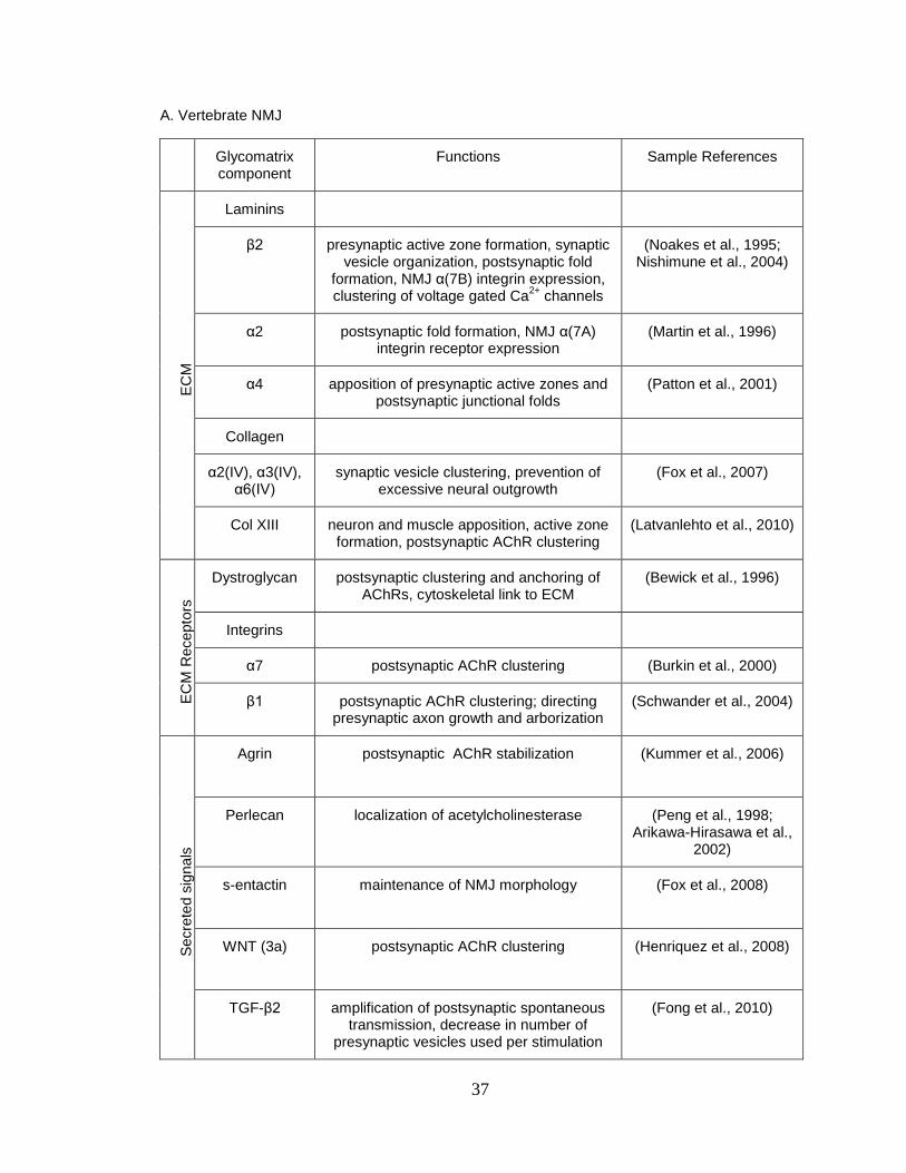
1. Neuromuscular junction synaptomatrix components ........................................ 37
2. Primary screen results ...................................................................................... 63
3. Secondary screen results ................................................................................. 69
4. Developmental phenotypes of neural glycogene knockdown ........................... 170
5. Screen targets associated with neurological disease ....................................... 173

x
LIST OF ABBREVIATIONS
GPI glycosyl phosphatidylinositol
CDG congenital disorders of glycosylation
NMJ neuromuscular junction
PSA polysialic acid
NCAM neural cell adhesion molecule
ECM extracellular matrix
SynCAM synaptic cell adhesion molecule
CAM cell adhesion molecule
GAG glycosaminoglycan
OB olfactory bulb
TRP transient receptor potential
NMDAR N-methyl-D-aspartate receptor
ERAD ER specific stress response
AMPAR α-amino-3-hydroxy-5-methyl-4-isoxazolepropionic acid receptor
LTD long term depression
LTP long term potentiation
NET norepinephrine transporter
SERT serotonin transporter
SNP single nucleotide polymorphism
DA dopamine
DAT dopamine transporter
PD Parkinson’s disease
GLYT2 glycine transporter

xi
GAT1 gamma-aminobutyric acid transporter 1
NKCC1 Na(+)-K(+)-2Cl(-) cotransporter-1
NGF nerve growth factor
AMP adenosine monophosphate
CREB cyclic AMP response element binding protein
IRS-1 insulin receptor
ALS Amyotrophic Lateral Sclerosis
NF neurofilament
AD Alzheimer’s disease
PHF paired helical filament
GSK-3β glycogen synthase kinase 3β
AGE advanced glycation end products
ADCC antibody-dependent cellular cytotoxicity
PCD para-neoplastic cerebellar degeneration
HL Hodgkin lymphoma
DNER Delta/Notch-like epidermal growth factor related receptor
GB granzyme B
HSPG heparan sulfate proteoglycan
SSR sub-synaptic reticulum
AZ active zone
BM basement membrane
GalNAc N-acetyl galactosamine
HRP horse radish peroxidase
DGC dystroglycan
LGMD limb-girdle muscular dystrophy

xii
CT carbohydrate antigen
HS heparan sulfate
WNT wingless-type MMTV integration site family TGF transforming growth factor BMP bone morphogenic protein UDPG uridyl-diphosphate-6-glucose dehydrogenase Ttv tout velu DLG discs large PSD postsynaptic density PS position specific JNK jun N-terminal kinase PIX PAK-interacting exchange factor GIT1 G protein couple receptor kinase interacting target Tkv thick veins Jeb jelly belly Gbb glass bottom boat GluR glutamate receptor Wit wishful thinking Sax saxophone Alk anaplastic lymphoma kinase Mad mothers against decapentaplegic Erk extracellular signal-regulated kinases CamKII calcium calmodulin kinase Fz2 frizzled receptor 2

xiii
MTG mind the gap RNA ribonucleic acid GAG glycosaminoglycan VDRC Vienna drosophila RNAi center TEVC two electrode voltage clamp KEGG Kyoto encyclopedia of genes and genomes EJC evoked junctional current GFP green fluorescence protein Sdc syndecan FGF fibroblast growth factor P-mad phosphorylated mothers against decapentaplegic FasII fasciclin PI propidium iodide VNC ventral nerve cord RGD arginine glycine aspartate PSP postsynaptic pocket SV synaptic vesicles PTP post tetanic potentiation VVA Vicia villosa agglutinin HPL Helix pomatia lectin CHIEF channelrhodopsin IEF variant RAD arginine alanine aspartate peptide RGD arginine glycine aspartate peptide

xiv
Brp bruchpilot ER endoplasmic reticulum MOGS mannosyl-oligosaccharide glucosidase GWAS genome-wide association studies CRISPR clustered regulatory interspaced short palindromic repeat TALEN transcription activator-like effector nucleases WGA wheat germ agglutinin DBA Dolichos biflorus agglutinin FMRP fragile X mental retardation protein FAK focal adhesion kinase

1
Chapter I
INTRODUCTION
Glycans in the nervous system: A primer for the glyco-skeptic
Glycans (carbohydrates or oligosaccharides) are sugar modifications on
glycoproteins and glycolipids that richly populate all nerve cell membranes
(Figure 1). Glycans are known to play important, non-exclusive roles as ligands,
modulators and co-receptors, but nevertheless remain enormously understudied
in the context of neurobiology (Matani et al., 2007; Dityatev et al., 2010b;
Soleman et al., 2013). This oversight has inhibited dissection of the fascinating
glycan-mediated mechanisms that regulate neural development and synapse
biology, including synaptic adhesion, neurotransmission and plasticity. The need
to explore glycan mechanisms is underscored by the growing list of human
‘glycanopathies’, with a new disorder reported every 17 days on average (Freeze
et al., 2014). Indeed, well over a 100 heritable genetic disorders result from
mutations in genes encoding products that catalyze and regulate glycans,
including O-fucosylation, O-GalNAcylation, O-GlcNAcylation, N- glycosylation,
glycosaminoglycans, GPI-Anchors and dystroglycans (Figure 2). Surprisingly,
there is a clear predominance of neural defects in congenital disorders of
glycosylation (CDG) disease states (Freeze et al., 2014). Here I illustrate, glycan-
mediated regulation of the nervous system, from molecules to behavior.

2
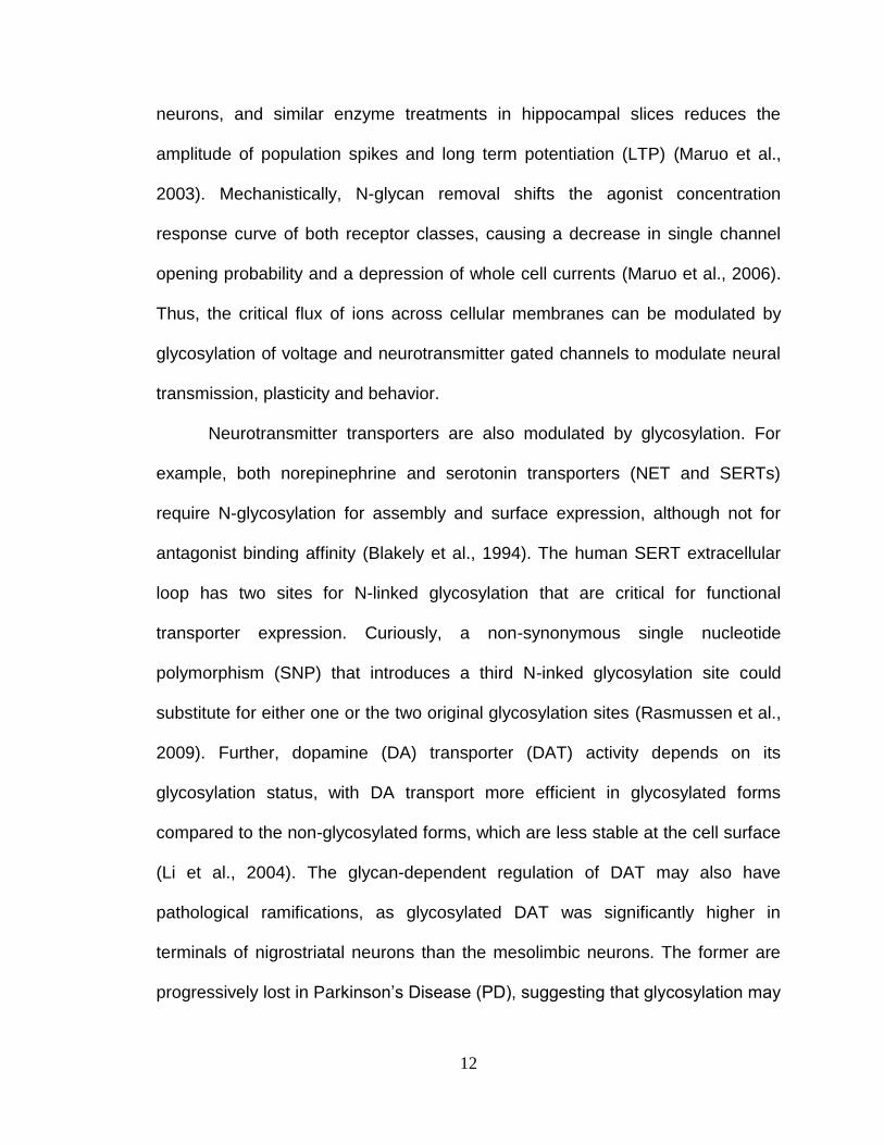
Figure 1. Glycocalyx of the cell membrane. Sugars boxed in teal are added
in the endoplasmic reticulum. Other sugars are added during passage through
the golgi. Abbreviations: mannose (Man), galactose (Gal), glucose (Glc), N-
acetylglucosamine (GlcNAc), glucosamine (GlcNH2), glucuronic acid (GlcA),
iduronic acid (IdoA), N-acetylgalactosamine (GalNAc), xylose (Xyl), fucose
(Fuc), sialic acid (Sia), 3-O-sulfated (3S), 6-O-sulfated (6S) and phosphate
(PO4-). (Modified from Stanley, 2011)

3
Figure 2. Neuroglycobiology publications and glycanopathies. (A)
Number of papers with the search terms ‘glycosylation’ and ‘neuron’ in
PubMed from 1971 to 2013. (B) Distribution shows the rate of identified
dystroglycanopathies per year. (Adapted from Freeze et al. 2014)

4
It is my hope that these illustrated glycan roles will convince the glyco-
skeptic, and illuminate the bounty that awaits explorers in the nascent field of
neuroglycobiology. A PubMed search including the terms ’glycosylation’ and
‘neuron’ yields a little over thousand articles from 1973 till present, clearly
highlighting the infancy of this field (Figure 2). Here, I review with specific
examples, some of the relatively well understood glycan-mediated effects on
neural cell adhesion, neurotransmission and mechanisms underlying neural
disease. This general overview (Chapter 1) is followed by a much more focused
discussion of glycan functions at peripheral neuromuscular synapses in
vertebrate and invertebrate systems (Chapter 2). The latter topic has been my
focus during characterization of glycogene effects on synapse structure, function
and plasticity at the Drosophila neuromuscular junction (NMJ) model synapse.
Glycosylation spatiotemporally regulates neural cell adhesion
Glycans form a dense glycocalyx layer on all cell surfaces (Varki et al.,
1999). Given their location, one would predict glycan macromolecules to be
obvious key regulators of cellular adhesion. One prime example of this function is
polysialic acid (PSA), a post-synthetic N-linked modification found on neural cell
adhesion molecule (NCAM), which decreases homophilic binding to attenuate
intercellular adhesion (Rutishauser, 1998). In vivo experiments show that
complete loss of PSA modification, by simultaneous deletion of two
polysialyltransferase genes (St8sia-II and St8sia-IV), produces severe brain
wiring defects, progressive hydrocephalus, postnatal growth retardation and

5
death. When NCAM was simultaneously deleted in this St8sia null background,
all observed phenotypes were restored to normal, identifying for the first time a
glycan to be more important than the glycoconjugate (glycan modified protein) as
a whole (Weinhold et al., 2005). Another example where glycans play a key role
in mediating adhesion is found on laminin, a major component of the extracellular
matrix (ECM) (Chen et al., 2003). Non-glycosylated laminins support cell
attachment but do not promote neural spreading or outgrowth, while glycosylated
laminins increase cell spreading is a dose dependent manner. Further,
proteolytic digestion of glycosylated laminin restores cell-spreading, suggesting
that the laminin carbohydrates provide the essential information
(Chandrasekaran et al., 1991). Glycans also control the activity of ECM receptor
integrins, which bind laminins. Expression of O-mannosyltransferases, protein O-
linked mannose N-acetylglucosaminyl-transferase 1 (PomGnT1) and N-
acetylglucosaminyltransferase-VB (GnT-Vb) all enhance β1-integrin dependent
neurite outgrowth on laminin (Abbott et al., 2006; Lee et al., 2006). Thus,
glycosylation of both ECM and ECM receptors can regulate neural adhesion,
spreading/migration and neurite outgrowth
Importantly, cell adhesion molecules (CAMs) can be multiply glycosylated
for specific roles. For example, synaptic cell adhesion molecules (SynCAMs)
mediating trans-synaptic adhesion come in multiple isoforms (SynCAM1,
SynCAM2, SynCAM3 and SynCAM4) that can form homophilic and heterophilic
complexes (Biederer, 2006). With expression of all 4 isoforms, removal of a
single N-glycosylation site (N290) in SynCAM3 increases adhesion (Gao et al.,

6
2008), while mutation of another N-glycosylation residue (Asn60) in SynCAM2
reduces adhesion (Fogel et al., 2010). Thus, N-glycans at specific positions have
differential effects on SynCAMs in regulating trans-synaptic adhesion. Likewise,
enzymatic removal of a single N-glycosylation at Asn303 in the extracellular
domain of postsynaptic Neuroligin-1 increases association with presynaptic
Neurexin-1β (Comoletti et al., 2003), showing a similar mechanism in other trans-
synaptic adhesion molecules. Moreover, modulation of glycosylation states of
secreted synaptic cleft resident proteins, such as acetylcholinesterase (AChE),
demonstrate an N-glycosylation requirement for interaction with Neurexin-1β.
Conversely, excessive glycosylation can competitively disrupt neurexin/neuroligin
adhesion to impair synapse adhesion (Xiang et al., 2014). Thus, glycans can
specifically and singularly regulate neural cell adhesion by influencing the
function of trans-synaptic, synaptic cleft resident or extracellular matrix
glycoproteins.
Glycans can mediate the malleability of cell adhesion during physiological
morphology changes associated with neural migration, axonal path finding and
plasticity. For example, the levels of sialylated PSA isoforms are temporally
regulated through development, with highly sialylated forms predominating in the
developing brain that are gradually replaced by adult isoforms with lower silaic
acid levels (Edelman, 1984). Consistently, hippocampal brain regions that
maintain morphological plasticity during learning activity, as well as regenerating
neurons, retain high density sialic acid modified NCAMs (Kiss and Rougon,
1997). Conversely, some glycans are progressively lost through development.

7
For example, in rat fetal neural cells, the N-linked glycosylated, polysialylated
and sulfated D2 CAM progressively loses sulfated forms in postnatal stages
(Lyles et al., 1984). Another example is Neuroglycan C, a brain specific
proteoglycan involved in adhesion, that loses chondroitin sulfate
glycosaminoglycan (GAG) chains during cerebellum and retinal development
(Aono et al., 2000; Oohira et al., 2004).
Glycan modifications may also be transiently present during development.
Levels of dolichyl phosphate mannose synthases that catalyzes formation of Dol-
P-P-GlnCNAc2Man9Glc3, a major substrate of N-glycosylation, are higher at day
36 than day 15 in postnatal mouse development, with a peak coincident with
synapse formation in the cerebral cortex (Idoyaga-Vargas and Carminatti, 1982).
Similarly, the ECM glycoprotein Tenascin-R associated with Purkinje neuron cell
bodies and dendrites in the molecular layer of cerebellum carries N-linked
oligosaccharides that terminate with β1,4-linked GalNAc-4-SO(4) that are
temporally regulated, increasing through cerebellar development between
postnatal days 14 and 21, corresponding to a period of Purkinje cell dendrite
extension and synaptogenesis (Woodworth et al., 2002). In mouse olfactory
epithelium sensory neurons that extend into the olfactory bulb (OB), mutants
deficient in glycosyltransferase β1,3 N-acetyl glucosaminyl transferase 1
(β3GnT1), a key enzyme in lactosamine glycan synthesis, exhibit disorganized
OB innervation and postnatal smell perception deficit (Henion et al., 2005).
However, at two weeks of age, lactosamine is unexpectedly re-expressed in
sensory neurons of mutant mice through a secondary pathway, accompanied by

8
regrowth of axons in to the OB glomerular layer and a return of smell perception
(Henion et al., 2005). Thus, glycans can spatiotemporally regulate neural cell
adhesion with increases, decreases or transient peaks of specific glycan
expression during brain development, particularly during synaptogenesis.
Glycosylation effects on neurotransmission
In addition to regulating neural development, glycans directly modulate
neurotransmission strength. For example, synaptic vesicle associated proteins
Synapsin I and II contain terminal N-acetylglucosamine (GlcNAc), and Synapsin I
is also modified by O-GlcNAc addition (Lüthi et al., 1991). When the single O-
GlcNAc site Thr-87 is mutated to alanine in primary hippocampal neurons,
Synapsin I increasingly localizes to synapses, which increases synaptic vesicle
clustering and vesicle reserve pool size (Skorobogatko et al., 2014). Similarly,
the Ca2+ sensor Synaptotagmin1 bears both N and O-linked glycosylation, and
mutational analysis reveals that the O-linked glycosylation partially targets the
protein to dense core vesicles (Kanno and Fukuda, 2008). Mutation of the N-
terminal N-glycosylation site re-directs Synaptotagmin 1 from vesicles to plasma
membrane, while transplanting this same site onto Synaptotagmin 7 re-directs
from plasma membrane to secretory vesicles (Han et al., 2004). In contrast, a
more recent analysis showed clear requirements for N-glycosylation of integral
synaptic vesicle protein SV2 in synaptic vesicle sorting, but no effects of
glycosylation on Synaptotagmin1 (Kwon and Chapman, 2012). Thus, amino acid

9
residue specific glycans can instruct cellular localization and trafficking of
synaptic vesicle proteins.
Glycans can also regulate the activity of channels to potently modulate
synaptic transmission. Sialylation of β1 subunit of voltage-gated Na+ channels
induces a uniform hyperpolarizing shift of steady state membrane potential and
kinetic gating of two alpha subunits, and reducing sialylation and N-glycosylation
impairs β1-induced gating effects (Johnson et al., 2004). Sialylation also controls
K+ channel function, as Kv1.1 sialylation causes abnormal macroscopic
activation and C-type inactivation kinetics producing a depolarized shift and
shallower voltage slope (Sutachan et al., 2005). Removal of N-glycosylated
chains from Kv12.2 in the mouse brain also causes a depolarizing shift in steady
state activation, and unglycosylated Kv12.2 is not trafficked to the cell surface
(Noma et al., 2009). Removal of glycosylation by site-directed mutagenesis of
Asn220 and Asn229 N-glycan sites on yet another K+ channel, Kv3.1, causes
differential channel distribution and the generation of outward ionic currents with
slower activation and deactivation rates than the glycosylated form (Hall et al.,
2011).
Glycans have also been increasingly identified to regulate ion channels
involved in sensory function. Insights into residue-specific effects of glycan loss
on channel function is seen in the N-glycosylated Transient Receptor Potential
Vanilloid 1 (TRPV1), which is the major determinant of capsaicin-evoked sensory
responses (Veldhuis et al., 2012). Specific de-glycosylation or site-directed
mutagenesis at residue N604 leads to rapid de-sensitization and loss of ion

10
selectivity of the TRPV1 channel (Veldhuis et al., 2012). Similarly, channel
properties are also affected in conditions of specific loss of glycosylation in
Transient Receptor Potential Melastatin 8 (TRPM8) channels (N934Q), leading to
a shift in the threshold of temperature activation and reduced response to
menthol/cold stimuli (Pertusa et al., 2012). Glycosylation also affects the
trafficking of sensory receptors. For example, in human bitter taste receptors
(TAS2R), non-glycosylated forms lacking N-glycosylation show substantially
lower cell surface localization, potentially due to reduced association with
chaperone calnexin (Reichling et al., 2008). Interestingly, site-specific
glycosylation can also differentially regulate activity-dependent function. In the
acid-sensing ion channel-1a (AISC1a), N-linked glycosylation at Asn393 and
Asn366 residues produce differential effects: Asn393 mutations increase cell
surface/dendrite trafficking, pH sensitivity and current density, and increase
dendritic targeting in N366Q mutants under conditions of acidosis-induced spine
loss, whereas N393Q mutants display the opposite effect (Jing et al., 2012).
Thus, TRP channels that respond to numerous sensory modalities are subject to
site-specific glycan-mediated control of their properties leading to perturbed
neural responses.
The Ca2+ influx fundamental to neurotransmission and plasticity is also
subject to glycan-mediated modulation(Frank, 2014). For example, voltage-gated
Ca2+ channels are modulated by alpha-2 delta subunits in which N-glycan
removal by N glycosidase F affects the current amplitude (Gurnett et al., 1996).
Intracellular Ca2+ release is also subject to glycan-mediated regulation by inositol

11
1,4,5 triphosphate (InsP3) receptor type I, which is modified by O-GlcNAc
glycosylation. Altering O-GlcNAc levels via oligosaccharyl transferase or loading
with UDP-GlcNac decreases Ca2+ channel activity, which is reversed by sugar
removal (Rengifo et al., 2007). N-glycosylation of Ca(V)3.2 T-type voltage-gated
Ca2+ channels affects function by accelerating current kinetics, increasing current
density and augmenting channel membrane expression, while de-glycosylating
this channel inhibits T-currents and reverses hyperalgesia in diabetic ob/ob mice
(Orestes et al., 2013). Thus, glycan-mediated effects on Ca2+ channel function
play critical functions across a range of channel families.
Neurotransmitter-gated channels are also modulated by glycosylation. The
cell surface expression of glutamatergic N-methyl-D-aspartate receptor (NMDAR)
can be repressed by tunicamycin treatment through regulation of NR1 but not
NR2A subunit synthesis. The inhibition of N-glycosylation activates Endoplasmic-
reticulum-associated protein degradation(ERAD), which degrades non-
glycosylated NR1 via ubiquitination and proteasome delivery (Gascón et al.,
2007). The NMDAR GluN2 subunit is also subject to glycan-mediated regulation
for synaptic targeting of the receptor, with GluN2B recruited in an activity-
dependent manner requiring N-linked glycosylation (Storey et al., 2011). The α-
amino-3-hydroxy-5-methyl-4-isoxazolepropionic acid receptor (AMPAR), another
ionotropic glutamate receptor class, is similarly regulated by O-linked GlcNAc
modification of the GluA2 subunit to modulate hippocampal long term depression
(LTD) (Taylor et al., 2014). Further, acute inhibition of N-glycosylation depresses
both NMDA and AMPA receptor currents by 30% in cultured hippocampal
12
neurons, and similar enzyme treatments in hippocampal slices reduces the
amplitude of population spikes and long term potentiation (LTP) (Maruo et al.,
2003). Mechanistically, N-glycan removal shifts the agonist concentration
response curve of both receptor classes, causing a decrease in single channel
opening probability and a depression of whole cell currents (Maruo et al., 2006).
Thus, the critical flux of ions across cellular membranes can be modulated by
glycosylation of voltage and neurotransmitter gated channels to modulate neural
transmission, plasticity and behavior.
Neurotransmitter transporters are also modulated by glycosylation. For
example, both norepinephrine and serotonin transporters (NET and SERTs)
require N-glycosylation for assembly and surface expression, although not for
antagonist binding affinity (Blakely et al., 1994). The human SERT extracellular
loop has two sites for N-linked glycosylation that are critical for functional
transporter expression. Curiously, a non-synonymous single nucleotide
polymorphism (SNP) that introduces a third N-inked glycosylation site could
substitute for either one or the two original glycosylation sites (Rasmussen et al.,
2009). Further, dopamine (DA) transporter (DAT) activity depends on its
glycosylation status, with DA transport more efficient in glycosylated forms
compared to the non-glycosylated forms, which are less stable at the cell surface
(Li et al., 2004). The glycan-dependent regulation of DAT may also have
pathological ramifications, as glycosylated DAT was significantly higher in
terminals of nigrostriatal neurons than the mesolimbic neurons. The former are
progressively lost in Parkinson’s Disease (PD), suggesting that glycosylation may

13
dictate differential vulnerability of midbrain dopaminergic cells in this
neurodegenerative disease (Afonso-Oramas et al., 2009). Disruption of the N-
glycosylation sites on the Glycine transporter (GLYT2), that removes glycine from
the inhibitory synaptic cleft, reduces activity by 35-40% (Martínez-Maza et al.,
2001). Likewise, mutations of two of the three N-glycosylation sites in the
extracellular loop of gamma-aminobutyric acid transporter 1 (GAT1) at inhibitory
synapses reduces transporter turnover (Liu et al., 1998). Inhibiting N-linked
glycosylation of the Na(+)-K(+)-2Cl(-) cotransporter-1 (NKCC1), normalizes
GABA reversal potentiation and restores GABA inhibition of presympathetic
neurons in spontaneously hypertensive rats (SHRs), and restores GABAergic
inhibition by maintaining chloride homeostasis (Ye et al., 2012). Thus,
neurotransmitter transporters represent another class of synaptic proteins
sensitive to specific glycosylation states, largely by affecting trafficking and cell
surface expression.
Interestingly, for some synaptic proteins, glycan modifications can
suppress function. For example, in neurotrophic factors responsible for neural
growth, survival and plasticity (Thoenen, 1995), the nerve growth factor (NGF)
Tyrosine Kinase Receptor 1 (TrkA) contains four N-glycosylation sites necessary
to prevent ligand independent activation and correctly localize TrkA to the cell
surface. Non-glycosylated forms are trapped intracellularly and are unable to
activate the Ras/MAP kinase signaling pathway (Watson et al., 1999). Moreover,
downstream signaling factors such as cyclic AMP response element binding
protein (CREB) known to contribute to synapse development and plasticity, also

14
exhibit glycan-mediated regulation. In response to neuronal activity, CREB is
dynamically modified by O-linked N-acetyl glucosamine and this glycosylation
represses CREB-dependent transcription (Rexach et al., 2012). Thus, glycan
modification can affect neurotransmission by positively or negatively influencing a
wide range of synaptic targets including synaptic vesicle proteins, voltage-gated
ion channels, ligand-gated ion channels, neurotransmitter transporters,
neurotrophic factors and associated downstream signaling pathways. This
modulation may arise from defects in protein folding, trafficking or expression of
the glycan-modified targets.
Novel mechanisms revealed by studying glycan related diseases
While a complete description of diseases arising from aberrant
glycosylation is beyond the scope of this overview, I will briefly discuss novel
modes of glycan-mediated regulation that are aberrant in specific disease
conditions. One such mechanism is observed in proteins where glycan
modification can affect subsequent post-translational modifications (Seet et al.,
2006). This interaction is best understood for cytosolic O-GlcNAc modifications
and phosphorylation at serine/threonine residues. The mechanism of cross-talk
can include alternative/competitive occupancy of the same residue, alternative
and reciprocal occupancy at different sites, simultaneous occupancy at different
sites, or site-dependent reciprocal (O-glycosylation or phosphorylation) or
simultaneous (O-glycosylation and phosphorylation) occupancy (Zeidan and
Hart, 2010). These mechanisms regulate neural transcription factor C/EBPβ,

15
insulin receptor IRS-1 and calcium-dependent kinase CaMKIV, affecting DNA-
binding capacity, turnover and enzymatic activity, respectively (Yang et al., 2008;
Dias et al., 2009; Li et al., 2009). Similar cross-regulation also occurs in
Amyotrophic Lateral Sclerosis (ALS) disease models, where neurofilament (NF)
proteins form intermediate filaments that are modified and regulated by
competing post-translational modifications. On a single NF subunit, O-GlcNAc
levels on the tail domain decrease with reciprocal increases in phosphorylation,
suggesting that synchronous regulation of glycosylation and
hyperphosphorylation may underlie the pathophysiological contribution
(Lüdemann et al., 2005).
Cross-talk between glycosylation and phosphorylation also appears in
Alzheimer’s disease (AD) models, in neurofibrillary plaques composed of post-
translationally modified microtubule associated Tau protein. Tau is known to form
abnormal bundles of straight filaments under conditions of hyperphosphorylation
and de- glycosylation (Arnold et al., 1996). However, restoration of normal
microtubule polymerization activity occurs only when Tau is both
dephosphorylated and de-glycosylated. Hence, hyper-phosphorylation appears
to promote aggregation of Tau and inhibit assembly of microtubules, while
glycosylation appears to stabilize the abnormal Tau paired helical filament (PHF)
structure (Wang et al., 1996). Increased non-enzymatic glycosylation of PHFs
decreases ability to bind tubules and leading to the pathological aggregations.
Further evidence of cross talk comes from in vitro studies where de-glycosylation
of aberrantly glycosylated tau decreases subsequent phosphorylation of Tau at
16
Ser214, Ser262 and Ser356 by protein kinase A. Interestingly, this de-
glycosylation of Tau positively modulates further de-phosphorylation by protein
phosphatase 2A and protein phosphatase 5 at another set of residues Ser198,
Ser199 and Ser202 (Ledesma et al., 1994). Tau protein can also be regulated by
kinase pair Cdk2/GSK-3β, such that phosphorylation of neighboring residues
S396 and S404 significantly decreases S400 O-GlcNAcylation. Reciprocally,
S400 O-GlcNacylation reduces S404 phosphorylation by Cdk2/Cyclin A3 kinase
and interrupts GSK3-β mediated sequential phosphorylation (Smet-Nocca et al.,
2011). Additionally, Tau can also be non-enzymatically glycosylated, which is
characterized by reducing sugars condensing with free amino groups of proteins,
leading to rearrangement and dehydration to forming unsaturated pigments and
cross-linked products called advanced glycation end products (AGEs) (Monnier
and Cerami, 1981; Vlassara et al., 1983; Peppa et al., 2003). AGEs are routinely
found in neurodegenerative diseases including Alzheimer’s (Smith et al., 1994),
Parkinson’s (Castellani et al., 1996), Pick’s (Kimura et al., 1996) diseases, ALS
(Kato et al., 2000) and diabetic conditions (Garlick et al., 1984), but it remains to
be identified if cross-regulatory mechanisms are also involved in these
conditions. Taken together, these studies show that glycosylation modifications
at specific residues can lead to a number of compounding effects.
Glycans can also be important for the detection of neural disorders, and
used as biomarkers for diagnosis. For example, in ALS high levels of sialylated
glycans, low levels of core fucosylated glycans and the expression of specific
glycan A2BG2 is observed in patient sera. These glycan changes increase the

17
affinity of IgG type antibodies to CD16 of effector cells leading to Antibody-
Dependent Cellular Cytotoxicity (ADCC) in brain and spinal cord tissue (Edri-
Brami et al., 2012). In this way, glycan changes correlated with ALS can serve as
an effective biomarker. Similarly, diagnostic glycan patterns in the brain occur in
para-neoplastic cerebellar degeneration (PCD) combined with Hodgkin
lymphoma (HL). In this neurological condition, anti-Tr antibodies are generated
against the Delta/Notch-like epidermal growth factor related receptor (DNER),
with the antibodies recognizing a N-glycosylation epitope (de Graaff et al., 2012).
A similar situation also arises in Rasmussen’s encephalitis, a severe form of
pediatric epilepsy, in which granzyme B(GB) serine protease is released by
activated immune cells generating the GluR3B autoantigenic peptide, as long as
no N-linked glycosylation is present within GluR3-GB recognition site (Gahring et
al., 2001). This change may serve as a prime candidate for the development of
antibodies against the N-linked glycosylation, allowing us to exploit the activation
of the particular glycan modification as a biomarker for diagnosis.
In summary, these few examples illustrate that glycans can widely
regulate neural properties at molecular, synaptic, circuit, developmental and
behavioral levels. Through these examples, we can make some general
conclusions about glycan-mediated neural effects. First, the neural proteins that
mediate these effects can be modulated either by virtue of their own glycan
modifications or by glycan-mediated regulation of their interacting partners.
Second, the same classes of glycan modification can have positive or negative
regulatory effects when attached to protein A, while being completely

18
dispensable for the function of protein B, indicating molecule-specific effects of
glycosylation. Third, multiply glycosylated proteins show position-specific effects
of loss or gain of glycan modifications, which can also influence other post-
translational modifications. Fourth, glycan modifications are spatiotemporally
regulated during normal development and in neuropathological conditions.
Moving forward, studying neuroglycobiology, particularly in genetically tractable
models, will allow for novel mechanistic characterization of these critically
important non-template driven macromolecules.

19
Chapter II
Glycosylated Synaptomatrix Regulation of Trans-Synaptic Signaling
This paper has been published under the same title in Developmental
Neurobiology, 2012
Neil Dani and Kendal Broadie
Departments of Biological Sciences, and Cell and Developmental Biology,
Kennedy Center for Research on Human Development,
Vanderbilt University, Nashville, TN 37232 USA

20
Abstract
Synapse formation is driven by precisely orchestrated intercellular
communication between the presynaptic and the postsynaptic cell, involving a
cascade of anterograde and retrograde signals. At the neuromuscular junction
(NMJ), both neuron and muscle secrete signals into the heavily glycosylated
synaptic cleft matrix sandwiched between the two synapsing cells. These signals
must necessarily traverse and interact with the extracellular environment, for the
ligand-receptor interactions mediating communication to occur. This complex
synaptomatrix, rich in glycoproteins and proteoglycans, comprises
heterogeneous, compartmentalized domains where specialized glycans
modulate trans-synaptic signaling during synaptogenesis and subsequent
synapse modulation. The general importance of glycans during development,
homeostasis and disease is well established, but this important molecular class
has received less study in the nervous system. Glycan modifications are now
understood to play functional and modulatory roles as ligands and co-receptors
in numerous tissues, however roles at the synapse are relatively unexplored. We
highlight here properties of synaptomatrix glycans and glycan-interacting proteins
with key roles in synaptogenesis, with a particular focus on recent advances
made in the Drosophila NMJ genetic system. We discuss open questions and
interesting new findings driving the current investigation of the complex, diverse
and largely understudied glycan mechanisms at the synapse.

21
Introduction
Electrically excitable cells (neurons and muscles) are precisely connected
via chemical synapses to form functional networks. Study of the neuromuscular
junction (NMJ) synapse between motor neuron and muscle cell has been
particularly instrumental in elucidating molecular mechanisms that drive
synaptogenesis, both in vertebrate and invertebrate models (Sanes and
Lichtman, 2001; Marques, 2005; Kummer et al., 2006; Collins and DiAntonio,
2007; Korkut and Budnik, 2009). Secreted glycoproteins (GPs) and
proteoglycans (PGs) interface with presynaptic and postsynaptic cell surfaces
within the NMJ synaptic cleft and in adjacent perisynaptic domains. These highly
compartmentalized extracellular environments harbor heavily glycosylated
extracellular matrix (ECM) proteins as well as glycosylated transmembrane
receptors and outer-leaflet glycolipids, which together form the ‘synaptomatrix’
(Dityatev et al., 2010c; Vautrin, 2010). All of these sugar-coated molecules
potentially interact with the multiple bidirectional trans-synaptic signals,
themselves highly glycosylated, which must necessarily traverse this extracellular
landscape to induce and modulate synaptic development, homeostasis, plasticity
and disease (Akins and Biederer, 2006; Margeta and Shen, 2010; Shen and
Cowan, 2010; Wu et al., 2010). Recent studies have begun to reveal the
importance of glycans in enabling and directing intercellular signaling in a wide
variety of cellular contexts (Hynes, 2009; Dityatev et al., 2010c). An appreciation
of the extracellular glycan environment, including knowledge of the many glycan
22
classes and their biochemical properties, is becoming essential for the
understanding of many areas of developmental neurobiology (Varki et al., 1999).
To date, the function of glycosylated ECM components has primarily been
studied in non-neuronal cells (Kalluri, 2003; Nelson and Bissell, 2006; Hynes,
2009; Sorokin, 2010); however, a range of glycan functions are increasingly
being appreciated at both vertebrate and invertebrate synapses (Dityatev et al.,
2010a, 2010c). We have known that the synaptomatrix is rich in glycan
modifications (Vautrin, 2010), but are only now beginning to more fully
understand the function of glycans during synaptogenesis and synaptic
modulation. Glycan modifications such as glycosaminoglycans (GAGs) have well
established roles in differentiation, tissue morphogenesis and organogenesis
(Kramer, 2010). Genetic studies in mice, Drosophila and C. elegans have also
revealed developmental requirements for numerous specific monosaccharide
and polysaccharide sugar modifications including O-fucose, O-mannose (Man),
mucin-type O-glycans and N-glycans (Haltiwanger and Lowe, 2004). A true
testament to the importance of glycans arises from the growing list of human
diseases attributed to mutations in glycan biosynthetic genes (Jaeken and
Matthijs, 2007; Jaeken et al., 2009), homologs of which are actively being studied
in genetic model organisms (Altmann et al., 2001; Hewitt, 2009). For example, N-
glycan biosynthesis defects induce disease states collectively categorized as
congenital diseases of glycosylation (CDGs), with common disorders such as
metabolic syndrome and autoimmunity also tied to this glycan class (Dennis et
al., 2009). Similarly, O-linked glycosylation defects give rise to numerous

23
diseases that include the muscular dystrophy class of neuromuscular disorders
(Wopereis et al., 2006). Mechanistically, glycan modifications feature prominently
in intercellular signaling, with cell surface organization and receptor clustering
dependent on specific glycans being recognized and organized by glycan-binding
lectin proteins (Martin, 2002; Yamaguchi, 2002; Kleene and Schachner, 2004;
Patnaik et al., 2006). These precedents warrant scrutiny of these same molecular
classes during physiological processes of synaptogenesis and synaptic
modulation, as well as in synaptic disease states, which are all highly dependent
on intercellular signaling.
One way forward in the exploration of glycans and glycan-mediated
mechanisms at the synapse is to exploit the genetically-tractable Drosophila
NMJ, a reduced genetic redundancy for the inherently complex glycan
modification pathways (Hagen et al., 2009). Mammalian glycan modifications
including hybrid and sialylated N-glycans are found in Drosophila, albeit at lower
concentrations, with the majority of modifications being high- or paucimannosidic
glycans (Koles et al., 2007). Further, Drosophila and mammalian glycan
biosynthetic galactosaminyltransferases enzymes show similar substrate
preferences and share preferred sites of O-linked N-acetylgalactosamine
(GalNAc) sugar modifications on target proteins (Ten Hagen et al., 2003a).
Unbiased forward genetic Drosophila screens have already contributed to
understanding of heparan sulfate proteoglycan (HSPG) biosynthetic pathways,
which have subsequently been shown to be important for cell-signaling,
morphogenesis, metabolism and tissue repair in mammals (Bishop et al., 2007).

24
Based on the confidence of conserved glycan pathways, investigations using the
Drosophila NMJ are now poised to make significant contributions to the
systematic in vivo study of glycan functions involved in synapse formation and
modulation. The aim of this review article is to highlight synaptomatrix glycans,
glycan-interacting proteins, glycosylated ligands and their receptors, focusing on
their recently discovered roles in synapse assembly at the Drosophila NMJ. Such
studies should be of interest not only to synapse biologists, but also within other
fields of neuroscience and developmental biology, as insights derived from
glycan roles in synaptogenesis are likely to be directly relevant to other arenas of
intercellular communication in the nervous system and during global
development.
The glycosylated synaptomatrix at the neuromuscular junction
Architecture of the NMJ synaptomatrix
At the vertebrate NMJ, the primary (1°) synaptic cleft is the space between
the motor neuron and the muscle that is continuous with secondary (2°) synaptic
clefts formed by muscle cell membrane invaginations that lie apposed to the
innervating motor neuron (Patton, 2003). The Drosophila NMJ cleft has a similar
architecture, however the postsynaptic muscle folds form the sub-synaptic
reticulum (SSR) that opens into the synaptic junctional cleft adjacent to
presynaptic active zones (AZ) (Prokop, 2006). The vertebrate cholinergic NMJ 1°
cleft is generally 40-50 nm wide and contains a clearly-defined synaptic basal
lamina, or basement membrane (BM), that also occupies the 2° clefts and is

25
continuous with the ensheathing muscle lamina (Patton, 2003). In comparison,
the Drosophila glutamatergic NMJ 1° cleft is only 15-20 nm wide, and in place of
a synaptic lamina there is an electron-dense specialization found only between
the apposing presynaptic AZ and postsynaptic density (Prokop, 2006). In cross-
section, this synaptic cleft domain contains periodic densities, and in freeze-
fracture displays a highly-ordered honeycomb pattern (Prokop, 1999). At the
vertebrate NMJ, the synaptic basal lamina provides mechanical support, harbors
signaling factors and serves as a substratum during synaptogenesis (Patton,
2003). At the Drosophila NMJ, loss of the cleft synaptomatrix causes catastrophic
failure of postsynaptic assembly and a near complete silencing of functional
synapses during embryonic synaptogenesis (Rohrbough et al., 2007). These
animals are consequentially paralyzed and die as mature embryos unable to
escape the eggcase.
Synaptomatrix contains glycosylated ECM protein isoforms
Glycosylation at the vertebrate NMJ has long been studied using
fluorescently-conjugated lectins (Ribera et al., 1987; Scott et al., 1988;
Crnefinderle and Sketelj, 1993), which bind specific carbohydrates, and to a
lesser extent, with anti-carbohydrate monoclonal antibodies that detect specific
carbohydrates such as β-linked GalNAc (Martin et al., 1999b) and cytotoxic T cell
(CT) carbohydrate antigens (Lefrancois and Bevan, 1985). Both approaches
reveal restriction of synaptic carbohydrate modifications to different presynaptic
(e.g. CT1) and postsynaptic (e.g. CT2) compartments, suggesting localized
requirements for specific glycan modifications (Martin et al., 1999b). Likewise,

26
anti–heparan sulfate antibodies that recognize HSPG glycosaminoglycan
modifications show clearly distinguishable synaptic and extrasynaptic (on the
muscle, but away from the synapse) glycan environments(Jenniskens et al.,
2000). Plant and fungal lectins have been especially useful for revealing localized
sugar modifications at the vertebrate NMJ. For example, Wheat Germ Agglutinin
(WGA), Soy bean agglutinin (SBA), Concanavilin A (ConA), Griffonia simplicifolia
1 isolectin B-4 (GS-1), Limax flavus agglutinin (LFA), Peanut agglutinin (PNA)
and Dolichos biflorus agglutinin (DBA) lectins all show strong labeling of NMJ
synaptic regions compared to low labeling of extrasynaptic regions (Iglesias et
al., 1992). In addition to highlighting spatially localized glycan modifications,
these studies provide insight into specialization of the ECM associated with the
NMJ (Lis and Sharon, 1986; Iglesias et al., 1992).
Similar localized carbohydrate distributions are also seen in Drosophila.
Studies show that embryonic neuronal somata bind ConA and Limulus
polyphemus agglutinin (LPA); central and peripheral neuronal processes bind
WGA, PNA, Ulex Europeus agglutinin 1 (UEA-1) and Bauhina purpura agglutinin
(BPA) lectins; while SBA labeling is completely excluded from the nervous
system (Fredieu and Mahowald, 1994; Damico and Jacobs, 1995). At the
Drosophila NMJ, WGA and Vicia villosa agglutinin (VVA) lectins show clearly
enriched synaptic labeling (Fig. 3) (Haines and Stewart, 2007; Rushton et al.,
2009). WGA labels clearly defined extracellular punctae that are widely
distributed over the muscle surface, but is much more intense, densely-spaced
and organized immediately adjacent to presynaptic boutons (Fig. 3A). These

27
WGA domains clearly indicate that the extracellular space is compartmentalized
into glycan-specialized regions. VVA labeling is almost wholly restricted to the
NMJ, with little or no labeling in extrasynaptic domains (Fig. 3B). In clear contrast
to WGA, VVA labels a more contiguous synaptomatrix domain closely associated
with NMJ boutons. Importantly, the NMJ synaptomatrix is defined as much by the
absence of carbohydrates as their presence. PNA (Fig. 3C) and DBA (Fig. 3D)
lectins clearly and intensely label non-synaptic areas but are effectively excluded
from the NMJ. This is in contrast to vertebrate NMJ lectin labeling, where DBA
exclusively labels rat synaptic domains (Iglesias et al., 1992), indicating some
species-specific differences. DBA recognizes trisaccharide-linked GalNAc, and
does label other Drosophila neuronal tissues such as the omatidia in the
developing eye (Yano et al., 2009). The lack of DBA labeling at the Drosophila
NMJ indicates the presence of a regulated and controlled synaptic environment
that expresses specific arrangement of sugars. These studies indicate
conservation of glycan modifications, as well as the fact that differences exist in
glycan expression between vertebrate and invertebrate NMJs.
Besides charting the NMJ glycan landscape, lectins have been used to
directly identify glycan modifications on synaptic proteins. In vitro studies show
that purified synaptic laminin (s-laminin) binds WGA, ConA, Maackia amurensis
agglutinin (MAA), Ricinus communis agglutinin (RCA120), Datura stramonium
agglutinin (DSA) and Aleuria aurantia agglutinin (AAA) ((1-6)-fucose) lectins,
without binding PNA (Chiu et al., 1992). These findings illustrate the specific but
heterogeneous nature of glycan modifications present on just a single

28
Figure 3. Glycan and glycan-interacting lectin expression domains at the Drosophila NMJ.
The Drosophila wandering third instar NMJ probed with a range of lectins and antibodies in detergent-free conditions to maintain synaptomatrix integrity. A) NMJ probed with WGA lectin (red) and anti-HRP (green), which recognizes glycans associated exclusively with the presynaptic neuronal membrane. The inset shows WGA domains in the synaptomatrix surrounding a single NMJ bouton. B) VVA lectin (red) and HRP (green). The VVA labeling occupies a different domain than WGA labeling, and is very highly enriched in the NMJ synaptomatrix. C) PNA lectin (red); HRP (green). D) DBA lectin (red); HRP (green). Note that both PNA and DBA lectins do not detectably label the NMJ synaptomatrix, although strong labeling is present in adjacent tissues (not shown). E) The MTG:GFP fusion protein (green) co-labeled with anti-HRP (blue). The MTG lectin localizes to synaptomatrix punctae (arrows) surround NMJ synaptic boutons. F) Triple labeling of MTG:GFP (green), βPS integrins (red) and HRP (blue). Note the three overlapping domains in the synaptomatrix. Scale bars = 5 μm.

29
synaptomatrix molecule required for NMJ development (Table IA) (Maselli et al.,
2009). Lectin staining also has helped to visualize sugar modifications on
dystroglycan, an ECM receptor found both at vertebrate and Drosophila NMJs. At
the Drosophila synapse, VVA lectin co-localizes with dystroglycan (Haines and
Stewart, 2007). More recently, lectins such as Galanthus rivalis agglutinin (GNA),
Nicotiana tabacum (Nictaba) and Rhizoctoni solani agglutinin (RSA) have been
utilized in lectin-affinity chromatography coupled to mass spectrometric analysis
to elucidate N- and O- linked glycosylation of a large number of Drosophila
synaptomatrix components such as lamininB2, LamininA, terribly reduced optic
lobes (trol; homolog of vertebrate perlecan) and the HSPG division abnormally
delayed (dally) (Vandenborre et al., 2010). Lectins are a powerful tool for glycan
investigation with enormous scope for increased use in future Drosophila NMJ
studies.
In addition to the above mentioned glycan distributions, the specialization
of NMJ synaptomatrix stems from the presence of specific isoforms of otherwise
ubiquitous ECM glycoproteins and proteoglycans, including laminin, collagen (IV)
and perlecan. At the vertebrate NMJ, global basal lamina glycoproteins such as
lamininα2/γ1, entactin, fibronectin and perlecan are present, along with
synaptically-specialized isoforms such as laminin α4/α5/β2, collagen
α3(IV)/α4(IV)/α5(IV), neuregulin α2, synaptic entactin (s-entactin), perlecan and
agrin (Table IA). Synaptic cleft specific proteins like laminin α4, β2 and α5 are N-
glycosylated, and recent proteomic studies have established two
galactosyltransferases involved in core glycosylation of s-collagen (Chen et al.,

30
2009c; Schegg et al., 2009). GalNAc sugar modifications found exclusively in
synaptic basal lamina further indicate the importance of glycosylation of NMJ
synaptomatrix proteins (Scott et al., 1988; Hall and Sanes, 1993; Patton, 2003).
In contrast, the molecular composition of the cleft material in the Drosophila NMJ
is only poorly characterized (Table IB). Indirect evidence for vertebrate-like NMJ
specialization comes from the a number of orthologous proteins to laminin B2
(Montell and Goodman, 1989), collagen IV (Borchiellini et al., 1996), dystroglycan
(Bogdanik et al., 2008) and perlecan (Voigt et al., 2002) which have been
identified at the Drosophila NMJ. However, clear roles in Drosophila
synaptogenesis have only been tested for laminin A (Prokop et al., 1998);
mutations cause a decrease in the extent of interaction between the motor
neuron and muscle (Table 1B). Clearly, there is enormous scope for further
studies using the Drosophila genetic model.
Synaptomatrix bounding cell membranes bear glycosylated proteins
In addition to secreted ECM protein glycosylation, most of the
transmembrane proteins involved in cell adhesion and signaling during
synaptogenesis carry extensive carbohydrate modifications. For example,
Drosophila cell adhesion molecules (CAMs), such as fasciclins I-III (e.g. fasII
homolog of vertebrate neural cell adhesion molecule (NCAM)) and neuroglian
(homolog of vertebrate L1), are developmentally-regulated glycoproteins involved
in homophilic recognition, adhesion and maintenance functions during
synaptogenesis (Table IB) (Bastiani et al., 1987; Patel et al., 1987; Harrelson and
Goodman, 1988; Bieber et al., 1989; Elkins et al., 1990; Halpern et al., 1991).
31
NCAM and L1 are decorated with specific L2/HNK-1 carbohydrate moieties
(Kruse et al., 1984), and their Drosophila homologs are similarly modified and
recognized by an antibody against the horse radish peroxidase (HRP) epitope, a
fucosylated N-glycan (Fig. 3) (Jan and Jan, 1982). Interestingly, fasciclins
expressed outside developing neural tissue are not bound by HRP antibodies,
indicating these are neural-specific glycosylation pathways (Snow et al., 1987).
NCAM is also modified by polysialic acid (PSA) addition, which inhibits cell
adhesive activities (Sadoul et al., 1983). Indeed, sialic acid modifications are
particularly important in modulating the activities of membrane proteins involved
in vertebrate intercellular signaling (Rutishauser, 2008), and similarly during
Drosophila development (Roth et al., 1992). In vertebrate synapses, sialylated
glycans are present in the synaptic cleft extracellular space, where they are
involved in cell adhesion and intercellular communication (Varki and Varki, 2007;
Rutishauser, 2008; Muhlenhoff et al., 2009), although the function of synaptic
sialylation remains poorly characterized.
Conserved Drosophila sialylation biosynthetic pathways include sialic acid
phosphate synthase (Kim et al., 2002), CMP-sialic acid synthetase (Viswanathan
et al., 2006) and Drosophila sialyltransferase (DSiaT) with homology to
mammalian ST6Gal sialyltransferases (Koles et al., 2004). At the Drosophila
NMJ, DSiaT plays roles in synaptogenesis that affect the manifestation of
locomotory behavior (Repnikova et al., 2010). One aspect of this requirement is
that DSiaT modulates voltage-gated sodium channels. In vertebrates, similar
regulation involves negative charge due to polysialylation, but in Drosophila the

32
mechanism appears dependent on monosialylation (Koles et al., 2004). Sialic
acid modifications also modulate synaptogenesis independently through
regulation of CAM homophilic interactions. For example, addition of polysialic
acid to NCAM attenuates adhesion and also interferes with other CAMs, such as
L1 (Rutishauser, 1998). Recently, a screen for synaptic mutants in Drosophila
uncovered fuseless (fusl), the putative homologue of the mammalian Sialin 8-
pass transmembrane sialic acid transporter (Long et al., 2008). In vertebrates,
the monosaccharide sialic acid cleaved from sialoglycoconjugates is exported
across membranes by the Sialin transporter (Morin et al., 2004; Wreden et al.,
2005) and two inherited cognitive dysfunction diseases occur in humans when
the sialin gene is mutant (Verheijen et al., 1999). At the Drosophila NMJ, fusl
mutants display >75% reduction in evoked synaptic transmission due to a
presynaptic requirement in localizing Cacophony Ca2+ channels (Kawasaki et al.,
2000; Xing et al., 2005). The homologous vertebrate Ca2+ channel (α-1 subunit)
binds ECM laminins, to facilitate organization of presynaptic active zones (Table
IA) (Nishimune et al., 2004). At the Drosophila NMJ, the Bruchpilot protein
stabilizes active zone formation by integrating Cacophony Ca2+ channels with
intracellular components and, just like fusl mutants (Long et al., 2008), bruchpilot
mutants show reduced Ca2+ channel clustering and impaired vesicular release
(Kittel et al., 2006; Wagh et al., 2006). Since sialic acid modifications are typically
added as the terminal residue of cell surface oligosaccharides, one attractive
model is that such a carbohydrate addition to the extracellular domain of the

33
presynaptic Ca2+ channel provides a critical link to the synaptic cleft ECM, driving
active zone assembly during synaptogenesis.
Another important synaptomatrix component that is required to tether the
muscle to the ECM is dystroglycan (Dg), part of the dystrophin associated
glycoprotein complex (DGC) (Pilgram et al., 2010). In addition to binding the
intracellular cytoskeleton, the α-dystroglycan in the DGC binds multiple
extracellular synaptomatrix components such as secreted agrin (Sugiyama et al.,
1994) and perlecan (Talts et al., 1999), and transmembrane neurexin (Sugita et
al., 2001). At the Drosophila NMJ, dystroglycan facilitates the organization of
glutamate receptor (GluR) clusters in the postsynaptic domain (Table IB), and
plays roles in the organization of muscle costameres and attachment sites to the
epidermal tendon cells (Bogdanik et al., 2008). In vertebrates, mutations in at
least three glycan biosynthetic genes (POMT1 (de Bernabe et al., 2002), POMT2
(van Reeuwijk et al., 2005) and POMGnT1 (Yoshida et al., 2001)), produce hypo-
glycosylation of α-dystroglycan. Dysfunctional glycosyltransferases give rise to a
range of diseases termed dystroglycanopathies that give rise to congenital
muscular dystrophies (CGDs) and limb-girdle muscular dystrophy (LGMD)
(Martin, 2007). In vertebrates, dystroglycan serine/threonine residues are
modified by glycans Neu5Ac(α2-3)Gal(β1-4)GlcNAc(β1-2)Man(α1-O-S/T) and a
core 1 O-linked structure Gal(β1-3)GalNAc(α1-O-S/T) (Endo, 1999). Other sugar
modifications include CT carbohydrate antigen (Hoyte et al., 2002), HNK-1
antigen (Smalheiser and Kim, 1995) and Lewis-X antigen (Martin, 2003a). At the
Drosophila NMJ, mutation of the two mannosyltransferase enzymes that target

34
dystroglycan for glycosylation, tw (POMT1) and rt (POMT2) (Nakamura et al.,
2010), recapitulate synaptic phenotypes of reduced Dg function (Table IB)
(Haines et al., 2007; Shcherbata et al., 2007; Wairkar et al., 2008). These studies
highlight the utility of the Drosophila NMJ model for further study of the
glycobiology at the synapse, and as a model system for human neuromuscular
diseases arising from defects in glycan biosynthesis.
Glycosylated synaptomatrix interaction with trans-synaptic signals
The immediately obvious signal that traverses the glycosylated
synaptomatrix is the neurotransmitter itself: acetylcholine (ACh) at the vertebrate
NMJ and glutamate at the Drosophila NMJ. It was first shown in Drosophila that
neurotransmitter release from the presynaptic terminal suppresses the surface
presentation and localized clustering of its postsynaptic receptors, so that the
neurotransmitter inhibits its own receptor during synapse formation and
modulation (Featherstone et al., 2000, 2002; Augustin et al., 2007). ACh at the
vertebrate NMJ has the same effect, acting as a negative regulator of
acetylcholine receptor (AChR) surface maintenance and clustering (Misgeld et
al., 2002; Brandon et al., 2003). Recent evidence suggests that glycans like
polysialic acid can interact directly with such neurotransmitters, indicating a
putative modulatory role for these glycans with this classical trans-synaptic
signaling (Sato et al., 2010). At the vertebrate NMJ, negative ACh function is
counteracted by the action of the secreted signal agrin (Bezakova and Ruegg,

35
2003; Misgeld et al., 2005), a key player in synaptogenesis and the founding
example of secreted trans-synaptic signaling ligands.
HSPG trans-synaptic signaling
The HSPG Agrin secreted by the motor neuron is 50% sugar by weight
due to glycan modifications that include heparan sulfate chains (Tsen et al.,
1995), O-linked glycans (Parkhomovskiy et al., 2000) and N-linked glycans
(Rupp et al., 1991). Agrin induces phosphorylation of the muscle-specific kinase
(MuSK) receptor that can be inhibited by glycans Gal(β1,4)GlcNAc and
Gal(β1,3)GalNAc (Parkhomovskiy et al., 2000). MuSK also binds
Gal(β1,4)GlcNAc, which suggests that this glycan modification is required for
agrin mediated AChR stabilization during synaptogenesis (Table IA)
(Parkhomovskiy et al., 2000; Kummer et al., 2006). Other glycans such as
heparin, heparan sulfate and sialic acid show inhibitory effects that perturb agrin-
mediated AChR stabilization (Wallace, 1990; Grow and Gordon, 2000).
Treatment with enzymes that cleave sugars, such as neuraminidase (exposes
glycans Gal(β1,4)GlcNAc or Gal(β1,3)GalNAc) (Martin and Sanes, 1995) or α-
galactosidase (removes α-galactose sugars to expose lactosamines or N-
acetyllactosamines) (Parkhomovskiy and Martin, 2000), causes agrin-
independent MuSK activation and AChR stabilization (Grow et al., 1999).
Besides regulation of synaptogenesis and associated signal transduction, other
glycans attached to synaptomatrix components such as laminin-1 and -2 can
bind to agrin by both heparan-sulfate glycan-dependent and -independent
mechanisms (Table IA) (Hall et al., 1997). Further, agrin not only presents glycan

36
chains, but also binds to carbohydrates of other glycoconjugates through its
lectin domain, extending its capacity to form an inter-connected
compartmentalized meshwork at the synapse (Kleene and Schachner, 2004).
Agrin is not identifiable in the Drosophila genome. However, other secreted
HSPGs such as syndecan, as well as the GPI-anchored HSPG dally-like protein
(dlp), have been identified at the Drosophila NMJ (Table IB) (Johnson et al.,
2006; Ren et al., 2009) where they mediate axon guidance and synapse
formation (Yamaguchi, 2001; Lee and Chien, 2004; Holt and Dickson, 2005; Van
Vactor et al., 2006). The basic structure shared by HSPGs is a protein core to
which heparan sulfate glycosaminoglycan (HS) chains are attached (Bernfield et
al., 1999). GAG chains are attached to serine/threonine residues on proteins via
a linker (GlcA-Gal-Gal-Xyl) by alternate addition of glucuronic acid (GlcA) and N-
acetylglucosamine (GlcNAc) via 1,4- linkages (Lind et al., 1993). HS saccharides
are further modified by addition of sulfate groups to diversify GAG chains that
direct HSPG functions. These modifications are catalyzed by N-deacetylase/N-
sulfotransferase (Ndst), which replaces the N-acetyl group of GlcNAc with a
sulfate group (Aikawa and Esko, 1999), and then by substrate-specific iduronosyl
2-O-sulfotransferase (hs2st), glucosaminyl 6-O-sulfotransferase (hs6st) and
glucosaminyl 3-O-sulfotransferase (hs3st) (Rosenberg et al., 1997; Habuchi et
al., 2000). Along the HS chains, sulfate modifications can be concentrated into
highly sulfated domains (S domains) of 6-10 disaccharides that resemble heparin
(Maccarana et al., 1996). Only 10% of the disaccharide units are S domains,
indicating a possible spatial encoding of information by the sulfate positions on
37
A. Vertebrate NMJ
Glycomatrix component
Functions Sample References
EC
M
EC
M
Laminins
β2 presynaptic active zone formation, synaptic vesicle organization, postsynaptic fold
formation, NMJ α(7B) integrin expression, clustering of voltage gated Ca
2+ channels
(Noakes et al., 1995; Nishimune et al., 2004)
α2 postsynaptic fold formation, NMJ α(7A) integrin receptor expression
(Martin et al., 1996)
α4 apposition of presynaptic active zones and postsynaptic junctional folds
(Patton et al., 2001)
Collagen
α2(IV), α3(IV), α6(IV)
synaptic vesicle clustering, prevention of excessive neural outgrowth
(Fox et al., 2007)
Col XIII neuron and muscle apposition, active zone formation, postsynaptic AChR clustering
(Latvanlehto et al., 2010)
EC
M R
ecepto
rs
Ec
Dystroglycan postsynaptic clustering and anchoring of AChRs, cytoskeletal link to ECM
(Bewick et al., 1996)
Integrins
α7 postsynaptic AChR clustering (Burkin et al., 2000)
β1 postsynaptic AChR clustering; directing presynaptic axon growth and arborization
(Schwander et al., 2004)
Secre
ted s
ignals
Agrin postsynaptic AChR stabilization
(Kummer et al., 2006)
Perlecan localization of acetylcholinesterase (Peng et al., 1998; Arikawa-Hirasawa et al.,
2002)
s-entactin maintenance of NMJ morphology
(Fox et al., 2008)
WNT (3a) postsynaptic AChR clustering
(Henriquez et al., 2008)
TGF-β2 amplification of postsynaptic spontaneous transmission, decrease in number of
presynaptic vesicles used per stimulation
(Fong et al., 2010)

38
acetylcholine neurotransmission, negative regulator of postsynaptic AChR clustering
(Misgeld et al., 2002; Brandon et al., 2003)
B. Drosophila NMJ
Glycomatrix component
Functions Sample References
EC
M Laminin A formation of appropriate contact area
between neuron and muscle (Prokop et al., 1998)
EC
M R
ecepto
rs
Ec
Dystroglycan regulation of postsynaptic GluR subunit composition, decrease in presynaptic release of glutamate neurotransmitter,
postsynaptic protein assembly
(Bogdanik et al., 2008)
Integrins
βPS synaptic bouton formation, NMJ synapse specification, localization of postsynaptic
proteins, postsynaptic assembly
(Beumer et al., 1999, 2002)
αPS1, αPS2 formation of appropriate contact sites between nerve and muscle
(Prokop et al., 1998)
αPS3 NMJ synapse specification, synaptic bouton formation, regulation of neurotransmission strength and activity-dependent modulation
(Rohrbough et al., 2000)
Fasciclins
Fas I presynaptic arborization control, neurotransmission strength
(Zhong and Shanley, 1995)
Fas II synaptic patterning, specificity, growth, stabilization, presynaptic functional plasticity
(Davis et al., 1996; Schuster et al., 1996)
Fas III homophilic synaptic target recognition
(Kose et al., 1997)
Syndecan presynaptic terminal growth regulation
(Johnson et al., 2006)
Dally-like protein (dlp)
presynaptic active zone morphology, synaptic transmission strength
(Johnson et al., 2006)
Secre
ted
sig
na
ls
Mind-the-Gap (Mtg)
organization of synaptic cleft matrix, postsynaptic GluR localization, integrin localization, Jeb/Alk signaling regulation
(Rohrbough et al., 2007; Rushton et al., 2009; Rohrbough K. et al.,
2010)

39
Jelly-belly (Jeb)
regulation of cell adhesion proteins, BMP signaling pathway interaction
(Englund et al., 2003; Rohrbough K. et al.,
2010)
Wingless
(Wg)
presynaptic active zone formation, postsynaptic GluR distribution, activity dependent synaptic bouton formation,
regulation of spontaneous release function
(Packard et al., 2002; Mathew et al., 2005; Ataman et al., 2008; Korkut and Budnik,
2009)
Glass Bottom Boat (Gbb)
localization of presynaptic active zones, regulation of cell adhesion molecules,
regulation of spontaneous release function and neurotransmission strength
(Aberle et al., 2002; Haghighi et al., 2003; McCabe et al., 2003; Nahm et al., 2010)
Glutamate neurotransmission, negative regulator of postsynaptic GluR clustering
(Jan and Jan, 1976; Featherstone et al.,
2000, 2002; Augustin et al., 2007; Chen et al.,
2009a)
Table I: Neuromuscular junction synaptomatrix components
A) The vertebrate NMJ. The table summarizes only the major synaptomatrix components discussed in this review. B) The Drosophila NMJ. Synaptomatrix components are listed in the same order as for the vertebrate NMJ; including ECM, ECM receptors and secreted trans-synaptic signals.

40
the HS (Nakato and Kimata, 2002).
Once added, the sulfate groups can be cleaved by sulfatases (e.g. sulf1),
which selectively removes sulfates attached to particular disaccharide units
(Lamanna et al., 2007). The resultant HS sulfation profiles dictate HSPG co-
receptor functions that modulate ligand-receptor interactions (Dreyfuss et al.,
2009). For example, Fibroblast growth factor (FGF) ligand dimerization occurs on
characteristically sulfated HS sequences of 10-14 sugars (Walker et al., 1994),
and interaction of the dimerized ligand with its receptor (FGFR) is dependent on
sulfated HS (Springer et al., 1994). Structural studies confirm HS mediated
stabilization in a 2:2 tetrameric assembly between the FGF1 and FGFR2 dimers
associated with HS chains (Schlessinger et al., 2000). The role of such
modifications in directing HSPG functions during synaptic development and
modulation is a critical area for future investigation.
WNT-Wingless signaling
Numerous morphogens that are required during many phases of
development are also found to play important roles at synapses, including the
WNTs (Hall et al., 2000; Packard et al., 2002; Salinas, 2005; Henriquez et al.,
2008), fibroblast growth factors (FGFs) (Umemori et al., 2004), and Transforming
growth factor/Bone morphogenic proteins (TGF-β/BMPs) (Packard et al., 2003;
Salinas, 2003). Glycan modifications have an intimate relationship with such
classical morphogens, and there is great potential for HS modifications regulating
trans-synaptic signaling. Drosophila forward genetic screens of WNT signaling

41
pathways have identified genetic interactions with heparan sulfate (HS)
biosynthetic enzymes (Hacker et al., 2005). For example, screens for mutants
phenocopying the founding WNT wingless (Wg) mutant identified sugarless (sgl),
a uridyl-diphosphate-6-glucose dehydrogenase (UDPG) that synthesizes
glucuronic acid building blocks of HS chains, and tout-velu (ttv), a polymerase
that extends these chains (Bellaiche et al., 1998). These findings indicate a link
between HS sulfation and WNT-wingless signaling. At the Drosophila NMJ,
trans-synaptic Wg plays an important role in synaptogenesis (Fig. 4), where it
has recently been shown to mediate anterograde signaling via an unusual
exosome delivery mechanism (Korkut et al., 2009). Phenotypes of loss-of-
function mutations in this pathway include reduced number of synaptic boutons,
disrupted organization of the postsynaptic scaffold protein Discs Large (DLG), a
postsynaptic density 95 kDa (PSD-95) homolog, and glutamate receptor (GluR)
mislocalization (Fig. 4) (Ataman et al., 2008). Likewise, WNT signaling in C.
elegans regulates GluR-1 abundance in the ventral nerve cord (Juo and Kaplan,
2004). WNT signaling similarly operates at mammalian synapses, where Wnt7a
enhances synapsin I clustering and branching in cultured granule cells and
cerebellar synapses (Table IA) (Lucas and Salinas, 1997). The cognate receptor
for WNT-Wg is Frizzled 2 (Fz2), which is endocytosed upon ligand binding and
then transported to the nuclear region where its cleaved C-terminal region
translocates into the nucleus to induce transcription (Fig. 4) (Mathew et al.,
2005). WNT-Wg can also function as a retrograde signal by modulating futsch
(Drosophila microtubule associated protein 1B) via inhibition of Shaggy

42
Figure 4. Diagram of trans-synaptic signaling pathways at the Drosophila NMJ. Presynaptic: The active zone (AZ) is indicated by a T-bar. Cell membrane components include PS integrins (α3 and βPS subunits), homophilic CAM Fasciclin II (FasII) and Cacophony calcium channels (Cac). Cytoplasmic proteins include kinases JNK and Shaggy, and the MAP1B Futsch. Postsynaptic: The glutamate receptor (GluR) domain includes two GluR classes (GluRIIA and B) and potassium channels (K+). Cell membrane components include PS integrins (α1/2 and βPS) and FasII. Membrane associated and cytoplasmic proteins include scaffolding proteins Discs large (DLG) and Dock, kinase PAK and regulators PIX and GIT1, and calmodulin kinase II (CamKII). Trans-synaptic pathways: Secreted signals Wingless (Wg), Glass bottom boat (Gbb) and Jelly belly (Jeb), and their respective membrane cognate receptors Frizzled 2 (Fz2), Thickveins (Tkv)/Wishful Thinking (Wit)/Saxophone (Sax) and Anaplastic lymphoma kinase (Alk). The Frizzled nuclear import pathway is indicated as FNI. The known downstream transcription factor for Gbb is Mothers against decapentaplegic (Mad; P-Mad indicating phosphorylated form), and for Jeb is ERK (P-ERK indicating phosphorylated form). Extracellular synaptomatrix components are indicated as ECM between the presynaptic neuron and postsynaptic muscle cells.

43
(Drosophila homolog of GSK3β), hence affecting microtubule function that
modulates NMJ formation (Franco et al., 2004; Franciscovich et al., 2008)
WNT-Wg signaling also prevents ectopic synapse formation on non-target
muscle cells hence directing appropriate synaptogenesis (Table IB) (Inaki et al.,
2007). The potential role of glycans in mediating such signaling stems from
studies in Drosophila wing disc showing that WNT-Wg localization, as well as
activation of the downstream signaling pathway, is dependent on the precise
extent of HS sulfation (Reichsman et al., 1996). For example, sulf1 mutants that
cannot cleave sulfate modifications show increased Wg expression along the
dorso-ventral axis of the wing disc, supporting a role for sulfated HS chains in
regulating Wg signaling (Kleinschmit et al., 2010). By extension, HS at the
Drosophila NMJ could sequester Wg and other trans-synaptic signals, or present
them as co-receptors to their transmembrane receptors to initiate downstream
signaling processes driving synaptogenesis and subsequent synapse maturation
(Fig. 4). In support of this hypothesis, quail sulfatase (Qsulf1) can positively
influence the ability of WNT ligand to associate with its cognate receptor Frizzled
(Ai et al., 2003). A high priority is to assay for similar glycan mechanisms
regulating trans-synaptic signaling.
TGFβ/BMP signaling
TGFβ/BMP signaling is also modulated by heparan sulfate glycan
modifications (Chen et al., 2006). In mammals, BMP proteins have roles in neural
crest formation (Kishimoto et al., 1997; Dick et al., 2000; Nguyen et al., 2000)

44
and migration (Graham et al., 1994; Shah et al., 1996; Marazzi et al., 1997; Sela-
Donenfeld and Kalcheim, 2000), axon guidance (Augsburger et al., 1999; Butler
and Dodd, 2003), neurite outgrowth and synaptogenesis (Meng et al., 2002;
Endo et al., 2003). At the Drosophila NMJ, the retrograde BMP signal Glass
Bottom Boat (Gbb) is similarly required for synapse stabilization (Fig. 4) (Eaton
and Davis, 2005). Null gbb mutants, as well as mutations in its BMP receptors
(type I receptors Thickveins (Tkv) and Saxophone (Sax), type II receptor Wishful
thinking (Wit)) and BMP pathway regulators (e.g. the Cdc42 selective GAP Rich),
produce gross synaptic defects that include distorted pre- and postsynaptic
membranes, mislocalized presynaptic T-bars, reduced active zone number, and
decreased synaptic transmission at the NMJ (Fig. 4) (Marques et al., 2002;
McCabe et al., 2003, 2004; Rawson et al., 2003; Nahm et al., 2010). As above,
sulf1 activity has an activating effect on BMP signaling, suggesting that HS
modifications may have a role in this trans-synaptic signaling pathway as well.
For example, sulf1 regulates release of the BMP antagonist noggin, allowing
BMP to interact with its cognate receptor in cell culture (Viviano et al., 2004). It
should be noted that glycan regulation of these signaling pathways are based
mostly on in vitro data and are highly context specific; hence directly predicting
stimulatory and/or inhibitor roles of glycan modifications at the synapse is not
straightforward. In addition to binding, presenting and/or sequestering WNT-
wingless and BMP molecules bound to HS, these signaling molecules can be
released by the enzymatic activity of matrix metalloproteinases (MMPs) that shed
ecto-domains of HSPGs (Kainulainen et al., 1998). Other glycosylated synaptic

45
ECM components can similarly bind and present signaling factors. Thus, glycans
in the synaptomatrix could potentially serve as a repository for trans-synaptic
signaling molecules, and likely modulate known WNT and BMP signaling
pathways by sequestering away or presenting signals to cognate receptors.
Glycan-binding lectins regulate trans-synaptic signaling
Mind-the-gap: secreted lectin that organizes cell surface receptors
An exciting idea that has arisen in recent years is that endogenous lectins
(glycan-binding proteins) play critical roles in development, immunity and
intercellular signaling (Drickamer and Taylor, 1993; Marth and Grewal, 2008;
Dam and Brewer, 2010). At the Drosophila NMJ, a prime example is the N-
acetylglucosamine (GlcNAc)-binding lectin encoded by mind-the-gap (mtg)
(Rohrbough et al., 2007; Rushton et al., 2009; Rohrbough K. et al., 2010). The
MTG protein is secreted from the presynaptic terminal to reside within the
synaptic cleft, and in perisynaptic extracellular domains, where it co-localizes
with WGA (Fig. 3E,F). Consistently, both proteins share binding affinity for
GlcNAc sugar residues. Transgenic MTG-GFP is trafficked to synapses, where
the secreted protein remains near the cell surface (Fig. 3E). Ultrastructural
immunogold labeling shows secreted MTG adjacent to active zones in the
presynaptic terminal, and in the extracellular lumen of the postsynaptic SSR
(Rohrbough et al., 2007). During embryonic development, the dynamic mtg
expression pattern correlates closely with key periods of NMJ synaptogenesis,
with the expression peak coinciding with the presynaptic induction of

46
postsynaptic GluR domain assembly (Broadie and Bate, 1993a, 1993b, 1993c).
Null mtg mutants exhibit severe abrogation of the glycosylated synaptomatrix
between the presynaptic active zone and postsynaptic GluR domains, and
greatly reduced GluR localization with a corresponding loss of GluR function
(Table IB) (Rohrbough et al., 2007). Moreover, all known postsynaptic
signaling/scaffold proteins functioning upstream of GluR localization are grossly
reduced or severely mislocalized in mtg mutants, including the dPix–dPak–Dock
cascade (Parnas et al., 2001; Ang et al., 2003) and the DLG/PSD-95 scaffold
(Fig. 4) (Thomas et al., 2000; Ataman et al., 2006b; Gorczyca et al., 2007).
Neuronally-targeted mtg RNA-interference (RNAi) likewise reduces postsynaptic
assembly, whereas postsynaptically-targeted RNAi has no effect (Rohrbough et
al., 2007). Similarly, neuronally-targeted wildtype mtg in the null mutant rescues
the postsynaptic assembly loss. These data conclusively indicate that
presynaptic MTG is required for the induction of the postsynaptic pathways
driving GluR domain formation; hence it serves as an anterograde trans-synaptic
signal.
It was recently shown that direct loss of GalNAc transferase alters
Drosophila NMJ structure and function, as well as locomotory behavior (Haines
and Stewart, 2007). This work independently demonstrates that GlcNAc-
mediated interactions have key roles in synaptic maturation. Thus, together this
recent work indicates that both the GlcNAc sugar itself and GlcNAc-binding
lectins modulate synaptogenesis. Since GlcNAc is a repeating sugar unit of HS, it
seems probable that MTG regulates localization of HS-carrying proteins and

47
thereby any trans-synaptic signaling proteins bound to this lattice. With this
mechanism, MTG could coordinately regulate multiple trans-synaptic pathways
via binding GlcNAc moieties on both HSPGs and receptors such as integrins
(Fig. 3F). The fact that null mtg mutants exhibit a gross reduction or complete
absence of electron-dense synaptic matrix (Rohrbough et al., 2007) certainly
suggests disorganization/loss of multiple synaptic ECM components and ECM-
binding proteins. Consistent with this prediction, targeted presynaptic mtg
knockdown strongly decreases the level of position specific (PS) integrin synaptic
expression, present in both pre- and postsynaptic membranes (Fig. 4; Table IB)
(Beumer et al., 1999; Rohrbough et al., 2000), causing a loss of NMJ localization
of this ECM binding receptor (Rushton et al., 2009). Thus, interaction between
synaptic membranes and the ECM are weakened in the absence of MTG.
Moreover, since PS integrin receptors exist in a physical complex with the DLG
scaffold and control calcium/calmodulin dependent kinase II (CaMKII) activation
(Beumer et al., 2002), this MTG-dependent pathway also provides a mechanism
to regulate localization of postsynaptic proteins in the GluR domain during
synaptogenesis (Fig. 4). Similarly in mammals, endogenous galactose binding
lectins (galectins) bind lactosamine residues (Hirabayashi et al., 2002), such as
those found in the extracellular domain of β1 integrin chains, to organize lattice
signaling microdomains at the cell surface (Perillo et al., 1995; Chung et al.,
2000; Brewer et al., 2002; Braccia et al., 2003). Although this mechanism has not
been studied at the vertebrate NMJ, it supports the possibility of a conserved
glycan-dependent organizing event.

48
Mind-the-gap: modulator of trans-synaptic signaling
Integrins are just one component of MTG-regulated trans-synaptic
signaling. The working hypothesis is that secreted MTG organizes a GlcNAc
glycomatrix that coordinately regulates the multiple bidirectional signals that
traverse the synaptic cleft between neuron and muscle (Fig. 4). For example, it
was just recently shown that MTG strongly modulates the secreted signaling
ligand Jelly Belly (Jeb) and its receptor tyrosine kinase Anaplastic Lymphoma
Kinase (Alk), an anterograde signaling pathway from neuron to muscle (Bazigou
et al., 2007; Palmer et al., 2009; Rohrbough K. et al., 2010). In the Drosophila
nervous system, this Jeb-Alk signaling activates transcription of downstream
genes, including cell adhesion proteins and the TGFβ/BMP signal Dpp (Loren et
al., 2001; Englund et al., 2003; Lee et al., 2003; Shirinian et al., 2007). At the
Drosophila NMJ, Jeb is presynaptically secreted to reside in punctate domains
closely associated with presynaptic boutons, while its Alk receptor exhibits a
more uniform expression in the postsynaptic domain (Fig. 4) (Rohrbough K. et
al., 2010). This signaling array is set up during embryonic synaptogenesis and
maintained throughout postembryonic development. In mtg null synapses, the
Jeb signal is grossly over-expressed, with elevated levels, increased size of
extracellular domains and an increased number of these domains throughout the
synaptomatrix (Rohrbough K. et al., 2010). Conversely, the postsynaptic Alk
receptor expression is decreased, albeit to a lesser degree, potentially a
reflection of the general postsynaptic disorganization (Table IB). Although direct
mutation of the Jeb-Alk pathway has not, as yet, been demonstrated to cause

49
overt synaptogenesis defects, it is predicted that this pathway must regulate
transcription modulating synapse formation and/or maintenance. Consistent with
this hypothesis, Alk receptor signaling regulates TGFβ/BMP-dependent
transcription in C. elegans (Reiner et al., 2008). By extension, at the Drosophila
NMJ Jeb-Alk signaling could potentially regulate the TGFβ/BMP Gbb retrograde
pathway involved in synaptic modulation (Aberle et al., 2002; Haghighi et al.,
2003; McCabe et al., 2003, 2004). Of course, such a mechanism would be in
addition to the prediction that MTG directly regulates WNT-Wg and Gbb trans-
synaptic signals as they navigate the synaptomatrix. Putative interactions with
these signaling pathways have yet to be tested.
Unanswered Questions and Future Directions
The extracellular synaptomatrix is densely packed with glycoproteins and
proteoglycans in highly compartmentalized domains (Figs. 4 and 5). There are
numerous unanswered questions about the development and organization of this
environment. What is the origin of these different molecules? They could be
expressed locally (e.g. neuron, muscle and associated glia) or trapped from
distant sources (e.g. haemocytes and fat body). How are these molecules
packed into such a constrained space, and spatially organized? Synaptomatrix-
resident molecules seem prohibitively large for the narrow cleft, and show clear
regional domain distributions. What dictates selective posttranslational
modification of glycans on particular synaptomatrix proteins? Possibly the three
dimensional structure of proteins effectively masks potential glycosylation sites,
allowing only certain exposed domains to be modified. Is the glycosylation status

50
of synaptomatrix proteins locally regulated within the synaptic cleft? If so, what is
the nature of these modifications, and what mechanisms control such dynamic
shifts? Such mechanisms could lead to differential glycosylation of identical
isoforms of synaptomatrix proteins in a spatial and/or temporal manner. It must
also be remembered that protein glycosylation is likely only part of the story, as
lipids are also heavily glycosylated. For example, two Drosophila genes,
egghead (egh) and braniac (brn) (Goode et al., 1996), encode enzymes
responsible for glycosphingolipid biosynthesis, and mutation of both genes
causes clear behavioral phenotypes (Chen et al., 2007). Together, all of these
glycosylation mechanisms could affect trans-synaptic signaling.
At the Drosophila NMJ, multiple trans-synaptic signals, including WNT
(Wg) and TGF-β/BMP (Gbb), navigate through the heavily glycosylated
synaptomatrix to bind their respective receptors (Fig. 4). We propose here that
the glycan-rich environment in the synaptomatrix likely directly modulates these
trans-synaptic pathways during synaptogenesis, as has been clearly
demonstrated in other arenas of development. For example, the HSPG Dlp that
binds WNT-wingless to regulate its extracellular distribution in the developing
Drosophila wing disc (Han et al., 2005). Dlp is enriched at the NMJ, along with
the HSPG syndecan, where they control NMJ growth and presynaptic active
zone assembly (Table I) (Johnson et al., 2006). Thus, HSPGs through their
glycan modifications are here hypothesized to modulate WNT signals, likely
jointly with TGF-β/BMP signals (Rider, 2006), suggesting significant intersection
between these pathways within the synaptomatrix. In this scenario, glycan

51
structures could bind and modulate multiple trans-synaptic signals
simultaneously to coordinately regulate interactions with their cognate receptors.
These signals could individually be stimulatory or inhibitory to the process of
synaptogenesis, and therefore extracellular glycans may serve as platforms for
signal integration that allow only the net effect of bound signaling molecules to
determine synapse formation and subsequent modulation.
If trans-synaptic signals are modulated by glycans, then what modulates
the glycans? We propose here that endogenous lectins are enormously
important in shaping glycan distribution within the synaptomatrix environment.
Mind-the-gap is one such lectin secreted into the Drosophila NMJ synaptomatrix,
where it binds GlcNAc moieties (Rohrbough et al., 2007; Rushton et al., 2009).
Interestingly, HS glycan modifications are abundant in GlcNAc residues, as this
monosaccharide is repeated several times to form the final HS glycan chains
(Lamanna et al., 2007). HS chains are also be modified by sulfate groups, with
periodic highly sulfated regions (S-domains) interspersed by regions with almost
no sulfation. We propose here that sulfation could provide an additional level of
information processing to regulate trans-synaptic signaling (Lamanna et al.,
2007). By binding GlcNAc-rich HS chains, MTG is hypothesized to organize
secreted and/or membrane-bound HSPGs in the synaptomatrix, to regulate
synapse formation. Does MTG interact directly with WNT and/or TGF-β/BMP
trans-synaptic pathways through such a mechanism? Although this has not yet
been tested, these pathway’s binding site preferences may be similar to other
signaling molecules such as FGFs (Wu et al., 2003) and Antithrombin III

52
(Kuberan et al., 2002), which bind sulfate rich domains. This leaves the GlcNAc
moieties in non-sulfated regions open for recognition by MTG, which could then
drive organization of HS glycans carrying HSPGs. We have just recently shown
that Mtg strongly modulates the newly defined Jeb/Alk trans-synaptic signaling
pathway (Rohrbough K. et al., 2010), in direct support of our core hypothesis.
Clearly, the Jeb/Alk pathway could be regulated by similar glycan-dependent
mechanisms, perhaps in a combinatorial manner with other trans-synaptic
pathways. The combinatorial regulation hypothesis warrants direct investigation.
It should be abundantly clear from the above discussion that glycan
modification of the synaptomatrix plays critical roles in the formation of new
synapses and the subsequent modulation of synaptic properties. This densely-
packed extracellular compartment provides an interface environment where
glycan-glycan and glycan-protein interactions occur in a restricted space,
providing exciting scope for crosstalk between different trans-synaptic signaling
pathways. The molecular mechanisms that govern synaptogenesis may very well
intimately depend on the cumulative effects of integrated and intersecting
pathways within the synaptic glycomatrix, where glycosylation determines ligand
life-time and either facilitates or thwarts the presentation of ligand to receptor.
Further studies of the synaptic glycomatrix in the context of synaptogenesis are
expected to reveal mechanisms of cross-talk between established signaling
pathways, and to yield insights into novel signaling mechanisms that direct
synapse formation.

53
Acknowledgements
We thank Jeffrey Rohrbough and Emma Rushton for Figure 3 contributions and
help with figure construction, and for critical input on the manuscript. This work
was supported by NIH R01 grant

54
Chapter III
A Targeted Glycan-Related Gene Screen Reveals Heparan Sulfate
Proteoglycan Sulfation Regulates WNT and BMP Trans-Synaptic Signaling
This paper has been published under the same title in PLoS Genetics, 2012
Neil Dani†, Minyeop Nahm¥, Seungbok Lee¥ and Kendal Broadie†
†Departments of Biological Sciences, Cell and Developmental Biology, Kennedy
Center for Research on Human Development, Vanderbilt University, Nashville,
TN 37232, USA
¥Department of Cell and Developmental Biology, Seoul National University,
Seoul 110-740, Republic of Korea
Shortened Title: HSPG regulated trans-synaptic signaling
Keywords: Drosophila, glycobiology, transgenic RNAi, synaptogenesis,
transcription

55
Abstract
A Drosophila transgenic RNAi screen targeting the glycan genome, including all
N/O/GAG-glycan biosynthesis/modification enzymes and glycan-binding lectins,
was conducted to discover novel glycan functions in synaptogenesis. As proof-of-
product, we characterized functionally-paired heparan sulfate (HS) 6-O-
sulfotransferase (hs6st) and sulfatase (sulf1), which bidirectionally control HS
proteoglycan (HSPG) sulfation. RNAi knockdown of hs6st and sulf1 causes
opposite effects on functional synapse development, with decreased (hs6st) and
increased (sulf1) neurotransmission strength confirmed in null mutants. HSPG
co-receptors for WNT and BMP intercellular signaling, Dally-like Protein and
Syndecan, are differentially misregulated in the synaptomatrix of these mutants.
Consistently, hs6st and sulf1 nulls differentially elevate both WNT (Wingless;
Wg) and BMP (Glass Bottom Boat; Gbb) ligand abundance in the synaptomatrix.
Anterograde Wg signaling via Wg receptor dFrizzled2 C-terminus nuclear import,
and retrograde Gbb signaling via synaptic MAD phosphorylation and nuclear
import, are differentially activated in hs6st and sulf1 mutants. Consequently,
transcriptional control of presynaptic glutamate release machinery and
postsynaptic glutamate receptors is bidirectionally altered in hs6st and sulf1
mutants, explaining the bidirectional change in synaptic functional strength.
Genetic correction of the altered WNT/BMP signaling restores normal synaptic
development in both mutant conditions, proving that altered trans-synaptic
signaling causes functional differentiation defects.

56
Author summary
Glycans are sugar additions to proteins. Surrounding all eukaryotic cells,
secreted and membrane glycans form a glycocalyx that regulates cell-cell
signaling. However, the mechanisms controlling glycan-dependent intercellular
communication are largely unknown. In the nervous system, glycans play
important roles in the development and regulation of synapses mediating
intercellular communication. The Drosophila neuromuscular junction serves as a
genetically tractable synapse in which expression of glycan-related genes can be
systematically knocked down to investigate effects on synaptic morphology and
function. This study employs a transgenic RNAi screen to characterize the
synaptic requirements of 130 glycan-related genes. From this screen, two
functionally paired genes (hs6st and sulf1) that add or remove a sulfate at the 6-
O position on heparan sulfate proteoglycans (HSPGs) were identified as being
critically important for synaptic functional development. Removal of each gene
produces an opposite effect on neurotransmission strength, weakening and
strengthening communication, respectively. This mechanism controls the
synaptic expression of two HSPGs, which act as co-receptors to control the
abundance of anterograde WNT and retrograde BMP signals, which drive
intracellular signal transduction pathways regulating gene transcription to control
synaptic functional development. This screen serves as a platform for systematic
investigation of glycan mechanisms regulating synaptic development.

57
Introduction
Glycans coat cell surfaces, and glycosylation decorates secreted
molecules of the pericellular space and extracellular matrix (ECM) (Iozzo, 1998;
Varki, 2011). It is well known that glycan modifications mediate critical functions
of intercellular signaling and regulate interactions of numerous growth factors
with the ECM (Kleene and Schachner, 2004; Dityatev and Schachner, 2006).
The synthesis, modification and degradation of glycoconjugates, including O/N-
linked glycoproteins, glycosaminoglycan (GAG) proteoglycans and glycan-
binding lectins, is controlled by a dedicated cadre of genes (Varki et al., 1999;
Hagen et al., 2009). In the nervous system, these glycan-related genes play key
roles in development, including neuron fate specification, migration, formation of
axon tracts and synapse maturation (Barros et al., 2011). At synapses,
glycosylated ECM molecules, membrane receptors and outer-leaflet glycolipids
together form the highly specialized synaptomatrix interface (Dityatev and
Schachner, 2006; Vautrin, 2010), which interacts with trans-synaptic signals to
modulate synaptogenesis (Dani and Broadie, 2012).
A prime example is the classic Agrin proteoglycan, which bears heparan
sulfate (HS) chains, O/N-linked glycans and also a glycan-binding lectin domain
that binds other glycoconjugates (Rupp et al., 1991; Tsim et al., 1992; Tsen et
al., 1995). Reduction of GAG sulfation perturbs the Agrin signaling that drives
postsynaptic acetylcholine receptor (AChR) cluster maintenance at the
neuromuscular synapse (McDonnell and Grow, 2004). Likewise,
Galbeta1,4GlcNAc and Galbeta1,3GalNAc glycans inhibit Agrin signaling by

58
suppressing muscle specific kinase (MuSK) autophosphorylation, a key step
during synaptogenesis (Parkhomovskiy et al., 2000). Analogous glycan-
dependent mechanisms at the Drosophila neuromuscular synapse involve the
secreted Mind-the-Gap (Mtg) lectin, which assembles the glycosylated
synaptomatrix between presynaptic active zone and postsynaptic glutamate
receptor (GluR) domains (Rohrbough et al., 2007). This glycan mechanism
induces GluR clustering, synaptic localization of integrin ECM receptors, and
shapes trans-synaptic signaling by controlling ligand/receptor abundance
(Rushton et al., 2009, 2012; Rohrbough K. et al., 2010). Thus, many long-term
studies in vertebrate and invertebrate genetic models suggest that glycan
mechanisms are a core foundation of synapse development.
In the current study, we conducted a broad transgenic RNA interference
(RNAi) screen of synaptic glycan function, assaying requirements in both
structural and functional development of the Drosophila neuromuscular junction
(NMJ). We tested 130 genes from 8 functional categories: N-glycan, O-glycan
and GAG biosynthesis; glycosyltransferases and glycan modifying/degrading
enzymes; glycoprotein and proteoglycan core proteins; sugar transporters and
glycan-binding lectins. We found that RNAi-knockdown of genes in all eight
categories affects synaptic morphological development, with gene-specific
effects on branching, bouton differentiation and synapse area. Likewise, all eight
categories regulate synaptic functional development, with gene-specific effects
both weakening and strengthening neurotransmission. Interestingly, only a few
genes affect both structure and function, suggesting separable roles for glycans

59
in regulating these synaptogenic pathways. The results of this genomic
transgenic screen are presented as a platform from which to pursue systematic
investigation of glycan mechanisms in synaptic development.
Two genes were selected for screen validation and mechanistic
characterization; functionally-paired HS 6-O-endosulfatase (sulf1) and HS 6-O-
sulfotransferase (hs6st). RNAi knockdown and null mutants identically alter
synaptic functional development in a bidirectional manner; loss of sulf1 elevates
neurotransmission strength, whereas loss of hs6st weakens it. Heparan sulfate
proteoglycan (HSPG) targets Dally-like Protein (Dlp) and Syndecan (Sdc) (Carey,
1997; Dejima et al., 2011) are mislocalized in sulf1 and hs6st null synapses. In
other developmental contexts, the sulfation state of these HSPG co-receptors
strongly regulates WNT and BMP intercellular signaling (Yan and Lin, 2009;
Kleinschmit et al., 2010; Dejima et al., 2011). At Drosophila synapses, WNT (Wg)
is a key anterograde (Packard et al., 2002; Korkut and Budnik, 2009) and BMP
(Gbb) a key retrograde (McCabe et al., 2003; Keshishian and Kim, 2004) trans-
synaptic signal. Consistently, loss of sulf1 and hs6st differentially changes
synaptomatrix levels of Wg and Gbb, and downstream signaling into muscle and
motor neuron nuclei, respectively. Glutamate release and receptor machinery is
thereby bidirectionally altered in the two nulls. Genetic restoration of Wg/Gbb
signaling to control levels restores the bidirectional changes in synaptic functional
strength and pre-/post- synaptic differentiation in both sulf1 and hs6st nulls. We
conclude that extracellular HSPG sulfation state in the synaptomatrix is a point of
intersection between WNT/BMP trans-synaptic signaling pathways that drive

60
functional development of the neuromuscular synapse.
Results
RNAi screen of glycan-related genes identifies multiple synaptogenesis
defects
Synaptic glycans play important roles as ligands, modulators and co-
receptors regulating cell-matrix and intercellular communication (Kleene and
Schachner, 2004; Holt and Dickson, 2005; Matani et al., 2007). Differential
glycan distribution on pre- and postsynaptic surfaces, and in the cleft, of
numerous protein classes, strongly suggests that glycan mechanisms mediate
synaptic structural and functional development (Martin, 2002, 2003b; Yamaguchi,
2002). To test the genomic scope of this requirement, we used confocal imaging
and electrophysiological recording at the well-characterized Drosophila
glutamatergic neuromuscular junction (NMJ) (Keshishian et al., 1996; Gramates
and Budnik, 1999; Ruiz-Canada and Budnik, 2006) to screen the Vienna
Drosophila RNAi Center (VDRC) library of glycan-related genes (Dietzl et al.,
2007). We induced UAS-RNAi knockdown using the ubiquitous UH1-GAL4 driver
(Wodarz et al., 1995; Rohrbough et al., 2007) . We assayed morphological
defects by co-labeling for pre- and postsynaptic markers, and assayed functional
defects with two-electrode voltage clamp (TEVC) recording of neurotransmission
strength. A summary of the screen results is shown in Figure 5. Full numerical
results of the screen are shown in Table II.

61
Candidate glycan-related genes were identified and classified into eight
functional categories using the Kyoto Encyclopedia of Genes and Genomes
(KEGG) database (Kanehisa and Goto, 2000) [37] (Fig. 5). Additional genes
were added to the screen based on ortholog identification using the Information
Hyperlinked over Proteins (iHOP) database (Hoffmann and Valencia, 2004). The
candidate gene list was expanded and verified using Flybase (Tweedie et al.,
2009). From this list, genes were cross-referenced with available VDRC UAS-
RNAi transgenic lines to generate a final candidate list containing 130 genes
within eight functionally-defined categories (Fig. 5): N-glycan, O-glycan and
glycosaminoglycan (GAG) biosynthesis; glycan core proteins (HSPG core
proteins/glycoproteins); sugar transporters; glycosyltransferases; glycan
modification genes (modification and degradation of glycans); and glycan-binding
lectins. On genetic knockdown, 103 lines were viable until the wandering 3rd
instar, whereas 27 lines showed developmental lethality at embryonic and early
larval stages of development. From the 103 genetic lines characterized by
confocal microscopy and TEVC electrophysiology in the 3rd instar (Fig. 5), 21
exhibited pupal stage developmental lethality. Interestingly, >50% of pupal lethal
lines displayed statistically significant defects in NMJ synaptic morphology and
function. For all 103 larval-viable lines, synapse morphology and function was
quantified at the wandering 3rd instar NMJ (Fig. 5; Table II). Each UAS-RNAi line
driven by UH1-GAL4 in the w1118 background was compared to the genetic
control of w1118 crossed to UH1-GAL4 (UH1-GAL4 x w1118) (Dietzl et al., 2007).

62
Figure 5. Glycan-related gene RNAi screen for synapse structure/function defects. Transgenic RNAi screen interrogating effects of glycan-related gene knockdown on the morphology and function of the Drosophila neuromuscular junction (NMJ) synapse. All VDRC UAS-RNAi lines were crossed to the UH1-GAL4 driver line. Target genes are indicated by Drosophila genome CG annotation number and categorized by function. Confocal imaging of co-labeled pre- and postsynaptic markers was used to quantify NMJ architecture, including branch number, bouton number and synaptic area. TEVC electrophysiology was used to quantify evoked excitatory junctional current (EJC) amplitudes. The magnitude of fold changes compared to control (w1118×UH1-GAL4) is shown on a color scale (see legend below the two columns). Statistical significance was calculated using one-way ANOVA analysis, and displayed as p<0.05 (*), p<0.01 (**).

63
Bouton Mean Branch NMJ area EJC
Human gene Drosophila gene CG Mean SD Mean SD Mean SD Mean SD
control 22.103 4.828 3.185 0.933 190.67 57.291 279.391 53.344
MOGS n/d 1597 30 6.066 3.333 0.516 169.205 48.809 400.270 26.083
n/d Ady43A 1851 18 5.196 2.333 0.577 148.064 8.454 189.310 28.793
FUT10 FucTB 4435 35.667 1.527 3 1.155 186.213 23.67 321.383 68.022
ALG8 n/d 4542 27 4.583 4 0.633 159.583 43.879 444.660 36.252
ST6GAL1 ST6Gal 4871 28.833 7.305 3.333 0.985 213.39 67.511 365.230 119.690
FUT5 FucTA 6869 26.333 4.633 3.8 1.304 147.817 36.392 305.748 117.470
MGAT2 Mgat2 7921 22 3.742 3.5 0.837 189.773 22.672 246.187 3.809
n/d FucTD 9169 37 4 2.25 0.500 194.396 25.843 272.132 64.077
n/d egghead 9659 39 6.244 3.5 1.000 195.263 19.213 355.295 17.430
ALG11 n/d 11306 25.333 3.512 3.333 0.577 230.813 29.049 225.406 125.710
MAN1B1 n/d 11874 34 10.52 3.167 0.408 181.955 74.303 395.570 102.630
GANAB n/d 14476 28.667 33.966 3.667 0.577 216.678 33.966 223.710 22.690
n/d n/d 17173 28.5 6.807 3.6 0.548 124.356 46.355 307.180 57.973
ALG10 Alg10 32076 31 4.561 3.667 0.516 197.429 17.163 279.570 46.372
MAN1A2 α-Man-I 32684 34 4.582 2.75 0.500 169.013 33.821 333.670 66.085
GALNT10 pgant6 2103 21.5 3.834 2.833 0.577 194.424 25.064 287.176 71.023
GALNT2 pgant2 3254 24.167 7.408 3.222 0.943 204.517 16.806 329.211 74.372
n/d pgant3 4445 22.363 6.253 3.583 0.900 204.088 46.144 283.633 71.781
GALNT7 GalNAc-T2 6394 26.667 7.202 3.333 0.985 267.491 28.555 346.103 70.061
GALNT11 Pgant35A 7480 30.75 5.514 3.4 0.548 225.928 54.736 384.920 113.470
n/d GalNAc-T1 8182 26 2.898 2.833 1.169 204.585 27.409 262.726 36.814
C1GALT1 C1GalTA 9520 25.5 5.671 3.083 0.830 190.985 47.339 287.106 47.779
GALNT1 pgant5 31651 22.833 1.835 2.833 1.169 193.122 15.031 463.473 114.000
n/d pgant4 31956 21.167 4.355 3.667 1.211 216.217 3.565 241.505 58.796
OGT sxc 10392 19.833 3.43 3 0.707 193.308 44.025 241.217 7.358
RFNG fng 10580 22.167 6.853 3.333 0.516 191.667 38.482 312.147 21.118
POMT2 tw 12311 22.867 3.623 3.33 1.033 192.855 42.817 253.687 51.372
POFUT1 O-fut1 12366 23.667 0.577 3 0.817 147.801 41.089 258.148 58.650
POFUT2 O-fut2 14789 27 5.329 2.75 0.957 136.323 30.379 341.320 41.460
POGLUT1 rumi 31152 23 2.708 4.333 1.506 128.274 29.862 295.523 22.455
B3GNT1 n/d 3253 27.333 7.257 4.167 0.983 140.891 52.097 285.230 72.183
n/d GlcAT-S 3881 25.5 6.654 3.364 0.809 193.28 20.529 326.828 97.482
CHPF n/d 4351 32.333 9.688 3 0.603 206.671 40.309 292.107 47.009
HS6ST3 Hs6st 4451 32.3 6.195 3.182 0.982 160.382 51.968 221.445 76.016
SULF1 Sulf1 6725 31.614 5.335 3.1667 0.753 182.304 41.794 390.791 77.178
NDST2 sfl 8339 27.667 7.371 2.5 0.548 151.417 13.814 233.830 28.840
EXT2 Ext2 8433 24 4.328 3.25 1.422 173.777 36.755 222.140 47.690
CHSY1 n/d 9220 27.833 4.956 3.5 0.548 183.432 23.112 295.953 103.380
EXT1 ttv 10117 24.167 6.145 3.333 0.817 133.221 29.341 321.280 73.667
UGDH sgl 10072 29.5 4.68 2.5 0.548 173.36 58.236 284.297 16.373
HS2ST1 Hs2st 10234 18.5 3.391 2.5 0.837 158.557 30.162 256.106 42.834
B4GALT7 β4GalT7 11780 31 3.464 2.667 0.817 241.78 33.859 280.748 80.240
CSGALNACT1 n/d 12913 47.333 4.367 3.833 0.983 237.477 12.936 255.450 15.958
EXTL3 botv 15110 25.667 2.887 3.667 0.577 208.774 20.338 237.780 54.332
XYLT1 oxt 32300 22 5.586 3.333 0.516 178.204 47.231 271.733 28.785

64
B3GAT3 GlcAT-I 32775 26.833 5.913 3.8333 0.753 138.585 27.628 357.973 57.666
n/d Lamp1 3305 21.8 9.418 2.833 0.753 162.125 86.057 415.217 93.494
n/d Sdc 10497 28.2 5.404 3.2 1.095 175.03 43.881 369.440 23.913
BMPR1A tkv 14026 23.4 4.722 3.6 1.140 153.965 44.908 318.610 41.332
DAG1 Dg 18250 32.333 2.887 3 0.894 179.973 25.716 247.725 32.376
GPC4 dlp 32146 23.167 4.579 2.667 0.516 178.265 44.432 320.060 27.625
FUT8 FucT6 2448 27.5 8.167 3.6 0.548 154.319 61.493 339.230 58.474
PGM1 Pgm 5165 21.833 3.71 2.916 0.515 206.119 39.981 252.970 50.370
NANS Sas 5232 31.333 9.873 3.3 0.823 153.102 35.941 294.317 44.404
PIGA n/d 6401 42 8.509 2.667 0.516 206.846 46.72 344.273 52.533
UGCG GlcT-I 6437 26.667 8.083 4 1.000 216.773 16.353 233.829 79.773
n/d veg 6657 30.167 7.25 3.167 1.169 205.144 27.966 398.740 35.558
GYS1 n/d 6904 32.333 10.504 3 0.000 207.118 57.579 269.623 30.786
GMPPA n/d 8207 24.833 2.787 4.333 1.211 177.999 27.549 268.644 41.288
GPI Pgi 8251 24.333 1.528 2.333 0.577 194.383 19.923 201.820 19.608
B3GALT5 n/d 8673 27.5 3.017 3 1.095 146.754 41.517 303.453 82.266
GALT Galt 9232 30.667 3.011 3.2 0.837 153.632 28.734 293.016 25.469
UAP1 mmy 9535 25.167 2.483 2.667 0.985 182.304 28.075 315.563 82.648
PIGM n/d 9865 22.5 5.244 3.5 1.049 144.827 59.411 242.390 26.816
PIGB n/d 12006 31.167 5.115 3.667 1.211 134.796 28.835 291.793 58.065
UGT8 n/d 30438 27.5 5.822 2.833 0.753 181.873 30.45 306.521 101.000
n/d n/d 33145 17.167 4.622 2.833 0.753 128.98 32.529 319.993 113.290
n/d Hexo1 1318 25.333 2.16 3.667 0.516 215.715 25.786 364.250 31.756
n/d n/d 2135 31 3.578 3.5 1.225 167.263 53.965 320.120 63.546
HK2 Hex-A 3001 16 5.586 3.5 1.643 140.193 63.045 332.046 53.046
n/d α-Man-IIb 4606 30 5.727 3.2 0.447 190.397 34.896 250.280 38.711
FUCA2 Fuca 6128 27.333 5.033 3.75 0.500 171.796 31.487 289.288 38.343
IDUA n/d 6201 27.667 3.445 3.25 0.754 201.597 14.957 376.125 59.557
n/d n/d 6206 23.4 2.074 3.333 1.211 214.18 20.927 380.517 17.713
ARSB n/d 7402 25.166 3.189 3.333 1.506 212.965 24.657 467.046 64.949
GCK Hex-C 8094 24.4 2.51 3.667 1.033 180.536 20.949 267.080 86.281
GLB1 Gal 9092 19.25 3.304 3.636 1.027 177.423 49.938 337.420 26.981
PMM2 n/d 10688 19.33 9.416 3.167 0.753 182.76 73.797 341.740 52.213
IDS n/d 12014 21.167 4.07 2.8733 0.753 166.262 44.156 376.323 51.119
GUSB n/d 15117 22.167 4.75 3.833 1.169 147.51 35.494 295.890 43.041
GBE1 n/d 33138 30.667 7.371 3 1.000 220.125 18.167 249.926 20.682
HK1 Hex-t2 32849 31 5.568 3.333 0.577 278.224 36.517 306.070 41.116
n/d fw 1500 21.857 5.551 2.5 0.548 127.544 25.087 308.248 92.674
n/d lectin-46Cb 1652 23.333 0.5744 3 0.000 190.972 23.371 248.513 21.387
n/d lectin-21Ca 2826 31.583 6.142 3.667 1.303 180.812 30.264 246.890 12.486
n/d lectin-24Db 2958 30.333 2.733 3.167 0.753 195.685 35.978 296.626 45.587
n/d lectin-24A 3410 28.167 3.71 3.167 0.753 144.841 13.427 254.893 36.372
n/d glec 6575 28 1 2.5 0.577 161.402 48.768 237.911 40.374
n/d Nrx-1 7050 19.5 5.958 3.167 0.753 173.703 50.319 230.490 40.784
n/d lectin-28C 7106 33.833 5.492 3.167 0.983 166.873 46.092 319.500 26.068
n/d Lectin-galC1 9976 24.667 9.866 3.333 0.577 192.248 75.2809 460.623 52.526
LGALS9B galectin 11372 29.667 3.011 2.5 0.548 177.635 39.298 239.073 33.651
n/d lectin-21Cb 13686 38.167 8.377 4 0.894 248.003 106.58 300.010 38.486

65
n/d lectin-33A 16834 28 3.536 4.8 1.483 168.059 37.381 303.560 38.403
n/d lectin-30A 17011 27.667 7.767 3.333 0.577 187.305 61.135 298.137 62.090
n/d lectin-29Ca 17799 29.667 3.386 3.667 1.862 207.744 31.663 327.926 88.079
n/d lectin-37Da 33532 25.5 4.506 3.167 0.753 154.475 27.428 287.150 33.351
n/d lectin-37Db 33533 35.667 5.428 3.167 0.408 210.426 34.634 268.326 60.086
SLC2A1 Glut1 1086 25.5 3.873 3.833 0.718 181.751 30.84 358.287 24.932
SLC35A2 Csat 2675 26.5 5.648 4.167 1.169 226.021 30.701 373.160 21.740
SLC35D1 frc 3874 27.833 2.563 3.5 1.314 219.609 40.526 299.290 43.799
SLC35B2 sll 7623 27.333 3.502 3.667 0.516 195.407 35.123 277.510 10.289
SLC35C1 Gfr 9620 19.167 3.764 2.667 0.817 142.467 26.185 225.942 25.866
Table II. Primary screen results. Raw number values of the RNAi screen indicated by human ortholog name, Drosophila gene name and CG number. Mean value and standard deviation (SD) included for NMJ morphology parameters of bouton number, branch number and synaptic area, and for NMJ functional parameter of evoked excitatory junctional current (EJC) amplitude. Sample sizes ≥6 NMJs and ≥3 animals for morphology and function measurements.

66
All morphological and functional assays were done blind to genotype, with values
reported as fold-change compared to genetic control, as well as statistical
significance calculated using one-way ANOVA analyses (see color scheme;
P<0.05 (*), P<0.01 (**); Fig. 5). The data represents >6 NMJs from >3 animals
from every genotype. Synapse morphology was imaged by co-labeling with
presynaptic marker anti-horse radish peroxidase (HRP) and postsynaptic marker
anti-Discs Large (DLG). A synaptic bouton was defined as a varicosity of >2 µm
in minimum diameter labeled by both HRP and DLG, and a synaptic branch was
defined as a process containing at least two boutons (Gatto and Broadie, 2008).
NMJ branch number was the least affected morphological parameter, with only 2
of 103 genes showing a statistically significant change (Fig. 5). Many more genes
were involved in bouton development. All 27 genes showing a statistically
significant change compared to genetic control exhibited elevated bouton
numbers (Fig. 5), suggesting that glycan mechanisms primarily limit
morphological growth. Synapse area was determined by outlining the terminal
area labeled by DLG using the thresholding function in ImageJ. The majority of
gene knockdown conditions showed a decrease in NMJ area compared to
control (Fig. 5). 7 RNAi lines exhibited a statistically significant decrease in area,
whereas only 2 lines exhibited a statistically significant increase in synaptic area.
All raw values of measured morphological parameters are included in Table I.
To assay functional differentiation, the motor nerve was stimulated with a
suction electrode while the evoked excitatory junctional current (EJC) was
recorded in the muscle (Fig. 5) (Beumer et al., 1999). Nerve stimulation was

67
applied at 4V for 0.5 ms at a frequency of 0.2 Hz, with the muscle clamped at -60
mV. EJC amplitudes were calculated from recorded traces in the ubiquitously-
driven RNAi lines (w1118 background) compared to the w1118; UH1-GAL4/+
control. Recordings were obtained from >3 independent trials for each RNAi
knockdown condition. All electrophysiological screening was done blind to
genotype, with values reported as fold-change and statistical significance
calculated by one-way ANOVA analyses (see color scheme; P<0.05 (*), P<0.01
(**); Fig. 5). Genes from all eight glycan classes were identified to produce
changes in neurotransmission strength upon genetic knockdown. For the 103
larval-viable lines tested, 26 lines showed a trend towards increased
transmission strength, and 12 were statistically elevated compared to genetic
control (Fig. 5). 4 gene knockdowns showed a trend towards decreased
transmission strength, of which only 1 line reached statistical significance. 73 of
the 103 lines tested showed no change in functional strength (Fig. 5).
Interestingly, only 6 RNAi lines showed statistically significant effects on both
NMJ morphology parameters and EJC amplitude: CG1597, CG6657, CG7480,
CG4451, CG6725 and CG11874 (Fig. 5). This suggests that glycan effects on
synapse morphological and functional development are largely separable. All raw
values of EJC measurements are included in Table II.
To validate results, a secondary screen was conducted using independent
RNAi lines obtained from the VDRC and Harvard TRiP collections (Table III). Of
the 44 genes that showed morphological and functional defects in the primary
screen, 33 were retested using independent RNAi lines, with the others lacking

68
available secondary lines from any source. Using the same screen of
morphological and functional characterization, we determined that ~80% of
retested secondary lines showed the reported structural (bouton number) and
functional (EJC) phenotypes consistent with primary screen (Table II). These
primary and secondary RNAi screen results now represent a resource for the
systematic characterization of glycan mechanisms underlying synaptic structural
and functional development. Screen results were further studied by comparing
synaptogenesis phenotypes of RNAi knockdown with defined genetic nulls for
two genes, CG6725 and CG4451, from the glycosaminoglycan biosynthesis
class (Fig. 5). The RNAi screen of functional strength as measured by EJC
amplitudes indicated opposite effects for these two lines, with CG6725 (RNAi-
sulf1) knockdown exhibiting an increase in transmission strength and CG4451
(RNAi-hs6st) knockdown producing a decrease (Fig. 5). Along with our goal to
identify interesting glycan-related genes involved in synapse development, we
show here characterization of null alleles of two genes obtained from screen
results and define the associated mechanisms driving the bidirectional regulation
of synaptic functional development.
Synaptogenesis is bidirectionally regulated by paired sulf1 and hs6st
genes
The RNAi screen identified two functionally-paired genes, sulf1 (CG6725) and
hs6st (CG4451), with similar effects on morphological development but opposite
effects on synaptic functional differentiation (Fig. 5). Our goal was to use these

69
Secondary Screen - Morphology (Bouton number)
Human Gene
Drosophila Gene CG ID Fold ID Fold Confirmed
control 1 1
FUT10 FucTB 4435 GD40519 1.613 GD40520 1.162 N
n/d FucTD 9169 CG27008 1.673 CG27009 1.301 Y
n/d egghead 9659 GD10137 1.764 GD405160 1.567 Y
MAN1B1 n/d 11874 GD101661 1.538 GD4919 1.553 Y
MAN1A2 α-Man-I 32684 GD39572 1.538 GD5528 1.447 Y
PIGA n/d 6401 KK107714 1.900 GD39552 1.294 N
GYS1 n/d 6904 GD35136 1.462 GD35137 1.693 Y
GALT Galt 9232 KK100025 1.387 GD29087 1.494 Y
n/d n/d 2135 KK103338 1.402 GD16628 1.241 N
n/d α-Man-IIb 4606 GD42652 1.357 KK108043 1.338 Y
HK1 Hex-t2 32849 KK100218 1.402 GD47331 1.640 Y
n/d lectin-24Db 2958 KK105118 1.372 GD45294 1.4403 Y
n/d lectin-21Cb 13686 KK106450 1.726 GD32507 1.513 Y
n/d lectin-37Db 33533 KK107567 1.613 GD51100 1.617 Y
Secondary Screen - EJC
Human Gene
Drosophila Gene CG ID Fold ID Fold
Confirmed
control
1
1
ALG8 n/d 4542 KK104870 1.782 GD7132 1.333 Y
ST6GAL1 ST6Gal 4871 KK100284 1.464 GD47955 0.978 N
MAN1B1 n/d 11874 KK101661 1.586 GD4419 1.214 Y
GALNT1 pgant5 31651 GD2629 1.858 KK110647 1.090 N
HS6ST3 hs6st 4451 KK101636 0.796 GD42658 0.658 Y
SULF1 sulf1 6725 GD37362 1.389 GD37361 1.326 Y
B3GAT3 GlcAT-I 32775 KK107840 1.435 TRiP.HMS00289 1.280 Y
n/d Lamp1 3305 GD7309 1.664 TRiP.GLV21040 1.404 Y
n/d Lectin-galC1 9976 GD38002 1.846 KK100935 1.680 Y
Table III. Secondary screen results. Raw number values for the secondary
screen results indicated by human ortholog name, Drosophila gene name and CG
number. The two independent IDs for RNAi lines are shown. For all retested lines,
morphological quantification for NMJ bouton number (top) and evoked excitatory
junctional current (EJC) amplitude (bottom). All results are shown as fold-changes
compared to genetic control. Sample sizes are ≥6 individual animals per
genotype. Replication of primary screen result is indicated in the final column as
Y, and failure to replicate indicated as N.

70
genes as a test case from the completed glycan screen, by assaying phenotypes
in recently characterized null mutants of both genes (Kamimura et al., 2001; You
et al., 2011). The gene products Sulfated (Sulf1), an HS 6-endosulfatase, and
Hs6st, an HS 6-O-sulfotransferase, drive opposing changes in sulfation state of
the same C6 carbon of the repeated glucosamine unit in GAG modified heparan
sulfate proteoglycans (Kamimura et al., 2001; Ai et al., 2003). Viable null mutants
are available for both genes, e.g. sulf1 (sulf1∆1) and hs6st (hs6std770) (Kamimura
et al., 2001; You et al., 2011), but requirements have never been assayed in the
nervous system or neuromusculature. We therefore first compared phenotypes of
RNAi knockdown and null alleles at the NMJ synapse by confocal imaging of
synaptic morphogenesis and TEVC recording of synaptic functional
neurotransmission.
Using double-labeling for HRP (presynaptic) and DLG (postsynaptic), NMJ
structural parameters including bouton number, branch number and synaptic
area were quantified in sulf1 and hs6st null alleles. The mutant results closely
recapitulated the RNAi knockdown findings from the screen (Table II, III). To
consistently compare RNAi and null mutant conditions, both animal groups were
simultaneously reared and processed to visualize the NMJ (Fig. 6). Structural
quantification showed an increased bouton number with RNAi-mediated sulf1
knockdown (sulf1-RNAi x UH1-GAL4; 36.4±1.6, n=10) and hs6st knockdown
(hs6st-RNAi x UH1-GAL4; 35.1±1.96, n=10) compared to the transgenic control
(w1118 x UH1-GAL4; 21.9±1.84, n=10 p<0.001, n=10; Fig. 6A,B). Consistently,
increased bouton number was observed in both sulf1 (31.9 ± 1.37, n=10) and

71
hs6st (36.25 ± 2.58, n=8) null mutants compared to genetic control (w1118, 19.3 ±
1.69, p<0.001, n=10; Fig. 6C,D). In contrast, no significant change in branch
number was exhibited with sulf1 knockdown (3.22 ± 0.28, p>0.05, n=9) or hs6st
knockdown (3.22 ± 0.22, p>0.05, n=9) compared to control (w1118 x UH1-GAL4;
2.64 ± 0.06, n=11). Similarly, no significant change was observed in the synaptic
branch number in sulf1 (2.8 ± 0.33, p=0.27, n=10,) and hs6st (3.63 ± 0.38,
p=0.115, n=10) nulls compared to control (w1118; 3.4 ± 0.46, n=8). Further, there
was no significant difference in synaptic area in sulf1 (138.16 ± 5.82, p>0.05,
n=10,) and hs6st (138.48 ± 13.38, p>0.05, n=8,) mutants compared to the control
(w1118 ; 118.04 ± 8,38, n=10), however a slight increase in synaptic area was
observed in sulf1 knockdown (178.68 ± 10.64, p<0.05, n=9), while no change
was observed for hs6st knockdown (164 ± 8.47, p>0.05, n=10) as compared to
control (w1118 x UH1-GAL4; 134.57 ± 11.95, n=10). Based on these imaging
studies, we conclude morphological differences in synaptic architecture observed
in both sulf1 and hs6st null allele conditions is consistent with both RNAi
knockdown conditions.
Functional development was next tested with electrophysiological
recording to compare RNAi and null mutant phenotypes (Fig. 7). Representative
TEVC records are shown as an average of 10 consecutive nerve stimulus
responses in 1.0 mM extracellular Ca2+ for each transgenic genotype in Figure
7A; sulf1 knockdown (UH1-GAL4 x sulf1-RNAi), hs6st knockdown (UH1-GAL4 x
hs6st-RNAi) and genetic control (UH1-GAL4 x w1118). There was a striking ~80%
difference in EJC amplitude between sulf1 and hs6st knockdown conditions, with

72
Figure 6 NMJ synaptic bouton number in sulf1 and hs6st mutants. (A) Representative NMJ images from muscle 4 in segment A3 showing anti-horseradish peroxidase (HRP; red) and anti-Discs Large (DLG; green) in control (w1118×UH1-GAL4), sulf1 RNAi (UH1-GAL4×UAS-CG6725) and hs6st RNAi (UH1-GAL4×UAS-CG4451). (B) Quantification of synaptic bouton number in RNAi-knockdown conditions for sulf1 and hs6st, normalized to genetic control (w1118×UH1-GAL4). Sample sizes are ≥10 animals per indicated genotypes. (C) Representative NMJ images of anti-HRP (red) and anti-DLG (green) in w1118 control, sulf1 and hs6st null mutants. (D) Quantification of synaptic bouton number in mutant conditions normalized to genetic control. Sample sizes are ≥8 animals per indicated genotype. Statistically significant differences were calculated using student's t-test and indicated as ***p<0.001. Error bars indicate S.E.M.

73
Figure 7. Loss of sulf1/hs6st causes opposite effects on transmission strength. (A) Representative excitatory junctional current (EJC) traces from control (w1118×UH1-GAL4), sulf1 RNAi (UH1-GAL4×UAS-CG6725) and hs6st RNAi (UH1-GAL4×UAS-CG4451). The nerve was stimulated (arrows) in 1.0 mM external Ca2+, with TEVC records (−60 mV holding potential) from muscle 6 in segment A3. Each trace averaged from 10 consecutive recordings. (B) Quantified mean EJC amplitudes (nA) for the three genotypes shown in panel A. (C) Representative traces from control (w1118), sulf1Δ1 and hs6std770 null alleles under the same conditions described in panel A. (D) Quantified mean EJC amplitudes (nA) for the three genotypes shown in panel C. Sample sizes are at least 11 animals per indicated genotype. Statistically significant differences calculated using student's t-test, ** p<0.01, *** p<0.001. Error bars indicate S.E.M.

74
sulf1 elevated by ~30% and hs6st reduced by ~30% compared to control.
Quantification of EJC amplitudes showed both knockdown conditions to
be highly significantly different from control and each other (control, 286.22 ±
8.56 nA; sulf1-RNAi, 365.01 ± 9.502 nA, p<0.001; hs6st-RNAi, 199.19 ± 11.84
nA, p<0.001; sulf1-RNAi vs. hs6st-RNAi, p<0.001; Fig. 7B). These opposite
effects on neurotransmission strength were confirmed in characterized null
alleles for both genes [42,43]. Representative traces from sulf1∆1 and hs6std770
null mutants compared to w1118 control are shown in Figure 7C. Quantification of
EJC amplitudes showed null mutants to be highly significantly different from
control and each other (w1118, 256.14 ± 7.38 nA; sulf1∆1, 372.86 ± 18.49 nA,
n=11, p<0.001; hs6st, 209.66 ± 13.44 nA, n=14, p<0.01; sulf1∆1 vs. hs6st,
p<0.001; Fig. 7D). These results were confirmed in an independent sulf1 null
allele (sulf1∆P1), which shows comparable elevation compared to control (w1118,
244.91 ± 9.04 nA; sulf1∆P1, 282.28 ± 13.59, p<0.05, n=22), as well as the hs6st
null (hs6std770) over deficiency (Df(3R)ED6027), which shows comparable
depression compared to control (w1118, 256.14 ± 7.38 nA; hs6st/Df(3R)ED6027,
224.06 ± 7.65 nA, p<0.05, n=18). These results reveal a critical role for sulf1 and
hs6st genes in synaptic functional development.
Given the functionally-paired nature of sulf1 and hs6st activities on 6-O-S
modification, and the epistatic function of hs6st to sulf1, we predicted that
knocking both genes down would produce a phenotype similar to knockdown of
hs6st alone. Consistently, hs6st and sulf1 double knockdown produced EJC
amplitudes significantly lower than control (w1118 x hs6st-RNAi; sulf1-RNAi

75
(control), 225.17 ± 6.28 nA, n=12; hs6st-RNAi, sulf1-RNAi x UH1-GAL4, 198.22
± 9.77 nA, n=15, p<0.05; Fig. 8). Cell-specific knockdown in neural (elav-GAL4),
muscle (24B-GAL4) and glia (repo-GAL4) also support the observed opposite
effects in neurotransmission strength. With sulf1 knockdown in muscle, EJC
amplitude was significantly elevated compared to control (w1118 x sulf1-RNAi
(control), 199.97 ± 21.86 nA; 24B-GAL4 x sulf1-RNAi (knockdown), 222.88 ±
25.78 nA, p<0.01, n=10), but no change occurred with neural knockdown (elav-
GAL4 x sulf1-RNAi, 196.09 ± 25.08 nA, p=0.72, n=10) or glial knockdown (repo-
GAL4 x sulf1-RNAi, 208.40 ± 32.45 nA, p=0.53, n=7). Moreover, only neural
knockdown of hs6st caused a decrease in EJC amplitude (w1118 x hs6st-RNAi
(control), 211.496 ± 22.142 nA, elav-GAL4 x hs6st-RNAi (knockdown), 184.68 ±
28.97 nA, p<0.05, n=16), while no change occurred with muscle knockdown
(24B-GAL4 x hs6st-RNAi, 209.92 ± 24.74 nA, p=0.88, n=9) or glial knockdown
(repo-GAL4 x hs6st-RNAi, 216.38 ± 37.80 nA, p=0.32, n=7). We conclude that
HSPG sulfation state strongly modulates NMJ functional development, with
contributions from both motor neuron and muscle, but not glia. The clear next
step was to test for differences in the localization and abundance of synaptic
HSPG targets known to regulate NMJ synaptogenesis.
HSPG abundance at the synaptic interface is dependent on sulf1 and hs6st.
Both GPI-anchored HSPG glypican Dally-like (Dlp) and transmembrane HSPG
Syndecan (Sdc) are clearly expressed at the Drosophila NMJ (Fig. 9), where they
are known to regulate synaptogenesis (Johnson et al., 2006). We detect no
enrichment of the secreted HSPG perlecan (Trol) at the NMJ, although

76
Figure 8. Double knockdown of sulf1 and hs6st measure of EJC amplitude. (A) Representative evoked excitatory junctional current (EJC) traces from control (w1118×UH1-GAL-4) and double knockdown with both sulf1 and hs6st RNAi transgenic lines (UH1-GAL4×UAS-sulf1-RNAi; UAS-hs6st-RNAi). (B) Quantified mean EJC amplitudes (nA) for the two genotypes shown in panel A normalized to control. Sample sizes are ≥12 animals per indicated genotype. Statistically significant differences calculated using student's t-test, * p<0.05, Error bars indicate S.E.M.

77
it is abundantly expressed in the motor nerve leading up to the synaptic terminal
and present in lower levels throughout the muscle (Fig. 10). We therefore
hypothesized that membrane-associated Dlp and Sdc HSPGs are targeted by
sulf1 and hs6st activity to regulate their synaptic distribution and/or function. To
test this hypothesis, we assayed both Dlp and Sdc under non-permeabilized,
detergent-free conditions to examine their cell surface expression at the NMJ
synaptic interface of sulf1 and hs6st null mutants compared to control. These
data are summarized in Figure 11.
In the genetic background control (w1118), Dlp shows a punctate
expression pattern strongly concentrated in a halo-like array around the anti-HRP
labeled presynaptic membrane (Fig. 9, Fig. 11, top). In sulf1 mutants there was a
clear and consistent increase in Dlp abundance, with more numerous and
intense punctae at the synaptic interface surrounding NMJ boutons, while at
hs6st mutant synapses there was an opposing decrease in Dlp abundance (Fig.
11). This bidirectional and differential effect on Dlp abundance was quantified as
fluorescence intensity normalized to the internal HRP labeling control. There was
a significant Dlp increase in sulf1 compared to control (~40% elevated over
control; p<0.05; n=11), and a significant Dlp decrease in the hs6st null synapse
(~15% reduced compared to control; p<0.05; n = 11; Fig. 11B). Importantly, the
difference between sulf1 and hs6st nulls was very highly significant (p<0.001). In
comparison, cell surface Sdc labeling also showed a dense halo-like localization
around NMJ synaptic boutons labeled with cell adhesion marker Fasciclin II

78
Figure 9. NMJ synaptic localization of Dally-like and Syndecan HSPGs. Representative confocal images showing HSPG synaptic localization at the larval NMJ. (A) Single channel images of presynaptic anti-horseradish peroxidase (anti-HRP, blue), Dally-like Protein (anti-Dlp, green) and postsynaptic glutamate receptor subunit IID (anti-GluRIID, red). (B) Single channel images showing presynaptic anti-horseradish peroxidase (anti-HRP, blue), syndecan (anti-Sdc, red) and postsynaptic Discs Large (anti-DLG, green). (C) Merged image showing Dlp localization with respect to presynaptic HRP, postsynaptic GluRIID and the triple-labeled terminal. (D) Merged image showing Sdc localization with respect to presynaptic HRP, postsynaptic DLG and the triple-labeled terminal.

79
Figure 10. HSPG Perlecan (Trol) is absent from the NMJ synaptic terminal. (A) Representative confocal image showing Perlecan expression at the wandering third instar larval NMJ using the Trol-GFP Flytrap line ZCL1700 from the Flytrap GFP Resource. Single channel and merged images show presynaptic anti-horseradish peroxidase (anti-HRP, red) and Trol-GFP (green). (B) Representative confocal image showing Perlecan (anti-PcanV) antibody staining, shown at a much higher confocal gain than in A to emphasize muscle expression. Perlecan is strongly expressed in the motor nerve, and clearly present on the muscle surface, but is never detectably enriched at the NMJ terminal. In many cases, as in the example shown, Perlecan appears at lower levels in the perisynaptic region surrounding the NMJ than elsewhere on the muscle.
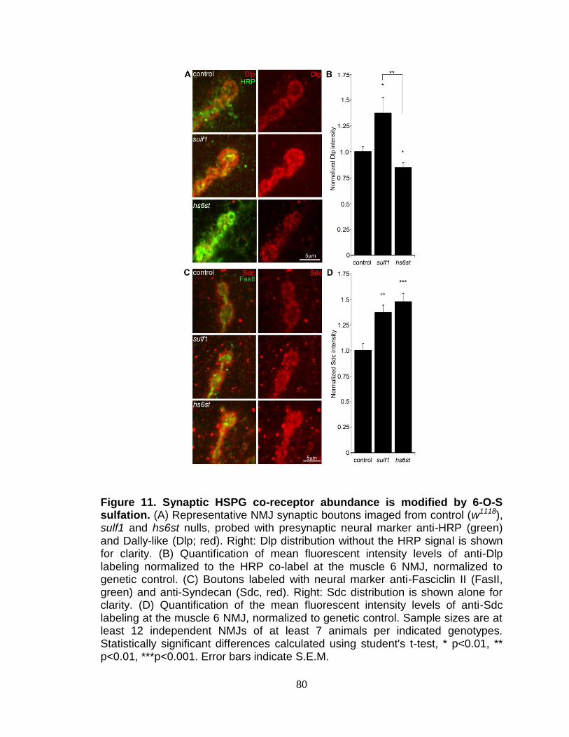
80
Figure 11. Synaptic HSPG co-receptor abundance is modified by 6-O-S sulfation. (A) Representative NMJ synaptic boutons imaged from control (w1118), sulf1 and hs6st nulls, probed with presynaptic neural marker anti-HRP (green) and Dally-like (Dlp; red). Right: Dlp distribution without the HRP signal is shown for clarity. (B) Quantification of mean fluorescent intensity levels of anti-Dlp labeling normalized to the HRP co-label at the muscle 6 NMJ, normalized to genetic control. (C) Boutons labeled with neural marker anti-Fasciclin II (FasII, green) and anti-Syndecan (Sdc, red). Right: Sdc distribution is shown alone for clarity. (D) Quantification of the mean fluorescent intensity levels of anti-Sdc labeling at the muscle 6 NMJ, normalized to genetic control. Sample sizes are at least 12 independent NMJs of at least 7 animals per indicated genotypes. Statistically significant differences calculated using student's t-test, * p<0.01, ** p<0.01, ***p<0.001. Error bars indicate S.E.M.

81
(FasII; Fig. 9, Fig. 11C). Synaptic Sdc labeling intensity was consistently greater
in both sulf1 and hs6st nulls compared to control (Fig. 11). Quantification of
fluorescence intensity normalized to HRP revealed that Sdc abundance was
greatly increased in sulf1 null synapses compared to control (~35% elevated over
control; p<0.01; n=17) and, to a greater degree, also in hs6st nulls (~50%
elevated over control; p<0.001; n=12; Fig. 11D). Thus, both Dlp and Sdc HSPGs
are strongly altered in sulf1 and hs6st null NMJ synapses, with Dlp bidirectionally
misregulated and Sdc differentially elevated in the two mutant conditions.
HSPGs act as co-receptors for WNT and BMP intercellular signaling
ligands in many developmental contexts, acting to modulate extracellular ligand
abundance and downstream signaling (Lin and Perrimon, 2000; Hacker et al.,
2005). Drosophila WNT Wingless (Wg) distribution and signaling is known to be
modulated by Dlp, which retains Wg at the cell surface in a mechanism that is
enhanced by HS GAG chains (Yan et al., 2009). Specifically, Wg ligand
abundance and signaling activity along the dorso-ventral axis of the developing
Drosophila wing disc is elevated in sulf1 mutants (Kleinschmit et al., 2010).
Likewise, BMP ligands in other cellular contexts are closely regulated by HSPG
co-receptors (Dejima et al., 2011). Specifically, Dlp has been suggested to
similarly regulate Drosophila BMP Glass Bottom Boat (Gbb) (Dejima et al.,
2011). We therefore hypothesized that altered HSPG co-receptors Dlp and/or
Sdc in sulf1 and hs6st null synapses regulate Wg and Gbb abundance to drive
differentially altered trans-synaptic signaling across the synaptic cleft.

82
HSPG sulfation regulates abundance of WNT/BMP trans-synaptic ligands
Classical WNT and BMP morphogens act locally at synapses to fine tune
synaptogenesis (Salinas, 2003; Marques, 2005). At the Drosophila NMJ, the
WNT Wg is well-characterized as an anterograde trans-synaptic signal
modulating synaptogenesis (Packard et al., 2002, 2003; Korkut and Budnik,
2009). Similarly, the BMP Gbb is well-characterized as a retrograde signal driving
synaptic development (McCabe et al., 2003; Rawson et al., 2003; Keshishian
and Kim, 2004). A third trans-synaptic signaling pathway, presynaptically-
secreted Jelly Belly (Jeb) to postsynaptic Alk receptor (Rohrbough K. et al.,
2010), has no known interaction with HSPGs and therefore would not be
expected to be affected in sulf1 and hs6st nulls, providing a comparison for
specificity. To test the hypothesis that the observed alterations of HSPG co-
receptor abundance will drive specific changes in WNT and BMP intercellular
pathways, we labeled NMJ synapses with antibodies under non-permeablized
conditions to reveal extracellular trans-synaptic signaling ligands (Fig. 12), and
compared protein abundance and distribution in controls, sulf1 and hs6st null
mutants. The data are summarized in Figure 13.
NMJ synapses were first labeled with Wg antibody (green) together with
anti-HRP (red) to label the presynaptic membrane (Fig. 13A). In control animals
(w1118), external Wg localized at large type Ib synaptic boutons in a dynamic
pattern of punctuate distribution at the synaptic interface between motor neuron
and muscle (Fig. 12, Fig. 13A, top). In sulf1 and hs6st mutants, Wg was
consistently elevated and concentrated uniformly in the extracellular domain

83
Figure 12. Permeablized versus non-permeabilized Wg and Gbb labeling. Representative NMJ images of muscle 6/7 in segment A3 from the wandering third instar. Merged and single channel images of (A) anti-horseradish peroxidase (HRP; red) and anti-Wingless (Wg; green), and (B) anti-Fasciclin II (FasII; green) and anti-glass bottom boat (Gbb; red), in non-permeablized labeling conditions in the absence of detergent. Note strong localization of both Wg and Gbb at the NMJ terminal. Merged and single channel images of (C) anti-HRP (red) and anti-Wg (green), and (D) anti-FasII (green) and anti-Gbb (red) in permeablized labeling conditions with 4% paraformaldehyde added to all antibody incubations. Note that most of the synaptic localization of Wg and Gbb is lost.

84
adjacent to, and overlapping with, the anti-HRP-labeled presynaptic membrane
(Fig. 13A, middle and bottom). The elevated Wg levels in mutants were clearly
observed at the level of individual synaptic boutons, as shown in the magnified
insets in Figure 13A. To examine changes in Wg spatial distribution, cross-
sectional planes were examined in single confocal line scans through the
diameter of individual synaptic boutons (Fig. 13A, white lines). Representative
distribution plots for membrane-marker HRP (red) and external Wg (green) are
shown in Figure 13B. In all genotypes, extracellular Wg was closely associated
with the HRP-labeled presynaptic membrane, but both sulf1 and hs6st nulls
displayed a consistent increase in Wg label intensity and broadening of the
spatial domain occupied by the secreted Wg ligand (Fig. 13B, middle and
bottom). To quantify changes in extracellular Wg abundance, the mean
fluorescent signal intensity was normalized to the internal HRP co-label, and then
normalized to analogous control intensity ratios. In sulf1∆1 nulls, there was very
highly significant elevation of Wg compared to control (~90% increased; p<0.001;
n=16; Fig. 13C). A similar increase was observed in the independent sulf1∆P1 null
(p<0.001; n=11). The hs6st null displayed a smaller significant increase in Wg
abundance (~40% increased; p<0.001; n=15; Fig. 13C), which was again
recapitulated in hs6st null over deficiency (Df(3R)ED6027) condition. Importantly,
Wg abundance is differentially elevated in sulf1 vs. hs6st mutants (p<0.01, Fig.
13C). To test whether the sulf1/hs6st mechanism might coordinately regulate
multiple trans-synaptic signals, we next assayed the BMP Gbb, a muscle-derived
retrograde signal. A barrier to previous Gbb analyses has been the absence

85
Figure 13. Synaptic WNT and BMP ligand abundance is modified by 6-O-S sulfation. Images show muscle 6 NMJ in segment A3 probed in non-detergent conditions, so that only extracellular protein distributions are detected. The white lines indicate cross-section planes for spatial measurements. Insets indicate single synaptic boutons at higher magnification. (A) Representative NMJ boutons from control (w1118), sulf1 and hs6st null genotypes, labeled for presynaptic anti-horseradish peroxidase (HRP, red) and anti-wingless (Wg, green). (B) Extracellular distribution of Wg across the diameter of a synaptic bouton. The Y-axis indicates intensity and the X-axis shows distance in microns. The HRP intensity profile is indicated in red; Wg intensity is shown in green. (C) Quantification of Wg mean intensity levels normalized to the HRP co-label, and to genetic control. Sample sizes are at least 15 animals per indicated genotypes. (D) Representative synaptic boutons labeled with presynaptic anti-Fasciclin II (FasII; green) and anti-Glass Bottom Boat (Gbb; red). (E) Gbb distribution across the diameter of a synaptic bouton. Y-axis indicates intensity and the X-axis shows distance in microns. FasII intensity profile is indicated in green; Gbb intensity is shown in red. (F) Quantification of Gbb mean intensity levels normalized to genetic control. Sample sizes are at least 11 independent NMJs of at least 7 animals per indicated genotypes. Statistically significant differences calculated using student's t-test and Mann-Whitney test for non-parametric data, ** p<0.01, *** p<0.001. Error bars indicate S.E.M.

86
of an anti-Gbb antibody. We therefore generated a specific anti-Gbb antibody for
this study (see Methods). As above, labeling was done under non-permeabilized
conditions to reveal only the extracellular Gbb, together with labeling for HRP or
the cell adhesion molecule marker FasII to reveal the presynaptic membrane
(Fig. 12). In the control (w1118), extracellular Gbb concentrated in a ring of
punctate domains around boutons (Fig. 13D, top). Gbb was similarly punctate in
sulf1 and hs6st nulls, but consistently more extensive and denser (Fig. 13D,
middle and bottom; see magnified insets). To examine Gbb spatial distribution,
cross-sectional planes of confocal line scans were made through individual
synaptic boutons (Fig. 13D, white lines). Representative plots for FasII (green)
and Gbb (red) show extracellular Gbb closely associated with the FasII-labeled
presynaptic membrane in all genotypes (Fig. 13E). However, sulf1 and hs6st
nulls consistently displayed increased Gbb intensity and broadened expression
compared to the control. Upon quantifying signal intensity of Gbb normalized to
HRP co-label, sulf1∆1 exhibited a significantly higher Gbb abundance than control
(65% increased; p<0.01; n=12; Fig. 13F). The independent sulf1∆P1 null allele
showed a similar increase (p<0.001; n=12). The hs6st null also showed Gbb
elevation compared to control (59% increased; p<0.01; n=11; Fig. 13E), which
was confirmed in hs6st null over deficiency (Df(3R)ED6027; p<0.05; n=23).
To test further whether extracellular Wg and Gbb abundance was sensitive to the
sulfation state of GAGs, a biochemical approach was next used to determine
effects on Wg and Gbb trans-synaptic signals (Fig. 14). Specifically, NMJs were
acutely exposed to heparin, the most sulfated form of GAG, and then synaptic

87
Wg and Gbb abundance was measured by immunolabeling as above. We found
that both trans-synaptic signals were rapidly altered by heparin incubation in a
dose-dependent manner. Specifically, incubation with increasing concentrations
of heparin caused a reciprocal decrease in Wg labeling intensity in the NMJ
synaptic domain (Fig. 14A,C), with a significant decrease first detected with
0.315 mg/ml heparin incubation (~50% less than control, p<0.01, n=4).
Interestingly, incubation with heparin caused the opposing loss of Gbb from the
synaptic domain. In a dose-dependent manner, increasing heparin
concentrations caused a parallel increase in Gbb abundance in the NMJ synaptic
domain (Fig. 14B,C), with significant increases again first detected at 0.315
mg/ml heparin (~25% greater than control, p<0.05) and rising further at 0.625
mg/ml heparin (~40% greater than control, p<0.001). These results indicate that
HSPG sulfation state does indeed affect trans-synaptic signal abundance,
supporting the observed alterations in Wg and Gbb abundance in mutants of
heparan sulfate modifying genes, sulf1 and hs6st.
To examine effects on other trans-synaptic signaling pathways in the sulf1
and hs6st mutant synapses, we also assayed for changes in Jeb and FGF
signaling (Rohrbough K. et al., 2010). In both control and mutants, extracellular
Jeb labeling was tightly associated with NMJ type Ib boutons and, like other
trans- synaptic ligands, occupied an extracellular domain closely associated with
the presynaptic membrane (Fig. 15). However, in stark contrast to Wg and Gbb
ligands in the same extracellular synaptomatrix domain, no change was

88
Figure 14. NMJ retention of Wg/Gbb altered by highly-sulfated heparin. Confocal imaging of Wg and Gbb trans-synaptic ligand abundance at the wandering third instar NMJ (muscle 4, segment A3) following acute incubation with highly-sulfated heparin. (A) Single channel and merged images of anti-horseradish peroxidase (HRP; red) and anti-Wingless (Wg; green) following control (no heparin), 0.156 mg/ml, 0.315 mg/ml and 0.625 mg/ml heparin treatments. (B) Single channel and merged images of anti-HRP (red) and anti-glass bottom boat (Gbb; green) following control, 0.156 mg/ml, 0.315 mg/ml and 0.625 mg/ml heparin treatments. (C) Quantification of fluorescence intensity of Wg and Gbb normalized to the internal HRP co-label for the control and indicated heparin concentrations. Individual data points are an average of ≥3 animals. Dotted line shows fitted linear trend lines. Statistically significant differences calculated using student's t-test and indicated as ***p<0.001, ** p<0.01, * p<0.05. Error bars indicate S.E.M.

89
observed in Jeb abundance or spatial distribution in sulf1 null (p=0.99, n=10) or
hs6st null (p=0.36, n=8) compared to control (w1118) NMJ synapses (Fig. 15).
FGF signaling is also well established to be affected by HSPGs (Shimokawa et
al., 2011), and one pioneering study has investigated roles for FGF signaling at
the Drosophila NMJ (Sen et al., 2011). The probe used in the previous study was
an antibody against the FGF receptor Heartless (Htl) (Shishido et al., 1997).
Using this antibody, we confirmed that the Htl receptor beautifully localizes to
NMJ boutons to mediate FGF signaling (Fig. 16). However, Htl receptor synaptic
abundance and distribution was very similar for the sulf1 (p=0.89, n=9) and hs6st
(p=0.69, n=7) mutants compared to control (w1118) (Fig. 16B). Unfortunately, no
antibody probes are available for Drosophila FGF ligands, so these signals have
not yet been queried. Together, these results show that both WNT (Wg) and
BMP (Gbb) ligand abundance is coordinately upregulated by the sulf1 and hs6st
mechanism at the NMJ synapse, but that a spatially overlapping signaling ligand
(Jeb) and at least FGF receptor expression are unaffected. These results
strongly predict that Wg and Gbb trans-synaptic signaling controlled by sulf1 and
hs6st activity regulates synaptic functional development.
Trans-synaptic WNT/BMP signaling is regulated by HSPG sulfation
Wg and Gbb serve as anterograde and retrograde trans-synaptic signals,
respectively, activating cognate receptors to initiate downstream signaling
cascades and nuclear import pathways in muscles and motor neurons,
respectively (Keshishian & Kim, 2004; Marques, 2005; M Packard et al., 2003).

90
Figure 15. NMJ expression of Jeb ligand unchanged in sulf1/hs6st nulls. (A) Representative NMJ images at the wandering third instar NMJ on muscle 6 in segment A3 from control (w1118), sulf1 and hs6st nulls, labeled with neural marker anti-horseradish peroxidase (HRP; red) and anti-Jelly belly (Jeb; green). Merged images show Jeb tightly localized at synaptic boutons. (B) Quantification of anti-Jeb mean fluorescence intensity levels normalized to HRP co-label and the genetic control. Sample sizes are ≥8 animals per indicated genotypes. Statistically significant differences calculated using student's t-test. N.S. indicates no significant difference. Error bars indicate S.E.M.

91
Figure 16. NMJ expression of FGF receptor unchanged in sulf1/hs6st nulls. (A) Representative NMJ images at the wandering third instar NMJ on muscle 6 in segment A3 from control (w1118), sulf1 and hs6st nulls, labeled with neural marker anti-horseradish peroxidase (HRP; red) and anti-Heartless (Htl; green). Merged images show the Htl FGF receptor tightly localized at synaptic boutons. (B) Quantification of Htl mean fluorescence intensity levels normalized to HRP co-label and the genetic control. Sample sizes are ≥7 animals per indicated genotypes. Statistically significant differences calculated using student's t-test. N.S. indicates no significant difference. Error bars indicate S.E.M.

92
The anterograde Wg signal drives dFrizzled-2 (dFz2) receptor
internalization in the postsynaptic domain followed by cleavage of the receptor C-
terminus, which then enters the muscle nuclei (Mathew et al., 2005). The muscle-
derived retrograde Gbb signal activates presynaptic receptors to drive
phosphorylation of the Mothers Against Decapentaplegic (Mad) transcription
factor, and then P-Mad enters the motor neuron nuclei to regulate transcription
(McCabe et al., 2003; Keshishian and Kim, 2004; Kim G., 2010). Given the
differential change in both HSPG co-receptor and Wg/Gbb ligand abundance in
sulf1 vs. hs6st mutants, we hypothesized that these signaling pathways would be
differentially affected during synaptogenesis. We therefore quantitatively assayed
the paired muscle and motor neuron nuclear import pathways to determine
whether and how trans-synaptic signaling may be modulated by sulf1 and hs6st
at the NMJ synapse.
Characterized antibodies specifically recognizing the N- and C-termini of
the WgdFz2 receptor allow measurements of the receptor at the NMJ synapse
(dFz2N; Fig. 17) and the cleaved fragment (dFz2C; Fig. 18) imported into muscle
nuclei (Mathew et al., 2005; Mosca and Schwarz, 2010). We first assayed dFz2
receptor abundance at the NMJ with the N-terminal specific antibody. The dFz2
receptor is closely associated with the synaptic cell membrane marker FasII and
occupies a domain that envelopes all type Ib boutons (Fig. 17). In hs6st nulls,
the dFz2 receptor domain was spatially extended as compared to controls,
however sulf1 alleles showed no detectable change in the receptor. Likewise,
fluorescence intensity measurements showed no significant difference between

93
control and sulf1 nulls, but hs6st null synapses displayed a ~25% increase in
dFz2 receptor abundance, a very significant elevation (p<0.01, n=12; Suppl. Fig.
17B) in synaptic dFz2 abundance. Thus, importantly (see Discussion),
significantly more dFz2 receptors occur in the hs6st null compared to sulf1 null
synapse.
To assay downstream signal transduction, the cleaved Fz2C fragment
imported into muscle nuclei was quantified using the established method of
counting dFz2C-positive punctae in nuclei proximal to the NMJ (Fig. 18) (Mosca
and Schwarz, 2010) [59]. In genetic control (w1118), most muscle nuclei contained
a small number (1-3) of detectable dFz2C punctae, but some nuclei contained
more and others were devoid of detectable dFz2C (Fig. 18A, top). More than 100
muscle nuclei were quantified in >7 different animals to determine the control
level of dFz2C nuclear import. In sulf1 and hs6st mutants, there was a clear and
consistent bidirectional difference in the number and size of dFz2C punctae in
muscle nuclei (Fig. 18A, middle and bottom). Null sulf1 nuclei showed a highly
significant decrease in number of dFz2C punctae per nuclei (>50% decreased;
p<0.01; n=163; Fig. 18B). In contrast, hs6st nulls had an opposing highly
significant increase in dFz2C punctae per nuclei (>60% increased; p<0.01;
n=163; Fig. 18B). The difference between sulf1 and hs6st null mutants was very
highly significant (p<0.001), with a differential change in signaling paralleling the
bidirectional change in synaptic functional differentiation (Fig. 7). A characterized
antibody specifically recognizing phosphorylated Mad (P-Mad) allowed
independent measurements of Gbb signaling in the presynaptic terminal and

94
Figure 17. Synaptic Frizzled-2 receptor levels in sulf1 and hs6st nulls. Frizzled-2 receptor N-terminus (dFz2-N) specific antibody shows localized expression surrounding synaptic boutons at the NMJ. (A) Representative wandering third instar NMJ images from muscle 6 in segment A3 for control (w1118), sulf1 and hs6st null mutants, double-labeled with presynaptic neural marker anti-Fasciclin II (FasII, red) and dFz2-N (green). Right: dFz2-N shown alone for clarity. (B) Quantification of dFz2-N mean fluorescence intensity for the indicated genotypes, normalized to the genetic control. Sample sizes are ≥12 animals per genotype. Statistically significant differences calculated using student's t-test, ** p<0.01. Error bars indicate S.E.M.

95
Figure 18. Loss of sulf1 and hs6st causes opposite effects on WNT signaling. (A) Representative images of muscle nuclei from control (w1118), sulf1 and hs6st nulls, labeled with nuclear marker propidium iodide (PI, red) and for the C-terminus of the Wingless receptor Frizzled 2 (dFz2-C, green). Arrows indicate punctate dFz2-C nuclear labeling. Nuclei shown from muscle 6 in segment A3. (B) Quantification of the number of dFz2-C punctae per nuclei, normalized to genetic control. The total number of nuclei analyzed is indicated in each column; 119 for control (w1118) and 163 nuclei each for sulf1 and hs6st null mutants. Sample sizes are ≥9 animals per indicated genotypes. Statistically significant differences calculated using student's t-test; ** p<0.01 *** p<0.001. Error bars indicate S.E.M.

96
P-Mad import into the motor neuron nuclei as a transcriptional regulator (Fig. 19)
(Persson et al., 1998; McCabe et al., 2003). To assay this transduction pathway,
P-Mad fluorescent intensity normalized to FasII was first assayed in presynaptic
boutons (Higashi-Kovtun et al., 2010; Nahm et al., 2010). In the genetic control
(w1118), P-Mad labeling was bounded by the synaptic cell adhesion molecule
marker FasII, with P-Mad localized in numerous punctate domains (Fig. 19A,
arrows). In sulf1 and hs6st nulls, both the intensity and size of P-Mad positive
punctae were obviously and consistently greater than in controls (Fig. 19A,
middle and bottom). In fluorescence intensity quantification, sulf1 null synapses
displayed a significant increase in synaptic P-Mad (45% increased; p<0.05;
n=10; Fig. 19C). An increase in P-Mad was also observed in the hs6st null
boutons (42% greater than control; p<0.01; n=15; Fig. 19C). The motor neuron
nuclei at the ventral nerve cord (VNC) midline accumulate P-Mad transcription
factor downstream of Gbb signaling at the NMJ (McCabe et al., 2003; Higashi-
Kovtun et al., 2010; Nahm et al., 2010). In genetic control (w1118), P-Mad nuclear
labeling was consistently detected in these motor neuron nuclei (Fig. 19B,
arrows). A similar P-Mad distribution was observed in motor neuron nuclei of
sulf1 and hs6st nulls, but the intensity of P-mad expression was clearly and
consistently elevated in both mutants compared to control (Fig. 19B, middle and
bottom). In fluorescence intensity quantification, sulf1 null neuronal nuclei
displayed a very significant increase in P-Mad accumulation (15% increased;
p<0.01; n=14; Fig. 19D), paralleling increased P-Mad signaling at the NMJ (Fig.
19C). Likewise, hs6st null motoneuron nuclei

97
Figure 19. Loss of sulf1 and hs6st causes differential effects on BMP signaling. (A) Representative NMJ synaptic boutons on muscle 6 in segment A3 from control (w1118), sulf1 and hs6st nulls, labeled with neural marker anti-Fasciclin II (FasII, red) and for phosphorylated Mothers against decapentaplegic (P-Mad; green) activated downstream of Gbb signaling. Arrows indicate representative P-Mad punctae in the indicated genotypes. (B) Representative ventral nerve cord (VNC) midlines from the same 3 genotypes, labeled with anti-FasII (red) and P-Mad (green). Labeled motor neuron nuclei are indicated by arrows. Quantification of the mean fluorescent intensity level of P-Mad labeling normalized to FasII co-label at the NMJ synapse (C) and in motor neuron nuclei (D), normalized to genetic control. Sample sizes are ≥14 animals per indicated genotypes. Statistically significant differences calculated using the Mann-Whitney test for non-parametric data, * p<0.05, ** p<0.01. Error bars indicate S.E.M.

98
exhibited a smaller but still significant elevation in P-Mad accumulation (9%
elevated over control; p<0.05; n=21; Fig. 19D), again paralleling the observed P-
Mad signaling change at the NMJ (Fig. 19C). We conclude that both anterograde
WNT (Wg) and retrograde BMP (Gbb) trans-synaptic signaling in muscle and
motor neuron nuclei, respectively, is differentially regulated by the sulf1 and
hs6st HSPG sulfation mechanism.
Trans-synaptic WNT/BMP signals genetically interact with sulf1 and hs6st
nulls
In the sulf1 and hs6st nulls we identified a bi-directional change in
synaptic functional differentiation, measured as evoked junction current
amplitudes increased in sulf1 and decreased in hs6st null synapses (Fig. 7). We
therefore hypothesized that these functional changes are driven by the
differential Wg and Gbb trans-synaptic signaling defects characterized above in
sulf1 and hs6st mutants (Figs. 12, 14, 19). We reasoned that correcting Wg and
Gbb levels in sulf1 and hs6st nulls should restore neurotransmission to control
levels. To test this hypothesis, we crossed heterozygous wg/+ and gbb/+ mutants
into both sulf1 and hs6st homozygous null backgrounds, both singly and in
combination, and compared them to both positive and negative controls. The
resulting 9 genotypes were all assayed with TEVC electrophysiology to compare
EJC transmission strength. A summary of these data is given in Figure 20.
Representative transmission records are shown as an average of 10 consecutive
EJC responses (1.0 mM extracellular Ca2+) for the genotypes in Figure 20A, with
quantification of mean peak amplitudes in all genotypes shown in Figure 20B.

99
First testing sulf1 nulls, we examined the consequences of heterozygous genetic
reduction of Wg and Gbb, alone and in combination. Compared to the elevated
EJC amplitude of the sulf1 null condition (381.28 ±1 62.24 nA, p<0.01, n=9; Fig.
20B), genetic reduction of Wg (wg/+; sulf1/sulf1) caused very significantly
reduced transmission, similar to genetic reduction of Gbb (gbb/+; sulf1/sulf1)
with a comparable effect, restoring EJC amplitude to control levels (267.16 ±
16.33, p<0.01, n=9; Fig. 20B). Combinatorial genetic reduction of both Wg and
Gbb in the sulf1 null (wg/gbb;sulf1/sulf1) similarly returned EJC amplitudes to
control levels (278.78 ± 23.17, n=7; Fig. 20B). Secondly testing hs6st nulls,
genetic reduction of either Wg or Gbb alone was not sufficient to significantly
change the depressed synaptic function (Fig. 20B). In this case, combinatorial
genetic reduction of both Wg and Gbb in the hs6st null (wg/gbb;hs6st/hs6st) was
required to raise the depressed EJC amplitude, a very significant increase back
to control levels (272.98 ± 18.58, p<0.01, n=8; Fig. 20B). Therefore, we conclude
that combinatorial Wg and Gbb trans-synaptic signaling defects are causative for
the observed bi-directional effects on synaptic functional differentiation in the
sulf1 and hs6st null mutant conditions.
The sulf1 and hs6st mechanism regulates pre- and postsynaptic
differentiation
The consequence of WNT (Wg) and BMP (Gbb) trans-synaptic signaling
is nuclear import and transcriptional regulation in both synaptic partner cells
(Packard et al., 2003; Salinas, 2003). We therefore hypothesized that sulf1 and
hs6st null mutants would show bidirectional changes in pre- and postsynaptic
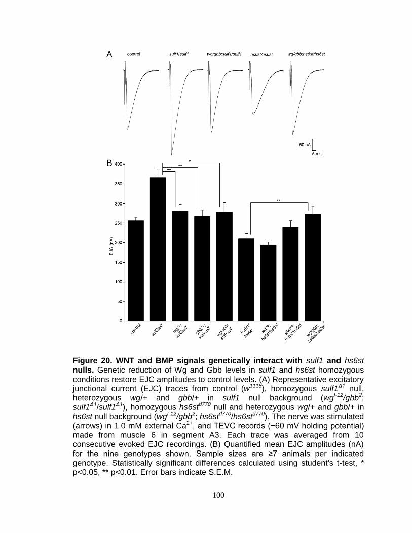
100
Figure 20. WNT and BMP signals genetically interact with sulf1 and hs6st nulls. Genetic reduction of Wg and Gbb levels in sulf1 and hs6st homozygous conditions restore EJC amplitudes to control levels. (A) Representative excitatory junctional current (EJC) traces from control (w1118), homozygous sulf1Δ1 null, heterozygous wg/+ and gbb/+ in sulf1 null background (wgI-12/gbb2; sulf1Δ1/sulf1Δ1), homozygous hs6std770 null and heterozygous wg/+ and gbb/+ in hs6st null background (wgI-12/gbb2; hs6std770/hs6std770). The nerve was stimulated (arrows) in 1.0 mM external Ca2+, and TEVC records (−60 mV holding potential) made from muscle 6 in segment A3. Each trace was averaged from 10 consecutive evoked EJC recordings. (B) Quantified mean EJC amplitudes (nA) for the nine genotypes shown. Sample sizes are ≥7 animals per indicated genotype. Statistically significant differences calculated using student's t-test, * p<0.05, ** p<0.01. Error bars indicate S.E.M.

101
molecular components that would explain the bidirectional change in synaptic
functional differentiation (Figs. 8 and 21). To test this hypothesis, we examined a
key component of the presynaptic active zone (Bruchpilot; Brp) (Wagh et al.,
2006), and an essential subunit of the postsynaptic glutamate receptor (Bad
Reception (Brec); GluRIID) (Featherstone et al., 2005). In parallel, we also
performed a miniature EJC (mEJC) analysis to compare functional presynaptic
vesicle release probability and postsynaptic response amplitude. A summary of
these data is shown in Figure 21.
First, NMJ synapses were double-labeled for GluRIID recognized with
anti-Brec (green) and Brp recognized with anti-nc82 (red) to compare genetic
control (w1118) with sulf1 and hs6st nulls (Fig. 21A). We found that GluRIID was
very significantly elevated at sulf1 synapses compared to control (~30%
increased; p<0.01, n=20; Fig. 21B). In the opposing direction, hs6st null
synapses showed a significant decrease in GluRIID abundance (~15% reduced;
p<0.05, n=21; Fig. 21B). The GluRIID field area per bouton and number of
GluRIID punctae normalized to field area per synaptic bouton were also
bidirectionally altered in the sulf1 and hs6st nulls (Fig. 21C,D). GluRIID receptor
field area was increased in sulf1 (~30% greater; p<0.01, n=47) but decreased in
hs6st (~25% reduced; p<0.01, n=51). Conversely, measurements of GluRIID
puncta normalized to field area per synaptic bouton were decreased in sulf1
(~15% lower; p<0.05, n=47), but increased in hs6st nulls (~40% greater; p<0.01,
n=51, Fig. 21D). The bi-directional differences between sulf1 and hs6st were very
highly significant (p<0.001). The active zone protein Brp also showed opposite

102
Figure 21. Bi-directional effects of sulf1 and hs6st nulls on synaptic assembly. (A) Representative NMJ boutons from control (w1118), sulf1 and hs6st null genotypes, labeled for postsynaptic Bad Reception (Brec) glutamate receptor IID subunit (GluRIID, green) and presynaptic active zone Bruchpilot (anti-nc82, red). Quantification of GluRIID mean fluorescent intensity (≥18 animals per indicated genotype) (B), GluRIID field area (≥40 boutons from ≥9 animals per indicated genotype) (C), and GluRIID punctae number per synaptic bouton (≥40 boutons from ≥9 animals per indicated genotype) (D), all normalized to genetic control. (E) Representative mEJC traces from control (w1118), sulf1Δ1 and hs6std770 null alleles. Quantified mean mEJC amplitude (nA) (F), mean mEJC frequency (Hz) (G) and mean quantal content (H), with genetic control levels indicated as a dotted red line in each case. Sample sizes ≥15 recordings per indicated genotype. Statistically significant differences calculated using student's t-test or Mann-Whitney test for non-parametric data and indicated as, * p<0.05, ** p<0.01, *** p<0.001. Error bars indicate S.E.M.

103
effects (Fig. 21A). Although the difference between sulf1 null and control was not
quite significant (p>0.05, n=20), hs6st null synapses showed a very significant
decrease in Brp compared to control (~20% reduced; p<0.01, n=21; Fig. 21A).
Based on these results, we next tested pre- (Brp) and postsynaptic
(Brec/GluRIID) changes in sulf1 and hs6st mutants with genetic reduction of Wg
and Gbb (wg/ gbb;sulf1/sulf1 and wg/gbb;hs6st/hs6st), as in Figure 21.
Distribution changes of both pre- and postsynaptic components were assayed as
measurements of glutamate receptor field and active zone areas (Fig. 22). To
measure glutamate receptor distribution comparing wg/gbb;sulf1/sulf1 to
matched control, we counted the number of GluRIID punctae per bouton (p=0.73,
n=48; Fig. 22B) and GluRIID area (p=0.92, n=48; Fig. 22C), and found both
corrected back to control levels. Likewise, for wg/gbb;hs6st/hs6st compared to
control, GluRIID puncta number (p=0.88, n=48) and area (p=0.41, n=58) were
both corrected to control levels. To measure Brp-positive presynaptic active
zones comparing wg/gbb;sulf1/sulf1 to matched control, we counted the number
of Brp punctae per bouton (p=0.43, n=48; Suppl. Fig. 22D) and Brp area (p=0.39,
n=48; Suppl. Fig. 22E), and found both corrected back to control levels. Likewise,
for wg/gbb;hs6st/hs6st compared to control, Brp number (p=0.54, n=58) and
area(p=0.19, n=58) were also corrected back to control levels. These results
provide strong genetic evidence that Wg and Gbb trans-synaptic signaling
changes are causative for the pre- and postsynaptic molecular differentiation
defects in the sulf1 and hs6st null mutants. These bidirectional pre- and
postsynaptic molecular changes parallel functional transmission changes in sulf1

104
Figure 22. Wg and Gbb signals genetically interact with sulf1 and hs6st nulls. Genetic reduction of Wg and Gbb levels in sulf1 and hs6st homozygous conditions restores molecular synaptic assembly to control levels. (A) Representative NMJ boutons from control (w1118), heterozygous wg/+ and gbb/+ in sulf1 null background (wgI-12/gbb2; sulf1Δ1/sulf1Δ1) and hs6st null background (wgI-12/gbb2; hs6std770/hs6std770) labeled for postsynaptic Bad Reception (Brec) glutamate receptor IID subunit (GluRIID, green) and presynaptic active zone Bruchpilot (anti-nc82, red). Quantification of GluRIID punctae/bouton (B), total GluRIID area (C), Brp punctae/bouton (D) and total Brp area (E), all normalized to the genetic control. All multiply mutant conditions are restored to control levels for all parameters, with no significant differences remaining.

105
and hs6st mutants (Fig. 21). To assay function at the single synapse level, we
finally assayed spontaneous synaptic vesicle fusion events. Representative
mEJC traces for control compared to sulf1 and hs6st nulls are shown in Figure
21E. Consistent with observed bidirectional changes in evoked transmission,
mEJC amplitudes in hs6st were ~25% lower than in sulf1 nulls (hs6st, 0.60 ±
0.02 nA vs. sulf1, 0.76 ± 0.05 nA; p<0.5, n=34; Fig. 21F). Moreover, hs6st nulls
had a ~100% elevated mEJC frequency compared to sulf1 nulls (hs6st, 2.56 ±
0.27 vs. sulf1, 1.30 ± 0.09; p<0.001, n=34; Fig. 21G). Based on these mEJC
measurements, there was a highly significant bidirectional change in quantal
content between the two mutant conditions, with sulf1 quantal content ~50%
greater than hs6st (sulf1, 539.98 ± 22.02 vs. hs6st, 350.69 ± 8.92; p<0.001,
n=34; Fig. 21H). Taken together, these results show a bi-directional change in
presynaptic glutamate release machinery and vesicle fusion probability, as well
as postsynaptic glutamate receptor levels and functional responsiveness. We
conclude that these changes underlie the bi-directional switch in
neurotransmission strength characterizing sulf1 and hs6st mutants.
Discussion
It is well known that synaptic interfaces harbor heavily-glycosylated membrane
proteins, glycolipids and ECM molecules, but understanding of glycan-mediated
mechanisms within this synaptomatrix is limited (Dani and Broadie, 2012). Our
genomic screen aimed to systematically interrogate glycan roles in both
structural and functional development in the genetically-tractable Drosophila NMJ
synapse. 130 candidate genes were screened, classified into 8 functional

106
families: N-glycan biosynthesis, O-glycan biosynthesis, GAG biosynthesis,
glycoprotein/proteoglycan core proteins, glycan modifying/degrading enzymes,
glycosyltransferases, sugar transporters and glycan-binding lectins. From this
screen, 103 RNAi knockdown conditions were larval viable, whereas 27 others
produced early developmental lethality. 35 genes had statistically significant
effects on different measures of morphological development: 27 RNAi-mediated
knockdowns increased synaptic bouton number, 9 affected synapse area (2
increased, 7 decreased) and 2 genes increased synaptic branch number. These
data suggest that overall glycan mechanisms predominantly serve to limit
synaptic morphogenesis. 13 genes had significant effects on the functional
differentiation of the synapse, with 12 increasing transmission strength and only
1 decreasing function upon RNAi knockdown. Thus, glycan-mediated
mechanisms also predominantly limit synaptic functional development. A very
small fraction of tested genes (CG1597; pgant35A, CG7480; veg, CG6657;
hs6st, CG4451; sulf1, CG6725 and CG11874) had effects on both morphology
and function. A large percentage of genes (~30%) showed morphological defects
with no corresponding effect on function, while only 7% of genes showed
functional alterations without morphological defects, and <5% of all genes affect
both. These results suggest that glycans have clearly separable roles in
modulating morphological and functional development of the NMJ synapse.
A growing list of neurological disorders linked to the synapse are attributed to
dysfunctional glycan mechanisms, including muscular dystrophies, cognitive
impairment and autism spectrum disorders (Inlow and Restifo, 2004; Muntoni et

107
al., 2008; Schachter, 2009). Drosophila homologs of glycosylation genes
implicated in neural disease states include ALG3 (CG4084), ALG6 (CG5091),
DPM1 (CG10166), FUCT1 (CG9620), GCS1 (CG1597), MGAT2 (CG7921),
MPDU1 (CG3792), PMI (CG33718) and PPM2 (CG12151) (Inlow and Restifo,
2004). Two of these genes, Gfr (CG9620) and CG1597, showed synaptic
morphology phenotypes in our RNAi screen. Given that connectivity defects are
clearly implicated in cognitive impairment and autism spectrum disorders
(Belmonte et al., 2004; Gatto and Broadie, 2011), it would be of interest to
explore the glycan mechanism affecting synapse morphology in Drosophila
models of these disease states. Glycans are well known to modulate extracellular
signaling, including ligands of integrin receptors, to regulate intercellular
communication (Zhang et al., 2008, 2010). In our genetic screen, several O-
glycosyltransferases mediating this mechanism were identified to show
morphological (GalNAc-T2, CG6394; pgant35A, CG7480, O-fut2, CG14789;
rumi, CG31152) and functional (pgant5, CG31651; pgant35A, CG7480) synaptic
defects upon RNAi knockdown. These findings suggest that known integrin-
mediated signaling pathways controlling NMJ synaptic structural and functional
development (Beumer et al., 1999, 2002; Rohrbough et al., 2000; Rushton et al.,
2009) are modulated by glycan mechanisms. Our screen showed CG6657 RNAi
knockdown affects functional differentiation, consistent with reports that this gene
regulates peripheral nervous system development (Prokopenko et al., 2000). The
corroboration of our screen results with published reports underscores the utility
of RNAi-mediated screening to identify glycan mechanisms, and supports use of

108
our screen results for bioinformatic/meta-analysis to link observed phenotypes to
neurophysiological/pathological disease states and to direct future glycan
mechanism studies at the synapse.
From our screen, the two functionally-paired genes sulf1 and hs6st were
selected for further characterization. As in the RNAi screen, null alleles of these
two genes had opposite effects on synaptic functional differentiation but similar
effects on synapse morphogenesis, validating the corresponding screen results.
The two gene products have functionally-paired roles; Hs6st is a heparan sulfate
(HS) 6-O-sulfotransferase (Kamimura et al., 2001), and Sulf1 is a HS 6-O-
endosulfatase (Dhoot et al., 2001). These activities control sulfation of the same
C6 on the repeated glucosamine moiety in HS GAG chains found on heparan
sulfate proteoglycans (HSPGs). At the Drosophila NMJ, two HSPGs are known
to regulate synapse assembly; the GPI-anchored glypican Dally-like protein
(Dlp), and the transmembrane Syndecan (Sdc) (Johnson et al., 2006). In
contrast, the secreted HSPG Perlecan (Trol) is not detectably enriched at the
NMJ (Morin et al., 2001), and indeed appears to be selectively excluded from the
perisynaptic domain. In other developmental contexts, the membrane HSPGs
Dlp and Sdc are known to act as co-receptors for WNT and BMP ligands,
regulating ligand abundance, presentation to cognate receptors and therefore
signaling (Yan et al., 2009; Dejima et al., 2011). Importantly, the regulation of
HSPG co-receptor abundance has been shown to be dependent on sulfation
state mediated by extracellular sulfatases (Lai et al., 2008). Consistently, we
observed upregulation of Dlp and Sdc in sulf1 null synapses, whereas Dlp was

109
reduced in hs6st null synapses. In the developing Drosophila wing disc, HSPG
co-receptors increase levels of the Wg ligand due to extracellular stabilization
(Han et al., 2005), and the primary function of Dlp in this developmental context
is to retain Wg at the cell surface (Yan and Lin, 2009). Likewise, in developing
Drosophila embryos, a significant fraction of Wg ligand is retained on the cell
surfaces in a HSPG-dependent manner (Pfeiffer et al., 2002), with the HSPG
acting as an extracellular co-receptor. Syndecan also modulates ligand-
dependent activation of cell-surface receptors by acting as a co-receptor (Carey,
1997; Dejima et al., 2011). At the NMJ, regulation of both these HSPG co-
receptors occurs in the closely juxtaposed region between presynaptic bouton
and muscle subsynaptic reticulum, in the exact same extracellular space
traversed by the secreted trans-synaptic Wg and Gbb signals (Johnson et al.,
2006). We therefore proposed that altered Dlp and Sdc HSPG co-receptors in
sulf1 and hs6st mutants differentially trap/stabilize Wg and Gbb trans-synaptic
signals at the interface between motor neuron and muscle, to modulate the
extent and efficacy of intercellular signaling driving synaptic development.
HS sulfation modification is linked to modulating the intercellular signaling
driving neuronal differentiation (Gorsi and Stringer, 2007). In particular, WNT and
BMP ligands are both regulated via HS sulfation of their extracellular co-
receptors, and both signals have multiple functions directing neuronal
differentiation, including synaptogenesis (Packard et al., 2003; Salinas, 2003;
Marques, 2005). In the Drosophila wing disc, extracellular WNT (Wg) ligand
abundance and distribution was recently shown to be strongly elevated in sulf1

110
null mutants (Kleinschmit et al., 2010). Moreover, sulf1 has also recently been
shown to modulate BMP signaling in other cellular contexts (Otsuki et al., 2010).
Consistently, we have shown here increased WNT Wg and the BMP Gbb
abundance and distribution in sulf1 null NMJ synapses. The hs6st null also
exhibits elevated Wg and Gbb at the synaptic interface, albeit the increase is
lower and results in differential signaling consequences. In support of this
contrasting effect, extracellular signaling ligands are known to bind HSPG HS
chains differentially dependent on specific sulfation patterns (Baeg and Perrimon,
2000; Baeg et al., 2001; Mohammadi et al., 2005). It is important to note that the
sulf1 and hs6st modulation of trans-synaptic signals is not universal, as Jelly
Belly (Jeb) ligand abundance and distribution was not altered in the sulf1 and
hs6st null conditions (Rohrbough K. et al., 2010). This indicates that discrete
classes of secreted trans-synaptic molecules are modulated by distinct glycan
mechanisms to control NMJ structure and function.
At the Drosophila NMJ, Wg is very well characterized as an anterograde
trans-synaptic signal (Packard et al., 2002; Ataman et al., 2006a; Korkut and
Budnik, 2009) and Gbb is very well characterized as a retrograde trans-synaptic
signal (Marques et al., 2002; McCabe et al., 2003; Keshishian and Kim, 2004;
Marques, 2005). In Wg signaling, the dFz2 receptor is internalized upon Wg
binding and then cleaved so that the dFz2-C fragment is imported into muscle
nuclei (Mathew et al., 2005; Ataman et al., 2006a; Mosca and Schwarz, 2010). In
hs6st nulls, increased Wg ligand abundance at the synaptic terminal corresponds
to an increase in dFz2C punctae in muscle nuclei as expected. In contrast, the

111
increase in Wg at the sulf1 null synapse did not correspond to an increase in the
dFz2C-terminus nuclear internalization, but rather a significant decrease. One
explanation for this apparent discrepancy is the ‘exchange factor’ model based
on the biphasic ability of the HSPG co-receptor Dlp to modulate Wg signaling
(Yan et al., 2009). In the Drosophila wing disc, this model suggests that the
transition of Dlp co-receptor from an activator to repressor of signaling depends
on Wg cognate receptor dFz2 levels, such that a low ratio of Dlp:dFz2
potentiates Wg-dFz2 interaction, whereas a high ratio of Dlp:dFz2 prevents dFz2
from capturing Wg (Yan et al., 2009). In sulf1 null synapses, we observe a very
great increase in Dlp abundance (~40% elevated) with no significant change in
the dFz2 receptor. In contrast, at hs6st null synapses there is a decrease in Dlp
abundance (15% decreased) together with a significant increase in dFz2 receptor
abundance (~25% elevated). Thus, the higher Dlp:dFz2 ratio in sulf1 nulls could
explain the decrease in Wg signal activation, evidenced by decreased dFz2-C
terminus import into the muscle nucleus. In contrast, the Dlp:Fz2 ratio in hs6st is
much lower, supporting activation of the dFz2-C terminus nuclear internalization
pathway. This previously proposed competitive binding mechanism dependent
on Dlp co-receptor and dFz2 receptor ratios predicts the observed synaptic Wg
signaling pathway modulation in sulf1 and hs6st dependent manner (Yan et al.,
2009).
At the Drosophila NMJ, Gbb is very well characterized as a retrograde
trans-synaptic signal, with muscle-derived Gbb causing the receptor complex
Wishful thinking (Wit), Thickveins (Tkv) and Saxophone (Sax) to induce

112
phosphorylation of the transcription factor mothers against Mothers against
decapentaplegic (P-Mad) (Wharton et al., 1999; McCabe et al., 2003; Keshishian
and Kim, 2004). Mutation of Gbb ligand, receptors or regulators of this pathway
have shown that Gbb-mediated retrograde signaling is required for proper
synaptic differentiation and functional development (Marques et al., 2002;
McCabe et al., 2003, 2004; Rawson et al., 2003). Further, loss of Gbb signaling
results in significantly decreased levels of P-Mad in the motor neurons (McCabe
et al., 2003). We show here that accumulation of Gbb in sulf1 and hs6st null
synapses causes elevated P-Mad signaling at the synapse and P-Mad
accumulation in motor neuron nuclei. Importantly, sulf1 null synapses show a
significantly higher level of P-Mad signaling compared to hs6st null synapses,
and this same change is proportionally found in P-Mad accumulation within the
motor neuron nuclei. These findings indicate differential activation of Gbb trans-
synaptic signaling dependent on the HS sulfation state controlled by the sulf1
and hs6st mechanism, similar to the differential effect observed on Wg trans-
synaptic signaling. Our genetic interaction studies show that these differential
effects on trans-synaptic signaling have functional consequences, and exert a
causative action on the observed bi-directional functional differentiation
phenotypes in sulf1 and hs6st nulls. Genetic correction of Wg and Gbb defects in
the sulf1 null background restores elevated transmission back to control levels.
Similarly, genetic correction of Wg and Gbb in hs6st nulls restores the decreased
transmission strength back to control levels. These results demonstrate that the

113
Wg and Gbb trans-synaptic signaling pathways are differentially regulated and, in
combination, induce opposite effects on synaptic differentiation.
Both Wg and Gbb pathway mutants display disorganized and mislocalized
presynaptic components at the active zone (e.g. Bruchpilot; Brp) and
postsynaptic components including glutamate receptors (e.g. Bad reception;
Brec/GluRIID) (Aberle et al., 2002; Marques et al., 2002; Packard et al., 2002).
Consistently, the bi-directional effects on neurotransmission strength in sulf1 and
hs6st mutants are paralleled by dysregulation of these same synaptic
components. Changes in presynaptic Brp and postsynaptic GluR
abundance/distribution causally explain the bi-directional effects on synaptic
functional strength between sulf1 and hs6st null mutant states. Alterations in
active zone Brp and postsynaptic GluRs also agree with assessment of
spontaneous synaptic activity. Null sulf1 and hs6st synapses showed opposite
effects on miniature evoked junctional current (mEJC) frequency (presynaptic
component) and amplitude (postsynaptic component). Further, quantal content
measurements also support the observation of bidirectional synaptic function in
the two functionally paired nulls. Genetic correction of Wg and Gbb defects in
both sulf1 and hs6st nulls restores the molecular composition of the pre- and
postsynaptic compartments back to wildtype levels. When both trans-synaptic
signaling pathways are considered together, these data suggest that HSPG
sulfate modification under the control of functionally-paired sulf1 and hs6st jointly
regulates both WNT and BMP trans-synaptic signaling pathways in a differential
manner to modulate synaptic functional development on both sides of the cleft.

114
We present here the first systematic investigation of glycan roles in the
modulation of synaptic structural and functional development. We have identified
a host of glycan-related genes that are important for modulating neuromuscular
synaptogenesis, and these genes are now available for future investigations, to
determine mechanistic requirements at the synapse, and to explore links to
neurological disorders. As proof for the utilization of these screen results, this
study has identified extracellular heparan sulfate modification as a critical
platform of the intersection for two secreted trans-synaptic signals, and
differential control of their downstream signaling pathways that drive synaptic
development. Other trans-synaptic signaling pathways are independent and
unaffected by this mechanism, although it is of course possible that a larger
assortment of signals could be modulated by this or similar mechanisms. This
study supports the core hypothesis that the extracellular space of the synaptic
interface, the heavily-glycosylated synaptomatrix, forms a domain where glycans
coordinately mediate regulation of trans-synaptic pathways to modulate
synaptogenesis and subsequent functional maturation.
Materials and Methods
Drosophila stocks and genetics
The glycan-related gene collection was generated using the KEGG glycan
databases and Flybase annotation. The 163 UAS-RNAi lines tested were
obtained from the Vienna Drosophila RNAi Center (VDRC) and Harvard TRiP
collection. Transgenic UAS-RNAi males were crossed to GAL4 driver females,

115
with progeny raised at 25°C on standard food, controlling for density (3 ♀
crossed to 2 ♂). The UH1-GAL4 driver was used for ubiquitous knockdown of
target gene expression (Rohrbough et al., 2007). Neural specific elav-GAL4 (Lin
and Goodman, 1994a), muscle specific 24B-GAL4 (Brand and Perrimon, 1993)
and glia specific repo-GAL4 lines (Sepp et al., 2001) from Bloomington stock
center were used to assay cell-targeted knockdown. The two sulf1 null alleles
used were sulf1∆1 (You et al., 2011) and sulf1∆P1 (Kamimura et al., 2001). The
two hs6st null alleles used were hs6std770 and the deficiency Df(3R)ED6027
(Kamimura et al., 2006). The wg allele wgI-12 (Marie et al., 2010) and gbb alleles
gbb1 and gbb2 were used (Wharton et al., 1999; McCabe et al., 2003). Multiply
mutant animals were made using standard genetic crosses. The trol-GFP line
was obtained from Flytrap (Morin et al., 2001).
Antibody production
We generated a rabbit polyclonal anti-Gbb antibody using a 1:1 combination of
two Gbb-specific peptides (SHHRSKRSASHP, NDENVNLKKYRNMIVKSC)
corresponding to amino acids 319-330 and 435-452 of Gbb (Young-In Frontier,
Seoul, Korea). The antibody was purified by Protein A affinity chromatography,
and antibody specificity demonstrated by examining immunoreactivity in the
wandering third instar neuromusculature with gbb mutants and by expressing
UAS-gbb9.1 under the control of the muscle driver BG57-GAL4 (Figure 23).
Immunoreactivity in the wandering third instar neuromusculature was severely
reduced in a strong hypomorphic gbb allele (gbb1/gbb2, UAS-gbb9.9), which has
leaky expression of UAS-gbb9.9 in a null allelic combination [25,87,95]. In sharp

116
contrast, the anti-Gbb signal was strongly elevated in BG57-GAL4/UAS-gbb9.1
relative to wildtype larvae.
Immunocytochemistry
Wandering third instars were dissected in Ca2+-free saline and then immediately
fixed in either 4% paraformaldehyde for 10 minutes (all labels except anti-Dlp) or
Bouin’s fixative for 30 mins (anti-Dlp). Preparations were then washed in
permeabilizing PBST (PBS + 0.1% Triton-X) or detergent-free PBS for
extracellular labeling only (Rushton et al., 2009) . The following primary
antibodies were used: rabbit or goat anti-HRP (1:250; Jackson ImmunoResearch
Laboratories); mouse anti-DLG (4F3; 1:250; Developmental Studies Hybridoma
Bank (DSHB)); mouse anti–Fasciclin II (1D4; 1:5; DSHB); mouse anti-Dlp (13G8,
1:5; DSHB) and rabbit anti-Syndecan (1:200) (Spring et al., 1994); mouse anti-
Wg (4D4; 1:2 DSHB) and rabbit anti-Gbb (1:100); rabbit anti-PcanV (1:1000)
(Friedrich et al., 2000) guinea pig anti-Jeb (1:100) (Rohrbough K. et al., 2010);
rabbit anti-dFz2-C (1:500) and rabbit anti-dFz2-N (1:100) (Mathew et al., 2005);
rabbit anti-Htl (1:100) (Shishido et al., 1997); rabbit anti-P-Mad (PS1; 1:1000)
(Persson et al., 1998); rabbit anti-GluRIID (1:500) (Featherstone et al., 2005) and
mouse anti-BRP (1:100; DSHB). Primary antibodies were incubated at 4°C
overnight. Alexa-conjugated secondary antibodies (Jackson ImmunoResearch
Laboratories) were used at 1:250 dilutions for 2 hours at room temperature.
Staining with propidium iodide (Sigma Aldrich) to visualize cell nuclei was done at
1:100 dilution of 1 mg/ml propidium iodide incubated for 30 minutes at room
temperature.

117
Imaging quantification
Images were taken with on an upright Zeiss LSM 510 META laser-scanning
confocal using a Plan Apo 63X oil objective. For structural quantification,
including NMJ synapse branch number, bouton number and area, preparations
were double-labeled with anti-HRP and anti-DLG, with counts made at muscle 4
in segment A3. For nuclear import studies, nuclei were identified by propidium
iodide staining with fluorescent punctae counted and intensity quantified [59]. For
synaptic functional protein quantitation, glutamate receptor and Brp punctae were
quantified for muscle 4, segment 3. Glutamate receptor number and field area
was quantified in consecutive boutons of >3µm diameter. All preparations were
fixed, stained and processed simultaneously to allow for intensity comparisons.
All analyses were done with ImageJ software (National Institutes of Health) using
the threshold function to outline areas and Z-stacks made using the maximum
projection function. Statistics were done with one-way ANOVA analysis followed
by Dunnett’s post-test. All analyses were done blind to genotypes during all
stages of experimentation and analysis. All figure images were projected in LSM
Image Examiner (Zeiss) and exported to Adobe photoshop.
Heparin treatment
Stock solution of heparin (Sigma, H3393) in 1XPBS was prepared and serially
diluted to obtain concentrations (e.g. 0.625, 0.315 and 0.156 mg/ml). Dissected
wandering third instar larvae were incubated with these heparin concentrations
for 5 minutes at RT, followed by a 1 minute wash with 1XPBS and then 10

118
minute fix with 4% paraformaldehyde in 1XPBS. After fixation, anti-Wg or anti-
Gbb antibodies were used as above with appropriate secondary antibodies.
Processed animals were analyzed for changes in intensity measurements as
above in the image quantification section. All fluorescence intensity
measurements were compared to preparations treated identically with only
1XPBS and no heparin, and the processed simultaneously for immunolabeling,
microscopy and quantification.
Electrophysiology
Two-electrode voltage-clamp (TEVC) records were made from the wandering
third instar NMJ as previously described (Beumer et al., 1999). In brief, staged
control, mutant and transgenic RNAi animals were secured on sylgard-coated
coverslips with surgical glue (liquid suture), dissected longitudinally along the
dorsal midline, and glued flat. The segmental nerves were cut near the base of
the ventral nerve cord. Recording was performed in 128 mM NaCl, 2 mM KCl, 4
mM MgCl2, 1.0 mM CaCl2, 70 mM sucrose, and 5 mM Hepes. Recording
electrodes (1-mm outer diameter capillaries; World Precision Instruments) were
filled with 3 M KCl and had resistances of >15 MΩ. Spontaneous mEJCs were
collected using continuous (gap-free) recording and evoked EJC recordings were
made from the voltage-clamped (Vhold = −60 mV) muscle 6 in segment A3 with a
TEVC amplifier (Axoclamp 200B; MDS Analytical Technologies). The cut
segmental nerve was stimulated with a glass suction electrode at a
suprathreshold voltage level (50% above baseline threshold value) for a duration
of 0.5 ms. Records were made with 0.2 Hz nerve stimulation in episodic

119
acquisition setting and analyzed with Clampex software (version 7.0; Axon
Instruments). Each n=1 represents a recording from a different animal. Statistical
comparisons were performed using student’s t-test or the Mann-Whitney test for
non-parametric data.
Acknowledgments
We are particularly grateful to Hiroshi Nakato (sulf1∆P1 and hs6std770), Xinhua Lin
(sulf1∆1), Kristy Wharton (gbb1/gbb2, UAS-gbb9.9 and UAS-gbb9.1), the Vienna
Drosophila RNAi Center (VDRC), the Harvard TriP Collection and the
Bloomington Drosophila Stock Center for providing essential Drosophila stocks.
We also particularly thank Joachim Schulz (Sdc), Peter ten Dijke (P-Mad), Vivian
Budnik (dFz2-C and dFz2-N), Stefan Baumgartner (PcanV) and the Iowa
Hybridoma Bank for essential antibodies. We thank Samuel Friedman for
providing a representative image of anti-Perlecan antibody staining at the NMJ.
We thank William Parkinson, Emma Rushton, Lane Coffee and Sean Schaffer for
technical assistance.

120
Chapter IV
Two pgant O-GalNAc transferases regulate synaptic plasticity by activity-
dependent regulation of integrin signaling
This paper has been published under the same title in Journal of Neuroscience,
2014
Neil Dani, He Zhu and Kendal Broadie
Departments of Biological Sciences, Cell & Developmental Biology and
Pharmacology, and The Vanderbilt Kennedy Center for Research on Human
Development, Vanderbilt University, Nashville, TN 37232, USA

121
Abstract
Using a Drosophila whole-genome transgenic RNAi screen for glycogenes
regulating synapse function, we have identified two protein α-N-
acetylgalactosaminyltransferases (pgant3 and pgant35A) that regulate synaptic
O-linked glycosylation (GalNAcα1-O-S/T). Loss of either pgant alone elevates
pre-/postsynaptic molecular assembly and evoked neurotransmission strength,
but synapses appear restored to normal in double mutants. Likewise, activity-
dependent facilitation, augmentation and post-tetanic potentiation are all
suppressively impaired in pgant mutants. In non-neuronal contexts, pgant
function regulates integrin signaling, and we show here that the synaptic αPS2
integrin receptor and transmembrane tenascin ligand (ten-m) are both
suppressively down-regulated in pgant mutants. Channelrhodopsin-driven activity
rapidly (<1 minute) drives integrin signaling in wildtype synapses, but is
suppressively abolished in pgant mutants. Optogenetic stimulation in pgant
mutants alters presynaptic vesicle trafficking and postsynaptic pocket size during
the perturbed integrin signaling underlying synaptic plasticity defects. Critically,
acute blockade of integrin signaling acts synergistically with pgant mutants to
eliminate all activity-dependent synaptic plasticity.

122
Introduction
The heavily glycosylated transmembrane and extracellular synaptomatrix
at the synaptic interface plays pivotal roles in synaptogenesis, neurotransmission
and synaptic plasticity (Dityatev and Schachner, 2003; Broadie et al., 2011; Dani
and Broadie, 2012). Neurological disease states arising from aberrant
glycosylation occur in numerous congenital disorders of glycosylation (CDGs)
and dystroglycanopathies (Freeze, 2006). However, the mechanisms by which
synaptomatrix glycan modifications regulate normal synapse function and
dysfunction in heritable disease states remain poorly understood (Ohtsubo and
Marth, 2006). Drosophila is a powerful genetic model to pursue these synaptic
glycan mechanisms, given the conservation of glycan pathways, reduced
glycogene genomic redundancy in this system, and host of techniques available
at the well-characterized glutamatergic neuromuscular synapse (Keshishian et
al., 1996; Gagneux and Varki, 1999). Using this model, we have recently shown
that endogenous glycan-binding lectin (mind the gap; Rushton et al., 2009),
heparan sulfate proteoglycan (HSPG) modifiers (sulf1/hs6st; Dani et al., 2012)
and N-linked glycosylation (mgat1; Parkinson et al., 2013) glycan mechanisms all
act as potent regulators of trans-synaptic integrin, WNT and BMP signaling.
To systematically pursue synaptic glycan mechanisms, we undertook a
Drosophila whole-genome screen of glycogenes using RNAi-mediated
knockdown of all N-/O-/glycosaminoglycan-linked enzymes, glycosaminoglycans,
glycosyltransferases and glycan-binding lectins, characterizing effects on NMJ
structure and function using confocal microscopy and two-electrode voltage-

123
clamp electrophysiology, respectively (Dani et al., 2012). This screen identified
two α-N-acetylgalactosaminyltransferases, pgant3 and pgant35A, which catalyze
transfer of GalNAc monosaccharides onto serine/threonine residues (GalNAcα1-
O-S/T) to form Tn antigens, as found within mucin-like O-linked glycans (Ten
Hagen et al., 2003a). This most complexly regulated glycosylation is orchestrated
by multiple GalNAc‐transferases (12 pgants in Drosophila) with distinct and
overlapping peptide specificities (Yoshida et al., 2008; Tran and Ten Hagen,
2013). Pursuing our screen results with well-characterized pgant3 and pgant35A
loss-of-function mutants (see Methods), we found elevated synaptic O-linked
glycosylation, pre-/postsynaptic molecular assembly, pre-/postsynaptic
ultrastructural elaborations and neurotransmission strength, which are all
corrected in double mutants that show none of these synaptic defects, identifying
a novel suppressive genetic interaction.
In non-neuronal tissues, Drosophila pgants regulate integrin signaling and
intercellular adhesion (Zhang and Ten Hagen, 2011). Importantly, we have
shown position specific (PS) integrins, localized both pre- and postsynaptically,
regulate NMJ morphogenesis (Beumer et al., 1999), synaptic
scaffold/synaptomatrix adhesion molecules (Beumer et al., 2002), functional
differentiation (Rohrbough et al., 2007) and activity-dependent plasticity
(Rohrbough et al., 2000). We therefore hypothesized that pgants regulate
integrin signaling at the synapse, and consistently find suppressive down-
regulation of αPS2-containing integrin receptors (Beumer et al., 1999), RGD-
containing tenascin (ten-m) ligand (Mosca et al., 2012), and postsynaptic

124
membrane adhesion defects in pgant mutants. Furthermore, we find integrin- and
activity-dependent functional synaptic plasticity is suppressively (reciprocal
suppression) regulated in pgant mutants. Importantly, we find that
channelrhodopsin activity stimulation (Wang et al., 2011) disrupts downstream
integrin association with talin and pFAK signaling, and elevates postsynaptic
membrane adhesion defects. RGD peptide blockade of integrin function
synergistically abolishes all activity-dependent synaptic plasticity in pgant
mutants. These data show two pgants suppressively regulate synaptic O-GalNAc
glycosylation, synapse molecular assembly, neurotransmission strength and
activity-dependent plasticity via trans-synaptic integrin-tenascin signaling.
Materials and Methods
Drosophila genetics
All stocks were maintained at 25°C on standard food. Two independent mutant
alleles isolated by EMS mutagenesis were employed for pgant3; 1) pgant3m1, a
C>T transition changing conserved arginine to cysteine at amino acid 130
resulting in failure to glycosylate substrates in enzymatic activity tests, and 2)
pgant3m2, a G>A transition that creates a stop codon at amino acid 609, thereby
deleting the C-terminal 59 amino acids and resulting in an unstable protein
(Zhang et al., 2010). Similarly, the pgant35A mutations used were 1)
pgant35AHG8, a C>T transition at nucleotide 265 resulting in a glutamine to stop
codon change at amino acid 89, and 2) pgant35A3775, a T>A transversion at
nucleotide 584 resulting in a premature stop codon at amino acid 195, both fully

125
eliminating the catalytic domain (Ten Hagen and Tran, 2002). All mutants were
placed in the w1118 genetic background, and w1118 was therefore used as the
wildtype control. Rescue and overexpression experiments were performed with
UAS-pgant3 and UAS-pgant35A (Zhang et al., 2008) wildtype transgenes driven
by neural (elav-gal4) (Lin and Goodman, 1994a), muscle (24B-gal4) (Brand and
Perrimon, 1993) and ubiquitous (UH1-gal4) (Wodarz et al., 1995) drivers.
Standard genetic techniques were used to generate recombinant and multiply
mutant animals. Optogenetic studies were performed with the UAS-ChIEF-
tdTomato channelrhodopsin transgene (Wang et al., 2011) driven by the neural-
specific elav-gal4 driver in animals raised on 0.25 mM ATR (all-trans retinal;
Sigma) supplemented food. Animals used for experimentation were of either sex.
Immunocytochemistry
Wandering third instars were dissected in Ca2+-free saline and then fixed in 4%
paraformaldehyde for 10 mins. Preparations were then washed in either
permeabilizing PBST (PBS+0.1% Triton-X) or detergent-free PBS for
extracellular labeling (Rushton et al., 2009). O-GalNAc glycans were visualized
with TRITC-conjugated VVA (1:250; EY Laboratories) and HPL (1:250;
Invitrogen) lectins (Chia et al., 2014) . Mouse antibodies obtained from the
Developmental Studies Hybridoma Bank (DSHB) included: anti-βPS (1:500),
anti-αPS1 (1:200), anti-αPS2 (1:500), anti-scab (1:200), anti-talin (1:10) and anti-
DLG (4F3; 1:250). Other sourced primary antibodies included: mouse anti-Ten-m
(1:3000) (Levine et al., 1994), mouse anti-Tig (1:200) (Fogerty et al., 1994),
guinea pig anti-LanA (1:200) (Inoue and Hayashi, 2007), rat anti-Tsp (1:200)

126
(Subramanian et al., 2007), rabbit Wb-N (1:500) (Martin et al., 1999a), rabbit
anti-βν (1:300) (Yee and Hynes, 1993) and rabbit anti-pFAK (pY397; 1:50;
Invitrogen). All antibodies were incubated at 4°C overnight. Alexa-conjugated
Fluor 647-goat anti-HRP and secondary antibodies (Jackson ImmunoResearch
Laboratories) were incubated at 1:250 for 2 hours at RT.
Image quantification
Control and mutant preparations for antibody and lectin studies were processed
simultaneously for all intensity comparisons (Dani et al., 2012). To allow for direct
comparisons of signal intensity levels, all genotypes were dissected, fixed,
labeled and imaged in parallel at the same time, with identical confocal settings
and intensity measurements also made at the same time for all compared
genotypes. Imaging was done on an upright Zeiss LSM 510 META laser-
scanning confocal using a Plan Apo 63x oil objective. NMJ structural
quantification was done with anti-HRP imaging at muscle 6/7 in segment A3. All
intensity analyses were done with ImageJ software (NIH) using the threshold
function to outline Z-stack areas with the maximum projection function. All
statistical comparisons were performed with one-way ANOVA analysis followed
by Dunnett’s or Dunn’s post-test for non-parametric data using Instat Graphpad
software. All data are presented as mean ± SEM. All images were projected in
LSM Image Examiner (Zeiss) and exported to Adobe Photoshop.
Electrophysiology
Two-electrode voltage-clamp (TEVC) records were made from NMJs of paired
control and mutant wandering third instars as reported previously (Beumer et al.,

127
1999). Briefly, recordings were performed in 128 mM NaCl, 2 mM KCl, 4 mM
MgCl2, 1.0 mM CaCl2, 70 mM sucrose, and 5 mM Hepes saline (PH 7.1).
Recording electrodes (1-mm outer diameter capillaries; World Precision
Instruments) filled with 3M KCl had resistances of >15 MΩ. Evoked excitatory
junction currents (EJCs) were recorded at 18oC using episodic recording from
voltage-clamped (Vhold: −60 mV) muscle 6 in segment A3 with a TEVC amplifier
(Axoclamp 2B; Axon). Excitatory junctional potentials (EJPs) were also recorded
in parallel. Segmental nerves were stimulated with a glass suction electrode at a
suprathreshold voltage (50% above threshold) for 0.5 ms duration at 0.5 Hz. For
synaptic plasticity studies, the nerve was stimulated at 10 Hz for 60 seconds in
0.2 mM CaCl2 saline (Rohrbough et al., 2000). EJCs were acquired via Clampex
(Axon) and analyzed using Clampfit 9.0 by averaging 10 (during initial/PTP) to 20
(during tetanus) consecutive responses. Gly-Arg-Gly-Asp-Ser-Pro (GRGDSP)
integrin inhibition and Gly-Arg-Ala-Asp-Ser-Pro (GRADSP) control peptides
(Sigma) were used at 0.2 mM, incubated for 1 hour at 18oC. Statistical
comparisons were done using one-way ANOVA analysis followed by Dunnett’s
post-test with Instat Graphpad software. Each N=1 represents a recording from a
different animal. All data are presented as mean ± SEM.
Electron microscopy
Ultrastructural analyses were performed as reported previously
(Beumer et al., 1999). Briefly, staged third instar preparations were fixed in 1.6%
paraformaldehyde/2% glutaraldehyde (20 mins), washed in 1XPBS (10 mins)
and transferred to 2.5% glutaraldehyde in cacodylate buffer (12 hrs) with washes

128
in the same buffer (30 mins). Preparations were postfixed in 1% OsO4 in
cacodylate buffer (2 hrs) and then dehydrated in an ethanol series followed by
propylene oxide (30 mins). Segment A3 muscle 6/7 was dissected free of the
preparations and separately embedded in araldite resin. Ultrathin (40 nm)
sections were cut using a Leica ultracut UCT 54 ultramicrotome and then
transferred to formvar-coated slot grids. Sections were imaged using a Phillips
CM10 transmission Electron Microscopy at 80 kV, with images collected on a 4
megapixel CCD camera. Sample sizes are >10 independent NMJs, with the
statistical analyses calculated using unpaired t-tests. Images acquired from AMT
Image Capture Software were exported to Adobe Photoshop. All data are
presented as mean ± SEM.
Optogenetics
Wandering third instars were dissected in 0.2 mM Ca2+ saline on Sylgard-coated
plates with the nervous system kept intact. An LEDD1B LED driver, M470L2
mounted LED at 470nm affixed with LA1951-A lens was used to stimulate
channelrhodopsin activity (Gruntman and Turner, 2013). Preparations were
subjected to a 60 sec train of light stimulation at 10 Hz, with a pulse duration of
60 ms, followed by immediate fixation and processing during continual
stimulation, using the methods described above. >8 independent NMJs were
analyzed for each genotype and condition, with statistical tests for activity-
dependent changes in fluorescence intensity and ultrastructure performed as
described above in the immunocytochemistry and electron microscopy sections.

129
Results
Pgants regulate synapse composition and transmission strength
An unbiased genetic screen of glycogenes identified synaptic function
defects using inducible RNAi-mediated down regulation of two pgants (pgant3
and pgant35A). This screen tested 130 glycan-related genes defined in 8
function categories: N-glycan, O-glycan and glycosaminoglycan biosynthesis;
glycosyltransferases, glycan degrading/ modifying enzymes; glycoprotein and
proteoglycan core proteins; sugar transporters and glycan-binding lectins. Using
a combination of confocal microscopy and two-electrode voltage-clamp (TEVC)
electropyhysiology, NMJ morphology and functional transmission defects were
tested in Drosophila wandering third instar larvae following ubiquitous (UH1-gal4)
RNAi knockdown. From this screen, 31 genes affected synapse structure (27
increased bouton number, 2 increased branching and 2 increased NMJ area)
and 13 affected synapse function (12 increased and 1 decreased). Only 6 gene
knockdowns affected both structure and function. To investigate mucin-type O-
linked glycosylation, 9 available RNAi lines were used to test 6 pgant genes
(pgant2, pgant3, pgant4, pgant5, pgant6 and pgant35A) and 3 additional GalNAc
transferases (GalNAcT-1, GalNAcT-2 and C1GalTA). Of these, 3 pgant genes
(pgant3, pgant5 and pgant35A) were identified to have increased
neurotransmission strength upon knockdown, and GalNAc-T2 showed increased
NMJ area. The other 5 gene knockdowns caused no detectable NMJ
phenotypes. Well-characterized mutants are available for only pgant3 and
pgant35A (see Methods), which have been extensively studied in heteroallelic

130
null combinations (Ten Hagen and Tran, 2002; Zhang et al., 2010). In this study,
we pursued a full characterization of these two pgant genes using the same
conditions.
To characterize synaptic mechanisms in pgant3 and pgant35A null single
and double mutant larvae, we performed nerve-evoked excitatory junction current
(EJC) recordings in the TEVC paradigm (Dani et al., 2012). Sample traces of 10
consecutive, superimposed responses are shown for 4 genotypes; the genetic
background control (w1118), pgant3 (pgant3m1/pgant3m2) and pgant35A
(pgant35AHG8/pgant35A3775) single mutants in w1118 background, and the double
null mutant (pgant3m1,pgant35AHG8/ pgant3m2,pgant35A3775). Neurotransmission
is clearly and consistently elevated in both pgant mutants, increased 25-40%
compared to controls (Fig. 23A). Quantification of mean EJC amplitudes shows
that synapse strength is very significantly elevated in both pgant3
(255.46±8.12nA, n=26, p<0.001) and pgant35A (277.62±11.88nA, n=22,
p<0.001) single mutants compared to control (198.73±7.77nA, n=17; Fig.24A,
right). Surprisingly, however, neurotransmission in the recombinant double null
mutant is not significantly elevated compared to control (231.64±7.24nA, n=21,
p>0.05; Fig.24A. right), which behaves like the control. Thus, a similar phenotype
occurs in the two pgant single mutants, which is absent in the double mutant. We
use the term ‘suppression’ throughout this study, as the simplest genetic term
describing the observed interaction. Importantly, there is a synaptic function
defect only, with no differences in NMJ morphology in either of the pgant
mutants. In quantifying synaptic branching, neither pgant3 (5.00±0.28 decrease,

131
Figure 23. Null pgant mutants suppressively elevate neurotransmission strength. (A) Representative evoked excitatory junction current (EJC) records from genetic control (w1118), pgant3 (pgant3m1/pgant3m2) and pgant35A (pgant35AHG8/pgant35A3775) single mutants, and double mutants (pgant3m1,pgant35AHG8/pgant3m2,pgant35A3775). Ten consecutive EJCs traces shown at 0.2 Hz stimulation from the muscle 6 NMJ in segment A3. Scale bar indicates EJC amplitude (50nA, Y-axis) and time (10ms, X-axis). Right: Histogram of mean EJC amplitudes, with sample sizes >17 for each genotype. (B) Representative NMJ boutons for the above four genotypes labeled for postsynaptic glutamate receptors (GluRIID, green) and presynaptic active zone Bruchpilot (Brp, red), with split channels shown for clarity. White arrows indicate GluRIID/Brp paired punctae in single mutants. Scale bar: 2µm. (C) Quantification of GluRIID/Brp areas and punctae number per NMJ terminal. Statistical differences are calculated using one-way ANOVA with Dunnett’s post-test; *p<0.05, **p<0.01, ***p<0.001, N.S. indicates no significance.

132
n=28) or pgant35A (5.79±0.26 n=24) single mutants, or the double mutants
(6.26±0.41, n=23) showed any significant difference from controls (5.58±0.22,
n=26). Likewise, NMJ bouton number is also not significantly affected in pgant3
(90.89±4.25, n=28) or pgant35A (92.29±5.78, n=24) single mutants or double
mutants (77.46±4.18, n=24) compared to controls (85.96±4.18, n=28). This
finding was the first discovery of the suppressive action of pgant genes on
synapse function.
To begin to determine how pgant co-repressive regulation arises at the
synapse, we labeled NMJs for presynaptic active zones with Bruchpilot (Brp),
and postsynaptic glutamate receptors (GluRIID), marking the two sides of each
individual synapse (Fig.24B). There is a clear and consistent increase in
Brp/GluRIID punctae in both pgant3 and pgant35A single mutants, indicating a
co-operative change on both pre- and postsynaptic sides of the synapse.
Importantly, however, the double null mutant does not show any detectable
increase in either synaptic marker (Fig. 23B). Quantification reveals significantly
increased glutamate receptor field area and punctae number in pgant3
(82.59±6.77µm2 (p<0.05) and 358.0±16.20 (p<0.01); n=14) and pgant35A
(81.02±6.95µm2 and 302.73±15.61; n=15, p<0.05) single mutants, but with no
differences in the double mutants in either parameter compared to controls
(61.03±3.99µm2 and 233.25±12.33, n=16; Fig.24C). Likewise, presynaptic Brp
active zone area and punctae number are increased at pgant3 (54.80±4.80µm2
and 357.46±18.89; n=13, p<0.05) and pgant35A (52.96±4.30µm2 and
305.4±14.86; n=15, p<0.05) single mutants, with no differences in

133
pgant3,pgant35A double mutants compared to controls (38.56±3.03µm2 and
242±31.22; n=16; Fig. 23C). Both pgant mutants show no significant change in
spontaneous miniature EJC (mEJC) frequencies (pgant3, 2.29±0.17Hz, n=10;
pgant35A, 2.06±0.11Hz, n=9) compared to control (1.93±0.13Hz, n=10), but do
show small, significant decreases in mEJC amplitudes (pgant3, 0.68±0.03nA,
n=10, p<0.05; pgant35A, 0.69±0.03nA, n=9; p<0.05) compared to control
(0.80±0.02nA, n=10). These results show that pgant3 and pgant35A both up-
regulate neurotransmission strength through elevated pre- and postsynaptic
assembly via a mutually suppressive mechanism that predominantly impacts
evoked function.
Pgants regulate presynaptic vesicles and postsynaptic pocket size
The synaptic ultrastructure of the Drosophila NMJ has been well
characterized by transmission electron microscopy (TEM), categorizing multiple
synaptic vesicle (SV) pools in the presynaptic bouton and the complex
architecture of the expansive sub-synaptic reticulum (SSR) of the postsynaptic
membrane (Rohrbough et al., 2007). As no gross morphology differences were
associated with observed neurotransmission elevations in pgant mutants, we
next investigated synapse ultrastructure. On the presynaptic side, we measured
bouton area, active zone architecture, overall SV density and SV distribution in
concentric rings (e.g. 250 nm, 500 nm) extending from each active zone (Fig.
24A,B; white arrows). On the postsynaptic side, we assayed SSR area, thickness
on major and minor axes, density (folds/unit length) and postsynaptic pocket

134
Figure 24. Null pgant mutants suppressively alter pre/postsynaptic ultrastructure. (A) Representative transmission electron microscopy (TEM) images of synaptic boutons from muscle 6 NMJ in segment A3 of genetic control (w1118, top), pgant3 (pgant3m1/pgant3m2) and pgant35A (pgant35AHG8/pgant35A3775) single mutants (middle), and double mutants (pgant3m1,pgant35AHG8/3775/pgant3m2,pgant35A3775, bottom). Labels indicate synaptic vesicle (SV) pools in presynaptic bouton, subsynaptic reticulum (SSR) of muscle membrane, and presynaptic active zones (arrowheads). Scale: 500nm. (B) High magnification images of single active zone synapses in all four genotypes. Labels indicate presynaptic t-bars (arrowheads) and postsynaptic pockets (white dotted lines). Scale: 100nm. (C) Quantification of SV pools (<250 nm (top), <500 nm (middle) from active zone t-bar) and postsynaptic pocket depth (bottom). Sample size >10 boutons for each of the four genotypes. Statistical differences calculated using one-way ANOVA with Dunnett’s post-test; *p<0.05, **p<0.01, N.S. indicates no significance.

135
(PSP) size by measuring the distance from presynaptic active zone to the first
SSR membrane layer (Fig. 24A,B; dotted white lines). Expansion of the
postsynaptic pocket is a hallmark of mutants defective in synaptomatrix resident,
trans-synaptic signaling ligands (Packard et al., 2002) and heparan sulfate
proteoglycan (HSPG) extracellular regulators of trans-synaptic signaling
(Kamimura et al., 2013).
Presynaptic bouton appearance (Fig. 24A) and area in both pgant mutants
(pgant3, 7.53±0.85 µm2, n=10 pgant35A, 7.76±0.80 µm2, n=12,) are not
significantly different from the genetic control (w1118, 9.51±1.78 µm2, n=7),
although there is a trend towards smaller boutons. Likewise, active zone
architecture and t-bar dimensions are not measurably affected by loss of pgant
activity (Fig. 24B). In contrast, the density and distribution of SV pools is clearly
aberrant in both pgant single mutants, although the double null mutant is not
detectably different from the control (Fig. 24A,B). Immediately adjacent to active
zone t-bars (Fig. 24B, arrows), SV clustering is increased in both pgant single
mutants, but not in the double mutant combination. Quantifying SV number within
250 nm of the t-bar shows a consistent density in controls (10.5±0.91), which is
significantly increased in both pgant3 (15.22±0.99, p<0.01) and pgant35A
(15.53±0.78, p<0.01) single mutants, but back at control level in double null
mutants (9.4±0.67; n=15; Fig. 24C, top). Likewise, at a distance of 500 nm from
the active zone t-bar, SV number increases in pgant3 (36.3±1.66, p<0.01) and
pgant35A (43.31±1.21, p<0.01) single mutants, but not in double mutants
(26.06±1.44, n=15) compared to controls (26±2.18; Fig. 24C, middle). Thus,

136
presynaptic vesicle pool distribution is suppressively regulated by pgant3 and
pgant35A, in line with changes in synaptic function.
Postsynaptic SSR appearance, area, thickness and density are normal in
all pgant mutants (Fig. 24A). Quantification of SSR area (pgant3, 1.26±0.16;
pgant35A, 1.13±0.17; pgant3,pgant35A, 1.09±0.17), thickness (pgant3,
1.12±0.10; pgant35A, 0.84±0.08; pgant3,pgant35A, 0.80±0.07) and density
(pgant3, 0.83±0.05; pgant35A,1.10±0.08; pgant3,pgant35A, 1.11±0.08) all show
no significant changes normalized to controls (Fig. 24A). In contrast, however,
there is a striking expansion in both pgant mutants of the postsynaptic pocket
(PSP) (Packard et al., 2002; Kamimura et al., 2013). This compartment has been
defined as “a postsynaptic area immediately apposed to an active zone
containing amorphous material” (Packard et al., 2002), which is spatially
localized between postsynaptic membrane and SSR (Ren et al., 2009). The PSP
compartment has been shown to be expanded in trans-synaptic signaling
disrupted mutants including WNT wingless (wg), BMP glass bottom boat (gbb),
HSPG perlecan (trol) and HSPG sulfateless (sfl) mutants (Packard et al., 2002;
Tian and Ten Hagen, 2007; Ren et al., 2009; Nahm et al., 2010; Kamimura et al.,
2013). Both pgant3 and pgant35A single mutants similarly display an enlarged
PSP compartment, although the double null mutant is not detectably different
from the control (Fig. 24B; dotted white lines). As a quantifiable PSP parameter,
pocket depth from presynaptic active zone to the next adjacent postsynaptic SSR
membrane was measured in all four genotypes. Compared to controls (mean
PSP depth 121.17±4.95nm, n=10), both pgant single mutants display a >2-fold

137
expanded PSP (pgant3, 254.7±35.25nm, p<0.01; pgant35A, 233.96±41.83nm,
p<0.05) (Fig. 24B,C bottom). In sharp contrast, the double null mutants show no
significant increase in PSP depth compared to controls (169.09±15.46nm; n=12;
Fig. 24C, bottom). Thus, we observe suppressive regulation by pgant genes of
SV pools in the presynaptic compartment as well as postsynaptic compartment
expansion, paralleling the changes in neurotransmission strength.
Neuronal and muscle pgant3 and pgant35A modulate neurotransmission
To determine cell-specific requirements of pgant3 and pgant35A, we used
the inducible Gal4-UAS binary system (Brand and Perrimon, 1993) to express
UAS-pgant3 or UAS-pgant35A wildtype transgenes in neurons (elav-gal4) or
muscles (24B-gal4) in respective single mutant backgrounds, and assayed for
phenotype rescue (Fig. 25). Sample EJC traces show an average of 10
consecutive nerve-evoked responses for both rescue conditions in both pgant
nulls (Fig. 25A). We find that functional neurotransmission strength is restored to
control levels when pgant3 is expressed in either neurons (pgant3m1/pgant3m2;
UAS-pgant3/elav, 181.61±11.85 nA, n=14) or muscles (pgant3m1/pgant3m2; UAS-
pgant3/24B, 187.47±12.72 nA, n=11) in the otherwise pgant3 null background, as
compared to controls (w1118, 193.34±8.69 nA, n=14; Fig. 25C, top panel).
Similarly, pgant35A expression in neurons (pgant35AHG8/pgant35A3775; UAS-
pgant35A/elav, 197.50±14.26 nA, n=9) or muscles (pgant35AHG8/pgant35A3775;
UAS-pgant35A/24B, 211.42±17.06 nA, n=10) in the pgant35A mutant
background likewise rescues neurotransmission strength to control level (Fig.

138
Figure 25. Pgants function in neurons and muscle to regulate neurotransmission. (A) Representative evoked junctional current (EJC) records from genetic control (w1118), pgant3 mutant with neuronal (pgant3m1/pgant3m2; UAS-pgant3/elav) and muscle (pgant3m1/pgant3m2; UAS-pgant3/24B) pgant3 expression, pgant35A mutant with neuronal (pgant35AHG8/pgant35A3775; UAS-pgant35A/elav) and muscle (pgant35AHG8/pgant35A3775; UAS-pgant35A/24B) pgant35A expression. 10 consecutive EJC traces are shown at 0.2 Hz stimulation from the muscle 6 NMJ in segment A3. Scale bar indicates EJC amplitude (50 nA, Y-axis) and time (10 ms, X-axis). Sample size n≥8 animals for all conditions. (B) Representative NMJ boutons for the above five genotypes co-labeled for both postsynaptic glutamate receptors (GluRIID, green) and presynaptic active zone Bruchpilot (Brp, red), with split channels shown for clarity. Scale bar: 2µm. (C) Histograms show normalized EJC amplitude, punctae number and GluRIID/Brp area per NMJ terminal for the above five genotypes. Sample size n≥10 animals for all conditions. Statistical differences are calculated using one-way ANOVA with Dunnett’s post-test. N.S. indicates no significance.

139
25C, top panel).
To test if this functional rescue correlates with corrected synaptic
molecular assembly, NMJs were labeled for presynaptic Brp and postsynaptic
GluRIID (Fig.26B). Brp punctae number in pgant3 neuronal (392.56±22.86, n=9)
or muscle (321.13±17.84, n=15) rescue conditions, as well as pgant35A neuronal
(340±25.63, n=8) or muscle (409.58±27.71, n=12) rescue conditions, does not
differ significantly from control (358.84±18, n=19; Fig. 25B,C). Similarly, GluRIID
punctae number is also restored to control levels (360.73±19.16, n=19), with
pgant3 neuronal (395.89±23.95, n=9) or muscle (327.6±18.22, n=15) rescue, as
well as pgant35A neuronal (336±24.71, n=8) or muscle (414.17±26.98, n=12)
rescue. The same result is reflected in Brp area measurements, where pgant3
neuronal (85.67±6.78, n=10) or muscle (75.66±7.99, n=15) rescue, as well as
pgant35A neuronal (79.12±8.23 µm2, n=10) or muscle (88.80±8.29 µm2, n=12)
rescue is similar to control values (74.35±4.69 µm2, n=18; Fig. 25B,C). Similarly,
postsynaptic GluRIID area measurements in pgant3 neuronal (173.75±10.38
µm2, n=10) or muscle (142.47±10.80 µm2, n=15) rescue, as well as pgant35A
neuronal (155.61±9.97 µm2, n=10) or muscle (156.99±11.89 µm2, n=12) rescue,
are not significantly different from control (148.20±7.01 µm2, n=18; Fig. 25C).
These results show that both pgant3 and pgant35A can function either pre- or
postsynaptically to regulate synaptic assembly and neurotransmission strength.
Pre-/postsynaptic balance of pgant3 and pgant35A regulate
neurotransmission
Given the pgant suppressive mechanism and coupled pre/postsynaptic roles of

140
pgant3 and pgant35A, we next tested whether the balance of pgant3 and
pgant35A is required to properly regulate neurotransmission. We generated
allelic combinations for UAS-pgant3 wildtype transgene expression in neurons
(elav-gal4), muscles (24B-gal4) or ubiquitously (UH1-gal4) in the
pgant3,pgant35A double mutant background, and tested effects on
neurotransmission strength. Representative EJC traces for each genotype are
shown in Fig. 26A. Compared to the control mean EJC amplitude
(pgant3m1,pgant35AHG8/pgant3m2,pgant35A3775; UAS-pgant3/+, 211.42±11.94 nA,
n=8), neuronal (pgant3m1,pgant35AHG8/pgant3m2,pgant35A3775; elav/UAS-pgant3,
258.99±9.59 nA, n=9, p<0.05), muscle
(pgant3m1,pgant35AHG8/pgant3m2,pgant35A3775; 24B/UAS-pgant3, 276.52±11.19
nA, n=9, p<0.05) as well as ubiquitous
(pgant3m1,pgant35AHG8/pgant3m2,pgant35A3775; UAS-pgant3/UH1+,
254.47±13.59 nA, n=13, p<0.05) pgant3 expression in the double mutant
background, all significantly elevated neurotransmission strength (Fig. 26B).
Thus, restoring pgant3 to either neuron or muscle effectively reveals the
pgant35A single mutant phenotype (Fig. 23A).
In parallel, we overexpressed both pgant genes alone to test the effect on
neurotransmission strength. Representative EJC traces for each genotype are
shown in Fig. 26C. As compared to control (UAS-pgant3/+, 205.55±8.77 nA,
n=22), pgant3 overexpression in neurons (UAS-pgant3/elav, 154.99±11.99 nA,
n=11 p<0.01), muscles (pgant3/24B, 165.62±11.13 nA, n=10, p<0.05) or
ubiquitous (pgant3/UH1, 164.80±9.79 nA, n=10, p<0.05) all similarly decreased

141
Figure 26. Pre/postsynaptic pgant3/35A balance regulates neurotransmission. Representative EJC records (A) and mean amplitudes (B) for pgant3 expression in the double mutant background. The four genotypes include genetic control (pgant3m1,pgant35AHG8/pgant3m2,pgant35A3775;+/UAS-pgant3), neuronal (pgant3m1, pgant35AHG8/pgant3m2,pgant35A3775;elav/UAS-pgant3), muscle (pgant3m1,pgant35AHG8/ pgant3m2,pgant35A3775;24B/UAS-pgant3) and ubiquitous (pgant3m1,pgant35AHG8/ pgant3m2,pgant35A3775;UH1+/UAS-pgant3) pgant3 expression in the double mutant. Sample size n≥10 animals for all conditions. (C) Representative EJC records and (D) mean amplitudes for pgant3 and pgant35A overexpression conditions. The eight genotypes include pgant3 genetic control (UAS-pgant3/+), neuronal (UAS-pgant3/elav), muscle (UAS-pgant3/24B) and ubiquitous (UAS-pgant3/UH1) pgant3 overexpression; and pgant35A genetic control (UAS-pgant35A/+), neuronal (UAS-pgant35A/elav), muscle (UAS-pgant35A/24B) and ubiquitous (UAS-pgant35A/UH1) pgant35A overexpression. Sample size ≥10 animals for each genotype. Statistical differences calculated using one-way ANOVA with Dunnett’s post-test; *p<0.05, **p<0.01, N.S. indicates no significance.

142
mean EJC amplitudes (Fig 27D, left panel). Likewise, as compared to control
(UAS-pgant35A/+, 235.15±10.77 nA, n=19), pgant35A overexpression in
neurons (UAS-pgant35A/elav, 193.50±13.39 nA, n=18, p<0.05) and ubiquitous
overexpression (UAS-pgant35A/UH1, 187.52±9.43 nA, n=10, p<0.05) both
decrease neurotransmission transmission, although muscle overexpression
alone has no significant effect (UAS-pgant35A/24B, 224.99±8.77 nA, n=20; Fig.
26D, right panel). Overall, pgant overexpression has the opposite consequence
of pgant loss of function (Fig. 23A). Thus, the proper balance of pgant3/pgant35A
in neurons and muscle bidirectionally regulates the strength of synaptic
transmission.
Activity-dependent synaptic plasticity is impaired in pgant mutants
In the non-neuronal context of the Drosophila wing disc, pgant mutants
specifically impair integrin signaling to cause intercellular de-adhesion (Zhang et
al., 2010). Similarly, the above synaptic ultrastructure defects in pgant mutants
recalls synaptic integrin signaling, which we have shown is required for activity-
dependent synaptic plasticity (Rohrbough et al., 2000). We therefore next
investigated the multiple phases of activity-dependent plasticity in pgant mutants,
including immediate facilitation and maintained augmentation during a tetanic
stimulus train (10 Hz, 60 seconds), and initiation and maintenance of post-tetanic
potentiation (PTP) following return to basal stimulation. In this paradigm, EJCs
are recorded initially at 0.5 Hz for 30 seconds, followed by the tetanic train, and
then returned to basal 0.5 Hz for a total of 5 minutes recording (Rohrbough et al.,
2000). Figure 27A shows representative traces for control (w1118), single

143
Figure 27. Impaired activity-dependent synaptic plasticity in pgant mutants. (A) Representative two-electrode voltage-clamp (TEVC) records from genetic control (w1118, top), pgant3 (pgant3m1/pgant3m2) and pgant35A (pgant35AHG8/pgant35A3775) single mutants (middle two traces), and double mutants (pgant3m1, pgant35AHG8/pgant3m2,pgant35A3775, bottom). The stimulation paradigm is 0.5 Hz for 30 seconds (solid bar), 10 Hz for 60 seconds (hatched bar), and then a return to 0.5 Hz (solid bar) for 5 minutes total recording time. Scale bars indicate 20nA (y-axis) and 10s (x-axis). (B) Mean EJC amplitudes over time normalized to initial mean EJC amplitude for control (solid circle), pgant3 (hollow square) and pgant35A (hollow triangle) single mutants, and double mutant (hollow circle). Bars labeled “C” and “D” are expanded below, showing normalized EJC amplitudes during tetanic stimulation (C) and early post-tetanic stimulation periods (D). Sample sizes >10 independent NMJs for each of the four genotypes. Statistical differences calculated using one-way ANOVA with Dunnett’s post-test; *p<0.05, **p<0.01, ***p<0.001, N.S. indicates no significance.

144
(pgant3m1/pgant3m2 and pgant35AHG8/pgant35A3775) and double mutants
(pgant3m1,pgant35AHG8/pgant3m2,pgant35A3775).
For quantification, consecutive EJCs are averaged throughout the
stimulation phases to display mean amplitudes normalized to the starting level
(Fig. 27B). Controls show immediate, rapid facilitation leading to a 6-fold
augmentation during tetanic stimulation, followed by 2-fold initial PTP phase,
later maintained as a ~50% elevation for the duration of the recording (Fig.
27A,B). In contrast, both pgant single mutants show very significantly impaired
initial facilitation and blunted 4-fold augmentation during tetanic stimulation (Fig.
27B, solid bar labeled C). For example, at 20 seconds into the tetanic train, EJC
amplitudes show augmentation decreases of ≤65% in pgant3 (p<0.01, n=10)
and ≤55% in pgant35A (p<0.01, n=11) mutants. In contrast, the double null
mutant is clearly less impaired than either single pgant mutant (Fig. 27A,B). At 20
seconds into the tetanic train, the double mutants exhibit a reduced impairment
of ≤40% compared to controls (p<0.05, n=11; Fig. 27C). Following this initial
facilitation phase, double mutants reach control levels of augmentation, whereas
the single pgant mutants remain impaired (Fig, 28A,B), showing a suppressive
interaction. Following the tetanic train, potentiation in double mutants is
indistinguishable from controls, whereas both single mutants (pgant3 and
pgant35A) show strong loss of PTP initiation (Fig. 27B, solid bar labeled D).
Quantification shows >50% decrease in pgant3 (p<0.001, n=10) and >35%
decrease in pgant35A (p<0.05, n=11) single mutants compared to controls, but
no detectable decrease in the double null mutants (Fig. 27D). Thus, pgant

145
mechanisms regulate activity-dependent facilitation, augmentation and
potentiation.
Pgants suppressively regulate integrin signaling
Synapses sandwich heavily-glycosylated transmembrane and extracellular
proteins that regulate synaptic function and plasticity (Dani and Broadie, 2012).
For example, we have previously shown that O-linked heparan sulfate
glycosaminoglycans bidirectionally regulate WNT and BMP trans-synaptic
signaling to modulate neurotransmission strength (Dani et al., 2012). To directly
visualize changes in synaptic O-GalNAc glycosylation in pgant mutants, we used
fluorescently-conjugated VVA-TRITC and HPL-488 lectins to label NMJ terminals
(Fig. 28). Non-detergent conditions were used throughout to examine only the
glycosylation state of the extracellular synaptomatrix (Dani et al., 2012).
Representative images show the halo-like VVA (Fig. 28A, top left) and HPL (Fig.
28A, middle left) labeling surrounding the anti-HRP marked synaptic boutons. In
comparison to controls, O-linked glycan expression is very significantly increased
in both pgant3 (31.65±5.61%, n=9, p<0.01) and pgant35A (58.50±4.39%, n=14,
p<0.01) single mutants, however there is no significant change in the double
mutants (13.54±5.04%, n=6, p>0.05) (Fig. 28B). Similarly, quantified HPL
labeling is very significantly elevated in both pgant3 (33.13±6.39%, n=21,
p<0.01) and pgant35A (41.06±7.83%, n=18, p<0.01) single mutants, but no
significant difference occurs in the double null mutants (7.49±6.85%, n=18,
p>0.05) compared to controls (Fig. 28B). Thus, two independent approaches
highlight the suppressive regulation of synaptic O-GalNAc modification by these

146
Figure 28. Synaptomatrix O-glycan and integrin signaling defects in pgant mutants. (A) NMJ synaptic boutons from control (w1118, left column), pgant3 (pgant3m1/pgant3m2) and pgant35A (pgant35AHG8/pgant35A3775) single mutants (middle two columns) and the double mutant (pgant3m1,pgant35AHG8/pgant3m2,pgant35A3775; right column) show co-labeling for synaptic O-linked glycosylation markers (VVA and HPL), integrin receptor (αPS2) and transmembrane integrin ligand (ten-m) relative to the presynaptic
marker anti-HRP. Scale bar: 2 m. (B) Histograms show fluorescence intensities for all four labels (VVA, HPL, αPS2 and ten-m) normalized to each genetic control (dotted red line). Sample size ≥10 independent NMJs for each label for all four genotypes. Statistical differences calculated using one-way ANOVA with Dunnett’s post-test; **p<0.01.

147
two pgant genes.
Studies in non-neuronal tissues have shown that pgant mutants
misregulate integrin signaling (Zhang et al., 2010). Consistently, we have
previously identified pre-/postsynaptically localized Position Specific (PS) integrin
receptors at the Drosophila NMJ, containing multiple different α and β subunits
(Beumer et al., 1999, 2002; Rohrbough et al., 2000, 2007; Rushton et al., 2009).
We therefore tested the multiple integrin receptor subunits, including αPS1
(mew), αPS2 (if), αPS3 (scab/volado), βPS (mys) (Brower et al., 1984) and βv
(Yee and Hynes, 1993). The two β subunits show an interesting pgant-specific
change, with βPS increased in pgant35A (1.39±0.08, n=14, p<0.01) and βv
increased in pgant3 (1.21±0.03, n=18, p<0.05) single mutants, but no significant
change of either β subunit in the double mutant (βPS, 1.17±0.64, n=10, p>0.05;
βv, 0.91±0.06, n=10, p>0.05) normalized to control. Most of the α receptor
subunits show no consistent changes in the pgant mutants, including αPS1
(pgant3, 1.27±0.07, n=18, pgant35A, 1.10±0.05, n=18) and αPS3 (pgant3,
1.05±0.02, n=8, pgant35A, 1.15±0.06, n=12; all p>0.05 with respect to control).
The sole exception is αPS2, which sharply decreases in both pgant3
(39.19±6.75%, n=10, p<0.01) and pgant35A (34.37±5.69%, n=11, p<0.01) single
right). We next examined a host of characterized integrin ligands for changes
in pgant single and double mutants (Zhang and Ten Hagen, 2011), including
Tiggrin (Fogerty et al., 1994), laminin α subunits LanA (Inoue and Hayashi, 2007)
and Wing-blister (Wb) (Martin et al., 1999a), Thrombospondin (Tsp)
(Subramanian et al., 2007) and Tenascin (ten-m) (Levine et al., 1994). Most of

148
these ligands show no consistent changes in pgant single and double mutants as
compared to control: Tiggrin (pgant3, 1.08±0.05, n=11, pgant35A, 1.03±0.05,
n=14, pgant3,pgant35A, 1.07±0.03, n=8, as compared to control, p>0.05), LanA
(pgant3, 1.25±0.07, n=11, p<0.05, pgant35A, 1.16±0.07, n=14, p>0.05, double
mutant, 1.00±0.05, n=8, p>0.05), Wb-N terminus (pgant3, 1.00±0.05, n=10,
pgant35A, 1.12±0.03, n=11, double mutant, 1.11±0.04, n=7, all p>0.05), and Tsp
(pgant3, 1.02±0.07, n=9, pgant35A, 1.12±0.06, n=10, double mutant, 1.13±0.05,
n=8, p>0.05), all normalized to control. The sole exception was the RGD domain-
containing, transmembrane ten-m (Levine et al., 1994). Ten-m localizes in a
halo-like ring around HRP-labeled synaptic boutons in controls, but is
consistently reduced in both pgant single mutants (Fig. 28A, middle). Compared
to controls, ten-m levels are very significantly decreased in pgant3
(21.88±3.47%, n=15, p<0.01) and pgant35A (20.84±3.91%, n=16, p<0.01) single
mutants, but show no change in double null mutants (Fig. 28A, right). Thus, the
two pgant genes suppressively downregulate αPS2 integrin/ten-m ligand at the
synapse.
Neuronal and muscle pgants regulate O-glycosylation and integrin
signaling
To determine if changes in synaptic O–linked glycosylation and trans-synaptic
ten-m/αPS2 integrin signaling are directed by pre- or postsynaptic pgant function,
we next tested both pgant3 and pgant35A rescue in neurons and muscle in their
respective null mutant backgrounds (Fig. 29). Representative NMJs showing
VVA lectin, αPS2 integrin and ten-m ligand labeling are shown for all genotype

149
conditions in Fig. 29A. Both pgant3 neuronal (1.02±0.05, n=15) and muscle
(1.05±0.05, n=15), as well as pgant35A neuronal (1.13±0.03, n=13) and muscle
(1.01±0.03, n=22) expression restored VVA lectin labeling to control levels
(1.00±0.03, n=18; Fig. 29B). Similarly, αPS2 integrin abundance is also rescued
with pgant3 neuronal (0.94±0.05, n=19) or muscle (0.99±0.06, n=20) expression,
as well as pgant35A neuronal (0.90±0.04, n=21) or muscle (0.86±0.04, n=25)
expression, as compared to control (1.00±0.03, n=20; Fig. 29B). Interestingly,
only neuronal pgant3 (0.99±0.04, n=10) and pgant35A (0.97±0.04, n=8)
expression could restore synaptic ten-m levels to control levels (1.00±0.03,
n=21), whereas muscle pgant3 (0.88±0.04, n=17, p<0.05) and pgant35A
(0.82±0.03, n=14, p<0.01) remained significantly decreased normalized to control
(Fig. 29B, right panel). Thus, both pre- and postsynaptic pgant3 and pgant35A
are sufficient to properly regulate synaptic O-linked glycosylation and integrin
levels, but regulation of the ten-m ligand requires pgant function in the
presynaptic neuron.
Pgants regulate activity-dependent integrin signaling at the synapse
With striking activity-dependent effects on synaptic plasticity in pgant mutants,
we next queried activity-dependent changes in integrin signaling (Fig. 30)
presynaptic activity, followed by confocal microscopy examination for molecular
changes at the NMJ synapse. The neuronal driver (elav-gal4) (Lin and Goodman,
1994b) was used to target UAS-CHiEF-tdTomato (Wang et al., 2011) in genetic
control, single and double mutants. Channelrhodopsin targeting was confirmed
by visualizing tdTomato expression (Fig. 30A) and eliciting evoked junctional

150
Figure 29. Pre/postsynaptic pgant3/35A regulate O-GalNAc and integrin signaling. (A) NMJ synaptic boutons co-labeled for synaptic O-linked glycosylation marker (VVA), integrin receptor (αPS2) (top three panels) and ten-m relative to the presynaptic marker anti-HRP (bottom two panels), with split channels shown for clarity. The five genotypes shown include genetic control (w1118), neuronal (pgant3m1/pgant3m2; UAS-pgant3/elav) and muscle (pgant3m1/pgant3m2; UAS-pgant3/24B) pgant3 expression, and neuronal (pgant35AHG8/pgant35A3775; UAS-pgant35A/elav) and muscle (pgant35AHG8/ pgant35A3775; UAS-pgant35A/24B) pgant35A expression conditions. Scale bar: 2
m. (B) Histograms showing the relative fluorescence intensities for all three labels (VVA, αPS2 and ten-m) normalized to each genetic control (dotted red line). Sample size ≥8 independent NMJs for each label for all five genotypes. Statistical differences calculated using one-way ANOVA with Dunnett’s post-test; *p<0.05, **p<0.01.

151
Figure 30. Activity-dependent integrin signaling changes in pgant mutants. (A) Representative image of neurally-targeted (elav-gal4) channelrhodopsin expression (ChIEF-tdTomato) in third instar ventral nerve cord. The native fluorescence of the tagged light-activated channel (red) is shown without
amplification. Scale bar: 50 m. (B) Representative evoked junctional potential (EJP) trace from the muscle 6 NMJ induced by blue light (470nm) stimulation of ChiEF-expressing neurons. (C) Schematic of the optogenetic stimulation paradigm used to assay changes in integrin signaling. Basal stimulation at 0.5 Hz

152
for 30 seconds to establish baseline, followed by 10 Hz blue light pulses for 60 seconds to induce-activity-dependent changes. Samples were then immediately fixed and labeled for analyses. (D) Representative images of pFAK (top) and talin (bottom) labeling, with HRP co-labeling, showing split channels to compare unstimulated control (left) and light-stimulated (10Hz, 60 seconds) synaptic boutons. The genotypes are ChIEF-expressing control (w1118; UAS-ChIEF/elav-gal4, top), pgant3 (pgant3m1/pgant3m2; UAS-ChIEF/elav) and pgant35A (pgant35AHG8/pgant35A3775; UAS-ChIEF/elav-gal4) single mutants (middle two rows), and the double mutant (pgant3m1, pgant35AHG8/pgant3m2,pgant35A3775; UAS-ChIEF/elav-gal4 (far right). (E) Histograms show relative fluorescence intensities of pFAK (left) and talin (right) normalized to genetic control for all four genotypes. Sample size >8 independent NMJs for each label and condition for all genotypes. Statistics calculated using one-way ANOVA with Dunnett’s post-test; *p<0.05.

153
potentiations (EJPs) with 5 Hz blue light (λ=460 nm, 60 ms duration) stimulation
(Fig. 30B). Guided by the plasticity stimulation paradigm (Fig. 27), preparations
were illuminated with 60 ms light pulses at 10 Hz for 60 seconds, then
immediately fixed for imaging (Fig. 30C). VVA-TRITC O-GalNac labeling did not
detectably change in unstimulated controls compared to optogenetically-
stimulated preparations (pgant3, 1.09±0.04, pgant35A, 1.09±0.03, double
mutant, 0.96±0.04 as normalized to controls, n≥13, all p>0.05). Similarly, we
observe no change in levels of integrin ligand ten-m (pgant3, 1.00±0.04,
pgant35A, 1.02±1.04, double mutant, 1.14±0.05, n≥17, all p>0.05) or integrin
receptor αPS2 levels (pgant3, 0.94±0.04, pgant35A, 0.88±0.04, double mutant,
0.84±0.07 as normalized to controls, n≥8, all p>0.05). We therefore investigated
integrin downstream signaling by assaying talin and pFAK abundance
(Devenport et al., 2007; Tsai et al., 2012a).
To determine if activity-dependent integrin signal transduction is affected,
we investigated channelrhodopsin-dependent changes in Talin recruitment and
downstream pFAK production (Fig. 30D). Interestingly, when compared to
respective unstimulated genotype controls, optogenetic stimulation drives a
striking increase in Talin levels in both control (49.41±18.96%, n=8 p<0.05) and
pgant double mutants (80.1±7.93%, n=13, p<0.05) compared to unstimulated
conditions, whereas neither pgant3 nor pgant35A single mutants showed any
significant activity-dependent change in Talin recruitment to the synapse (Fig.
30D, bottom row; Fig. 30E, left panel). Moreover, we find an activity-dependent
decrease pFAK levels in stimulated controls (UAS-CHIEF/elav, 17.85±4.23%,

154
n=49, p<0.05), with no change in stimulated pgant3 (pgant3m1/pgant3m2; UAS-
CHIEF/elav 8.15±6.28%, n=43, p>0.05), pgant35A (pgant35AHG8/pgant35A3775;
UAS-CHIEF/elav 7.76±3.94%, n=61, p>0.05) and double mutant
pgant3,pgant35A (pgant3m1, pgant35AHG8/pgant3m2,pgant35A3775; UAS-
CHIEF/elav, 3.61±2.61%, n =27, p>0.05) conditions (Fig. 30D top row; Fig. 27E,
right). We conclude that both integrin recruitment of talin and downstream
production of pFAK is activity-dependent and under pgant-dependent
suppressive regulation.
Pgants regulate activity-dependent postsynaptic pocket size
Misregulated integrin signaling leads to intercellular de-adhesion and
subsequent wing blistering in pgant mutant wing discs (Zhang et al., 2010).
Moreover, mutants in trans-synaptic WNT/BMP and HSPG extracellular
pathways manifest enlarged postsynaptic pockets at the NMJ (Packard et al.,
2002; Kamimura et al., 2013). As we have shown that pgant mutants
suppressively regulate basal and activity-dependent integrin signaling and
postsynaptic pocket expansion, we next examined optogenetic activity-
dependent synaptic ultrastructural changes, with a particular focus on the
postsynaptic pocket. In the above channelrhodopsin-expressing mutants and
controls, we adopted the same light stimulation paradigm, followed by fixation
and transmission electron microscopy examination of synapse ultrastructure (Fig.
31).
In optogenetically-stimulated synaptic terminals, there is an obvious
decrease in SV density in all four genotypes compared to unstimulated controls

155
(Fig. 31A). At <250 nm away from the active zone, quantification of SV number
shows a ~30% decrease in controls, and a similar ~30% decrease in stimulated
pgant3 mutants (n=13, p<0.001 compared to unstimulated condition; Fig. 31B).
Both the pgant35A single mutant (n=19) and the double null mutant (n=16)
behave similarly. Further, there are no significant differences in suppressive
regulation under basal conditions (Fig. 24) or unstimulated UAS-ChIEF carrying
lines (Fig. 31) i.e. single mutants (pgant3m1/pgant3m2, 15.22±0.99 vesicles;
pgant35AHG8/pgant35A3775, 15.54±0.78; p<0.05) are elevated compared to
control (w1118, 10.5±0.91) and the double mutant (pgant3m1,pgant35AHG8/
pgant3m2,pgant35A37758, 9.4±0.67). Similarly, in unstimulated single mutants
carrying the channelrhodopsin transgene, synaptic vesicles are elevated
(pgant3m1/pgant3m2; UAS-CHIEF/elav, 9.53±0.63;, p<0.01;
pgant35AHG8/pgant35A3775; UAS-CHIEF/elav, 8.95±0.49, p<0.05) with respect to
control (UAS-CHIEF/elav, 7.13±0.57) and double mutant
(pgant3m1,pgant35AHG8/pgant3m2,pgant35A3775; UAS-CHIEF/elav, 7.68±0.39).
Thus, activity drives SV cycling in all four genotypes comparably (Fig.32B). In
contrast, optogenetically-stimulated control NMJ synapses show an activity-
dependent increase in PSP depth that does not occur in either pgant3 and
pgant35A single mutant, although the double mutant is indistinguishable from
control (Fig. 31A, dotted lines). Quantification of these differences reveal an
activity-dependent PSP depth increase of >50% in control (p<0.05 compared to
unstimulated condition) and ~35% increase in the double null mutants (n=14,
p<0.05), but no significant change in either single pgant mutant (Fig. 31C).

156
Figure 31. Activity-dependent changes in synapse ultrastructure in pgant mutants. (A) Representative active zone synapses of control (w1118; UAS-ChIEF/elav-gal4, top), pgant3 (pgant3m1/m2; UAS-ChIEF/elav) and pgant35A (pgant35AHG8/3775; UAS-ChIEF/elav-gal4) single mutants (middle two rows) and double mutant (pgant3m1, pgant35AHG8/pgant3m2,pgant35A3775; UAS-ChIEF/elav-gal4, bottom), comparing the unstimulated (left column) and light-stimulated (10Hz, 60 seconds; right column) conditions. Dotted white line indicates postsynaptic pocket (PSP) depth for each condition and genotype. Scale bar: 100 nm. (B, C) Histograms showing quantification of activity-dependent changes for normalized synaptic vesicle number (B) and PSP depth (C) for all four genotypes under stimulated (black bars) and unstimulated (gray bars) conditions. Sample size >15 independent boutons for each genotype and condition. Statistical differences calculated using one-way ANOVA with Dunnett’s post-test; *p<0.05, **p<0.01, ***p<0.001, N.S. indicates no significance.

157
Further, comparing basal genotypes with unstimulated ChIEF carrying controls
and mutants show no significant difference in PSP depth. For controls (w1118,
120.47±7.46 nm, n=12 vs. unstimulated control, 147.41±15.69 nm, n=14,
p>0.05), single mutants (pgant3, 272.36±45.83 nm, n=13 vs. unstimulated
pgant3, 253.26±37.70 nm, n=14, p>0.05; pgant35A, 246.84±46.63 nm, n=12 vs.
unstimulated pgant35A, 232.97±24.88 nm, n=15, p>0.05) and the double mutant
(pgant3,pgant35A, 182.13±23.76 nm, n=14 vs. unstimulated pgant3,pgant35A,
227.31±18.56 nm, n=25, p>0.05). Thus, presynaptic vesicle number decreases in
all genotypes with acute optogenetic stimulation, but pgants suppressively
regulate activity-dependent postsynaptic pocket expansion, consistent with the
dysregulated integrin-mediated signaling.
Integrin inhibition blocks activity-dependent synaptic plasticity in pgant
mutants
We have previously shown that blocking integrin signaling with RGD
peptides interferes with synaptic plasticity at the Drosophila NMJ, comparably to
integrin mutations (Bahr et al., 1997; Rohrbough et al., 2000). Further, the ten-m
integrin ligand that is found to be suppressively regulated by pgants, contains an
RGD sequence. Hence, as a direct test of integrin signaling requirements in
pgant-dependent facilitation, augmentation and potentiation phases of tetanic
stimulus train induced synaptic plasticity, we utilized RGD integrin inhibitory
peptides and scrambled RAD controls in the genetic background control, pgant
single mutants and the double mutant (Rohrbough et al., 2000). Using our
established protocols for peptide incubation (Rohrbough et al., 2000), we

158
recorded EJCs using the same stimulation paradigm employed above (Fig. 24).
Recordings were normalized to the mean basal EJC amplitude in
sham/RGD/RAD-treated controls (Fig. 32A), pgant3 (Fig. 32B) and pgant35A
(Fig. 32C) single mutants, and pgant3,pgant35A (Fig.33D) double mutants.
Consecutive EJCs were averaged during the 0.5Hz and 10Hz stimulation
phases, respectively, for data presentation and quantification.
In RGD compared to RAD-treated control (w1118) synapses, a >50% elevation
occurs in synaptic augmentation during the tetanic stimulus train, and >30%
increase occurs in PTP following stimulation (Fig. 32A). In striking contrast, pgant
single and double mutants show a synergistic interaction with integrin blockade
to exhibit a loss of both phases of activity-dependent plasticity (Fig. 32B-D, left).
Quantification of EJC amplitudes during the tetanic phase shows a significant
increase in RGD-treated compared to RAD-treated control synapses (p<0.05,
n≥9; Fig. 32A, right). However, EJC amplitudes actually decrease ~60% in
pgant3 (p<0.05, n≥9; Fig. 32B, right), pgant35A (p<0.05, n≥6; Fig. 32C right) and
pgant3,pgant35A (p<0.05, n≥4; Fig. 32D) following RGD treatment. During PTP
phases, RGD-treatment again causes a highly significant EJC amplitude
increase compared to RAD-treated controls (p<0.05, n≥9; Fig. 32A right).
Remarkably, RGD-treatment instead causes >50% decreases in pgant3 (p<0.05,
n≥9; Fig. 32B), pgant35A (p<0.05, n≥6; Fig. 32C) and pgant3, pgant35A
mutants (p<0.05, n≥4; Fig. 32D), compared to RAD-treated synapses.
Importantly there are no significant differences between RAD-treated synapses
and sham controls(Fig. 32A-D). We conclude that integrin signaling blockade

159
Figure 32. Integrin inhibition blocks all synaptic plasticity in pgant mutants. TEVC recordings from sham treated, RAD control and RGD integrin-blocking peptide applications in genetic control (w1118, A), pgant3 (pgant3m1/pgant3m2, B) and pgant35A (pgant35AHG8/pgant35A3775, C) single mutants, and the double mutant (pgant3m1, pgant35AHG8/pgant3m2,pgant35A3775, D). Left column: The stimulation paradigm is indicated on the X-axis with EJC amplitudes normalized to the basal EJC amplitude in each condition for sham control (solid circle), RAD control (hollow square) and integrin-blocking RGD (hollow circle) peptide applications. Right column: Histograms show normalized EJC amplitudes for sham/RAD/RGD peptide treatments for the indicated time-periods. Samples size: ≥5 independent NMJs for each genotype and treatment condition (>60 recordings total). Statistical differences calculated using one-way ANOVA with Dunnett’s post-test; *p<0.05.

160
coupled to the loss of pgant function causes a complete loss of activity-
dependent facilitation, augmentation and potentiation, consistent with a
requirement of pgant activity in integrin-mediated functional synaptic plasticity.
Discussion
Across species, glycans are increasingly being recognized as key
regulators of synaptic function and plasticity (Dani and Broadie, 2012; Scott and
Panin, 2014). Classically, Gal(β1,4)GlcNAc, Gal(β1,3)GalNAc, CT carbohydrate
antigen, heparin, heparan sulfate and sialic acid are all known to modulate the
trans-synaptic agrin signal mediating postsynaptic acetylcholine receptor
stabilization at mammalian NMJs (Wallace, 1990; Parkhomovskiy et al., 2000).
Similarly, the Drosophila Mind-the-Gap (Mtg) glycan-binding lectin regulates the
stabilization/organization of postsynaptic glutamate receptors and establishes the
extracellular matrix-integrin interface at the NMJ (Rohrbough et al., 2007;
Rushton et al., 2009). Other Drosophila glycan regulating genes including
sialyltransferase (DSiaT), sialic acid transporter Fuseless (Fusl) and UDP-
GlcNAc:α-3-D-mannoside-β1,2-N-acetylglucosaminyl-transferase I (Mgat1) also
modulate ion channels, pre/postsynaptic organization and neurotransmission
strength at the NMJ (Long et al., 2008; Repnikova et al., 2010). Our RNAi
glycogene screen recently identified a pair of genes (hs6st and sulf1) that
regulate HSPG sulfation state to modulate the bidirectional trans-synaptic
WNT/BMP signaling driving pre/postsynaptic assembly and synapse function
(Dani et al., 2012; Parkinson et al., 2013). Another gene pair, pgant3 and
pgant35A, catalyzing early steps of mucin O-glycan (GalNAcα1-O-S/T)

161
posttranslational modification as N-acetylgalactosaminyl transferases
(Schwientek et al., 2002; Ten Hagen et al., 2003b), was identified to have
neurotransmission effects in the same screen.
In Drosophila, pgant3 is characterized to regulate integrin-ligand secretion
and intercellular adhesion, and pgant35A for appropriate intercellular septate
junction formation (Tian and Ten Hagen, 2007; Zhang et al., 2008). Microarray
analyses have identified pgant3 and pgant35A transcripts in the developing
nervous system and musculature (Tian and Ten Hagen, 2006; Chintapalli et al.,
2007), and our lectin analyses show NMJ O-GalNAc modifications dependent on
both pgant3 and pgant35A. Null mutants display increased presynaptic active
zone bruchpilot (brp, ELKS/CAST) and postsynaptic glutamate receptor bad
reception (brec, GluRIID) assembly (Featherstone et al., 2005; Wagh et al.,
2006), and elevated evoked neurotransmission strength, and genetic rescue
experiments show pgant3 and pgant35A function both in neurons and muscle. All
synaptic defects occurring in single pgant nulls are absent in double mutants,
which are largely indistinguishable from controls. Similar observations have been
described as ‘co-repression’ and ‘reciprocal suppression’ in the context of
transcriptional regulation and physically interacting proteins, respectively.
However, as the basis of the pgant3/pgant35A interaction is as yet unknown, we
have opted here for the conservative ‘suppression’ genetic interaction definition.
This suppressive regulation presumably arises from balanced pgant3/pgant35A
function. Consistently, when a single wildtype transgene (UAS-pgant3) is
expressed (pre- or postsynaptically) in the double mutant (pgant3,pgant35A), the

162
other mutant phenotype (pgant35A) re-emerges. Moreover, overexpression of
either pgant3 or pgant35A individually in neuron or muscle decreases
neurotransmission strength, which is the opposite consequence of single loss-of-
function. These results reveal a pgant3/pgant35A suppressive mechanism
dependent on the balance between these two genes on both sides of the
synapse.
The pgant suppressive mechanism regulates synaptic ultrastructural
organization, including presynaptic vesicle pools and postsynaptic pocket size.
Like other synaptic phenotypes, postsynaptic pocket size is elevated in single
pgant3/pgant35A mutants, but normal in double mutants. Importantly,
postsynaptic pocket compartments apposed to presynaptic active zones are
expanded in trans-synaptic WNT/BMP signaling ligand mutants (Packard et al.,
2002; Tian and Ten Hagen, 2007; Ren et al., 2009; Nahm et al., 2010; Kamimura
et al., 2013) as well as mutants affecting extracellular HSPG regulators of trans-
synaptic signaling (Packard et al., 2002; Tian and Ten Hagen, 2007; Ren et al.,
2009; Nahm et al., 2010; Kamimura et al., 2013). Consistently, we identified the
trans-synaptic ten-m/αPS2 integrin signaling pair (Mosca et al., 2012) to be
suppressively regulated by the pgant3/pgant35A mechanism. Ten-m/αPS2
integrin interactions are known to drive intercellular adhesion (Graner et al.,
1998), and pgant3 is known to regulate integrin-ligand secretion and promote
adhesion in the developing Drosophila wing (Zhang et al., 2010). At the
Drosophila NMJ, both ten-m ligand and αPS2 integrin are localized pre- and
postsynaptically (Mosca et al., 2012). Based on these extensive established

163
interactions, we interpret the enlarged PSP in pgant3 and pgant35A single
mutants to a consequence of impaired ten-m/integrin signaling. As the spacing
between pre- and postsynaptic membranes is not affected, and normally
apposed pre-/post-synaptic membranes occur with enlarged PSPs, we consider
this to be a postsynaptic defect. This is not surprising as αPS2/ten-m are both
transmembrane proteins, and integrin signaling is well known to bridge to the
cytoskeleton (Delon and Brown, 2007). Thus, an enlarged PSP can manifest on
the inside of the postsynaptic membrane due to impaired integrin signaling. The
levels of ten-m and αPS2, as well as postsynaptic pocket size, are all
suppressively regulated by the pgant3/pgant35A mechanism.
Synaptic O-GalNAc abundance is likewise suppressively regulated by
pgant3 and pgant35A, with levels elevated in single mutants and normal in
double mutants. Like mammalian pgants (GalNAc-Ts or ppGalNAcTs),
Drosophila pgants (12 total) are thought to function hierarchically, competing for
naked or glycosylated substrates to regulate final O-GalNAc density (Ten Hagen
et al., 2003a). The observed suppressive mechanism suggests pgant3 and
pgant35A may function at the same tier of glycosylation. Alternatively, with the
imbalance induced by pgant mutations, other pgant family members may be
dysregulated, leading to increased O-GalNAc synaptic glycosylation. Normally
Golgi-resident pgants relocated to the ER are known to increase O-GalNAc
glycosylation (Gill et al., 2010), dependent on Src activation downstream of
integrin signaling (Mitra and Schlaepfer, 2006), which is misregulated in pgant
mutants. In addition to well-described α/β-integrins functions at the mammalian

164
NMJ, α3 integrin affects hippocampal dendrite stability and function (Kerrisk et
al., 2013), whereas β3 integrin associates with GluA2 AMPA receptors (Pozo et
al., 2012). In Drosophila, we have shown that αPS1-3 and βPS regulate synapse
assembly and neurotransmission strength (Beumer et al., 1999; Rohrbough et
al., 2000), agreeing with pgant roles shown here in presynaptic vesicle pool and
postsynaptic glutamate receptor regulation. In synaptic plasticity, α3/5/8 and β1
integrin knockdown all impair hippocampal long term potentiation (LTP) (Chan et
al., 2003, 2006). Similarly, Drosophila αPS3 (Volado) and βPS mutants show
impaired augmentation and post-tetanic potentiation (PTP) (Rohrbough et al.,
2000), agreeing with pgant roles shown here in maintaining both plasticity
phases. In addition to the joint ten-m/αPS2 downregulation in pgant3 and
pgant35A, each mutant also displays distinct misregulation of integrin signaling
components (βv and βPS, respectively), with roles in neurotransmission and
synaptic plasticity (Rohrbough et al., 2000; Tsai et al., 2012a; Tran and Ten
Hagen, 2013).
All phases of synaptic plasticity (facilitation, augmentation and
potentiation) are suppressively regulated by pgant3 and pgant35A. To investigate
mechanisms of these activity-dependent changes, we employed optogenetic
stimulation to test acute subcellular ultrastructure and integrin signaling effects
(Fenno et al., 2011). Classical studies coupling traditional electrical nerve
stimulation to ultrastructural analysis at frog NMJ revealed dynamic vesicle fusion
after single stimuli (Heuser and Reese, 1981) and vesicle depletion after a
prolonged train of 10 Hz stimulation (Ceccarelli et al., 1972). Recent studies

165
using channelrhodopsin (ChIEF) optogenetic stimulation identified an ultrafast
endocytic mechanism at the C. elegans NMJ (Watanabe et al., 2013a), which
was subsequently validated in hippocampal synapses (Watanabe et al., 2013b),
but did not assay effects on vesicle pools. Utilizing the same ChIEF optogenetic
tool in Drosophila, we find that a brief, high frequency light train (10Hz 60ms
pulses for 60 seconds) drives a depression of vesicles in distinct pools around
presynaptic active zones. We also find activity-dependent expansion of
postsynaptic pockets in controls, which fails in both pgant single mutants but is
restored in double mutants, again showing a suppressive mechanism.
Consistently, we identify suppressive activity-dependent elevation of integrin
downstream talin signaling in only control and double mutant conditions,
supported by known roles of talin-mediated αPS2 integrin signaling (Devenport et
al., 2007). Moreover, we find a lack of activity-dependent pFAK regulation,
supported by previous studies showing activity-dependent decreases in pFAK
signaling at the Drosophila NMJ (Tsai et al., 2012a). Importantly, RGD treatment
perturbing integrin-signaling and synaptic plasticity, also alters synaptic pFAK
levels (Staubli et al., 1998; Rohrbough et al., 2000; Russo et al., 2013).
Consistent with this mechanism, RGD treatment acts synergistically with pgant
mutations to prevent the manifestation of a synaptic plasticity.
In summary, this is the first investigation of synaptic pgant roles, which
combines molecular, electrophysiological, electron microscopy and optogenetic
approaches. We identify here a novel suppressive mechanism between two
pgant family members (pgant3 and pgant35A) regulating synaptomatrix O-

166
GalNAc glycosylation state, coupled presynaptic active zone and postsynaptic
glutamate receptor assembly, transmission strength, integrin signaling and
synaptic adhesion, and the appearance of activity-dependent plasticity. Future
studies will seek to determine whether Ca2+ and/or CaMKII signaling
mechanisms (Tsai et al., 2012a) are misregulated during pgant synaptic
dysfunction, as the leading causal link between activity and observed synaptic
changes. Based on recent reports that show O-GalNac levels regulate proteolytic
cleavage and ligand secretion (Zhang et al., 2014), we will test whether the pgant
suppressive mechanism may reflect interactions between pgants or within other
enzymatic classes. A final priority will be investigation of pgant-mediated
regulation of disease-related synaptic proteins, including Dystroglycan (Dg)
(Henry et al., 2001) and Neurofimbrin (NF1) (Tsai et al., 2012b), to test
hypotheses that heritable neurological and neuromuscular disorders are causally
related to the pgant synaptic mechanisms.
Acknowledgements
We are particularly grateful to Kelly Ten Hagen for pgant mutant and transgenic
lines (pgant3m1, pgant3m2, pgant35AHG8, pgant35A3775, UAS-pgant3 and UAS-
pgant35A), Zhuoren Wang for the optogenetic line (UAS-ChIEF-tdTomato) and
the Bloomington Drosophila Stock Center for providing other essential stocks.
We also particularly thank the following for essential antibodies; Ron Wides (Ten-
m), John Fessler (Tig), Talila Volk (LanA, Tsp), Stephan Baumgartner (Wb-N),
Richard Hynes (βν) and the Iowa Hybridoma Bank. This work was fully supported
by NIMH grant MH096832 to K.B.

167
Chapter V
CONCLUSIONS AND FUTURE DIRECTIONS
The foundation of this work was a genome-level investigation of glycan
related genes to identify mechanisms that regulate NMJ structure and function.
By transgenic RNAi knockdown, I broadly investigated glycogenes involved in
N/O/glycosaminoglycan biosynthesis; such as glycosyltransferases, glycosidases
and glycan transporters; and encoding glycan-binding lectins and glycan targeted
core-proteins (Fig 34). By validating the primary screen results with a secondary
screen and the use of loss of function mutants, I have developed a resource that
can guide targeted investigation of glycogenes in various synaptic contexts.
Knockdown of 37 genes across the 8 tested groups showed statistically
significant differences in morphological development, with 27 that increase
synaptic bouton number and 2 that increase branch numbers (Fig 34). These
findings suggest that glycans may generally serve as a mechanism to restrict
NMJ growth. Knockdown of 13 genes had effects on functional differentiation,
with 12 knockdowns showing an increase and only 1 showing a decrease in
neurotransmission strength (Fig 34). These findings suggest that glycans
generally limit synaptic transmission as well. A further key result is that
glycogenes appear to regulate NMJ architecture and function in largely separable
mechanisms. In only rare cases does the same glycogene affect both structure
and function (Fig 34).

168
Figure 33. Glycogene screen results. Transgenic RNAi-mediated
knockdown of glycogenes in the following gene families (clockwise from
top): lectins, sugar transporters, O-glycan biosynthesis, glycan modification,
core protein, glycosyltransferases, N-glycan biosynthesis and
glycosaminoglycan (GAG) biosynthesis. The total number of genes tested in
each glycan family listed in bold parentheses. The number of genes that
affect NMJ morphology, function or both is indicated in regular parentheses.
169
All measured parameters of morphology and function are ensemble
properties. For example, the comparator used to measure neurotransmission
(evoked excitatory junction current; EJC) is a function of action potential
propagation, Ca2+ influx/sensing, synaptic vesicle cycle, factors limiting
neurotransmitter diffusion and postsynaptic responsiveness, among other
factors. To pin-point the step specifically regulated by a targeted glycogene
requires further analysis of spontaneous release measurements, ultrastructural
analysis, FM1-43 dye labeling and measurements of postsynaptic receptor
distribution and function, among other analyses. Post hoc analysis on collected
EJC to assay rise/fall times might reveal altered kinetics. Likewise, morphological
parameters quantitated from confocal microscopy can now be subject to further
quantification, including subsynaptic reticulum (SSR) architecture, differences in
number of mini-boutons to identify immature/newly developing boutons, or inter-
bouton distance/clustering that may suggest pre/postsynaptic cytoskeletal
defects. Coupling these studies with glycoproteomic, glycotranscriptomic and
glycoanalytical approaches will be needed to define the underlying molecular
regulation. These studies may also reveal any cross-talk or redundancy between
glycogene products, giving us further insight into the emergent hypothesis that
glycans largely limit synapse structure and function.
Also tested in the RNAi screen was the characterization of developmental
defects caused by neural knockdown of glycogenes. Of the 120 glycan related
genes tested, only 17 showed a developmental lethality phenotype (Table IV).
Interestingly, the majority (13) showed post-embryonic

170
Gene name Gene function CG
Vienna ID
Embryonic lethal Tim8
P-P-bond-hydrolysis-driven protein transmembrane transporter activity 1728 2609
N/A alkaline sulfatase 8646 38092
mitochondrial carrier protein 18022 20580
vermiform chitin deacetylase 8756 47128
N-acetyllactosaminide beta-1,3-N-acetylglucosaminyltransferase activity 11149 7882
alpha-1,3-mannosylglycoprotein 4-beta-N-acetylglucosaminyltransferase activity 9384 109285
UDP-galactose:beta-N-acetylglucosamine beta-1,3-galactosyltransferase activity 8668 33156
Oligosaccharide biosynthesis protein Alg14-like 6308 30273
nervana
sodium:potassium-exchanging ATPase activity 8863 35904
α-Man-Ib
mannosyl-oligosaccharide 1,2-alpha-mannosidase activity 11874 101661
lectin-30A C-type lectin 17011 107218
comm3 - 32209 15466
1st instar lethal Thor 4E-BP 8846 35439
Mgat1 Alpha-1,3-mannosylltransferase 13431 103609
myospheroid BPS integrin 1560 103704
minute lethal/necrotic Ost48 Oligosaccharyltransferase 48kD subunit 9022 105881
lethal (2) k12914 dolichyl-diphosphooligosaccharide-protein glycotransferase activity 13393 33166
Alpha-1,3/1,6-mannosyltransferase ALG2 1291 32116
OstΔ Oligosaccharide transferase Δ subunit 6370 107068
lipase activity 10116 13731
phospho-N-acetylmuramoyl-pentapeptide-transferase activity 5287 51882
CG34238
8437 10641
beta-1,4-mannosyltransferase activity 18012 20580
alpha-1,2-mannosyltransferase activity 11306 104286
voltage-gated chloride channel activity 5284 51882
2nd/3rd instar lethal
glycosyltransferase (hexosyl) 14512 108957
pupal lethal
mannosyl-oligosaccharide glucosidase activity 1597 108675
alpha-1,3-mannosylglycoprotein 4-beta-N-acetylglucosaminyltransferase activity 17173 100347
lectin-28C C-type lectin 7106 104801
Galt
Galactose-1-phosphate uridylyltransferase 9232 100025
PIG-V 6657 10441

171
Gene name Gene function CG Vienna ID
pupal lethal
NADH:ubiquinone oxidoreductase-like, 20kDa subunit 2014 108457
furrowed C-type lectin 1500 106656
Ext2 (sotv)
EXTL2, alpha-1,4-N-acetylhexosaminyltransferase 8433 109949
GalNAc-T2
polypeptide N-acetylgalactosaminyltransferase activity 6394 105160
trol perlecan 33950 24259
egghead beta-1,4-mannosyltransferase activity 9659 45160
meltrin peptidase/ADAM 7649 102641
medial glomeruli
UDP-galactose transmembrane transporter activity 5802 103753
Cuticular protein 50Cb cuticular protein 6305 30274
tkv TGF-beta receptor 14026 105834
alpha-glucosidase activity 14476 48374
botv
glucuronyl-galactosyl-proteoglycan 4-alpha-N-acetylglucosaminyltransferase activity 15110 108262
weak animals lectin-33A C-type lectin 16834 108412
disfigured non-inflated wings Stubble serine type endopeptidase 4316 108455
disfigured wings Rac1 Small GTPase 2448 109432
hyperactive adults AGBE
1,4-alpha-glucan branching enzyme activity 33138 108087
wings and eyes deformed
alpha-1,3-mannosylglycoprotein 4-beta-N-acetylglucosaminyltransferase activity 9384 109285
aggressive adult activity trol perlecan 33950 22642
extended proboscis rumi glucosyltransferase activity 31152 14480
hyperactive, spastic animals Hex-t1 hexokinase 33102 46574
Table IV. Developmental phenotypes of neural glycogene knockdown

172
lethality in the 1st instar larval stage (11) or 2nd instar (2), compared to relatively
rare embryonic lethality (4). Orthologs of a number of the screen hits are
implicated in neurological diseases. For example, mannosyl-oligosaccharide
glucosidase (MOGS) was recently identified in congential disorder of
glycosylation type IIb (CDG-IIb), which presents with multiple neurological
complications (Sadat et al., 2014). Interestingly, the Drosophila homolog
(CG1597) identified in the screen produced increased NMJ architecture and very
significantly elevated neurotransmission with RNAi knockdown. As new
glycogene-associated diseases are rapidly being discovered, the screening
results from this study may help identify and prioritize disease genes regulating
synaptic properties (Table V). Alternatively, screen-derived glycogene targets
may be cross-referenced to genome-wide association studies (GWAS) to
generate new disease models in genetically malleable systems using newly
identified gene disruption tools such as zinc-finger nucleases (ZFNs), clustered
regulatory interspaced short palindromic repeat (CRISPR) and transcription
activator-like effector nucleases (TALEN) tools (Gaj et al., 2013) .
Synaptic organization of glycans, glycoproteins and proteoglycans
This work has also revealed the spatial organization of a number of
glycans/glycoproteins and proteoglycans in the Drosophila NMJ. Although this
landscape is relatively well studied at vertebrate NMJs, this information is lacking
in Drosophila (Martin et al., 1999b; Van Vactor et al., 2006; Ren et al., 2009;
Parkinson et al., 2013). I have identified glycans, glycan modified

173
CG Gene name molecular dysfunction Disease Symptoms Ref.
1597 - mannosyl-oligosaccharide glucosidase needed for N-
glycosylation CDG-IIb
Multiple neurologic symptoms
Sadat et al., 2014
4542 xiantuan dolichyl pyrophosphate
Glc1Man9GlcNAc2 alpha-1,3-glucosyltransferase
CDG-Ih reduced fetal
movement
Schollen et al. 2004
11874 Mannosidase Ib mannosyl-oligosaccharide 1,2-
alpha-mannosidase activity
Mental retardation, Autosomal recessive
15
Multiple symptoms
Rafiq et al., 2011
32076 - α-3 glucosyltransferase Congenital long QT
syndrome ventricular
arrhythmias
Jongbloed et al. 1999
32775 GlcAT-I galactosylgalactosylxylosylprote
in 3-beta-glucuronosyltransferase
TBD developmental
delay
von oettingen
et al. 2014
6401 - GPI-anchor synthesis Multiple Congenital
anomalies - hypotonia - seizures syndrome 2
epilepsy, developmental
delay
Kato et al. 2014
6657 PIG-V mannosyltransferase Hyperphosphatasia
with MRS 1 mental
retardation Krawitz et al. 2010
9232 Galactose-1-phosphate
uridyltransferase
UDP-glucose:hexose-1-phosphate uridylyltransferase
Galactosemia neurologic movement disorders
Lucioni et al. 2014
2135 - beta glucuronidase Mucopolysaccharidosi
s, Type VII mental
impairment Shipley et al. 1993
6128 α-L-fucosidase α-L-fucosidase Fucosidosis psychomotor retardation
Kousseff et al. 1976
7402 - N-acetylgalactosamine-4-
sulfatase activity Mucopolysaccharidosi
s, Type VI
central nervous
system defects
Giugliani et al. 2007
33138 1,4-α-Glucan
Branching Enzyme
1,4-α-glucan branching enzyme activity
Polyglucosan body disease
CNS/PNS dysfunction
Lossos et al. 1998
32849 Hex-t2 Hexokinase Charcot-Marie-Tooth
disease, Type 4G Motor/sensory
neuropathy Rogers et al. 2000
9620 neuronally
altered carbohydrate
GDP-fucose transmembrane transporter
CDG-IIc developmental
delay Dauber et al. 2014
Table V. Screen targets associated with neurological disease
174
glycoproteins/proteoglycans and secreted trans-synaptic glycoprotein signals
spatially organized in discrete compartments on the extracellular surfaces of
synapsing neuron and muscle. I have employed non-detergent conditions for
labeling (immunohistochemistry) and visualization (confocal microscopy) of these
signals (Chapter 3 and Chapter 4). Importantly, the routinely used anti-
horseradish peroxidase (HRP) antibody that is used to mark presynaptic
neurons, recognizes α(1,6) and α(1,3) fucose modifications on N-linked
glycosylation and shows detergent sensitive spatial organization (Rendic et al.,
2010). As routinely visualized under detergent conditions, anti-HRP closely labels
the presynaptic bouton membranes. However, under non-detergent conditions I
observe a diffuse and wispy anti-HRP pattern extending over a larger area of the
muscle. These structures may form trans-synaptic connections, similar to
cytoneme in developing wing disc (Roy et al., 2014). It would be of significant
interest to determine if cytonemal markers localize to these structures,
particularly because classic synaptic genes such as neuroglian that are shown to
reduce cytoneme-mediated transport are also known to be recognized by anti-
HRP (Desai et al., 1994). This approach may identify another mode of synaptic
communication in addition to secreted/membrane-bound trans-synaptic signaling
and neurotransmission.
I have also used fluorescently-labeled lectin proteins that recognize
glycans to determine expression patterns at the neuromuscular junction. Many
lectins reveal a halo-like domain bounding presynaptic boutons that envelopes
presynaptic and postsynaptic regions and can extend well into the muscle,

175
depending on the particular lectin (for example: VVA, WGA (Fig. 3, Chapter 2).
However, other lectin-defined glycans are completely absent from the NMJ
synapse (for example: DBA (Fig. 3, Chapter 2), supporting the hypothesis that
glycans are differentially expressed with cell-specific roles. Despite these
interesting findings, it should be noted that this imaging approach is inherently
limited. First, glycans are formed by monosaccharides linked together by
glycosidic bonds, while lectin specificity is usually demonstrated by incubation
with cognate monosaccharides (Simionescu et al., 1982). It is usually unknown if
successively added glycans alter or mask the cognate monosaccharide
recognition. Second, lectins are known to be sensitive to the density of the
particular glycan, hence sparsely expressed glycans may not be identified at
specific lectin concentrations (Godula and Bertozzi, 2012). It is also possible that
endogenous lectins may compete with fluorescently tagged exogenous lectin
labels. These caveats highlight the need for further investigation with novel tools
and suggest guarded interpretation of lectin staining patterns upon glycogene
manipulation at synapses and other tissues. These limitations may be partially
circumvented by using metabolic labeling with chemically modified glycans, as
applied in the zebrafish model system for in vivo imaging of membrane-
associated glycans through development (Laughlin et al., 2008). However, my
preliminary tests with these reagents provided mixed results. Optimizing
metabolic labeling, including dosage and appropriate imaging techniques, may
allow for better real-time investigation of glycan expression/turnover dynamics.

176
Overlapping with the extracellular glycan domain are trans-synaptic
signaling glycoprotein ligands (e.g. WNT and BMP) that reside in a halo-like
pattern around NMJ terminals (Fig 14, Chapter 3). Secreted Wg (Drosophila
WNT- Wingless) is enriched at specific boutons, whereas Gbb (Drosophila BMP-
glass bottom boat) shows relatively uniform expression pattern (Fig 14, Chapter
3). This differential pattern is not yet understood, but Wg-positive boutons may
be sites of imminent morphological growth, or regions of suppressed neural
activity as WNTs are actively endocytosed to mediate downstream signaling
(Ataman et al., 2008). These differential patterns may also be regulated by
heparan sulfate proteoglycan (HSPG) co-receptors, that are known to bind these
signals via sulfate glycosaminoglycan side chains (Ren et al., 2009). My results
in chapter 3 show that the relative abundance of Wg and Gbb is clearly sensitive
to HSPG sulfation state regulated by heparan sulfate-6-sulfotransferases (hs6st)
and heparan sulfate-6-endosulfatase (sulf1). As seen in Fig 35 the sulfation
patterns of HSPG glycosaminoglycans (GAG) in the two mutants show an
elevation of the GlcNS and the 2SGlcNS levels with complete loss of GlcNAc6S,
GlcNS6S and 2SGlcNS6S. Null sulf1 mutants have GlcNS and 2SGlcNS
decreased while 2SGlcNS6s levels are increased, with no changes in GlcNAc6S
and GlcNS6S compared to controls (Fig 35). While this may explain the
magnitude of Wg and Gbb signal differences, it does not address observed
differences in spatial distribution across the NMJ (Fig 13, Chapter 3). This could
be addressed by using available HS sulfation-state specific phage display

177
Figure 34. HSPG sulfation in hs6st and sulf1 mutants.(A) Relative abundance of sulfated monosaccharides (top row) of HSPGs in hs6st and sulf1 mutants compared to controls. (B) Model of HSPG Glypican (left) and Syndecan (right). Adapted from Kleinshmit et al. 2013, Deijma et al. 2013 and Johnson et al. 2006.

178
antibodies to determine overlap of particular sulfation states with Wg/Gbb signal
abundance (van Kuppevelt et al., 1998).
Interestingly, we observe differential effects on Dlp and Sdc HSPGs in
sulf1 and hs6st mutants. While there is a bi-directional effect on synaptic Dlp
levels, which is elevated in sulf1 and decreased in hs6st, Sdc is elevated in both
mutant conditions (Fig. 11, Chapter 2). There are at least three differences in the
architecture of these HSPGs that may account for this difference. First, Dlp is a
glypican that is GPI-anchored to the membrane, while Sdc is a trans-membrane
proteins (Bar-Shavit et al., 1996; Chen et al., 2009b) . Secondly, the HS
glycosaminoglycan chain is attached relatively close to the membrane in the Dlp
glypican, while transmembrane Sdc has HS chains farther from the cell surface
(Filmus and Selleck, 2001; Couchman, 2003). Finally, Sdc also bears an
additional chondroitin sulfate glycosaminoglycan, and there is potential for
antagonistic/synergistic effects of the ligands bound by each of these sulfated
glycosaminoglycans (Fig. 34) (Johnson et al., 2006).
Screen-derived target validation using pairs of glycogenes
Drosophila RNAi screens have successfully identified novel gene
mechanisms in multiple cellular contexts (Mummery-Widmer et al., 2009; Neely
et al., 2010; Pospisilik et al., 2010; Schnorrer et al., 2010). However, unique to
my screening approach was the subsequent targeted investigation of gene pairs
operating in the same pathway. For the work described in Chapter 3, I focused
on one gene pair (hs6st and sulf1) that catalyzes the transfer and removal of a

179
single HSPG sulfate group. As a result, I uncover an ‘exchange factor
mechanism’ regulating the availability of trans-synaptic Wg signals. Similarly for
the work described in Chapter 4, I focused on a second gene pair (pgant3 and
pgant35A) that catalyzes the transfer of O-linked GalNAc sugars, and uncover a
suppressive mechanism that regulates integrin signaling to neurotransmission
strength and plasticity. Neither mechanism would have been identified without
these gene pairs being studied in conjunction. Alternative interpretations, testable
hypotheses and conclusions for these two mechanisms are outlined in the
following sections.
Exchange factor mechanism regulates synaptic WNT signaling
Low-affinity interactions between signaling ligands and
glycosaminoglycans have long been known to regulate intercellular signaling
(Kjellén and Lindahl, 1991). A classic example is HS glycans that oligomerize
Fibroblast Growth Factors (FGF) and also dictate FGF receptor dimerization,
activation and cell proliferation (Spivak-Kroizman et al., 1994). Seminal studies in
the developing Drosophila wing disc similarly show WNT signals are modulated
by HSPG Dally and Dlp by virtue of its GAG modification (Lin and Perrimon
1999). This interaction strongly shapes the extracellular WNT morphogen
gradient and signaling essential for normal development (Yan and Lin, 2009). It is
known that the Dally:Dlp ratio affects the gradient, with Dlp both sequestering
and presenting WNT ligands to the cognate dFz2 receptor (Han Lin 2005).
Consistent with this HSPG ‘exchange factor model’ proposed in the developing
wing disc, sulfation state regulation by hs6st/sulf1 controls relative Dlp co-

180
receptor and dFz2 receptor abundance to dictate the corresponding Wg
downstream signaling at the NMJ (Figure 35). Through this mechanism, sulf1
and hs6st have opposite effects on dFz2 import, followed by C-terminus
cleavage of the receptor for import into the nucleus and subsequent association
with mRNP granules (Speese et al., 2012).
Interestingly, working with Sam Friedman, we found that the Dlp co-
receptor mechanism also interfaces with the Fragile X mental retardation protein
(FMRP) translational regulator (Friedman et al., 2013). Loss of FMRP causes
Fragile X Syndrome (FXS), the most common monogenic form
of inherited intellectual disability and autism spectrum disorders (Gatto and
Broadie, 2011). The Drosophila FXS disease model recapitulates neurological
symptoms in the human condition, including both synaptic and behavioral defects
(Tessier and Broadie, 2008; Gatto and Broadie, 2011; Kanellopoulos et al., 2012)
. In loss of function mutant alleles of dfmr1 HSPG co-receptor expression at the
NMJ is grossly elevated (Dlp, ~90%; Sdc ~50%) (Friedman et al., 2013).
Correspondingly, Wg levels are also elevated along with a decrease in
intracellular dFz2C levels, consistent with the above ‘exchange factor model’
model (Fig 36). Importantly, genetic correction of the Dlp levels, in the dfmr1
mutant background restores all synaptic structural phenotypes. Functional
phenotypes include an elevation in neurotransmission strength the dfmr1 null
alleles, is also restored upon genetic correction of the elevated levels of both
HSPG co-receptors (Dlp and Sdc) (Figure 32). Independent studies have also
identified HSPG transcripts to be FMRP-bound targets via high throughput

181
Figure 35. Exchange Factor Model. (A) Exchange Factor Model depicts
interaction between heparan sulfate proteoglycan (HSPG) Dally like protein
(Dlp), trans-synaptic signal WNT (Wg), cognate receptor Frizzled2 (Fz2) and
downstream Frizzled 2 C-terminus (Fz2C). (B) Ratio between Dlp and dFz2
dictates level of downstream Wg signaling (dFz2C) levels. Green arrows
indicate level of each of the described components while lack of arrows
indicate no difference as compared to control.

182
sequencing of mRNA isolated via crosslinking immunopreciptation (HITS-CLIP)
(Darnell et al., 2011). Based on the combination of these results, we propose that
HSPG-mediated co-receptor abundance and consequent co-receptor activity can
be regulated by HS GAG sulfation and translational mechanisms to regulate
synapse structure and function (Friedman et al., 2013).
Non-exchange factor model regulates synaptic BMP signaling
The elevation in the trans-synaptic BMP signal Glass bottom boat (Gbb)
may not be regulated by the HSPG co-receptor mechanism. Neither Dlp nor Sdc
are known to interact with this signaling ligand. Further, unlike the reciprocal
regulation of WNT downstream signaling, hs6st and sulf1 mutants exhibit
elevated Gbb downstream signaling through phosphorylated Mothers against
decapentaplegic (p-MAD) activation (Chapter 3). Altered Gbb signaling has been
shown to affect NMJ synaptic morphology (McCabe et al., 2003), and we
observe that the morphology parameters of bouton number, branch number and
NMJ area are all elevated in both mutant conditions. It is therefore possible that
synaptic morphology may be primarily regulated by BMP signaling in non-
exchange factor mechanism. Further support for this hypothesis arises from p-
MAD dependent downstream regulation of Trio, which has Rho-guanyl nucleotide
exchange factor activity (Newsome et al., 2000). Importantly, Trio is known to
activate Rac GTPase, which leads to changes in the cytoskeleton and
modulation of NMJ growth (Ball et al., 2010). Thus, one model is that increased
p-MAD leads to increased transcription of Trio, which in turn positively regulate
actin polymerization and NMJ growth. This idea suggests that sulf1/hs6st

183
regulation of Wg and Gbb affect neurotransmission and growth respectively.
Alternatively, my data also show elevated and depressed neurotransmission
strength in sulf1 and hs6st mutants, while Wg and Gbb levels remain elevated in
both. Intriguingly, only when Wg and Gbb are simultaneously reduced in both
sulf1/hs6st mutants is neurotransmission restored to wildtype levels. This
suggests that there may also be a level of cross-talk between the Wg and Gbb
signaling pathways. This forms a strong foundation for further investigation of
glycan-regulated effects on coordinated WNT/BMP trans-synaptic signaling.
Suppressive regulation of O-glycosylation, neurotransmission and plasticity
In the second phase of this work (Chapter 4), I investigate synaptic O-
linked glycosylation catalyzed by the family of protein-N-acetylgalactosaminyl
transferases (pgants). Embarking on the investigation of two screen-identified
genes, pgant3 and pgant35A, I uncovered a unique suppressive interaction
regulating NMJ functional properties (Figure 36). This suppressive interaction is
defined by the observed result that the double mutants do not show any of the
single mutant phenotypes and are essentially unchanged from control. This type
of interaction is rare, but has been described previously, particularly in yeast
studies. For example, mutations in the yeast homolog of cytoskeletal fimbrin
(Sac6p) dominantly and reciprocally suppress phenotypes of the temperature-
sensitive yeast actin mutation (act1-1), such that double mutants are essentially
indistinguishable from controls (Adams and Botstein, 1989). In this case, the two
proteins physically interact (Honts, 1994). Whether or not pgants physically
interact is unknown, but multiple pgants are known to be co-distributed within the

184
Figure 36. Pgant3 and Pgant35A suppressively regulate O-
glycosylation. Pgant3 and Pgant35A suppressively regulate expression of
GalNAc (blue), but independently regulate GalNAc (red) and GalNAc (green).
Total GalNAc levels indicated in each mutant condition of single mutants
pgant3 (pgant3-/-), pgant35A (pgant35A-/-) and double mutant (pgant3-/-
,pgant35A-/-).

185
cis and medial-Golgi compartments (Stanley, 2011). They function in a
hierarchical manner such that early addition of O-GalNAc modifications can
influence subsequent O-GalNAc additions as the protein is trafficked through the
Golgi (Tian and Ten Hagen, 2009). Given that our studies of pgant3 and
pgant35A single mutants shown identical loss of function phenotypes for synaptic
O-GalNAc glycosylation, it is possible that these proteins are localized in the
same Golgi compartment, function at the same tier of glycosylation and may also
physically interact. Efforts directed towards testing these hypotheses will further
mechanistic insight into O-GalNAc synaptic glycosylation.
While this suppressive mechanism explains the observation that O-
GalNAc levels remain unchanged in the double mutant condition, and is elevated
only in the pgant3 and pgant35A single mutant conditions, it does not account for
how single mutants would elevate levels of O-GalNAc modification. At least three
possibilities can be tested in this regard. First, it is known that pgants catalyze
the transfer of monosaccharide GalNAc to naked serine/threonine residues to
form the Tn antigen, which can be modified by addition of galactose to form the T
antigen, and further modified by sialic acid to form SiaT antigens (Tian and Ten
Hagen, 2009) . Given that glycosyltransferases that catalyze these reactions are
sensitive to substrate concentrations and donor sugars that provide the
monosaccharide, and in the presence of limited GalNAc in the single mutant
conditions of either pgant3 and pgant35A the critical levels of substrates may not
be available for further modification (Bowles et al., 2006). Hence, Tn antigen
GalNAcs may remain unmodified and available for further detection by the two

186
GalNAc recognizing lectin tools used in this study. This possibility could be tested
by comparing relative levels of galactose and sialic acid, to determine if they are
reciprocally reduced in the single mutant conditions and elevated in the control
and double mutants.
A second possibility arises from the organization of the pgants within the
Golgi, which is known to be sensitive to the activation of Src signaling (Gill et al.,
2010). Activation of this pathway leads to the re-arrangement of the pgants from
the cis/medial Golgi to the ER, leading to excessive O-GalNAc modifications
added onto protein substrates, consequently elevating the level of glycosylation
on the cell surface (Gill et al., 2010). Src is known to be activated downstream of
integrin signaling. Critically, I have identified that an integrin receptor and
Tenascin ligand combination, along with downstream Talin signaling, is
suppressively regulated by both pgants (Chapter 4). It would therefore be of
great interest to test the status of Src signaling in these contexts (Arias-Salgado
et al., 2003). A third possibility arises from the fact that pgants are part of a family
of genes with at least 12 members in Drosophila (Tian and Ten Hagen, 2009).
Loss of either pgant3 or pgant35A may potentially lead to an imbalance in the
function of the other pgants or overcompensation by the other family members.
While these changes may manifest at the gene expression level, it is known that
pgants can modify both naked and pre-glycosylated substrates, and earlier
disruption in O-GalNAc modifications (in the pgant3 or pgant35A mutants) may
affect subsequent glycosylation by the other family members, potentially leading
to the observed elevation of synaptic O-GalNAc (Ten Hagen and Tran, 2002) .

187
Targets of the suppressive regulation: the integrin signaling pathway
I have identified integrin signaling to be regulated by the pgant dependent
suppressive regulation (Chapter 4). This result is supported by studies in the
Drosophila wing disc, where pgant mutations modulate the secretion of the
integrin ligand Tiggrin, to regulate intercellular adhesion between the two wing
disc cell layers (Zhang et al., 2008). However, there is no significant difference in
Tiggrin levels at the NMJ in either of pgant3 and pgant35A mutant conditions.
This suggests that pgants show cell-specific regulation of integrin signaling,
which is supported by the wing disc studies in which pgant35A mutants alone do
not have any effects on wing disc integrin signaling and pgant3 mutant effects
cannot be rescued by pgant35A (Zhang et al., 2008). At the NMJ, the pgant-
dependent neural effects suppressively regulates the integrin ligand/receptor pair
Ten-m and αPS2, which are expressed pre-and postsynaptically and known to be
potent regulators of the synaptic development and function (Beumer et al., 1999;
Mosca et al., 2012). While Ten-m is known to form trans-synaptic adhesions with
its synaptic partner Ten-a (Mosca et al., 2012), I show that Ten-m/αPS2 signaling
occurs at intercellular adhesions at the neuromuscular synapse.
Both αPS2 and Ten-m contain strings of consecutive serine/threonine
residues predicted to be recognized and modified by the pgant gene family.
Importantly, Ten-m has been shown to be a predominant target of both pgant3
and pgant35A in glycosyltransferase activity measurements in vitro (Zhang et al.,
2008). Thus, Ten-m may plausibly be a direct target of O-GalNAc modification at
the neuromuscular synapse. However, in both pgant3 or pgant35A mutant

188
conditions, αPS2 and Ten-m levels are down-regulated, while O-GalNAc levels
are elevated. The straight-forward interpretation is that pgant dysregulation
decreases abundance of the integrin ligand/receptor pair. Alternatively increased
O-linked glycosylation may perturb epitope recognition by antibodies against
ligand/receptor. Another possibility is that increases in O-linked glycosylation,
lead to greater interaction between the ligand and receptor to cause
conformation changes that decrease epitope availability for subsequent detection
by Ten-m and αPS2 antibodies. These options could be tested by use of
appropriate de-glycosylating enzymes and lectin-pull down experiments, as well
as the generation of direct epitope-tagged Ten-m and αPS2 transgenes.
Using cell-specific rescue experiments, I identify pre- and postsynaptic
requirements for pgant3 and pgant35A (Chapter 4). Both Ten-m ligand and αPS2
receptor are found pre and postsynaptically, with relatively greater expression of
both targets in the postsynaptic membrane (Beumer et al., 1999; Mosca et al.,
2012). Importantly, postsynaptic Ten-m has been shown to form a trans-synaptic
pair with presynaptic Ten-a, with unknown roles for presynaptic Ten-m (Mosca et
al., 2012). As only presynaptic rescue of pgant3 and pgant35A can rescue the
downregulated Ten-m levels in corresponding loss of function mutant
backgrounds, pgant dependent suppressive mechanism in the presynaptic
neuron may specifically regulate the Ten-m/αPS2 signaling. I identify both pre-
and postsynaptic subcellular defects in pgant3 and pgant35A mutants. Of
particular interest is the regulation of the postsynaptic pocket (PSP) space, which
is found apposing presynaptic active zones (Fig. 24, Chapter 4). The PSP is

189
significantly larger in pgant3 and pgant35A single mutant conditions, but double
mutants return to the control condition. The PSP compartment was first
described by Estes et al. 1996 in shibire endocytosis mutants. Packard et al.
2002 later reported PSP enlargement in WNT signaling mutants. Enlarged PSPs
have subsequently been reported in BMP Gbb mutants (Nahm et al., 2010), sfl
mutants regulating HSPGs in the extracellular space (Ren et al., 2009), and
Perlecan secreted HSPG mutants (Kamimura et al., 2013). Hence, PSPs are
sensitive to defective glycosylation and trans-synaptic signaling.
Consistently, we find defective signaling at the Ten-m/αPS2 signaling axis.
Ten-m/PS2 integrin signaling drives cell adhesion (Graner et al., 1998) and
pgants also promote adhesion and matrix secretion (Zhang and Ten Hagen,
2010). Both pgant mutants down-regulate synaptic Ten-m/PS2 levels at the
NMJ synapse, with no differences in pgant double mutants. Based on this
extensive literature, we interpreted enlarged PSPs to be a consequence of
impaired synaptic adhesion. Indeed, normally apposed pre- and postsynaptic
membranes occur with pgant enlarged PSPs. This is not surprising as PS2/Ten-
m are both transmembrane, and integrin signaling is well known to bridge to the
cytoskeleton. Thus, an adhesion defect can manifest on either side of the
postsynaptic membrane due to impaired integrin signaling. Further investigation
of the cytoskeletal differences associated with these using markers for
presynaptic microtubules, and postsynaptic alpha-spectrin would further support
this idea. Of further interest would be an ultrastructural analysis with gold-labeled
antibodies against postsynaptic scaffold proteins and glutamate receptors given

190
the expansion of PSP in the pgant mutants and the single and double mutants
under activity-dependent conditions (Chapter 4). Thus, I identify pgant dependent
suppressive mechanism to control synaptic function, subcellular distribution and
synaptic integrin signaling.
Integrins are also known to potently regulate synaptic plasticity. Earlier
work shows significant depression of facilitation and augmentation phases in
integrin mutants (Rohrbough et al., 2000). Consistent with identified
dysregulation of integrin signaling in pgant mutants, I have found differences in
the facilitation and augmentation phases during high frequency stimulation. I find
that suppressive mechanisms controlling neurotransmission strength during high
frequency are time-dependent, as single and double mutants largely behave
similarly during this phase, but there is a restoration of suppressive effects of
neurotransmission in the post-tetanic phases (Chapter 4). Using optogenetic
strategies, I show that these differences in phasic properties can be explained by
suppressive and non-suppressive regulation of integrin dependent downstream
signaling. In response to tetanic stimulation, I find pFAK levels in the
single/double mutants behave similarly, while in the post-tetanic phase, Talin
levels are depressed in single mutants while double mutants are similar to
controls (Fig. 30, Chapter 4). Further insight into these dynamics could be
determined by also visualizing total FAK levels with FAK antibodies to determine
if there are suppressive interactions on the FAK levels or if they are reciprocally
regulated as compared to the pFAK levels.

191
I also show activity-dependent expansion of the PSP is controls and
double mutants, which is largely absent in the single mutants (Fig. 31, Chapter
4). This change may arise from the altered dynamics of the Talin/pFAK
activation. The interaction between Talin and pFAK remains an area of intense
research where Talin is considered to be an obligate pFAK interactor. However,
recent reports have shown that pFAK may in turn regulate Talin levels (Lawson
et al., 2012). In these studies, pFAK and Talin are co-regulated in stable points of
intercellular adhesion, while they are differentially regulated in adhesions at
actively spreading ‘environmental probing’ extensions of the cell. Given that the
PSP expansion is also a result of dynamic membrane changes, elicited by only
60 seconds of nerve stimulation, the observed expansion of the PSP
compartment could arise from uncoupled pFAK and Talin dynamics. This
question remains unresolved, but would be of significant interest to cell biologists
and neurobiologists given the common interests in inter-cellular and cell-matrix
adhesion. In summary, I was fortunate to discover novel synaptic glycan-
mediated mechanisms, mediated by coupled gene pairs that are part of the same
glycan pathway. I would strongly advocate using this paradigm for investigation
of other complementary and inhibitory pairs of genes.

192
REFERENCES
Abbott KL, Troupe K, Lee I, Pierce M (2006) Integrin-dependent neuroblastoma cell adhesion and migration on laminin is regulated by expression levels of two enzymes in the O-mannosyl-linked glycosylation pathway, PomGnT1 and GnT-Vb. Exp Cell Res 312:2837–2850.
Aberle H, Haghighi AP, Fetter RD, McCabe BD, Magalhaes TR, Goodman CS (2002) wishful thinking encodes a BMP type II receptor that regulates synaptic growth in Drosophila. Neuron 33:545–558.
Adams AE, Botstein D (1989) Dominant suppressors of yeast actin mutations that are reciprocally suppressed. Genetics 121:675–683.
Afonso-Oramas D, Cruz-Muros I, Alvarez de la Rosa D, Abreu P, Giráldez T, Castro-Hernández J, Salas-Hernández J, Lanciego JL, Rodríguez M, González-Hernández T (2009) Dopamine transporter glycosylation correlates with the vulnerability of midbrain dopaminergic cells in Parkinson’s disease. Neurobiol Dis 36:494–508.
Ai XB, Do AT, Lozynska O, Kusche-Gullberg M, Lindahl U, Emerson CP (2003) QSulf1 remodels the 6-O sulfation states of cell surface heparan sulfate proteoglycans to promote Wnt signaling. J Cell Biol 162:341–351.
Aikawa J, Esko JD (1999) Molecular cloning and expression of a third member of the heparan sulfate/heparin GlcNAc N-deacetylase/N-sulfotransferase family. J Biol Chem 274:2690–2695.
Akins MR, Biederer T (2006) Cell-cell interactions in synaptogenesis. Curr Opin Neurobiol 16:83–89.
Altmann F, Fabini G, Ahorn H, Wilson IBH (2001) Genetic model organisms in the study of N-glycans. Biochimie 83:703–712.
Ang LH, Kim J, Stepensky V, Hing H (2003) Dock and Pak regulate olfactory axon pathfinding in Drosophila. Development 130:1307–1316.
Aono S, Keino H, Ono T, Yasuda Y, Tokita Y, Matsui F, Taniguchi M, Sonta S, Oohira A (2000) Genomic organization and expression pattern of mouse neuroglycan C in the cerebellar development. J Biol Chem 275:337–342.
Arias-Salgado EG, Lizano S, Sarkar S, Brugge JS, Ginsberg MH, Shattil SJ (2003) Src kinase activation by direct interaction with the integrin beta cytoplasmic domain. Proc Natl Acad Sci U S A 100:13298–13302.

193
Arikawa-Hirasawa E, Rossi SG, Rotundo RL, Yamada Y (2002) Absence of acetylcholinesterase at the neuromuscular junctions of perlecan-null mice. Nat Neurosci 5:119–123.
Arnold CS, Johnson G V, Cole RN, Dong DL, Lee M, Hart GW (1996) The microtubule-associated protein tau is extensively modified with O-linked N-acetylglucosamine. J Biol Chem 271:28741–28744.
Ataman B, Ashley J, Gorczyca D, Gorczyca M, Mathew D, Wichmann C, Sigrist SJ, Budnik V (2006a) Nuclear trafficking of Drosophila Frizzled-2 during synapse development requires the PDZ protein dGRIP. Proc Natl Acad Sci U S A 103:7841–7846.
Ataman B, Ashley J, Gorczyca M, Ramachandran P, Fouquet W, Sigrist SJ, Budnik V (2008) Rapid activity-dependent modifications in synaptic structure and function require bidirectional Wnt signaling. Neuron 57:705–718.
Ataman B, Budnik V, Thomas U (2006b) Scaffolding proteins at the Drosophila neuromuscular junction. Fly Neuromuscul Junction Struct Funct Second Ed 75:181–216.
Augsburger A, Schuchardt A, Hoskins S, Dodd J, Butler S (1999) BMPs as mediators of roof plate repulsion of commissural neurons. Neuron 24:127–141.
Augustin H, Grosjean Y, Chen KY, Sheng Q, Featherstone DE (2007) Nonvesicular release of glutamate by glial xCT transporters suppresses glutamate receptor clustering in vivo. J Neurosci 27:111–123.
Baeg GH, Lin XH, Khare N, Baumgartner S, Perrimon N (2001) Heparan sulfate proteoglycans are critical for the organization of the extracellular distribution of Wingless. Development 128:87–94.
Baeg GH, Perrimon N (2000) Functional binding of secreted molecules to heparan sulfate proteoglycans in Drosophila. Curr Opin Cell Biol 12:575–580.
Bahr BA, Staubli U, Xiao P, Chun D, Ji ZX, Esteban ET, Lynch G (1997) Arg-Gly-Asp-Ser-selective adhesion and the stabilization of long-term potentiation: pharmacological studies and the characterization of a candidate matrix receptor. J Neurosci 17:1320–1329.
Ball RW, Warren-Paquin M, Tsurudome K, Liao EH, Elazzouzi F, Cavanagh C, An B-S, Wang T-T, White JH, Haghighi AP (2010) Retrograde BMP signaling controls synaptic growth at the NMJ by regulating trio expression in motor neurons. Neuron 66:536–549.

194
Barros CS, Franco SJ, Müller U, Mueller U (2011) Extracellular Matrix: Functions in the Nervous System. Cold Spring Harb Perspect Biol 3:a005108.
Bar-Shavit R, Maoz M, Ginzburg Y, Vlodavsky I (1996) Specific involvement of glypican in thrombin adhesive properties. J Cell Biochem 61:278–291.
Bastiani MJ, Harrelson AL, Snow PM, Goodman CS (1987) Expression of fasciclin-I and fasciclin-II glycoproteins on subsets of axon pathways during neurona development in the grasshopper. Cell 48:745–755.
Bazigou E, Apitz H, Johansson J, Loren CE, Hirst EMA, Chen PL, Palmer RH, Salecker I (2007) Anterograde jelly belly and Alk receptor tyrosine kinase signaling mediates retinal axon targeting in Drosophila. Cell 128:961–975.
Bellaiche Y, The I, Perrimon N (1998) Tout-velu is a Drosophila homologue of the putative tumour suppressor EXT-1 and is needed for Hh diffusion. Nature 394:85–88.
Belmonte MK, Allen G, Beckel-Mitchener A, Boulanger LM, Carper RA, Webb SJ (2004) Autism and abnormal development of brain connectivity. J Neurosci 24:9228–9231.
Bernfield M, Gotte M, Park PW, Reizes O, Fitzgerald ML, Lincecum J, Zako M (1999) Functions of cell surface heparan sulfate proteoglycans. Annu Rev Biochem 68:729–777.
Beumer K, Matthies HJG, Bradshaw A, Broadie K (2002) Integrins regulate DLG/FAS2 via a CaM kinase II-dependent pathway to mediate synapse elaboration and stabilization during postembryonic development. Development 129:3381–3391.
Beumer KJ, Rohrbough J, Prokop A, Broadie K (1999) A role for PS integrins in morphological growth and synaptic function at the postembryonic neuromuscular junction of Drosophila. Development 126:5833–5846.
Bewick GS, Young C, Slater CR (1996) Spatial relationships of utrophin, dystrophin, beta-dystroglycan and beta-spectrin to acetylcholine receptor clusters during postnatal maturation of the rat neuromuscular junction. J Neurocytol 25:367–379.
Bezakova G, Ruegg MA (2003) New insights into the roles of agrin. Nat Rev Mol Cell Biol 4:295–308.
Bieber AJ, Snow PM, Hortsch M, Patel NH, Jacobs JR, Traquina ZR, Schilling J, Goodman CS (1989) Drosophila neuroglian - a member of the

195
immunoglobulin superfamily with extensive homology to the vertebrate neural adhesion molecule L1. Cell 59:447–460.
Biederer T (2006) Bioinformatic characterization of the SynCAM family of immunoglobulin-like domain-containing adhesion molecules. Genomics 87:139–150.
Bishop JR, Schuksz M, Esko JD (2007) Heparan sulphate proteoglycans fine-tune mammalian physiology. Nature 446:1030–1037.
Blakely RD, De Felice LJ, Hartzell HC (1994) Molecular physiology of norepinephrine and serotonin transporters. J Exp Biol 196:263–281.
Bogdanik L, Framery B, Froelich A, Franco B, Mornet D, Bockaert J, Sigrist SJ, Grau Y, Parmentier ML (2008) Muscle Dystroglycan Organizes the Postsynapse and Regulates Presynaptic Neurotransmitter Release at the Drosophila Neuromuscular Junction. PLoS One 3:e2084.
Borchiellini C, Coulon J, LeParco Y (1996) The function of type IV collagen during Drosophila muscle development. Mech Dev 58:179–191.
Bowles D, Lim E-K, Poppenberger B, Vaistij FE (2006) Glycosyltransferases of lipophilic small molecules. Annu Rev Plant Biol 57:567–597.
Braccia A, Villani M, Immerdal L, Niels-Christiansen LL, Nystrom BT, Hansen GH, Danielsen EM (2003) Microvillar membrane Microdomains exist at physiological temperature - Role of galectin-4 as lipid raft stabilizer revealed by “superrafts.” J Biol Chem 278:15679–15684.
Brand AH, Perrimon N (1993) Targeted gene expression as a means of altering cell fates and generating dominant phenotypes. Development 118:401–415.
Brandon EP, Lin WC, D’Amour KA, Pizzo DP, Dominguez B, Sugiura Y, Thode S, Ko CP, Thal LJ, Gage FH, Lee KF (2003) Aberrant Patterning of neuromuscular synapses in choline acetyltransferase-deficient mice. J Neurosci 23:539–549.
Brewer CF, Miceli MC, Baum LG (2002) Clusters, bundles, arrays and lattices: novel mechanisms for lectin-saccharide-mediated cellular interactions. Curr Opin Struct Biol 12:616–623.
Broadie K, Bate M (1993a) Innervation directs receptor synthesis and localization in drosophila embryo synaptogenesis. Nature 361:350–353.
Broadie K, Bate M (1993b) Activity-dependent development of the neuromuscular synapse during embryogenesis. Neuron 11:607–619.

196
Broadie K, Baumgartner S, Prokop A (2011) Extracellular matrix and its receptors in Drosophila neural development. Dev Neurobiol 71:1102–1130.
Broadie KS, Bate M (1993c) Development of the embryonic neuromuscular synapse of drosophila-melanogaster. J Neurosci 13:144–166.
Brower DL, Wilcox M, Piovant M, Smith RJ, Reger LA (1984) Related cell-surface antigens expressed with positional specificity in Drosophila imaginal discs. Proc Natl Acad Sci 81:7485–7489.
Burkin DJ, Kim JE, Gu MJ, Kaufman SJ (2000) Laminin and alpha 7 beta 1 integrin regulate agrin-induced clustering of acetylcholine receptors. J Cell Sci 113:2877–2886.
Butler SJ, Dodd J (2003) A role for BMP heterodimers in roof plate-mediated repulsion of commissural axons. Neuron 38:389–401.
Carey DJ (1997) Syndecans: Multifunctional cell-surface co-receptors. Biochem J 327:1–16.
Castellani R, Smith MA, Richey PL, Perry G (1996) Glycoxidation and oxidative stress in Parkinson disease and diffuse Lewy body disease. Brain Res 737:195–200.
Ceccarelli B, Hurlbut WP, Mauro A (1972) Depletion of vesicles from frog neuromuscular junctions by prolonged tetanic stimulation. J Cell Biol 54:30–38.
Chan C-S, Weeber EJ, Kurup S, Sweatt JD, Davis RL (2003) Integrin requirement for hippocampal synaptic plasticity and spatial memory. J Neurosci 23:7107–7116.
Chan C-S, Weeber EJ, Zong L, Fuchs E, Sweatt JD, Davis RL (2006) Beta 1-integrins are required for hippocampal AMPA receptor-dependent synaptic transmission, synaptic plasticity, and working memory. J Neurosci 26:223–232.
Chandrasekaran S, Dean JW, Giniger MS, Tanzer ML (1991) Laminin carbohydrates are implicated in cell signaling. J Cell Biochem 46:115–124.
Chen CL, Huang SS, Huang JS (2006) Cellular heparan sulfate negatively modulates transforming growth factor-beta(1) (TGF-beta(1)) responsiveness in epithelial cells. J Biol Chem 281:11506–11514.

197
Chen KY, Augustin H, Featherstone D (2009a) Effect of ambient extracellular glutamate on Drosophila glutamate receptor trafficking and function. J Comp Physiol a-Neuroethology Sens Neural Behav Physiol 195:21–29.
Chen P, Abacherli LE, Nadler ST, Wang Y, Li Q, Parks WC (2009b) MMP7 shedding of syndecan-1 facilitates re-epithelialization by affecting alpha(2)beta(1) integrin activation. Vij N, ed. PLoS One 4:e6565.
Chen R, Jiang XN, Sun DG, Han GH, Wang FJ, Ye ML, Wang LM, Zou HF (2009c) Glycoproteomics Analysis of Human Liver Tissue by Combination of Multiple Enzyme Digestion and Hydrazide Chemistry. J Proteome Res 8:651–661.
Chen YW, Pedersen JW, Wandall HH, Levery SB, Pizette S, Clausen H, Cohen SM (2007) Glycosphingolipids with extended sugar chain have specialized functions in development and behavior of Drosophila. Dev Biol 306:736–749.
Chen Z-L, Indyk JA, Strickland S (2003) The hippocampal laminin matrix is dynamic and critical for neuronal survival. Mol Biol Cell 14:2665–2676.
Chia J, Tham KM, Gill DJ, Bard-Chapeau EA, Bard FA (2014) ERK8 is a negative regulator of O-GalNAc glycosylation and cell migration. Elife 3:e01828.
Chintapalli VR, Wang J, Dow JAT (2007) Using FlyAtlas to identify better Drosophila melanogaster models of human disease. Nat Genet 39:715–720.
Chiu AY, Ugozolli M, Meiri K, Ko J (1992) Purification and lectin-binding properties of s-laiminin, a synaptic isoform of the laminin-B1 chain. J Neurochem 59:10–17.
Chung CD, Patel VP, Moran M, Lewis LA, Miceli MC (2000) Galectin-1 induces partial TCR zeta-chain phosphorylation and antagonizes processive TCR signal transduction. J Immunol 165:3722–3729.
Collins CA, DiAntonio A (2007) Synaptic development: insights from Drosophila. Curr Opin Neurobiol 17:35–42.
Comoletti D, Flynn R, Jennings LL, Chubykin A, Matsumura T, Hasegawa H, Südhof TC, Taylor P (2003) Characterization of the interaction of a recombinant soluble neuroligin-1 with neurexin-1beta. J Biol Chem 278:50497–50505.
Couchman JR (2003) Syndecans: proteoglycan regulators of cell-surface microdomains? Nat Rev Mol Cell Biol 4:926–937.

198
Crnefinderle N, Sketelj J (1993) Congruity of acetylcholine-receptor, acetylcholinesterase, and Dolichos-bifluorus lectin binding glycoprotein in postsynaptic-like sarcolemmal specializations in noninnervated regenerating rat muscles. J Neurosci Res 34:67–78.
Dam TK, Brewer FC (2010) Maintenance of cell surface glycan density by lectin-glycan interactions: A homeostatic and innate immune regulatory mechanism. Glycobiology 20:1061–1064.
Damico P, Jacobs JR (1995) Lectin histochemistry of the drosophila embryo. Tissue Cell 27:23–30.
Dani N, Broadie K (2012) Glycosylated synaptomatrix regulation of trans-synaptic signaling. Dev Neurobiol 72:2–21.
Dani N, Nahm M, Lee S, Broadie K (2012) A targeted glycan-related gene screen reveals heparan sulfate proteoglycan sulfation regulates WNT and BMP trans-synaptic signaling. PLoS Genet 8:e1003031.
Darnell JC, Van Driesche SJ, Zhang C, Hung KYS, Mele A, Fraser CE, Stone EF, Chen C, Fak JJ, Chi SW, Licatalosi DD, Richter JD, Darnell RB (2011) FMRP stalls ribosomal translocation on mRNAs linked to synaptic function and autism. Cell 146:247–261.
Davis GW, Schuster CM, Goodman CS (1996) Genetic dissection of structural and functional components of synaptic plasticity .3. CREB is necessary for presynaptic functional plasticity. Neuron 17:669–679.
De Bernabe DB V, Currier S, Steinbrecher A, Celli J, van Beusekom E, van der Zwaag B, Kayserili H, Merlini L, Chitayat D, Dobyns WB, Cormand B, Lehesjoki AE, Cruces J, Voit T, Walsh CA, van Bokhoven H, Brunner HG (2002) Mutations in the O-mannosyltransferase gene POMT1 give rise to the severe neuronal migration disorder Walker-Warburg syndrome. Am J Hum Genet 71:1033–1043.
De Graaff E, Maat P, Hulsenboom E, van den Berg R, van den Bent M, Demmers J, Lugtenburg PJ, Hoogenraad CC, Sillevis Smitt P (2012) Identification of delta/notch-like epidermal growth factor-related receptor as the Tr antigen in paraneoplastic cerebellar degeneration. Ann Neurol 71:815–824.
Dejima K, Kanai MI, Akiyama T, Levings DC, Nakato H (2011) Novel Contact-dependent Bone Morphogenetic Protein (BMP) Signaling Mediated by Heparan Sulfate Proteoglycans. J Biol Chem 286:17103–17111.

199
Delon I, Brown NH (2007) Integrins and the actin cytoskeleton. Curr Opin Cell Biol 19:43–50.
Dennis JW, Nabi IR, Demetriou M (2009) Metabolism, Cell Surface Organization, and Disease. Cell 139:1229–1241.
Desai CJ, Popova E, Zinn K (1994) A Drosophila receptor tyrosine phosphatase expressed in the embryonic CNS and larval optic lobes is a member of the set of proteins bearing the “HRP” carbohydrate epitope. J Neurosci 14:7272–7283.
Devenport D, Bunch TA, Bloor JW, Brower DL, Brown NH (2007) Mutations in the Drosophila αPS2 integrin subunit uncover new features of adhesion site assembly. Dev Biol 308:294–308.
Dhoot GK, Gustafsson MK, Ai XB, Sun WT, Standiford DM, Emerson CP (2001) Regulation of Wnt signaling and embryo patterning by an extracellular sulfatase. Science 293:1663–1666.
Dias WB, Cheung WD, Wang Z, Hart GW (2009) Regulation of calcium/calmodulin-dependent kinase IV by O-GlcNAc modification. J Biol Chem 284:21327–21337.
Dick A, Hild M, Bauer H, Imai Y, Maifeld H, Schier AF, Talbot WS, Bouwmeester T, Hammerschmidt M (2000) Essential role of Bmp7 (snailhouse) and its prodomain in dorsoventral patterning of the zebrafish embryo. Development 127:343–354.
Dietzl G, Chen D, Schnorrer F, Su KC, Barinova Y, Fellner M, Gasser B, Kinsey K, Oppel S, Scheiblauer S, Couto A, Marra V, Keleman K, Dickson BJ (2007) A genome-wide transgenic RNAi library for conditional gene inactivation in Drosophila. Nature 448:151–156.
Dityatev A, Schachner M (2003) Extracellular matrix molecules and synaptic plasticity. Nat Rev Neurosci 4:456–468.
Dityatev A, Schachner M (2006) The extracellular matrix and synapses. Cell Tissue Res 326:647–654.
Dityatev A, Schachner M, Sonderegger P (2010a) The dual role of the extracellular matrix in synaptic plasticity and homeostasis. Nat Rev Neurosci 11:735–746.
Dityatev A, Seidenbecher CI, Schachner M (2010b) Compartmentalization from the outside: the extracellular matrix and functional microdomains in the brain. Trends Neurosci 33:503–512.

200
Dityatev A, Seidenbecher CI, Schachner M (2010c) Compartmentalization from the outside: the extracellular matrix and functional microdomains in the brain. Trends Neurosci 33:503–512.
Dreyfuss JL, Regatieri C V, Jarrouge TR, Cavalheiro RP, Sampaio LO, Nader HB (2009) Heparan sulfate proteoglycans: structure, protein interactions and cell signaling. An Acad Bras Cienc 81:409–429.
Drickamer K, Taylor ME (1993) Biology of animal lectins. Annu Rev Cell Biol 9:237–264.
Eaton BA, Davis GW (2005) LIM Kinase1 controls synaptic stability downstream of the type II BMP receptor. Neuron 47:695–708.
Edelman GM (1984) Modulation of cell adhesion during induction, histogenesis, and perinatal development of the nervous system. Annu Rev Neurosci 7:339–377.
Edri-Brami M, Rosental B, Hayoun D, Welt M, Rosen H, Wirguin I, Nefussy B, Drory VE, Porgador A, Lichtenstein RG (2012) Glycans in sera of amyotrophic lateral sclerosis patients and their role in killing neuronal cells. PLoS One 7:e35772.
Elkins T, Hortsch M, Bieber AJ, Snow PM, Goodman CS (1990) Drosophila fasciclin-I is a novel homophilic adhesion molecule that along with fasciclin-III can mediate cell sorting. J Cell Biol 110:1825–1832.
Endo M, Ohashi K, Sasaki Y, Goshima Y, Niwa R, Uemura T, Mizuno K (2003) Control of growth cone motility and morphology by LIM kinase and slingshot via phosphorylation and dephosphorylation of cofilin. J Neurosci 23:2527–2537.
Endo T (1999) O-mannosyl glycans in mammals. Biochim Biophys Acta-General Subj 1473:237–246.
Englund C, Loren CE, Grabbe C, Varshney GK, Deleuil F, Hallberg B, Palmer RH (2003) Jeb signals through the Alk receptor tyrosine kinase to drive visceral muscle fusion. Nature 425:512–516.
Featherstone DE, Rushton E, Broadie K (2002) Developmental regulation of glutamate receptor field size by nonvesicular glutamate release. Nat Neurosci 5:141–146.
Featherstone DE, Rushton E, Rohrbough J, Liebl F, Karr J, Sheng Q, Rodesch CK, Broadie K (2005) An essential Drosophila glutamate receptor subunit

201
that functions in both central neuropil and neuromuscular junction. J Neurosci 25:3199–3208.
Featherstone DE, Rushton EM, Hilderbrand-Chae M, Phillips AM, Jackson FR, Broadie K (2000) Presynaptic glutamic acid decarboxylase is required for induction of the postsynaptic receptor field at a glutamatergic synapse. Neuron 27:71–84.
Fenno L, Yizhar O, Deisseroth K (2011) The development and application of optogenetics. Annu Rev Neurosci 34:389–412.
Filmus J, Selleck SB (2001) Glypicans: proteoglycans with a surprise. J Clin Invest 108:497–501.
Fogel AI, Li Y, Giza J, Wang Q, Lam TT, Modis Y, Biederer T (2010) N-glycosylation at the SynCAM (synaptic cell adhesion molecule) immunoglobulin interface modulates synaptic adhesion. J Biol Chem 285:34864–34874.
Fogerty FJ, Fessler LI, Bunch TA, Yaron Y, Parker CG, Nelson RE, Brower DL, Gullberg D, Fessler JH (1994) Tiggrin, a novel Drosophila extracellular matrix protein that functions as a ligand for Drosophila alpha PS2 beta PS integrins. Development 120:1747–1758.
Fong SW, McLennan IS, McIntyre A, Reid J, Shennan KIJ, Bewick GS (2010) TGF-beta 2 alters the characteristics of the neuromuscular junction by regulating presynaptic quantal size. Proc Natl Acad Sci U S A 107:13515–13519.
Fox MA, Ho MSP, Smyth N, Sanes JR (2008) A synaptic nidogen: Developmental regulation and role of nidogen-2 at the neuromuscular junction. Neural Dev 3:24.
Fox MA, Sanes JR, Borza DB, Eswarakumar VP, Fassler R, Hudson BG, John SWM, Ninomiya Y, Pedchenko V, Pfaff SL, Rheault MN, Sado Y, Segal Y, Werle MJ, Umemori H (2007) Distinct target-derived signals organize formation, maturation, and maintenance of motor nerve terminals. Cell 129:179–193.
Franciscovich AL, Mortimer AA V, Freeman AA, Gu J, Sanyal S (2008) Overexpression Screen in Drosophila Identifies Neuronal Roles of GSK-3 beta/shaggy as a Regulator of AP-1-Dependent Developmental Plasticity. Genetics 180:2057–2071.

202
Franco B, Bogdanik L, Bobinnec Y, Debec A, Bockaert JRL, Parmentier ML, Grau Y (2004) Shaggy, the homolog of glycogen synthase kinase 3, controls neuromuscular junction growth in Drosophila. J Neurosci 24:6573–6577.
Frank CA (2014) How voltage-gated calcium channels gate forms of homeostatic synaptic plasticity. Front Cell Neurosci 8:40.
Fredieu JR, Mahowald AP (1994) Glycoconjugate expression during drosophila embryogenesis. Acta Anat (Basel) 149:89–99.
Freeze HH (2006) Genetic defects in the human glycome. Nat Rev Genet 7:537–551.
Freeze HH, Chong JX, Bamshad MJ, Ng BG (2014) Solving glycosylation disorders: fundamental approaches reveal complicated pathways. Am J Hum Genet 94:161–175.
Friedman SH, Dani N, Rushton E, Broadie K (2013) Fragile X mental retardation protein regulates trans-synaptic signaling in Drosophila. Dis Model Mech 6:1400–1413.
Friedrich M V, Schneider M, Timpl R, Baumgartner S (2000) Perlecan domain V of Drosophila melanogaster. Sequence, recombinant analysis and tissue expression. Eur J Biochem 267:3149–3159.
Gagneux P, Varki A (1999) Evolutionary considerations in relating oligosaccharide diversity to biological function. Glycobiology 9:747–755.
Gahring L, Carlson NG, Meyer EL, Rogers SW (2001) Granzyme B proteolysis of a neuronal glutamate receptor generates an autoantigen and is modulated by glycosylation. J Immunol 166:1433–1438.
Gaj T, Gersbach CA, Barbas CF (2013) ZFN, TALEN, and CRISPR/Cas-based methods for genome engineering. Trends Biotechnol 31:397–405.
Gao J, Chen T, Hu G, Gong Y, Qiang B, Yuan J, Peng X (2008) Nectin-like molecule 1 is a glycoprotein with a single N-glycosylation site at N290KS which influences its adhesion activity. Biochim Biophys Acta 1778:1429–1435.
Garlick RL, Mazer JS, Chylack LT, Tung WH, Bunn HF (1984) Nonenzymatic glycation of human lens crystallin. Effect of aging and diabetes mellitus. J Clin Invest 74:1742–1749.
Gascón S, García-Gallo M, Renart J, Díaz-Guerra M (2007) Endoplasmic reticulum-associated degradation of the NR1 but not the NR2 subunits of the

203
N-methyl-D-aspartate receptor induced by inhibition of the N-glycosylation in cortical neurons. J Neurosci Res 85:1713–1723.
Gatto CL, Broadie K (2008) Temporal requirements of the fragile X mental retardation protein in the regulation of synaptic structure. Development 135:2637–2648.
Gatto CL, Broadie K (2011) Drosophila modeling of heritable neurodevelopmental disorders. Curr Opin Neurobiol 6:834–841.
Gill DJ, Chia J, Senewiratne J, Bard F (2010) Regulation of O-glycosylation through Golgi-to-ER relocation of initiation enzymes. J Cell Biol 189:843–858.
Godula K, Bertozzi CR (2012) Density variant glycan microarray for evaluating cross-linking of mucin-like glycoconjugates by lectins. J Am Chem Soc 134:15732–15742.
Goode S, Melnick M, Chou TB, Perrimon N (1996) The neurogenic genes egghead and brainiac define a novel signalling pathway essential for epithelial morphogenesis during Drosophila oogenesis. Development 122:3863–3879.
Gorczyca D, Ashley J, Speese S, Gherbesi N, Thomas U, Gundelfinger E, Gramates LS, Budnik V (2007) Postsynaptic membrane addition depends on the discs-large-interacting t-SNARE gtaxin. J Neurosci 27:1033–1044.
Gorsi B, Stringer SE (2007) Tinkering with heparan sulfate sulfation to steer development. Trends Cell Biol 17:173–177.
Graham A, Franciswest P, Brickell P, Lumsden A (1994) The signaling molecule BMP4 mediates apoptosis in the rhombencephalic neural crest. Nature 372:684–686.
Gramates LS, Budnik V (1999) Assembly and maturation of the Drosophila larval neuromuscular junction. Neuromuscul Junctions Drosoph 43:93–+.
Graner MW, Bunch TA, Baumgartner S, Kerschen A, Brower DL (1998) Splice variants of the Drosophila PS2 integrins differentially interact with RGD-containing fragments of the extracellular proteins tiggrin, ten-m, and D-laminin 2. J Biol Chem 273:18235–18241.
Grow WA, Ferns M, Gordon H (1999) Agrin-independent activation of the agrin signal transduction pathway. J Neurobiol 40:356–365.

204
Grow WA, Gordon H (2000) Sialic acid inhibits agrin signaling in C2 myotubes. Cell Tissue Res 299:273–279.
Gruntman E, Turner GC (2013) Integration of the olfactory code across dendritic claws of single mushroom body neurons. Nat Neurosci 16:1821–1829.
Gurnett CA, De Waard M, Campbell KP (1996) Dual function of the voltage-dependent Ca2+ channel alpha 2 delta subunit in current stimulation and subunit interaction. Neuron 16:431–440.
Habuchi H, Tanaka M, Habuchi O, Yoshida K, Suzuki H, Ban K, Kimata K (2000) The occurrence of three isoforms of heparan sulfate 6-O-sulfotransferase having different specificities for hexuronic acid adjacent to the targeted N-sulfoglucosamine. J Biol Chem 275:2859–2868.
Hacker U, Nybakken K, Perrimon N (2005) Heparan sulphate proteoglycans: The sweet side of development. Nat Rev Mol Cell Biol 6:530–541.
Hagen KGT, Zhang LP, Tian E, Zhang Y (2009) Glycobiology on the fly: Developmental and mechanistic insights from Drosophila. Glycobiology 19:102–111.
Haghighi AP, McCabe BD, Fetter RD, Palmer JE, Hom S, Goodman CS (2003) Retrograde control of synaptic transmission by postsynaptic CaMKII at Drosophila neuromuscular. Neuron 39:255–267.
Haines N, Seabrooke S, Stewart BA (2007) Dystroglycan and protein O-mannosyltransferases 1 and 2 are required to maintain integrity of Drosophila larval muscles. Mol Biol Cell 18:4721–4730.
Haines N, Stewart BA (2007) Functional roles for beta 1,4-N-acetlygalactosaminyltransferase-A in Drosophila larval neurons and muscles. Genetics 175:671–679.
Hall AC, Lucas FR, Salinas PC (2000) Axonal remodeling and synaptic differentiation in the cerebellum is regulated by WNT-7a signaling. Cell 100:525–535.
Hall H, Deutzmann R, Timpl R, Vaughan L, Schmitz B, Schachner M (1997) HNK-1 carbohydrate-mediated cell adhesion to laminin-1 is different from heparin-mediated and sulfatide-mediated cell adhesion. Eur J Biochem 246:233–242.
Hall MK, Cartwright TA, Fleming CM, Schwalbe RA (2011) Importance of glycosylation on function of a potassium channel in neuroblastoma cells. PLoS One 6:e19317.

205
Hall ZW, Sanes JR (1993) Synaptic structure and development - the neuromuscular junction. Cell 72:99–121.
Halpern ME, Chiba A, Johansen J, Keshishian H (1991) Growth cone behavior underlying the development of stereotypic synaptic connections in drosophila embryos. J Neurosci 11:3227–3238.
Haltiwanger RS, Lowe JB (2004) Role of glycosylation in development. Annu Rev Biochem 73:491–537.
Han C, Yan D, Belenkaya TY, Lin XJ (2005) Drosophila glypicans Dally and Dally-like shape the extracellular Wingless morphogen gradient in the wing disc. Development 132:667–679.
Han W, Rhee J-S, Maximov A, Lao Y, Mashimo T, Rosenmund C, Südhof TC (2004) N-glycosylation is essential for vesicular targeting of synaptotagmin 1. Neuron 41:85–99.
Harrelson AL, Goodman CS (1988) Growth cone guidance in insects: fasciclin-II is a member of the immunoglobulin superfamily. Science 242:700–708.
Henion TR, Raitcheva D, Grosholz R, Biellmann F, Skarnes WC, Hennet T, Schwarting GA (2005) Beta1,3-N-acetylglucosaminyltransferase 1 glycosylation is required for axon pathfinding by olfactory sensory neurons. J Neurosci 25:1894–1903.
Henriquez JP, Webb A, Bence M, Bildsoe H, Sahores M, Hughes SM, Salinas PC (2008) Wnt signaling promotes AChR aggregation at the neuromuscular synapse in collaboration with agrin. Proc Natl Acad Sci U S A 105:18812–18817.
Henry MD, Satz JS, Brakebusch C, Costell M, Gustafsson E, Fässler R, Campbell KP (2001) Distinct roles for dystroglycan, beta1 integrin and perlecan in cell surface laminin organization. J Cell Sci 114:1137–1144.
Heuser JE, Reese TS (1981) Structural changes after transmitter release at the frog neuromuscular junction. J Cell Biol 88:564–580.
Hewitt JE (2009) Abnormal glycosylation of dystroglycan in human genetic disease. Biochim Biophys Acta-Molecular Basis Dis 1792:853–861.
Higashi-Kovtun ME, Mosca TJ, Dickman DK, Meinertzhagen IA, Schwarz TL (2010) Importin-beta 11 Regulates Synaptic Phosphorylated Mothers Against Decapentaplegic, and Thereby Influences Synaptic Development and Function at the Drosophila Neuromuscular Junction. J Neurosci 30:5253–5268.

206
Hirabayashi J, Hashidate T, Arata Y, Nishi N, Nakamura T, Hirashima M, Urashima T, Oka T, Futai M, Muller WEG, Yagi F, Kasai K (2002) Oligosaccharide specificity of galectins: a search by frontal affinity chromatography. Biochim Biophys Acta-General Subj 1572:232–254.
Hoffmann R, Valencia A (2004) A gene network for navigating the literature. Nat Genet 36:664.
Holt CE, Dickson BJ (2005) Sugar codes for axons? Neuron 46:169–172.
Honts JE (1994) Actin mutations that show suppression with fimbrin mutations identify a likely fimbrin-binding site on actin. J Cell Biol 126:413–422.
Hoyte K, Kang C, Martin PT (2002) Definition of pre- and postsynaptic forms of the CT carbohydrate antigen at the neuromuscular junction: ubiquitous expression of the CT antigens and the CT GalNAc transferase in mouse tissues. Mol Brain Res 109:146–160.
Hynes RO (2009) The Extracellular Matrix: Not Just Pretty Fibrils. Science 326:1216–1219.
Idoyaga-Vargas V, Carminatti H (1982) Postnatal changes in dolichol-pathway enzyme activities in cerebral cortex neurons. Biochem J 202:87–95.
Iglesias M, Ribera J, Esquerda JE (1992) Treatment with digestive agents reveals several glycoconjugates specifically associated with rat neuromuscular-junction. Histochemistry 97:125–131.
Inaki M, Yoshikawa S, Thomas JB, Aburatani H, Nose A (2007) Wnt4 is a local repulsive cue that determines synaptic target specificity. Curr Biol 17:1574–1579.
Inlow JK, Restifo LL (2004) Molecular and comparative genetics of mental retardation. Genetics 166:835–881.
Inoue Y, Hayashi S (2007) Tissue-specific laminin expression facilitates integrin-dependent association of the embryonic wing disc with the trachea in Drosophila. Dev Biol 304:90–101.
Iozzo R V (1998) Matrix proteoglycans: From molecular design to cellular function. Annu Rev Biochem 67:609–652.
Jaeken J, Matthijs G (2007) Congenital disorders of glycosylation: A rapidly expanding disease family. Annu Rev Genomics Hum Genet 8:261–278.
207
Jaeken J, Matthijs G, Schachter H, Freeze HH (2009) Glycosylation diseases: Quo vadis? Biochim Biophys Acta - Mol Basis Dis 1792:925–930.
Jan LY, Jan YN (1976) L-glutamate as an excitatory transmitter at Drosophila larval neuromuscular junction. J Physiol 262:215–236.
Jan LY, Jan YN (1982) Antibodies to horseradish-peroxidase as specific neuronal markers in drosophila and in grasshopper embryos. Proc Natl Acad Sci United States Am Sci 79:2700–2704.
Jenniskens GJ, Oosterhof A, Brandwijk R, Veerkamp JH, van Kuppevelt TH (2000) Heparan sulfate heterogeneity in skeletal muscle basal lamina: Demonstration by phage display-derived antibodies. J Neurosci 20:4099–4111.
Jing L, Chu X-P, Jiang Y-Q, Collier DM, Wang B, Jiang Q, Snyder PM, Zha X-M (2012) N-glycosylation of acid-sensing ion channel 1a regulates its trafficking and acidosis-induced spine remodeling. J Neurosci 32:4080–4091.
Johnson D, Montpetit ML, Stocker PJ, Bennett ES (2004) The sialic acid component of the beta1 subunit modulates voltage-gated sodium channel function. J Biol Chem 279:44303–44310.
Johnson KG, Tenney AP, Ghose A, Duckworth AM, Higashi ME, Parfitt K, Marcu O, Heslip TR, Marsh JL, Schwarz TL, Flanagan JG, Van Vactor D (2006) The HSPGs syndecan and dallylike bind the receptor phosphatase LAR and exert distinct effects on synaptic development. Neuron 49:517–531.
Juo P, Kaplan JM (2004) The anaphase-promoting the abundance of GLR-1 complex regulates glutamate receptors in the ventral nerve cord of C-elegans. Curr Biol 14:2057–2062.
Kainulainen V, Wang HM, Schick C, Bernfield M (1998) Syndecans, heparan sulfate proteoglycans, maintain the proteolytic balance of acute wound fluids. J Biol Chem 273:11563–11569.
Kalluri R (2003) Basement membranes: Structure, assembly and role in tumour angiogenesis. Nat Rev Cancer 3:422–433.
Kamimura K, Fujise M, Villa F, Izumi S, Habuchi H, Kimata K, Nakato H (2001) Drosophila heparan sulfate 6-O-sulfotransferase (dHS6ST) gene - Structure, expression, and function in the formation of the tracheal system. J Biol Chem 276:17014–17021.

208
Kamimura K, Koyama T, Habuchi H, Ueda R, Masu M, Kimata K, Nakato H (2006) Specific and flexible roles of heparan sulfate modifications in Drosophila FGF signaling. J Cell Biol 174:773–778.
Kamimura K, Ueno K, Nakagawa J, Hamada R, Saitoe M, Maeda N (2013) Perlecan regulates bidirectional Wnt signaling at the Drosophila neuromuscular junction. J Cell Biol 200:219–233.
Kanehisa M, Goto S (2000) KEGG: Kyoto Encyclopedia of Genes and Genomes. Nucleic Acids Res 28:27–30.
Kanellopoulos AK, Semelidou O, Kotini AG, Anezaki M, Skoulakis EMC (2012) Learning and memory deficits consequent to reduction of the fragile X mental retardation protein result from metabotropic glutamate receptor-mediated inhibition of cAMP signaling in Drosophila. J Neurosci 32:13111–13124.
Kanno E, Fukuda M (2008) Increased plasma membrane localization of O-glycosylation-deficient mutant of synaptotagmin I in PC12 cells. J Neurosci Res 86:1036–1043.
Kato S, Horiuchi S, Liu J, Cleveland DW, Shibata N, Nakashima K, Nagai R, Hirano A, Takikawa M, Kato M, Nakano I, Ohama E (2000) Advanced glycation endproduct-modified superoxide dismutase-1 (SOD1)-positive inclusions are common to familial amyotrophic lateral sclerosis patients with SOD1 gene mutations and transgenic mice expressing human SOD1 with a G85R mutation. Acta Neuropathol 100:490–505.
Kawasaki F, Felling R, Ordway RW (2000) A temperature-sensitive paralytic mutant defines a primary synaptic calcium channel in Drosophila. J Neurosci 20:4885–4889.
Kerrisk ME, Greer CA, Koleske AJ (2013) Integrin α3 is required for late postnatal stability of dendrite arbors, dendritic spines and synapses, and mouse behavior. J Neurosci 33:6742–6752.
Keshishian H, Broadie K, Chiba A, Bate M (1996) The drosophila neuromuscular junction: a model system for studying synaptic development and function. Annu Rev Neurosci 19:545–575.
Keshishian H, Kim YS (2004) Orchestrating development and function: retrograde BMP signaling in the Drosophila nervous system. Trends Neurosci 27:143–147.

209
Kim G. N. M (2010) Identification of downstream targets of the Bone Morphogenetic Protein pathway in the Drosophila nervous system. Dev Dyn 239:2413–2425.
Kim K, Lawrence SM, Park J, Pitts L, Vann WF, Betenbaugh MJ, Palter KB (2002) Expression of a functional Drosophila melanogaster N-acetylneuraminic acid (Neu5Ac) phosphate synthase gene: evidence for endogenous sialic acid biosynthetic ability in insects. Glycobiology 12:73–83.
Kimura T, Ikeda K, Takamatsu J, Miyata T, Sobue G, Miyakawa T, Horiuchi S (1996) Identification of advanced glycation end products of the Maillard reaction in Pick’s disease. Neurosci Lett 219:95–98.
Kishimoto Y, Lee KH, Zon L, Hammerschmidt M, Schulte-Merker S (1997) The molecular nature of zebrafish swirl: BMP2 function is essential during early dorsoventral patterning. Development 124:4457–4466.
Kiss JZ, Rougon G (1997) Cell biology of polysialic acid. Curr Opin Neurobiol 7:640–646.
Kittel RJ, Hallermann S, Thomsen S, Wichmann C, Sigrist SJ, Heckmann M (2006) Active zone assembly and synaptic release. Biochem Soc Trans 34:939–941.
Kjellén L, Lindahl U (1991) Proteoglycans: structures and interactions. Annu Rev Biochem 60:443–475.
Kleene R, Schachner M (2004) Glycans and neural cell interactions. Nat Rev Neurosci 5:195–208.
Kleinschmit A, Koyama T, Dejima K, Hayashi Y, Kamimura K, Nakato H (2010) Drosophila heparan sulfate 6-O endosulfatase regualtes Wingless morphogen gradient formation. Dev Biol 345:204–214.
Koles K, Irvine KD, Panin VM (2004) Functional characterization of Drosophila sialyltransferase. J Biol Chem 279:4346–4357.
Koles K, Lim JM, Aoki K, Porterfield M, Tiemeyer M, Wells L, Panin V (2007) Identification of n-glycosylated proteins from the central nervous system of Drosophila melanogaster. Glycobiology 17:1388–1403.
Korkut C, Ataman B, Ramachandran P, Ashley J, Barria R, Gherbesi N, Budnik V (2009) Trans-Synaptic Transmission of Vesicular Wnt Signals through Evi/Wntless. Cell 139:393–404.

210
Korkut C, Budnik V (2009) WNTs tune up the neuromuscular junction. Nat Rev Neurosci 10:627–634.
Kose H, Rose D, Zhu XM, Chiba A (1997) Homophilic synaptic target recognition mediated by immunoglobulin-like cell adhesion molecule Fasciclin III. Development 124:4143–4152.
Kramer KL (2010) Specific sides to multifaceted glycosaminoglycans are observed in embryonic development. Semin Cell Dev Biol 21:631–637.
Kruse J, Mailhammer R, Wernecke H, Faissner A, Sommer I, Goridis C, Schachner M (1984) Neural cell-adhesion molecules and myelin-associated glycoprotein share a common carbohydrate moiety recognized by monoclonal antibody-L2 and antibody-HNK-1. Nature 311:153–155.
Kuberan B, Lech M, Zhang LJ, Wu ZLL, Beeler DL, Rosenberg RD (2002) Analysis of heparan sulfate oligosaccharides with ion pair-reverse phase capillary high performance liquid chromatography-microelectrospray ionization time-of-flight mass spectrometry. J Am Chem Soc 124:8707–8718.
Kummer TT, Misgeld T, Sanes JR (2006) Assembly of the postsynaptic membrane at the neuromuscular junction: paradigm lost. Curr Opin Neurobiol 16:74–82.
Kwon SE, Chapman ER (2012) Glycosylation is dispensable for sorting of synaptotagmin 1 but is critical for targeting of SV2 and synaptophysin to recycling synaptic vesicles. J Biol Chem 287:35658–35668.
Lai J-P, Sandhu DS, Yu C, Han T, Moser CD, Jackson KK, Guerrero RB, Aderca I, Isomoto H, Garrity-Park MM, Zou H, Shire AM, Nagorney DM, Sanderson SO, Adjei AA, Lee J-S, Thorgeirsson SS, Roberts LR (2008) Sulfatase 2 up-regulates glypican 3, promotes fibroblast growth factor signaling, and decreases survival in hepatocellular carcinoma. Hepatology 47:1211–1222.
Lamanna WC, Kalus I, Padva M, Baldwin RJ, Merry CLR, Dierks T (2007) The heparanome - The enigma of encoding and decoding heparan sulfate sulfation. J Biotechnol 129:290–307.
Latvanlehto A, Fox MA, Sormunen R, Tu HM, Oikarainen T, Koski A, Naumenko N, Shakirzyanova A, Kallio M, Ilves M, Giniatullin R, Sanes JR, Pihlajaniemi T (2010) Muscle-Derived Collagen XIII Regulates Maturation of the Skeletal Neuromuscular Junction. J Neurosci 30:12230–12241.

211
Laughlin ST, Baskin JM, Amacher SL, Bertozzi CR (2008) In vivo imaging of membrane-associated glycans in developing zebrafish. Science 320:664–667.
Lawson C, Lim S-T, Uryu S, Chen XL, Calderwood DA, Schlaepfer DD (2012) FAK promotes recruitment of talin to nascent adhesions to control cell motility. J Cell Biol 196:223–232.
Ledesma MD, Bonay P, Colaço C, Avila J (1994) Analysis of microtubule-associated protein tau glycation in paired helical filaments. J Biol Chem 269:21614–21619.
Lee HH, Norris A, Weiss JB, Frasch M (2003) Jelly belly protein activates the receptor tyrosine kinase Alk to specify visceral muscle pioneers. Nature 425:507–512.
Lee I, Guo H-B, Kamar M, Abbott K, Troupe K, Lee J-K, Alvarez-Manilla G, Pierce M (2006) N-acetylglucosaminyltranferase VB expression enhances beta1 integrin- dependent PC12 neurite outgrowth on laminin and collagen. J Neurochem 97:947–956.
Lee JS, Chien CB (2004) When sugars guide axons: Insights from heparan sulphate proteoglycan mutants. Nat Rev Genet 5:923–935.
Lefrancois L, Bevan MJ (1985) Functional modifications of cyto-toxic T-lymphocyte T200 glycoprotein recognized by monoclonal antibodies. Nature 314:449–452.
Levine A, Bashan-Ahrend A, Budai-Hadrian O, Gartenberg D, Menasherow S, Wides R (1994) Odd Oz: a novel Drosophila pair rule gene. Cell 77:587–598.
Li L-B, Chen N, Ramamoorthy S, Chi L, Cui X-N, Wang LC, Reith MEA (2004) The role of N-glycosylation in function and surface trafficking of the human dopamine transporter. J Biol Chem 279:21012–21020.
Li X, Molina H, Huang H, Zhang Y-Y, Liu M, Qian S-W, Slawson C, Dias WB, Pandey A, Hart GW, Lane MD, Tang Q-Q (2009) O-linked N-acetylglucosamine modification on CCAAT enhancer-binding protein beta: role during adipocyte differentiation. J Biol Chem 284:19248–19254.
Lin DM, Goodman CS (1994a) Ectopic and increased expression of fasciclin II alters motoneuron growth cone guidance. Neuron 13:507–523.
Lin DM, Goodman CS (1994b) Ectopic and increased expression of Fasciclin II alters motoneuron growth cone guidance. Neuron 13:507–523.
212
Lin XH, Perrimon N (2000) Role of heparan sulfate proteoglycans in cell-cell signaling in Drosophila. Matrix Biol 19:303–307.
Lind T, Lindahl U, Lidholt K (1993) Biosynthesis of heparin heparan-sulfate - identification of a 70-kDa protein catalyzing both the D-glucuronosyltransferase and the N-acetyl-D-glucosaminyl transferase reactions. J Biol Chem 268:20705–20708.
Lis H, Sharon N (1986) Lectins as molecules and as tools. Annu Rev Biochem 55:35–67.
Liu Y, Eckstein-Ludwig U, Fei J, Schwarz W (1998) Effect of mutation of glycosylation sites on the Na+ dependence of steady-state and transient currents generated by the neuronal GABA transporter. Biochim Biophys Acta 1415:246–254.
Long AA, Kim E, Leung H-T, Woodruff E, An L, Doerge RW, Pak WL, Broadie K (2008) Presynaptic calcium channel localization and calcium-dependent synaptic vesicle exocytosis regulated by the Fuseless protein. J Neurosci 28:3668–3682.
Loren CE, Scully A, Grabbe C, Edeen PT, Thomas J, McKeown M, Hunter T, Palmer RH (2001) Identification and characterization of DAlk: a novel Drosophila melanogaster RTK which drives ERK activation in vivo. Genes to Cells 6:531–544.
Lucas FR, Salinas PC (1997) WNT-7a induces axonal remodeling and increases synapsin I levels in cerebellar neurons. Dev Biol 192:31–44.
Lüdemann N, Clement A, Hans VH, Leschik J, Behl C, Brandt R (2005) O-glycosylation of the tail domain of neurofilament protein M in human neurons and in spinal cord tissue of a rat model of amyotrophic lateral sclerosis (ALS). J Biol Chem 280:31648–31658.
Lüthi T, Haltiwanger RS, Greengard P, Bähler M (1991) Synapsins contain O-linked N-acetylglucosamine. J Neurochem 56:1493–1498.
Lyles JM, Linnemann D, Bock E (1984) Biosynthesis of the D2-cell adhesion molecule: post-translational modifications, intracellular transport, and developmental changes. J Cell Biol 99:2082–2091.
Maccarana M, Sakura Y, Tawada A, Yoshida K, Lindahl U (1996) Domain structure of heparan sulfates from bovine organs. J Biol Chem 271:17804–17810.

213
Marazzi G, Wang YQ, Sassoon D (1997) Msx2 is a transcriptional regulator in the BMP4-mediated programmed cell death pathway. Dev Biol 186:127–138.
Margeta MA, Shen K (2010) Molecular mechanisms of synaptic specificity. Mol Cell Neurosci 43:261–267.
Marie B, Pym E, Bergquist S, Davis GW (2010) Synaptic Homeostasis Is Consolidated by the Cell Fate Gene gooseberry, a Drosophila pax3/7 Homolog. J Neurosci 30:8071–8082.
Marques G (2005) Morphogens and synaptogenesis in Drosophila. J Neurobiol 64:417–434.
Marques G, Bao H, Haerry TE, Shimell MJ, Duchek P, Zhang B, O’Connor MB (2002) The Drosophila BMP type II receptor wishful thinking regulates neuromuscular synapse morphology and function. Neuron 33:529–543.
Marth JD, Grewal PK (2008) Mammalian glycosylation in immunity. Nat Rev Immunol 8:874–887.
Martin D, Zusman S, Li X, Williams EL, Khare N, DaRocha S, Chiquet-Ehrismann R, Baumgartner S (1999a) wing blister, a new Drosophila laminin alpha chain required for cell adhesion and migration during embryonic and imaginal development. J Cell Biol 145:191–201.
Martin PT (2002) Glycobiology of the synapse. Glycobiology 12:1R–7R.
Martin PT (2003a) Dystroglycan glycosylation and its role in matrix binding in skeletal muscle. Glycobiology 13:55R–66R.
Martin PT (2003b) Glycobiology of the neuromuscular junction. J Neurocytol 32:915.
Martin PT (2007) Congenital muscular dystrophies involving the O-mannose pathway. Curr Mol Med 7:417–425.
Martin PT, Kaufman SJ, Kramer RH, Sanes JR (1996) Synaptic integrins in developing, adult, and mutant muscle: Selective association of alpha 1, alpha 7A, and alpha 7B integrins with the neuromuscular junction. Dev Biol 174:125–139.
Martin PT, Sanes JR (1995) Role for a synapse-specific carbohydrate in agrin-induced clustering of acetylcholine-receptors. Neuron 14:743–754.

214
Martin PT, Scott LJ, Porter BE, Sanes JR (1999b) Distinct structures and functions of related pre- and postsynaptic carbohydrates at the mammalian neuromuscular junction. Mol Cell Neurosci 13:105–118.
Martínez-Maza R, Poyatos I, López-Corcuera B, N úñez E, Giménez C, Zafra F, Aragón C (2001) The role of N-glycosylation in transport to the plasma membrane and sorting of the neuronal glycine transporter GLYT2. J Biol Chem 276:2168–2173.
Maruo K, Nagata T, Yamamoto S, Nagai K, Yajima Y, Maruo S, Nishizaki T (2003) Tunicamycin inhibits NMDA and AMPA receptor responses independently of N-glycosylation. Brain Res 977:294–297.
Maruo K, Yamamoto S, Kanno T, Yaguchi T, Maruo S, Yashiya S, Nishizaki T (2006) Tunicamycin decreases the probability of single-channel openings for N-methyl-D-aspartate and alpha-amino-3-hydroxy-5-methyl-4-isoxazole propionic acid receptors. Neuroreport 17:313–317.
Maselli RA, Ng JJ, Anderson JA, Cagney O, Arredondo J, Williams C, Wessel HB, Abdel-Hamid H, Wollmann RL (2009) Mutations in LAMB2 causing a severe form of synaptic congenital myasthenic syndrome. J Med Genet 46:203–208.
Matani P, Sharrow M, Tiemeyer M (2007) Ligand, modulatory, and co-receptor functions of neural glycans. Front Biosci 12:3852–3879.
Mathew D, Ataman B, Chen JY, Zhang YL, Cumberledge S, Budnik V (2005) Wingless signaling at synapses is through cleavage and nuclear import of receptor DFrizzled2. Science 310:1344–1347.
McCabe BD, Hom S, Aberle H, Fetter RD, Marques G, Haerry TE, Wan H, O’Connor MB, Goodman CS, Haghighi AP (2004) Highwire regulates presynaptic BMP signaling essential for synaptic growth. Neuron 41:891–905.
McCabe BD, Marques G, Haghighi AP, Fetter RD, Crotty ML, Haerry TE, Goodman CS, O’Connor MB (2003) The BMP homolog Gbb provides a retrograde signal that regulates synaptic growth at the Drosophila neuromuscular junction. Neuron 39:241–254.
McDonnell KMW, Grow WA (2004) Reduced glycosaminoglycan sulfation diminishes the agrin signal transduction pathway. Dev Neurosci 26:1–10.
Meng YH, Zhang Y, Tregoubov V, Janus C, Cruz L, Jackson M, Lu WY, MacDonald JF, Wang JY, Falls DL, Jial ZP (2002) Abnormal spine

215
morphology and enhanced LTP in LIMK-1 knockout mice. Neuron 35:121–133.
Misgeld T, Burgess RW, Lewis RM, Cunningham JM, Lichtman JW, Sanes JR (2002) Roles of neurotransmitter in synapse formation: Development of neuromuscular junctions lacking choline acetyltransferase. Neuron 36:635–648.
Misgeld T, Kummer TT, Lichtman JW, Sanes JR (2005) Agrin promotes synaptic differentiation by counteracting an inhibitory effect of neurotransmitter. Proc Natl Acad Sci U S A 102:11088–11093.
Mitra SK, Schlaepfer DD (2006) Integrin-regulated FAK-Src signaling in normal and cancer cells. Curr Opin Cell Biol 18:516–523.
Mohammadi M, Olsen SK, Goetz R (2005) A protein canyon in the FGF-FGF receptor dimer selects from an a la carte menu of heparan sulfate motifs. Curr Opin Struct Biol 15:506–516.
Monnier VM, Cerami A (1981) Nonenzymatic browning in vivo: possible process for aging of long-lived proteins. Science 211:491–493.
Montell DJ, Goodman CS (1989) Drosophila laminin - sequence of B2 subunit and expression of all three subunits during embryogenesis. J Cell Biol 109:2441–2453.
Morin P, Sagne C, Gasnier B (2004) Functional characterization of wild-type and mutant human sialin. Embo J 23:4560–4570.
Morin X, Daneman R, Zavortink M, Chia W (2001) A protein trap strategy to detect GFP-tagged proteins expressed from their endogenous loci in Drosophila. Proc Natl Acad Sci U S A 98:15050–15055.
Mosca TJ, Hong W, Dani VS, Favaloro V, Luo L (2012) Trans-synaptic Teneurin signalling in neuromuscular synapse organization and target choice. Nature 484:237–241.
Mosca TJ, Schwarz TL (2010) The nuclear import of Frizzled2-C by Importins-beta 11 and alpha 2 promotes postsynaptic development. Nat Neurosci 13:935–U50.
Muhlenhoff M, Oltmann-Norden I, Weinhold B, Hildebrandt H, Gerardy-Schahn R (2009) Brain development needs sugar: the role of polysialic acid in controlling NCAM functions. Biol Chem 390:567–574.

216
Mummery-Widmer JL, Yamazaki M, Stoeger T, Novatchkova M, Bhalerao S, Chen D, Dietzl G, Dickson BJ, Knoblich JA (2009) Genome-wide analysis of Notch signalling in Drosophila by transgenic RNAi. Nature 458:987–992.
Muntoni F, Torelli S, Brockington M (2008) Muscular dystrophies due to glycosylation defects. Neurotherapeutics 5:627–632.
Nahm M, Long AA, Paik SK, Kim S, Bae YC, Broadie K, Lee S (2010) The Cdc42-selective GAP Rich regulates postsynaptic development and retrograde BMP transsynaptic signaling. J Cell Biol 191:661–675.
Nakamura N, Stalnaker SH, Lyalin D, Lavrova O, Wells L, Panin VM (2010) Drosophila Dystroglycan is a target of O-mannosyltransferase activity of two protein O-mannosyltransferases, Rotated Abdomen and Twisted. Glycobiology 20:381–394.
Nakato H, Kimata K (2002) Heparan sulfate fine structure and specificity of proteoglycan functions. Biochim Biophys Acta-General Subj 1573:312–318.
Neely GG et al. (2010) A Genome-wide Drosophila Screen for Heat Nociception Identifies alpha 2 delta 3 as an Evolutionarily Conserved Pain Gene. Cell 143:628–638.
Nelson CM, Bissell MJ (2006) Of extracellular matrix, scaffolds, and signaling: Tissue architecture regulates development, homeostasis, and cancer. Annu Rev Cell Dev Biol 22:287–309.
Newsome TP, Schmidt S, Dietzl G, Keleman K, Asling B, Debant A, Dickson BJ (2000) Trio combines with dock to regulate Pak activity during photoreceptor axon pathfinding in Drosophila. Cell 101:283–294.
Nguyen VH, Trout J, Connors SA, Andermann P, Weinberg E, Mullins MC (2000) Dorsal and intermediate neuronal cell types of the spinal cord are established by a BMP signaling pathway. Development 127:1209–1220.
Nishimune H, Sanes JR, Carlson SS (2004) A synaptic laminin-calcium channel interaction organizes active zones in motor nerve terminals. Nature 432:580–587.
Noakes PG, Gautam M, Mudd J, Sanes JR, Merlie JP (1995) Aberrant differentiation of neuromuscular-junctions in mice lacking s-laminin β2. Nature 374:258–262.
Noma K, Kimura K, Minatohara K, Nakashima H, Nagao Y, Mizoguchi A, Fujiyoshi Y (2009) Triple N-glycosylation in the long S5-P loop regulates the

217
activation and trafficking of the Kv12.2 potassium channel. J Biol Chem 284:33139–33150.
Ohtsubo K, Marth JD (2006) Glycosylation in cellular mechanisms of health and disease. Cell 126:855–867.
Oohira A, Shuo T, Tokita Y, Nakanishi K, Aono S (2004) Neuroglycan C, a brain-specific part-time proteoglycan, with a particular multidomain structure. Glycoconj J 21:53–57.
Orestes P, Osuru HP, McIntire WE, Jacus MO, Salajegheh R, Jagodic MM, Choe W, Lee J, Lee S-S, Rose KE, Poiro N, Digruccio MR, Krishnan K, Covey DF, Lee J-H, Barrett PQ, Jevtovic-Todorovic V, Todorovic SM (2013) Reversal of neuropathic pain in diabetes by targeting glycosylation of Ca(V)3.2 T-type calcium channels. Diabetes 62:3828–3838.
Otsuki S, Hanson SR, Miyaki S, Grogan SP, Kinoshita M, Asahara H, Wong C-H, Lotz MK (2010) Extracellular sulfatases support cartilage homeostasis by regulating BMP and FGF signaling pathways. Proc Natl Acad Sci U S A 107:10202–10207.
Packard M, Koo ES, Gorczyca M, Sharpe J, Cumberledge S, Budnik V (2002) The Drosophila Wnt, wingless, provides an essential signal for pre- and postsynaptic differentiation. Cell 111:319–330.
Packard M, Mathew D, Budnik V (2003) Wnts and TGF beta in synaptogenesis: Old friends signalling at new places. Nat Rev Neurosci 4:113–120.
Palmer RH, Vernersson E, Grabbe C, Hallberg B (2009) Anaplastic lymphoma kinase: signalling in development and disease. Biochem J 420:345–361.
Parkhomovskiy N, Kammesheidt A, Martin PT (2000) N-acetyllactosamine and the CT carbohydrate antigen mediate agrin-dependent activation of MuSK and acetylcholine receptor clustering in skeletal muscle. Mol Cell Neurosci 15:380–397.
Parkhomovskiy N, Martin PT (2000) alpha-galactosidase stimulates acetylcholine receptor aggregation in skeletal muscle cells via PNA-binding carbohydrates. Biochem Biophys Res Commun 270:899–902.
Parkinson W, Dear ML, Rushton E, Broadie K (2013) N-glycosylation requirements in neuromuscular synaptogenesis. Development 140:4970–4981.

218
Parnas D, Haghighi AP, Fetter RD, Kim SW, Goodman CS (2001) Regulation of postsynaptic structure and protein localization by the Rho-type guanine nucleotide exchange factor dPix. Neuron 32:415–424.
Patel NH, Snow PM, Goodman CS (1987) Characterization and cloning of fasciclin-III - a glycoprotein expressed on a subset of neurons and axon pathways in drosophila. Cell 48:975–988.
Patnaik SK, Potvin B, Carlsson S, Sturm D, Leffler H, Stanley P (2006) Complex N-glycans are the major ligands for galectin-1, -3, and -8 on Chinese hamster ovary cells. Glycobiology 16:305–317.
Patton BL (2003) Basal lamina and the organization of neuromuscular synapses. J Neurocytol 32:883–903.
Patton BL, Cunningham JM, Thyboll J, Kortesmaa J, Westerblad H, Edstrom L, Tryggvason K, Sanes JR (2001) Properly formed but improperly localized synaptic specializations in the absence of laminin alpha 4. Nat Neurosci 4:597–604.
Peng HB, Ali AA, Daggett DF, Rauvala H, Hassell JR, Smalheiser NR (1998) The relationship between perlecan and dystroglycan and its implication in the formation of the neuromuscular junction. Cell Adhes Commun 5:475–489.
Peppa M, Uribarri J, Vlassara H (2003) Glucose, Advanced Glycation End Products, and Diabetes Complications: What Is New and What Works. Clin Diabetes 21:186–187.
Perillo NL, Pace KE, Seilhamer JJ, Baum LG (1995) Apoptosis of T-cells mediated by galectin-1. Nature 378:736–739.
Persson U, Izumi H, Souchelnytskyi S, Itoh S, Grimsby S, Engstrom U, Heldin CH, Funa K, ten Dijke P (1998) The L45 loop in type I receptors for TGF-beta family members is a critical determinant in specifying Smad isoform activation. Febs Lett 434:83–87.
Pertusa M, Madrid R, Morenilla-Palao C, Belmonte C, Viana F (2012) N-glycosylation of TRPM8 ion channels modulates temperature sensitivity of cold thermoreceptor neurons. J Biol Chem 287:18218–18229.
Pfeiffer S, Ricardo S, Manneville JB, Alexandre C, Vincent JP (2002) Producing cells retain and recycle wingless in Drosophila embryos. Curr Biol 12:957–962.

219
Pilgram GSK, Potikanond S, Baines RA, Fradkin LG, Noordermeer JN (2010) The Roles of the Dystrophin-Associated Glycoprotein Complex at the Synapse. Mol Neurobiol 41:1–21.
Pospisilik JA et al. (2010) Drosophila Genome-wide Obesity Screen Reveals Hedgehog as a Determinant of Brown versus White Adipose Cell Fate. Cell 140:148–160.
Pozo K, Cingolani LA, Bassani S, Laurent F, Passafaro M, Goda Y (2012) β3 integrin interacts directly with GluA2 AMPA receptor subunit and regulates AMPA receptor expression in hippocampal neurons. Proc Natl Acad Sci U S A 109:1323–1328.
Prokop A (1999) Integrating bits and pieces: synapse structure and formation in Drosophila embryos. Cell Tissue Res 297:169–186.
Prokop A (2006) Organization of the efferent system and structure of neuromuscular junctions in Drosophila. Fly Neuromuscul Junction Struct Funct Second Ed 75:71–90.
Prokop A, Martin-Bermudo MD, Bate M, Brown NH (1998) Absence of PS integrins or laminin A affects extracellular adhesion, but not intracellular assembly, of hemiadherens and neuromuscular junctions in Drosophila embryos. Dev Biol 196:58–76.
Prokopenko SN, He YC, Lu Y, Bellen HJ (2000) Mutations affecting the development of the peripheral nervous system in drosophila: A molecular screen for novel proteins. Genetics 156:1691–1715.
Rasmussen TN, Plenge P, Bay T, Egebjerg J, Gether U (2009) A single nucleotide polymorphism in the human serotonin transporter introduces a new site for N-linked glycosylation. Neuropharmacology 57:287–294.
Rawson JM, Lee M, Kennedy EL, Selleck SB (2003) Drosophila neuromuscular synapse assembly and function require the TGF-beta type I receptor saxophone and the transcription factor mad. J Neurobiol 55:134–150.
Reichling C, Meyerhof W, Behrens M (2008) Functions of human bitter taste receptors depend on N-glycosylation. J Neurochem 106:1138–1148.
Reichsman F, Smith L, Cumberledge S (1996) Glycosaminoglycans can modulate extracellular localization of the wingless protein and promote signal transduction. J Cell Biol 135:819–827.

220
Reiner DJ, Ailion M, Thomas JH, Meyer BJ (2008) C-elegans anaplastic lymphoma kinase ortholog SCD-2 controls dauer formation by modulating TGF-beta signaling. Curr Biol 18:1101–1109.
Ren Y, Kirkpatrick CA, Rawson JM, Sun M, Selleck SB (2009) Cell Type-Specific Requirements for Heparan Sulfate Biosynthesis at the Drosophila Neuromuscular Junction: Effects on Synapse Function, Membrane Trafficking, and Mitochondrial Localization. J Neurosci 29:8539–8550.
Rendic D, Sharrow M, Katoh T, Overcarsh B, Nguyen K, Kapurch J, Aoki K, Wilson IBH, Tiemeyer M (2010) Neural-specific alpha 3-fucosylation of N-linked glycans in the Drosophila embryo requires Fucosyltransferase A and influences developmental signaling associated with O-glycosylation. Glycobiology 20:1353–1365.
Rengifo J, Gibson CJ, Winkler E, Collin T, Ehrlich BE (2007) Regulation of the inositol 1,4,5-trisphosphate receptor type I by O-GlcNAc glycosylation. J Neurosci 27:13813–13821.
Repnikova E, Koles K, Nakamura M, Pitts J, Li HW, Ambavane A, Zoran MJ, Panin VM (2010) Sialyltransferase Regulates Nervous System Function in Drosophila. J Neurosci 30:6466–6476.
Rexach JE, Clark PM, Mason DE, Neve RL, Peters EC, Hsieh-Wilson LC (2012) Dynamic O-GlcNAc modification regulates CREB-mediated gene expression and memory formation. Nat Chem Biol 8:253–261.
Ribera J, Esquerda JE, Comella JX (1987) Phylogenetic polymorphism on lectin binding to junctional and nonjunctional basal lamina at the vertebrate neuromuscular-junction. Histochemistry 87:301–307.
Rider CC (2006) Heparin/heparan sulphate binding in the TGF-beta cytokine superfamily. Biochem Soc Trans 34:458–460.
Rohrbough K. J. B, Rohrbough J, Broadie K (2010) Anterograde jelly belly ligand to Alk receptor signaling at developing synapses is regulated by mind the gap. Development 137:3523–3533.
Rohrbough J, Grotewiel MS, Davis RL, Broadie K (2000) Integrin-mediated regulation of synaptic morphology, transmission, and plasticity. J Neurosci 20:6868–6878.
Rohrbough J, Rushton E, Woodruff E, Fergestad T, Vigneswaran K, Broadie K (2007) Presynaptic establishment of the synaptic cleft extracellular matrix is required for post-synaptic differentiation. Genes Dev 21:2607–2628.

221
Rosenberg RD, Shworak NW, Liu J, Schwartz JJ, Zhang LJ (1997) Heparan sulfate proteoglycans of the cardiovascular system - Specific structures emerge but how is synthesis regulated? J Clin Invest 100:S67–S75.
Roth J, Kempf A, Reuter G, Schauer R, Gehring WJ (1992) Occurence of sialic acids in drosophila melanogaster. Science 256:673–675.
Roy S, Huang H, Liu S, Kornberg TB (2014) Cytoneme-mediated contact-dependent transport of the Drosophila decapentaplegic signaling protein. Science 343:1244624.
Ruiz-Canada C, Budnik V (2006) Introduction on the use of the Drosophila embryonic/larval neuromuscular junction as a model system to study synapse development and function, and a brief summary of pathfinding and target recognition. Fly Neuromuscul Junction Struct Funct Second Ed 75:1–31.
Rupp F, Payan DG, Magillsolc C, Cowan DM, Scheller RH (1991) Structure and expression of rat agrin. Neuron 6:811–823.
Rushton E, Rohrbough J, Broadie K (2009) Presynaptic secretion of mind-the-gap organizes the synaptic extracellular matrix-integrin interface and postsynaptic environments. Dev Dyn 238:554–571.
Rushton E, Rohrbough J, Deutsch K, Broadie K (2012) Structure-function analysis of endogenous lectin mind-the-gap in synaptogenesis. Dev Neurobiol.
Russo MA, Paolillo M, Sanchez-Hernandez Y, Curti D, Ciusani E, Serra M, Colombo L, Schinelli S (2013) A small-molecule RGD-integrin antagonist inhibits cell adhesion, cell migration and induces anoikis in glioblastoma cells. Int J Oncol 42:83–92.
Rutishauser U (1998) Polysialic acid at the cell surface: biophysics in service of cell interactions and tissue plasticity. J Cell Biochem 70:304–312.
Rutishauser U (2008) Polysialic acid in the plasticity of the developing and adult vertebrate nervous system. Nat Rev Neurosci 9:26–35.
Sadat MA et al. (2014) Glycosylation, hypogammaglobulinemia, and resistance to viral infections. N Engl J Med 370:1615–1625.
Sadoul R, Hirn M, Deagostinibazin H, Rougon G, Goridis C (1983) Adult and embryonic mouse neural cell-adhesion molecules have different binding properties. Nature 304:347–349.

222
Salinas PC (2003) Synaptogenesis: Wnt and TGF-beta take centre stage. Curr Biol 13:R60–R62.
Salinas PC (2005) Retrograde signalling at the synapse: a role for Wnt proteins. Biochem Soc Trans 33:1295–1298.
Sanes JR, Lichtman JW (2001) Induction, assembly, maturation and maintenance of a postsynaptic apparatus. Nat Rev Neurosci 2:791–805.
Sato C, Yamakawa N, Kitajima K (2010) Measurement of glycan based interactions by frontal affinity chromatography and surface plasmon resonance. Methods Enzymol Vol 478 Glycomics 478:219–232.
Schachter H (2009) Paucimannose N-glycans in Caenorhabditis elegans and Drosophila melanogaster. Carbohydr Res 344:1391–1396.
Schegg B, Hulsmeier AJ, Rutschmann C, Maag C, Hennet T (2009) Core Glycosylation of Collagen Is Initiated by Two beta(1-O)Galactosyltransferases. Mol Cell Biol 29:943–952.
Schlessinger J, Plotnikov AN, Ibrahimi OA, Eliseenkova A V, Yeh BK, Yayon A, Linhardt RJ, Mohammadi M (2000) Crystal structure of a ternary FGF-FGFR-heparin complex reveals a dual role for heparin in FGFR binding and dimerization. Mol Cell 6:743–750.
Schnorrer F, Schönbauer C, Langer CCH, Dietzl G, Novatchkova M, Schernhuber K, Fellner M, Azaryan A, Radolf M, Stark A, Keleman K, Dickson BJ (2010) Systematic genetic analysis of muscle morphogenesis and function in Drosophila. Nature 464:287–291.
Schuster CM, Davis GW, Fetter RD, Goodman CS (1996) Genetic dissection of structural and functional components of synaptic plasticity .1. Fasciclin II controls synaptic stabilization and growth. Neuron 17:641–654.
Schwander M, Shirasaki R, Pfaff SL, Muller U (2004) beta 1 integrins in muscle, but not in motor neurons, are required for skeletal muscle innervation. J Neurosci 24:8181–8191.
Schwientek T, Bennett EP, Flores C, Thacker J, Hollmann M, Reis CA, Behrens J, Mandel U, Keck B, Schäfer MA, Haselmann K, Zubarev R, Roepstorff P, Burchell JM, Taylor-Papadimitriou J, Hollingsworth MA, Clausen H (2002) Functional conservation of subfamilies of putative UDP-N-acetylgalactosamine:polypeptide N-acetylgalactosaminyltransferases in Drosophila, Caenorhabditis elegans, and mammals. One subfamily composed of l(2)35Aa is essential in Drosophila. J Biol Chem 277:22623–22638.

223
Scott H, Panin VM (2014) The role of protein N-glycosylation in neural transmission. Glycobiology 24:407–417.
Scott LJC, Bacou F, Sanes JR (1988) A synapse-specific carbohydrate at the neuromuscular junction: association with both acetylecholinesterase and a glycolipid. J Neurosci 8:932–944.
Seet B, Dikic I, Zhou M, Pawson T (2006) Reading protein modifications with int... [Nat Rev Mol Cell Biol. 2006] - PubMed - NCBI. Nat Rev Mol Cell Biol:473–483 Available at: http://www.ncbi.nlm.nih.gov/pubmed/16829979 [Accessed August 27, 2014].
Sela-Donenfeld D, Kalcheim C (2000) Inhibition of noggin expression in the dorsal neural tube by somitogenesis: a mechanism for coordinating the timing of neural crest emigration. Development 127:4845–4854.
Sen A, Yokokura T, Kankel MW, Dimlich DN, Manent J, Sanyal S, Artavanis-Tsakonas S (2011) Modeling spinal muscular atrophy in Drosophila links Smn to FGF signaling. J Cell Biol 192:481–495.
Sepp KJ, Schulte J, Auld VJ (2001) Peripheral glia direct axon guidance across the CNS/PNS transition zone. Dev Biol 238:47–63.
Shah NM, Groves AK, Anderson DJ (1996) Alternative neural crest cell fates are instructively promoted by TGF beta superfamily members. Cell 85:331–343.
Shcherbata HR, Yatsenko AS, Patterson L, Sood VD, Nudel U, Yaffe D, Baker D, Ruohola-Baker H (2007) Dissecting muscle and neuronal disorders in a Drosophila model of muscular dystrophy. Embo J 26:481–493.
Shen K, Cowan CW (2010) Guidance Molecules in Synapse Formation and Plasticity. Cold Spring Harb Perspect Biol 2:a001842.
Shimokawa K, Kimura-Yoshida C, Nagai N, Mukai K, Matsubara K, Watanabe H, Matsuda Y, Mochida K, Matsuo I (2011) Cell Surface Heparan Sulfate Chains Regulate Local Reception of FGF Signaling in the Mouse Embryo. Dev Cell 21:257–272.
Shirinian M, Varshney G, Loren CE, Grabbe C, Palmer RH (2007) Drosophila Anaplastic Lymphoma Kinase regulates Dpp signalling in the developing embryonic gut. Differentiation 75:418–426.
Shishido E, Ono N, Kojima T, Saigo K (1997) Requirements of DFR1/heartless, a mesoderm-specific Drosophila FGF-receptor, for the formation of heart, visceral and somatic muscles, and ensheathing of longitudinal axon tracts in CNS. Development 124:2119–2128.

224
Simionescu M, Simionescu N, Palade GE (1982) Differentiated microdomains on the luminal surface of capillary endothelium: distribution of lectin receptors. J Cell Biol 94:406–413.
Skorobogatko Y, Landicho A, Chalkley RJ, Kossenkov A V, Gallo G, Vosseller K (2014) O-linked β-N-acetylglucosamine (O-GlcNAc) site thr-87 regulates synapsin I localization to synapses and size of the reserve pool of synaptic vesicles. J Biol Chem 289:3602–3612.
Smalheiser NR, Kim E (1995) Purification of cranin, a laminin-binding membrane protein: identity with dystroglycan and reassessment of its carbohydrate moieties. J Biol Chem 270:15425–15433.
Smet-Nocca C, Broncel M, Wieruszeski J-M, Tokarski C, Hanoulle X, Leroy A, Landrieu I, Rolando C, Lippens G, Hackenberger CPR (2011) Identification of O-GlcNAc sites within peptides of the Tau protein and their impact on phosphorylation. Mol Biosyst 7:1420–1429.
Smith MA, Richey PL, Taneda S, Kutty RK, Sayre LM, Monnier VM, Perry G (1994) Advanced Maillard reaction end products, free radicals, and protein oxidation in Alzheimer’s disease. Ann N Y Acad Sci 738:447–454.
Snow PM, Patel NH, Harrelson AL, Goodman CS (1987) Neural-specific carbohydrate moiety shared by many surface glycoproteins in drosophila and grasshopper embryos. J Neurosci 7:4137–4144.
Soleman S, Filippov MA, Dityatev A, Fawcett JW (2013) Targeting the neural extracellular matrix in neurological disorders. Neuroscience 253:194–213.
Sorokin L (2010) The impact of the extracellular matrix on inflammation. Nat Rev Immunol 10:712–723.
Speese SD, Ashley J, Jokhi V, Nunnari J, Barria R, Li Y, Ataman B, Koon A, Chang Y-T, Li Q, Moore MJ, Budnik V (2012) Nuclear envelope budding enables large ribonucleoprotein particle export during synaptic Wnt signaling. Cell 149:832–846.
Spivak-Kroizman T, Lemmon MA, Dikic I, Ladbury JE, Pinchasi D, Huang J, Jaye M, Crumley G, Schlessinger J, Lax I (1994) Heparin-induced oligomerization of FGF molecules is responsible for FGF receptor dimerization, activation, and cell proliferation. Cell 79:1015–1024.
Spring J, Painesaunders SE, Hynes RO, Bernfield M (1994) Drosophila Syndecan - conservation of a cell-surface heparan-sulfate proteoglycan. Proc Natl Acad Sci U S A 91:3334–3338.

225
Springer BA, Pantoliano MW, Barbera FA, Gunyuzlu PL, Thompson LD, Herblin WF, Rosenfeld SA, Book GW (1994) Identification and concerted function of 2 receptor-binding surfaces on basic fibroblast growth-factor required for mitogenesis. J Biol Chem 269:26879–26884.
Stanley P (2011) Golgi glycosylation. Cold Spring Harb Perspect Biol 3:a005199–.
Staubli U, Chun D, Lynch G (1998) Time-Dependent Reversal of Long-Term Potentiation by an Integrin Antagonist. J Neurosci 18:3460–3469.
Storey GP, Opitz-Araya X, Barria A (2011) Molecular determinants controlling NMDA receptor synaptic incorporation. J Neurosci 31:6311–6316.
Subramanian A, Wayburn B, Bunch T, Volk T (2007) Thrombospondin-mediated adhesion is essential for the formation of the myotendinous junction in Drosophila. Development 134:1269–1278.
Sugita S, Saito F, Tang J, Satz J, Campbell K, Sudhof TC (2001) A stoichiometric complex of neurexins and dystroglycan in brain. J Cell Biol 154:435–445.
Sugiyama J, Bowen DC, Hall ZW (1994) Dystroglycan binds nerve and muscle agrin. Neuron 13:103–115.
Sutachan JJ, Watanabe I, Zhu J, Gottschalk A, Recio-Pinto E, Thornhill WB (2005) Effects of Kv1.1 channel glycosylation on C-type inactivation and simulated action potentials. Brain Res 1058:30–43.
Talts JF, Andac Z, Gohring W, Brancaccio A, Timpl R (1999) Binding of the G domains of laminin alpha 1 and alpha 2 chains and perlecan to heparin, sulfatides, alpha-dystroglycan and several extracellular matrix proteins. Embo J 18:863–870.
Taylor EW, Wang K, Nelson AR, Bredemann TM, Fraser KB, Clinton SM, Puckett R, Marchase RB, Chatham JC, McMahon LL (2014) O-GlcNAcylation of AMPA receptor GluA2 is associated with a novel form of long-term depression at hippocampal synapses. J Neurosci 34:10–21.
Ten Hagen KG, Fritz TA, Tabak LA (2003a) All in the family: the UDP-GalNAc:polypeptide N-acetylgalactosaminyltransferases. Glycobiology 13:1R–16R.
Ten Hagen KG, Tran DT (2002) A UDP-GalNAc:polypeptide N-acetylgalactosaminyltransferase is essential for viability in Drosophila melanogaster. J Biol Chem 277:22616–22622.

226
Ten Hagen KG, Tran DT, Gerken TA, Stein DS, Zhang Z (2003b) Functional characterization and expression analysis of members of the UDP-GalNAc:polypeptide N-acetylgalactosaminyltransferase family from Drosophila melanogaster. J Biol Chem 278:35039–35048.
Tessier CR, Broadie K (2008) Drosophila fragile X mental retardation protein developmentally regulates activity-dependent axon pruning. Development 135:1547–1557.
Thoenen H (1995) Neurotrophins and neuronal plasticity. Science 270:593–598.
Thomas U, Ebitsch S, Gorczyca M, Koh YH, Hough CD, Woods D, Gundelfinger ED, Budnik V (2000) Synaptic targeting and localization of Discs-large is a stepwise process controlled by different domains of the protein. Curr Biol 10:1108–1117.
Tian E, Ten Hagen KG (2006) Expression of the UDP-GalNAc: polypeptide N-acetylgalactosaminyltransferase family is spatially and temporally regulated during Drosophila development. Glycobiology 16:83–95.
Tian E, Ten Hagen KG (2007) A UDP-GaINAc : polypeptide N-acetylgalactosaminyltransferase is required for epithelial tube formation. J Biol Chem 282:606–614.
Tian E, Ten Hagen KG (2009) Recent insights into the biological roles of mucin-type O-glycosylation. Glycoconj J 26:325–334.
Tran DT, Ten Hagen KG (2013) Mucin-type O-glycosylation during development. J Biol Chem 288:6921–6929.
Tsai P-I, Wang M, Kao H-H, Cheng Y-J, Lin Y-J, Chen R-H, Chien C-T (2012a) Activity-dependent retrograde laminin A signaling regulates synapse growth at Drosophila neuromuscular junctions. Proc Natl Acad Sci U S A 109:17699–17704.
Tsai P-I, Wang M, Kao H-H, Cheng Y-J, Walker JA, Chen R-H, Chien C-T (2012b) Neurofibromin mediates FAK signaling in confining synapse growth at Drosophila neuromuscular junctions. J Neurosci 32:16971–16981.
Tsen G, Halfter W, Kroger S, Cole GJ (1995) Agrin is a heparan-sulfate proteoglycan. J Biol Chem 270:3392–3399.
Tsim KWK, Ruegg MA, Escher G, Kroger S, McMahan UJ (1992) cDNA that encodes active agrin. Neuron 8:677–689.

227
Tweedie S, Ashburner M, Falls K, Leyland P, McQuilton P, Marygold S, Millburn G, Osumi-Sutherland D, Schroeder A, Seal R, Zhang H (2009) FlyBase: enhancing Drosophila Gene Ontology annotations. Nucleic Acids Res 37:D555–D559.
Umemori H, Linhoff MW, Ornitz DM, Sanes JR (2004) FGF22 and its close relatives are presynaptic organizing molecules in the mammalian brain. Cell 118:257–270.
Van Kuppevelt TH, Dennissen MA, van Venrooij WJ, Hoet RM, Veerkamp JH (1998) Generation and application of type-specific anti-heparan sulfate antibodies using phage display technology. Further evidence for heparan sulfate heterogeneity in the kidney. J Biol Chem 273:12960–12966.
Van Reeuwijk J, Janssen M, van den Elzen C, de Bernabe DB V, Sabatelli P, Merlini L, Boon M, Scheffer H, Brockington M, Muntoni F, Huynen MA, Verrips A, Walsh CA, Barth PG, Brunner HG, van Bokhoven H (2005) POMT2 mutations cause alpha-dystroglycan hypoglycosylation and Walker-Warburg syndrome. J Med Genet 42:907–912.
Van Vactor D, Wall DP, Johnson KG (2006) Heparan sulfate proteoglycans and the emergence of neuronal connectivity. Curr Opin Neurobiol 16:40–51.
Vandenborre G, Van Damme EJM, Ghesquiere B, Menschaert G, Hamshou M, Rao RN, Gevaert K, Smagghe G (2010) Glycosylation Signatures in Drosophila: Fishing with Lectins. J Proteome Res 9:3235–3242.
Varki A (2011) Evolutionary Forces Shaping the Golgi Glycosylation Machinery: Why Cell Surface Glycans Are Universal to Living Cells. Cold Spring Harb Perspect Biol 3:a005462.
Varki A, Cummings RD, Esko JD, Freeze HH, Stanley P, Bertozzi CR, Hart GW, Etzler ME (1999) Essentials of glycobiology. Essentials Glycobiol:xvii+653p.
Varki NM, Varki A (2007) Diversity in cell surface sialic acid presentations: implications for biology and disease. Lab Investig 87:851–857.
Vautrin J (2010) The synaptomatrix: A solid though dynamic contact disconnecting transmissions from exocytotic events. Neurochem Int 57:85–96.
Veldhuis NA, Lew MJ, Abogadie FC, Poole DP, Jennings EA, Ivanusic JJ, Eilers H, Bunnett NW, McIntyre P (2012) N-glycosylation determines ionic permeability and desensitization of the TRPV1 capsaicin receptor. J Biol Chem 287:21765–21772.

228
Verheijen FW, Verbeek E, Aula N, Beerens C, Havelaar AC, Joosse M, Peltonen L, Aula P, Galjaard H, van der Spek PJ, Mancini GMS (1999) A new gene, encoding an anion transporter, is mutated in sialic acid storage diseases. Nat Genet 23:462–465.
Viswanathan K, Tomiya N, Park J, Singh S, Lee YC, Palter K, Betenbaugh MJ (2006) Expression of a functional Drosophila melanogaster CMP-sialic acid synthetase - Differential localization of the drosophila and human enzymes. J Biol Chem 281:15929–15940.
Viviano BL, Paine-Saunders S, Gasiunas N, Gallagher J, Saunders S (2004) Domain-specific modification of heparan sulfate by Qsulf1 modulates the binding of the bone morphogenetic protein antagonist noggin. J Biol Chem 279:5604–5611.
Vlassara H, Brownlee M, Cerami A (1983) Excessive nonenzymatic glycosylation of peripheral and central nervous system myelin components in diabetic rats. Diabetes 32:670–674.
Voigt A, Pflanz R, Schafer U, Jackle H (2002) Perlecan participates in proliferation activation of quiescent Drosophila neuroblasts. Dev Dyn 224:403–412.
Wagh DA, Rasse TM, Asan E, Hofbauer A, Schwenkert I, Dürrbeck H, Buchner S, Dabauvalle M-C, Schmidt M, Qin G, Wichmann C, Kittel R, Sigrist SJ, Buchner E (2006) Bruchpilot, a protein with homology to ELKS/CAST, is required for structural integrity and function of synaptic active zones in Drosophila. Neuron 49:833–844.
Wairkar YP, Fradkin LG, Noordermeer JN, DiAntonio A (2008) Synaptic defects in a Drosophila model of congenital muscular dystrophy. J Neurosci 28:3781–3789.
Walker A, Turnbull JE, Gallagher JT (1994) Specific heparan-sulfate saccharides mediate the activity of basic activity of basic fibroblast growth-factor. J Biol Chem 269:931–935.
Wallace BG (1990) Inhibition of agrin-induced acetylcholine-receptor aggregation by heparin, heparan sulfate, and other polyanions. J Neurosci 10:3576–3582.
Wang JZ, Grundke-Iqbal I, Iqbal K (1996) Glycosylation of microtubule-associated protein tau: an abnormal posttranslational modification in Alzheimer’s disease. Nat Med 2:871–875.

229
Wang K, Liu Y, Li Y, Guo Y, Song P, Zhang X, Zeng S, Wang Z (2011) Precise spatiotemporal control of optogenetic activation using an acousto-optic device. Samuel A, ed. PLoS One 6:e28468.
Watanabe S, Liu Q, Davis MW, Hollopeter G, Thomas N, Jorgensen NB, Jorgensen EM (2013a) Ultrafast endocytosis at Caenorhabditis elegans neuromuscular junctions. Elife 2:e00723.
Watanabe S, Rost BR, Camacho-Pérez M, Davis MW, Söhl-Kielczynski B, Rosenmund C, Jorgensen EM (2013b) Ultrafast endocytosis at mouse hippocampal synapses. Nature 504:242–247.
Watson FL, Porcionatto MA, Bhattacharyya A, Stiles CD, Segal RA (1999) TrkA glycosylation regulates receptor localization and activity. J Neurobiol 39:323–336.
Weinhold B, Seidenfaden R, Röckle I, Mühlenhoff M, Schertzinger F, Conzelmann S, Marth JD, Gerardy-Schahn R, Hildebrandt H (2005) Genetic ablation of polysialic acid causes severe neurodevelopmental defects rescued by deletion of the neural cell adhesion molecule. J Biol Chem 280:42971–42977.
Wharton KA, Cook JM, Torres-Schumann S, de Castro K, Borod E, Phillips DA (1999) Genetic analysis of the bone morphogenetic protein-related gene, gbb, identifies multiple requirements during Drosophila development. Genetics 152:629–640.
Wodarz A, Hinz U, Engelbert M, Knust E (1995) Expression of crumbs confers apical character on plasma membrane domains of ectodermal epithelia of Drosophila. Cell 82:67–76.
Woodworth A, Fiete D, Baenziger JU (2002) Spatial and temporal regulation of tenascin-R glycosylation in the cerebellum. J Biol Chem 277:50941–50947.
Wopereis S, Lefeber DJ, Morava E, Wevers RA (2006) Mechanisms in protein O-glycan biosynthesis and clinical and molecular aspects of protein O-glycan biosynthesis defects: A review. Clin Chem 52:574–600.
Wreden CC, Wlizla M, Reimer RJ (2005) Varied mechanisms underlie the free sialic acid storage disorders. J Biol Chem 280:1408–1416.
Wu HT, Xiong WC, Mei L (2010) To build a synapse: signaling pathways in neuromuscular junction assembly. Development 137:1017–1033.

230
Wu ZLL, Zhang LJ, Yabe T, Kuberan B, Beeler DL, Love A, Rosenberg RD (2003) The involvement of heparan sulfate (HS) in FGF1/HS/FGFR1 signaling complex. J Biol Chem 278:17121–17129.
Xiang Y-Y, Dong H, Yang BB, Macdonald JF, Lu W-Y (2014) Interaction of Acetylcholinesterase with Neurexin-1β regulates Glutamatergic Synaptic stability in Hippocampal neurons. Mol Brain 7:15.
Xing B, Long AA, Harrison DA, Cooper RL (2005) Developmental consequences of neuromuscular junctions with reduced presynaptic calcium channel function. Synapse 57:132–147.
Yamaguchi Y (2001) Heparan sulfate proteoglycans in the nervous system: their diverse roles in neurogenesis, axon guidance, and synaptogenesis. Semin Cell Dev Biol 12:99–106.
Yamaguchi Y (2002) Glycobiology of the synapse: the role of glycans in the formation, maturation, and modulation of synapses. Biochim Biophys Acta 1573:369–376.
Yan D, Lin X (2009) Shaping Morphogen Gradients by Proteoglycans. Cold Spring Harb Perspect Biol 1:a002493.
Yan D, Wu Y, Feng Y, Lin S-C, Lin X (2009) The Core Protein of Glypican Daily-Like Determines Its Biphasic Activity in Wingless Morphogen Signaling. Dev Cell 17:470–481.
Yang X, Ongusaha PP, Miles PD, Havstad JC, Zhang F, So WV, Kudlow JE, Michell RH, Olefsky JM, Field SJ, Evans RM (2008) Phosphoinositide signalling links O-GlcNAc transferase to insulin resistance. Nature 451:964–969.
Yano H, Yamamoto-Hino M, Goto S (2009) Spatial and temporal regulation of glycosylation during Drosophila eye development. Cell Tissue Res 336:137–147.
Ye Z-Y, Li D-P, Byun HS, Li L, Pan H-L (2012) NKCC1 upregulation disrupts chloride homeostasis in the hypothalamus and increases neuronal activity-sympathetic drive in hypertension. J Neurosci 32:8560–8568.
Yee GH, Hynes RO (1993) A novel, tissue-specific integrin subunit, beta nu, expressed in the midgut of Drosophila melanogaster. Development 118:845–858.
Yoshida A, Kobayashi K, Manya H, Taniguchi K, Kano H, Mizuno M, Inazu T, Mitsuhashi H, Takahashi S, Takeuchi M, Herrmann R, Straub V, Talim B,

231
Voit T, Tapaloglu H, Toda T, Endo T (2001) Muscular dystrophy and neuronal migration disorder caused by mutations in a glycosyltransferase, POMGnT1. Dev Cell 1:717–724.
Yoshida H, Fuwa TJ, Arima M, Hamamoto H, Sasaki N, Ichimiya T, Osawa K-II, Ueda R, Nishihara S (2008) Identification of the Drosophila core 1 beta 1,3-galactosyltransferase gene that synthesizes T antigen in the embryonic central nervous system and hemocytes. Glycobiology 18:1094–1104.
You J, Belenkaya T, Lin X (2011) Sulfated Is a Negative Feedback Regulator of Wingless in Drosophila. Dev Dyn 240:640–648.
Zeidan Q, Hart GW (2010) The intersections between O-GlcNAcylation and phosphorylation: implications for multiple signaling pathways. J Cell Sci 123:13–22.
Zhang L, Syed ZA, van Dijk Härd I, Lim J-M, Wells L, Ten Hagen KG (2014) O-glycosylation regulates polarized secretion by modulating Tango1 stability. Proc Natl Acad Sci U S A 111:7296–7301.
Zhang L, Ten Hagen KG (2010) Dissecting the biological role of mucin-type O-glycosylation using RNA interference in Drosophila cell culture. J Biol Chem 285:34477–34484.
Zhang L, Ten Hagen KG (2011) The cellular microenvironment and cell adhesion: a role for O-glycosylation. Biochem Soc Trans 39:378–382.
Zhang L, Tran DT, Ten Hagen KG (2010) An O-glycosyltransferase promotes cell adhesion during development by influencing secretion of an extracellular matrix integrin ligand. J Biol Chem 285:19491–19501.
Zhang L, Zhang Y, Hagen KG Ten (2008) A mucin-type O-glycosyltransferase modulates cell adhesion during Drosophila development. J Biol Chem 283:34076–34086.
Zhong Y, Shanley J (1995) Altered nerve-terminal arborization and synaptic transmission in Drosophila mutants of cell-adhesion molecule fasciclin-1. J Neurosci 15:6679–6687.