genetic engineering - u.s. wheat and barley scab initiative
TRANSCRIPT

GENETIC ENGINEERING
Chairperson: Kay Walker-Simmons

237
Genetic Engineering
WHEAT TRANSFORMATION: A NEEDED TOOL FOR WHEATGENETICS AND GERMPLASM IMPROVEMENT
P. S. Baenziger1*, M. Dickman2, T. Clemente1, S. Sato1, A. Mitra2, J. Watkins2, B. Langston2, B. LaVallee1, J. Schimelfenig2,
S. Mitra1, J. Bohlmann1 and M. Montgomery1
1Department of Agronomy and Horticulture; and 2Department of Plant Pathology,University of Nebraska at Lincoln (UNL), Lincoln, NE, 68503-0915, USACorresponding Author: PH: (402) 472-1538; E-mail: [email protected]
ABSTRACT
Fusarium head blight (FHB) is a major disease of wheat in the north central and eastern United States thataffects both grain yield and end-use quality. It is generally agreed that the most cost effective way of preventingthis disease would be through the release of FHB resistant cultivars. A major limitation to developing FHBresistant cultivars is the limited availability of resistant germplasm. Our project has attempted to increasegenetic resources expressing FHB resistance through wheat transformation. Though microprojectile bom-bardment is commonly used, wheat transformation using Agrobacterium tumefaciens is preferred because ahigher probability of simple, low copy integration events and ease in generating ‘marker-free’ transgenic linesby simultaneous delivery of two T-DNA elements. Currently we are using two strategies for increasing FHBresistance. The first strategy relies upon anti-fungal protein expression, such as bovine lactoferrin along with itsderivative bovine lactoferricin, and the synthetic lytic peptide D4E1. The second strategy is to develop linesexpressing inhibitors of programmed cell death (anti-apoptotic genes; Bcl-xL, Op-IAP, Sf-IAP, and ced9).FHB and its toxin, deoxynivalenol, have been suggested to regulate programmed cell death during pathogeninfection. By transforming wheat with inhibitors of programmed cell death we hope to not only increasetolerance to FHB in wheat, but inhibit necrotrophic pathogen infection in general. The expression of inhibitorsof programmed cell death may result in other agronomically beneficial traits as well. Preliminary results suggestimproved tolerance to high levels of salinity and cold stress, as transgenic plants demonstrate reduced levels ofDNA fragmentation and increased hardiness, respectively. We have also demonstrated the ability ofdeoxynivalenol to induce increased levels of programmed cell death via TUNEL staining of treated leaf sec-tions. Finally, while our interest lays in the augmentation of FHB resistance present in wheat and its relatives,the importance of wheat transformation as a key genomics tools is clear. Transformation technology providesthe ability to insert beneficial genes as well as silence existing genes in order to elucidate host-pathogen inter-actions during necrotrophic infection. For example, a newly discovered extremely efficient gene silencingsystem called DRIGS (direct repeat induced post transcriptional gene silencing) is currently being tested inwheat.
ACKNOWLEDGEMENT
This material is based upon work supported by the U.S. Department of Agriculture, under Agreement No. 59-0790-9-026. This is a cooperative project with the U.S. Wheat & Barley Scab Initiative. Any opinions,findings, conclusions, or recommendations expressed in this publication are those of the author(s) and do notnecessarily reflect the view of the U.S. Department of Agriculture.
238
Genetic Engineering
MAPPING FHB RESISTANCE QTL IN A BARLEY POPULATIONDERIVED FROM AN ATAHUALPA X M81 CROSS
K.A. Beaubien1, L.M. Nduulu1 and K.P. Smith1*
1Dept. of Agronomy and Plant Genetics, University of Minnesota, St. Paul, MN, USA*Corresponding Author: PH: (612) 624-1211; E-mail: [email protected]
ABSTRACT
Previous barley mapping studies, using four sources of resistance (Chevron, Frederickson, Gobernadora andZhedar), report four major Fusarium head blight (FHB) quantitative trait loci (QTLs): one on chromosome 2near the centromere, one on chromosome 2 near the Vrs1 locus, one on chromosome 2 near the southerntelomere and one on chromosome 6. The first objective of this study was to identify QTL for resistance toFHB in an Atahualpa x M81 (AxM81) population in hopes that they will complement those previously identi-fied. Atahualpa was chosen because it is genetically dissimilar to other sources of resistance in barley. Thesecond objective was to investigate the effect of major spike morphology traits on the detection of FHB QTLs.To accomplish this, we created a selected subset of the AxM81 population that was fixed for two single genetraits that have a phenotypic association with FHB. The nud1 locus, located on chromosome 1, determinescovered v. hulless kernels. The Vrs1 locus, located on chromosome 2, determines two-rowed v. six-rowedspike type. The random subset contains 102 individuals segregating at both the nud1 and Vrs1 loci. Theselected subset contains 67 individuals fixed at both the nud1 (for covered kernels) and Vrs1 (for six-rowedspike type) loci. Phenotypic data for FHB severity was collected from four environments; deoxynivalenol(DON) accumulation data was collected from one environment. A simple sequence repeat (SSR) linkage mapwas constructed using JoinMap 3.0. The map currently covers approximately 60% of the barley genome.Composite interval mapping QTL analysis with PlabQTL 3.0 has located a major FHB QTL using the randompopulation on chromosome 2 associated with the Vrs1 locus at Crookston 2003 (LOD=13.9; R2=46.9%),China 2003 (LOD=19.6; R2=59.1%) and China 2004 (LOD=9.6; R2=35.5%). A single DON QTL locatedin the random population was coincident with the major Vrs1 QTL at Crookston 2002 (LOD=3.7; R2=15.4%).One FHB QTL, identified in a single environment, was located in the random population on chromosome 2associated with GBM1062 (approximately 15 cM distal to Vrs1 ) at Crookston 2002 (LOD=3.3; R2=14.1%).Three FHB QTLs, identified in single environments, were located using the selected population; two arelocated on chromosome 1 associated with HvCMA at China 2004 (LOD=3.2; R2=20.2%), and Bmag0321at Crookston 2003 (LOD=3.1; R2=19.5%), and one is located on chromosome 2 associated with Bmac0093at Crookston 2003 (LOD=4.0; R2=23.9%). One DON QTL was located in the selected population onchromosome 1 associated with Bmag0321 at Crookston 2002 (LOD=3.7; R2=22.9%). Preliminary resultsindicate several QTL for FHB resistance were identified using the selected subsets that were not identifiedusing the random subset.
ACKNOWLEDGEMENT
This material is based upon work supported by the U.S. Department of Agriculture, under Agreement No. 59-0790-4-120. This is a cooperative project with the U.S. Wheat & Barley Scab Initiative. Any opinions,findings, conclusions, or recommendations expressed in this publication are those of the authors and do notnecessarily reflect the view of the U.S. Department of Agriculture.

239
Genetic Engineering
ROLE OF DIOXYGENASES IN FUNGAL SPORULATIONMarion Brodhagen, Dimitrios Tsitsigiannis and Nancy Keller*
Department of Plant Pathology, University of Wisconsin, Madison, WI, 53706, USA*Corresponding Author: PH: 608-262-9795; E-mail: [email protected]
ABSTRACT
Oxylipins comprise a family of structurally related oxygenated long chain fatty acid-derived molecules thatexhibit crucial biological activities as signals of intra- and inter-cellular communication in mammals, plants andfungi. Oxylipin production is ubiquitous among pathogenic and saprophytic fungi and appears to play a role inlife cycle control particularly in sexual and asexual development. For instance, in various members of Muco-rales, immunofluorescence microscopy showed that 3-OH oxylipins are associated with asexual reproductivestructures (e.g. sporangium, columella and aggregating sporangiospores), and in the yeast Dipodascopsisuninucleata with the sexual reproductive phase of the life cycle (e.g. gametangia, asci and matrix of releasedaggregating ascospores). We have recently identified three fatty acid oxygenases (PpoA, PpoB and PpoC) inthe model fungus Aspergillus nidulans. Deletion of the encoding genes were correlated with changes in theasexual to sexual spore development, alterations in mycotoxin biosynthesis and decreased virulence as mea-sured by spore reduction on host seed. Phylogenetic analyses showed that ppo genes are present in bothsaprophytic and pathogenic Ascomycetes and Basidiomycetes, suggesting a conserved role for Ppo enzymesin the life cycle of fungi. We have identified four putative Ppo proteins in Fusarium graminearum and willdescribe strategies in inactivating these genes.
240
Genetic Engineering
SATURATION MAPPING OF THE FHB RESISTANCE QTLQFHS-NDSU-3A IN TETRAPLOID WHEAT
X. Chen1, J. Hu2, S. Kianian1 and X. Cai1*
1Dept. of Plant Sciences, North Dakota State University, Fargo, ND 58105; and2USDA-ARS, Northern Crop Science Lab, Fargo, ND 58105
*Corresponding Author: PH: (701) 231-7404; E-mail: [email protected]
ABSTRACT
Fusarium head blight (FHB), a destructive disease of wheat, has posed a significant threat to wheat produc-tion, processing, and consumption. Sources of effective resistance to FHB have not been found in durumwheat (Triticum turgidum var. durum L., 2n=4x=28, AABB). A major FHB resistance quantitative traitlocus (QTL) Qfhs.ndsu-3AS was identified from a wild tetraploid wheat accession (T. dococcoides L.,2n=4x=28, AABB) and mapped within a 29.3 cM interval on chromosome 3A. A mapping population of 83recombinant inbred chromosome lines (RICLs) derived from a cross between the T. turgidum var. durum cv.Langdon (LDN)-T. dicoccoides substitution line 3A and LDN has been used for saturation mapping of thisQTL region in the present study. To date, we have assigned 30 new molecular markers to the QTL region,which extended the map distance from 155.2 cM to 248.4 cM. These markers, including SSR, STS, TRAP(target region amplification polymorphism), SSCP (single-strand conformation polymorphism), and CAPS(cleaved amplified polymorphic sequence), were generated from the ESTs mapped within the deletion bin3AS-4 where the microsatellite marker closely linked with the peak of the QTL, Xgwm2, was assigned. Wehave identified new markers flanking the QTL and placed the QTL within a 9.4 cM chromosomal interval thatis over three times smaller than the previous interval (29.3 cM). Thermal asymmetric interlaced PCR (TAIL-PCR) has been employed to extend DNA sequences surrounding the loci of interest. Single or low-copyTAIL-PCR products have been used to screen BAC libraries of LDN and T. tauschii (2n=2x=14, DD) andgenerate more markers to saturate this QTL region. A large F2 population (over 1,000 individuals) was devel-oped from a cross between LDN and a RICL with a smaller T. dicoccoides chromosomal fragment containingQfhs.ndsu-3AS. This population has been used to generate more recombinants for fine mapping of the QTLregion. F3 offspring of the heterozygous recombinant F2 individuals were produced to generate homozygousrecombinants for FHB evaluation. Comparative mapping suggested that the FHB resistance QTL Qfhs.ndsu-3AS and Qfhs.ndsu-3BS localized on the short arm of chromosome 3A and 3B respectively, are not homoeoloci.
241
Genetic Engineering
TISSUE SPECIFIC EXPRESSION OF A CHITINASE GENEFROM TRICHODERMA ATROVIRIDE CONFERS
FUSARIUM RESISTANCE TO GM-BARLEYJ.L. Clarke, S.S. Klemsdal* and O. Elen
Norwegian Crop Research Institute, Plant Protection Centre, Norway*Corresponding Author: PH: (47) 64949400; E-mail: [email protected]
ABSTRACT
Since many Fusarium-mycotoxins are heat stable, these compounds cannot be removed through the chain offood processing, and once present in the grains at harvest, they will also be present in the final product. Thereduction of the original infection of Fusarium will thus be the only way to reduce the amount of Fusarium-produced mycotoxins in the final food products. Cereal genes conferring resistance to Fusarium infectionhave not yet been identified, but in some wheat cultivars, resistance loci have been mapped. Trichodermagenes encoding chitindegrading enzymes have been introduced into several plant species and have been shownto increase the plants’ resistance against fungal pathogens. At the Norwegian Crop Research Institute we haveproduced GM-barley where a fungal endochitinase gene, ech42 from T. atroviride regulated by the barleypromoter Ltp2, has been inserted resulting in increased resistance towards Fusarium infection of the seeds.The advantage of the Ltp2 promoter is that it permits a gene to be expressed only in the aleurone layer ofdeveloping seeds, corresponding to the point of time when Fusarium infects the spikes of barley. One of theresulting transformed plant lines, PL9, seemed to be especially promising. The copy number was estimated bythe real-time PCR method to be low. Study on the inheritance of the transgenes in T1 progeny revealed a 3:1segregation. The expression of the chitinase gene, ech42, was studied in the T1 generation using quantitativereal-time RT-PCR assay. Some T1 progenies showed very high ech42 expression while others had either verylow or no detectable expression. After inoculation with Fusarium culmorum, all ech42 containing T1 prog-enies coming from PL9 showed high resistance. The amount of F. culmorum present after point inoculation ofthe spikes was quantified by real-time PCR analysis. Extremely low amounts or no F. culmorum could bedetected in seeds located at the same spike close to the point inoculated grains compared to the huge amountsfound in wild type control plants. Further studies will be performed on plants from the T2 generation currentlygrown in the greenhouse.
242
Genetic Engineering
A TRUNCATED FORM OF RIBOSOMAL PROTEIN L3 ELIMINATESRIBOSOME DEPURINATION AND CELL DEATH CAUSED
BY POKEWEED ANTIVIRAL PROTEIN AND CONFERSRESISTANCE TO TRICHOTHECENE MYCOTOXINS
Rong Di and Nilgun E. Tumer*
Biotechnology Center for Agriculture and the Environment, and the Department of Plant Biology andPathology, Cook College, Rutgers University, New Brunswick, New Jersey 08901-8520, USA
*Corresponding Author: PH: 732-932-8165 x215; E-mail: [email protected]
ABSTRACT
The contamination of important agricultural products, such as wheat, barley or maize with the trichothecenemycotoxin, deoxynivalenol (DON) due to infection with Fusarium graminearum or Fusarium culmorum isa worldwide problem. Trichothecenes inhibit translation by targeting ribosomal protein L3. We have previ-ously shown that pokeweed antiviral protein (PAP), a single chain ribosome inactivating protein, depurinatesribosomes by binding to L3. Co-expression of a truncated form of yeast L3 (L3D), which contains only thefirst 100 amino acids, together with wild type PAP in transgenic tobacco plants led to a dramatic increase inPAP mRNA and protein expression. Unlike plants expressing PAP alone, transgenic plants expressing wildtype PAP and yeast L3D were phenotypically normal. Ribosomes from these plants were not depurinated,even though high levels of PAP was associated with ribosomes. Expression of the endogenous tobaccoribosomal protein L3 was upregulated in transgenic lines containing L3D and PAP. Transgenic lines thatshowed high level of PAP and L3 protein expression were resistant to the Fusarium mycotoxins, DON and4,15-diacetoxyscirpenol (DAS). These results demonstrate that co-expression of yeast L3D and PAP elimi-nates ribosome depurination, mRNA destabilization and cell death caused by PAP and increases endogenousL3 expression. High levels of PAP and L3 expressed in these plants confer resistance to trichothecene myc-otoxins.

243
Genetic Engineering
OVEREXPRESSION OF ANTIFUNGAL PROTEINS INCREASES THERESISTANCE OF WHEAT TO FUSARIUM HEAD BLIGHT
C.A. Mackintosh1, J.M. Lewis1, S.J. Heinen1, L.E. Radmer1, R. Dill-Macky2,C.K. Evans2, G.D. Baldridge2, R.J. Zeyen2 and G. J. Muehlbauer1*
1Department of Agronomy and Plant Genetics, 411 Borlaug Hall; and 2Department of Plant Pathology, 495Borlaug Hall, 1991 Upper Buford Circle, St Paul, MN 55108, USA
*Corresponding Author: PH: 612-625-6228; E-mail: [email protected]
ABSTRACT
We are developing and testing transgenic wheat for resistance to Fusarium Head Blight (FHB). Anti-fungalproteins (AFPs) such as ß-1,3-glucanases, thionins, chitinases, thaumatin-like proteins (tlps) and ribosome-inactivating proteins (RIPs) are thought to inhibit fungal growth via different mechanisms. Chitinases and ß-1,3-glucanases degrade fungal cell walls, tlps and thionins degrade fungal cell membranes and RIPs inhibitfungal protein synthesis. Transgenic wheat lines over-expressing these AFPs in the cultivar Bobwhite weregenerated using micro-projectile bombardment. In transgenic lines carrying a ß-1,3-glucanase, an α-puro-thionin and a tlp-1, our previous results showed statistically significant reductions in scab severity comparedto the nontransgenic Bobwhite controls in the greenhouse. These lines were evaluated in field trials in thesummer of 2004 and five lines exhibited statistically significant reductions in scab severity compared tonontransgenic Bobwhite controls. We also developed seventeen and eight lines carrying a barley chitinaseand barley RIP, respectively. In addition, we developed four, eleven and six lines expressing a combinationof chitinase/RIP, chitinase/tlp-1 and RIP/tlp-1, respectively. These combinations each employ two of thethree different mechanisms of fungal growth inhibition. We screened these lines for FHB resistance in thegreenhouse three to four times. Eight chitinase, one RIP, three chitinase/tlp-1, one chitinase/RIP and threeRIP/tlp-1 lines consistently show enhanced resistance towards FHB when compared to Bobwhite, theuntransformed control. Western blot analyses of these lines are discussed.

244
Genetic Engineering
EXPRESSION OF ARABIDOPSIS NPR1 IN WHEAT CONFERSRESISTANCE TO FUSARIUM HEAD BLIGHT
Ragiba Makandar1, Harold N. Trick2 and Jyoti Shah1*
1Division of Biology; and 2Department of Plant Pathology,Kansas State University, Manhattan, KS 66506 USA
*Corresponding Author: PH: 785-532-6360; E-mail: [email protected];
ABSTRACT
Plant productivity and quality is severely limited by Fusarium Head Blight (FHB) or scab, which has re-emerged as a devastating disease of wheat and barley. Breeding has been at the forefront in developing wheatwith improved FHB resistance. Biotechnology provides an alternative approach for augmenting resistance toFHB. The NPR1 gene is a key regulator of basal and inducible defense responses in Arabidopsis to severalpathogens. Moreover, over expression of NPR1 in Arabidopsis and rice was found to confer resistance tobiotrophic pathogens. However, the impact of NPR1 on resistance to necrotrophs remains to be determined.We provide evidence that expression of NPR1 confers resistance to FHB in wheat. The Arabidopsis NPR1gene was expressed in wheat plants from the ubiquitously expressed maize Ubi1 promoter. A strong type IIresistance to FHB was observed in two independently derived Ubi1::AtNPR1 transgenic lines. FHB resis-tance was inherited as a dominant trait; significant correlation was observed between the FHB resistancephenotype and the expression of the Ubi1::AtNPR1 transgene. Expression of the transgene and FHB resis-tance was stably maintained in the T2 and T3 generations. Comparisons of grain yield between a transgenic lineand the non-transgenic control plant revealed no detrimental effects of the Ubi1::AtNPR1 transgene expres-sion on grain yield in healthy green house grown plants. These two promising lines are being readied for fieldstudies on yield, the durability of FHB resistance and broad-spectrum resistance to other pathogens.
Previously, we had cloned a partial cDNA for a wheat homolog (WhNPR1) of the Arabidopsis and rice NPR1genes from a rust-infected Lr21 wheat cDNA library. The predicted WhNPR1 protein exhibits 80% similarityto the Rice NPR1 protein. We have used RACE to clone the 5‘end of the WhNPR1 gene. Using this RACEproduct, we are currently reconstructing the full-length WhNPR1 cDNA. Since NPR1 function in plant de-fense requires its interaction with other proteins, we hypothesize that increased expression of WhNPR1 will bemore effective than the Arabidopsis NPR1 in conferring durable resistance to FHB in wheat. To test thehypothesis, we will generate transgenic plants that overexpress WhNPR1.

245
Genetic Engineering
MICROARRAY ANALYSIS OF BARLEY INFECTEDWITH FUSARIUM GRAMINEARUM
Gary J. Muehlbauer1*, Jayanand Boddu1, Warren Kruger1 and Seungho Cho1
1Department of Agronomy and Plant Genetics, University of Minnesota, St. Paul, MN 55108, USA*Corresponding Author: PH: (612) 625-6228; E-mail: [email protected]
ABSTRACT
Fusarium head blight (FHB), caused by Fusarium graminearum, is a serious problem for barley and wheatcultivation. The objectives of this study were to identify barley defense response mechanisms operating againstF. graminearum. The Barley1 Affymetrix GeneChip provides a means for evaluating the differential transcriptaccumulation in barley from large sets of genes under defined conditions. We used the Barley1 GeneChip tostudy the differential transcript accumulation from barley genes in spikes challenged with F. graminierum andmock inoculation water controls. We also examined F. graminearum infection structures and deoxynivalenol(DON) accumulation. Four replicate experiments were conducted at five different time points, 24h, 48h, 72h,96h and 144h. Two classes of genes were identified namely, quantitatively expressing and qualitatively ex-pressing genes. Genes exhibiting quantitative differences in transcript accumulation were defined as thosetranscripts that accumulated in the Fusarium-treated spikes at a statistically significant higher level than thewater controls. A total of 186 such genes were found. Genes exhibiting qualitative differences in transcriptaccumulation are those that are exclusively found in the water controls or the Fusarium treated samples. Atotal of 389 such genes were found. Twenty genes were randomly selected and validated on the northernblots, indicating the GeneChip data are robust. Based on the defined patterns of differential transcript accumu-lation, histology and DON accumulation, we classified the disease progression into four broad classifications:preinfection (24h), early (48h), middle (72h and 96h) and late phase (144h). Functional classification of theidentified genes was done based on annotation, number and mean expression values and attempts were madeto tag the biological significance to these groups. One major pathway showing significant induction duringFusarium infection was found to be tryptophan metabolism. Our results show the power of the Barley1GeneChip to identify patterns and pathways of genes expression during F. graminearum infection.

246
Genetic Engineering
HIGH RESOLUTION MAPPING OF FUSARIUM HEAD BLIGHT RESISTANCE AND HEADING DATE QTL ON
CHROMOSOME 2H OF BARLEYLexingtons M. Nduulu, A. Mesfin, G.J. Muehlbauer and K.P. Smith*
Dept. of Agronomy and Plant Genetics, University of Minnesota, St. Paul, MN 55108, USA*Corresponding Author: PH: (612) 624-1211; E-mail: [email protected].
OBJECTIVES
To identify precise locations of Fusarium head blightresistance and heading date QTL on chromosome 2Hand determine if the association between the two traitsis due to linkage or pleiotropy.
INTRODUCTION
A major QTL for Fusarium head blight (FHB) resis-tance, discovered in the Chevron x M69 mappingpopulation was located in a 45 centimorgan (cM) ge-nomic region of chromosome 2 (2H) (de la Pena etal., 1999). The resistant allele at this QTL was alsoassociated with late heading. A follow-up validationstudy comparing the Chevron x M69 mapping popu-lation with two other Chevron derived populationsconfirmed the coincidence of HD and FHB QTL inthis region (Canci et al., 2004). Subsequently, amarker-assisted selection (MAS) study using markerscarrying the Chevron allele at the chromosome 2H QTLregion resulted in a 43% reduction in FHB severityand a 2-day increase in heading date (HD) (Gustus etal., 2001). In a more recent study, six backcross(BC3) near-isogenic lines carrying the chevron allelesfor markers in the chromosome 2 FHB QTL regionwere found to reduce FHB by 44% in St. Paul and41% in Crookston (Nduulu et al., 2002). This sameQTL region also increased HD by six days; thus cre-ating uncertainty as to whether the association is dueto linkage or pleiotropy.
In this current study, we report the construction of afine map for the chromosome 2H target QTL regionusing an F2 population derived from a cross betweena BC5 line carrying the Chevron alleles in the QTLregion and the recurrent parent M69. Because these
backcross-derived F2 lines are isogenic for the entiregenome and only segregate at the target QTL region,evaluating the recombinant lines allows us to more pre-cisely estimate QTL positions for FHB and HD.
MATERIALS AND METHODS
Development of the parental near isogenic line(pNIL): To develop the parental NIL, a progeny fromthe 101 F4:7 mapping population (de La Pena et al.,1999) was crossed with an elite line M69. Subse-quently, a marker-assisted backcrossing scheme us-ing M69 as the recurrent parent was used to advancelines to the BC4F2 generation. A BC4F2 line carry-ing the FHB-resistance Chevron alleles at the targetQTL region was selected as the pNIL.
Development of recombinant NILs (rNILs): Wederived an F2 population of 532 plants from a crossbetween the pNIL and M69. The F2s were screenedwith SSR markers Bmag0140 and Ebmac0521 flank-ing the target QTL region and 40 rNILs were identi-fied that had a recombination event between the flankingmarkers. These 40 putative recombinants were fur-ther screened with 11 additional SSR markers previ-ously mapped at the Bmag0140-EBmac0521 inter-val. Using marker data from the entire F2 population,a linkage map for the target QTL region was createdusing the GMendel 3.0 program (Holloway andKnapp, 1994). The 40 rNILs were further advancedto the F2:4 generation and used for field testing.
Field evaluations of rNILs: The rNILs and theparental lines Chevron, M69, and pNIL were evalu-ated at St. Paul and Crookston, MN, in the summerof 2003 and 2004. The experimental design at eachenvironment was a randomized complete block de-sign with 3 replications. Entries were planted in 2.4 m

247
Genetic Engineering
long single-row plots, spaced at 30 cm apart. At St.Paul, a macroconidia inoculation technique was usedwhereas at Crookston a grain-spawn inoculation tech-nique was used (Mesfin et al., 2003). Nurseries weremist-irrigated daily to enhance disease. Entries werescored for % FHB severity by examining 20 randomspikes from each plot and the number of infected spike-lets from each spike counted and expressed as a per-cent of the total spikelets present. Heading date wasscored as the number of days after planting to 50%emergence from the boot.
Statistical Analysis: The genotype x environmentinteraction effects were determined using Proc GLM(SAS Institute, 2000). The analysis revealed a signifi-cant G x E effect for both HD and FHB among rNILs.Further analyses were, therefore, performed on a perenvironment basis. Trait means for parental lines andrNILs were compared using protected LSD. Theassociation between specific markers in the target QTLregion and measured traits was determined by simpleinterval mapping (SIM) using PlabQTL (Utz andMelchinger, 1996).
RESULTS AND DISCUSSION
The rNILs carrying the Chevron allele at different seg-ments at the chromosome 2H QTL region differed sig-nificantly for FHB and HD, indicating that the QTL forHD and FHB resistance were segregating amongstthe rNILs. The pNIL used to develop the fine map-ping population did not differ from the Chevron forFHB severity, but was slightly earlier than Chevronfor HD (Table 1). This suggests the QTL for FHB onchromosome 2H is responsible for most of the resis-tance from Chevron. However, additional alleles atloci for HD outside the chromosome 2H target regionlikely contribute to the late heading of Chevron.
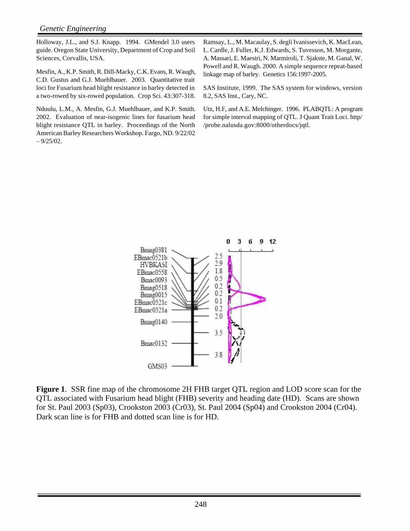
A total of 13 SSR markers, 9 from the linkage map ofCanci et al., (2004), and four additional markers pre-viously mapped in the same region by Ramsay et al.,(2000) were genotyped for fine mapping (Fig. 1). Ofthese, EBmac0849 mapped in the same location asBmac0093 and was subsequently dropped from theanalysis. For the most part, the marker order was con-sistent with the original map. The total distance cov-
ered by the new map is 17.4 cM compared with the44.9 cM distance covered by the updated Chevron xM69 map of Canci et al., 2004.
A major QTL for HD was detected between markersHVBKASI and Bmag0015 (1 cM apart) at all the fourenvironments tested and explained 40-80% of thephenotypic variation (Fig. 1; Table 2). A separate QTLfor FHB flanked by markers Bmag0140 andBmac0132 (3.5 cM apart) was detected 2 cM awayfrom the HD QTL. This FHB QTL was detected in 2of the 4 environments tested and explained 40-50%of the variance. Failure to detect FHB QTL in allenvironments was most likely due to poor disease levelsexperienced in the environments where QTL were notdetected (Table 1). In conclusion, these data indicatethat the association between FHB and HD is due tolinkage (2 cM apart) rather than pleiotropy.
ACKNOWLEDGEMENTS
We thank Ed Schiefelbein, Guillermo Velasquez, andCharlie Gustus for assistance in the field and green-house operations. This material is based upon worksupported by the U.S. Department of Agriculture, un-der Agreement No.59-0790-4-120. This is a coop-erative project with the U.S.Wheat & Barley ScabInitiative. Any opinions, findings, conclusions, or rec-ommendations expressed in this publication are thoseof the author(s) and do not necessarily reflect the viewof the U.S. Department of Agriculture.
REFERENCES
Canci, P.C., Nduulu, L.M., Muehlbauer, G.J, Dill-Macky, R.,Rasmusson, D.C., and Smith, K.P. 2004. Validation of Quan-titative Trait Loci for Fusarium head blight and kernel discol-oration in barley. Molecular Breeding. 91-104
de la Pena, R. C., Smith, K. P., Capettini, F., Muehlbauer, G. J,Gallo-Meagher, M., Dill-Macky, R., Somers, D. A., andRasmusson D. C. 1999. Quantitative trait loci associatedwith resistance to fusarium head blight and kernel discolora-tion in barley. Theor. App. Genet. 99:561-569.
Gustus, C. and K. P. Smith. 2001. Evaluating Phenotypic andMarker Assisted Selection in the F2 Generation for Chevron-derived FHB Resistance in Barley. Proceedings of the 2001National Fusarium Head Blight Forum. Erlanger, KY 12/8/01 -12/10/01.
248
Genetic Engineering
Holloway, J.L., and S.J. Knapp. 1994. GMendel 3.0 usersguide. Oregon State University, Department of Crop and SoilSciences, Corvallis, USA.
Mesfin, A., K.P. Smith, R. Dill-Macky, C.K. Evans, R. Waugh,C.D. Gustus and G.J. Muehlbauer. 2003. Quantitative traitloci for Fusarium head blight resistance in barley detected ina two-rowed by six-rowed population. Crop Sci. 43:307-318.
Nduulu, L.M., A. Mesfin, G.J. Muehlbauer, and K.P. Smith.2002. Evaluation of near-isogenic lines for fusarium headblight resistance QTL in barley. Proceedings of the NorthAmerican Barley Researchers Workshop. Fargo, ND. 9/22/02– 9/25/02.
Ramsay, L., M. Macaulay, S. degli Ivanissevich, K. MacLean,L. Cardle, J. Fuller, K.J. Edwards, S. Tuvesson, M. Morgante,A. Massari, E. Maestri, N. Marmiroli, T. Sjakste, M. Ganal, W.Powell and R. Waugh. 2000. A simple sequence repeat-basedlinkage map of barley. Genetics 156:1997-2005.
SAS Institute, 1999. The SAS system for windows, version8.2, SAS Inst., Cary, NC.
Utz, H.F, and A.E. Melchinger. 1996. PLABQTL: A programfor simple interval mapping of QTL. J Quant Trait Loci. http//probe.nalusda.gov:8000/otherdocs/jqtl.
Figure 1. SSR fine map of the chromosome 2H FHB target QTL region and LOD score scan for the QTL associated with Fusarium head blight (FHB) severity and heading date (HD). Scans are shown for St. Paul 2003 (Sp03), Crookston 2003 (Cr03), St. Paul 2004 (Sp04) and Crookston 2004 (Cr04). Dark scan line is for FHB and dotted scan line is for HD.

249
Genetic Engineering
Table 1. Means of the parents (pNIL, Chevron and M69) and recombinant near isogenic lines (rNILs) for percent Fusarium head blight severity (FHB) and heading date (HD). Trait Environment pNIL Chevron M69 rNIL Mean Range FHB Sp2003 1.8a 1.3a 3.7b 2.4 1.0-4.4 Cr2003 2.3a 3.8a 7.2b 5.5 0.7-13.1 Sp2004 11.3a 8.5a 12.3a 9.4 1.9-20.6 Cr2004 6.6a 1.6a 21.5b 17.6 4.6-59.7 HD
Sp2003
58.5b
60.0c
55.0a
58.0
54.0-61.0
Cr2003 58.5b 61.0c 56.5a 58.1 54.0-61.0 Sp2004 55.0b 55.7b 49.3a 56.7 48.3-57.0 Cr2004 66.7b 68.7c 64.3a 66.0 62.3-68.7 Means within the same row followed by the same letter are not significantly different (P?0.05). Sp2003 = St. Paul, MN, 2003; Cr2003 = Crookston, MN, 2003; Sp2004 = St. Paul, MN, 2004; Cr2004 = Crookston, MN, 2004.
Table 2. Significant QTL (LOD>3.0) associated with Fusarium head blight (FHB) severity and heading date (HD) at four environments in the Chevron/M69 fine mapping population of recombinant near isogenic lines. Trait/ Pos1
Marker interval
St. Paul 2003 Crookston 2003 St. Paul 2004 Crookston 2004
LOD %Exp2 Add3 LOD %Exp Add LOD %Exp Add LOD %Exp Add FHB 11,13 Bmag0140-
Bmac132 3.43 43.2 -2.82 4.21 50.0 -14.37
HD 6 HVBKASI-
EBmac0558 3.05 44.3 1.63
7 Bmag0518-Bmag0015
8.74 76.3 2.59 10.02 80.8 2.75 9.62 79.5 3.47
1Pos = centimorgan position. 2% Exp = Percent phenotypic variance explained by QTL. 3Add = Additive effect of the Chevron allele on FHB severity and heading date expressed as regression coefficient.

250
Genetic Engineering
A GFP REPORTER STRAIN FOR MONITORING TRI5 GENEACTIVITY IN FUSARIUM GRAMINAERUM
L. Niessen*, G. Kalinowski, S. Theisen and R.F. Vogel
Technische Universität München, Lehrstuhl für Technische Mikrobiologie,Weihenstephaner Steig 16, D-85350 Freising, Germany
*Corresponding Author: PH: 49 8161 715496; E-mail: [email protected]
ABSTRACT
Fusarium graminearum is the most serious pathogen within the Fusarium Head Blight complex of fungalspecies. It produces the trichothecene mycotoxin Deoxynivalenol (DON) as a major virulence factor in thehost-pathogen interaction. The TRI5 gene has a key role in the biosynthesis of trichothecene mycotoxins. Wehave developed a reporter system by transformation of F. graminearum TMW 4.0122 with the eGFP geneunder control of the TRI5 promotor. A 926 bp fragment upstream of the TRI5 start codon containing thepromotor region as well as a 342 bp portion upstream from the 3´end of the TRI5 coding region of a singlespore isolate of F. graminearum TMW 4.0122 were cloned in E. coli DH5±. Fragments were excised andligated via HindIII restriction sites newly introduced by modified primers. The ligation product was cloned intothe pSM2 vector via PstI and ClaI restriction sites to result in transformation vector pSM2GK1871, whichwas linearized by restriction of a singular HindIII site. Protoplasts of F. graminearum TMW 4.0122 wereobtained by treatment of germinated conidia with driselase (Interspex) at 30 °C for 3 h. Protoplasts wereseparated and transformed with linearized pSM2GK1871. Selection on hygromycin B agar (150 µg/ml) re-vealed 88 transformants. Clones were subcultured on GYEP agar plates and inspected for expression of eGFPunder the fluorescence microscope (Olympus). One clone (10/2/1) displayed intense green fluorescence emis-sion at 510-550 nm upon excitation at 470-490 nm after 15 d of incubation at 25 °C. No such fluorescencewas seen in the wild type strain grown under the same conditions. Fluorescence in the transformant was limitedto a specific type of cells, which showed a characteristic yellow pigmentation when inspected under the lightmicroscope. Such cells were also present in the wild type mycelium, with no green fluorescence emitted uponexcitation at 470-490 nm. We are currently investigating whether trichothecene production in F. graminearumTMW 4.0122 might be restricted to specialized cells (“toxocytes”). Based on the results obtained during thecurrent study we are using the TRI5 reporter strain to investigate the role of that gene in the barley/wheat-F.graminearum interaction and to learn more about the factors affecting and regulating production of DONunder the conditions prevailing in the field and at processing of cereals, e.g. during malt production.

251
Genetic Engineering
GLUCOSYLTRANSFERASES FROM ARABIDOPSISTHALIANA INACTIVATING THE FUSARIUM TOXINS
DEOXYNIVALENOL AND ZEARALENONEB. Poppenberger1,3, F. Berhiller2, D. Lucyshyn1, R. Mitterbauer1,
W. Schweiger1, R. Schuhmacher2, R. Krska2 and G. Adam1*
1BOKU – University of Natural Resources and Applied Life Sciences, Department of Applied Plant Sciences and Plant Biotechnology, Institute of Applied Genetics and Cell Biology,Vienna, Austria; 2BOKU – University of Natural Resources and Applied Life Sciences,
Department IFA-Tulln, Center for Analytical Chemistry, Tulln, Austria; and 3Present Address:CNAP, Department of Biology, University of York, York YO10 5YW, UK
*Corresponding Author: PH: 0043-1-36006-6380; E-mail: [email protected]
ABSTRACT
During the infection of small grain cereals and maize Fusarium graminearum produces the mycotoxinsdeoxynivalenol (DON) and zearalenone (ZON). It has been demonstrated that the production of the trichotheceneDON, which acts as an inhibitor of eukaryotic protein synthesis, contributes to the virulence of Fusarium(presumably by interfering with the expression of plant defense genes). ZON, which has very high estrogenicactivity in animals, also seems to play a role in plant-pathogen interaction. We have searched for Arabidopsisgenes which can inactivate these Fusarium toxins. A yeast strain highly sensitive to DON was used as a hostfor an Arabidopsis thaliana expression library and a UDP-glucosyltransferase (UGT) gene conferring resis-tance to DON (DOGT1) was identified (Poppenberger et al., 2003). Overexpression of the DOGT1 gene inArabidopsis led to increased DON resistance of seedlings. The metabolite DON-3O-glucoside is inactive ininhibiting protein synthesis (tested with a wheat germ extract in vitro translation system). DOGT1 is located ina cluster of 6 highly similar genes, but surprisingly the protein with the most closely related sequence is notprotecting against DON. By making hybrid proteins and functional testing in yeast we characterized structuralfeatures essential for substrate specificity of these UGTs. Interestingly nivalenol, which has just one additionalhydroxyl group, escapes detoxification.
We have also cloned an Arabidopsis UGT which converts ZON into ZON-4O-glucoside. While ZON showsstrong binding to the estrogen receptor in vitro, this is not observed with ZON-glucoside. Expession of thisUGT in an engineered yeast strain expressing the human estrogen receptor (hER), interferes with ZON-induced activation of hER-dependent reporter genes. This remarkable affinity can be exploited to produceZON-glucoside in high yield by feeding ZON to the recombinant yeast.
We propose that the glucosides of DON and ZON produced by plants are a currently overlooked source of“masked mycotoxin”. While the mycotoxin-glucosides escape standard analytical procedures, the toxic aglycacan be easily reactivated in the digestive tract.
ACKNOWLEDGEMENT
This work was supported by the the Austrian Genome Programme GEN-AU and the Austrian Academy ofSciences (DOC fellowship B.P.).

252
Genetic Engineering
REFERENCES
Poppenberger, B., Berthiller, F., Lucyshyn, D., Sieberer, T., Schuhmacher, R., Krska, R., Kuchler, K., Glössl,J., Luschnig, C., and Adam, G. (2003) Detoxification of the Fusarium mycotoxin deoxynivalenol by a UDP-glucosyltransferase from Arabidopsis thaliana. J. Biol. Chem. 278:47905-47914.
253
Genetic Engineering
TRANSGENES IN WHEATS. Sato1, X. Ye1, B. Langston2, J. Bohlmann1, B. LaVallee1, A. Mitra1,
S. Baenziger1, M. Dickman2 and T. Clemente1*
1Department of Agronomy and Horticulture; and 2Department of Plant Pathology,University of Nebraska, Lincoln, NE, 68503-0915, USA
*Corresponding Author: PH: 402-472-1428; E-mail: [email protected]
ABSTRACT
Our wheat transformation team at the University of Nebraska-Lincoln (UNL) employs an Agrobacterium-mediated protocol to deliver transgenes to the crop. We recently completed a survey of 30 spring wheatgenotypes for enhanced transformation frequencies. From this work a hard white genotype Xin chun 9 wasidentified that displayed improved transformability over Bobwhite. Current efforts are focused on evaluating aseries of novel Agrobacterium tumefaciens strains in a comparative study with both Bobwhite andXin chun 9.
We have been using this transformation system to evaluate potential antifungal transgenes in support of acollaborative effort targeting Fusarium Head Blight (FHB) resistance at UNL. To this end a total of 48transgenic wheat lines harboring three novel negative regulators of programmed cell death, or a ribosomalinactivating protein have been handed-off to our wheat breeding program. More recently transgenic wheatlines carrying two additional negative regulators of programmed cell death and a synthetic antifungal lyticpeptide have been produced. Field trials were conducted in the spring of 2004 on 28 transgenic lines carrythree negative regulators of programmed cell death genes, ced9, IAP and Bcl-xl, along with lines harboring theribosomal inactivating protein. Field plots were inoculated with F. garminearum just prior to anthesis. Datahas been ascertained on agronomic parameters, days to anthesis, vigor and plant height, in addition to FHBseverity and incidence.

254
Genetic Engineering
OPTIMIZATION OF AN AGROBACTERIUM-MEDIATEDTRANSFORMATION SYSTEM FOR DURUM WHEAT
Valluri V. Satyavathi1, Prem P. Jauhar2* and Lynn S. Dahleen2
Dept. of Plant Sciences, North Dakota State University1, USDA-ARS, Northern CropScience Laboratory2, Fargo, ND 58105, USA
*Corresponding Author: PH: (701) 239-1309; E-mail: [email protected]
OBJECTIVES
To optimize an Agrobacterium-mediated transforma-tion system for durum wheat to facilitate incorporationof antifungal genes for resistance against Fusarium headblight.
INTRODUCTION
Durum wheat (Triticum turgidum L., 2n = 4x = 28;AABB genomes) is an important cereal crop grown inthe United States, Canada, and in some Europeancountries. Several methods have been used for itsgenetic improvement. In recent years, genetic engi-neering has opened up new avenues for crop improve-ment and is a useful adjunct to conventional breeding.A prerequisite for application of such modern tech-niques is an efficient and reliable in vitro plant regen-eration system. Gene transfers in plants have beenachieved through direct DNA uptake, electroporation,microinjection, particle bombardment andAgrobacterium-mediated methods. We standardizedan efficient regeneration system for commercial du-rum wheat cultivars (Bommineni and Jauhar, 1996;Satyavathi et al., 2004) and by using particle bom-bardment produced transgenic durum with markergenes (Bommineni et al., 1997) and antifungal genes(Satyavathi and Jauhar, 2003). Transgenic durum hasnow been produced in other laboratories (He et al.,1999; Pellegrineschi et al., 2002). In bread wheat,partial FHB resistance was achieved by expressingpathogenesis-related proteins using particle bombard-ment, but this technique was hampered by multiplecopy gene insertions and gene silencing (Anand et al.,2003). Compared to direct gene transfer techniques,Agrobacterium-mediated transformation offers a num-ber of advantages, including potentially low copy num-ber and preferential integration into transcriptionally
active regions of the chromosome (Hu et al., 2003).So far, an Agrobacterium-mediated transformationsystem has not been reported for durum wheat. There-fore, we attempted to optimize the conditions forAgrobacterium mediated transformation of the com-mercial durum wheat cv. Maier using marker genes.
MATERIALS AND METHODS
Plant material and preculture - An agronomicallysuperior durum cultivar, Maier was used for transfor-mation. Spikes were harvested 14 days post anthesisand the spikelets were surface sterilized and culturedas described by Bommineni and Jauhar (1996). Thecallus induction medium was supplemented with 2.0mg L-1 dicamba. The cultures were incubated in thedark at 25 ± 2°C for 1-14 days depending on theexperiment performed.
Agrobacterium tumefaciens strain, plasmid, andculture - A disarmed Agrobacterium tumefaciensstrain AGL1 harboring pDM805 was provided byCSIRO Plant Industry, Canberra, Australia (Fig. 1).The binary vector pDM805 contains thephosphinothricin acetyltransferase (bar) gene underthe control of the promoter from the maize ubiquitin 1(Ubi1) gene and a terminator sequence from the A.tumefaciens nopaline synthase (nos) gene; the β-glu-curonidase gene uidA (gus) under the control of thepromoter from the rice actin 1 (Act1) gene and a ter-minator sequence from the rice ribulose bis-phosphatecarboxylase/oxygenase (rbcS) gene. A full strengthAgrobacterium suspension was obtained a day be-fore transformation as described by Tingay et al.(1997).
Acetosyringone treatment and particle bombard-ment - Acetosyringone effects on trans-formation ef-
255
Genetic Engineering
ficiency were studied by comparing Agrobacteriumsuspension without the chemical to suspension treatedwith 200 µM acetosyringone prior to infection. Theexplants were then infected for about 1 h. To studythe effect of particle bombardment on the extent ofinfection, the scutella were precultured for 14 daysand wounded by bombardment with gold particles.About 30-50 explants were bombarded with 0.3 mgof gold particles (1.0 µM) using a BioRad PDS-1000biolistic device with 1,100 psi rupture disc and com-pared to unwounded explants.
Inoculation and co-cultivation - Isolated scutellaprecultured for 1 day and 14 days were used for trans-formation. The explants were immersed in full strengthAgrobacterium suspension in a Petri dish for half anhour (1 h when acetosyringone was used in the sus-pension) and then transferred without rinsing, withscutellar surface placed in contact with the callus in-duction medium. Co-cultivation was carried out at 25± 2°C in darkness for about 2-3 days.
Selection and plant regeneration - After co-culti-vation, the explants were washed thrice with steriledistilled water in a Petri dish and blotted on sterileWhatman filter paper. The explants were then platedon selection medium, which was same as callus induc-tion medium but was supplemented with 150 mg L-1
Timentin and 5.0 mg L-1 bialaphos. Explants weremaintained on selection medium for 3-4 weeks at 25± 2°C in darkness, after which they were transferredto regeneration medium (selection medium withoutgrowth regulators). The culture conditions and regen-eration procedure were same as described bySatyavathi et al. (2004).
Histochemical GUS assay - T-DNA delivery intoexplant tissues was determined after 1-3 weeks ofculture on selection medium using the histochemicalGUS assay according to Bommineni et al. (1997).Explants with blue spots and the number of blue spotsper explant were counted under a stereomicroscope.
Statistical analyses - For studying the effect ofacetosyringone and bombardment on DNA delivery,we compared GUS expression among the explants.Each treatment had three replications and at least 50
explants (4 from each Petri dish) per treatment weresacrificed for GUS assays. As the data on the numberof explants with GUS spots and the number of spotsper explant for each treatment were not normally dis-tributed, analysis of variance was done using PROCCATMOD (SAS version 8.2, 2001).
RESULTS
Preliminary tests were performed to compare the re-sponses of the scutella that were precultured for 1 dayvs 10-14 days. After 1-2 days of co-cultivation withAgrobacterium and on transfer to selection medium,only 10% of the scutella precultured for 1-day initi-ated calli. In the case of 10-14 day preculturedscutella, about 70% of them developed callus overthe cut surface within a week and later developed cal-lus around the periphery of the scutellum. After 3weeks on selection medium, 511 of 725 scutella co-cultivated were resistant to bialaphos selection. GUSassays done 7 days after co-cultivation showed GUSspots all over the scutellum surface but most of thespots were localized on the areas starting to form cal-lus, usually at the periphery of the scutellum (Fig. 2A& B). In subsequent subcultures, the proliferating calluswas selected and brown callus was discarded. After4 weeks on selection medium, the embryogenic calluswas transferred to regeneration medium supplementedwith 5.0 mg L-1 bialaphos. Out of 725 scutella in-fected, only 3 plantlets were regenerated at the end of3-4 weeks with a transformation frequency of 0.4%.Various treatments like increasing co-culture duration,adding acetosyringone in the Agrobacterium suspen-sion, and bombarding the explants with gold particlesbefore infection, had differential effects on transfor-mation efficiency as follows:
Duration of co-culture - About 82% of the scutellathat were co-cultivated for 2 days were resistant tobialaphos selection. When the co-cultivation was ex-tended to 3 days, an overgrowth of the bacteria wasobserved on 50% of the scutella and the percentageof resistant scutella decreased to 56%.
Effect of acetosyringone - GUS expression wasdetected in all the tissues 3 wk after co-cultivation ei-ther in the presence or absence of the pretreatment.
256
Genetic Engineering
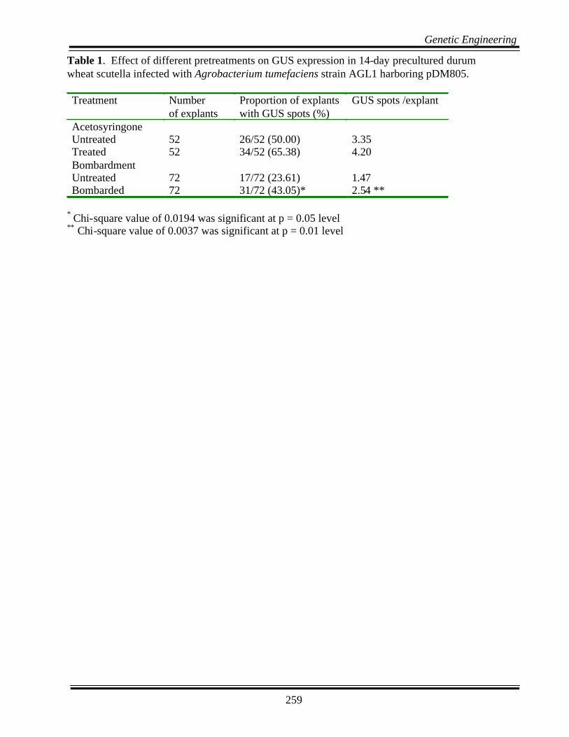
The scutella that were treated with acetosyringone didnot differ for GUS expression in terms of the numberof explants with GUS spots and the number of GUSspots per explant compared to those infected withAgrobacterium suspension without acetosyringone(Table 1).
Effect of bombardment - The explants that were in-jured by bombarding with gold particles showed sig-nificantly greater number of explants with GUS spots(p < 0.05) and also significantly greater GUS spotsper explant (p < 0.01) than those that were notwounded prior to infection (Table 1).
DISCUSSION
This work presents the first report on Agrobacterium-mediated transformation of durum wheat. A prereq-uisite for development of Agrobacterium-mediatedtransformation is the establishment of optimal condi-tions for T-DNA delivery into tissue from which wholeplants can be regenerated. Based on our previousfindings, we selected durum cultivar Maier for trans-formation with Agrobacterium. In general, modelgenotypes amenable to tissue culture or tomicroprojectile transformation have worked well forAgrobacterium-mediated transformation in severalcrops like wheat, maize, barley, and sugar cane (Chenget al., 2004). The isolated scutella are known to bechoice explants for many cereals including durum wheatand have been successfully used for regeneration andtransformation experiments. In the present study, wefound that 10-14 days preculture of explants increasestransformation efficiency. Similar results were observedin wheat where longer precultures resulted in efficientT-DNA delivery (Cheng et al., 1997; Wu et al., 2003).We obtained a transformation frequency of 0.4% whichis comparable to that reported in other cereals. Inwheat, transformation frequencies ranged from 0.3-4.3% and were increased to 10.5% by desiccation ofprecultured embryos (Cheng et al., 2004).
Chemicals such as acetosyringone for vir gene induc-tion are recommended in most of the monocot trans-formation protocols. We used a 200 µMacetosyringone treatment prior to infection. The pres-ence of acetosyringone did not increase GUS expres-
sion. The addition of acetosyringone at a concentra-tion of 150 to 200 µM during preculture or co-cultureincreased the number of transformed cells in rice (Hieiet al., 1994), barley (Tingay et al., 1997), and wheat(Cheng et al., 1997). In the present study, woundingprecultured scutella with gold particles before infect-ing the explants with Agrobacterium, increased theGUS expression significantly. Similar results wereobserved in barley (Tingay et al., 1997).
The advantage of Agrobacterium-mediated transfor-mation over particle bombardment is that this methodis simple and cost effective. We optimized conditionsfor Agrobacterium-mediated transformation usingscutella of the cultivar Maier and also studied the ef-fects of various pretreatments that could enhance in-fection and T-DNA delivery. Further experiments areneeded to increase regeneration from transformedcallus.
ACKNOWLEDGEMENT
We thank Mr. Terrance Peterson for technical help.
REFERENCES
Anand, A., Trick, H.N., Gill, B.S., and Muthukrishnan, S. 2003.Stable transgene expression and random gene silencing inwheat. Plant Biotechnology J. 2:241-251.
Bommineni, V.R., and Jauhar, P.P. 1996. Regeneration of plant-lets through isolated scutellum culture of durum wheat. PlantSci. 116:197-203.
Bommineni, V.R., Jauhar, P.P., and Peterson, T.S. 1997.Transgenic durum wheat by microprojectile bombardment ofisolated scutella. J. Hered. 88:475-481.
Cheng, M., Lowe, B.A., Spencer, T.M., Ye, X., and Armstrong,C.L. 2004. Factors influencing Agrobacterium-mediatedtransformation of monocotyledonous species. In Vitro Cell.Dev. Biol.- Plant. 40:31-45.
Cheng, M., Fry, J.E., Pang, S. et al. 1997. Genetic transforma-tion of wheat mediated by Agrobacterium tumefaciens. PlantPhysiol. 115:971-980.
He, G.Y., Rooke, L., Steele, S. et al. 1999. Transformation ofpasta wheat (Triticum turgidum L. var. durum) with high-molecular-weight glutenin subunit genes and modificationof dough functionality. Mol. Breeding 5:377-386.
257
Genetic Engineering
Hiei, Y., Ohta, S., Komari, T., and Kumashiro, T. 1994. Effi-cient transformation of rice (Oryza sativa L.) mediated byAgrobacterium and sequence analysis of the boundaries ofthe T-DNA. Plant J. 6:271-282.
Hu, T., Metz, S., Chay, C. et al. 2003. Agrobacterium-medi-ated large-scale transformation of wheat (Triticum aestivumL.) using glyphosate selection. Plant Cell Rep. 21:1010-1019.
Pellegrineschi, A., Brito, R.M., Velazquez, L. et al. 2002. Theeffect of pretreatment with mild heat and drought stresses onthe explant and biolistic transformation frequency of threedurum wheat cultivars. Plant Cell Rep. 20:955-960.
SAS. Copy righted 1999-2001. Procedure release 8.2. SASInstitute, Inc., SAS Campus Drive, Cary, NC.
Satyavathi, V.V., and Jauhar, P.P. 2003. In vitro regenerationof commercial durum cultivars and transformation with anti-fungal genes. Proc. of the 2003 National Fusarium Head BlightForum, Minneapolis, December 12-15, 2003. pp. 32-35.
Satyavathi, V.V., Jauhar, P.P., Elias, E.M., and Rao, M.B. 2004.Effects of growth regulators on in vitro plant regeneration indurum wheat. Crop Sci. 44:1839-1846.
Tingay, S., McElroy, D., Kalla, R., Fieg, S., Wang, M., Thornton,S., and Brettell, R. 1997. Agrobacterium tumefaciens-medi-ated barley transformation. The Plant J. 11:1369-1376.
Wu, H., Sparks, C., Amoah, B., and Jones, H.D. 2003. Factorsinfluencing successful Agrobacterium-mediated genetictransformation of wheat. Plant Cell Rep. 21:659-668.

258
Genetic Engineering
BL
Am
pr
ori
pSP
72rb
cSgu
sA
ct1
Ubi
1ba
rno
sB
R
K, Ss
Sn
X
N
B
Ss
X, No,
P
X
N
S
P
S
KB
B, No
S, No
S
Figu
re 1
.St
ruct
ure
and
rest
rictio
n m
ap o
f the
cer
eal t
rans
form
atio
n ve
ctor
pD
M80
5 (T
inga
yet
al
., 19
97).
BL
Am
pr
ori
pSP
72rb
cSgu
sA
ct1
Ubi
1ba
rno
sB
R
K, Ss
Sn
X
N
B
Ss
X, No,
P
X
N
S
P
S
KB
B, No
S, No
S BL
Am
pr
ori
pSP
72rb
cSgu
sA
ct1
Ubi
1ba
rno
sB
R
K, Ss
Sn
X
N
B
Ss
X, No,
P
X
N
S
P
S
KB
B, No
S, No
S
Figu
re 1
.St
ruct
ure
and
rest
rictio
n m
ap o
f the
cer
eal t
rans
form
atio
n ve
ctor
pD
M80
5 (T
inga
yet
al
., 19
97).
AB
Figu
re 2
.G
US
expr
essi
on in
the
scut
ellu
mon
e w
eek
afte
r co-
culti
vatio
n w
ith A
grob
acte
rium
. G
US
expr
essi
on in
cal
lus
that
was
resi
stan
t to
5.0
mg
L-1
bial
apho
sth
ree
wee
ks a
fter c
o-cu
ltiva
tion
with
A
grob
acte
rium
.
AB
AB
Figu
re 2
.G
US
expr
essi
on in
the
scut
ellu
mon
e w
eek
afte
r co-
culti
vatio
n w
ith A
grob
acte
rium
. G
US
expr
essi
on in
cal
lus
that
was
resi
stan
t to
5.0
mg
L-1
bial
apho
sth
ree
wee
ks a
fter c
o-cu
ltiva
tion
with
A
grob
acte
rium
.
259
Genetic Engineering
Table 1. Effect of different pretreatments on GUS expression in 14-day precultured durum wheat scutella infected with Agrobacterium tumefaciens strain AGL1 harboring pDM805. Treatment Number
of explants Proportion of explants with GUS spots (%)
GUS spots /explant
Acetosyringone Untreated 52 26/52 (50.00) 3.35 Treated 52 34/52 (65.38) 4.20 Bombardment Untreated 72 17/72 (23.61) 1.47 Bombarded 72 31/72 (43.05)* 2.54 **
* Chi-square value of 0.0194 was significant at p = 0.05 level ** Chi-square value of 0.0037 was significant at p = 0.01 level

260
Genetic Engineering
DEVELOPMENT OF TISSUE-SPECIFIC PROMOTERS FORTARGETING ANTI-FUSARIUM GENE EXPRESSIONR.W. Skadsen1*, M.L. Federico2, T. Abebe3 and M. Patel4
1U.S. Dept. of Agriculture, ARS, Cereal Crops Res. Unit, 501 Walnut St., Madison, WI 53711, USA;2University of Wisconsin, Agronomy Dept., 1575 Linden Dr., Madison, WI 53706-1590, USA; 3University of Northern Iowa, Dept. of Biology, 144 McCollum Science Hall, Cedar Falls, IA, USA; and 4University
of Wisconsin, Plant Pathology Dept., 1630 Linden Dr., Madison, WI 53706-1589, USA*Corresponding Author: PH: (608)262-3672; E-mail: [email protected]
ABSTRACT
We identified lemma and pericarp epithelium tissues as rapidly infected by Fusarium graminearum. Genesspecifically expressed in these tissues were identified and cloned so that promoters of selected genes could beused to express antifungal protein genes. These included a lipid transfer protein homologue (Ltp6), highlyexpressed in the pericarp epithelium but not in vegetative leaves, and a jacaline-like gene, Lem2, preferentiallyexpressed in the lemma/palea, compared with the flag leaf. Ltp6 is also expressed in coleoptiles and embryos;mRNA levels increase in response to salt, cold, abscisic acid and salicylic acid in a pattern distinct from otherbarley Ltps. Transient expression analysis of the promoter showed that 192 bp of upstream sequence conferstissue-specific expression and retains most promoter activity. Stable barley transformants have been pro-duced with a 247 bp promoter fused to a gfp reporter gene. In these, gfp expression is strong in the epicarp,embryo and coleoptile, but it is not found in other tissues. Gfp expression was detected during spike develop-ment, from early ovary differentiation through its final expression in the epicarp and during embyogenesis andgermination in the coleoptile, reproducing the expression pattern of the native gene. Lem2 is specificallyexpressed in the lemma/palea and coleoptile. SA induces Lem2 within 4 h, suggesting that it is a defensivegene. Promoter deletion studies showed that the tissue-specificity and promoter activity are conferred by ashort 5’ proximal region from -75 to +70. Stable transformants were produced with the “full-length” 1414 bppromoter and 5’ promoter deletions fused to gfp. Gfp expression occurred in the lemma/palea and coleoptile,but it also unexpectedly occurred in the epicarp and ligules. Lack of methylation in the epicarp may account forexpression in the epicarp.

261
Genetic Engineering
TARGETING OF ANTI-FUSARIUM GENE EXPRESSION IN BARLEYR.W. Skadsen1*, M.L. Federico2, T. Abebe3 and M. Patel4
1U.S. Dept. of Agriculture, ARS, Cereal Crops Res. Unit, 501 Walnut St., Madison, WI, 53711, USA; 2Univ.of Wisconsin, Agronomy Department, 1575 Linden Dr., Madison, WI, 53706-1590, USA; 3Univ. of Northern
Iowa, Department of Biology, 144 McCollum Science hall, Cedar Falls, IA, USA; and 4Univ. of Wisconsin,Plant Pathology Department, 1630 Linden Dr., Madison, WI 53706-1589, USA
*Corresponding Author: PH: (608)262-3672; E-mail: [email protected]
ABSTRACT
We identified lemma and pericarp epithelium (epicarp) tissues as rapidly infected by a strain of Fusariumgraminearum transformed with the green fluorescent protein gene (gfp). The fungus colonized the lemma in48 h, but it colonized the brush hairs at the seed tip within 7 h and rapidly grew downward along the epicarpand more slowly inward through the cross cells (Skadsen and Hohn, PMPP 64:45-53, 2004). Genes specifi-cally expressed in the lemma (Abebe et al., Crop Sci. 44:942-950, 2004) and epicarp were identified andcloned so that promoters of selected genes could be used to express antifungal protein genes in these suscep-tible tissues. Tissue-specific genes included a lipid transfer protein homologue (Ltp6), highly expressed in thepericarp epithelium but not in vegetative leaves, and a jacaline-like gene, Lem2, preferentially expressed in thelemma/palea, compared with the flag leaf. Ltp6 is also expressed in coleoptiles and embryos; mRNA levelsincrease in response to salt, cold, abscisic acid and salicylic acid (SA) in a pattern distinct from other barleyLtps. Transient expression analysis of the promoter showed that 192 bp of upstream sequence confers tissue-specific expression and retains most promoter activity. Stable barley transformants have been produced with a247 bp promoter fused to a gfp reporter gene (Federico et al., PMB, in press). In these, gfp expression isstrong in the epicarp, embryo and coleoptile, but it is not found in other tissues. Gfp expression was detectedduring spike development, from early ovary differentiation through its final expression in the epicarp, andduring embyogenesis and germination in the coleoptile, reproducing the expression pattern of the native gene.Lem2 is specifically expressed in the lemma/palea and coleoptile. SA induces Lem2 within 4 h, suggesting thatit is a defensive gene. Deletion studies showed that tissue-specificity is conferred by a short 5’ proximal regionfrom -75 to +70 (Abebe et al., Planta, in press) . Stable transformants were produced with the “full-length”1414 bp promoter and 5’ promoter deletions fused to gfp. Gfp expression occurred in the lemma/palea andcoleoptile, but it also unexpectedly occurred in the epicarp, perhaps due to a lack of methylation. In thelemmas, a developmental transition occurred wherein gfp was first expressed in the mesophyll cells; this wasgradually replaced by expression in specialized cork cells of the epidermis. An additional promoter, Lem1,was produced by Sathish Puthigae and showed lemma/palea-specific expression in transient assays (Skadsenet al., PMB, 49:545-555, 2002).
262
Genetic Engineering
A NOVEL STRATEGY FOR TRANSGENIC CONTROL OF FUSARIUM HEAD BLIGHT IN WHEAT
M. Somleva* and A. Blechl
USDA-ARS, Western Regional Research Center, Albany, CA 94710, USA*Corresponding Author: PH (510) 559-5673; E-mail: [email protected]
ABSTRACT
Changes in agricultural practices (e.g., minimal tilling) during the past two decades combined with changingclimate conditions have dramatically altered crop susceptibility to Fusarium head blight (FHB) or scab. FHBmay lead to direct yield losses of 5-20% worldwide in average epidemic years, but losses as high as 60-70%have also been reported. Host plant resistance, the most cost-effective way to fight the disease, in availablewheat germplasm is only partial and has been difficult to incorporate into cultivars adapted for regional growthin the U.S. Our goal is to achieve FHB resistance by employing plant genetic transformation, a potentiallypowerful tool for transgenic control of fungal diseases in cereals. We selected three candidate anti-Fusarium(AF) genes: Aspergillus glucose oxidase (GO) and barley peroxidases (Prx7 and Prx8) based on theirassociation with an array of naturally occurring plant defense mechanisms. Glucose oxidase is an apoplasticenzyme that catalyzes oxidation of β-D-glucose, generating H2O2, a compound with multiple functions in plantdefense. Induction of the peroxidases Prx7 and Prx8 has been correlated with the appearance of antifungalcompounds and papillae structures, respectively, in barley leaves exposed to powdery mildew. We insertedthe coding regions of these genes into our vector that contains the barley Lem1 promoter, which we havepreviously shown is active in the outer organs of transgenic wheat florets from anthesis to the soft dough stageof kernel development. This activity pattern makes it an excellent candidate for targeting AF gene expression tothe path of Fusarium invasion. We have generated 100 transgenic wheat lines carrying the Lem1::PRX and/orLem1::GO constructs. The in situ methods used for expression analyses of the primary transformants revealedthat the transgene-encoded proteins are accumulated either in the extracellular space (GO and Prx8) or in thecells (Prx7) of the spike tissues and were not present in developing grain. The possible synergistic effect ofthese enzymes on improving host resistance to initial fungal infection and pathogen spread will be discussed. Ifour strategy is successful, the lack of recombinant proteins in the grain will minimize concerns about the safetyof foods derived from these wheats and facilitate their approval by regulators and acceptance by consumers.
263
Genetic Engineering
CHARACTERIZATION OF ORGAN SPECIFICPROMOTERS IN TRANSGENIC WHEAT
M. Somleva* and A. Blechl
USDA-ARS, Western Regional Research Center, Albany, CA 94710, USA*Corresponding Author: PH (510) 559-5673; E-mail: [email protected]
OBJECTIVE
To identify promoters suitable for targeting anti-Fusarium gene expression to wheat tissues surround-ing the developing seed.
INTRODUCTION
Genetic engineering is the most promising approachto increase plant resistance to fungal pathogens, in-cluding Fusarium. The effectiveness of an antifungalgene in planta is determined by its expression levelsin the crucial host tissues and by the timing of its ex-pression such that suitable levels of the encoded pro-tein accumulate before the infection (Dahleen et al.,2001). At present, constitutive promoters are widelyused to achieve high expression levels throughout mosttissues of the plant. If only specific tissues need to beprotected or antifungal compounds need to be ex-pressed at certain targeted sites, the use of specificpromoters is recommended (Punja, 2001). Expres-sion of anti-Fusarium (AF) genes in the glume andlemma is desirable for both wheat and barley, becausethese organs comprise the outer most protective bar-rier encasing the reproductive organs. In this study wepresent the organ- and developmental specificity ofthe promoter of a maize glutamine synthase gene, GS,and the promoter of a barley floret-expressed gene,Lem1, in stable hexaploid wheat transformants.
MATERIALS AND METHODS
Vector and plasmid constructs: The following plas-mids were used for wheat transformation: pGS176and pGS177 carrying a 664-bp fragment of the pro-moter of the maize GS1-2 gene fused to the uidA cod-ing region (GUS) and the first introns of the nativegene and of the maize alcohol dehydrogenase (ADH)
gene, respectively (Muhitch et al., 2002); andpBSD5sGFP carrying a 1400-bp fragment containingthe Lem1 promoter and a partial N-terminal codingregion fused to the coding sequence for the green fluo-rescent protein (Lem1::GFP, Skadsen et al., 2002).For comparative studies of the promoter activity pat-terns, the plasmid pAHC15 carrying the uidA genedriven by the maize Ubi1 promoter and first intron(UBI::GUS) was also used (Christensen and Quail,1996).
Generation of primary transformants and moni-toring of the reporter gene expression: Transientexpression assays and wheat transformation were per-formed by particle bombardment of immature embryosof cv. Bobwhite. Stably transformed plants were iden-tified as described (Okubara et al., 2002). GFP fluores-cence in various tissues of transgenic plants and invitro cultures was monitored using an Olympus SZXstereomicroscope equipped with an SZX-RFL fluo-rescence attachment and a DP11 digital camera. GUSactivity was detected according to Hänsch et al.(1995).
Construction of cloning vectors: To makepBGS9Lem1, a 1067-bp fragment containing theLem1 promoter was PCR-amplified from the plasmidpBSD5sGFP (Skadsen et al., 2002) using Pfu Poly-merase (Stratagene) and primers 5’-GATAAGCTTGGGATGTC-3’ and 5’-ACGGATATCTGCGGTTGAAG-3’ with 5’ exten-sions to add HindIII and EcoRV restriction sites, re-spectively. After complete digestion with HindIII, theresulting fragment was ligated with a 3935-bp restric-tion fragment containing pBGS9 (Spratt et al., 1986)and the NOS 3’ transcriptional terminator sequence.The latter piece of DNA had been prepared from themonocot transformation vector pUBK (Okubara etal., 2002). To make pBGS9Lem1ADHi1, the first in-

264
Genetic Engineering
tron of the maize alcohol dehydrogenase gene, ADH,was PCR-amplified using the plasmid pGS177(Muhitch et al., 2002) as a template. The resulting 601-bp fragment was inserted into the EcoRV site ofpBGS9Lem1. A unique SmaI restriction site separatesthe ADH intron and the NOS 3’ region.
RESULTS AND DISCUSSION
In wheat, the period of susceptibility to head infectionby Fusarium lasts from anthesis (the time point whenthe anthers extrude from the spikes) through the doughstage of kernel development. To identify a promotersuitable for expression of anti-Fusarium genes, theactivities of reporter genes GUS and GFP fused to themaize GS and barley Lem1 promoters, respectively,were monitored during growth and development ofprimary wheat transformants and their progeny. TheGS promoter is only expressed in the pericarp and inthe scutellum of mature embryos (Fig. 1). Thus, it isnot suitable for use in anti-Fusarium constructs. Intransgenic plants carrying both Lem1::GFP andUBI::GUS, we compared the activity patterns of thetwo promoters by monitoring the expression of bothreporter genes during plant development (Fig. 2). Weobserved no GFP fluorescence in vegetative organs,indicating that the Lem1 promoter was not active inthese tissues. This is in accordance with the data forits organ- and developmental specificity in barley(Skadsen et al., 2002). In floret tissues, we detectedno GFP fluorescence before anthesis, demonstratingthat the Lem1 promoter did not function before thisstage (data not shown). In contrast, strong UBI-drivenGUS activity was detected in the young ovary andanthers (data not shown). At anthesis, UBI is active inthe reproductive organs (Fig. 2A), while the Lem1promoter drove high levels of gfp expression only inthe organs surrounding the developing floret (Fig. 2A).(The autofluorescence of the anthers was also seen incontrol plants.) These findings indicate that the Lem1promoter is active during the same period in spikedevelopment in transgenic wheat as it is in its nativecontext in barley. No GFP fluorescence was seen indeveloping seeds (Fig. 2B and C). In contrast, GUSactivity driven by the UBI promoter was detected inthe seed coat during the earliest stages of grain devel-
opment – watery ripe and soft dough stages (Fig. 2Band C, respectively).
The relative strength of the barley Lem1 promoter wasassessed in a transient assay (Fig. 3). Transient gfpexpression was first observed in wheat embryos 10 hafter bombardment (Fig. 3B), while uidA expressiondriven by maize UBI was detected within 2 h (Fig.3A). This finding indicates that that Lem1 is less ac-tive than UBI, which is one of the strongest of cerealpromoters characterized to date. Approximately thesame difference in gfp transient expression under thecontrol of Lem1 and UBI promoters was shown bySkadsen et al. (2002) in bombarded spikes of wheatand barley.
Results from these comparative studies suggest that,due to its organ specificity and moderate strength, thebarley Lem1 promoter would be an excellent choiceto target anti-Fusarium gene expression to wheat tis-sues surrounding the developing seed at anthesis, whileexcluding transgene-encoded foreign proteins from theedible grain. To facilitate its use for this and other pur-poses, we constructed the cloning vectorspBGS9Lem1 carrying the Lem1 promoter and theNOS 3’ terminator sequence (Fig. 4A) andpBGS9Lem1ADHi1, in which the first intron of themaize ADH gene was fused to the Lem1 promoter(Fig. 4B). Both vectors have unique blunt-end restric-tion sites that can be used for insertion of any codingsequence. Recently, we have successfully employedpBGS9Lem1 to express candidate anti-Fusariumgenes in transgenic wheat.
ACKNOWLEGEMENTS
This material is based upon work supported by theU.S. Department of Agriculture, under Agreement No.0405-BL-126. This is a cooperative project with theU.S. Wheat & Barley Scab Initiative.
REFERENCES
Christensen, A.H. and Quail, P.H. 1996. Ubiquitin promoter-based vectors for high-level expression of selectable and/orscreenable marker genes in monocotyledonous plants.Transgenic Res. 5:213-218.

265
Genetic Engineering
Dahleen, L.S., Okubara, P.A. and Blechl, A.E. 2001. Transgenicapproaches to combat Fusarium head blight in wheat andbarley. Crop Sci. 41:628-637.
Hänsch, R., Koprek, T., Mendel, R.R. and Schulze, J.1995. Animproved protocol for eliminating endogenous b- glucu-ronidase background in barley. Plant Sci. 105:63-69.
Muhitch, M.J., Liang, H., Rastogi, R. and Sollenberger, K.G.2002. Isolation of a promoter sequence from the glutaminesynthetase1-2 gene capable of conferring tissue-specific geneexpression in transgenic maize. Plant Sci. 163:865-872.
Okubara, P.A., Blechl, A.E., McCormick, S.P., Alexander, N.J.,Dill-Macky, R. and Hohn, T.M. 2002. Engineeringdeoxynivalenol metabolism in wheat through the expressionof a fungal trichothecene acetyltransferase gene. Theor. Appl.Genet. 106:74-83.
Punja, Z.K. 2001. Genetic engineering of plants to enhanceresistance to fungal pathogens – a review of progress andfuture prospects. Can. J. Plant Pathol. 23:216-235.
Skadsen, R.W., Sathish, P., Federico, M.L., Abebe, T., Fu J.and Kaeppler, H.F. 2002. Cloning of the promoter for a novelbarley gene, Lem1, and its organ-specific promotion of Gfpexpression in lemma and palea. Plant Mol. Biol. 49:545-555.
Spratt, B.G., Hedge, P.J., teHeesen, S., Edelman, A. andBroome-Smith, J.K. 1986). Kanamycin-resistant vectors thatare analogues of plasmids pUC8, pUC9, pEMBL8 andpEMBL9. Gene 41:337-342.

266
Genetic Engineering
Figure 2. Activity patterns of the barley Lem1 and maize UBI promoters during development of wheat florets and grain. Reporter gene expression was monitored in stable wheat transformants carrying Lem1::GFP and UBI::GUS, either by direct fluorescence (GFP) or by histochemical staining (GUS). Each panel shows developing florets and seeds from non-transformed plants under UV light (left) and GFP fluorescence (center) and GUS activity (right) in the same type of specimens from transgenic plants. (GFP fluorescence is visible as very light areas. GUS activity visible in color photographs is indicated by arrows.) A Florets at anthesis. Note the lack of GFP fluorescence in the outer floret organs of the control. B Developing kernels at a watery ripe stage. C Wheat grain at a soft dough stage. Note that the mature anthers and hairs of the caryopsis brush show strong autofluorescence under these conditions.
A B
C
A B
C
Figure 1. Activity of the GS promoter in developing florets and seeds in transgenic wheat. (GUS activity visible in color photographs is indicated by arrows.) A No GUS activity was observed in a young ovary and anthers. B GUS activity in a maturing ovary in a spikelet after pollination. There was no staining in the outer floral organs. C GUS activity in the pericarp. D and E No uidA expression was detected in immature embryos. F GUS activity in the scutellum of a mature embryo.
A B C D E F

267
Genetic Engineering
A B C D
Figure 3. Activity of Lem1 and UBI promoters during callus initiation and culture. Isolated zygotic wheat embryos 21 DAA were co-bombarded with Lem1::GFP and UBI::GUS. A GUS activity in an embryo 2 h after bombardment (dark spots). BExpression of gfp10 h after bombardment (lighter spots). C GFP fluorescence in a callus after one week of culture on the recovery medium. D The callus shown in Fig. 3C after staining for GUS activity.
A B C DA B C D
Figure 3. Activity of Lem1 and UBI promoters during callus initiation and culture. Isolated zygotic wheat embryos 21 DAA were co-bombarded with Lem1::GFP and UBI::GUS. A GUS activity in an embryo 2 h after bombardment (dark spots). BExpression of gfp10 h after bombardment (lighter spots). C GFP fluorescence in a callus after one week of culture on the recovery medium. D The callus shown in Fig. 3C after staining for GUS activity.
Figure 4. Linear diagrams of the cloning vectors. A The plasmid pBGS9Lem1 features the barley Lem1 promoter and nopaline synthase terminator (NOS). B The plasmid pBGS6Lem1ADH1 contains the ADH first intron inserted between the promoter and NOS. The unique EcoRV (A) and SmaI (B) sites were created to facilitate insertion of any coding region as a blunt-ended fragment. The locations of some other restriction sites are shown. The pBGS9 portion is not to scale.
Hin
d II
I (1)
Nco
I (14
0)
Xho
I (49
4)
Eco
RV
(105
7)
Sal I
(107
4)
Eco
RI (
1361
)
Lem1 promoter NOS
5001
pBGS9
A
Hin
d II
I (1)
Nco
I (14
0)
Xho
I (49
4)
Lem1 promoter NOSADH intron1
5602
pBGS9
Eco
RI (
1959
)
Sal I
(167
8)Sm
aI
(163
1)B
Hin
d II
I (11
67)
Figure 4. Linear diagrams of the cloning vectors. A The plasmid pBGS9Lem1 features the barley Lem1 promoter and nopaline synthase terminator (NOS). B The plasmid pBGS6Lem1ADH1 contains the ADH first intron inserted between the promoter and NOS. The unique EcoRV (A) and SmaI (B) sites were created to facilitate insertion of any coding region as a blunt-ended fragment. The locations of some other restriction sites are shown. The pBGS9 portion is not to scale.
Hin
d II
I (1)
Nco
I (14
0)
Xho
I (49
4)
Eco
RV
(105
7)
Sal I
(107
4)
Eco
RI (
1361
)
Lem1 promoter NOS
5001
pBGS9
A
Hin
d II
I (1)
Hin
d II
I (1)
Nco
I (14
0)
Xho
I (49
4)X
hoI (
494)
Eco
RV
(105
7)E
co R
V (1
057)
Sal I
(107
4)
Eco
RI (
1361
)E
co R
I (13
61)
Lem1 promoter NOS
5001
pBGS9
A
Hin
d II
I (1)
Nco
I (14
0)
Xho
I (49
4)
Lem1 promoter NOSADH intron1
5602
pBGS9
Eco
RI (
1959
)
Sal I
(167
8)Sm
aI
(163
1)B
Hin
d II
I (11
67)
Hin
d II
I (1)
Nco
I (14
0)
Xho
I (49
4)X
hoI (
494)
Lem1 promoter NOSADH intron1
5602
pBGS9
Eco
RI (
1959
)E
co R
I (19
59)
Sal I
(167
8)Sm
aI
(163
1)B
Hin
d II
I (11
67)

268
Genetic Engineering
COMBINED EXPRESSION OF CANDIDATE ANTI-FUSARIUM GENES IN WHEAT SPIKELETS
M. Somleva* and A. Blechl
USDA-ARS, Western Regional Research Center, Albany, CA 94710, USA*Corresponding Author: PH (510) 559-5673; E-mail: [email protected]
ABSTRACT
Fusarium head blight (FHB) or scab is one of the most destructive diseases of wheat, causing significantreductions in grain yield and quality. Although partial resistance has been identified in wheat varieties, nosources of immunity to Fusarium have yet been found. Genetic engineering is a promising method to createnew sources of wheat germplasm with host plant resistance to scab. Expression of genes conferring resistanceto FHB in wheat is desired in the glume and lemma, because these organs comprise the outer most protectivebarrier encasing the reproductive organs. Our objectives were to 1) introduce genes encoding recombinantantifungal proteins into bread wheat, cv. Bobwhite and 2) characterize their expression in in vitro cultures andstable transformants. A cloning vector carrying the barley Lem1 gene promoter was constructed. We havepreviously shown that the Lem1 promoter is active in wheat florets from anthesis to the soft dough stage ofkernel development, making it an excellent candidate for targeting antifungal gene expression to the path ofFusarium invasion. Coding regions of genes selected for their ability to induce an array of naturally occurringplant defense mechanisms – the Aspergillus glucose oxidase gene and two barley peroxidase genes, Prx7 andPrx8 - were fused to Lem1 and introduced into wheat immature embryos by particle bombardment. Func-tional analyses of the expression cassettes were performed by transient assays. The activities of the transgene-encoded proteins were studied in spike tissues of primary transformants and their progeny using enzymeassays. The employed in situ methods revealed that the recombinant peroxidases were present in the organssurrounding the developing floret at anthesis, but not in the developing grain.

269
Genetic Engineering
EXPRESSION PATTERNS OF CHITINASE AND THAUMATIN-LIKEPROTEINS IN THREE TRANSFORMATION EVENTS OF
BARLEY (HORDEUM VULGARE CV. CONLON)D.J. Tobias1*, C. Pritsch1, L.S. Dahleen2 and A.K. Jha1
1Department of Plant Sciences, North Dakota State University; and 2Cereal CropsResearch Unit,USDA-ARS NCSL, Fargo, ND 58105, USA
*Corresponding Author: PH: (701)239-1345; E-mail: [email protected]
OBJECTIVE
To analyze the protein expression patterns in barleytransformed with two antifungal genes.
INTRODUCTION
Fusarium head blight (FHB), predominantly causedby Fusarium graminearum, is a devastating diseasein cereal grains including barley. Resistant sourcesavailable to plant breeders are multigenic and provideonly partial protection from FHB. Resistance to FHBmight be achieved by overexpressing pathogenesis-related (PR) proteins that degrade structural compo-nents of the fungal pathogen. Currently, there are noreports of barley genotypes that are highly resistant toFHB. Combinations of antifungal and antitoxin genesare likely to offer a greater degree of resistance thantransgenic plants containing single genes. Delayed FHBdevelopment has been reported in transformed springwheat overexpressing tlp (Chen et al. 1999), and inplants co-expressing chitinase and glucanase genes(Zhu et al. 1994, Jongedijk et al. 1995, Anand et al.2003), demonstrating the potential role of antifungalproteins against FHB. The tlp is a membranepermeabilizing protein while chi catalyzes the degra-dation of chitin, a cell wall component of most fila-mentous fungi. Previously, the barley malting cultivarConlon was transformed with two antifungal genes,chi and tlp by particle bombardment (Dahleen andManoharan 2003) and 58 plants from three transfor-mation events were obtained and analyzed fortransgene integration and expression. In the the presentreport, T3 progenies from the three transformationevents were further analyzed for chitinase (chi) andthaumatin-like protein (tlp) expression in both leafand spike tissues.
MATERIALS AND METHODS
Transformation and regeneration - Immature em-bryos from the malting cultivar Conlon (two-rowedbarley) were cultured on callus induction medium(Dahleen and Bregitzer 2001) and transformed withthe antifungal genes, chi and tlp, by particle bombard-ment (Lemaux et al. 1996). Plasmids used for co-bom-bardment, pAHRC-tlp harboring the tlp gene andpAHG11 containing the chi gene, were provided byDr. S. Muthukrishnan (Kansas State Univ.). Both plas-mids contain the bar gene for bialaphos selection. Thetlp and chi genes are under the control of the maizeubil promoter and terminated by nos. Transgenic plantswere regenerated as described by Manoharan andDahleen (2002).
T3 progeny analyses - DNA from leaf and spike tis-sues was prepared using a modified CTAB method.PCR was carried out to determine the presence of chiand tlp genes in homozygous transgenic materials. PCRproducts were separated by agarose gel (1%) elec-trophoresis. Southern hybridization was carried out toconfirm the chi or tlp transgene integration pattern inthe three events. Genomic DNA was digested withHindIII, electrophoresed on a 1% agarose gel andtransferred onto Hybond N+ membrane. Probes usedwere full length inserts (both 1.1 kb) released from theplasmids by BamHI/HindIII (chi) or PstI (tlp) di-gests and labeled with 32P dCTP using the Redi-PrimeLabeling Kit (Amersham Pharmacia, Buckinghamshire,England). Total soluble protein was prepared fromyoung leaf and spike tissues of transgenic and controlplants. Western analyses were conducted using theImmun-Blot Colorimetric Assay (Bio-Rad, Hercules,CA). Antibodies for tlp and chi were provided by Dr.R. Skadsen (USDA-ARS, Madison, WI) and Dr. S.

270
Genetic Engineering
Muthukrishnan (Kansas State Univ.), respectively.Reverse-transcriptase (RT)-PCR was conducted us-ing the One Step RT-PCR System with Platinum TaqDNA polymerase (Invitrogen).
RESULTS AND DISCUSSION
PCR analysis for tlp on the T3 progenies of three trans-formation events (Events 1, 2 and 3) was in agree-ment with the previous report of Dahleen andManoharan (2003). All transgenic events showed thepresence of the tlp. While chi was detected in Event2, PCR detection of chi in Events 1 and 3 was notconsistent (data not shown). Southern hybridizationwas carried out to confirm the transgene integrationpattern in the three events. All transgenic lines testedfrom each event were found positive for tlp but onlyEvent 2 showed stable integration of the chi transgene(Fig. 1).
Previous Western analysis of T2 plants from three trans-formation events (Dahleen and Manoharan 2003) sug-gested that both transgenes were silenced in two events(Events 1 and 3). Our present results confirmed thepresence of a 26 kD rice chi in Event 2 leaves andspikes that was not detected in transgenic lines fromthe other two events (Fig. 2a). Aside from a 35-kDputative barley chi found in leaves from all transgenicplants and the wildtype, other chitinase bands of 25and 31 kD were detected. Although the 35-kD chiband found in leaves was also detected in Event 2spikes, a smaller band (ca. 34 kD) was present inEvents 1 and 3 spikes (Fig. 2b). In all three eventsincluding the non-transgenic Conlon spikes, at leastone band of chi close to 25 kD was present.
The expected 23-kD protein of the rice tlp was highlyexpressed in Event 2 leaves but had lower levels inspikes of the other two events (Fig. 2c). In spikes(Fig. 2d), a native tlp of approximately 23 kD is ex-pressed in wildtype Conlon and transgenics, which co-migrates with the rice tlp, making transgene expres-sion levels difficult to determine. A putative 24-kDprotein native to barley as well as smaller proteinsranging from 13 to 18 kD were detected in leaves andspikes of all transgenic events and the wildtype.
It is possible that the bands detected in leaves or spikesother than the 26 kD rice chi or 23 kD rice tlp couldbe either isoforms that are native to barley or couldhave been derived by proteolytic processing. To fur-ther analyze the differential protein expression andisoforms in leaf and spike tissues of transgenic andwildtype Conlon, isoelectric focusing gel electrophore-sis is currently underway. Northern blot analyses arebeing used to confirm the mRNA expression level ofchi and tlp in all transformation events using gene spe-cific probes. Initial RT-PCR showed chi transcripts inleaves for all events (data not shown) which could bedue to the presence of another chitinase endogenousto barley since the primer pairs amplify a short (372bp) region of the gene that contains 240 bases having84% sequence similarity with a barley (cv. NK1558)chitinase gene. RT-PCR analyses are being conductedusing pertinent primer pairs to discriminate rice chiand tlp transcripts from corresponding transcripts thatare endogenous to barley. Work is underway to opti-mize conditions for isoelectric focusing and RT-PCR.
ACKNOWLEDGEMENT
This material is based upon the work supported bythe U.S. Department of Agriculture. This is a coop-erative project with the U.S. Wheat & Barley ScabInitiative.
REFERENCES
Anand, A., T. Zhou, H.N. Trick, B.S. Gill, W.W. Bockus and S.Muthukrishnan. 2003. Greenhouse and field testing oftransgenic wheat plants stably expressing genes forthaumatin-like protein, chitinase and glucanase againstFusarium graminearum. J. Exp. Bot. 54: 1101-1111.
Chen, W.P., P.D. Chen, D.J. Liu, R. Kynast, B. Friebe, R.Velazhahan, S. Muthukrishnan and B.S. Gill. 1999. Develop-ment of wheat scab symptoms is delayed in transgenic wheatplants that constitutively express a rice thaumatin-like pro-tein gene. Theor. Appl. Genet. 99: 755-760.
Dahleen, L.S. and P. Bregitzer. 2001. An improved media sys-tem for high regeneration rates from barley immature embryo-derived callus of commercial cultivars. Crop Sci. 42: 934-938.
Dahleen, L.S. and M. Manoharan. 2003. Transformation ofbarley with two antifungal genes. In Vitro 39:36-A.

271
Genetic Engineering
Jongedijk, E., H. Tigelaar, J.S.C. Van Roeckel, S.A. Bres-Vloemans, I. Dekker, P.J.M. Van den Elzen, B.J.C. Cornelissenand L.S. Melchers. 1995. Synergistic activity of chitinasesand ²-1,3-glucanases enhances fungal resistance intransgenic tomato plants. Euphytica 85:173-180.
Lemaux, P.G., M.J. Cho, J. Louwerse, R. Williams and Y. Wan.1996. Bombardment-mediated transformation methods of bar-ley. Bio-Rad US/EG Bull. 2007: 1-6.
Manoharan, M. and L.S. Dahleen. 2002. Genetic transforma-tion of the commercial barley (Hordeum vulgare L.) cultivarConlon by particle bombardment of callus. Plant Cell Reports21: 76-80.
Zhu, Q., E.A. Maher, S. Masoud, R.A. Dixon and C.J. Lamb.1994. Enhanced protection against fungal attack by constitu-tive expression of chitinase and glucanase genes in transgenictobacco. Bio/technology 12:807-812.
chi tlp Event 1 Event 2 Event 3 + C
Figure 1. Southern analysis confirming the integration of (a) chi gene only in Event 2 and (b) tlp in all three transformation events. Thirty µg of genomic DNA was loaded per lane. chi = wildtype Conlon DNA plus 250 pg chi (1.1 kb); tlp = wildtype Conlon DNA plus 250 pg tlp (1.1 kb); + = positive control DNA; C = wildtype Conlon DNA.
A
B

272
Genetic Engineering
+L Cl Cs E1 E3 E2 E1 Cl +L
+L Cl Cs E1 E3 E2 E1 Cs Cl +L
Figure 2. Western blots showing the 26-kD rice chi protein in (a) leaves and (b) spikes of Event 2 (E2) plants which was not present in Events 1 and 3 (E1, E3) transgenic and in wildtype Conlon23-kD rice tlp protein was highly expressed in (c) leaves and (d) spikes of all Event 2 plants but a similar band was detected in Events 1 and 3 plants. Fifteen µg of total soluble protein was loaded per lane. +L = leaf, positive control; Cl = wildtype Conlon leaf; Cs = wildtype Conlon spike.
A
B
37 kD
25 kD
25 kD
20 kD
25 kD 20 kD
15 kD
15 kD
C
D
+S Cs E1 E3 E2 E1 +L 37 kD
25 kD
+S Cl Cs E1 E3 E2 E1 Cs Cl +L
273
Genetic Engineering
TOWARDS HIGH RESOLUTION TWO-DIMENSIONAL GELELECTROPHORESIS OF FHB INFECTED WHEAT SPIKE
PROTEINS USING A PROTEIN FRACTIONATOR ANDNARROW PH RANGE GELS
Wenchun Zhou*, François Eudes, André Laroche and Denis Gaudet
Research Centre, Agriculture and Agri-Food Canada, 5403 1st Ave. S., Lethbridge, AB T1J 4B1, Canada*Corresponding Author: PH: (403) 317-2269; E-mail: [email protected]
OBJECTIVES
To enhance resolution of two-dimensional gel electro-phoresis (2-D GE) and to improve detection of lowabundant proteins in wheat spikes.
INTRODUCTION
Identification of proteins related to different pheno-types has been carried out in an extensive range ofbiological tissues since the mid 1970s’ with the devel-opment of gel electrophoresis. Proteomics is the sys-tematic analysis of the protein expression in a tissue,cell, or subcellular compartment. Ultimately, aproteome analysis should include most if not all theproteins from a biological sample. Two-dimensionalgel electrophoresis (2-D GE) is the backbone tech-nique of current proteomics because it enables to si-multaneously separate complex mixtures of thousandsof proteins that can be found in common biologicalsamples. 2-D GE of proteins with broad range immo-bilized pH gradient (IPG) gel strips such as pH4-7and pH3-10 is commonly used for displaying proteinsA major setback of wide range IPGs is their limitationin visualizing less abundant proteins and in resolvingproteins with either similar pI or molecular weight(Hoving et al., 2002). To enhance resolution and im-prove detection of low abundant proteins in wheatspikes, proteins extracted from wheat spikes werefractionated with a Zoom Fractionator prior to 2-DGE and then narrow range IPG gel strips with pHranges matching that of each fraction of proteins wereused for isoelectric focusing (IEF) before size separa-tion.
MATERIALS AND METHODS
Plant materials - Wangshuibai, a Fusarium head blightresistant landrace from Jiangsu, P. R. China, was usedin this study. Growth and inoculation of plants aredescribed in the accompanying manuscript.
Protein extraction - Wheat spikelets inoculated witheither H2O or Fusarium graminearum were removedfrom spikes with a pair of forceps. About 15 treatedspikelets from different spikes (5-8) were mixed asone sample and were ground in pre-chilled mortar withliquid nitrogen. Finely ground powder was collectedinto 2 ml microcentrifuge tubes and weighed. One mlof 10% (w/v) trichloroacetic acid, 0.05% (v/v) 2-mercaptoethanol in cold (-20oC) acetone was addedto 0.3g of ground tissue. The samples were incubatedfor 2 h at -20oC to precipitate proteins and then cen-trifuged for 20 min at 16,000 g. The pellet of precipi-tated proteins was washed with 1 ml cold acetonecontaining 0.05% v/v 2-mercaptoethanol several timesuntil the pellet was colorless. A 10 min centrifugationat 16,000g was used to pellet the proteins after eachwash. Pellets were dried under vacuum for 10 min,and the proteins were resuspended with 1 ml of rehy-dration buffer (5 M urea, 2 M thiourea, 2% CHAPS,20 mM DTT, and 0.5% carrier ampholytes pH3-10(Invitrogen, Carlsbad, CA 92008, USA) for 1 h. Af-ter centrifugation at 16,000g for 10 min, the superna-tant was collected, and a 10ml sample was removedfor protein assay. The remaining supernatant was storedat -80oC until protein fractionation. Protein concen-tration of samples was determined using bovine serumalbumin with the Bradford method (Bradford, 1976).

274
Genetic Engineering
Protein fractionation - A Zoom® IEF Fractionatorfrom Invitrogen (Carlsbad, CA 92008, USA) was usedto fractionate isolated protein samples into differentpH range fractions on the basis of isoelectric points(Zuo and Speicher, 2000). Six Zoom® polyacrylamidedisks with pH values of 3.0, 4.6, 5.4, 6.2, 7.0, and10.0 were used to form five chambers with succes-sive pH ranges within the fractionation unit. A total of2 mg protein of was loaded in the five chambers andseparation was conducted as described in the users’manual. After fractionation, the five individual proteinsamples were removed from the individual chambersand stored to -80°C until loaded on IPGs gels
Protein Isoelectric Focusing and SDS-PAGE -Fractionated samples were loaded on IPGs gel stripson 11 cm pH3-10 or pH 4-7 IPGs gel strips accord-ing to the users’ manual (Bio-Rad Ltd.), electrofocusedand separated on small format polyacrylamide gels(10X12cm) for separation of samples based on theirmolecular size. On occasion, two 7 cm narrow pHrange IPGs were run on large format gel (20X20 cm)to permit side by side comparison of samples. Alter-natively when fractionation was omitted, 50 µg of solu-bilized proteins was mixed with 2 vol of rehydrationbuffer (6M urea, 2M thiourea, 2%CHAPS, 1% DTTand 0.5% ampholytes) and loaded on IEF gel stripsaccording to the user’s manual of the supplier. IEFwas carried out according to the supplier’s instruc-tion. After IEF, the strips were either kept at -20ºC ordirectly used in SDS-PAGE. The strips were equili-brated in equilibration buffer I (6M Urea, 2% SDS,0.05M Tris-HCl, (pH 8.8), 20% (w/v) glycerol, 2%(w/v) dithiothreitol) at ambient temperature for 15 min,and then in equilibration buffer II (6M Urea, 2% SDS,0.05M Tris-HCl, (pH 8.8), 20% (w/v) glycerol, 2.5%(w/v) iodoacetamide) for another 15 min. After equili-bration, the strips were positioned on top of the sec-ond-dimension gel and sealed with 1% agarose. SDS-PAGE was performed on 15% polyacrylamide gels.The small format gels were run for 1h at 200 V. Thelarge format gels were run for 30 min at 30 mA fol-lowed by 60 mA for 6 h.
RESULTS AND DISCUSSION
Efficient protein fractionation by ZOOM® IEFFractionator
About 2 mg protein extracted from spikes ofWangshuibai was applied on the ZOOM® IEF Frac-tionator. The 2D separation of fractionated sampleson broad IPGs showed that wheat spikes contain acomplex combination of polypeptides (Fig. 1A). Re-sults showed that the ZOOM® IEF Fractionator wasable to effectively separate this very complex mixtureof proteins into several pH ranges as showed in Fig.1B-E. Reproducible separation of complex proteinsamples into distinct liquid fractions was realized usingthis instrument. One disadvantage of ZOOM® IEFFractionator apparent in our experiments was a pro-tein loss of 40% during fractionation. This was likelydue to an entangling of proteins in the polyacrylamidedisks separating the different chambers.
Enrichment of low abundant proteins on narrowpH IPGs gel strips
The fractionated protein samples from Wangshuibaispikes were loaded on 7 cm IPGs gel strips with nar-row pH ranges that were 0.1 unit wider than the pHranges of each fraction at both ends. For example, 7cm IPGs with pH range of 6.1 to 7.1 were used toload proteins in the fraction of pH 6.2 to 7.0, and two7 cm IPGs were run on 20x20 cm PAGE in the sec-ond dimension (Fig.1F-G). Fractionation of proteinsamples using the ZOOM® IEF Fractionator enablesenrichment for less abundant polypeptides (eg. spot#2 in Fig. 1F) thereby making them visually detect-able. Spot 2 was not visible when 200 µg of wholeprotein sample was directly loaded on the same 11cm IPG gel strips during the 1st dimension separation(Fig. 1A). The fractionation protocol permitted theidentification of differentially expressed proteins be-tween FHB infected and H2O inoculated spikes suchas spots #1 and 2 in Fig.1F-G.
275
Genetic Engineering
In conclusion, the ZOOM® IEF Fractionator can sepa-rate wheat spike proteins into several fractions of dif-ferent pH ranges. Narrow pH ranges IPGs matchedwith all fractions were available for 2D-GE analysis.Higher resolution of protein profiling, enrichment forless abundant proteins and identification of differen-tially regulated proteins were achieved in this experi-ment.
ACKNOWLEDGEMENT
The research was funded in part by the Alberta Agri-cultural Research Funding Consortium awarded to FE.W. Zhou is an Agriculture and Agri-Food Canada Vis-iting Fellow.
REFERENCE
Bradford, M. 1976. A rapid and sensitive method for thequantitation of microgram quantities of protein utilizing theprinciple of protein-dye binding. Anal. Biochem. 72: 248-254.
Hoving, S., Gerrits B., Voshol, H., Müller, D., Roberts, R., andvan Oostrum J. 2002. Preparative two-dimensional gel elec-trophoresis at alkaline pH using narrow range immobilizedpH gradients. Proteomics 2: 127-134
Zuo, X., and Speicher, D. 2000. A method for global analysisof complex proteomes using sample prefractionation by so-lution isoelectrofocusing prior to two-dimensional electro-phoresis. Anal. Biochem. 284: 266-278.

276
Genetic Engineering
pH 4 7
(A) pH 3 10 3 10 4 7 4 7
(B) (C) (D) (E) pH 6.1 7.1 pH 6.1 7.1
(F) (G) Figure 1: Two dimensional electrophoregrams of wheat spike protein samples. A: a whole protein extract (200 µg) separated directly on a 11 cm pH4-7 IPG gel strip in the first dimension; B: fractionated sample (10 µg) from pH3-4.6 range separated on a 11 cm pH3-10 IPG gel strips; C: fractionated sample (50 µg) from pH4.6-5.4 range separated on a 11 cm pH3-10 IPG gel strips; D: fractionated sample (50 µg) from pH5.4-6.2 range separated on a 11 cm pH4-7 IPG gel strips; E: fractionated sample (50 µg) from pH6.2-7.0 range separated on a 11 cm pH4-7 IPG gel strips; F: fractionated protein (50 µg) from Wangshuibai spikes 3 days after control inoculation from pH6.2-7.0 range separated on a 7 cm pH6.1-7.1 IPG gel strip. G: fractionated protein (50 ug) from Wangshuibai spikes 3 days after inoculation with FHB pH6.2-7.0 range separated on a 7 cm pH 6.1-7.1 IPG gel strip. Arrow 1 indicates a protein only present on FHB infected spikes. Arrow 2 indicates one protein present only in H2O inoculated spikes. Gels A to E were stained with SYPRO-Ruby while gels F and G were stained with silver
? 1
?2
277
Genetic Engineering
TWO-DIMENSION DIFFERENTIAL DISPLAY OF PROTEINS ISOLATEDFROM FHB INFECTED AND HEALTHY SPIKES OF WANGSHUIBAI
Wenchun Zhou*, François Eudes, André Laroche and Denis Gaudet
Research Centre, Agriculture and Agri-Food Canada, 5403 1st Ave. S., Lethbridge, AB T1J 4B1, Canada*Corresponding Author: PH: (403) 317-2269; E-mail: [email protected]
OBJECTIVES
To identify proteins responsive to FHB infection fromspikes of Wangshuibai, a FHB resistant landrace fromChina.
INTRODUCTION
Fusarium head blight (FHB) or scab, caused byFusarium graminearum Schwabe, and sometimesby other Fusarium species, is a severe disease ofTriticum spp. and Hordeum vulgare, causing signifi-cant reductions in yield and quality in many wheat pro-duction regions around the world (McMullen et al.,1997). Although the genetics of FHB resistance havebeen well documented and resistant cereal cultivarshave been developed to minimize FHB impact, thereis a limited understanding of the molecular mechanismsinvolved in plant resistance against the infection andspread of Fusarium graminearum.
Proteomics techniques provide an important tool tostudy mechanisms of plant resistance against biotic andabiotic stress. Using two-dimensional electrophoresis(2D-GE) and proteomics techniques, specific proteinshave been shown to be differentially expressed in salt-and heat-stressed wheat (Majoul et al., 2000, 2003;Ouerghi et al., 2000). One of the major advantages ofthis technique is that differentially expressed proteinscan clearly and reproducibly be detected betweensensitive vs. tolerant lines, or between infected(stressed) vs. uninfected (non-stressed) conditions.Proteins that are qualitatively or quantitatively differ-ent in their expression levels among treatments have ahigh likelihood of playing an important role in the re-sponse of the plant to a given stress. Further identifi-cation of these differentially expressed proteins by LC-MS/MS can provide powerful insight into the molecular
mechanisms of resistance and underlying functions ofthese proteins in determining resistance or tolerance inplants.
MATERIALS AND METHODS
Plant materials - Wangshuibai, a Fusarium headblight resistant landrace from Jiangsu, P. R. China, wasused in this study. Seeds of Wangshuibai were germi-nated in plastic trays filled with vermiculite. After seed-lings emerged, plants were transferred to a vernaliza-tion chamber for 8 weeks at 4oC with a 16 hr photo-period. Vernalized plants were transplanted into 15cm pots and grown in a greenhouse. About 30 potswere placed randomly on a bench in a greenhousemaintained at 24oC with a 16 hr photoperiod (artificiallights were used to maintain light intensity over 300watts/m2 when it is necessary). The same water andfertilizer management were used for all materials dur-ing the entire growing period.
Wheat spikelets were inoculated with F. graminariumconidiospores or deionized water using two syringeson the morning when they were at the mid-anthesisdevelopmental stage. About 1000 conidiospores in avolume of 10 µl were injected into two flowering flo-rets of a spikelet. The same volume of deionized wa-ter was injected into flowering spikelets on a differentplant to serve as a control. The inoculated spikeletswere marked and the time and date of inoculation re-corded. Inoculated plants were placed into a mist roomimmediately after inoculation. The humidity in the mistroom was maintained at 90% using a computer con-trolled high-pressure mist system. The temperature inthe mist room was 24 oC and with the same light in-tensity as in the growth room. Following inoculation,spikes were harvested by cutting with a pair of scis-sors 24 h, 48h and 72 h after inoculation Harvested

278
Genetic Engineering
spikes were immediately placed on ice, and were trans-ferred into a -80oC freezer for storage until proteinextraction.
Protein extraction and quantitation - Standardprotein extraction and quantification methods de-scribed in the accompanying manuscript in these Pro-ceedings were employed.
Isoelectric focusing and SDS-PAGE - A solubilisedprotein sample (150 -500µg) was mixed with rehy-dration buffer from Bio-Rad (Hercules, CA, USA) toa total volume of 350 µl was loaded and focused on17 cm Bio-Rad Ready Gel Strips as described by themanufacturer’s manual. For the second dimensionseparation, the strips were positioned on top of thesecond-dimension gel and sealed with 1% agarose.SDS-PAGE was performed on 15% polyacrylamidegels. The gels were run for 30 min at 30 mA followedby 60 mA for 6 h. Sample separation was repeatedthree times.
Staining of PAGE gels - Three staining methodswere used in this experiment. The silver stainningmethod was used for analytical purpose. The SYPRORuby stain method was used for quantitative analysis.For preparative gels, Colloidal Coomassie Blue (CBB)G-250 was used. Induced and differentially regulatedprotein spots were excised from CBB stained gels forLC-MS/MS analysis.
RESULTS AND DISCUSSION
2-DE display of proteins from spikes -
The separation of protein samples from the spikes ofWangshuibai that were harvested 1-, 2-, and 3-daysafter inoculation with F. graminearum or water areshown on Fig. 1. Acidic proteins were displayed onimmobilized pH gradient (IPG) gel strips pH range 4-7, while basic proteins were displayed on IPG gel stripspH range 7-10. Collectively, these results representedproteins within pH range 4-10 that were extracted fromwheat spikes. Analyses of results showed that underour conditions the 2-DE technique used was highlyreproducible for proteins isolated from both FHB in-fected and healthy spikes.
Proteins displayed differentially between healthyand FHB infected spikes - Both qualitative andquantitative differences of protein expression wereobserved between healthy and FHB infected spikeson 3 time courses. In total, twelve protein spots rang-ing from 6 to 120 kilodaltons were detected only inproteins from FHB infected spikes. For example, twodifferent protein spots from isolated spikes sampled2- and 3-days after inoculation with FHB are shownin Fig. 2. More than twenty spots ranging in molecularsize from 6 to 120 kilodaltons were also detected ei-ther as being up- or down-regulated following inocu-lation with FHB infection. These protein spots havebeen excised and we are waiting for the LC-MS/MSresults to identify all these proteins with altered levelsof expression caused by FHB infection.
The utilization of 2D-GE has enabled the reproduc-ible identification of differentially regulated polypep-tides and mass spectrophotometry results will permitprotein identification whether these proteins originatefrom wheat or the pathogen and provide an indicationof which biochemical pathways are involved followinginfection of the FHB wheat land race Wangshuibai.
ACKNOWLEDGEMENT
The research was funded in part by the Alberta Agri-cultural Research Funding Consortium awarded to FE.W. Zhou is an Agriculture and Agri-Food Canada Vis-iting Fellow.
REFERENCE
Majoul, T., Chahed, K., Zamiti, E., Ouelhazi, L., Ghrir, R., 2000.Analysis by two-dimensional electrophoresis of the effect ofsalt stress on the polypeptide patterns in roots of a salt-tolerant and a salt-sensitive cultivar of wheat. Electrophore-sis 21: 2562-2565.
Majoul, T., Bancel, E., Triboi, E., Hamida, J. B., Branlard, G.,2003. Proteomic analysis of the heat stress on hexaploid wheatgrain: characterization of heat-responsive proteins from totalendosperm. Proteomics 3: 175-183.
McMullen, M., Jones, R., Gallenburg, D., 1997. Scab of wheatand barley: a re-emerging disease of devastating impact. PlantDis. 81: 1340-1348.

279
Genetic Engineering
Ouerghi, Z., Remy, R., Ouelhazi, L., Ayadi, A., Brulfert, J.,2000. Two-dimensional electrophoresis of soluble leaf pro-teins, isolated from two wheat species (Triticum durum andTriticum aestivum) differing in sensitivity towards NaCl. Elec-trophoresis 21: 2487-2491.
1- (A, B), 2-(C, D) and 3-day (E, F) after inoculation with Fusarium graminearum (B, D, F)or water (A, C, E). The small dashed box on 3-day after inoculation indicates the close-up region shown in Figure 2. A) Wangshuibai 1-day after control inoculation B) Wangshuibai 1-day after FHB inoculation
pH 4 7 pH 7 10 pH 4 7 pH 7 10
C) Wangshuibai 2-day after control inoculation D) Wangshuibai 2-day after FHB inoculation
pH 4 7 pH 7 10 pH 4 7 pH 7 10
E) Wangshuibai 3-day after control inoculation F) Wangshuibai 3-day after FHB inoculation
pH 4 7 pH 7 10 pH 4 7 pH 7 10
Figure 1. Two dimensional electrophoregrams of protein samples isolated from Wangshuibai wheat spike harvested
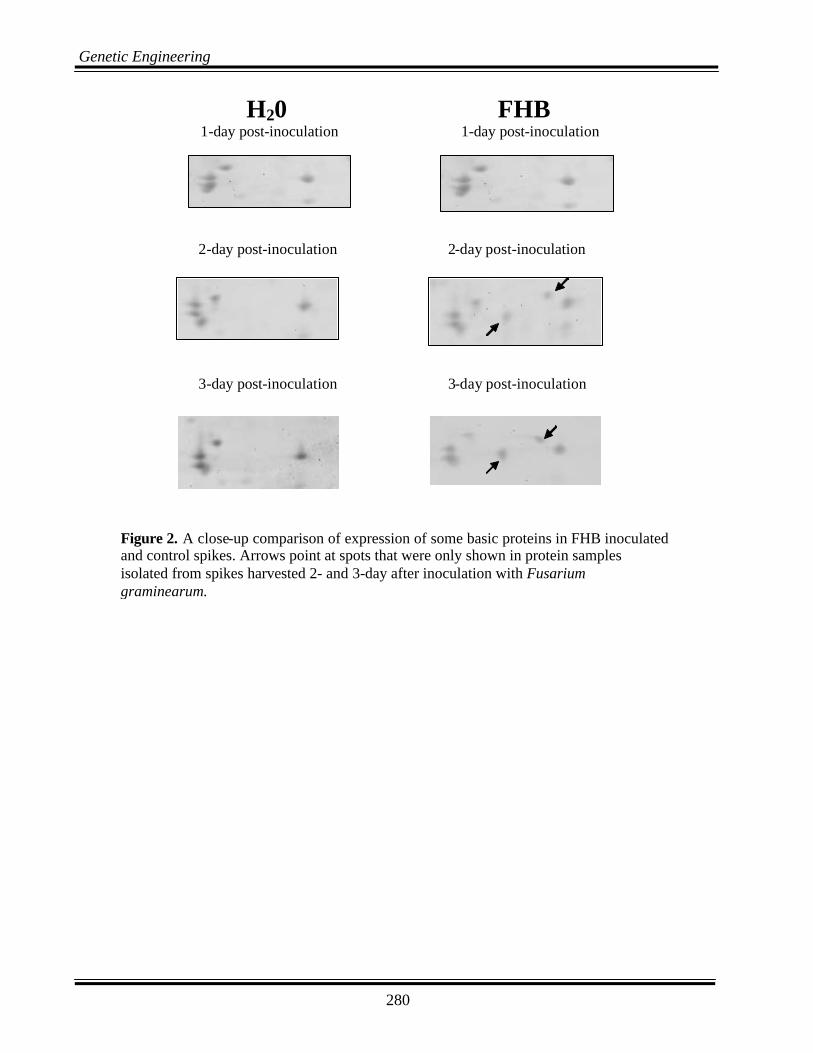
280
Genetic Engineering
H20 FHB 1-day post-inoculation 1-day post-inoculation
Figure 2. A close-up comparison of expression of some basic proteins in FHB inoculated and control spikes. Arrows point at spots that were only shown in protein samples isolated from spikes harvested 2- and 3-day after inoculation with Fusarium graminearum.
2-day post-inoculation 2-day post-inoculation
3-day post-inoculation 3-day post-inoculation