genetic testing in bleeding disorders
TRANSCRIPT
REVIEW ARTICLE
Genetic testing in bleeding disorders
C. DE BRASI,*† O. EL-MAARRI,‡ D. J. PERRY,§ J. OLDENBURG,‡ B. PEZESHKPOOR‡ and
A. GOODEVE¶***Instituto de Medicina Experimental (IMEX), CONICET-Academia Nacional de Medicina; †Instituto de Investigaciones
Hematologicas Mariano R Castex, Academia Nacional de Medicina, Buenos Aires, Argentina; ‡Institute of Experimental
Hematology and Transfusion Medicine, University of Bonn, Bonn, Germany; §Department of Haematology, Addenbrookes
Hospital, Cambridge; ¶Sheffield Diagnostic Genetics Service, Sheffield Children’s NHS Foundation Trust; and **Haemostasis
Research Group, Department of Cardiovascular Science, Sheffield University Medical School, Sheffield, UK
Summary. The aim of molecular genetic analysis infamilies with haemophilia is to identify the causativemutation in an affected male as this provides valuableinformation for the patient and his relatives. For thepatient, mutation identification may highlightinhibitor development risk or discrepancy betweendifferent factor VIII assays. For female relatives,knowledge of the familial mutation can facilitate carrierstatus determination and prenatal diagnosis. Recentadvances in understanding mutations responsible for
haemophilia and methods for their detection arepresented. For reporting of such mutations,participation in external quality assessment ensures thatessential patient and mutation details are routinelyincluded and that pertinent information is incorporatedin the interpretation.
Keywords: external quality assessment, genetic analysis,haemophilia A, haemophilia B, intrachromosomal inver-sion, missing mutations
Introduction
In families with haemophilia, identification of theunderlying mutation(s) in an affected male followedby its analysis in female relatives ‘at risk’ is themethod of choice for clarification of carrier status andfor prenatal diagnosis. In other inherited bleeding dis-orders, genetic analysis can help with the diagnosiswhen the phenotype remains unclear and can providedifferential diagnosis between similar disorders. Estab-lishing the underlying mutation may also enableprediction of the risk of inhibitor development.Haemophilia A (HA) and haemophilia B (HB) are
X-linked recessively inherited coagulopathies thatmanifest in hemizygous males with worldwide fre-quencies of 1:5000 and 1:25 000 respectively.Although heterozygous female carriers only rarelyexpress symptoms, haemophilia carrier diagnosis pro-vides valuable information for genetic counselling.
This article describes advances in understanding of thegenetics of haemophilia, particularly those made bylaboratories in Argentina and Germany, and thendiscusses the requirement for and utility of externalquality assessment (EQA) for bleeding disorder geneticanalysis.
Haemophilia genetic analysis – the Argentinianexperience: De Brasi
Since 1995, the Argentinian Molecular Genetics ofHaemophilia Laboratory has pursued two intertwinedobjectives: molecular diagnosis, including establishingnew approaches to investigate F8/F9 DNA markersand mutations, and study of the genotype-phenotyperelationship in an Argentinian series of haemophiliapatients and carriers.In 1993, the most common recurrent mutation in
HA, the F8 intron 22 inversion (Inv22) was described,which is implicated in 35–50% of severe-HA casesregardless of ethnic/geographical origin. Using South-ern blotting, molecular diagnosis of Inv22 has beenavailable in Argentina since 1995. Shortly after thesecond recurrent inversion affecting F8, intron 1(Inv1), was described, our series was reported alongwith a review of the literature estimating that Inv1causes <3% of severe-HA in Argentina [1]. Inv22
Correspondence: Prof. Anne Goodeve, BSc, PhD, Haemostasis
Research Group, Department of Cardiovascular Science, Facultyof Medicine, Dentistry and Health, Beech Hill Road, S10 2RX
Sheffield, UK.
Tel.: 0044 114 271 2679/7005; fax: 0044 114 271 1863;e-mail: [email protected]
Accepted 24 February 2014
54 © 2014 John Wiley & Sons Ltd
Haemophilia (2014), 20 (Suppl. 4), 54–58 DOI: 10.1111/hae.12409
originates from homologous recombination between a9.5 kb sequence located within F8 intron 22 (int22h-1) and one of two oppositely oriented extragenic cop-ies of int22h (int22h-2 and int22h-3) located by theXq-telomere. Similarly, Inv1 originates from homolo-gous recombination between intra- and extragenic900 bp homologs. Inv22 and Inv1 are occasionallyassociated with DNA gain/loss or altered DNAsequence, making their genotyping challenging. Liuet al. developed a rapid analysis of Inv22 based onlong-distance PCR (LD-PCR) [2]. Our variant ofinverse-PCR (inverse shifting-PCR, IS-PCR) thatavoids PCR amplification through the int22h regionwas devised in 2004. In this technique, genomic DNAis digested with BclI restriction enzyme, and self-ligated producing BclI-DNA circles that provide thetemplate sequence for conventional PCR analysis [3].The finished sequence of the human X-chromosomeindicated that int22h-2 and int22h-3 are inversely ori-ented to one another and it became clear that onlyone of these sequences generates inversions throughhead-to-head pairing with int22h-1. The other copymay generate deletions (Del22) or duplications (Dup22)but not inversions by recombining with equally orientedint22h-1. To support experimental evidence that Inv22type I results from recombination between int22h-1 andint22h-3 and type II between int22h-1 and int22h-2,Bagnall et al. hypothesized a non-deleterious 68 kbinversion mediated by large inverted repeats (50 kb)exchanging int22h-2/int22h-3 locations [4]. To distin-guish these genomic variants, including haemophilia-causing Inv22 and Del22 and non-causing Dup22,Bagnall et al. developed a LD-PCR-based approach [5].Our laboratory modified the previous IS-PCR-basedapproach, which now enables genotyping of both Inv1and Inv22 from the same template [6] and is applicableto chorionic villus extracted-DNA for prenatal diagnosis[7]. El-Hattab et al. found that hemizygous Dup22 andDel22 associate with intellectual disability and in uteromale lethality respectively [8]. The extreme severity ofDel22 in males resulting from loss of several genes sug-gests that reliable Del22 genotyping should be sup-ported by detecting each of the specific juxtaposedsequences of Del22, and the precise DNA loss associ-ated with the ~0.5 Mb deletion [9].Non-inversion HA- and HB-causative mutations
include large deletions of an exon or more that aredetected by a consistent absence of contiguous exon-specific PCR products. These mutations can be charac-terized by PCR amplification across deletion junctions,and include both those caused by non-homologousand by homeologous recombination, e.g. that betweenequally oriented AluSx sequences in introns 4 and 10of F8 [10]. For genotyping small F8 and F9 mutations,high-resolution conformation-sensitive gel electropho-resis (CSGE) on 37 and 8 amplicons, respectively, fol-lowed by Sanger sequencing of the selected exon(s)
showing anomalous CSGE-patterns detects mutationsin the majority of subjects. These procedures allowedcharacterization of insertions/deletions of 1–10 bp (in-dels) mostly associated with frameshifts, and nucleo-tide substitutions predicting missense, nonsense orRNA splicing defects [11,12]. Once a proband’ssequence variant has been determined, the genotype-phenotype correlation can be investigated followingthe Clinical Molecular Genetics Society (CMGS) Prac-tice Guideline for Unclassified Variants [13] alongwith 3D-structural modelling [14].In conclusion, the characterization of causative hae-
mophilia mutations is essential to provide the bestinformation for carrier and prenatal diagnosis, forgenetic counselling and to predict phenotypic charac-teristics, such as genotype-specific inhibitor risks.
Missing mutations in haemophilia A: El-Maarri, Pezeshkpoor and Oldenburg
In almost all HA patients, the deficiency of factorVIII (FVIII) activity can be traced to mutations in F8.With advances in molecular diagnostic techniquesand particularly in sequencing technology in the lastdecade, it has become possible to sequence all F8 ex-ons in all patients for an affordable cost, even insmall clinics. Therefore, it was expected that themolecular defect in F8 would be detected in everyHA patient. However, it became clear that this wasnot the case. At that point, different centres startedto characterize these patients and document theirclinical phenotypes.For ‘mutation-negative’ cases, the first step in the
investigation is to verify the HA phenotype. This ques-tion can been addressed in two ways; first, to verifythat only FVIII levels are decreased in these patients;second, to exclude combined FV/FVIII deficiency thatcould be caused by mutations in LMAN1 or MCFD2that may alter the secretion pathways of both FVIIIand factor V. In addition, defects in VWF should beexcluded, as any sub-optimal binding of FVIII to itsplasma carrier (von Willebrand factor) would lead toreduced FVIII activity as observed in von Willebranddisease type 2N. Finally, the two F8 inversions anddeletions, duplications and exonic mutations areexcluded by established tests [5,6]. Only after all theabove possibilities are excluded is further detailedanalysis described below recommended.The first molecular clue to identify the genetic
defects in mutation-negative patients was described in2008 [15]. Large duplications were identified in someof these patients [16]. Such duplications of entire ex-ons escape detection when individual exons aresequenced. Therefore, these duplications are only effi-ciently detected by multiplex ligation-dependent probeamplification [15], or possibly by array comparativegenomic hybridization.
© 2014 John Wiley & Sons Ltd Haemophilia (2014), 20 (Suppl. 4), 54--58
GENETIC TESTING IN BLEEDING DISORDERS 55
In 2011, Castaman et al. identified intronic muta-tions lying deep in F8 introns causing abnormal F8splicing leading to a decrease in levels of normallyspliced F8 mRNA [17]. They identified these mutationsbased on their effect on ectopic F8 mRNA only aftersequencing the neighbouring genomic regions. Recentlywe developed a detailed protocol for detecting themolecular defects in ‘mutation-negative’ patients[18,19]. A systematic stepwise investigation to detectall possible changes in the F8 locus is proposed. Thefirst step is to exclude gross rearrangements caused bygross duplications, recombinations or inversions. Suchrearrangements could leave the exons intact but in thewrong order. Such rearrangements can be excluded bylong-range (LR) amplification of overlapping ampli-cons that cover the whole F8 genomic locus. Using thisstrategy, one patient with a rearranged genomic struc-ture due to recombination between inverted repeatswas identified [20]. The second step is to search forabnormal splicing by RT-PCR that covers all exon–exon boundaries. Once abnormal splicing is detectedthen the intronic regions surrounding the breakpointsare sequenced to identify the intronic mutationsinvolved [17]. If no mutation is detected then a thirdstep is to sequence all the LR-PCR products using amassively parallel sequencing approach (next genera-tion sequencing). The advantage of this approach isthe rapid identification of all variants in the locus atonce [19]. Novel variants can then be further investi-gated for their effect on splicing (that may have beenmissed by previous RT-PCR) or for enhancer/silencereffect by functional assays. By undertaking these steps,mutations are expected to be identified in a proportionof previous ‘mutation-negative’ cases.
Quality assurance in genetic testing: DavidPerry on behalf of UK NEQAS BC
In contrast to phenotypic data, the results of genotypicassays are unequivocal with no borderline values.Accordingly, there is an acceptance of the accuracy ofsuch data by referring physicians. However, severalstudies have shown that mutation detection in com-mon with any analytical test has an intrinsic error rate[21,22]. A failure to correctly identify a mutation orto interpret its significance can have major implica-tions for an individual and their family members.In the UK, participation in a recognized EQA
scheme is a requirement for laboratory accreditationand a number of such schemes exist, coordinatedthrough UK National External Quality AssessmentService (NEQAS). The only EQA scheme for thegenetics of the heritable bleeding disorders in the EUis that administered by UK NEQAS for Blood Coagu-lation (UK NEQAS BC).In 1998, UK NEQAS BC established a pilot scheme
to assess the performance of laboratories in genetic
testing [23]. In 2003, a Special Advisory Group onHaemophilia Molecular Genetics for UK NEQAS BCwas established, with the remit of developing a robustEQA scheme for both UK and international partici-pants. The scheme was designed to address three fun-damental aspects of genetic testing: (i) Correctidentification of the patient and their reason for refer-ral; (ii) Correct identification of the causative geneticmutation(s); (iii) Interpretation and reporting ofgenetic data in the context of the any relevant clinicaland family data.Between 2003 and 2013, 18 exercises were under-
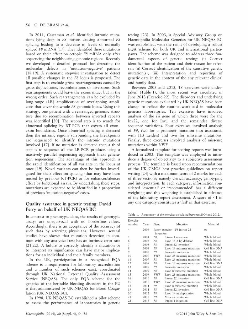
taken (Table 1), the most recent was circulated inJune 2013 (Exercise 22). The disorders and underlyinggenetic mutations evaluated by UK NEQAS have beenchosen to reflect the routine workload in moleculargenetics laboratories. Ten exercises have involvedanalysis of the F8 gene of which three were for theInv22, one for Inv1 and the remainder diversesequence variations. Four exercises involved analysisof F9, two for a promoter mutation (not associatedwith HB Leiden) and two for missense mutations.Finally, three exercises involved analysis of missensemutations within VWF.A formalized template for scoring reports was intro-
duced in 2003. This template was employed to intro-duce a degree of objectivity to a subjective assessmentprocess. The template is based upon recommendationsof the UK CMGS best practice guidelines on reportwriting [24] with a maximum score of 2 marks for eachof three sections; namely clerical accuracy, genotypingand interpretation. In each category, information con-sidered ‘essential’ or ‘recommended’ has a differentweighting and this weighting is established in advanceof the laboratory report assessment. A score of <1 inany one category constitutes a ‘fail’ in that exercise.
Table 1. A summary of the exercises circulated between 2004 and 2012.
Exercise
number Year Gene Mutation Material
4 2004 Paper exercise – F8 intron 22
inversion
na
5 2004 F8 Intron 1 inversion Whole blood
6 2005 F8 Exon 14 2 bp deletion Whole blood
7 2005 F8 Intron 22 inversion Whole blood
8 2006 F8 Exon 19 missense mutation Cell line DNA
9 2006 F9 Promoter mutation Whole blood
10 2007 VWF Exon 28 missense mutation Whole blood
11 2007 F8 Exon 25 missense mutation Whole blood
12 2008 F8 Exon 19 missense mutation Cell line DNA
13 2008 F9 Promoter mutation Whole blood
14 2009 F8 Exon 8 missense mutation Whole blood
15 2009 VWF Exon 28 missense mutation Whole blood
16 2010 F8 Intron 22 inversion Cell line DNA
17 2010 VWF Exon 46 missense mutation Whole blood
18 2011 F9 Exon 8 missense mutation Whole blood
19 2011 F8 Intron 22 inversion Cell line DNA
20 2012 F8 Exon 14 nt duplication Whole blood
21 2012 F9 Missense mutation Whole blood
22 2013 F8 Intron 1 inversion Cell line DNA
Haemophilia (2014), 20 (Suppl. 4), 54--58 © 2014 John Wiley & Sons Ltd
56 C. DE BRASI et al.

Reports are scored independently by four experi-enced individuals and a consensus subsequentlyreached. Laboratories that are registered with thescheme who either fail to submit a report or do sooutside the allocated turnaround time of 6 weeks(chosen to reflect UKHCDO recommendations) willalso fail. A fail in any exercise generates a letter fromthe Director of UK NEQAS BC with the offer of assis-tance. Each participating laboratory is assigned aunique identification number that allows the contin-uing performance of each laboratory to be reviewed.The identification of participating laboratories remainsunknown to the reviewers.All participating laboratories use the mutation
nomenclature system proposed by the Human GeneVariation Society (HGVS) [25] that requires allsequence variations to be defined in relation to a spec-ified reference sequence. The ‘A’ nucleotide of theATG-translation initiation codon to be numbered as+1 with the protein sequence representing the primarytranslation product numbered from the initiatormethionine and therefore, includes signal peptidesequence. For some genes and proteins, this requiresrenumbering and makes reference to previouslydescribed mutations challenging. Laboratories are,therefore, encouraged to include legacy nomenclatureas a number of published mutations including some ofthose listed in online locus-specific mutation databasesremain in the ‘legacy’ format.Of the 18 exercises circulated between 2004 and
2013, 13 involved the use of whole blood and fiveDNA derived from immortalized cell lines. Wholeblood samples distributed internationally yield suffi-cient quantity and quality of DNA for analysis evenwhen transport delays of several days occur.The majority of laboratories in each exercise achieve
full marks, and failing is unusual. Reasons for failingan exercise include clerical inaccuracies [e.g. a failureto include unique identifiers for each individual(s)];genotyping errors (e.g. incorrectly numbering themutation or predicted effect on the protein; failing toidentify a mutation that was present; identifying a sec-ond mutation that was not present) and finally inter-pretation errors. Many of the errors that have led to a
fail were based upon incorrect interpretation, e.g. fail-ure to answer the clinical question; incorrectly assign-ing carrier status (or not) to an ‘at-risk’ female; failingto establish the significance of a novel mutation andfailing to consider the possibility of mosaicism.The aim of EQA schemes is to highlight problems
and deficiencies in laboratory procedures. This EQAscheme has led to a more uniform inclusion of infor-mation into reports and a standardized use of muta-tion nomenclature. There are currently 27 laboratoriesregistered for this scheme: 24 in the EU of which 12are in the UK and three in non-EU countries. Thescheme has received very positive feedback from par-ticipants and is seen as a fundamental part of goodlaboratory practice.
Conclusion
This article has demonstrates (i) the continuing devel-opment of molecular genetic analysis of haemophiliadirected towards identifying the causative mutation invirtually all patients; and (ii) that for mutations identi-fied, participation in an EQA scheme promotes report-ing and interpretation of the effect of these mutationsto a recognized international standard.
Acknowledgements
This work was supported by a DFG grant (Deutsche Forschungsge-
meinschaft: EL499/2-1), a Baxter bioscience grant (number: H12-000820)
and the Bayer Haemophilia Awards Program.
Disclosures
Dr Carlos de Brasi has not received any commercial support during the
past 2 years. Dr El-Maarri has received support to attend meetings from
Bayer and Baxter. Dr Pezeshkpoor has received support to attend meet-
ings from Biotest and Baxter. Professor Oldenburg received reimburse-
ment for attending symposia/congresses and/or honoraria for speaking or
consulting, and/or funds for research from Baxter, Bayer, Biogen Idec,
Biotest, CSL Behring, Grifols, Inspiration, Novo Nordisk, Octapharma,
Swedish Orphan Biovitrum, and Pfizer. Professor Goodeve has received
honoraria for presentations given from Novo Nordisk and Octapharma
and receives support for the ISTH VWF mutation database from CSL
Behring. Dr Perry has received educational grants and support to attend
meetings from Baxter Healthcare and Novo Nordisk. He has also
received consultancy fees from Biogenidec and Amgen.
References
1 Rossetti LC, Candela M, Bianco RP et al.
Analysis of factor VIII gene intron 1 inver-
sion in Argentinian families with severe
haemophilia A and a review of the litera-
ture. Blood Coagul Fibrinolysis 2004; 15:
569–72.2 Liu Q, Nozari G, Sommer SS. Single-tube
polymerase chain reaction for rapid diagno-
sis of the inversion hotspot of mutation in
hemophilia A. Blood 1998; 92: 1458–9.
3 Rossetti LC, Radic CP, Larripa IB et al.
Genotyping the hemophilia inversion hot-
spot by use of inverse PCR. Clin Chem
2005; 51: 1154–8.4 Bagnall RD, Giannelli F, Green PM. Poly-
morphism and hemophilia A causing inver-
sions in distal Xq28: a complex picture. J
Thromb Haemost 2005; 3: 2598–9.5 Bagnall RD, Giannelli F, Green PM.
Int22 h-related inversions causing hemo-
philia A: a novel insight into their origin
and a new more discriminant PCR test for
their detection. J Thromb Haemost 2006;
4: 591–8.6 Rossetti LC, Radic CP, Larripa IB et al.
Developing a new generation of tests for
genotyping hemophilia-causative rearrange-
ments involving int22 h and int1 h hot-
spots in the factor VIII gene. J Thromb
Haemost 2008; 6: 830–6.7 Radic CP, Rossetti LC, Zuccoli JR et al.
Inverse shifting PCR based prenatal diagno-
sis of hemophilia-causative inversions
involving int22 h and int1 h hotspots from
© 2014 John Wiley & Sons Ltd Haemophilia (2014), 20 (Suppl. 4), 54--58
GENETIC TESTING IN BLEEDING DISORDERS 57
chorionic villus samples. Prenat Diagn
2009; 29: 1183–5.8 El-Hattab AW, Fang P, Jin W et al.
Int22 h-1/int22 h-2-mediated Xq28 rear-
rangements: intellectual disability associ-
ated with duplications and in utero male
lethality with deletions. J Med Genet 2011;
48: 840–50.9 Abelleyro MM, Rossetti LC, Radic CP
et al. Are int22 h-mediated deletions a
common cause of hemophilia? Ann Hema-
tol 2012; 91: 633–6.10 Rossetti LC, Goodeve A, Larripa IB et al.
Homeologous recombination between
AluSx-sequences as a cause of hemophilia.
Hum Mutat 2004; 24: 440.
11 Radic CP, Rossetti LC, Abelleyro MM
et al. Assessment of the F9 genotype-spe-
cific FIX inhibitor risks and characterisa-
tion of 10 novel severe F9 defects in the
first molecular series of Argentinian
patients with haemophilia B. Thromb Hae-
most 2013; 109: 24–33.12 Rossetti LC, Radic CP, Candela M et al.
Sixteen novel hemophilia A causative muta-
tions in the first Argentinian series of severe
molecular defects. Haematologica 2007;
92: 842–5.13 Bell J, Bodmer D, Sistermans E et al.
Practice Guidelines for the Interpretation
and Reporting of Unclassified Variants
(UVs) in Clinical Molecular Genetics.
Available at http://cmgsweb.shared.hosting.
zen.co.uk/BPGs/Best_Practice_Guidelines.
htm. Accessed January 3, 2014.
14 Rallapalli PM, Kemball-Cook G, Tud-
denham EG et al. An interactive muta-
tion database for human coagulation
factor IX provides novel insights into
the phenotypes and genetics of hemo-
philia B. J Thromb Haemost 2013; 11:
1329–40.15 Rost S, Loffler S, Pavlova A et al. Detection
of large duplications within the factor VIII
gene by MLPA. J Thromb Haemost 2008;
6: 1996–9.16 Zimmermann MA, Oldenburg J, Muller
CR et al. Characterization of duplication
breakpoints in the factor VIII gene. J
Thromb Haemost 2010; 8: 2696–704.17 Castaman G, Giacomelli SH, Mancuso ME
et al. F8 mRNA studies in haemophilia A
patients with different splice site mutations.
Haemophilia 2010; 16: 786–90.18 Pezeshkpoor B, Pavlova A, Oldenburg J
et al. F8 genetic anylsis strategies when
standard approches fail. Haemostaseologie
2013; 34. [Epub ahead of print].
19 Pezeshkpoor B, Zimmer N, Marquardt N
et al. Deep intronic ‘mutations’ cause
hemophilia A: application of next genera-
tion sequencing in patients without detect-
able mutation in F8 cDNA. J Thromb
Haemost 2013; 11: 1679–87.20 Pezeshkpoor B, Rost S, Oldenburg J et al.
Identification of a third rearrangement at
Xq28 that causes severe hemophilia A as a
result of homologous recombination
between inverted repeats. J Thromb Hae-
most 2012; 10: 1600–8.21 Dequeker E, Cassiman JJ. Genetic testing
and quality control in diagnostic laborato-
ries. Nat Genet 2000; 25: 259–60.22 Tripodi A, Peyvandi F, Chantarangkul V
et al. Relatively poor performance of clini-
cal laboratories for DNA analyses in the
detection of two thrombophilic mutations–a cause for concern. Thromb Haemost
2002; 88: 690–1.23 Perry DJ, Goodeve A, Hill M et al. The
UK National External Quality Assessment
Scheme (UK NEQAS) for molecular genetic
testing in haemophilia. Thromb Haemost
2006; 96: 597–601.24 Treacy RJL, Robinson DO. Best Practice
Guidelines for Reporting Molecular
Genetics Results. Available at http://www.
cmgs.org/BPGs/best_practice_guidelines.htm.
Accessed January 3, 2014.
25 HGVS. Nomenclature for the Description
of Sequence Variations Homepage. Avail-
able at http://www.hgvs.org/mutnomen/.
Accessed January 3, 2014.
Haemophilia (2014), 20 (Suppl. 4), 54--58 © 2014 John Wiley & Sons Ltd
58 C. DE BRASI et al.