genitourinary - university of miami...presentation outline 2 overview including anatomy and general...
TRANSCRIPT

1
FCDS 2011 Educational Webcast Series December 15, 2011
Susan Smith Pierce, CTR
Gema Midence, MBA, CTR
Steven Peace, BS, CTR
Genitourinary
Presentation Outline
2
Overview including Anatomy and General Information
Kidney Parenchyma
Kidney Renal Pelvis
Bladder
Prostate
Multiple Primary and Histology Coding Rules Refresher
Collaborative Stage Data Collection System (CSv02.03.02)
2011 FCDS Required CS Site Specific Factors (SSF)
Treatment Guidelines by Stage
Documentation

Genitourinary System
3 Source: http://medicaltrue.com/urinary-tract

Kidney Parenchyma
4

United States
2011 Incidence / Mortality
New Cancer Cases
1,596,670 all site
60,920 kidney & renal pelvis cancer cases
Cancer Deaths
571,950 all sites
13,120 kidney & renal pelvis cancer case
Source: American Cancer Society Cancer Facts and Figures 2011 5
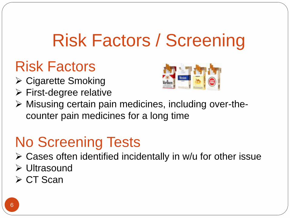
Risk Factors / Screening
6
Risk Factors Cigarette Smoking
First-degree relative
Misusing certain pain medicines, including over-the-
counter pain medicines for a long time
No Screening Tests Cases often identified incidentally in w/u for other issue
Ultrasound
CT Scan

Tumor Markers/Lab Tests
Elevated LDH levels
Hypercalcemia
Anemia
Thrombocytosis
Elevated ESR or CRP
7
Source: AJCC 7th Edition
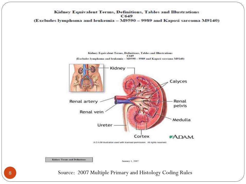
8 Source: 2007 Multiple Primary and Histology Coding Rules

Anatomy of the Kidney and Ureter
9
1. Parenchyma
2. Cortex
3. Medulla
4. Perirenal fat
5. Capsule
6. Ureter
7. Pelvis of kidney
8. Renal vessels
9. Hilum
10. Calyx
http://training.seer.cancer.gov/kidney/anatomy/

Anatomy Kidney
10
Figure I-2-13. Structures Adjacent to Kidney Adapted from: Medi-Clip: Grant’s Atlas Images I, Thorax and Abdomen. Williams and Wilkins, 1998.
Source: Collaborative Stage Data Collection System, Part I, Section 2

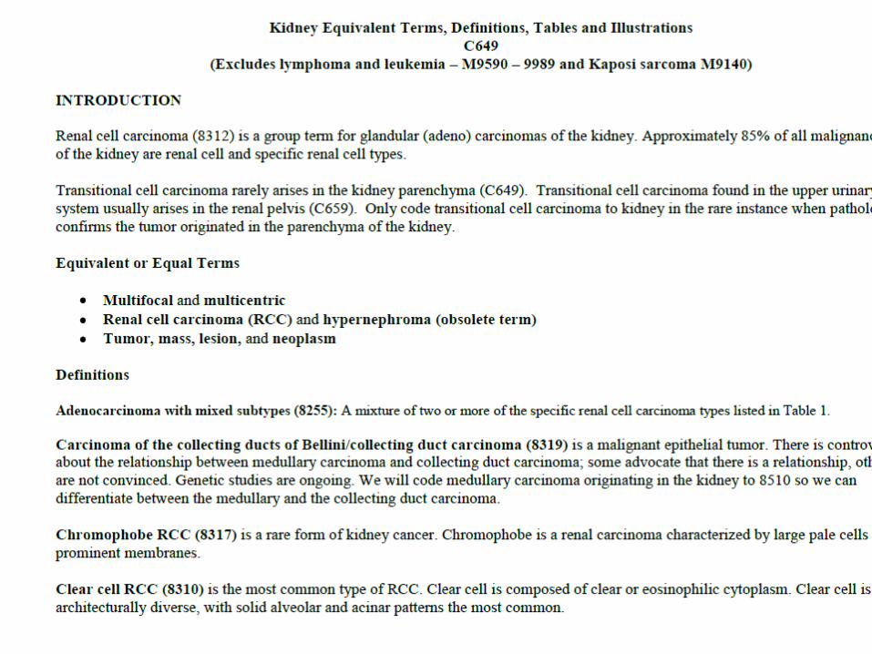
Histology
11
Specific Renal Cell Carcinoma Types
8255 Adenocarcinoma with mixed subtypes**
8260 Papillary (Chromophil)*
8310 Clear Cell
8316 Cyst associated, cystic
8317 Chromophobe*
8318 Sarcomatoid (Spindle cell)
8319 Collecting duct type (Bellini duct)
8320 Granular cell
8510 Medullary carcinoma, NOS; medullary adenocarcinoma
8959 Malignant cystic nephroma; malignant multilocular cystic nephroma
8312 Renal cell carcinoma is a GROUP term for glandular
(adeno) carcinoma of the kidney
* Note: Chromophil and chromophobe are different histologies
**Note: A mixture of two or more of the specific renal cell carcinoma types listed in this table.
Source: 2007 Multiple Primary & Histology Coding Rules

2007 Multiple Primary Rules
12
Kidney
Formats
• Flowchart Format
• Matrix Format
• Text Format

13
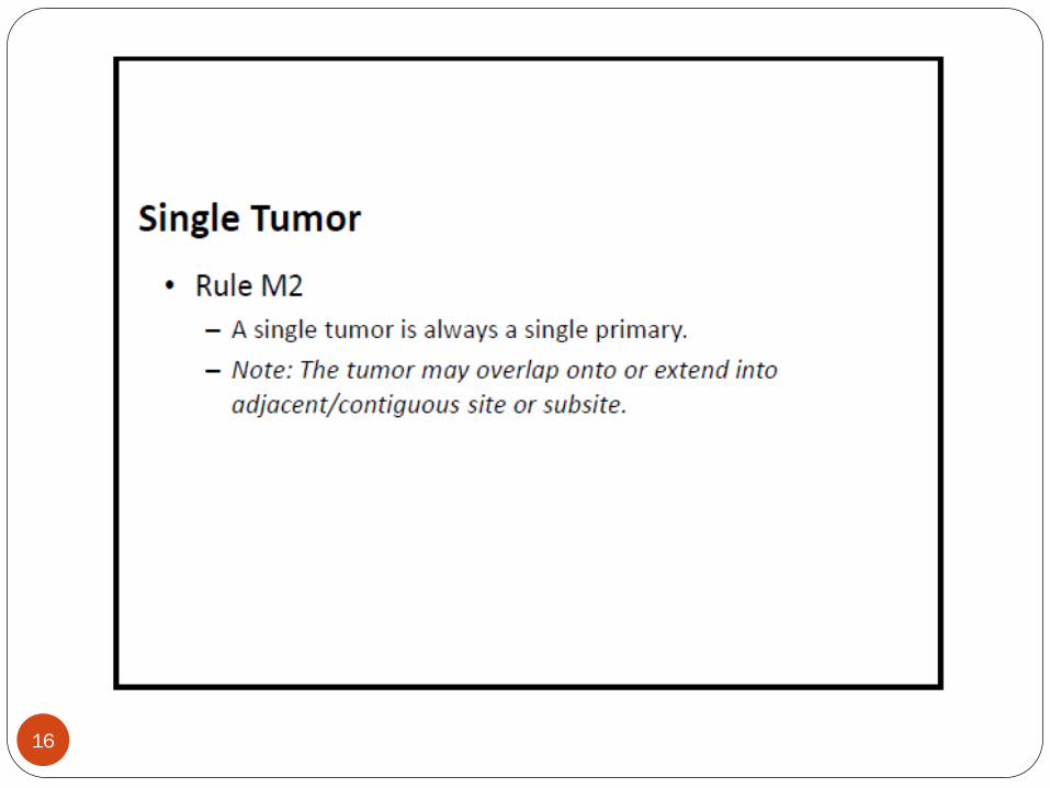
14

15
16
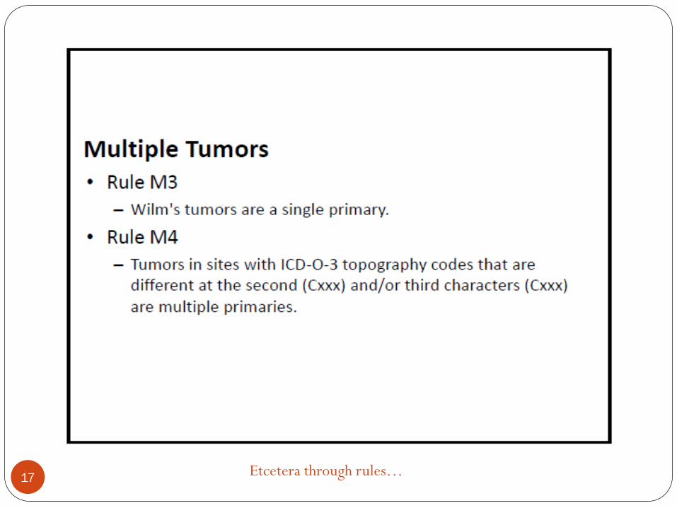
17 Etcetera through rules…
Collaborative Stage v02.03.02
Kidney Parenchyma C64.9
18

19
KidneyParenchyma
Version v.02.03
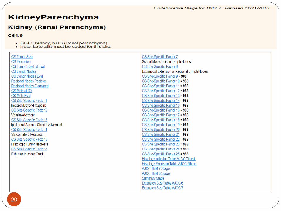
20
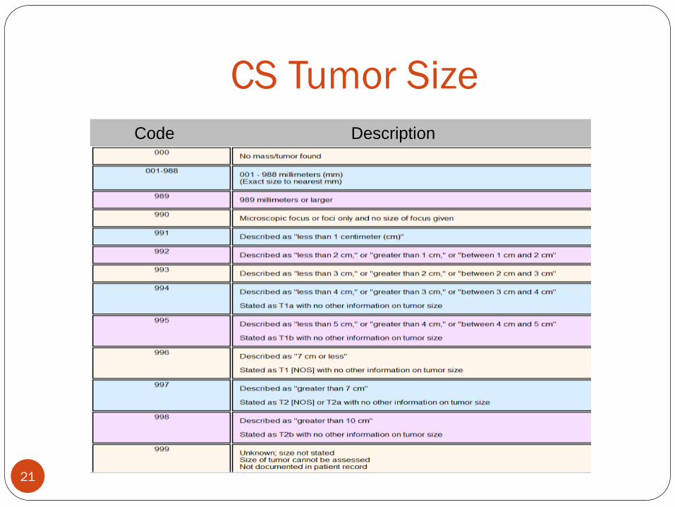
21
Code Description
CS Tumor Size
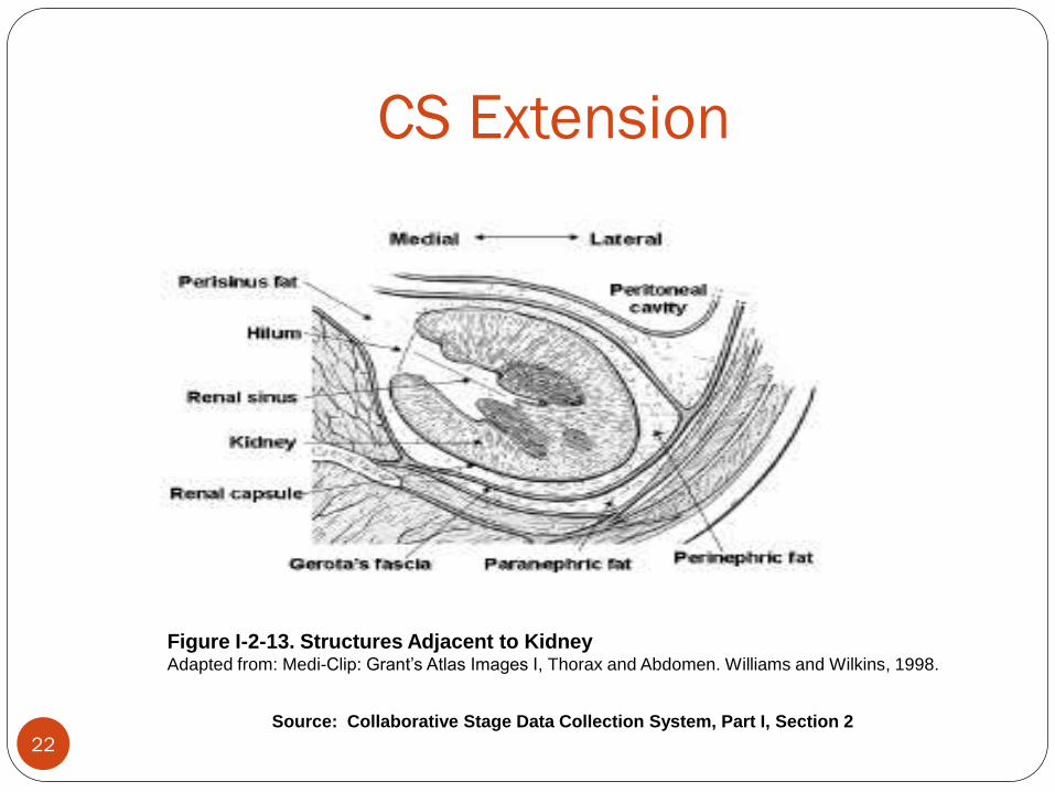
CS Extension
22
Figure I-2-13. Structures Adjacent to Kidney Adapted from: Medi-Clip: Grant’s Atlas Images I, Thorax and Abdomen. Williams and Wilkins, 1998.
Source: Collaborative Stage Data Collection System, Part I, Section 2

CS Extension
23
Note 2: Gerota’s fascia
Note 3: Invasion beyond the capsule
Note 4: “In situ of renal parenchyma”
Note 5: Use of code 300
Note 6: T1 and T2 tumors with tumor size
Note 7: Direct extension to other structures

24
Code(s) 630-645 – Ipsilateral
adrenal
now T4 - Contiguous Invasion
Code 601
Renal vein involvement now T3a
CS Extension

25 ^ For CS Extension codes 100-360 ONLY, the T category for AJCC 7 is assigned based on the value of CS Tumor Size, as
shown in the Extension Size AJCC 7 Table for this site.
“Stated as” T1a Code 310
“Stated as” T1b Code 320
“Stated as” T1NOS Code 330
“Stated as” T2a Code 340
“Stated as” T2b Code 350
“Stated as” T2NOS Code 360
CS Extension
26
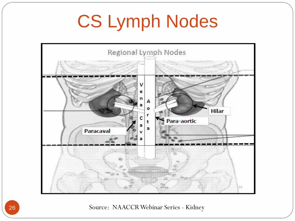
CS Lymph Nodes
Source: NAACCR Webinar Series - Kidney

CS Mets at Dx
27
Code 00: No distant mets
Code 10: Distant lymph nodes
Code 20: Extension to contralateral kidney
Code 40: Non contiguous ipsilateral adrenal
Code 50: OBSOLETE code
Code 55: (40 or 20) + 10
Code 60: Distant metastasis, NOS
Code 99: Unknown

CS Site-Specific Factors (CoC Required)
28
Not Required
Not Required
SSF1: Invasion Beyond Capsule
SSF2: Vein Involvement
SSF3: Ipsilateral Adrenal Gland Involvement
SSF4: Sarcomatoid Features
SSF5: Histologic Tumor Necrosis
SSF6: Fuhrman Nuclear Grade
SSF7: Size of Metastasis in Lymph Nodes
SSF8: Extranodal Extension
28

CS Site-Specific Factors FCDS
29
None Required by FCDS
29

30
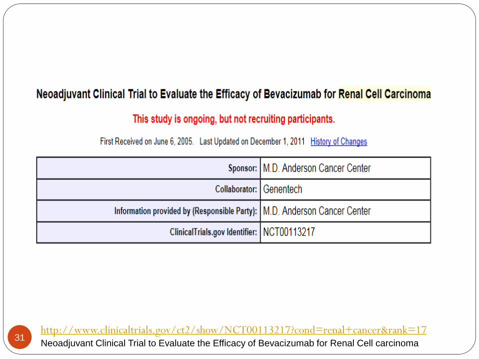
Kidney Cancer Treatment
31 http://www.clinicaltrials.gov/ct2/show/NCT00113217?cond=renal+cancer&rank=17 Neoadjuvant Clinical Trial to Evaluate the Efficacy of Bevacizumab for Renal Cell carcinoma
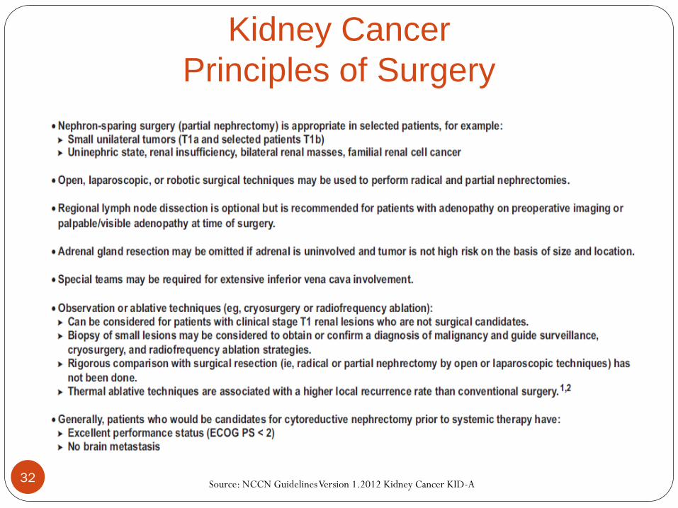
Kidney Cancer
Principles of Surgery
32 Source: NCCN Guidelines Version 1.2012 Kidney Cancer KID-A

Kidney Cancer
Primary Treatment Stage I-III
33 Source: NCCN Guidelines Version 1.2012 Kidney Cancer KID-1

Kidney Cancer
Primary Treatment Stage IV
34 Source: NCCN Guidelines Version 1.2012 Kidney Cancer KID-2
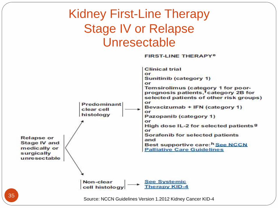
35 Source: NCCN Guidelines Version 1.2012 Kidney Cancer KID-4
Kidney First-Line Therapy
Stage IV or Relapse Unresectable
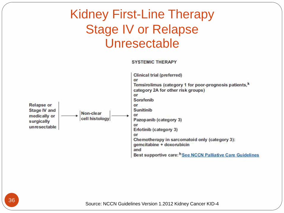
36 Source: NCCN Guidelines Version 1.2012 Kidney Cancer KID-4
Kidney First-Line Therapy
Stage IV or Relapse Unresectable
37
Renal Pelvis, Ureter, Bladder

38
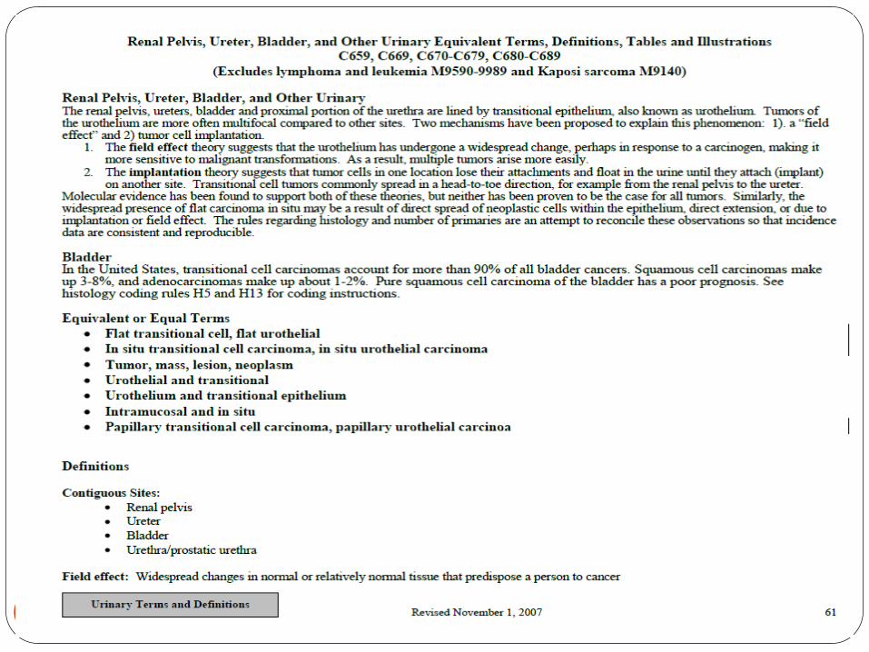
Field Effect Theory
The field effect theory suggests that the urothelium
has undergone a widespread change, perhaps in
response to a carcinogen, making it more sensitive
to malignant transformations. As a result, multiple
tumors arise more easily.

39
Implantation Theory
The implantation theory suggests that tumor cells
in one location lose their attachments and float in
the urine until they attach (implant) on another site.
Transitional cell tumors commonly spread in a
head-to-toe direction, for example from the renal
pelvis to the ureter.
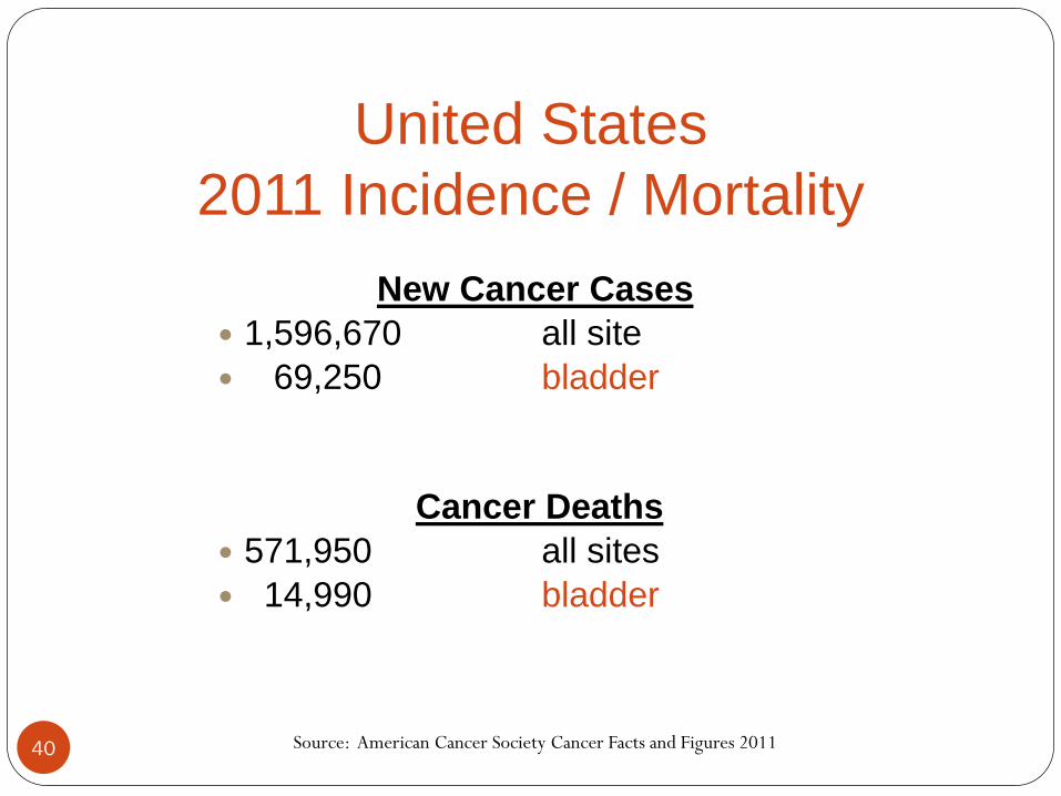
United States
2011 Incidence / Mortality
New Cancer Cases
1,596,670 all site
69,250 bladder
Cancer Deaths
571,950 all sites
14,990 bladder
Source: American Cancer Society Cancer Facts and Figures 2011 40
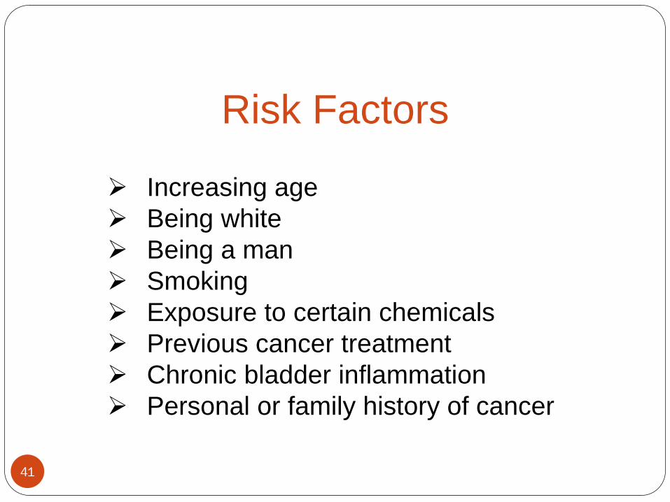
Risk Factors
41
Increasing age
Being white
Being a man
Smoking
Exposure to certain chemicals
Previous cancer treatment
Chronic bladder inflammation
Personal or family history of cancer

Symptoms and Screening
42
Signs & Symptoms Blood in urine (hematuria)
Frequent urination
Painful urination
Urinary tract infection
Abdominal pain
Back pain
Screening Tests
There is no standard or routine screening test for
bladder cancer
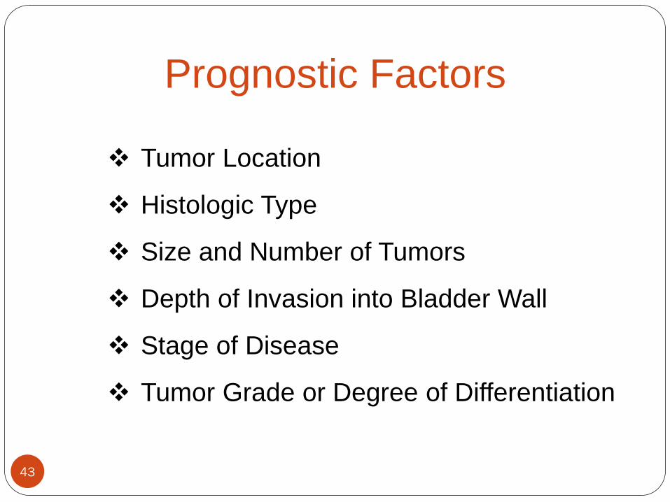
Prognostic Factors
43
Tumor Location
Histologic Type
Size and Number of Tumors
Depth of Invasion into Bladder Wall
Stage of Disease
Tumor Grade or Degree of Differentiation
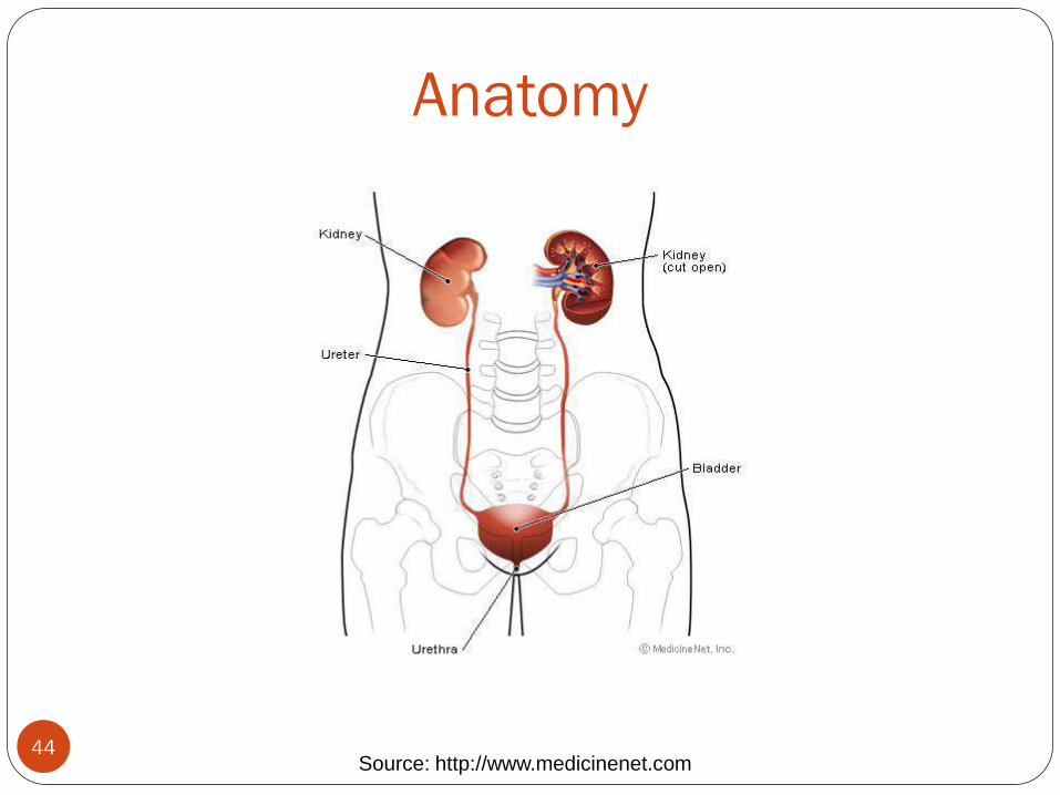
Anatomy
44 Source: http://www.medicinenet.com
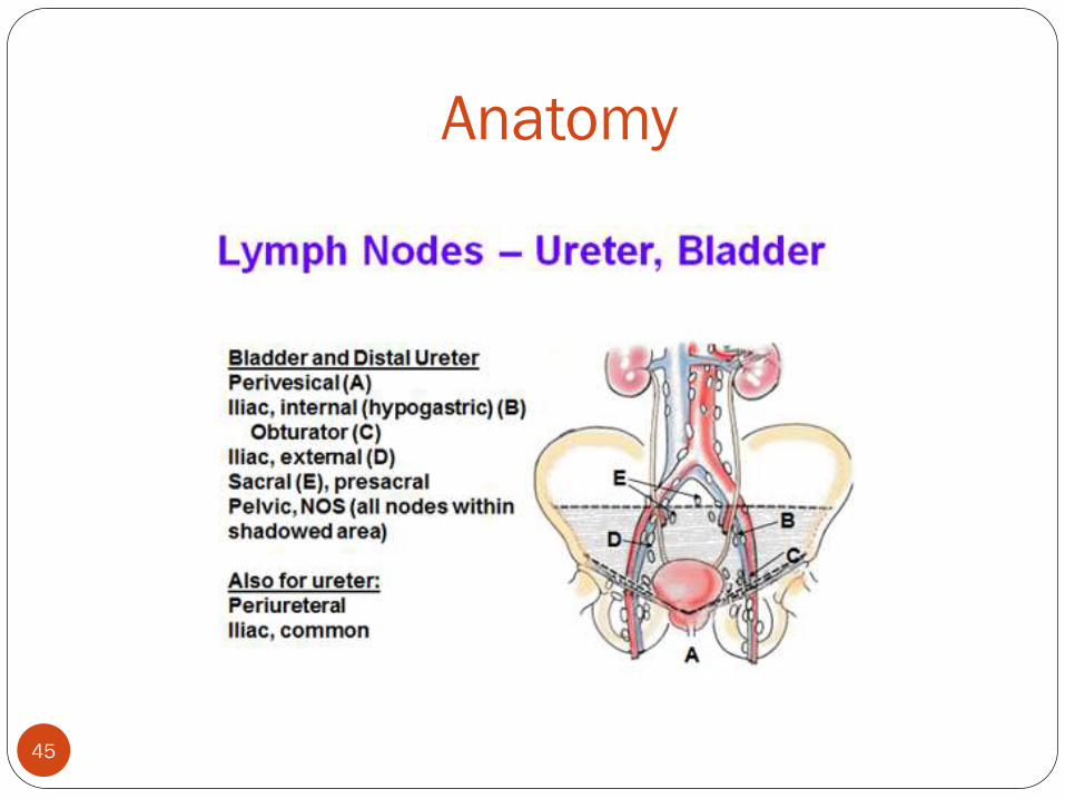
Anatomy
45
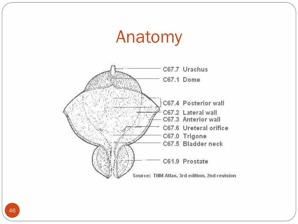
Anatomy
46
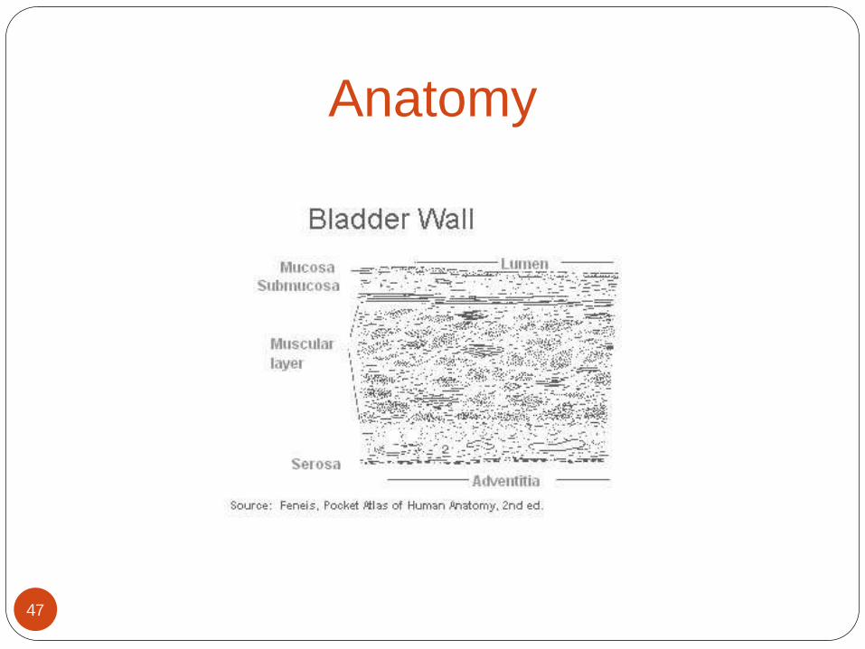
Anatomy
47
Histology
48
Urothelial or Transitional Cell Carcinoma
Squamous Cell Carcinoma
Adenocarcinoma
Carcinosarcoma

49 Source Multiple Primary & Histology Coding Rules - Table 1 – Urothelial Tumors Note: Excludes pure squamous carcinoma, glandular (adeno) carcinoma, or other bladder tumor histologies. Source: Multiple Primary & Histology Coding Rules; NCI - SEER
Histology Urothelial/Transitional Cell Tumors Code With squamous differentiation 8120
With glandular differentiation
With trophoblastic differentiation
Nested
Microcystic
Transitional cell, NOS
Papillary carcinoma 8130
Papillary transitional cell
Micropapillary 8131
Lymphoepithelioma-like 8082
Plasmacytoid
Sarcomatoid 8122
Giant cell 8031
Undifferentiated 8020

50
Grade
Grade is a prognostic factor for bladder cancer
High grade tumors have a worse prognosis
Low grade noninvasive tumors in young patients
have a better prognosis
Note: If the term low grade (LG) or high grade(HG)
is indicated for a urothelial primary, assume it is a
WHO/ISUP grade

Two-Grade System
Conversion Table
51
Source:
FORDS Page 11, SECTION ONE: Case Eligibility and Overview of Coding Principles; Coding Two-Grade
Systems
Terminology Histologic Grade
Low grade 1 / 2
High grade 2 / 2
Code
2
4

Multiple Primary Rules
Histology Coding Rules
52
Formats
• Flowchart Format
• Matrix Format
• Text Format
Renal Pelvis
Ureter
Bladder

53
54
55

56

57
Multiple Primary Rules
Rule M6
Bladder tumors with any combination of the
following histologies are a single primary:
Papillary carcinoma (8050)
Transitional cell carcinoma (8120-8124)
Papillary transitional cell carcinoma (8130-8131)

58
Multiple Primary Rules
One Per Lifetime
Each patient may only have one invasive urothelial bladder cancer per
lifetime.
Once a patient has an invasive urothelial bladder cancer,
subsequent non-invasive or invasive urothelial bladder cancer is
considered the same primary.
Each patient can only have one non-invasive urothelial bladder cancer
per lifetime.
Must occur prior to the invasive urothelial bladder cancer

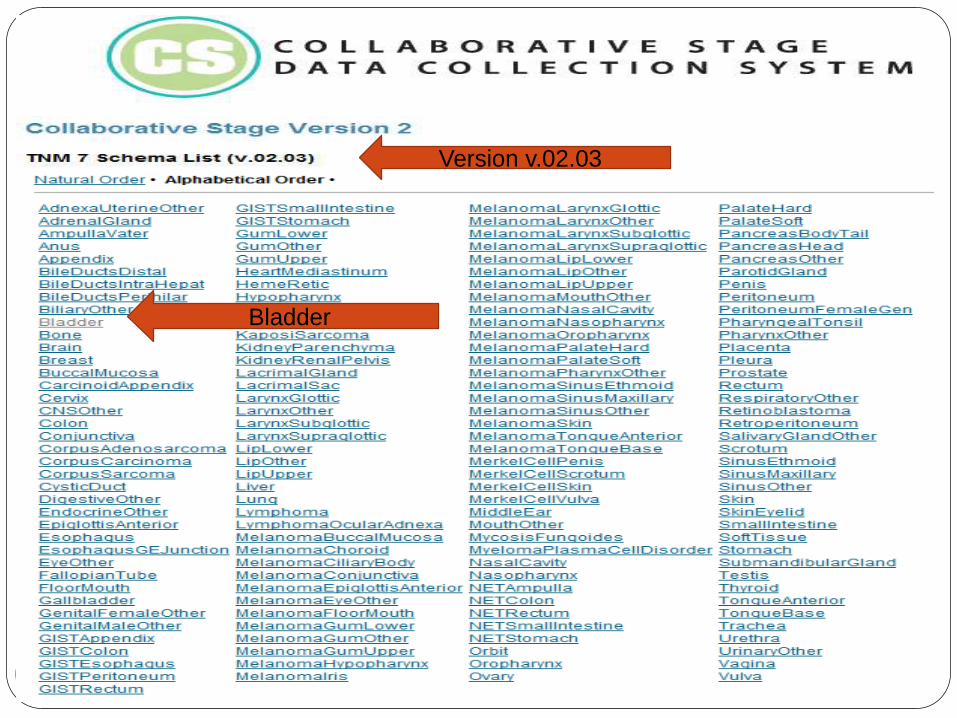
Collaborative Stage v02.03.02
Bladder
C67.0 – 67.9
59

Bladder Cancer Staging
60 Source: http://www.emoryhealthcare.org/urology/oncology/bladder-cancer

61
62
Bladder
Version v.02.03

63
64
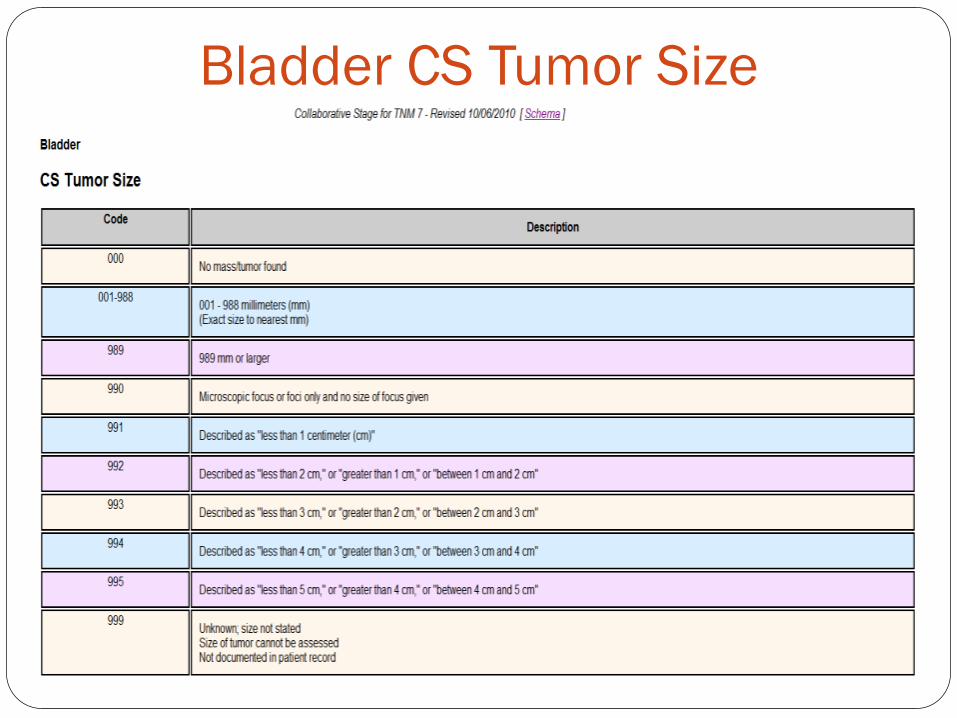
Bladder CS Tumor Size

65
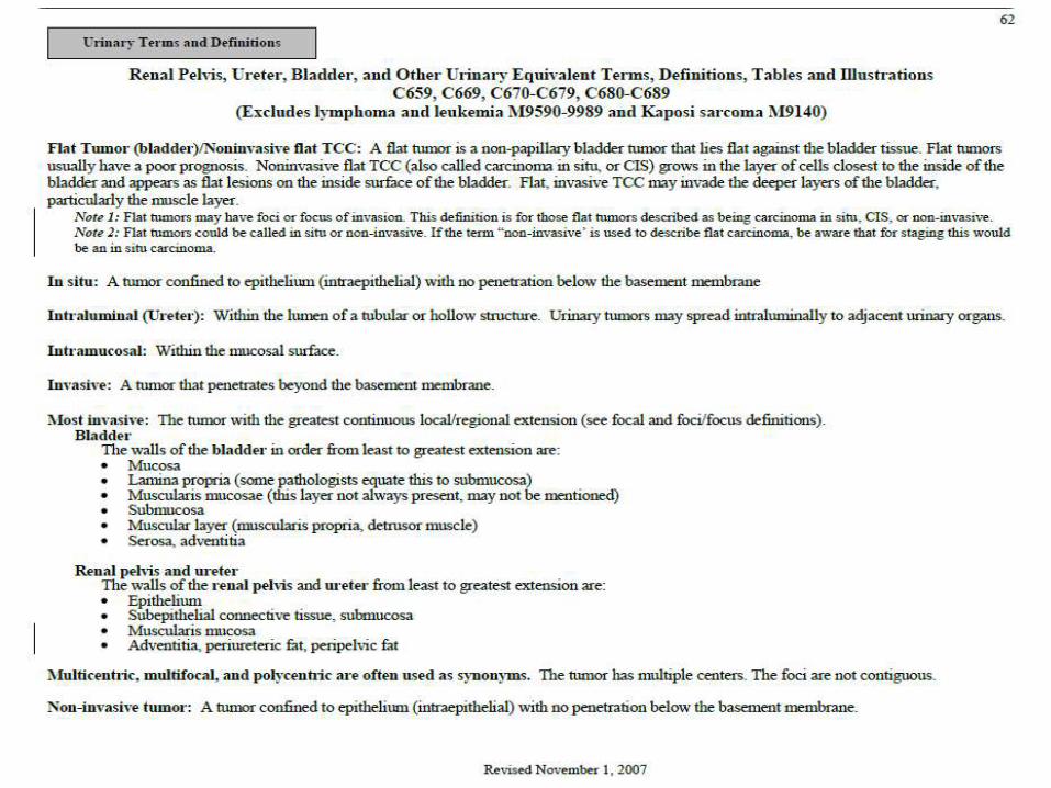
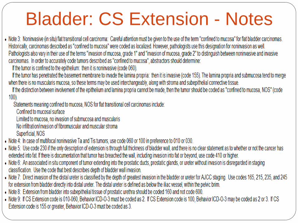
Bladder CS Extension Notes
Noninvasive papillary carcinomas
Listing of definite statements
Listing of inferred descriptions
Extended Note 3 for in situ
Extended Note 3 for locally invasive
Expanded notes for coding extension
Several notes moved around
Notes rewritten to clarify instructions

Bladder: CS Extension Notes
66
67
Bladder: CS Extension - Notes
68
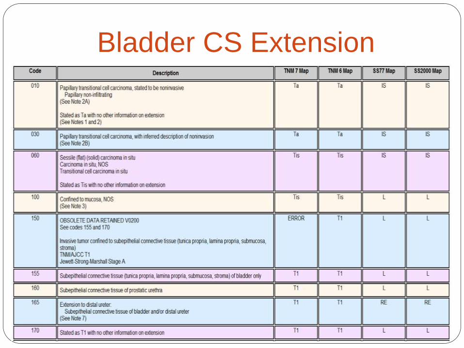
Bladder CS Extension

69
Bladder CS Lymph Nodes
CS Lymph Node
N1: single positive node
N2: multiple positive nodes
N3: common iliac node involvement
Common Iiac Nodes
Coded in CS Lymph nodes for 7th edition
Previously coded in CS Mets at Dx
70
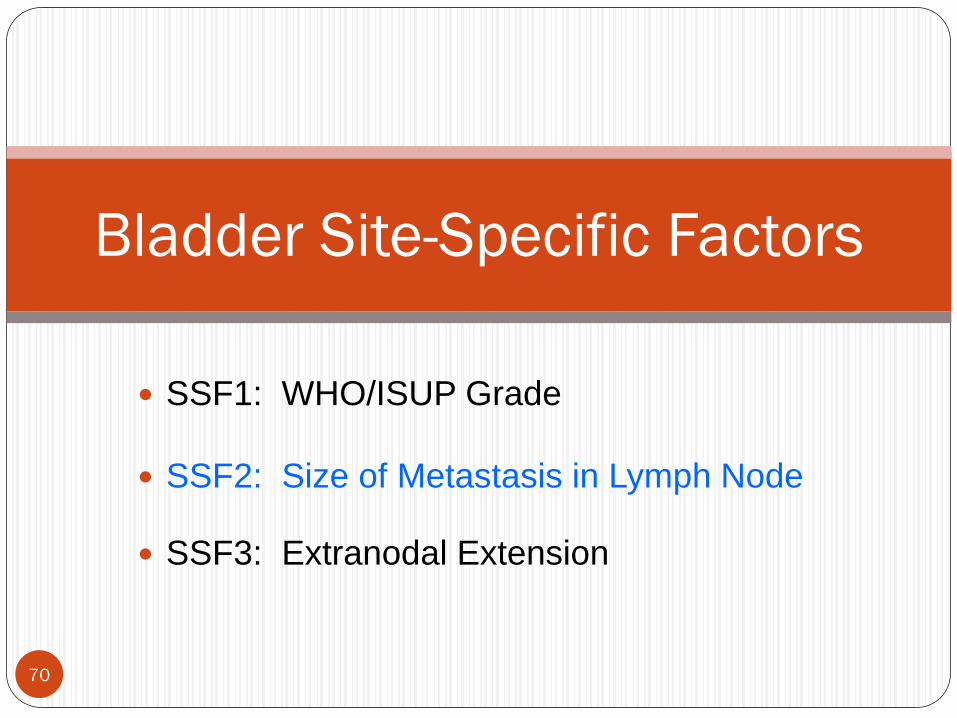
Bladder Site-Specific Factors
SSF1: WHO/ISUP Grade
SSF2: Size of Metastasis in Lymph Node
SSF3: Extranodal Extension

71
CS Site-Specific Factor 2
Survival impacted by size of lymph nodes
Applicable for clinical or pathologic Pathologic takes priority
Source documents: Clinical (imaging, physical exam) Pathologic (pathology report)
Collected for: Bladder, Kidney Parenchyma

72
Urothelial Cancer Treatment

73
Principles of Surgical Management
BL-A

74
Approximate Probability of Recurrence and Progression
BL-C

75
Principles of Intravesical Treatment
BL-F

76
BL-G
Principles of Chemotherapy Management

77
BL-H
Principles of Radiation Management of Invasive Disease

PROSTATE
78
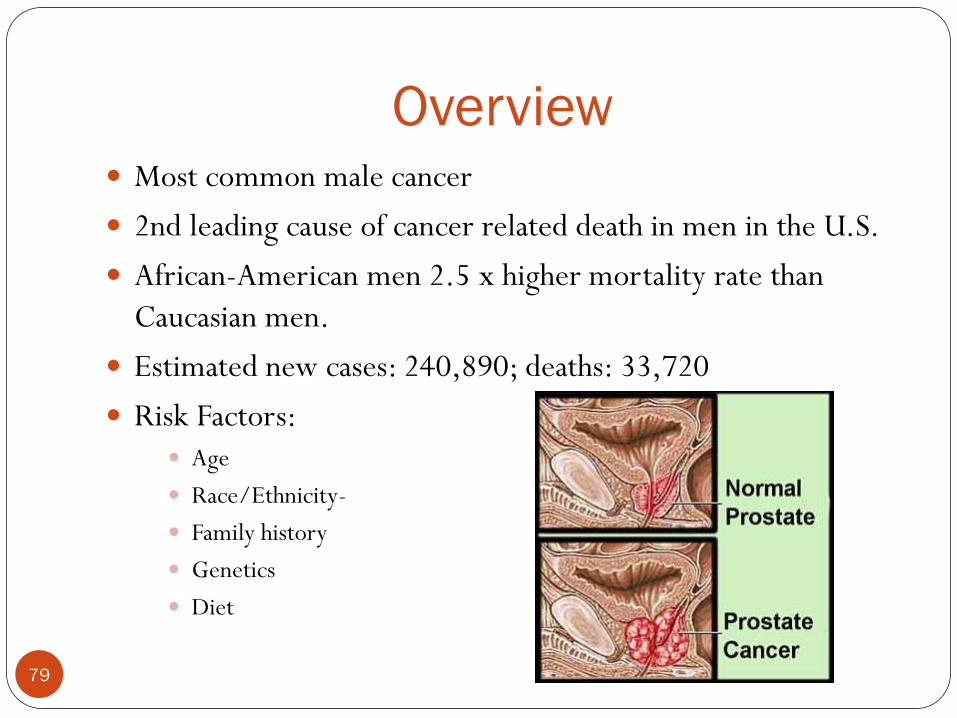
Overview Most common male cancer
2nd leading cause of cancer related death in men in the U.S.
African-American men 2.5 x higher mortality rate than
Caucasian men.
Estimated new cases: 240,890; deaths: 33,720
Risk Factors: Age
Race/Ethnicity-
Family history
Genetics
Diet
79

Age-Adjusted Cancer Death Rates,
Males by Site, US 1930-2007
80

Anatomy The prostate is a gland
found ONLY in men
It is located in front of the
rectum and under the
bladder
The size of a healthy
prostate gland is about the
size of a walnut
Source: http://www.abbottdiagnostics.com U.S. National Cancer Institute
81
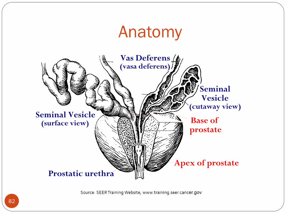
Anatomy
Vas Deferens (vasa deferens)
Seminal Vesicle (surface view)
Seminal Vesicle
(cutaway view)
Prostatic urethra
Apex of prostate
Base of prostate
82
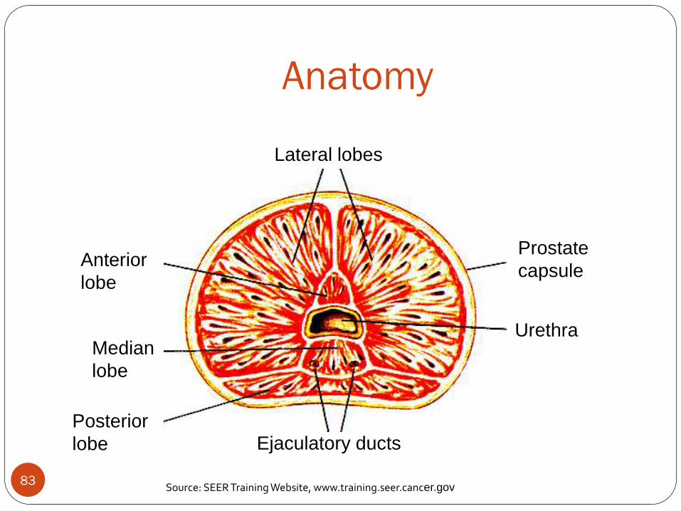
Anatomy
Source: SEER Training Website, www.training.seer.cancer.gov
Lateral lobes
Anterior
lobe
Median
lobe
Posterior
lobe Ejaculatory ducts
Prostate
capsule
Urethra
83

Diagnostic Procedures
PSA testing
DRE
TRUS
Biopsy
CT, MRI, Bone Scan
Evaluation for Metastases
84

Histology 99% Adenocarcinoma
Per MP/H, code acinar to 8140
1% Other
Sarcoma, small cell, other
PIN–Do NOT abstract*
30% men will go on to develop CaP
Close follow-up recommended for 2 years
* except reportable by agreement
Imag
e sou
rce: Natio
nal C
ancer In
stitute
85

Prognostic Factors
Clinical predictors
PSA – Prostate-specific antigen
Gleason score
Tumor stage
Pathologic factors
Number/percentage of positive biopsies
Surgical margin status
86

87

MPH Rules
Only ONE Prostate Cancer DX per patient lifetime
Dx of Acinar Carcinoma, Code to 8140 (Adenocarcinoma)
88

Prostate: Clinical Assessment
Clinically Apparent vs
Inapparent
89

Clinical Stage: Why Important??
The CS is logically divided into 4 major categories: T1, T2,
T3 and T4 stages.
Clinical Stages T1a and T1b
Incidentally detected during a TURP
Clinical stages T1c and T2
PSA test positive – detects earlier stage
Clinical Stage T3
DRE detects palpable disease sufficient to indicate that the
tumor has penetrated through the prostate capsule
90
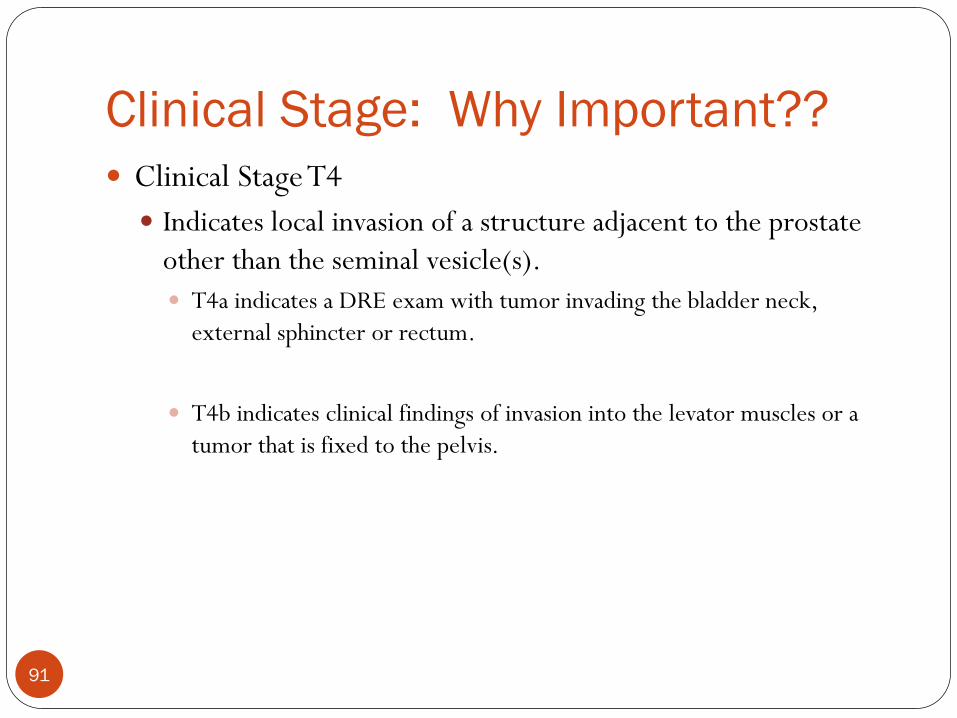
Clinical Stage: Why Important??
Clinical Stage T4
Indicates local invasion of a structure adjacent to the prostate
other than the seminal vesicle(s).
T4a indicates a DRE exam with tumor invading the bladder neck,
external sphincter or rectum.
T4b indicates clinical findings of invasion into the levator muscles or a
tumor that is fixed to the pelvis.
91

Clinical Stage Illustrations
92
T1c
T2 (a,b,c)
T3 (a,b,c)
T4 (a,b) Material provided by Prostate Cancer Research Institute (PCRI)
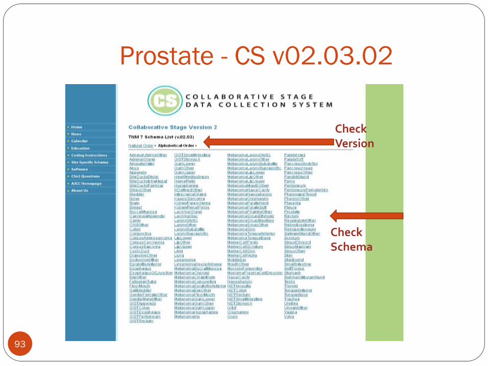
Prostate - CS v02.03.02
Check Version
Check Schema
93
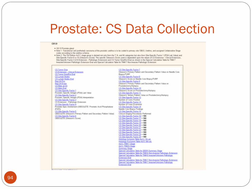
Prostate: CS Data Collection
94
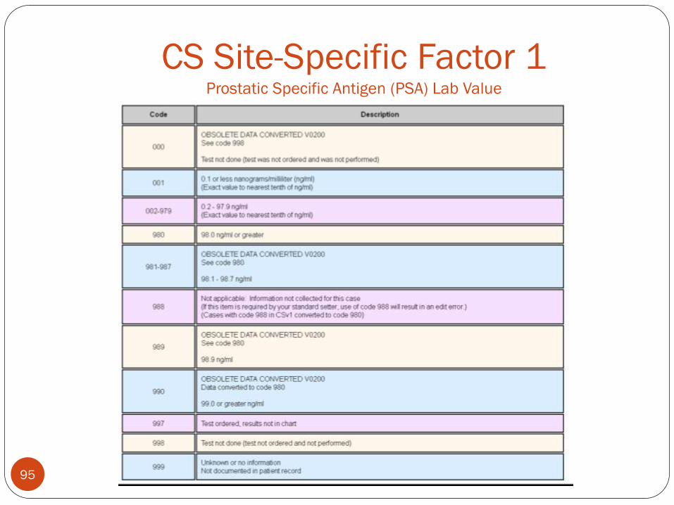
CS Site-Specific Factor 1 Prostatic Specific Antigen (PSA) Lab Value
95
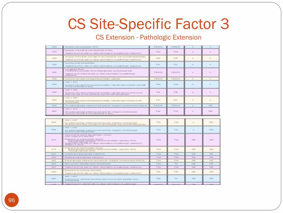
CS Site-Specific Factor 3 CS Extension - Pathologic Extension
96

CS Site-Specific Factor 8 Gleason's Score on Needle Core Biopsy/Transurethral Resection of Prostate
97

9
8
Gleason Pattern(s) and Score
http://www.stjohnprovidence.org

Grade Conversion
Code Gleason’s
Score
Terminology Histologic
Grade
1 2, 3, 4 Well differentiated I
2 5, 6 Moderately
differentiated
II
3 7, 8, 9, 10 Poorly
differentiated
III
99

CS Site-Specific Factor 10 Gleason's Score on Prostatectomy/Autopsy
100

Treatment Options
Observation
Seed RT Surgery
Experimental Hormone
Beam RT
101
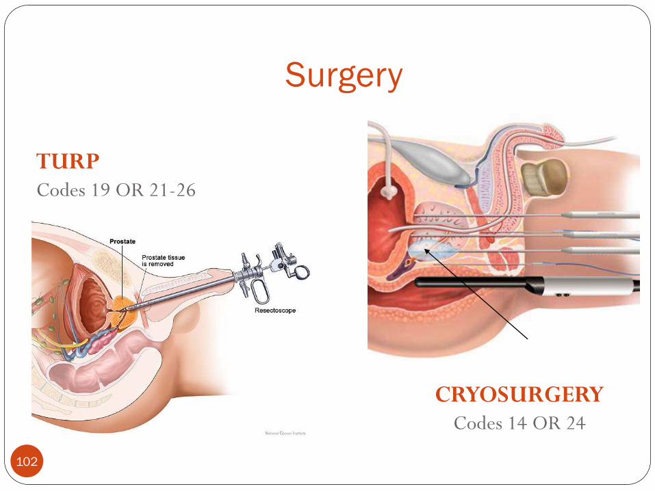
Surgery
TURP Codes 19 OR 21-26
CRYOSURGERY Codes 14 OR 24
Imag
e So
urce
: N
atio
nal
Can
cer
Inst
itut
e Ice ball
102

Prostatectomy Perineal, retropubic, suprapubic–
depends on patient’s anatomy and surgical history
Nerve-sparing
Robotic
Codes 30 – 80
Laparoscopic radical prostatectomy
constitutes less than 1% of all
prostatectomies performed in the
US.
103

Radical Perineal Prostatectomy
104

Beam Radiation
Prostate sitting on rectum
IMRT 3-D
Conformal
Images reproduced by permission of L.J.S.Bradbury - www.prostate-cancer-radiotherapy.org.uk
105

Brachytherapy (HDR)
Used with permission from Dr. Mark Scholz and www.PCRI.org
106

NCCN Guidelines
107

Initial Therapy By Stage Stage I (occult)
Observation without immediate treatment. If the patient is younger (age 50-60), immediate treatment may be considered.
External beam radiation therapy following transurethral resection
Radical prostatectomy with pelvic lymphadenectomy
Interstitial radioisotopes
Stage II (palpable prostate tumor at diagnosis)
Radical prostatectomy with pelvic lymphadenectomy
External beam radiation therapy following transurethral resection
Interstitial radioisotopes (under clinical evaluation)
108
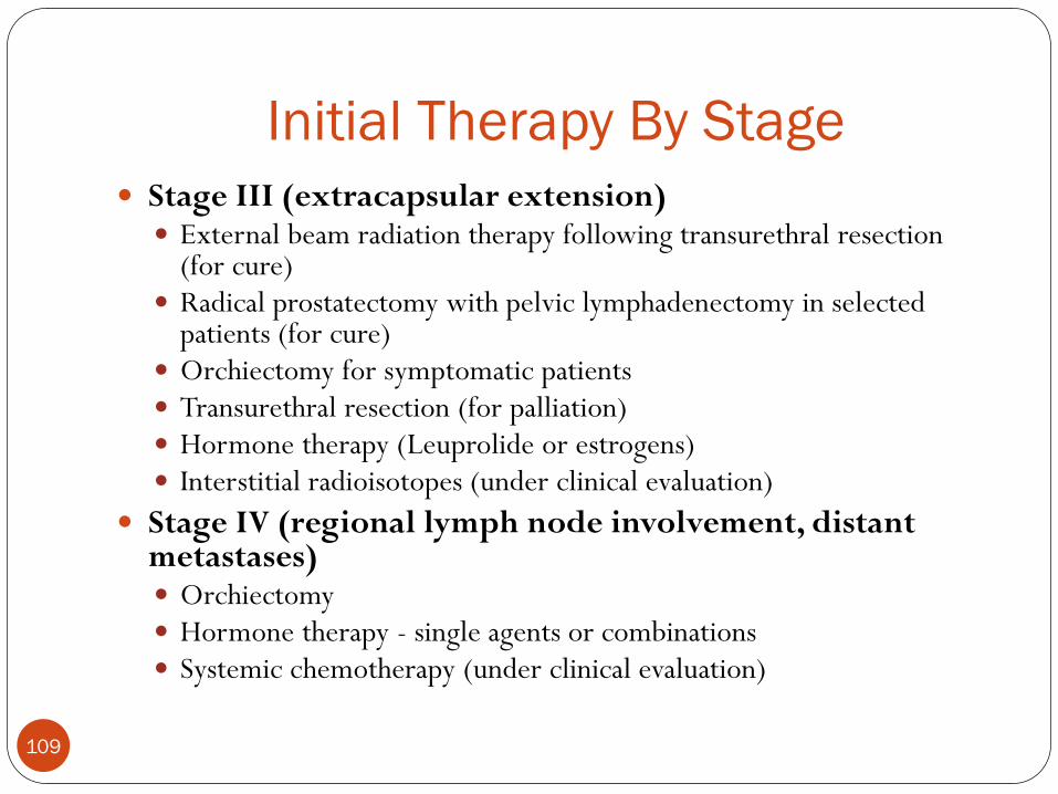
Stage III (extracapsular extension) External beam radiation therapy following transurethral resection
(for cure) Radical prostatectomy with pelvic lymphadenectomy in selected
patients (for cure) Orchiectomy for symptomatic patients Transurethral resection (for palliation) Hormone therapy (Leuprolide or estrogens) Interstitial radioisotopes (under clinical evaluation)
Stage IV (regional lymph node involvement, distant metastases) Orchiectomy Hormone therapy - single agents or combinations Systemic chemotherapy (under clinical evaluation)
109
Initial Therapy By Stage

Questions
110

111 NEXT WEBCAST: January 19, 2012 - Brain and CNS Tumors