genomic dna fingerprinting by restriction fragment end labeling
TRANSCRIPT
Proc. Natl. Acad. Sci. USAVol. 92, pp. 5572-5576, June 1995Microbiology
Genomic DNA fingerprinting by restriction fragment end labelingT. J. MARTIJN VAN STEENBERGEN*, SEAN D. COLLOMStt, PETER W. M. HERMANS§, JOHANNES DE GRAAFF*¶,AND RONALD H. A. PLASTERKt¶*Department of Oral Microbiology, Academic Centre for Dentistry Amsterdam, van der Boechorststraat 7, 1081 BT Amsterdam, The Netherlands; tNetherlandsCancer Institute, Plesmanlaan 121, 1066CX Amsterdam, The Netherlands; §Laboratory of Pediatrics, Erasmus University, P.O. Box 1738, 3000 DR Rotterdam,The Netherlands; and IDepartment of Medical Microbiology, Vrije Universiteit, van der Boechorststraat 7, 1081 BT Amsterdam, The Netherlands
Communicated by Melvin I. Simon, California Institute of Technology, Pasadena, CA, February 27, 1995
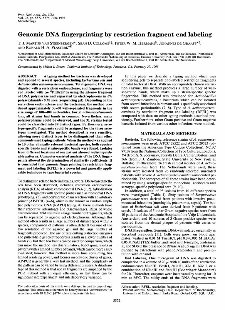
ABSTRACT A typing method for bacteria was developedand applied to several species, including Escherichia coli andActinobacillus actinomycetemcomitans. Total genomic DNA wasdigested with a restriction endonuclease, and fragments wereend labeled with [a-32PIdATP by using the Klenow fragmentof DNA polymerase and separated by electrophoresis in 6%polyacrylamide/8 M urea (sequencing gel). Depending on therestriction endonuclease and the bacterium, the method pro-duced approximately 30-50 well-separated fragments in thesize range of 100-400 nucleotides. For A. actinomycetemcomi-tans, all strains had bands in common. Nevertheless, manypolymorphisms could be observed, and the 31 strains testedcould be classified into 29 distinct types. Furthermore, sero-type-specific fragments could be assigned for the three sero-types investigated. The method described is very sensitive,allowing more distinct types to be distinguished than othercommohly used typing methods. When the method was appliedto 10 other clinically relevant bacterial species, both species-specific bands and strain-specific bands were found. Isolatesfrom different locations of one patient showed indistinguish-able patterns. Computer-assisted analysis of the DNA finger-prints allowed the determination of similarity coefficients. Itis concluded that genomic fingerprinting by restriction frag-ment end labeling (RFEL) is a powerful and generally appli-cable technique to type bacterial species.
To distinguish related bacterial strains, several DNA-based meth-ods have been described, including restriction endonucleaseanalysis (REA) ofwhole chromosomal DNA (1, 2), hybridizationof DNA fragments with specific probes such as ribosomal DNA(ribotyping) (3), and polymerase chain reaction with an arbitraryprimer (AP-PCR) (4-6), which is also known as random ampli-fied polymorphic DNA (RAPD) typing. All these methods havetheir respective advantages and disadvantages. REA of wholechromosomal DNA results in a large number of fragments, whichcan be separated by agarose gel electrophoresis. Although thismethod often results in a large number of distinct types within aspecies, comparison of patterns is often difficult because of thelow resolution of the agarose gel and the large number offragments produced. The use of rare-cutting restriction enzymesand pulsed-field gel electrophoresis results in a lower number ofbands (2), but then few bands can be used for comparison, whichcan make the method less discriminatory. Ribotyping results inpatterns with a limited number ofbands, which can be more easilyevaluated; however, this method is more time consuming, haslimited resolving power, and focuses on only one cluster of genes.AP-PCR is generally a very fast method, and the complexity ofthe pattern can be varied by using different primers. A disadvan-tage of this method is that not all fragments are amplified by thePCR method with an equal efficiency, so that there can besignificant interexperiment and interlaboratory variation.
The publication costs of this article were defrayed in part by page chargepayment. This article must therefore be hereby marked "advertisement" inaccordance with 18 U.S.C. §1734 solely to indicate this fact.
In this paper we describe a typing method which usessequencing gels to separate end-labeled restriction fragmentsof total bacterial DNA. With an appropriately chosen restric-tion enzyme, this method produces a large number of well-separated bands, which make up a strain-specific geneticfingerprint. This method was developed for Actinobacillusactinomycetemcomitans, a bacterium which can be isolatedfrom several infections in humans and is specifically associatedwith severe periodontitis (7, 8). Type of A. actinomycetem-comitans by restriction fragment end labeling (RFEL) wascompared with data on other typing methods described pre-viously. Furthermore, other Gram-positive and Gram-negativebacteria isolated from various other infections were studied.
MATERIALS AND METHODSBacteria. The following reference strains of A. actinomyce-
temcomitans were used: ATCC 29522 and ATCC 29523 (ob-tained from the American Type Culture Collection), NCTC9710 (from the National Collection ofType Cultures, London),Y4 (from S. S. Socransky, Forsyth Dental Center, Boston), and366 (from J. J. Zambon, State University of New York atBuffalo). Furthermore, 26 fresh clinical isolates of A. actino-mycetemcomitans from The Netherlands were used; thesestrains were isolated from 26 randomly selected, unrelatedpatients with severe A. actinomycetemcomitans-associated pe-riodontitis. The serotypes of all these strains have been deter-mined by using serotype-specific monoclonal antibodies andserotype-specific polyclonal sera (9, 10).
In addition, a total of 93 isolates from 10 different specieswere investigated (Table 1). Sixty isolates of Streptococcuspneumoniae were derived from patients with invasive pneu-mococcal infections (meningitis, pneumonia, sepsis). Ten iso-lates of Escherichia coli were derived from 9 patients withsepsis, 13 isolates of 5 other Gram-negative species were from10 patients of the Academic Hospital of the Vrije Universiteit,Amsterdam, and 10 isolates of 3 Gram-positive species wereisolated from the dental plaque of patients with caries orperiodontitis.DNA Preparation. Genomic DNA was isolated essentially as
described previously (11). Cells were grown on blood agarplates, washed in 0.01 M Tris HCl, pH 8.0/0.005 M EDTA/0.05 M NaCl (TES) buffer, and lysed with lysozyme, proteinaseK, and SDS in the presence of RNaseA at 0.5 [kg/ml. DNAwaspurified by extractions with phenol/chloroform and precipi-tation with ethanol.End Labeling. One microgram of DNA was digested to
completion in a volume of 20 ,ul with 10 units of the restrictionendonucleases Hindlll, EcoRI, BamHI, Xho II, Nde I, or acombination of Hindlll and BamHI (Boehringer Mannheim)for 2 h. Thereafter, enzymes were inactivated by heating for 10min at 65°C. The sticky ends of the DNA fragments were
Abbreviation: RFEL, restriction fragment end labeling.lPresent address: Microbiology Unit, Department of Biochemistry,University of Oxford, South Parks Road, Oxford OX1 3QU, U.K.
5572
Proc. Natl. Acad. Sci. USA 92 (1995) 5573
Table 1. Origin and suitable enzymes* for 11 bacterial species studied with genomic DNAfingerprinting by RFEL
No. of SuitableSpecies Origin isolates tested enzyme
A. actinomycetemcomitans Subgingival plaque 31 HindlllEscherichia coli Sepsis 10 HindIIIStreptococcus pneumoniae Invasive infections 60 EcoRIAcinetobacter baumannii Hospitalized patients 3 EcoRIXanthomonas maltophilia Hospitalized patients 3 EcoRIKlebsiella pneumoniae Hospitalized patients 3 HindIIIEnterobacter agglomerans Hospitalized patients 2 HindlIlSerratia marcescens Hospitalized patients 2 HindlllStreptococcus mutans Supragingival plaque 3 EcoRIStreptococcus sobrinus Supragingival plaque 5 HindIIIPeptostreptococcus micros Subgingival plaque 2 EcoRI*A suitable enzyme is defined as an enzyme which produces about 20-60 DNA fragments in the sizeregion of 100-400 nucleotides.
labeled with 28 nM [a-32P]dATP (2.5 ,tCi per reaction; 1 ,uCi= 37 kBq) by using 1 unit of the Klenow fragment of DNApolymerase (Boehringer Mannheim) in a volume of 30 ,l for30 min at room temperature. Unlabeled deoxynucleosidetriphosphates were added at a concentration of 1 mM ifnecessary for incorporation. Unincorporated label was re-moved by precipitation with ethanol. DNA was redissolved in10 mM Tris/1 mM EDTA buffer, pH 7.6. The amount ofradioactivity incorporated in the DNA was measured to allowloading of an equal amount in each lane on the gel. DNAsamples were mixed with an equal volume of loading buffer[xylene cyanol at 1 mg/ml, bromophenol blue at 1 mg/ml, and10 mM EDTA in 98% (vol/vol) formamide], denaturated byheating for 10 min at 85°C, and separated by electrophoresisfor 3 h at 1600 V in a vertical 6% polyacrylamide gel in 8 Murea in 0.045 M Tris-borate/0.001 M EDTA, pH 8.0 buffer.The polyacrylamide gel was dried onto Whatman 3M filterpaper and DNA fragments were detected by autoradiography.
Computer-Assisted Analysis. The DNA end labeling finger-prints of the A. actinomycetemcomitans strains were analyzedby using the Windows version of the GELCOMPAR softwareversion 3.10 (Applied Math, Kortrijk, Belgium) after imagingof the autoradiograms with a 190 dots per inch scanner (HPScanjet IIcx/T, Hewlett-Packard). DNA patterns were nor-malized by usingA. actinomycetemcomitans-specific bands thatwere present in the fingerprints of all strains. Comparison offingerprints was done by the unweighted pair-group method ofaverages (UPGMA) clustering method, using the Jaccardcoefficient applied to peaks, according to the instructions ofthe supplier of GELCOMPAR.
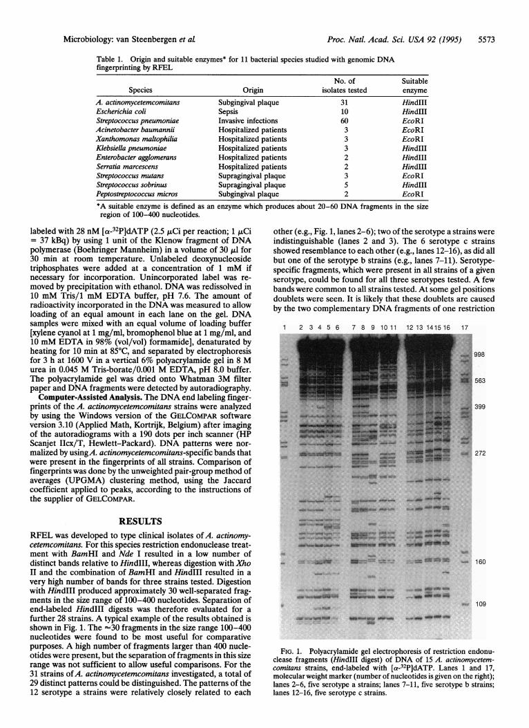
RESULTSRFEL was developed to type clinical isolates of A. actinomy-cetemcomitans. For this species restriction endonuclease treat-ment with BamHI and Nde I resulted in a low number ofdistinct bands relative to HindIll, whereas digestion with XhoII and the combination of BamHI and Hindlll resulted in avery high number of bands for three strains tested. Digestionwith HindIII produced approximately 30 well-separated frag-ments in the size range of 100-400 nucleotides. Separation ofend-labeled HindIII digests was therefore evaluated for afurther 28 strains. A typical example of the results obtained isshown in Fig. 1. The '30 fragments in the size range 100-400nucleotides were found to be most useful for comparativepurposes. A high number of fragments larger than 400 nucle-otides were present, but the separation of fragments in this sizerange was not sufficient to allow useful comparisons. For the31 strains ofA. actinomycetemcomitans investigated, a total of29 distinct patterns could be distinguished. The patterns of the12 serotype a strains were relatively closely related to each
other (e.g., Fig. 1, lanes 2-6); two of the serotype a strains wereindistinguishable (lanes 2 and 3). The 6 serotype c strainsshowed resemblance to each other (e.g., lanes 12-16), as did allbut one of the serotype b strains (e.g., lanes 7-11). Serotype-specific fragments, which were present in all strains of a givenserotype, could be found for all three serotypes tested. A fewbands were common to all strains tested. At some gel positionsdoublets were seen. It is likely that these doublets are causedby the two complementary DNA fragments of one restriction
1 2 3 4 5 6 7 8 9 10 11 12 13 141516 17
:.:
998
563
399
272
-..,.
- -NubW
e M*.WUiSit
* :: :._ ............. - _'..._........ ._...... .F .....:.-N.:um._b
*_
-_ " -
_ 160
109U.AW
:_ ___
FIG. 1. Polyacrylamide gel electrophoresis of restriction endonu-clease fragments (HindIII digest) of DNA of 15 A. actinomycetem-comitans strains, end-labeled with [a-32P]dATP. Lanes 1 and 17,molecular weight marker (number of nucleotides is given on the right);lanes 2-6, five serotype a strains; lanes 7-11, five serotype b strains;lanes 12-16, five serotype c strains.
Microbiology: van Steenbergen et al
5574 Microbiology: van Steenbergen et al
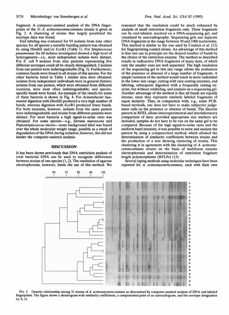
fragment. A computer-assisted analysis of the DNA finger-prints of the 31 A. actinomycetemcomitans strains is given inFig. 2. A clustering of strains that largely paralleled theserotype data was found.End labeling was evaluated for 93 isolates from nine other
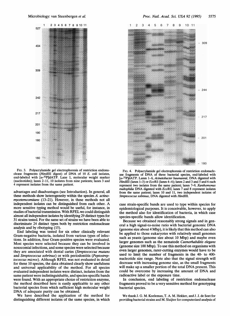
species; for all species a suitable banding pattern was obtainedby using HindIII and/or EcoRI (Table 1). For Streptococcuspneumoniae the 60 isolates investigated showed a high level ofheterogeneity-i.e., most independent isolates were distinct.For E. coli 9 isolates from nine patients representing fivedifferent serotypes could all be clearly distinguished; 2 isolatesfrom one patient were indistinguishable (Fig. 3). Furthermore,common bands were found in all strains of this species. For theother bacteria listed in Table 1 similar data were obtained:isolates from independent individuals were in general distinct;isolates from one patient, which were obtained from differentlocations, were most often indistinguishable; and species-specific bands were found. An example of the results for someof these bacteria is shown in Fig. 4. For Acinetobacter bau-mannii digestion with Hindlll produced a very high number ofbands, whereas digestion with EcoRI produced fewer bands.For both enzymes, however, isolates from the same patientwere indistinguishable and strains from different patients weredistinct. For most bacteria a high signal-to-noise ratio wasobtained. For some species-e.g., Serratia marcescens andPeptostreptococcus micros-some background label was foundover the whole molecular weight range, possibly as a result ofdegradation of the DNA during isolation; however, this did nothinder the computer-assisted analysis.
DISCUSSIONIt has been shown previously that DNA restriction analysis oftotal bacterial DNA can be used to recognize differencesbetween strains of one species (1, 2). The resolution of agaroseelectrophoresis, however, limits the use of this method. We
reasoned that the resolution could be much enhanced byanalysis of small restriction fragments only. These fragmentscan be end-labeled, resolved on a DNA-sequencing gel, andvisualized by autoradiography. Sequencing gels can separateDNA fragments in the range between 30 and 1000 nucleotides.This method is similar to the one used by Coulson et at (12)for fingerprinting cosmid clones. An advantage of this methodis that one can in principle set the desired number of bands bythe choice of the restriction enzyme. The method as describedresults in radioactive DNA fragments of many sizes, of whichonly the smaller ones are well separated. The high resolutionof the sequencing gel in this size range allows the evaluationof the presence or absence of a large number of fragments. Asimple variation of the method would result in more radiolabelin the lower size range: cutting with rare-cutting enzymes, endlabeling, subsequent digestion with a frequently cutting en-zyme, but without relabeling, and analysis on a sequencing gel.Another advantage of the method is that all bands are equallyintense, since they represent similarly labeled fragments ofequal molarity. Thus, in comparison with, e.g., some PCR-based methods, one does not have to make subjective judge-ment calls on the presence or absence of bands. The discretepattern in RFEL allows interexperimental and interlaboratorycomparison of data: provided appropriate size markers areincluded, samples do not have to be run on the same gel to becompared. Because of the high signal-to-noise ratio and theuniform band intensity, it was possible to store and analyze thepattern by using a computerized method, which allowed thedetermination of similarity coefficients between strains andthe production of a tree showing clustering of strains. Thisclustering is in agreement with the clustering of A. actinomy-cetemcomitans strains on the basis of multilocus enzymeelectrophoresis and determination of restriction fragmentlength polymorphisms (RFLPs) (13).
Several typing methods using molecular techniques have beenreported for A. actinomycetemcomitans, each with their own
60 70 80 90 100I, ,,II III II IIII IIIIIl||s i III IA-It III
I
i
I.
,.-f.,,..i, viig I,,. I,,
I l : f I I lI : :.,
.I.II
CC
CCCbbbbbbbbbbbcaaaaaaaaaaaab
X4'
.....
.I,. .:
II
.1 IFIG. 2. Genetic relationship among 31 strains ofA. actinomycetemcomitans as determined by computer-assisted analysis of DNA end-labeled
fingerprints. The figure shows a dendrogram with similarity coefficients, a computerized print of an autoradiogram, and the serotype designation(a, b, c).
Proc. Natl. Acad. Sci. USA 92 (1995)
Proc. Natl. Acad. Sci. USA 92 (1995) 5575
FIG. 3. Polyacrylamide gel electrophoresis of restriction endonu-clease fragments (Hindlll digest) of DNA of 10 E. coli isolates,end-labeled with [a-32P]dATP. Lane 1, molecular weight marker(nucleotides); lanes 2-11, 10 isolates from nine patients; lanes 3 and4 represent isolates from the same patient.
advantages and disadvantages (see Introduction). In general, allthese methods show heterogeneity within the species A. actino-mycetemcomitans (13-21). However, in these methods not allindependent isolates can be distinguished from each other. Amore sensitive typing method would be useful, for instance, instudies ofbacterial transmission. With RFELwe could distinguishalmost all independent isolates by identifying 29 distinct types for31 strains tested. For the same set of strains we have been able todiscriminate 24 distinct types both by restriction endonucleaseanalysis and by ribotyping (15).End labeling was tested for six other clinically relevant
Gram-negative bacteria, isolated from various types of infec-tions. In addition, four Gram-positive species were evaluated.Most species were selected because they can be involved innosocomial infections, and some species were selected becausethey are associated with dental caries (Streptococcus mutansand Streptococcus sobrinus) or with periodontitis (Peptostrep-tococcus micros). Although RFEL was not evaluated in detailfor these 10 species, the data obtained clearly show usefulnessand universal applicability of the method. For all speciesevaluated independent isolates were distinct, isolates from thesame patient were indistinguishable, and species-specific bandswere found. With an appropriate choice of restriction enzyme,the method described here is easily applicable to any otherbacterial species from which sufficient high molecular weightDNA of adequate purity can be obtained.We have described the application of the method for
distinguishing different isolates of the same species, in which
1 2 3 4 5 6 7 8 9 10 11
- 309
- 244
- 217
- 201
180
FIG. 4. Polyacrylamide gel electrophoresis of restriction endonucle-ase fragments of DNA of three bacterial species, end-labeled with[a-32P]dATP. Lanes 1-6, Acinetobacter baumannii, DNA digested withHindIlI (lanes 1-3) orEcoRI (lanes 4-6); lanes 2 and 3 and 5 and 6 eachrepresent two isolates from the same patient; lanes 7-9, Xanthomonasmaltophilia DNA digested with EcoRI; lanes 7 and 8 represent isolatesfrom the same patient; lanes 10 and 11, two independent isolates ofStreptococcus sobrinus, DNA digested with HindIIl.
case strain-specific bands are used to type within species forepidemiological purposes. It is conceivable, however, to applythe method also for identification of bacteria, in which casespecies-specific bands allow identification.Because we obtained reasonably strong signals and in gen-
eral a high signal-to-noise ratio with bacterial genomic DNA(genome size about 4 Mbp), it is likely that this method can alsobe applied to those eukaryotes with relatively small genomessuch as yeasts (genome size about 10 Mbp) and maybe evenlarger genomes such as the nematode Caenorhabditis elegans(genome size 100 Mbp). To use this method on organisms witheven larger genomes, rarer-cutting enzymes would have to beused to limit the number of fragments in the 40- to 400-nucleotide size range. Note also that the signal strength willdecrease with increasing genome size, as the small fragmentswill make up a smaller portion of the total DNA present. Thiscould be overcome by increasing the amount of DNA andradioactive label or the exposure time.
In conclusion, end labeling of restriction endonucleasefragments proved to be a very sensitive method for genotypingbacterial species.
We thank J. G. M. Koeleman, T. A. M. Hekker, and J. J. de Soet forproviding bacterial strains and M. Sluijter for computerized analysis of
1 2 3 4 5 6 7 8 9 10 11
...M527
404
309
242
217
201
190
180
160
Microbiology: van Steenbergen et at
"M I
Wviiim
.......
5576 Microbiology: van Steenbergen et at
the data. S.D.C. was supported by a long-term fellowship of theEuropean Molecular Biology Organization.
1. Owen, R. J. (1989) J. Med. Microbiol. 30, 89-99.2. Schwartz, D. C. & Cantor, C. R. (1984) Cell 37, 67-75.3. Grimont, F. & Grimont, P. A. D. (1986) Ann. Inst. Pasteurl
Microbiol. 137, 165-175.4. Williams, J. K. G., Kubelik, A. R., Livak, K. J., Rafalski, J. A. &
Tingey, S. V. (1990) Nucleic Acids Res. 18, 6531-6535.5. Welsh, J. & McClelland, M. (1990) Nucleic Acids Res. 18,
7213-7218.6. Wang, G., Whittam, T. S., Berg, C. M. & Berg, D. E. (1993)
Nucleic Acids Res. 21, 5931-5933.7. Zambon, J. J. (1985) J. Clin. Periodontol. 12, 1-20.8. Slots, J. & Listgarten, M. (1988) J. Clin. Periodontol. 15, 85-93.9. Gmur, R. & Guggenheim, B. (1990) Arch. Oral Bio. 35, Suppl.,
145-151.10. Gmur, R., McNabb, H., van Steenbergen, T. J. M., Baehni, P.,
Mombelli, A., van Winkelhoff, A. J. & Guggenheim, B. (1993)Oral Microbiol. Immunol. 8, 116-120.
11. van Steenbergen, T. J. M., van der Velden, U., Abbas, F. & deGraaff, J. (1991) J. Periodontol. 62, 235-241.
Proc. Natl. Acad. Sci. USA 92 (1995)
12. Coulson, A. R., Sulston, J. E., Brenner, S. & Karn, J. (1986) Proc.Natl. Acad. Sci. USA 83, 7821-7825.
13. Poulsen, K., Theilade, E., Lally, E. T., Demuth, D. R. & Kilian,M. (1994) Microbiology 140, 2049-2060.
14. Zambon, J. J., Sunday, G. J. & Smutko, J. S. (1990) J. Periodon-tol. 61, 75-80.
15. van Steenbergen, T. J. M., Bosch-Tijhof, C. J., van Winkelhoff,A. J., Gmur, R. & de Graaff, J. (1994) J. Clin. Microbiol. 32,2769-2774.
16. DiRienzo, J. M., Cornell, S., Kazoroski, L. & Slots, J. (1990) OralMicrobiol. Immunol. 35, 79S-84S.
17. Slots, J., Liu, Y. B., DiRienzo, J. M. & Chan, C. (1993) OralMicrobiol. Immunol. 8, 337-343.
18. Guthmiller, J. M., Kolodrubetz, D. & Kraig, E. (1993) Microb.Pathog. 14, 103-115.
19. Saarela, M., Asikainen, S., Jousimies-Somer, H., Asikainen, T.,von Troil-Linden, B. & Alaluusua, S. (1993) Oral Microbiol.Immunol. 8, 111-115.
20. Alaluusua, S., Saarela, M., Jousimies-Somer, H. & Asikainen, S.(1993) Oral Microbiol. Immunol. 8, 225-259.
21. Griffen, A. L., Leys, E. J. & Fuerst, P. A. (1992) Oral Microbiol.Immunol. 7, 240-243.