genotipi di pestivirus rna identificati in vaccini virali … · 2017-07-11 · (csfv) (hog cholera...
TRANSCRIPT

7
IntroduzioneI virus della Diarrea Virale
Bovina 1 (BVDV-1), DiarreaVirale Bovina 2 (BVDV-2), Bor-der disease (BDV) e Peste suinaclassica (PSCV) sono specie sta-bilite del genere Pestivirus dellafamiglia Flaviviridae, con la spe-cie proposta «Giraffa» (33), pato-geni degli ungulati domestici eselvatici, a distribuzione cosmo-polita, responsabili di vari tipi dimanifestazioni cliniche.
Ceppi non citopatici (NCP) diBVDV-1 e BVDV-2 sono statifrequentemente indicati comeresponsabili di contaminazione diprodotti biologici, colture cellula-ri, incluso cellule primarie e lineecellulari, anche di origine umana(2, 5, 16, 17, 28), siero fetalebovino (1, 3, 26) e vaccini ad usoveterinario (10, 22, 23, 32, 36).
Durante uno studio sperimen-tale in Giappone, RNA di Pestivi-rus è stato trovato anche in quat-
tro vaccini virali vivi ad uso uma-no (20). I vaccini erano stati pro-dotti da diverse ditte farmaceuti-che, regolarmente autorizzati ecommercializzati: due monova-lenti contro parotite e rosolia edue polivalenti contro morbillo,parotite e rosolia. L’analisi com-parativa della sequenza nucleoti-dica della 5’-UTR aveva identifi-cato RNA di BVDV quale conta-minante. Allo stesso modo, inter-ferone ad uso umano è stato tro-vato contaminato da RNA diBVDV (19). Uno studio realizza-to da Vilcek et al. (35) ha riporta-to risultati negativi su 30 vaccinivirali umani da ditte europeevalutati con PCR per la presenzadi RNA di Pestivirus. Gli Autoriavevano concluso che l’evenienzadi contaminazione di vaccini vira-li umani non rappresentava unfenomeno diffuso.
Ulteriori indagini sono stateintraprese su vaccini virali vivi ad
GENOTYPES OF PESTIVIRUS RNA
DETECTED IN ANTI INFLUENZAVIRUS VACCINES FOR HUMAN USE
SummaryNine polyvalent human influenza
virus vaccines were tested by reversetranscriptase-polymerase chainreaction (RT-PCR) for the presence ofpest ivirus RNA. Samples wereselected from manufacturers in Europeand the United States of America(USA).
Three samples of the nine vaccinestested (33.3%) gave positive resultsfor pestivirus RNA. The 5’-untranslatedgenomic region sequence of thecontaminant pestivirus RNA wasanalysed based on primary nucleotidesequence homology and onsecondary sequence structurescharacteristic to genotypes. Twosequences belonged to Pestivirus type-1(bovine viral diarrhoea virus [BVDV])species, genotypes BVDV-1b andBVDV-1e. These findings confirmprevious reports, suggest ing an
GENOTIPI DI PESTIVIRUS RNA IDENTIFICATI IN VACCINI VIRALI ANTI INFLUENZA AD USO UMANO
M. Giangaspero1, G. Vacirca1, R. Harasawa2, M. Buttner3, A. Panuccio4, C. De Giuli Morghen5, A. Zanetti6, A. Belloli1 & A. Verhulst71 Dipartimento di Scienze Cliniche Veterinarie, Facoltà di Medicina Veterinaria, Università degli Studi, Milano - Italia
2 Centro Animale per la Ricerca Biomedica, Facoltà di Medicina, Università di Tokyo - Giappone3 Istituto di Immunologia, Centro di Ricerche Federale per le Malattie Virali degli Animali, Tübingen - Germania
4 Centro Multizonale di Igiene e Prevenzione, Milano - Italia5 Istituto di Farmacologia, Facoltà di Medicina, Università degli Studi, Milano - Italia
6 Istituto di Virologia, Facoltà di Medicina, Università degli Studi, Milano - Italia7 Istituto di Medicina Tropicale Prince Léopold, Anversa - Belgio
L AV O R I S C I E N T I F I C I O R I G I N A L I / S C I E N T I F I C P A P E R S
RIASSUNTO
Vaccini ad uso umano, polivalenti contro virus dell’influenza, prodotti in Europa e USA, sono stati testaticon RT-nested PCR per la messa in evidenza di Pestivirus contaminanti. Tre campioni (33,3%), su 9 testati,sono risultati positivi per RNA di Pestivirus. La sequenza della regione genomica non tradotta 5' dell’RNA
dei Pestivirus contaminanti é stata analizzata sulla base di omologia della sequenza nucleotidica primaria edella struttura secondaria caratteristica dei genotipi. Due sequenze hanno mostrato la loro appartenenza
alla specie Pestivirus tipo 1 (diarrea virale bovina), genotipi BVDV-1b e BVDV-1e. I risultati ottenuti nel presente studio confermano precedenti osservazioni, suggerendo la necessità di poten-
ziare le misure per la prevenzione di contaminazioni nei prodotti biologici ad uso umano.
PAROLE CHIAVEContaminazione - Genotipi - Pestivirus - Vaccini.

M. GIANGASPERO E ALTRI VETERINARIA ITALIANA
8
uso umano selezionati da produt-tori Europei, Nord Americani eGiapponesi (14). Ventinovemonovalenti contro morbillo,parotite, rosolia o polio, otto poli-valenti contro morbillo, parotite erosolia e uno polivalente battericocontro Streptococcus pneumoniae,sono stati testati con transcriptasiinversa – nested PCR. Il 13% deicampioni testati (5 su 38) sonorisultati positivi per RNA di Pesti-virus. Tre vaccini (uno anti-roso-lia e due anti-morbillo) eranoEuropei e due (uno anti-parotite euno anti-rosolia) provenivano dalGiappone. La 5’-UTR dell’RNAdei Pestivirus contaminanti è stataamplificata e sequenzata. Le ana-lisi basate sull’omologia dellasequenza nucleotidica primaria esulla struttura secondaria hannorivelato che le sequenze isolateappartenevano al Pestivirus ditipo 1 (diarrea virale bovina).L’RNA di Pestivirus identificatodai campioni vaccinali Giappone-si anti-rosolia e anti-parotiteappartenevano rispettivamente aigenotipi BVDV-1c e BVDV-1a.Non è stato possibile geotipare lasequenza identificata da un cam-pione di vaccino anti-morbillo acausa della mancanza di una partedella 5’UTR. L’analisi di 2sequenze identificate da campioni
di vaccini Europei anti-morbillo eanti-rosolia ha evidenziato la loroappartenenza a un nuovo genotipodi Pestivirus BVDV-1d.
Data l’importanza della sicu-rezza dei prodotti biologici ad usoumano, lo studio attuale è statointrapreso allo scopo di fornireulteriori conferme dei precedentirisultati e al fine di valutare lacontaminazione da RNA di Pesti-virus in vaccini virali inattivatianti-influenzali ad uso umano.
Materiali e MetodiCampioni di vaccini
I tests sono stati effettuati sunove campioni di vaccini viraliinattivati polivalenti anti-influenzaad uso umano, da tre lotti prodottida due diverse ditte (qui menzio-nate come A e B) da Europa e StatiUniti (Tabella 1). In tre prove sonostati usati come controlli positivi iceppi di referenza BVDV Oregon(C24V) e NADL. In tutte le altreprove i controlli positivi sono statievitati al fine di ridurre i rischi dicontaminazione del sistema di ana-lisi. I campioni sono stati conser-vati a -70°C fino ad esecuzione deitests. Cinque Istituzioni di vastaesperienza in Belgio, Germania,Italia e Giappone e Centri di refe-renza Nazionale per l’identifica-zione dei Pestivirus, hanno esegui-to le analisi virologiche.
improvement in preventive measuresagainst contamination of biologicalproducts for human use.
KeywordsBovine viral diarrhoea - Contamination -Genotype - Pestivirus - Vaccine.
IntroductionBovine viral diarrhoea virus-1
(BVDV-1), BVDV-2, Border diseasevirus (BDV), classical swine fever virus(CSFV) (hog cholera virus) areestablished species of the Pestivirusgenus, Flaviviridae family, with atentative ‘giraffe’ species (33). Theyare cosmopolitan pathogens in cloven-hoofed ungulates, present a widerange of clinical manifestations andhave a significant impact onproduction.
Non-cytopathic (NCP) strains ofBVDV-1 and BVDV-2 have beenreported frequently as contaminants ofbiological products, cell cultures(including primary cell cultures and celllines), even of human origin (3, 5, 16,17, 28), bovine foetal serum (1, 2,26) and vaccines for veterinary use(10, 22, 23, 32, 36).
During an experimental studyconducted in Japan, pestivirus RNAwas also detected in four live humanvirus vaccines (20). The four vaccineswere produced by differentpharmaceutical companies whichwere correctly authorised andmarketed (two monovalent vaccinesagainst mumps and rubella (Germanmeasles) and two polyvalent vaccinesagainst measles, mumps and rubella).A comparative analysis of thenucleotide sequence at the 5’-untranslated region (UTR) revealedBVDV RNA as the contaminant.Similarly, interferon for human use wasfound contaminated by BVDV RNA(19). A study conducted by Vilcek etal. (35) reported negative results in 30human virus vaccines from Europeanproducers screened by polymerasechain reaction (PCR) for the detectionof pestivirus RNA. The authorsconcluded that contamination ofhuman virus vaccines is notwidespread. Further investigation wasundertaken on live virus vaccines forhuman use selected from European,North American and Japanese
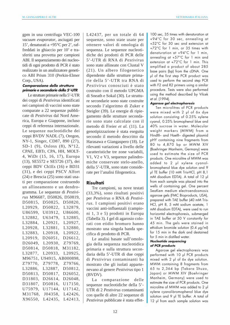
Tabella 1: Vaccini virali inattivati anti-influenza ad uso umano, prodotti su embrione di pollo, testati per PestivirusRNA con nested PCR. 1) numero del campione; 2) lotto di produzione; 3) produttore; 4) origine.Table 1: Inactivated human influenza virus vaccines produced in chicken embryos and tested for pestivirusRNA by nested PCR. 1: sample No. 2: Production batch 3: Manufacturer 4: Origin.
1 2 3 4
1 1 A Svizzera/Switzerland
2 1 A Svizzera/Switzerland
3 2 A Svizzera/Switzerland
4 2 A Svizzera/Switzerland
5 2 A Svizzera/Switzerland
6 2 A Svizzera/Switzerland
7 2 A Svizzera/Switzerland
8 3 B Stati Uniti d’America/United States of America
9 3 B Stati Uniti d’America/United States of America
VOL. 40 (1) GENOTIPI DI PESTIVIRUS RNA IDENTIFICATI IN VACCINI VIRALI ANTI INFLUENZA AD USO UMANO
9
Estrazione dell’RNAL’RNA virale è stato estratto da
ogni campione con il metodo iso-tiocianato di guanidina-fenolo-clo-roformio in singola fase (6) con ilkit di estrazione RNAzol B (Bio-tecx Laboratories Inc., USA) (a) oTRIZOL (Gibco BRL, USA) (b).
(a): 200 µl di ogni campionesono stati mescolati e agitati vigo-rosamente con 800 µl di RNAzolB, una soluzione contenente iso-tiocianato di guanidina-fenolo inuna provetta Eppendorf di 1,5 mlper 30”. Dopo l’aggiunta di 100 µldi cloroformio, la provetta è stataposta sotto agitazione per 30”,ottenendo una soluzione lattescen-te. La provetta è stata raffreddatain ghiaccio per 5’ e centrifugata a12.000 gpm per 10’. La faseacquosa (ca. 600 µ l di fluidosupernatante) è stata trasferita inun’altra provetta Eppendorf. Unµl di 20 mg/ml di glicogeno dimitilo (Boehringer MannheimGmbH, Germania) e 600 µl di iso-propanolo sono stati aggiunti e laprovetta è stata raffreddata inghiaccio per 30’. Il precipitato diRNA è stato raccolto per centrifu-gazione a 12.000 gpm per 10’. Ilpellet è stato lavato tre volte con600 µl di etanolo al 75% in acquatrattata con dietil pirocarbonato(DEPC) allo 0,1%. Il pellet è statoasciugato all’aria a +37°C per 10’,disciolto in 20 µl di acqua distilla-ta trattata con DEPC allo 0,1%, equindi riscaldata a +60°C per 10’.
(b): ogni campione è statomescolato a 1 ml di TRIZOL eincubato a temperatura ambienteper 5’. Sono stati aggiunti 200 µldi cloroformio e la provetta è sta-ta agitata vigorosamente e incu-bata a temperatura ambiente per3’. La soluzione è stata centrifu-gata a 12.000 gpm a +4°C per15’. La fase acquosa è stata rac-colta e 500 µ l di isopropanolosono stati aggiunti e la provetta èstata centrifugata a 12.000 gpm a+4°C per 10’. Il pellet è statolavato tre volte con 1 ml di etano-lo al 75% e ulteriormente centri-fugato a 12.000 gpm a +4°C per15’. Il pellet è stato asciugatoall’aria a +37°C per 5’, discioltoin 10 µl di acqua distillata trattatacon DEPC allo 0,1%. La soluzio-ne di RNA (16 µl) è stata sottopo-sta alla reazione di trascrizioneinversa (RT).Oligonucleotidi
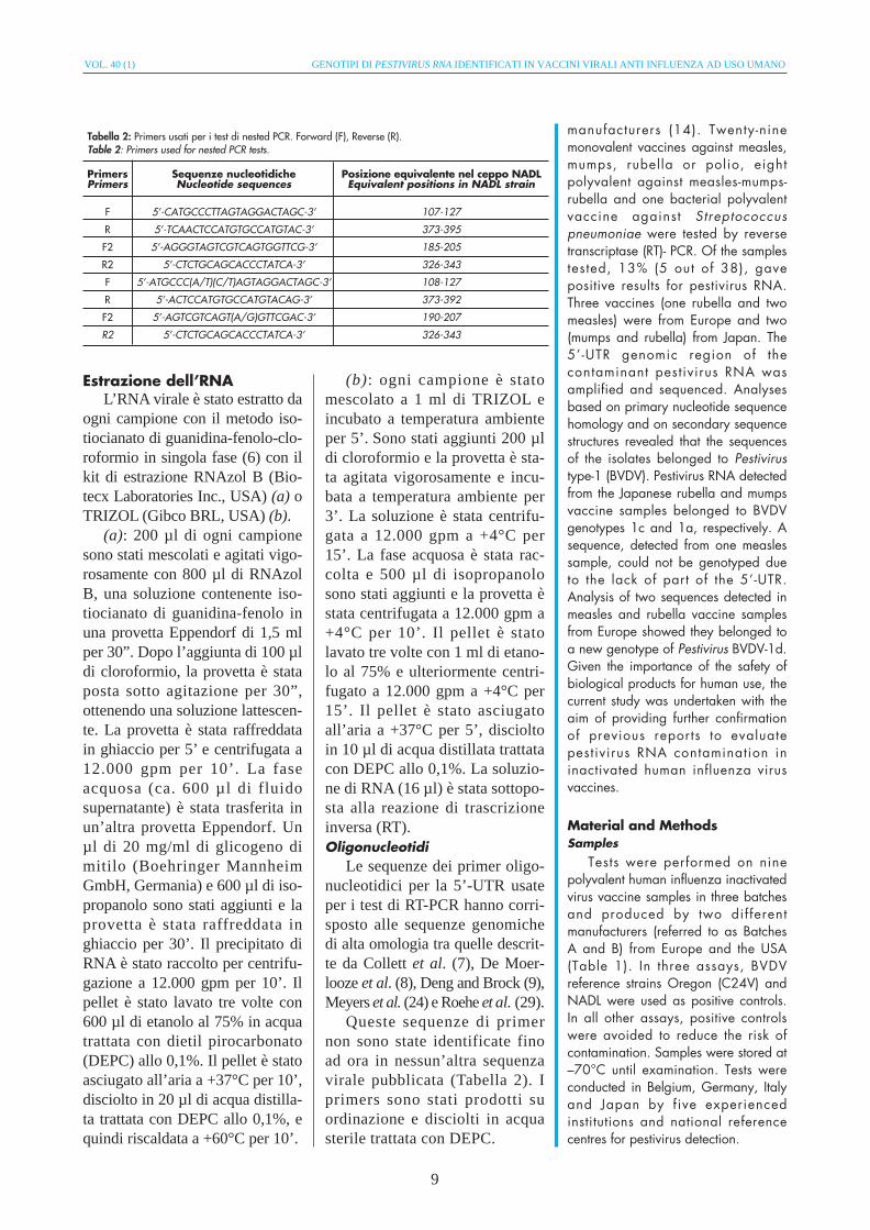
Le sequenze dei primer oligo-nucleotidici per la 5’-UTR usateper i test di RT-PCR hanno corri-sposto alle sequenze genomichedi alta omologia tra quelle descrit-te da Collett et al. (7), De Moer-looze et al. (8), Deng and Brock (9),Meyers et al. (24) e Roehe et al. (29).
Queste sequenze di primernon sono state identificate finoad ora in nessun’altra sequenzavirale pubblicata (Tabella 2). Iprimers sono stati prodotti suordinazione e disciolti in acquasterile trattata con DEPC.
manufacturers (14). Twenty-ninemonovalent vaccines against measles,mumps, rubella or polio, eightpolyvalent against measles-mumps-rubella and one bacterial polyvalentvaccine against Streptococcuspneumoniae were tested by reversetranscriptase (RT)- PCR. Of the samplestested, 13% (5 out of 38), gavepositive results for pestivirus RNA.Three vaccines (one rubella and twomeasles) were from Europe and two(mumps and rubella) from Japan. The5’-UTR genomic region of thecontaminant pestivirus RNA wasamplified and sequenced. Analysesbased on primary nucleotide sequencehomology and on secondary sequencestructures revealed that the sequencesof the isolates belonged to Pestivirustype-1 (BVDV). Pestivirus RNA detectedfrom the Japanese rubella and mumpsvaccine samples belonged to BVDVgenotypes 1c and 1a, respectively. Asequence, detected from one measlessample, could not be genotyped dueto the lack of part of the 5’-UTR.Analysis of two sequences detected inmeasles and rubella vaccine samplesfrom Europe showed they belonged toa new genotype of Pestivirus BVDV-1d.Given the importance of the safety ofbiological products for human use, thecurrent study was undertaken with theaim of providing further confirmationof previous reports to evaluatepestivirus RNA contamination ininactivated human influenza virusvaccines.
Material and MethodsSamples
Tests were performed on ninepolyvalent human influenza inactivatedvirus vaccine samples in three batchesand produced by two differentmanufacturers (referred to as BatchesA and B) from Europe and the USA(Table 1). In three assays, BVDVreference strains Oregon (C24V) andNADL were used as positive controls.In all other assays, positive controlswere avoided to reduce the risk ofcontamination. Samples were stored at–70°C until examination. Tests wereconducted in Belgium, Germany, Italyand Japan by five experiencedinstitutions and national referencecentres for pestivirus detection.
Primers Sequenze nucleotidiche Posizione equivalente nel ceppo NADLPrimers Nucleotide sequences Equivalent positions in NADL strain
F 5’-CATGCCCTTAGTAGGACTAGC-3’ 107-127
R 5’-TCAACTCCATGTGCCATGTAC-3’ 373-395
F2 5’-AGGGTAGTCGTCAGTGGTTCG-3’ 185-205
R2 5’-CTCTGCAGCACCCTATCA-3’ 326-343
F 5’-ATGCCC(A/T)(C/T)AGTAGGACTAGC-3’ 108-127
R 5’-ACTCCATGTGCCATGTACAG-3’ 373-392
F2 5’-AGTCGTCAGT(A/G)GTTCGAC-3’ 190-207
R2 5’-CTCTGCAGCACCCTATCA-3’ 326-343
Tabella 2: Primers usati per i test di nested PCR. Forward (F), Reverse (R).Table 2: Primers used for nested PCR tests.

M. GIANGASPERO E ALTRI VETERINARIA ITALIANA
10
Sintesi di DNA complementareLa soluzione per la reazione di
RT è stata costituita da 8 µ l ditampone 5x per la prima catena(250mM Tris-HCl, pH 8,3, 375mM KCl, 15 mM MgCl2), 4 µl di0,1 M ditiotritolo (DTT), 0,25 µl(25 U) di transcriptasi inversa delvirus della Leucemia murina(Gibco BRL, USA), 8 µl di deos-sinucleotidi trifosfato (dNTPs) auna concentrazione finale di 0,2mM ognuno (Gibco-BRL, USA),0,2 µl (110 U/µl) di inibitore diribonucleasi, 0,25 µl (40 pM/µl)di primer R e acqua trattata conDEPC ad un volume finale di 32µl. Appena prima dell’incubazio-ne, sono stati aggiunti 8 µ l disoluzione di RNA a 32 µl di solu-zione RT; in seguito, è stataaggiunta una goccia di olio mine-rale M-3516 (Sigma ChemicalsCo., USA). La sintesi della primacatena di DNA complementare(DNAc) è stata realizzata inbagnomaria a +37°C per 90’.PCR
L’amplificazione della 5’-UTRè stata realizzata secondo il meto-do descritto da Harasawa et al.(17). Sei µl di soluzione di DNAcsono stati aggiunti a 5 µl di tam-pone 10x (100 mM Tris-HCl, pH
8,9, 800 mM KCl, 15 mM MgCl2,5 mg/ml siero albumina bovina,1% sodio colato, 1% Triton X-100), 1 µl (1,25 U) di DNA poli-merasi Thermus thermophilus(Tth), 3 µ l di tampone Tth (10mM Tris HCl, pH 7,5, 300 mMKCl, 1 mM DTT, 0,1 mM disodioetilenediaminotetracetato (EDTA),50% glicerolo), dNTPs ad unaconcentrazione finale di 0,2 mMognuno, 0,25 µl di primers F e R(40 pM/µl ognuno) e acqua ad unvolume finale di 50 µl. Dopo averricoperto la mistura di reazionecon olio minerale, il protocollo direazione è stato eseguito in un ter-mociclatore: 30 cicli con denatu-razione a +94°C per 30”, annea-ling a +55°C per 100” ed esten-sione a +72°C per 100”, 35 voltecon denaturazione a +94°C per30”, annealing a +52°C per 30” eestensione a +72°C per 1’, o 35volte con denaturazione a +94°Cper 1’, annealing a +57°C per 1’,ed estensione a +72°C per 1’. E’stato amplificato un prodotto dicirca 285 paia di basi dal DNAc.Un µ l del prodotto della primafase di PCR è stato usato per rea-lizzare la seconda amplificazionedi PCR con i primers F2 e R2 conla stessa procedura. Le prove sono
RNA extractionViral RNA was extracted from
each sample by the single-stepguanidinium isothiocyanate-phenol-chloroform method (6) with theRNAzol B extraction kit (BiotecxLaboratories Inc., USA) (see a) below)or Trizol (Gibco BRL, USA) (see b)below), as follows:
a) 200 µl of each sample weremixed vigorously with 800 µl ofRNAzol B, a solution containingguanidinium isothiocyanate-phenol ina 1.5 ml Eppendorf tube for 30 sec.One hundred µl of chloroform wereadded and the tube was vigorouslyshaken by hand for 30 sec, until thesolution became milky. The tube waschilled in ice for 5 min and centrifugedat 12,000 rpm for 10 min. Theaqueous phase (approximately 600 µlof supernatant fluid) was transferredinto a fresh Eppendorf tube. One µl of20 mg/ml mussel glycogen(Boehringer Mannheim GmbH,Germany) and 600 µl of isopropanolwere added and the tube was chilledin ice for 30 min. The RNA precipitatewas collected by centrifugation at12,000 rpm for 10 min. The pelletwas washed three times with 600 µl of75% ethanol in water treated with0.1% diethyl pyrocarbonate (DEPC).The pellet was air-dried at +37°C for10 min, dissolved in 20 µl distilledwater treated with 0.1% DEPC, andthen heated at +60°C for 10 min
b) each sample pellet was mixedwith 1 ml of Trizol and incubated atroom temperature for 5 min. Twohundred µ l o f ch loro form wereadded and the tube was vigorouslyshaken and incubated a t roomtemperature for 3 min. The solutionwas centrifuged at 12,000 rpm at+4°C for 15 min. The aqueousphase was collected and 500 µl ofisopropanol were added and thetube was centrifuged at 12,000 rpmat +4°C for 10 min. The pellet waswashed three times with 1 ml of75% ethanol and centrifuged againat 12,000 rpm at +4°C for 15 min.The pellet was air-dried at +37°Cfor 5 min, dissolved in 10 µl ofdistilled water treated with 0.1%DEPC. Finally, RT was applied tothe RNA solution (16 µl).
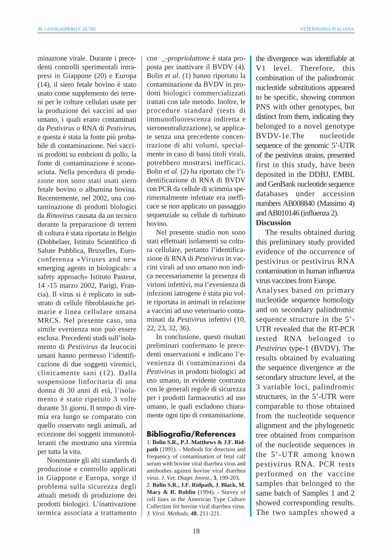
Tabella 3: Risultati di test nested PCR per pestivirus RNA in vaccini virali anti-influenza ad uso umano; ND:non determinato.Table 3: Results of nested PCR tests for adventitious pestivirus RNA in human influenza virus vaccines. ND:not determined.
N° 1° PCR 2° PCR Genotipo Nome/Numero d’accesso1st PCR 2nd PCR Genotype Name/Accession number
1 Negativo Positivo Ie Massimo4/AB008840Negative Positive
2 Negativo Positivo Ib Influenza2/AB010146Negative Positive
3 Negativo PositivoNegative Positive
4 Negativo PositivoNegative Positive
5 Positivo ND NDPositive
6 Negativo NegativoNegative Negative
7 Negativo NegativoNegative Negative
8 Negativo NegativoNegative Negative

VOL. 40 (1) GENOTIPI DI PESTIVIRUS RNA IDENTIFICATI IN VACCINI VIRALI ANTI INFLUENZA AD USO UMANO
11
state eseguite anche con il meto-do descritto da Vilcek et al. (34).Elettroforesi su gel di agarosio
Dieci µl di prodotto di PCRsono stati mescolati a 2 µ l disoluzione dye 6x costituita da0,25% xilene cianolo, 0,25% bludi bromofenolo e 40% di saccaro-sio in acqua. Per stimare la dimen-sione dei prodotti di PCR sono sta-ti usati markers di peso molecolare(MPM) da un plasmide pHY dige-rito HindIII- e HaeIII- contenentinove frammenti da 80 a 4.870 paiadi basi o MPM XIV (BoehringerMannheim, Germania). Un micro-litro di MPM è stato aggiunto a 2µ l di xilene cianolo-soluzionedye blu di bromofenolo e 9 µl ditampone TE (10 mM Tris-HCl,pH 8, 1mM EDTA disodico). I 12µ l di ogni campione ottenutosono stati posti nei pozzetti delgel. Gel di agarosio per elettroen-dosmosi all’1% di SeaKem (FMCBioproducts, USA), preparati contampone TAE (40mM Tris-HCl,pH8, 5 mM acetato di sodio,1mM EDTA disodico), sono statiusati per effettuare l’elettroforesi,in immersione in tampone TAE, a50 V costanti per 45’. I gel sonostati colorati in soluzione di eti-dio bromuro (0,4 µg/ml) per 15’,al buio, e scolorati 5’ in acquadistillata.Sequenziamento nucleotidico dei prodotti di PCR
L’elettroforesi dei gel di aga-rosio è stata realizzata con 10 µldi prodotto di PCR con l’aggiuntadi 2 µl di soluzione dye 6x. Perstimare la dimensione dei prodottidi PCR sono stati usati marker dipeso molecolare contenenti 8 fram-menti da 65 a 2.364 paia di basi(Takara Shuzo, Japan) o MWMXIV (Boehringer Mannheim, Ger-mania). Un microlitro di MPM èstato aggiunto a 2 µ l di xilenecianolo-soluzione dye blu di bro-mofenolo e 9 µl di tampone TE. I12 µl di soluzione di ogni cam-
pione sono stati posti nei pozzettidel gel. Per effettuare l’elettrofo-resi orizzontale, in immersione intampone TAE, a 100 V costantiper 25’ è stato usato un gel diagarosio per elettoendosmosibassa al 2% NuSieve 3:1 (FMCBioproducts, USA). I gel sonostati colorati con soluzione di eti-dio bromuro (2 µg/ml) per 10’ escolorati 3 volte con acqua distil-lata. I gel sono stati osservatiusando un UV transilluminator(Ultra Lum, USA) a 300 nanome-tri. Le porzioni di gel contenentibande positive sono state preleva-te e il DNA estratto con il kitGenetic II (BIO 100 Inc., USA).Tre volumi di soluzione NAIsono stati aggiunti e i gel sonostati liquefatti con un’incubazio-ne a +45°C per 5’. La sospensio-ne glass-milk è stata aggiunta eincubata per 5’. Dopo centrifuga-zione per 5”, il fluido supernatan-te è stato scartato e il pellet è sta-to lavato tre volte con acqua fre-sca e diluito in 10 µ l di acquadistillata. Un microlitro (equiva-lente a 3,2 pM) di primers R o Fsono stati addizionati a 30-90 ng(valutati mediante spettrofotome-tro) di DNA purificato; sono statiaggiunti 8 µl di pre-mix (Perkin-Elmer Corp., USA) consistente in4 µl di tampone di sequenziamen-to 5x, 1 µl di dNTP mix, 0,5 µl didye-deossi terminatori e 1 µl dipolimerasi Taq, e acqua distillatafino ad un volume finale di 20 µl.La soluzione di reazione conognuno dei primer è stata agitataper 1-2”, addizionata di una goc-cia di olio minerale e posta in umtermociclatore ABI 9600 (Perkin-Elmer Corp., USA), con inizio a+95°C per 10”, poi per 25 cicli a+96°C per 10”, +50°C per 5” e+60°C per 4’. I prodotti amplifi-cati (20 µl) sono stati purificati sucolonne reidratate Centri-Sep(Princeton Separations Inc., USA)con centrifugazione per 2’ a 3.000
OligonucleotidesOligonucleotide primer sequences ofthe 5’-UTR used for RT-PCR were basedon genomic sequences of highhomology among those described byCollett et al. (7), De Moerlooze et al.(8), Deng and Brock (9), Meyers et al.(24) and Roehe et al. (29). Theseprimer sequences have not been foundin any other viral sequences publishedto date (Table 2). Primers were custom-made, and dissolved in sterile DEPC-treated water.cDNA synthesis
The RT reaction solution wascomposed of 8 µl of 5x first strandbuffer (250 mM Tris-HCl, pH 8.3, 375mM KCl, 15 mM MgCl2), 4 µl of 0.1M dithiothreitol (DTT), 0.25 µl (25 U)of Moloney murine leukaemia virus RT(Gibco BRL, USA), 8 µldeoxynucleotide triphosphates (dNTPs)to a final concentration of 0.2 mMeach (Gibco-BRL), 0.2 µl (110 U/µl)ribonuclease inhibitor, 0.25 µl (40pM/µl) primer R and DEPC-treatedwater to a final volume of 32 µl. Justbefore the incubation, 8 µl of the RNAsolution were added to 32 µl of RTsolution and one drop of M-3516mineral oil (Sigma Chemicals Co.,USA) was added to the reactionmixture. The first strand synthesis ofcDNA was performed in a water bathat +37°C for 90 min.Polymerase chain reaction
The amplification of the 5’-UTR wasperformed according to the methoddescribed by Harasawa et al. (17). Sixmicrolitres of cDNA solution wereadded to 5 µl of 10x buffer (100 mMTris-HCl, pH 8.9, 800 mM KCl, 15 mMMgCl2, 5 mg/ml bovine serum albumin,1% sodium cholate, 1% Triton X-100), 1µl (1.25 U) Thermus thermophilus (Tth)DNA polymerase, 3 µl Tth buffer (10mM Tris HCl, pH 7.5, 300 mM KCl, 1mM DTT, 0.1 mM disodiumethylenediaminetetracetate(EDTA), 50%glycerol), dNTPs to a finalconcentration of 0.2 mM each, 0.25µl of F and R primers (40 pM/µl each)and water to a final volume of 50 µl.After the mixture was overlaid withmineral oil, the reaction cycle wasperformed in a thermal cycler,30 times with denaturation at +94°Cfor 30 sec, annealing at +55°C for100 sec and extension at +72°C for
M. GIANGASPERO E ALTRI VETERINARIA ITALIANA
12
gpm in una centrifuga VEC-100vacuum evaporator, asciugati per15’, denaturati a +95°C per 2’, raf-freddati in ghiaccio per 10’ e tra-sferiti una provetta per campioniABI. Il sequenziamento dei nucleo-tidi di ogni prodotto di PCR è statorealizzato in un analizzatore geneti-co ABI Prism 310 (Perkin-ElmerCorp., USA).Comparazione delle struttureprimarie e secondarie della 5’-UTR
Le strutture primarie nella 5’-UTRdei ceppi di Pestivirus identificatinei campioni di vaccini sono statecomparate a 22 sequenze pubbli-cate di Pestivirus dal Nord Ame-rica, Europa e Giappone, inclusoceppi di referenza internazionale.Le sequenze nucleotidiche deiceppi BVDV NADL (7), Oregon,NY-1, Singer, CD87, 890 (27),SD-1 (9), Osloss (8), N°12,CPAE, EBTr, CPA, HH, MOLT-4, WiDr (15, 16, 17), Europa(13), SE5572 e SE5726 (37), deiceppi BDV Ch1Es (16) e BD31(31), e dei ceppi PSCV Alfort(24) e Brescia (25) sono stati usa-ti per comparazione costruendoun allineamento e un dendro-gramma. Le sequenze di Pestivi-rus M96687, D50826, D50819,D50815, D50825, D50820,L20929, D50822, L32879,U86599, U03912, U86600,L32882, U63479, L32885,L32884, L20921, L20927,L20928, L32881, L32880,L32883, L20918, L20922,L20919, D26051, D26612,D26049, L20930, Z79769,D50814, D50818, M31182,L32877, L20933, L20925,M96751, U94915, AB000898,Z79770, Z79778, Z79766,L32886, L32887, D50812,D50813, D50817, D26052,D31803, D26614, D26048,D31807, D50816, U17150,U75979, U17144, U17142,M31768, J04358, L42426,X96550, L42435, L42413,
L42437, per un totale di 64sequenze, sono state usate perottenere valori di omologia disequenza. Le sequenze nucleoti-diche dei prodotti di PCR delle5’-UTR di RNA di Pestivirussono state allineate con Clustal V(21). Un albero filogeneticodipendente dalle strutture prima-rie della 5’-UTR tra RNA diPestivirus conosciuti è statocostruito con il metodo UPGMAdi Sneath e Sokal (30). Le struttu-re secondarie sono state costruitesecondo l’algoritmo di Zuker eSteigler (39). Le energie di ripie-gamento delle strutture seconda-rie sono state calcolate con ilmetodo di Freier et al. (11). Lagenotipizzazione è stata eseguitasecondo il metodo descritto daHarasawa e Giangaspero (18). Lerilevanti variazioni a livello dellecaratteristiche tre zone variabili,V1, V2 e V3, sequenze palindro-miche conservate stelo-anello,della 5’-UTR, sono state conside-rate per l’analisi filogenetica.
RisultatiTre campioni, su nove testati
(33,3%), sono risultati positiviper Pestivirus o RNA di Pestivi-rus. I campioni positivi eranovaccini anti-influenzali (campio-ni 1, 3 e 5) prodotti in Europa(Tabella 3). I gel di agarosio colo-rati con etidio bromuro hannomostrato una singola banda spe-cifica di prodotto di PCR.
Le analisi basate sull’omolo-gia della sequenza nucleotidicaprimaria e sulla struttura secon-daria della 5’-UTR di due ceppidi Pestivirus contaminanti hamostrato che gli isolati apparte-nevano al genere Pestivirus tipo 1(BVDV).
La comparazione dellesequenze nucleotidiche della 5’-UTR di 2 Pestivirus contaminanticon quelle di altre 22 sequenze diPestivirus pubblicate è stata effet-
100 sec, 35 times with denaturation at+94°C for 30 sec, annealing at+52°C for 30 sec and extension at+72°C for 1 min, or 35 times withdenaturation at +94°C for 1 min,annealing at +57°C for 1 min andextension at +72°C for 1 min. Thisamplified a product of about 285base pairs (bp) from the cDNA. Oneµl of the first step PCR product wasused to perform the second step PCRwith F2 and R2 primers using a similarprocedure. Tests were also performedusing the method described by Vilceket al. (1994).Agarose gel electrophoresis
Ten microlitres of PCR productswere mixed with 2 µl of 6x dyesolution consisting of 0.25% xylenecyanol, 0.25% bromophenol blue and40% sucrose in water. Molecularweight markers (MWM) from aHindIII– and HaeIII– digested plasmidpHY containing nine fragments from80 to 4,870 bp or MWM XIV(Boehringer Manheim, Germany) wereused to estimate the size of PCRproducts. One microlitre of MWM wasadded to 2 µl xylene cyanol-bromophenol blue dye solution and 9µl TE buffer (10 mM Tris-HCl, pH 8,1mM disodium EDTA). A total of 12 µlfrom each sample was placed into thewells of containing gel. One percentSeaKem medium electroendosmosisagarose gels (FMC Bioproducts, USA),prepared with TAE buffer (40 mM Tris-HCl, pH 8, 5 mM sodium acetate, 1mM disodium EDTA), were used to runhorizontal electrophoresis, submergedin TAE buffer at 50 V constantly for45 min. The gels were stained inethidium bromide solution (0.4 µg/ml)for 15 min in the dark and destainedfor 5 min in distilled water.Nucleotide sequencing of PCR products
Agarose gel electrophoresis wasperformed with 10 µl PCR productsmixed with 2 µl of 6x dye solution.MWM containing 8 fragments from65 to 2,364 bp (Takara Shuzo,Japan) or MWM XIV (BoehringerManheim, Germany) were used toestimate the size of PCR products. Onemicrolitre of MWM was added to 2 µlxylene cyanol-bromophenol blue dyesolution and 9 µl TE buffer. A total of12 µl from each sample solution was

VOL. 40 (1) GENOTIPI DI PESTIVIRUS RNA IDENTIFICATI IN VACCINI VIRALI ANTI INFLUENZA AD USO UMANO
13
Figura 1: Allineamento della sequenza nucleotidica dei prodotti di PCR dalle 5'-UTR per comparazione delle sequenze dei ceppi contaminanti dei vaccini virali con22 sequenze pubblicate di Pestivirus. Le sequenze nucleotidiche di ceppi di BVDV, BDV e PSCV sono state usate come referenze. Nucleotidi identici in due su tresequenze sono indicati in caratteri bianchi. I numeri di sequenze nucleotidiche sono dati per un allineamento di consenso. Trattini rappresentano spazi introdotti tranucleotidi adiacenti per un allineamento massimo.Figure 1: Nucleotide sequence alignment of the PCR products from the 5'-UTR for sequence comparison of the virus vaccine contaminant strains with twenty-twopublished pestivirus sequences. Nucleotide sequences of BVDV, BDV and CSFV strains are used for reference. Nucleotides that are identical in two of three sequen-ces are presented in white characters. The nucleotide sequence numbers are given from a consensus alignment. Dashes represent spacers between adjacent nucleo-tides introduced for maximum alignment.

M. GIANGASPERO E ALTRI VETERINARIA ITALIANA
14
tuata con il programma di allinea-mento Clustal V, usando il softwa-re DNASIS (Figura 1). Un alberofilogenetico basato sulla compara-zione della sequenza nucleotidicaprimaria è stato ottenuto con ilprogramma UPGMA, compresonel software DNASIS (Figura 2).
La comparazione del ceppo iso-lato dal campione 1 con 64 sequen-ze di ceppi di Pestivirus ha mostra-to un’omologia nella sequenzanucleotidica con i ceppi del genoti-
po 1b del 90-96% e dell’82-88%,84-85% e 69-71% con gli altrigenotipi di BVDV 1a, 1c e BVDV-2, rispettivamente. L’omologia del-la sequenza nucleotidica tra ilsecondo ceppo isolato dal campio-ne 2 e i genotipi di PestivirusBVDV 1a, 1b, 1c e BVDV-2 sonostati di 81-87%, 92-96%, 79-81% e70-72%, rispettivamente.
La variazione della sequenzatra le 5’-UTR dei ceppi dei Pestivi-rus identificati, principalmente
placed into the wells containing gel.Two percent NuSieve 3:1 lowelectroendosmosis agarose gel (FMCBioproducts, USA) in TAE buffer wasused to run horizontal electrophoresis,submerged in TAE buffer, at 100 Vconstantly for 25 min. The gel wasstained with ethidium bromide solution(2 µg/ml) for 10 min and destained 3times with distilled water. The gel wasobserved using a 300 nm UVtransilluminator (Ultra Lum, USA). Theportions of the gel that containedpositive bands were excised and DNAwas extracted using the Genetic II kit(BIO 100 Inc., USA). Three volumes ofNAI solution were added and the gelwas liquefied at +45°C incubation for5 min. Glass-milk suspension wasadded and incubated for 5 min. Aftercentrifugation for 5 sec, thesupernatant fluid was discarded andthe pellet was washed three times withfresh wash and eluted in 10 µl distilledwater. One microlitre (equivalent to3.2 pM) of primers R or F were mixedwith 30 ng-90 ng (determined byspectrophotometry) purified DNA, 8 µlpre-mix (Perkin-Elmer Corporation,USA) consist ing of 4 µl of 5xsequencing buffer, 1 µl dNTP mix, 0.5µl dye-deoxy terminators and 1 µl Taqpolymerase, and distilled water wasadded to 20 µl of the total volume. Themixture with each primer was vortexedfor 1-2 sec, with a drop of mineral oiladded and then placed in an ABI9600 thermal cycler (Perkin-ElmerCorporation, USA), with commencingat +95°C for 10 sec, followed by 25cycles of +96°C for 10 sec, +50°C for5 sec and +60°C for 4 min. Theamplified product (20 µl) was purifiedon Centri-Sep re-hydrated columns(Princeton Separations Inc., USA) withcentrifugation for 2 min at 3,000 rpmin a VEC-100 vacuum evaporatorcentrifuge, dried for 15 min,denatured at +95°C for 2 min, chilledon ice for 10 min and transferred toan ABI sampling tube. Nucleotidesequencing of each PCR product wasperformed in an ABI Prism 310Genetic Analyzer (Perkin-ElmerCorporation, USA).Comparison of primary andsecondary structures of 5’-UTR
Primary structures in the 5’-UTR ofthe pestivirus strains detected in
890
CD87
CPA
EBTr
CPAE
Ch1Es
BD31
Osloss
Massimo4
NY-1
Influenza2
WiDr
MOLT-4
Brescia
Alfort
SE5572
SE5726
Europa
HH
Oregon
No.12
SD-1
Singer
NADL
0.0236
0.0200
0.0000
0.02000.0000
0.0000
0.0000
0.0042
0.0180
0.0143
0.0042
0.0021
0.0180
0.0050
0.0032
0.00320.0280
0.0280
0.0133
0.0100
0.0100
0.0204
0.0135
0.0135
0.0289
0.0065
0.0065
0.0224
0.0069
0.0033
0.0183
0.0328
0.0098
0.0074
0.0055
0.0101
0.0135
0.0063
0.01450.0238
0.0399
0.0036
0.0488
0.0080
0.0013
0.0018
Figura 2: Albero filogenetico basato sulla comparazione della sequenza 5'-UTR di differenti ceppi di Pestivi-rus, ottenuto con UPGMA. I numeri indicati per i rami rilevanti sono riferiti a valori di bootstrap di 1.000replicazioni. La scala delle linee indica sostituzioni di 10 nucleotidi per 100 nucleotidi..Figure 2: Phylogenetic tree based on 5'-UTR sequence comparison from different pestivirus strains, obtainedby UPGMA. Numbers at the relevant branches refer to bootstrap values of 1,000 replications. The scale barindicates 10-nucleotide substitutions per 100 nucleotides.

VOL. 40 (1) GENOTIPI DI PESTIVIRUS RNA IDENTIFICATI IN VACCINI VIRALI ANTI INFLUENZA AD USO UMANO
15
limitata a tre regioni specifiche, lezone variabili V1, V2 e V3, tipichestrutture palindromiche, a forma distelo-anello, sono state identificateattraverso ricerca manuale di strut-ture potenzialmente elicoidali, cer-cando sostituzioni di un legame dibasi Watson-Crick per un altro.Sono stati identificati elementicomuni caratteristici dei Pestivirusa livello di V1 e V2. Le strutturestabili palindromiche stelo-anellohanno mostrato energie liberesostanzialmente negative: -12,92Kcal/mol nella struttura V1, -13,80Kcal/mol in V2 e -7,10 Kcal/molin V3 per il campione 1; -9,32
Kcal/mol in V1, -13,80 Kcal/molin V2 e -7,10 Kcal/mol in V3 per ilcampione 2.
Le rilevanti variazioni nucleo-tidiche sono state comparate conmembri rappresentativi di altrigenotipi di Pestivirus. I legami dibasi nucleotidiche componenti glisteli delle strutture palindromichea livello delle 3 zone variabili,V1, V2 e V3, della regione geno-mica 5’-UTR dei 2 ceppi di Pesti-virus hanno mostrato divergenzadai genotipi dei Pestivirus prece-dentemente descritti (campione 1)e chiare similitudini con i ceppidel genotipo 1b (campione 2),
vaccine samples were compared withtwenty-two published pestivirussequences from North America, Europeand Japan, including internationalreference strains. The nucleotidesequences of BVDV NADL strains (7),Oregon, NY-1, Singer, CD87, 890(27), SD-1 (9), Osloss (8), No. 12,CPAE, EBTr, CPA, HH, MOLT-4, WiDr(15, 16, 17), Europa (13), SE5572and SE5726 (37), of BDV strainsCh1Es (16) and BD31 (31), and ofHCV strains Alfort (24) and Brescia(25) were used for comparison byconstructing an alignment anddendrogram. Pestivirus sequencesM96687, D50826, D50819,D50815, D50825, D50820, L20929,D50822, L32879, U86599, U03912,U86600, L32882, U63479, L32885,L32884, L20921, L20927, L20928,L32881, L32880, L32883, L20918,L20922, L20919, D26051, D26612,D26049, L20930, Z79769, D50814,D50818, M31182, L32877, L20933,L20925, M96751, U94915,AB000898, Z79770, Z79778,Z79766, L32886, L32887, D50812,D50813, D50817, D26052,D31803, D26614, D26048,D31807, D50816, U17150,U75979, U17144, U17142,M31768, J04358, L42426, X96550,L42435, L42413, L42437, for a totalof 64 sequences, were used to obtainoverall sequence homology values.Nucleotide sequences of PCR productsfrom the 5’-UTR of pestivirus RNA werealigned by the Clustal V (21). Aphylogenetic tree according to theprimary structures of the 5’-UTR amongthe known pestivirus RNA wasconstructed by the unweighed pair-group method using arithmeticaverages (UPGMA) by Sneath andSokal (30). Secondary structures werepredicted according to the algorithm ofZuker and Steigler (39). Foldingenergies of the secondary structureswere calculated by the methoddescribed by Freier et al. (11).Genotyping was performed accordingto the method of Harasawa andGiangaspero (18). Relevant variationsin the characteristic three variable loci,V1, V2 and V3, conserved stem-looppalindromic sequences at the 5’-UTRand were used for the phylogeneticanalysis.
Figura 3: Strutture palindromiche (V1, V2 e V3) nella regione genomica 5'-UTR dei ceppi di Pestivirus isolati davaccini virali contro l’influenza (campione 1 colonna A, campione 2 colonna B). Il legame di basi Watson-Crické mostrato da un tratto (-) e il legame G:U tollerato nella struttura secondaria é indicato da un asterisco (*).Figure 3: Palindromic structures (V1, V2 and V3) in the 5'-UTR genomic region of the pestivirus strains isola-ted from influenza virus vaccines. (Sample 1: lane A, Sample 2: lane B). - : Watson-Crick base pairing. * :G:U pairing tolerated in secondary structures.
A B
V1
A GG GG AA A
U-AC-GC-GG-C
A-U . -C . -G
C C
5'-. -A-3'
G CU GG AU C
U-AU-AC-GG-C
A-UG-CC-G
C C
5'-U-A-3'
V2
G
5'-A-U-3'
V3
G G
G-CC-GA-UG*UG-CU-AC-GC-G
G U
G
5'-A-U-3'
G G
G-CC-GG*UA-UG-CU-AC-GC-G
G U
A
5'-A-U-3'
A C
U-AU-AG-CG-CC-G
U C
A C
A
5'-A-U-3'
A C
U-AU-AG-CG-CC-G
U C
A C

M. GIANGASPERO E ALTRI VETERINARIA ITALIANA
16
rispettivamente (Figura 3).Le sequenze nella 5’-UTR del
ceppo Massimo4 hanno mostratocaratteristiche paia di basi. La zonaV1 in posizione 8 aveva mostratoun legame di basi C-G, in V2, inposizione 7 un legame di basi A-Ue in V3, in posizione 4 un legamedi basi G-C.
Il legame di basi A-U in V2 eracondiviso con il genotipo BVDV-1b, dal quale la divergenza eraidentificabile al livello della V1.Per cui, tale combinazione di sosti-tuzioni nucleotidiche palindromi-che è apparsa essere specifica,mostrando PNS comuni con altrigenotipi, ma distinta da essi, indi-cando l’appartenenza a un nuovogenotipo BVDV-1e. Le sequenzenucleotidiche della 5’-UTR geno-mica dei ceppi di Pestivirus pre-sentati per la prima volta in questostudio sono state depositate nellebanche dati di sequenze nucleotidi-che DDBJ, EMBL e GenBank sotto inumeri di accesso AB008840 (Mas-simo4) e AB010146 (Influenza2).
DiscussioneI risultati ottenuti nel corso di
questo studio preliminare hannoevidenziato la contaminazione daPestivirus o da RNA di Pestivirusin vaccini virali anti-influenzaliad uso umano prodotti in Europa.
Le analisi basate sull’omologiadella sequenza nucleotidica prima-ria e sulla struttura secondaria del-le sequenze palindromiche nella5’-UTR hanno rivelato che l’RNAtestato con RT-PCR apparteneva alPestivirus di tipo 1 (BVDV). Irisultati ottenuti dalla valutazionedella divergenza della sequenza alivello della struttura secondarianelle 3 zone variabili, strutturepalindromiche, nella 5’-UTR era-no comparabili a quelli ottenutiattraverso l’allineamento dellesequenze nucleotidiche e l’alberofilogenetico ottenuto dalla compa-razione delle sequenze nucleotidi-
che nella 5’-UTR tra RNA di Pesti-virus noti.
I tests di PCR effettuati suicampioni di vaccino appartenenteallo stesso lotto, campioni 1 e 2,hanno mostrato risultati corri-spondenti. I due campioni hannomostrato una diversa intensitàdella banda specifica nel gel elet-troforetico. Il campione 1 hamostrato una banda positivaintensa e il campione 2 ha datouna banda debole. Questo potreb-be essere il risultato di una dege-nerazione dell’RNA nel campione2, testato più tardi, oltre la data divalidità. Inoltre, le loro sequenzenucleotidiche ottenute in 2 diversilaboratori usando una polimerasiTaq ad alta definizione di lettura,in doppio controllo, entrambeidentificate come BVDV-1, hannomostrato alcune differenze allivello del palindromo V1 (5nucleotidi della loop e un paio dibasi nello stelo in posizione 8) edel palindromo V2 (2 nucleotidinello stelo, in posizione 6 e 7;questi due cambi di nucleotidierano indicati come possibilisecondo il metodo PNS) (Figura3). Queste particolari sequenzepalindromiche sono state sospet-tate di essere siti strategici nella5’-UTR dei Pestivirus con funzio-ni regolatorie necessarie per l’e-spressione dei geni virali e lareplicazione del RNA (15). Que-sto aspetto potrebbe essere l’e-spressione di una contaminazionecombinata da parte di 2 differentiRNA di Pestivirus, appartenentialla stessa specie BVDV-1, a cau-sa di eterogeneità di fattori deter-minanti la contaminazione, o del-la mutazione dello stesso ceppo (ivaccini erano stati selezionati dal-lo stesso lotto).
Le paia di basi caratteristicheerano comuni a entrambi i genoti-pi BVDV-1a e BVDV-1b. In V1,C-G in posizione 14 era comune aBVDV-1a; in V2, A-U in posizio-
ResultsThree of the nine tested
samples (33.3%) gave positiveresults for pestivirus orpestivirus RNA. The positivespecimens were vaccinesagainst influenza (Samples 1, 2and 5) from Europe (Table 3).Ethidium bromide-stainedagarose gels showed a singlespecific band of PCR product.Analyses based on primarynucleotide sequence homologyand on secondary sequencestructure of the 5’-UTR of thecontaminant pestivirus strains,revealed that the isolatesbelonged to type 1 of thePestivirus genus (BVDV).Comparison of the 5’-UTRnucleotide sequences from thetwo pestivirus contaminantswith those from twenty-twopublished pestivirus sequenceswas performed by the ClustalV alignment program usingDNASIS software (Fig. 1). Aphylogenetic tree based onprimary nucleotide sequencecomparison was obtained bythe UPGMA program(DNASIS software) (Fig. 2).Comparison of the strain isolatedfrom Sample 1 with 64 pestivirusstrain sequences showed anoverall nucleotide sequencehomology with genotype 1bstrains of 90%-96%, and of 82%-88%, 84%-85% and 69%-71%with the other BVDV genotypes1a, 1c and BVDV-2, respectively.The overall nucleotide sequencehomology between the secondstrain isolated from Sample 2 andthe BVDV pestivirus genotypes1a, 1b, 1c and BVDV-2 were81%-87%, 92%-96%, 79%-81%and 70%-72%, respectively.

VOL. 40 (1) GENOTIPI DI PESTIVIRUS RNA IDENTIFICATI IN VACCINI VIRALI ANTI INFLUENZA AD USO UMANO
17
ne 7 era comune a BVDV-1b e inV3, G-C era comune a BVDV-1b.Rimane ancora non chiarita l’ori-gine evolutiva. La media dei valo-ri di divergenza di paia di basi eraleggermente più bassa con ilgenotipo BVDV-1b, 4, 7 invece di5, 1 per il genotipo BVDV-1a. Intermini di cambi evolutivi, lemutazione stabili avvenute hannogenerato caratteristiche paia dibasi, ibride tra quelle dei genotipiBVDV-1a e BVDV-1b o potreb-bero rappresentare un terzo grup-po tra BVDV-1a e BVDV-1b, rap-presentando prototipi della specie.Simili caratteristiche nucleotidiche,osservate nella sequenza del cam-pione 1, sono state osservate anchenel ceppo CRFK, contaminante diprodotti biologici, isolato in Giap-pone, e nel ceppo CP1885, isolatodal bovino in Belgio, classificaticome genotipo BVDV-1e (Gian-gaspero e Harasawa, dati nonpubblicati).
I ceppi di Pestivirus contami-nanti, identificati durante i prece-denti studi sulla contaminazione divaccini virali ad uso umano, sonostati allocati nei genotipi della spe-cie BVDV-1, BVDV-1a, BVDV-1b,BVDV-1c e BVDV-1d. Due ceppi,Rubella e MMR-T, appartenevanoal genotipo BVDV-2d della specieBVDV-2.
In tre esperimenti è stato usatoun controllo positivo, i ceppi direferenza Oregon C24V e NADL.Questo ha sollevato la necessità diaumentare l’accuratezza del testper evitare ogni rischio di contami-nazione. In un test realizzato con ilceppo di referenza Oregon C24V(genotipo BVDV-1a), la sequenzadi un ceppo isolato è risultataappartenente al genotipo 1b, esclu-dendo ogni dubbio di contamina-zione nel sistema diagnostico. Iltest con il ceppo NADL non hamostrato alcun campione positi-vo. Comunque, in future analisi,sarà piu semplice usare un con-
trollo positivo interno che conten-ga siti di legame per i primers mache produca prodotti piu larghiper discriminare chiaramente daspecifiche amplificazioni di Pesti-virus. I risultati ottenuti con PCRsono stati interpretati con partico-lare attenzione, confermati danested-PCR e sequenziamento,tenendo conto delle reazioni nonspecifiche osservate con RT-PCRper l’identificazione di RNA diPestivirus in due lotti di vaccinivivi polivalenti anti-poliovirus aduso umano (38). I campioni dientrambi i lotti avevano mostratouna banda di approssivamente 450paia di basi invece delle attese 300paia di basi del ceppo Pestivirus direferenza usato come controllopositivo. Il sequenziamento confer-mava l’aspecificità del risultato,rivelando un’omologia con unaregione del gene VP1 del Poliovi-rus di tipo 1.
La percentuale di campionipositivi riportata da Harasawa eTomyama (20), 4 positivi su 5testati (80%), non era rappresen-tativa dato il ridotto numero deicampioni valutati. In un secondoesperimento su interferone ad usoumano, la percentuale era più bas-sa, 30,4% su 46 campioni testati(19). Ulteriori indagini su vaccinivirali vivi ad uso umano seleziona-ti da produttori Europei, NordAmericani e Giapponesi (14) han-no mostrato il 13% di reazionipositive, su 38 campioni testati. Lapositività rilevata nello studioattuale è stata del 33,3%, ma relati-va a un limitato numero di campio-ni. I risultati negativi recentementeriportati da Vilcek et al. (35) suvaccini virali umani Europei nonhanno permesso ulteriori conclu-sioni. Solo attraverso un più ampiomonitoraggio sarà possibile ottene-re una chiara valutazione del pro-blema.
Un aspetto di interesse è l’iden-tificazione della fonte della conta-
Sequence variation within the5’-UTR of the identifiedpestivirus strains, mainlylimited to three specific regions,the variable loci V1, V2 andV3, typical palindromic stem-loop shaped structures werefound by manual search throughpotential helical structures,looking for substitutions of oneWatson-Crick base pair foranother. Pestivirus characteristicconsensus motifs at V1 and V2levels were identified. Thestable stem-loop palindromicstructures showed substantialnegative free energies, namely:–12.92 Kcal/mol in structureV1, –13.80 Kcal/mol in V2 and–7.10 Kcal/mol V3 for Sample1; –9.32 Kcal/mol V1, –13.80Kcal/mol V2 and –7.10Kcal/mol V3 for Sample 2.Relevant variations ofnucleotides were comparedwith representative members ofother pestivirus genotypes.Nucleotide base pairscomposing the palindromicstem structures, at the level ofthe three variable loci, V1, V2and V3 of the 5’-UTR genomicregion of the two pestivirusstrains, showed divergencefrom the previously describedgenotypes of pestivirus (Sample1) and clear similarities withgenotype 1b strains (Sample 2),respectively (Fig. 3).The sequences in the 5’-UTR ofthe strain Massimo 4 showedcharacteristic base pairings. TheV1 locus in position 8 showed aC-G base pair, in V2 in position 7,a base pairing A-U, and in V3 inposition 4, a base pair G-C. The A-U base pair in V2 was shared withgenotype BVDV-1b, from which
M. GIANGASPERO E ALTRI VETERINARIA ITALIANA
18
minazione virale. Durante i prece-denti controlli sperimentali intra-presi in Giappone (20) e Europa(14), il siero fetale bovino è statousato come supplemento dei terre-ni per le colture cellulari usate perla produzione dei vaccini ad usoumano, i quali erano contaminatida Pestivirus o RNA di Pestivirus,e questa è stata la fonte più proba-bile di contaminazione. Nei vacci-ni prodotti su embrioni di pollo, lafonte di contaminazione è scono-sciuta. Nella procedura di produ-zione non sono stati usati sierofetale bovino o albumina bovina.Recentemente, nel 2002, una con-taminazione di prodotti biologicida Rinovirus causata da un tecnicodurante la preparazione di terrenidi coltura è stata riportata in Belgio(Dobbelaer, Istituto Scientifico diSalute Pubblica, Bruxelles, Euro-conferenza «Viruses and newemerging agents in biologicals: asafety approach» Istituto Pasteur,14 -15 marzo 2002, Parigi, Fran-cia). Il virus si è replicato in sub-strato di cellule fibroblastiche pri-marie e linea cellulare umanaMRCS. Nel presente caso, unasimile evenienza non può essereesclusa. Precedenti studi sull’isola-mento di Pestivirus da leucocitiumani hanno permesso l’identifi-cazione di due soggetti viremici,clinicamente sani (12). Dallasospensione linfocitaria di unadonna di 30 anni di età, l’isola-mento è stato ripetuto 3 voltedurante 31 giorni. Il tempo di vire-mia era lungo se comparato conquello osservato negli animali, adeccezione dei soggetti immunotol-leranti che mostrano una viremiaper tutta la vita.
Nonostante gli alti standards diproduzione e controllo applicatiin Giappone e Europa, sorge ilproblema sulla sicurezza degliattuali metodi di produzione deiprodotti biologici. L’inattivazionetermica associata a trattamento
con _-propriolattone è stata pro-posta per inattivare il BVDV (4).Bolin et al. (1) hanno riportato lacontaminazione da BVDV in pro-dotti biologici commercializzatitrattati con tale metodo. Inoltre, leprocedure standard (tests diimmunofluorescenza indiretta esieroneutralizzazione), se applica-te senza una precedente concen-trazione di alti volumi, special-mente in caso di bassi titoli virali,potrebbero mostrarsi inefficaci.Bolin et al. (2) ha riportato che l’i-dentificazione di RNA di BVDVcon PCR da cellule di scimmia spe-rimentalmente infettate era ineffi-cace se non applicato un passaggiosequenziale su cellule di turbinatobovino.
Nel presente studio non sonostati effettuati isolamenti su coltu-ra cellulare, pertanto l’identifica-zione di RNA di Pestivirus in vac-cini virali ad uso umano non indi-ca necessariamente la presenza divirioni infettivi, ma l’evenienza diinfezioni iatrogene è stata piu vol-te riportata in animali in relazionea vaccini ad uso veterinario conta-minati da Pestivirus infettivi (10,22, 23, 32, 36).
In conclusione, questi risultatipreliminari confermano le prece-denti osservazioni e indicano l’e-venienza di contaminazioni daPestivirus in prodotti biologici aduso umano, in evidente contrastocon le generali regole di sicurezzaper i prodotti farmaceutici ad usoumano, le quali escludono chiara-mente ogni tipo di contaminazione.
Bibliografia/References1. Bolin S.R., P.J. Matthews & J.F. Rid-path (1991). - Methods for detection andfrequency of contamination of fetal calfserum with bovine viral diarrhea virus andantibodies against bovine viral diarrheavirus. J. Vet. Diagn. Invest., 3, 199-203.2. Bolin S.R., J.F. Ridpath, J. Black, M.Macy & R. Roblin (1994). - Survey ofcell lines in the American Type CultureCollection for bovine viral diarrhea virus.J. Virol. Methods, 48, 211-221.
the divergence was identifiable atV1 level. Therefore, thiscombination of the palindromicnucleotide substitutions appearedto be specific, showing commonPNS with other genotypes, butdistinct from them, indicating theybelonged to a novel genotypeBVDV-1e.The nucleotidesequence of the genomic 5’-UTRof the pestivirus strains, presentedfirst in this study, have beendeposited in the DDBJ, EMBLand GenBank nucleotide sequencedatabases under accessionnumbers AB008840 (Massimo 4)and AB010146 (influenza 2).Discussion
The results obtained duringthis preliminary study providedevidence of the occurrence ofpestivirus or pestivirus RNAcontamination in human influenzavirus vaccines from Europe.Analyses based on primarynucleotide sequence homologyand on secondary palindromicsequence structure in the 5’-UTR revealed that the RT-PCRtested RNA belonged toPestivirus type-1 (BVDV). Theresults obtained by evaluatingthe sequence divergence at thesecondary structure level, at the3 variable loci, palindromicstructures, in the 5’-UTR werecomparable to those obtainedfrom the nucleotide sequencealignment and the phylogenetictree obtained from comparisonof the nucleotide sequences inthe 5’-UTR among knownpestivirus RNA. PCR testsperformed on the vaccinesamples that belonged to thesame batch of Samples 1 and 2showed corresponding results.The two samples showed a

VOL. 40 (1) GENOTIPI DI PESTIVIRUS RNA IDENTIFICATI IN VACCINI VIRALI ANTI INFLUENZA AD USO UMANO
19
3 Bolin S.R. & J.F. Ridpath (1998). -Prevalence of bovine viral diarrhea Virusgenotypes and antibody those viralgenotypes in fetal bovine Serum. J. Vet.Diagn. Invest., 10, 135-139.4. Brock K.V., D.A. Brian, B.T. Rouse& L.N.D. Potgieter (1988). - Molecularcloning of a pneumopathic strain of bovi-ne viral diarrhea virus and its diagnosticapplication. Can. J. Vet. Res., 52, 451.5. Büttner M., A. Oehmig, F. Weiland,H.J. Rziha & E. Pfaff (1997). - Detec-tion of virus or virus specific nucleic acidin foodstuff or bioproducts-hazards andrisk assessment. Arch. Virol., 13, 57-66.6. Chomczynsky P. & N. Sacchi (1987). -Single-step method of RNA isolation by acidguanidinium thiocyanate-phenol-chloroformextraction. Annal. Biochem., 162, 156-159.7. Collett M.S., R. Larson, C. Gold, D.Strick, D.K. Anderson & A.F. Purchio(1988). - Molecular cloning and nucleoti-de sequence of the pestivirus bovine viraldiarrhea virus. Virology, 165, 191-199.8. De Moerlooze L., C. Lecomte, S.Brown-Shimmer, D. Schmetz, C.Guiot, D. Vandenbergh, D. Allaer, M.Rossius, G. Chappuis, D. Dina, A.Renard & J.A. Martial (1993). -Nucleotide sequence of the bovine viraldiarrhoea virus Osloss strain: comparisonwith related viruses and identification ofspecific DNA probes in the 5' untransla-ted region. J. Gen. Virol., 74, 1433-1438.9. Deng R. & K.V. Brock (1992). -Molecular cloning and nucleotidesequence of the pestivirus genome, non-cytopathic bovine viral diarrhea virusstrain SD-1. Virology, 191, 867-879.10. Falcone E., M. Conti & M. Tollis(2000). - Bovine Viral Diarrhea diseaseassociated with a contaminated vaccine.Vaccine, 18, 387-388.11. Freier S.M., R. Kierzek & J.A. Jaeger(1986). - Improved free-energy parametersfor predictions of RNA duplex stability.Proc. Nat. Acad. Sci. USA, 83, 9373-9377.12. Giangaspero M., G. Vacirca, M.Büttner, G. Wolf, E. Vanopdenbosch &G. Muyldermans (1993). - Serologicaland antigenical findings indicating pestivi-rus in man. Arch. Virol. (Suppl.), 7, 53-62.13. Giangaspero M., R. Harasawa & A.Verhulst (1997). - Genotypic characteri-stics of the 5’-untranslated region of apestivirus strain isolated from human leu-cocytes. Microbiol. Immunol., 40, 829-834.14. Giangaspero M., G. Vacirca, R.Harasawa, M. Büttner, A. Panuccio, C.De Giuli Morghen, A. Zanetti, A. Bello-li & A. Verhulst (2001). - Genotypes ofPestivirus RNA detected in live virus vac-cines for human use. J. Vet. Med. Sci., 63(7), 723-733.15. Harasawa R. (1996). - Phylogenetic
analysis of pestivirus based on the 5'-untranslated region. Acta Virol., 40, 49-54.16. Harasawa R. & H. Mizusawa(1995). - Demonstration and genotypingof Pestivirus RNA from mammalian celllines. Microbiol. Immunol., 39, 979-985.17. Harasawa R., K. Hikiji, H. Tanabe,Y. Takada & H. Mizusawa (1993). -Detection of adventitious Pestivirus incell cultures by polymerase chain reac-tion using nested-pair primers. TissueCult. Res. Commun., 12, 215-220.18. Harasawa R. & M. Giangaspero(1998). - A novel method for Pestivirusgenotyping based on palindromic nucleo-tide substitutions in the 5’-untranslatedregion. J. Virol. Methods, 70: 225-230.19. Harasawa R. & T. Sasaki (1995). -Sequence analysis of the 5'-untranslatedregion of Pestivirus RNA demonstratedin interferons for human use. Biologicals,23, 263-269.20. Harasawa R. & T. Tomiyama(1994). - Evidence of Pestivirus RNA inhuman virus vaccines. J. Clin. Microbiol.32, 1604-1605.21. Higgins D.G., A.J. Bleasby & R.Fouchs (1992). Clustal V: Improvedsoftware for multiple alignment. Comp.Appl. Biol. Sci., 8, 189-191.22. Kreeft H.A.J.G., I. Greser-Wilke, V.Moennig & M.C. Horzinek (1990). -Attempts to characterize bovine viraldiarrhea virus isolated from cattle afterimmunization with a contaminated vaccine.Deut. Tierarztl. Woch., 97, 63-65.23. Loken T., H. Krogsrud & I. Bjerkas(1991). - Outbreaks of border disease ingoats induced by a Pestivirus-contamina-ted orf vaccine, with virus transmission tosheep and cattle. J. Comp. Pathol., 104,195-209.24. Meyers G., T. Rümenapf & H.J.Thiel (1989). - Molecular cloning andnucleotide sequence of the genome of hogcholera virus. Virology, 171, 555-567.25. Moormann R.J.M., P.A.M. War-merdam, B. Van Der Meer, W.M.M.Schaaper, G. Wenswoort & M.M. Hul-st (1990). - Molecular cloning andnucleotide sequence of hog cholera virusstrain Brescia and mapping of the geno-mic region encoding envelope protein E1.Virology, 177, 812-815.26. Nuttal P.A., P.D. Luther & E.J. Stott(1977). - Viral contamination of bovine fœtalserum and cell cultures. Nature, 266, 835-837. 27. Pellerin C., J. Van Den Hurk, J.Lecomte & P. Tijssen (1994). - Identifi-cation of a new group of Bovine ViralDiarrhea virus strains associated withsevere outbreaks and high mortalities.Virology (203), 260-268.28. Potts B.J., M. Sawyer, I.C. Shekar-chi, T. Wismer & D. Huddleston
different intensity of thespecific band in theelectrophoretic gel. Sample 1showed an intense positiveband and Sample 2 revealed aweak band. This may be theresult of a degeneration of theRNA (Sample 2 was tested laterand the vaccine had expired).
Furthermore, the nucleotidesequences obtained from twolaboratories using a high proof-reading Taq polymerase indouble testing (both identified asBVDV-1), showed somedifferences at the palindrome V1level (5 nucleotides in the loopand base pairings in stemposition 8) and the palindromeV2 level (2 nucleotides in stempositions 6 and 7; these twonucleotide changes wereexpected according to the PNSmethod) (Fig. 3). Theseparticular palindromic loci weresuspected to be strategic sites inthe 5’-UTR of the pestiviruswith regulatory motifs necessaryfor viral gene expression andRNA replication (15). Thisaspect might have expressedcombined contamination by twodifferent pestivirus RNAs,belonging to the same BVDV-1species, due to heterogeneity ofsource factors of contamination,or a mutation of the same strain(the vaccines were selected fromthe same batch).
Nevertheless, thecharacteristic base pairsshared both genotypesBVDV-1a and BVDV-1b. InV1, C-G in position 14 wasshared with BVDV-1a; in V2,A-U in position 7 was sharedwith BVDV-1b and in V3, G-C in position 4 was shared

M. GIANGASPERO E ALTRI VETERINARIA ITALIANA
20
with BVDV-1b. Theevolutionary origin stillremains unclear. The meanbase pairs divergence valuewas slightly lower withgenotype BVDV-1b (4.7instead of 5.1) for genotypeBVDV-1a. In terms ofevolutionary changes, thestable mutations generatedcharacteristic hybrid basepairs between BVDV-1a andBVDV-1b genotypes or couldrepresent a third cluster withBVDV-1a and BVDV-1b,representing prototypes in thespecies. Similar nucleotidecharacteristics observed in thesequence of Sample 1 werealso reported in strain CRFK,adventitious contaminantsfrom biological products,isolated in Japan and strainCP1885 was recorded in acattle isolate from Belgium,determined as genotypeBVDV-1e (A. Giangasperoand R. Harasawa, unpublishedfindings).
The adventitious pestivirusstrains, identified during theprevious studies on humanvirus vaccine contamination,were allocated in genotypes ofBVDV-1 species BVDV-1a,BVDV-1b, BVDV-1c andBVDV-1d. Two strains(rubella and MMR-T),belonged to the BVDV-2species genotype BVDV-2d.In three experiments, theauthors used a positivecontrol, the BVDV referencestrains Oregon C24V andNADL. The accuracy of thetest was enhanced to avoidany risk of contamination. Apositive sample was detected
(1989). - Peroxidase-labeled primaryantibody method for detection of Pestivi-rus contamination in cell cultures. J.Virol. Methods, 26, 119-124.29. Roehe P.M., M.J. Woodward & S.Edwards (1992). - Characterization ofp20 gene sequences from a border disea-se-like Pestivirus isolated from pigs. Vet.Microbiol., 33, 231-238.30. Sneath P.H.A. & R.R. Sokal (1973).- Numerical Taxonomy. WH Freeman,San Francisco.31. Sullivan D.G., G. Chagan & R.K.Akkina (1997). - Genetic characteriza-tion of ruminant pestiviruses: Sequenceanalysis of viral genotypes isolated fromsheep. Virus Res. 47, 19-29.32. Vannier P., Y. Leforban, R. Carne-ro & R. Cariolet (1988). - Contamina-tion of a live virus vaccine against pseu-dorabies (Aujeszky’s disease) by an ovi-ne Pestivirus pathogen for the pig. Ann.Rech. Vet., 19, 283-290.33. Van Regenmortal M.H.V., C.M.Fauquet, D.H.L. Bishop, E. Carstens,M.K. Estes, S. Lemon, J. Maniloff,M.A. Mayo, D.J. McGeoch, C.R. Prin-gle & R. Wickner (2000). - Virus Taxo-nomy. Classification and Nomenclatureof Viruses. Academic Press, New York.34. Vilcek S., A.J. Herring, J.A. Her-ring, P.F. Nettleton, J.P. Lowings & D.J.Paton (1994). - Pestiviruses isolated frompigs, cattle and sheep can be allocated intoat least three genogroups using polymera-se chain reaction and restriction endonu-clease analysis. Arch. Virol., 136, 309-323.35.Vilcek S., D.J. Paton, P. Minor & M.Bentley (1999). - No confirmation ofPestivirus RNA in human virus vaccines.J. Clin. Microb., 37, 1653.36. Wensvoort G. & C. Terpstra(1988). - Bovine viral diarrhoea virusinfections in piglets born to sows vacci-nated against swine fever with contami-nated vaccine. Res. Vet. Sci., 45, 143-148.37. Wolfmeyer A., G. Wolf, M. Beer, W.Strube, N. Schmeer, H. Hehnen & O.R.Kaaden (1997). - Genomic (5’UTR) and sero-logical differences among German BVDVfield isolates. Arch. Virol., 142, 2049-2057.38. Zanotto C., M. Giangaspero, M.Büttner, A. Braun, C. De Giuli Mor-ghen, V. Elli, A. Panuccio & A. Radaelli(2002). - Evaluation of Poliovirus vacci-nes for Pestivirus contamination: Non-specific amplification of Poliovirussequences by pan-Pestivirus primers.Journal of Virological Methods, 102, 167-172.39.Zuker M. & P. Stiegler (1981). -Optimal computer folding of large RNAsequences using thermodynamics andauxiliary. Nucleic Acids Res., 9, 133-148.
in a test performed with theOregon C24V strain (BVDVgenotype 1a). The sequenceof the isolate was fromgenotype 1b. This clearlyexcluded any doubt ofcontamination in thediagnostic system. The testwith the NADL stain did notreveal positive samples.However, in further analyses,it will be easier to use aninternal positive controlwhich contains the primerbinding sites but will yield alarger product to clearlydiscriminate from pestivirus-specific amplifications.Maximum caution wasapplied when interpreting theresults obtained by PCR;these were confirmed bynested PCR and sequencing,taking into account non-specific reactions observedwith RT-PCR for thedetection of pestivirus RNAin two batches of polyvalenthuman live vaccines againstpoliovirus (38). Samples fromboth batches showed a bandof approximately 450 bpinstead of the expected 300bp for the reference pestivirusstrains used as positivecontrols. Sequencingconfirmed the non-specificityof the result and revealedhomology with a region inthe VP1 gene of Poliovirustype-1. The percentage ofpositive samples reported byHarasawa and Tomyama (20)(4 positive of 5 tested) (80%),cannot be consideredrepresentative because of thelow number of samplestested. In the secondexperiment on human

VOL. 40 (1) GENOTIPI DI PESTIVIRUS RNA IDENTIFICATI IN VACCINI VIRALI ANTI INFLUENZA AD USO UMANO
21
interferons, the percentagewas lower (30.4% of 46samples tested) (19). Furtherinvestigation on live humanvirus vaccines selected fromEuropean, North Americanand Japanese manufacturers(14) showed 13% of positivereactions, out of 38 samplestested. The positive levelrevealed in the current studywas 33.3%, but related to alimited number of samples.The negative results recentlyreported by Vilcek et al. (35)on European human virusvaccines did not lead tofurther conclusion. Onlythrough wider screening willit be possible to obtain a clearevaluation of the extent of theproblem.
A topic of interest is theidentification of sources ofviral contaminants. Duringexperimental controlsundertaken in Japan (20) andEurope (14), bovine foetalserum was used as a mediumsupplement for cell culturesused in the production ofhuman vaccines which werecontaminated by pestivirus orpestivirus RNA and it was themost probable source ofcontamination. However, thesource of contamination ofvaccines produced in chickenembryo is unknown. In thiscase, bovine foetal serum orbovine albumin were notinvolved. Recently (in 2002),
contamination of biologicalproducts by rhinovirus wasreported in Belgium; this wastraced to the preparation of themedia by a technician(Dobbelaer, Scientific Instituteof Public Health, Brussels,Euroconference «Viruses andnew emerging agents inbiologicals: a safety approach»,held at the Pasteur Institute inParis from 14 to 15 March2002). The virus replicated inprimary fibroblast cellsubstrate, human cell lineMRCS. Such an occurrencecould not be excluded as in thepresent case, given thatprevious investigations onpestivirus isolation fromhuman buffy coats revealedviraemia in two clinicallyhealthy people (12). From thebuffy coat cells of a 30-year-old woman, isolation wasrepeated three times for 31days. The period of viraemiawas long when compared tothat in animals, with theexception of immunotolerantsubjects with life-longviraemia.
Notwithstanding the highproduction and control standardsapplied in Japan and Europe, theproblem of safety of theproduction methods ofbiological product is raised.Thermic inactivation associatedwith b-propiolactone treatmentwas proposed to inactivateBVDV (4). Bolin et al. (1)
reported BVDV contaminationin biological products treatedusing the above method.Furthermore, standardprocedures (e.g. indirectimmunofluorescence and serumneutralisation tests), whenapplied without priorconcentration of high volumes,especially when low virus titresare involved, might beinefficient. Bolin et al. (1994)reported that BVDV RNAdetection by PCR fromexperimentally infected monkeycells was unsuccessful unlesssequential passages on turbinatebovine cells were made.
In the present study, isolationassays were not performed oncell culture. Therefore, thedetection of pestivirus RNA inhuman virus vaccines does notnecessarily indicate the presenceof infectious virions, but theoccurrence of iatrogenicinfection in animals has beenreported in relation to vaccinesfor veterinary use contaminatedby infectious pestivirus (10, 22,23, 32, 36).In conclusion, these preliminaryresults confirm previousobservations and demonstratethat the occurrence of pestiviruscontamination in biologicalproducts for human use contrastssignificantly with the generalsafety rules for pharmaceuticalproducts for human use, whichclearly exclude any kind ofcontamination.