geobacter sulfurreducens bioelectrochemistry
TRANSCRIPT

�������� ����� ��
Gene expression and deletion analysis of mechanisms for electron transferfrom electrodes to Geobacter sulfurreducens
Sarah M. Strycharz, Richard H. Glaven, Maddalena V. Coppi, Sarah M.Gannon, Lorrie A. Perpetua, Anna Liu, Kelly P. Nevin, Derek R. Lovley
PII: S1567-5394(10)00116-7DOI: doi: 10.1016/j.bioelechem.2010.07.005Reference: BIOJEC 6478
To appear in: Bioelectrochemistry
Received date: 8 April 2010Revised date: 6 July 2010Accepted date: 15 July 2010
Please cite this article as: Sarah M. Strycharz, Richard H. Glaven, MaddalenaV. Coppi, Sarah M. Gannon, Lorrie A. Perpetua, Anna Liu, Kelly P. Nevin,Derek R. Lovley, Gene expression and deletion analysis of mechanisms for electrontransfer from electrodes to Geobacter sulfurreducens, Bioelectrochemistry (2010), doi:10.1016/j.bioelechem.2010.07.005
This is a PDF file of an unedited manuscript that has been accepted for publication.As a service to our customers we are providing this early version of the manuscript.The manuscript will undergo copyediting, typesetting, and review of the resulting proofbefore it is published in its final form. Please note that during the production processerrors may be discovered which could affect the content, and all legal disclaimers thatapply to the journal pertain.

ACC
EPTE
D M
ANU
SCR
IPT
ACCEPTED MANUSCRIPT
1
Gene Expression and Deletion Analysis of Mechanisms for Electron Transfer from Electrodes to Geobacter sulfurreducens Sarah M. Strycharz*1, Richard H. Glaven1, Maddalena V. Coppi, Sarah M. Gannon, Lorrie A. Perpetua2, Anna Liu, Kelly P. Nevin, Derek R. Lovley Department of Microbiology, 203N Morrill Science Center IVN, University of Massachusetts Amherst, 639 N. Pleasant St., Amherst, Massachusetts 01003 1Present Address: Center for Bio/Molecular Science and Engineering, Naval Research Laboratory, 4555 Overlook Ave SW, Washington, DC 20375 2Present Address: Neag Comprehensive Cancer Center, University of Connecticut Health Care Center, 263 Farmington Ave, Farmington, Connecticut 06030 Abstract Geobacter sulfurreducens is one of the few microorganisms available in pure culture known to directly accept electrons from a negatively poised electrode. Microarray analysis was used to compare gene transcript abundance in biofilms of G. sulfurreducens using a graphite electrode as the sole electron donor for fumarate reduction compared with transcript abundance in biofilms growing on the same material, but not consuming current. Surprisingly, genes for putative cell-electrode connections, such as outer-surface cytochromes and pili, which are highly expressed in current-producing biofilms, were not highly expressed in current-consuming biofilms. Microarray analysis of G. sulfurreducens gene transcript abundance in current-consuming biofilms versus current-producing biofilms gave similar results. In both comparative studies current-consuming biofilms had greater transcript abundance for a gene (GSU3274) encoding a putative monoheme, c-type cytochrome. Deletion of genes for outer surface proteins previously shown to be essential for optimal electron transfer to electrodes had no impact on electron transfer from electrodes. Deletion of GSU3274 completely inhibited electron transfer from electrodes, but had no impact on electron transfer to electrodes. These differences in gene expression patterns and the impact of gene deletions suggests that the mechanisms for electron transfer from electrodes to G. sulfurreducens differ significantly from the mechanisms for electron transfer to electrodes. Keywords: microbial fuel cell, Geobacter sulfurreducens, cathode, electron donor, microarray *Center for Bio/Molecular Science and Engineering, Building 42, Room 304, Naval Research Laboratory, 4555 Overlook Ave SW, Washington, DC 20375, Phone: (202) 767-3822, Fax: (202) 404-1528, Email: [email protected]

ACC
EPTE
D M
ANU
SCR
IPT
ACCEPTED MANUSCRIPT
2
1. Introduction
A rapidly emerging area in microbe-electrode interaction studies is the possibility of driving beneficial microbial processes with electrons supplied with electrodes [1]. Potential applications include bioremediation of organic- [2-4], metal- [5] and nitrate- [6] contaminated water and the production of fuels and chemicals [1, 7-10]. Furthermore, microorganisms that can couple the oxidation of electrodes to the reduction of oxygen may enhance the cathode function of microbial fuel cells [8, 11, 12].
It has been known for some time that it is possible to donate electrons to microorganisms to influence microbial fermentation or to promote the reduction of organic compounds to more useful products by using electrodes to reduce soluble, redox-active molecules that can serve as an electron shuttle between the electrode and the microorganisms [1, 13, 14]. However, the need for mediators limits practical applications because mediators are often unstable and/or toxic. Furthermore, when production of fuels or chemicals is the goal it is necessary to separate mediators from the product(s) produced. Potential environmental applications such as microbial reduction of contaminants to more benign forms is not feasible with electron shuttles because it is too expensive or environmentally irresponsible to use redox-active mediators in open environments. Although abiotic production of hydrogen gas via the reduction of water with electrodes has been proposed for providing microbes with electrons, it is not practical for most desired applications because of the high energy demands required to catalyze this reaction [1]. Furthermore, electrodes poised at potentials low enough to produce significant hydrogen have the propensity to non-specifically reduce other redox-active species found in most environments, negatively impacting environmental quality [2, 15, 16].
The discovery that microorganisms attached to an electrode can directly accept electrons from electrodes [17] has broadened the potential applications for electrode-supplied reducing equivalents to promote beneficial microbial processes. Direct electron transfer negates the need for additional electron shuttling mediators. The fact that the cells are attached to the electrode makes it possible to co-localize microbes and their electron donor in specific contaminated zones for bioremediation, and facilitates biofilm separation from desired metabolic products in bioreactors. .
In initial studies examining the possibility for direct electron transfer, Geobacter metallireducens reduced nitrate to nitrite and Geobacter sulfurreducens reduced fumarate to succinate with a potentiostat-poised electrode as the sole electron donor [17]. Indirect electron transfer from the electrode to the Geobacter species via hydrogen was ruled out due to: 1) the lack of abiotic hydrogen production by the electrode; 2) the fact that G. metallireducens reduced nitrate even though it is unable to use hydrogen as an electron donor; and 3) the finding that a strain of G. sulfurreducens in which the capacity for hydrogen uptake was genetically deleted continued to reduce fumarate. Current consumption by G. metallireducens and G. sulfurreducens was dependent upon cells attached to the electrode [17, 18]. The ability of Geobacter species to use an electrode as the donor for the reduction of U(VI) [5] and chlorinated solvents [2] may have application in bioremediation of contaminated waters. More recently, the capacity for Anaeromyxobacter dehalogenans to reduce 2-chlorophenol with an electrode as the sole

ACC
EPTE
D M
ANU
SCR
IPT
ACCEPTED MANUSCRIPT
3
electron donor [3] and for Methanobacterium palustre to produce methane [7] with electrons derived from electrodes has been reported.
Indirect evidence for the ability of microorganisms to accept electrons from electrodes includes the finding that the presence of microorganisms on electrodes can promote the reduction of protons to produce hydrogen [8, 19] and the reduction of oxygen on the cathode of microbial fuel cells [11, 20]. However, in these instances, pure cultures capable of effectively accepting electrons from electrodes have yet to be described.
Understanding the mechanisms by which microorganisms can accept electrons from electrodes has been identified as a key research need to further develop potential applications [1, 21]. With the exception of M. palustre, all of the microorganisms that have been found to be capable of directly accepting electrons from electrodes are capable of transferring electrons to electrodes to generate current. Similar reversibility in electron flow has been previously noted in Geobacter species, which in addition to reducing Fe(III) to Fe(II), U(VI) to U(IV), and extracellular quinones to hydroquinones, can oxidize these reduced products with nitrate as the electron acceptor [22, 23]. This raises the possibility that electron transfer from electrodes to cells is simply a reverse of electron transfer to electrodes. Electron transfer to electrodes has been most thoroughly studied in strains of G. sulfurreducens [24-33], which has been the focus of study because: 1) it is closely related to the Geobacter species that colonize anodes in a diversity of environments [33-40]; 2) it produces current densities as high as any known pure culture [32, 41]; 3) the complete genome sequence [42] and a genome-scale metabolic model [43] are available; and 4) it can readily be genetically manipulated [44]. Analysis of gene expression of G. sulfurreducens growing with an electrode as the sole electron acceptor and evaluation of the impact of gene deletions on rates of electron transfer to electrodes have suggested that outer-surface c-type cytochromes, such as OmcS and OmcZ, as well as the electrically conductive pili referred to as microbial nanowires [45], are important components in extracellular electron transfer to electrodes [25, 26, 46, 47].
Here we report on studies designed to elucidate the mechanisms for electron transfer from electrodes to G. sulfurreducens. The results suggest that outer-surface components that are essential for optimal electron transfer to electrodes are not required for electron transfer in the reverse direction. 2. Experimental 2.1. Source of bacterium and culture conditions Geobacter sulfurreducens strain PCA (ATCC 51573, DSMZ 12127) was obtained from our laboratory culture collection, and maintained under anaerobic conditions in NBAF growth medium (0.04 g/L CaCl2*2H2O, 0.1 g/L MgSO4*7H2O, 1.8 g/L NaHCO3,
Na2CO3*H2O, 0.42 g/L KH2PO4, 0.22 g/L K2HPO4, 0.2 g/L NH4Cl, 0.38 g/L KCl, 0.36 g/L NaCl, vitamins and minerals) as previously described [44], with acetate (10 mM) as the electron donor and fumarate (40 mM) as the electron acceptor, and cysteine (1mM) added as a reductant. 2.2. Mutant construction and complementation

ACC
EPTE
D M
ANU
SCR
IPT
ACCEPTED MANUSCRIPT
4
G. sulfurreducens strain PCA ∆pilA [46], ∆omcB [48], ∆omcST [49], ∆omcE [49], and ∆omcZ [26] were obtained from our laboratory culture collection and are summarized in Table 1. The genes for SodA and GSU3274, were deleted with a previously described single-step gene replacement method [50]. Primers used for construction of deletion mutants for SodA and GSU3274 are summarized in Table 2.
To generate the mutagenic fragments for deletion of SodA and GSU3274, chromosomal DNA of wild type G. sulfurreducens was extracted (Epicenter Biotechnologies MasterPure DNA purification kit, Madison, WI) and used as a template. A 500-bp region upstream of SodA was amplified using primers GSsodA1 and Pablo2. A 500-bp region downstream of SodA was amplified using primers Pablo5 and Pablo6. The kanamycin resistance cassette from plasmid pBBR1MCS-2 was amplified as previously described [51] using primers Pablo3 and Pablo4. All three fragments were assembled using recombinant PCR, and the PCR product was gel purified and reamplified with primers GSsodA1 and Pablo6. The mutagenic fragment for deletion of GSU3274 was generated in a similar manner. A 500-bp region upstream of GSU3274 was amplified using primers ss3274up5’ and ss3274up3’ R1. A 500-bp region downstream of GSU3274 was amplified using primers ss3274dn5’ H3 and ss3274dn3’. The kanamycin resistance cassette from plasmid pBBR1MCS-2 was amplified using primers rgKan5’R1 and rgKan3’H3. All three fragments were combined and digested with enzymes EcoR1 and HindIII (New England Biolabs, Beverly, MA) and ligated (Epicenter Biotechnologies, Madison, WI). The ligation reaction was cleaned with the QIAquick PCR purification kit (Qiagen) and the 2.1kb mutagenic fragment was reamplified using distal primers ss3274up5’/ss3274dn3’. Electroporation, mutant isolation, and genotype confirmation for both deletion mutants was performed as previously described [44, 50].
For complementation of the deletion mutant of GSU3274 primers rg3274F and rg3274R were used to amplify gene GSU3274 and 20bp upstream of the start codon. This 443bp fragment along with expression vector pRG5 [52] were digested with enzymes EcoRI and HindIII (New England Biolabs, Beverly, MA), and ligated (Epicenter Biotechnologies, Madison, WI, 53713). The ligation reaction was purified with the QIAquick PCR purification kit (Qiagen), and electroporation, mutant isolation and genotype confirmation were performed as previously described [44, 50]. 2.3. Growth of Biofilms for Microarray Analysis Biofilms were grown on graphite electrodes as previously described [17, 24], in a dual-chambered electrode system (200 ml culture volume, 150 ml headspace), while continuously stirring (180 rpm) at 25°C. The working electrode was poised at either +300 mV (current-producing), or -500 mV (current-consuming) versus Ag/AgCl. As previously reported [2, 17], hydrogen evolution from abiotic reactions at the electrode surface was not detected using a reduction gas analyzer (data not shown)
Log-phase cultures of wild type or mutant strains of G. sulfurreducens were inoculated (10%) into the working electrode chamber containing freshwater medium (2.5 g/L NaHCO3, 0.25 g/L NH4Cl, 0.06 g/L NaHPO4*H2O, 0.1 g/L KCl, vitamins and minerals) [53] with both acetate (10 mM) and fumarate (40 mM) initially available for

ACC
EPTE
D M
ANU
SCR
IPT
ACCEPTED MANUSCRIPT
5
respiration in addition to the poised electrode [2, 5, 17]. Once cell density in the working electrode chamber reached A600 of ca. 0.2, the medium was replaced with fresh growth medium containing acetate (10 mM) only in the case of current-producing biofilms, or fumarate (40 mM) and a low (2 mM) concentration of acetate as a carbon source [17] in the case of current-consuming biofilms.
Current-consuming biofilms of wild type G. sulfurreducens were grown for microarray analysis (n=3) in batch mode until negative current flow was observed at a level greater than the background current (-30-40 µA). At that time, feeding was switched to a continuous flow mode containing fumarate (40 mM) and acetate (1 mM) as a carbon source at a dilution rate of 0.15 h-1. Once current became steady (ca. -0.5 mA) and a visible biofilm was present, medium was once again exchanged to medium containing fumarate only (40 mM) prior to harvesting for RNA extraction (ca. 3 days). Current-producing biofilms of wild-type G. sulfurreducens were grown for microarray analysis (n=3) in batch with acetate (10 mM) only until the current reached ca. 1 mA. The system was then switched to a continuous flow mode at a dilution rate of 0.15 h-1. Once current reached 10 mA, current-producing biofilms were harvested for RNA extraction. In order to produce biofilms that were neither producing nor consuming current for microarray analysis (n=3), designated no-current biofilms, biofilms were grown as described above but with the electrode left disconnected from the potentiostat and with both acetate (10 mM) and fumarate (40 mM) supplied in a continuous flow mode. No-current biofilms were harvested once a visible biofilm had formed on the electrode surface (ca. 4 days). Example images of current-producing and no-current biofilms [26], as well as current-consuming biofilms [17] can be found in previously published reports.
2.4. Microarray analysis of gene transcript abundance All biofilms were harvested from triplicate electrode systems as previously described [25, 41]. Briefly, electrodes were removed from the working electrode chamber and rinsed with RNA protect (Qiagen). Electrode surfaces were vigorously scraped into 100 ml of RNA protect, producing a graphite slurry. Graphite suspensions were centrifuged at 4 000 rpm for 15 minutes at 4°C. The supernatant was discarded and the remaining pellet was flash frozen in liquid nitrogen and stored at -80°C until extraction. RNA was extracted as previously described [25, 36]. RNA extracts were treated for DNA contamination with a DNA-free kit (Ambion) according to the manufacturer’s instructions. Treated RNA was tested for DNA contamination by polymerase chain reaction (PCR) using the following program: 1 cycle 95°C 3 min; 40 cycles 95°C 15 sec, 58°C 30 sec, 72°C 90 sec; and 72°C 10 min using G. sulfurreducens specific primers. PCR reaction products were analyzed with a 0.8% agarose/TAE gel stained with ethidium bromide. If genomic DNA contamination was detected, additional treatment with DNase was performed.
Equal quantities of RNA (10 µg) from each type of biofilm was chemically labeled with Cy3 or Cy5 (MicroMax ASAP® RNA Labeling Kit, Perkin Elmer, Wellesley, MA) according to manufacturer’s instruction and washed 4 times with RNase free H2O in MicroCon spin columns (Millipore, Billerica, MA). Washed RNA was subjected to fragmentation with the Fragmentation Reagent (Ambion, Austin, TX) in a 20 µl volume of nuclease-free H2O.

ACC
EPTE
D M
ANU
SCR
IPT
ACCEPTED MANUSCRIPT
6
Microarray analysis was performed as previously described [26, 54]. Customarray™ 12K arrays (Combimatrix, Mukilteo, WA) were hybridized according to the manufacturer’s instructions. Arrays were scanned and analyzed using GenePix 4000B scanner and GenePix and Acuity 4.0 software (Molecular Devices Inc., Sunnyvale, CA). Background effects were minimized by utilizing square outlines during spot finding to reduce variance in the contribution of the local background signal. Each outlined spot contains the same area, therefore, the total intensity from each spot for each channel is calculated as opposed to the more traditional mean intensity. Data was then exported into the R package for further statistical analysis. Total intensities were treated as previously described to generate the log2 ratios (M) where [M=log2 (Ex/Ct)] [54]. During data reprocessing, Ratio versus Intensity, or MA plots, before and after normalization of data for technical variations in the microarray procedure [55], and side-by-side box plots for all arrays were used to assess array quality [55]. LIMMA mixed model analysis (R-package LIMMA [56]) was applied to the normalized logged ratios to identify differentially expressed genes [54].
The microarray dataset for current-consuming biofilms versus no-current biofilms has been assigned GEO accession number GSE19150, and the microarray dataset for current-consuming versus current-producing biofilms has been assigned GEO accession number GSE19149.
2.5. Electron Consumption and Production in Mutant Strains In order to estimate the number of electrons transferred from the electrode for the reduction of fumarate to succinate, mutant strains of G. sulfurreducens were grown in batch mode only (duplicate reactors per mutant strain, triplicate in the case of the GSU3274 mutant). Following pregrowth as described above, medium was exchanged with fresh medium containing fumarate (40 mM) as the electron acceptor and a low concentration of acetate (2 mM) as a carbon source to further establish growth on the electrode surface once planktonic biomass was removed. For consistency between mutants, current consumption under these conditions was measured for 7 days and then the medium was exchanged to medium containing fumarate (40 mM) only and the electrode as the electron donor (-500 mV versus Ag/AgCl). Current consumption in the absence of the acetate carbon source was then monitored for an additional 7 days. The deletion mutant of GSU3274 was evaluated for its ability to produce current by growing it in an identical manner to wild type G. sulfurreducens current-producing biofilms described above. 2.6. Analytical methods Current measurements were collected as previously described using a Power Laboratory 4SP unit and CHART 4.0 software (AD Instruments) [17, 24]. For current-consuming biofilms, electrons transferred were calculated as using the conversions 1 Amp * second = 1 Coulomb (C), 1 C = 6.24 x 10¹⁸ electrons, and 1 mol = 6.23 x 10²³ electrons (96,500 C mol-1) [17].

ACC
EPTE
D M
ANU
SCR
IPT
ACCEPTED MANUSCRIPT
7
3. Results and Discussion Gene transcript abundance in cells growing on a graphite electrode serving as the sole electron donor for the reduction of fumarate was compared with transcript abundance in: 1) cells growing on the same graphite material, but with acetate as the electron donor as well as 2) cells growing on the same graphite material with the graphite serving as an electron acceptor for the oxidation of acetate. For brevity the three cell types are referred to as current-consuming, no-current, or current-producing cells, respectively. There were significant differences in the transcript abundance of a diversity of gene types in current-consuming cells versus no-current or current-producing cells (Tables 3 and 4). 3.1. Evidence for different electrical contacts in current-consuming versus current-producing cells. Surprisingly, none of the genes that had higher transcript abundance in the current-consuming cells versus the no-current cells encode apparent outer-membrane or outer-surface redox-active proteins (Table 3A). This contrasts with the increased transcript abundance for genes encoding a number of outer-surface c-type cytochromes and conductive pili in a previous comparison of current-producing cells versus a similar no-current control [26]. For example, two G. sulfurreducens outer-surface cell components are essential for optimal current production, the c-type cytochrome OmcZ, and PilA, the structural protein for the electrically conductive pili [26]. Neither omcZ nor pilA had greater transcript abundance in the current-consuming cells than in the no-current control (Table 3A) and expression of these genes, as well as associated genes (GSU2075, subtilisin and GSU1497, hypothetical protein), was much lower in the current-consuming cells than in the current-producing cells (Table 4B). Other genes previously shown to be highly expressed in current-producing cells [26], such as genes in operons containing the outer-membrane cytochrome genes omcB or omcC (GSU2737, GSU2739 and GSU2731, GSU2732, GSU2733), as well as the gene for another outer surface cytochrome, OmcE, were not more highly expressed in current-consuming cells than the no-current control (Table 3A) and current-consuming cells had a much lower abundance of transcripts for these genes than the current-producing cells (Table 4B). The outer surface cytochromes OmcS and OmcT, which have also been implicated in extracellular electron transfer [25, 49], had lower transcript abundance in current-consuming cells than no-current control cells (Table 3B). A putative phage tail sheath protein gene (GSU0975) which was previously shown to be more highly expressed in current-producing cells versus no-current cells [26] had lower expression levels in current-consuming versus current-producing cells (Table 4B).
These results suggested that some outer-surface proteins might be less important as potential electrical contacts in current-consuming cells than they are for electron transfer to electrodes. In order to further evaluate the role of these outer-surface components the current-consuming ability of strains in which one or more of these genes had been deleted was investigated (Figure 1). There was substantial variability in the extent of current consumption between replicates, presumably due to variability in the

ACC
EPTE
D M
ANU
SCR
IPT
ACCEPTED MANUSCRIPT
8
extent of biofilm growth, but it was possible to discern clear trends between different classes of mutants. Mutant strains deficient in the gene for PilA or OmcZ, the two proteins required for optimal current production [26], did not have a negative impact on electron transfer from the electrode with fumarate serving as the electron acceptor compared to wild type cells. Deleting genes for other abundant outer-surface c-type cytochromes also did not limit current consumption compared to wild type cells (Figure 1). Therefore, it is clear that none of these components is absolutely required for electron transfer from electrodes.
The concept that electrons are directly transferred from electrodes to G. sulfurreducens is based on several lines of evidence that suggest that hydrogen gas produced at the electrode surface is not an intermediate for electron transfer from the electrode to the cells [17]. If hydrogen was an important electron transfer intermediate then it would be expected that genes for subunits of the uptake hydrogenase, Hyb [57], would be more highly expressed in current-consuming biofilms than current-producing or no-current biofilms. The gene for HybS, which encodes the small Hyb subunit [57], did have slightly higher transcript abundance in current-consuming cells versus the no-current control. However, this increase was just above the threshold for significance (fold-change > 2.0) and the expression of the other Hyb subunits essential for a functional uptake hydrogenase were not upregulated in the current-consuming cells. Furthermore, if hydrogen was an important electron donor for the current-consuming cells then it would be expected that the current-consuming cells would have a higher transcript abundance for Hyb subunit genes than current-producing cells, but this was not the case. Thus, gene expression analysis suggests that hydrogen was not an important intermediate in electron transfer from electrodes. These results are consistent with previous studies from our group which demonstrated that a deletion mutant of G. sulfurreducens deficient in Hyb and G. metallireducens, which cannot utilize H2 as an electron donor, can still utilize a poised electrode as an electron donor for fumarate reduction [17]. Expression patterns that are similar for current-consuming and current-producing cells versus the no-current control cells might be expected to reveal commonalties in physiology when cells electronically interact with electrodes. GSU3406 was the only gene that had higher transcript abundance in current-consuming cells versus no-current cells that in a previous study [26] was also more highly expressed in current-producing cells versus no-current cells. GSU3406 encodes a protein involved in amino acid transport. Further study of the function of this gene may provide additional insight into whether it is actually required for transport of amino acids, and if so, how the higher expression of this gene might aid in microbe-electrode interactions. 3.2. An Intracellular Cytochrome Required for Current Consumption
The putative redox-active protein with the greatest increase in gene transcript abundance in current-consuming cells was GSU3274, which is predicted to encode a monoheme, c-type cytochrome. Higher expression of GSU3274 in current-consuming cells was observed in comparisons with both no-current (Table 3A), and current-producing cells (Table 4A). Transcripts for GSU3273, predicted to be in an operon with GSU3274 [58], were also more abundant in current-consuming cells. GSU3273 encodes a hypothetical protein.

ACC
EPTE
D M
ANU
SCR
IPT
ACCEPTED MANUSCRIPT
9
Deleting GSU3274 completely inhibited the capacity for electron transfer from electrodes (Figure 1). In contrast, with acetate as the electron donor the deletion mutant reduced fumarate and was routinely cultured in acetate-fumarate medium. Furthermore, this mutant produced current as well as wild-type cells (Figure 2). Complementation of the GSU3274 deletion mutation restored current-consuming electron transfer to a level similar to wild type cells (Figure 1).
The amino acid sequence of GSU3274 shares homology most closely with putative cytochrome c family proteins from Pelobacter propionicus, Thioalkalivibrio sp., Leptothrix cholodnii, Rhodoferax ferrireducens, and Polaromonas sp., all of which appear to contain a signal peptide cleavage domain suggesting they are translocated to the outer membrane. However, GSU3274 does not have a signal peptide cleavage site and is predicted to localize to the periplasm (PSORT http://psort.ims.u-tokyo.ac.jp/form.html). Therefore, it is unlikely that this protein serves as an electrical contact between the cells and the electrode. The structure of the protein encoded by GSU3274 is predicted to be similar to a cytochrome c2 from the photosynthetic bacterium, Rhodopila globiformis, with a high redox potential [59, 60].
Thus, one potential role for the GSU3274 cytochrome may be to serve as an intermediary in electron transfer between the outer cell surface and the inner membrane. However, it should be recognized that recent studies have demonstrated that deletion of c-type cytochrome genes in G. sulfurreducens can impact on the expression of other proteins through mechanisms other than mere polar effects. For example, deletion of the G. sulfurreducens gene for the outer-surface c-type cytochrome OmcF [47] or the inner-membrane cytochrome MacA [52] prevented proper transcription of the outer-surface cytochrome gene omcB. Deleting the gene for either of the outer-surface c-type cytochromes OmcG or OmcH prevented proper synthesis or posttranslational modification of OmcB [61]. Therefore, a full proteomic analysis of the GSU3274 deletion mutant would be required before further speculation on the function of the GSU3274 cytochrome is possible.
3.3. Other Genes with Consistent Differences in Transcript Abundance in Current-Consuming Cells versus Other Treatments
The gene with the greatest increase in abundance in current-consuming cells in comparison with the no-current control was GSU1906 (Table 3A). GSU1906 was also more highly expressed in current-consuming cells than in current-producing cells (Table 4A). GSU1906 encodes a putative 2-isopropylmalate synthase, which in other organisms [62-65] catalyzes the first step in the leucine biosynthesis pathway with the reaction:
Acetyl-CoA + α-Ketoisovalerate + H2O ⇔ (2S)-2-isopropylmalate + CoA 2-isopropylmalate synthase is the first enzyme specific to leucine biosynthesis and is feedback inhibited by the end product, leucine [62, 63]. Increased expression of 2-isopropylmalate synthase might indicate a demand for more leucine-rich proteins, but this is not apparent from the gene expression data and further proteomic studies are warranted. Alternatively, higher expression of 2-isopropylmalate synthase may be in response to other as yet to be determined changes in metabolism.
The second most highly upregulated gene in current-consuming cells versus no-current or current-producing cells was GSU0216. This is a highly conserved hypothetical protein unique to Geobacteraceae that includes 4 conserved cysteine residues. The

ACC
EPTE
D M
ANU
SCR
IPT
ACCEPTED MANUSCRIPT
10
protein product of GSU0216 is predicted to localize to the cytoplasm of the cell (PSORT http://psort.ims.u-tokyo.ac.jp/form.html). Its function also warrants further investigation.
Transcripts for sodA were in higher abundance in both comparative studies (Tables 3A and 4A). SodA has been implicated in oxygen stress in many microorganisms [66], but in G. sulfurreducens may also function as a general stress response protein [67-69]. However, a strain containing a deletion mutation for sodA did not eliminate the ability of cells to use the electrode as an electron donor (Figure 1). Genes with lower transcript abundance in current-consuming cells versus the no-current or current-producing cells included genes encoding hypothetical proteins (GSU0469, GSU1333, GSU1339, and GSU2780) and a putative ATP-binding protein of an ABC transporter for an unknown substrate (GSU1341). There were other differences in transcript abundance that were apparent only in the comparison of current-consuming cells versus the no-current control or in the comparison between the current-consuming cells and the current-producing cells, but did not appear in both comparisons. The fact that these differences were not consistent between current-producing cells and the two other treatments, suggests that these divergences do not specifically relate to the electron transfer from electrodes and are not considered further here. 4. Implications These results suggest that G. sulfurreducens has mechanisms for accepting electrons from electrodes that are substantially different than those for transferring electrons to electrodes. Differential gene expression in current-consuming biofilms versus current-producing biofilms may be dictated by the simple fact that the electrode potential influences the range of proteins for which electrode-cell interaction is energetically favorable. Furthermore, once electrons are transferred across the inner membrane, the remaining steps in electron transfer to electrodes does not require mechanisms for energy conservation, merely a pathway for electrons to flow down a potential gradient [9]. In contrast, the pathway for electron transfer from electrodes into the cell must be specifically linked to a mechanism for generating a proton-motive force. These are significantly different metabolic demands The fact that a diversity of microorganisms have been shown to accept electrons from a variety of solid phase electron donors such as ferrous iron minerals [70], metallic iron [71], and reduced humic substances [23] suggests that there are likely to be many microorganisms capable of using an electrode as an electron donor. Comparative analysis of such organisms as well as more detailed investigation of genetically tractable organisms like G. sulfurreducens are expected to yield further insights into this important pathway for microbe-electrode interactions. Acknowledgments This research was supported by the Office of Science (BER), U. S. Department of Energy, Cooperative Agreement No. DE-FC02-02ER63446 and Office of Naval Research Grant N00014-10-1-0084.

ACC
EPTE
D M
ANU
SCR
IPT
ACCEPTED MANUSCRIPT
11
References 1. Thrash, J.C. and J.D. Coates, Review: Direct and indirect electrical stimulation of
microbial metabolism. Environ. Sci. Technol., 2008. 42(11): p. 3921-3931. 2. Strycharz, S.M., et al., Graphite electrode as a sole electron donor for reductive
dechlorination fo tetrachloroethene by Geobacter lovleyi. Appl. Environ. Microbiol., 2008.
3. Strycharz, S.M., et al., Reductive dechlorination of 2-chlorophenol by Anaeromyxobacter dehalogenans with an electrode serving as the electron donor. Environ Microbiol Rep, 2010. 2(2): p. 289-294.
4. Aulenta, F., et al., Microbial Reductive dechlorination of trichloroethene with electrodes serving as electron donors without the external addition of redox mediators. Biotechnol. Bioeng., 2009. 103(1): p. 85-91.
5. Gregory, K.B. and D.R. Lovley, Remediation and recovery of uranium from contaminated subsurface environments with electrodes. Environ. Sci. Technol., 2005. 39(22): p. 8943-8947.
6. Virdis, B., et al., Electron fluxes in a microbial fuel cell performing carbon and nitrogen removal. Environ. Sci. Technol., 2009. 43: p. 5144-5149.
7. Cheng, S., et al., Direct biological conversion of electrical current into methane by electromethanogenesis. Environ. Sci. Technol., 2009. 43: p. 3953-3958.
8. Rozendal, R.A., et al., Hydrogen production with a microbial biocathode. Environ. Sci. Technol., 2008. 42: p. 629-634.
9. Lovley, D.R., The microbe electric: Conversion of organic matter to electricity Curr. Opin. Biotechnol., 2008. 19(6): p. 564-571.
10. Nevin, K.P., et al., Microbial Electrosynthesis: Feeding microbes electricity to convert carbon dioxide and water to multi-carbon extracellular organic compounds. mBio, 2010. 1(2): p. e00103-10.
11. He, Z. and L.T. Angenent, Application of bacterial biocathodes in microbial fuel cells. Electroanalysis, 2006. 18(19-20): p. 2009-2015.
12. Clauwaert, P., et al., Minimizing losses in bio-electrical systems: the road to applications. Appl. Microbiol. Biotechnol., 2008. 79: p. 901-913.
13. Park, D.H., et al., Microbial utilization of electrically reduced neutral red as the sole electron donor for growth and metabolite production. Appl. Environ. Microbiol., 1999. 65(7): p. 2912-2917.
14. Park, D.H. and J.G. Zeikus, Utilization of electrically reduced neutral red by Actinobacillus succinogenes: Physiological function of neutral red in membrane-driven fumarate reduction and energy conservation. J. Bacteriol., 1999. 181(8): p. 2403-2410.
15. Shimomura, T. and R.A. Sanford, Reductive dechlorination of tetrachloroethene in a sand reactor using a potentiostat. J. Environ. Qual., 2005. 34: p. 1435-1438.
16. Skadberg, B., et al., Influence of pH, current and copper on the biological dechlorination of 2,6-dichlorophenol in an electrochemical cell. Water Res., 1999. 33: p. 1997-2010.
17. Gregory, K.B., D.R. Bond, and D.R. Lovley, Graphite electrodes as electron donors for anaerobic respiration. Environ. Microbiol., 2004. 6(6): p. 596-604.

ACC
EPTE
D M
ANU
SCR
IPT
ACCEPTED MANUSCRIPT
12
18. Dumas, C., R. Basseguy, and A. Bergel, Microbial electrocatalysis with Geobacter sulfurreducens biofilm on stainless steel cathodes. Electrochim. Acta, 2008. 53: p. 2494-2500.
19. Cheng, S. and B.E. Logan, Sustainable and efficient biohydrogen production via electrohydrogenesis. Proc. Natl. Acad. Sci. USA, 2007. 104(47): p. 18871-18873.
20. Rabaey, K., et al., Cathodic oxygen reduction catalyzed by bacteria in microbial fuel cells. ISME J, 2008. 2: p. 519-527.
21. Logan, B.E., Exoelectrogenic bacteria that power microbial fuel cells. Nat. Rev. Microbiol., 2009. 7: p. 375-381.
22. Finneran, K.T., M.E. Housewright, and D.R. Lovley, Multiple influences of nitrate on uranium solubilitry during bioremediation of uranium contaminated subsurface sediments. Environ. Microbiol., 2002. 4: p. 510-516.
23. Lovley, D., et al., Humics as an electron donor for anaerobic respiration. Environ. Microbiol., 1999. 1: p. 89-98.
24. Bond, D.R. and D.R. Lovley, Electricity production by Geobacter sulfurreducens attached to electrodes. Appl. Environ. Microbiol., 2003. 69: p. 1548-1555.
25. Holmes, D.E., et al., Microarray and genetic analysis of electron transfer to electrodes in Geobacter sulfurreducens. Environ. Microbiol., 2006. 8(10): p. 1805-1815.
26. Nevin, K.P., et al., Anode biofilm transcriptomics reveals outer surface components essential for high density current production in Geobacter sulfurreducens fuel cells. PloS ONE, 2009. 4(4): p. e5628.
27. Richter, H., et al., Electricity generation by Geobacter sulfurreducens attached to gold electrodes. Langmuir, 2008. 24(8): p. 4376-4379.
28. Richter, H., et al., Cyclic voltammetry of biofilms of wild type and mutant Geobacter sulfurreducens on fuel cell anodes indicates possible roles of OmcB, OmcZ, type IV pili, and protons in extracellular electron transfer. Energy Environ. Sci., 2009. 2: p. 506-516.
29. Dumas, C., R. Basseguy, and A. Bergel, DSA to grow electrochemically active biofilms of Geobacter sulfurreducens. Electrochim. Acta, 2008. 53: p. 3200-3209.
30. Dumas, C., R. Basseguy, and A. Bergel, Electrochemical activity of Geobacter sulfurreducens biofilms on stainless steel anodes. Electrochim. Acta, 2008. 53: p. 5235-5241.
31. Fricke, K., F. Harnisch, and U. Schroder, On the use of cyclic voltammetry for the study of anodic electron transfer in microbial fuel cells. Energy Environ. Sci., 2008. 1: p. 144-147.
32. Yi, H., et al., Selection of a variant of Geobacter sulfurreducens with enhanced capacity of current production in microbial fuel cells. Biosens Bioelectron, 2009. 24: p. 3498-3503.
33. Ishii, S., et al., Comparison of electrode reduction activities of Geobacter sulfurreducens and an enriched consortium in an air-cathode microbial fuel cell. Appl. Environ. Microbiol., 2008. 74(23): p. 7348-7355.
34. Bond, D.R., et al., Electrode-reducing microorganisms that harvest energy from marine sediments. Science, 2002. 295(5554): p. 483-5.
35. Tender, L.M., et al., Harnessing microbially generated power on the seafloor. Nat. Biotechnol., 2002. 20(8): p. 821-5.

ACC
EPTE
D M
ANU
SCR
IPT
ACCEPTED MANUSCRIPT
13
36. Holmes, D.E., et al., Microbial communities associated with electrodes harvesting electricity from a variety of aquatic sediments. Microb. Ecol., 2004. 48: p. 178-190.
37. Jung, S. and J.M. Regan, Comparison of anode bacterial communities and performance in microbial fuel cells with different electron donors. Appl. Microbiol. Biot., 2007. 77: p. 393-402.
38. Lee, H.-S., et al., Evaluation of energy-conversion efficiencies in microbial fuel cells (MFCs) utilizing fermentable and non-fermentable substrates. Water Res., 2008. 42: p. 1501-1510.
39. Liu, Y., et al., Improvement of the anodic bioelectrocatalytic activity of mixed culture biofilms by a simple consecutive electrochemical selection procedure. Biosens Bioelectron, 2008. 24: p. 1006-1011.
40. Torres, C.I., et al., Selecting anode-respiring bacteria based on anode potential: phylogenetic, electrochemical, and microscopic characterization. Environ. Sci. Technol., 2009. 43(24): p. 9519-9524.
41. Nevin, K.P., et al., Power Output and Columbic Efficiencies from Biofilms of Geobacter sulfurreducens Comparable to Mixed Community Microbial Fuel Cells. Environ. Microbiol., 2008. 10(10): p. 2505-2514.
42. Methé, B.A., et al., The genome of Geobacter sulfurreducens: insights into metal reduction in subsurface environments. Science, 2003. 302: p. 1967-1969.
43. Mahadevan, R., et al., Characterization of metabolism in the Fe(III)-reducing organism Geobacter sulfurreducens by constraint-based modeling. Appl. Environ. Microbiol., 2006. 72(2): p. 1558-1568.
44. Coppi, M.V., et al., Development of a genetic system for Geobacter sulfurreducens. Appl. Environ. Microbiol., 2001. 67: p. 3180-3187.
45. Reguera, G., et al., Extracellular electron transfer via microbial nanowires. Nature, 2005. 435: p. 1098-1101.
46. Reguera, G., et al., Biofilm and nanowire production leads to increased current in Geobacter sulfurreducens fuel cells. Appl. Environ. Microbiol., 2006. 72: p. 7345-7348.
47. Kim, B.-C., et al., Insights into genes involved in electricity generation in Geobacter sulfurreducens via whole genome microarray analysis of the OmcF-deficient mutant. Bioelectrochemistry, 2008. 73: p. 70-75.
48. Leang, C., M.V. Coppi, and D.R. Lovley, OmcB, a c-type polyheme cytochrome, involved in Fe(III) reduction in Geobacter sulfurreducens. J. Bacteriol., 2003. 185: p. 2096-2103.
49. Mehta, T., et al., Outer Membrane c-type cytochromes required for Fe(III) and Mn(IV) oxide reduction in Geobacter sulfurreducens. Appl. Environ. Microb., 2005. 71(12): p. 8634-8641.
50. Lloyd, J.R., et al., Biochemical and genetic characterization of PpcA, a periplasmic c-type cytochrome in Geobacter sulfurreducens. Biochem. J., 2003. 369: p. 153-161.
51. Kovach, M.E., et al., Four new derivatives of the broad-host-range cloning vector pBBR1MCS, carrying different antibiotic-resistance cassettes. Gene 1995. 166: p. 175-176.

ACC
EPTE
D M
ANU
SCR
IPT
ACCEPTED MANUSCRIPT
14
52. Kim, B.-C. and D.R. Lovley, Investigation of direct vs. indirect involvement of the c-type cytochrome MacA in Fe(III) reduction by Geobacter sulfurreducens. FEMS Microbiol. Lett., 2008. 286: p. 39-44.
53. Lovley, D.R. and E.J. Phillips, Organic matter mineralization with reduction of ferric iron in anaerobic sediments. Appl. Environ. Microbiol., 1986. 51(4): p. 683-689.
54. Postier, B.L., et al., Benefits of electrochemically synthesized oligonucleotide microarrays for analysis of gene expression in understudied microorganisms. J. Microbiol. Meth., 2008. 74(1): p. 26-32.
55. Smyth, G.K. and T. Speed, Normalization of cDNA microarray data. Methods, 2003. 31: p. 265–273.
56. Smyth, G.K., Linear models and empirical Bayes methods for assessing differential expression in microarray experiments. Stat Appl Genet Mol Biol, 2004. 3.
57. Coppi, M.V., R.O. O'Neil, and D.R. Lovley, Identification of an uptake hydrogenase required for hydrogen-dependent reduction of Fe(III) and other electron acceptors by Geobacter sulfurreducens. J. Bacteriol., 2004. 186: p. 3022-3028.
58. Krushkal, J., et al., Genome-wide expression profiling in Geobacter sulfurreducens: identification of Fur and RpoS transcription regulatory sites in a relGsu mutant. Funct. Integr. Genomics, 2007. 7: p. 229-255.
59. Benning, M.M., T.E. Meyer, and H.M. Holden, Molecular structure of a high potential cytochrome c2 isolated from Rhodopila globiformis. Arch. Biochem. Biophys., 1996. 333(2): p. 338-348.
60. Kelley, L.A., R.M. MacCallum, and M.J.E. Sternberg, Enhanced genome annotation using structural prfiles in the program 3D-PSSM. J. Mol. Biol., 2000. 299: p. 499-520.
61. Kim, B.C., et al., Two putative c-type multiheme cytochromes required for the expression of OmcB, an outer membrane protein essential for optimal Fe(III) reduction in Geobacter sulfurreducens. J. Bacteriol., 2006. 188(8): p. 3138-3142.
62. Koon, N., C.J. Squire, and E.N. Baker, Crystal structure of LeuA from Mycobacterium tuberculosis, a key enzyme in leucine biosynthesis. Proc. Natl. Acad. Sci. USA, 2004. 101(22): p. 8295-8300.
63. Patek, M., et al., Leucine synthesis in Corynebacterium glutamicum: Enzyme activities, structure of leuA, and effect of leuA inactivation on lysine synthesis. Appl. Environ. Microbiol., 1994. 60(1): p. 133-140.
64. Stieglitz, B.I. and J.M. Calvo, Distribution of the isopropylmalate pathway to leucine among diverse bacteria. J. Bacteriol., 1974. 118(3): p. 935-941.
65. Kohlhaw, G.B. and T.R. Leary, alpha-Isopropylmalate synthase from Salmonella typhimurium. J. Biol. Chem., 1969. 244(8): p. 2218-2225.
66. Zuber, P., Management of oxidative stress in Bacillus. Annu. Rev. Microbiol., 2009. 63: p. 575-597.
67. Petersohn, A., et al., Global analysis of the general stress response of Bacillus subtilis. J. Bacteriol., 2001. 183(19): p. 5617-5631.
ACC
EPTE
D M
ANU
SCR
IPT
ACCEPTED MANUSCRIPT
15
68. Lin, W.C., M.V. Coppi, and D.R. Lovley, Geobacter sulfurreducens can grow with oxygen as a terminal electron acceptor. Appl. Environ. Microbiol., 2004. 70(4): p. 2525-8.
69. Mouser, P.J., et al., Quantifying expression of Geobacter spp. oxidative stress genes in pure culture and during in situ uranium bioremediation. ISME J, 2009. 3: p. 454-465.
70. Harrison, A.P., The acidophilic thiobacilli and other acidophilic bacteria that share their habitat. Annu. Rev. Microbiol., 1984. 38(1): p. 265-292.
71. Dinh, H.T., et al., Iron corrosion by novel anaerobic microorganisms. Nature, 2004. 427: p. 829-832.

ACC
EPTE
D M
ANU
SCR
IPT
ACCEPTED MANUSCRIPT
16
Table 1. G. sulfurreducens deletion mutant strains used in this study. Table 2. Primers used to generate deletion mutations in G. sulfurreducens strain PCA sodA and GSU3274. Table 3A. Gene transcripts with higher transcript abundance in current-consuming versus no-current biofilms. Table 3B. Gene transcripts with lower transcript abundance in current-consuming versus a no-current biofilms. Table 4A. Gene transcripts with higher transcript abundance current-consuming versus current-producing biofilms. Table 4B. Genes transcripts with lower transcript abundance in current-consuming versus current-producing biofilms.

ACC
EPTE
D M
ANU
SCR
IPT
ACCEPTED MANUSCRIPT
17
Figure 1. Current consumption rates of wild type biofilms and various mutants. Current flux was measured for seven days in the presence of an acetate carbon source (black bars) and then for seven days in the absence of the carbon source (gray bars). Bars represent averages of replicate electrode systems. Diamonds (◊) represent the total amount (mmoles) of electrons transferred for each replicate.
Figure 1 - Current consumption rates of wild type biofilms and various mutants. Current flux was measured for seven days in the presence of an acetate carbon source (black bars) and then for seven days in the absence of the carbon source (gray bars). Bars represent averages of replicate electrode systems. Diamonds (◊) represent the total amount (mmoles) of electrons transferred for each replicate.

ACC
EPTE
D M
ANU
SCR
IPT
ACCEPTED MANUSCRIPT
18
Figure 2. ∆GSU3274 deletion mutant grown as a current-producing biofilm with acetate (10 mM) as the electron donor and a poised graphite electrode (+300 mV vs. Ag/AgCl) as the electron acceptor in a continuous flow system. Representative plots of duplicate electrode cells for both the ∆GSU3274 deletion mutant and wild type G. sulfurreducens are shown.
Figure 2 - ∆GSU3274 deletion mutant grown as a current-producing biofilm with acetate (10 mM) as the electron donor and a poised graphite electrode (+300 mV vs. Ag/AgCl) as the electron acceptor in a continuous flow system. Representative plots of duplicate electrode cells for both the ∆GSU3274 deletion mutant and wild type G. sulfurreducens are shown.

ACC
EPTE
D M
ANU
SCR
IPT
ACCEPTED MANUSCRIPT
19
Table 1. G. sulfurreducens deletion mutant strains used in this study.
Table 2. Primers used to generate deletion mutations in G. sulfurreducens strain DL1 sodA and GSU3274. Gene Purpose Primer
Name Primer Sequence (5 '- 3')a,b
GSU3274
mutant construction
ss3274up5’ TGTGCACTCGGTCGCATCGC
ss3274up3’ R1 GCATAGAATT CCATAACAAGCGAAAG
ss3274dn5’ H3 GCATCAAGCTT TACTGACGAGGGAAAG
ss3274dn3’ GCCGCCACATCTTTAAGG
rgKan5’R1
GCATGAGAATT CCTGACGGAACAGCGGGAAGTCCAGC
rgKan3’H3
GCTATGAAGCTT TCATAGAAGGCGGCGGTGGAATCGAA
complementation rg3274F
GCATAGAATT CCAACTATTCTTCGCTTGTTATG
rg3274R GCATCAAGCTT TCAGTACGTGACCGTGATGGC
sodA mutant construction GSsodA1 TATCCGCGACGTGATCCCGTTC
Pablo2 GGTTGCTTCCTCCTTGAC
Pablo5 AACCCAGTTCAGATCGGACAC
Pablo6 CCACCACCACTTCACTATCC
Pablo3 GTCAAGGAGGAAGCAACCATGTCAGCTACTGGGCTATCTGG
Pablo4 GTGTCCGATCTGAACTGGGTTATCGAAATCTCGTGATGG
aPrimer sequences highlighted in bold designate restriction sites.
Strain Reference pilA deletion mutant (∆pilA) 54 omcZ deletion mutant (∆omcZ) 45 omcB deletion mutant (∆omcB) 32 omcST deletion mutant (∆omcST) 42 omcE deletion mutant (∆omcE) 42 sodA deletion mutant (∆sodA) This study GSU3274 deletion mutant (∆GSU3274)
This study
∆GSU3274 complement (∆GSU3274/pRG5-GSU3274)
This study

ACC
EPTE
D M
ANU
SCR
IPT
ACCEPTED MANUSCRIPT
20
bPrimer sequences in italics indicate the kanamycin cassette sequence. Table 3A. Gene transcripts with higher transcript abundance in current-consuming versus no-current biofilms. Locus IDa Common
Name Role Category/Main Role
Subrole Predicted localization
Fold changeb
GSU1906 leuA, 2-isopropylmalate synthase
Amino acid biosynthesis
Pyruvate family
cytoplasm 6.17
GSU0216 conserved hypothetical protein
Hypothetical proteins
Conserved cytoplasm 5.34
GSU3274 cytochrome c family protein, putative
Energy metabolism
Electron transport
periplasm 5.07
GSU3268.1
Feo-A Transport and binding proteins
Cations inner membrane
3.95
GSU3273 hypothetical protein
cytoplasm 3.42
GSU1158 sodA, superoxide dismutase
Cellular processes
Detoxification
cytoplasm 3.07
GSU3406 amino acid ABC transporter, periplasmic amino acid-binding protein
Transport and binding proteins
Amino acids, peptides and amines
periplasm/outer membrane
2.79
GSU1305 gdhA, Glu/Leu/Phe/Val dehydrogenase family protein
Energy metabolism
Amino acids and amines
cytoplasm 2.7
GSU1649 cytochrome b/b6
Energy metabolism
Electron transport
inner membrane
2.24
GSU1700 maeB, NADP-dependent malic enzyme
Energy metabolism
TCA cycle inner membrane
2.23
GSU0782 hybS, nickel-dependent hydrogenase, small subunit
Energy metabolism
Electron transport
inner membrane
2.18
GSU3128 hypothetical cytoplasm 2.16
ACC
EPTE
D M
ANU
SCR
IPT
ACCEPTED MANUSCRIPT
21
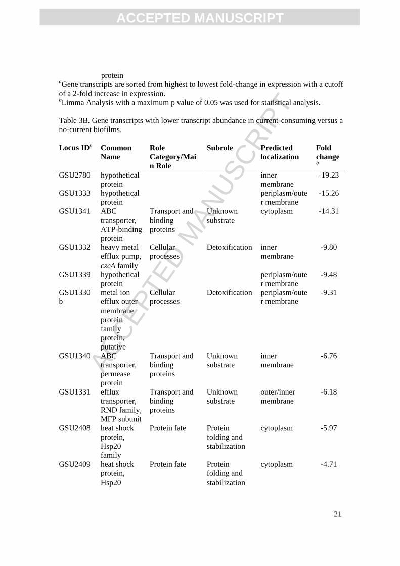
protein aGene transcripts are sorted from highest to lowest fold-change in expression with a cutoff of a 2-fold increase in expression. bLimma Analysis with a maximum p value of 0.05 was used for statistical analysis. Table 3B. Gene transcripts with lower transcript abundance in current-consuming versus a no-current biofilms. Locus IDa Common
Name Role Category/Main Role
Subrole Predicted localization
Fold changeb
GSU2780 hypothetical protein
inner membrane
-19.23
GSU1333 hypothetical protein
periplasm/outer membrane
-15.26
GSU1341 ABC transporter, ATP-binding protein
Transport and binding proteins
Unknown substrate
cytoplasm -14.31
GSU1332 heavy metal efflux pump, czcA family
Cellular processes
Detoxification inner membrane
-9.80
GSU1339 hypothetical protein
periplasm/outer membrane
-9.48
GSU1330b
metal ion efflux outer membrane protein family protein, putative
Cellular processes
Detoxification periplasm/outer membrane
-9.31
GSU1340 ABC transporter, permease protein
Transport and binding proteins
Unknown substrate
inner membrane
-6.76
GSU1331 efflux transporter, RND family, MFP subunit
Transport and binding proteins
Unknown substrate
outer/inner membrane
-6.18
GSU2408 heat shock protein, Hsp20 family
Protein fate Protein folding and stabilization
cytoplasm -5.97
GSU2409 heat shock protein, Hsp20
Protein fate Protein folding and stabilization
cytoplasm -4.71

ACC
EPTE
D M
ANU
SCR
IPT
ACCEPTED MANUSCRIPT
22
family
GSU3410 hypothetical protein
inner membrane
-4.59
GSU2410 heat shock protein, Hsp20 family
Protein fate Protein folding and stabilization
cytoplasm -4.45
GSU0538 heat shock protein, Hsp20 family
Protein fate Protein folding and stabilization
cytoplasm -3.90
GSU2406 dnaJ domain protein
Unknown function
General cytoplasm -3.78
GSU0658 clpB, ClpB protein
Protein fate Degradation of proteins, peptides, and glycopeptides
cytoplasm -3.77
GSU2503 omcT, cytochrome c family protein
Energy metabolism
Electron transport
periplasm/outer membrane
-3.38
GSU3409 hypothetical protein
inner membrane
-3.11
GSU2938 hypothetical protein
periplasm/outer membrane
-3.06
GSU2407 hypothetical protein
cytoplasm -3.03
GSU0033 dnaK, chaperone protein dnaK
Protein fate Protein folding and stabilization
cytoplasm -2.99
GSU1018 hypothetical protein
periplasm/outer membrane
-2.65
GSU1337 hypothetical protein
outer/inner membrane
-2.46
GSU0469 hypothetical protein
periplasm/outer membrane
-2.46
GSU2504 omcS, cytochrome c family protein
Energy metabolism
Electron transport
periplasm/outer membrane
-2.31
GSU0364 ppcB, cytochrome c3
Energy metabolism
Electron transport
periplasm/outer membrane
-2.26
GSU0919 hypothetical protein
outer/inner membrane
-2.23

ACC
EPTE
D M
ANU
SCR
IPT
ACCEPTED MANUSCRIPT
23
GSU2751 dcuB, C4-dicarboxylate transporter, anaerobic
Transport and binding proteins
Carbohydrates, organic alcohols, and acids
inner membrane
-2.21
aGene transcripts are sorted from highest to lowest fold-change in expression with a cutoff of a 2-fold increase in expression. bLimma Analysis with a maximum p value of 0.05 was used for statistical analysis. Table 4A. Gene transcripts with higher transcript abundance current-consuming versus current-producing biofilms. Locus ID a
Common Name
Role Category/Main Role
Subrole Predicted localization
Fold changeb
GSU3274
cytochrome c family protein, putative
Energy metabolism
Electron transport
periplasm 6.76
GSU0216
conserved hypothetical protein
Hypothetical proteins
Conserved cytoplasm 5.84
GSU1395
hypothetical protein
periplasm/outer membrane
5.81
GSU3272
hypothetical protein
cytoplasm 5.29
GSU1906
leuA, 2-isopropylmalate synthase
Amino acid biosynthesis
Pyruvate family
cytoplasm 5.26
GSU1079
hypothetical protein
inner membrane
5.12
GSU3273
hypothetical protein
cytoplasm 4.71
GSU3271
hypothetical protein
periplasm/outer membrane
3.72
GSU1401
dnaE, DNA polymerase III, alpha subunit
DNA metabolism
DNA replication, recombination, and repair
cytoplasm 3.47
GSU0317
conserved hypothetical protein
Hypothetical proteins
Conserved periplasm/outer membrane
3.10
GSU0542
GGDEF domain protein
Unknown function
General cytoplasm 3.01
GSU2515
cytochrome c family protein,
Energy metabolism
Electron transport
periplasm/outer membrane
2.92

ACC
EPTE
D M
ANU
SCR
IPT
ACCEPTED MANUSCRIPT
24
putative
GSU1709
smpB, SsrA-binding protein
Protein synthesis
Other cytoplasm 2.71
GSU0848
ferredoxin family protein, putative
Energy metabolism
Electron transport
cytoplasm 2.67
GSU1158
sodA, superoxide dismutase
Cellular processes
Detoxification cytoplasm 2.67
GSU0534
Rrf2 family protein
Unknown function
General cytoplasm 2.61
GSU2750
conserved domain protein
Hypothetical proteins
Domain cytoplasm 2.57
GSU0089
heterodisulfide reductase subunit
Energy metabolism
Electron transport
periplasm/outer membrane
2.36
GSU1877
oxidoreductase, 2-nitropropane dioxygenase family
Unknown function
Enzymes of unknown specificity
inner membrane
2.35
GSU1024
ppcD, cytochrome c3
Energy metabolism
Electron transport
periplasm/outer membrane
2.35
GSU0971
peptidyl-tRNA hydrolase domain protein
Unknown function
General cytoplasm 2.35
GSU1857
hypothetical protein
periplasm/outer membrane
2.34
GSU2751
dcuB, C4-dicarboxylate transporter, anaerobic
Transport and binding proteins
Carbohydrates, organic alcohols, and acids
inner membrane
2.23
GSU3268
feoB-2, ferrous iron transport protein B
Transport and binding proteins
Cations inner membrane
2.17
GSU1118
universal stress protein family
Cellular processes
Adaptations to atypical conditions
cytoplasm 2.10
GSU1398
SCO1/SenC family protein
Unknown function
General inner membrane
2.09
GSU2828
GGDEF domain protein
Unknown function
General inner membrane
2.09
GSU2507
sensor histidine kinase
Signal transduction
Two-component systems
inner membrane
2.04

ACC
EPTE
D M
ANU
SCR
IPT
ACCEPTED MANUSCRIPT
25
aGene transcripts are sorted from highest to lowest fold-change in expression with a cutoff of a 2-fold increase in expression. bLimma Analysis with a maximum p value of 0.05 was used for statistical analysis. Table 4B. Genes transcripts with lower transcript abundance in current-consuming versus current-producing biofilms. Locus ID a
Common Name
Role Category/Main Role
Subrole Predicted localization
Fold changeb
GSU0469
hypothetical protein
periplasm/outer membrane
-11.25
GSU2076
omcZ, cytochrome c family protein
Energy metabolism
Electron transport outer membrane
-10.31
GSU0975
phage tail sheath protein, putative
Other categories
Prophage functions
inner membrane
-8.29
GSU1497
hypothetical protein
pilA operon periplasm/outer membrane
-7.64
GSU2737
omcB, polyheme membrane-associated cytochrome c
Energy metabolism
Anaerobic outer membrane
-7.56
GSU0976
conserved hypothetical protein
Hypothetical proteins
Conserved cytoplasm -5.13
GSU2780
hypothetical protein
inner membrane
-5.07
GSU2887
cytochrome c family protein
Energy metabolism
Electron transport periplasm/outer membrane
-4.65
GSU2897
hypothetical protein
n/a, sequence too short
-4.47
GSU1496
pilA, pilin domain protein
Unknown function
General inner membrane
-4.37
GSU2640
hypothetical protein
inner membrane
-3.96
GSU2731
omcC, polyheme membrane-
Energy metabolism
Anaerobic outer/inner membrane
-3.86

ACC
EPTE
D M
ANU
SCR
IPT
ACCEPTED MANUSCRIPT
26
associated cytochrome c
GSU2739
orf1-1, hypothetical protein
omcB operon periplasm/outer membrane
-3.77
GSU1538
methylamine utilization protein MauG, putative
Energy metabolism
Amino acids and amines
periplasm/outer membrane
-3.71
GSU0974
hypothetical protein
cytoplasm -3.63
GSU2075
subtilisin Protein fate Degradation of proteins, peptides, and glycopeptides
periplasm/outer membrane
-3.56
GSU0983
conserved hypothetical protein
Hypothetical proteins
Conserved cytoplasm -3.35
GSU0468
hypothetical protein
periplasm/outer membrane
-3.27
GSU1341
ABC transporter, ATP-binding protein
Transport and binding proteins
Unknown substrate
cytoplasm -3.24
GSU2733
orf1-2, hypothetical protein
omcC operon periplasm/outer membrane
-3.11
GSU1333
hypothetical protein
periplasm/outer membrane
-2.89
GSU0618
omcE, cytochrome c family protein
Energy metabolism
Electron transport periplasm/outer membrane
-2.79
GSU0591
cytochrome c family protein
Energy metabolism
Electron transport inner membrane
-2.73
GSU0979
conserved hypothetical protein
Hypothetical proteins
Conserved cytoplasm -2.66
GSU0590
hypothetical protein
inner membrane
-2.63
GSU0988
conserved hypothetical protein
Hypothetical proteins
Conserved cytoplasm -2.55

ACC
EPTE
D M
ANU
SCR
IPT
ACCEPTED MANUSCRIPT
27
GSU1337
hypothetical protein
outer/inner membrane
GSU0972
ATPase, AAA family
Unknown function
General cytoplasm -2.44
GSU1339
hypothetical protein
periplasm/outer membrane
-2.44
GSU0986
tail lysozyme, putative
Other categories
Prophage functions
cytoplasm -2.37
GSU2074
PPIC-type PPIASE domain protein
Unknown function
General periplasm/outer membrane
-2.25
GSU2732
orf2-2, cytochrome c family protein
Energy metabolism
Electron transport, omcC operon
periplasm/outer membrane
-2.16
GSU2808
cytochrome c family protein
Energy metabolism
Electron transport periplasm/outer membrane
-2.11
GSU2912
cytochrome c family protein
Energy metabolism
Electron transport periplasm/outer membrane
-2.09
GSU0991
glycosyl transferase, group 1 family protein
Cell envelope Biosynthesis and degradation of surface polysaccharides and lipopolysaccharides
inner membrane
-2.08
GSU1099
pstS, phosphate ABC transporter, periplasmic phosphate-binding protein
Transport and binding proteins
Anions periplasm/outer membrane
-2.01
aGene transcripts are sorted from highest to lowest fold-change in expression with a cutoff of a 2-fold increase in expression. bLimma Analysis with a maximum p value of 0.05 was used for statistical analysis.