gerhard scholtz . gregory d. edgecombe the evolution of
TRANSCRIPT

Dev Genes Evol (2006) 216: 395–415DOI 10.1007/s00427-006-0085-4
REVIEW
Gerhard Scholtz . Gregory D. Edgecombe
The evolution of arthropod heads: reconciling morphological,
developmental and palaeontological evidence
Received: 7 December 2005 / Accepted: 1 May 2006 / Published online: 28 June 2006# Springer-Verlag 2006
Abstract Understanding the head is one of the greatchallenges in the fields of comparative anatomy, develop-mental biology, and palaeontology of arthropods. Numer-ous conflicting views and interpretations are based on anenormous variety of descriptive and experimental ap-proaches. The interpretation of the head influences viewson phylogenetic relationships within the Arthropoda aswell as outgroup relationships. Here, we review currenthypotheses about head segmentation and the nature of headstructures from various perspectives, which we try tocombine to gain a deeper understanding of the arthropodhead. Though discussion about arthropod heads showssome progress, unquestioned concepts (e.g., a presegmen-tal acron) are still a source of bias. Several interpretationsare no longer tenable based on recent results fromcomparative molecular developmental studies, improvedmorphological investigations, and new fossils. Current dataindicate that the anterior arthropod head comprises threeelements: the protocerebral/ocular region, the deutocereb-ral/antennal/cheliceral segment, and the tritocerebral/ped-ipalpal/second antennal/intercalary segment. The labrumand the mouth are part of the protocerebral/ocular region.Whether the labrum derives from a former pair of limbs
remains an open question, but a majority of data support itsbroad homology across the Euarthropoda. From thealignment of head segments between onychophorans andeuarthropods, we develop the concept of “primary” and“secondary antennae” in Recent and fossil arthropods,posit that “primary antennae” are retained in some fossileuarthropods below the crown group level, and proposethat Trilobita are stem lineage representatives of theMandibulata.
Keywords Segmentation . Labrum . Primary antennae .Secondary antennae . Trilobita . Cambrian
Introduction
For more than a century, the problems of the number andnature of segments and other elements constituting thearthropod head have been a hotly debated issue (e.g.,Goodrich 1897; Weber 1952; Siewing 1963; Rempel 1975;Weygoldt 1985; Scholtz 1995; Scholtz and Edgecombe2005). Models, theories, and hypothesis building havebecome an intellectual challenge unmatched by otherproblems of arthropod morphology. Numerous articles dealwith head segmentation and even national schools ortraditions of views on arthropod heads evolved. Accord-ingly, we find typical French (Chaudonneret 1987;Casanova 1996), German (Heymons 1901; Siewing1969, Paulus and Weygoldt 1996) or Swedish (Holmgren1916; Hanström 1928; Dahl 1956) heads (see Rempel1975) with different segmental compositions which were,and sometimes still are, discussed in their respectivetraditional environment. The views on heads influencedreconstructions of arthropod phylogenetic relationshipsand our general views of arthropod evolution. This debateon heads finds an interesting parallel in the discussionabout the vertebrate head where similar problems ofsegment numbers and nature and transformations of partsoccur (e.g., Starck 1963; Kuratani 2003; Northcutt 2005;Olsson et al. 2005). Embryologists, anatomists, palaeon-tologists, and molecular developmental biologists are
Communicated by guest editors Jean Deutsch and Gerhard Scholtz
Electronic Supplementary Material Supplementary material isavailable for this article at http://dx.doi.org/10.1007/s00427-006-0085-4 and is accessible for authorized users.
G. Scholtz (*)Humboldt-Universität zu Berlin, Institut für Biologie/Vergleichende Zoologie,Philippstr. 13,10115 Berlin, Germanye-mail: [email protected].: +49-30-20936005Fax: +49-30-20936002
G. D. EdgecombeAustralian Museum,6 College Street,Sydney, NSW, 2010, Australiae-mail: [email protected]

concerned by this problem, and in particular, the latter twogroups revived the controversy in recent times, based onnew fossil finds and new comparative molecular tech-niques (Schmidt-Ott et al. 1994a,b; Rogers and Kaufman1997; Budd 2002, Urbach and Technau 2003a, Waloszek etal. 2005).
Why is this debate so long lasting and so vivid? What isso important about heads? In particular, the answer to thelatter question is self-evident, if one thinks even cursorilyabout the meaning of a head. Furthermore, cephalizationmight be a clue to understand the evolutionary success ofarthropods. Given this long-standing concerted multi-national, multidisciplinary effort, the question arises,“Why has this discussion not come to an end or to asatisfying solution?” There are at least two reasons for this.On the one hand, the discussion is sometimes hampered byconcepts based on assumed phylogenetic relationships orevolutionary scenarios that bias the interpretation ofstructures. On the other hand, there are real problems inthe interpretation of certain structures in the head becausewe can rightly assume that the needs to form a head and abrain led to evolutionary transformations of structures to adegree that makes homologization very problematic. Thiseffect is even amplified by the approach of someresearchers dealing with the head problem, who do notintegrate the available evidence but rely excessively upon arestricted set of data. In contrast, we want to propose a kindof “total evidence approach” to the arthropod headproblem. The organization, combination, and interpretationof the diverse kinds of data from morphology, develop-mental biology, molecular biology, phylogenetics, andpalaeontology form the intellectual challenge mentionedby Snodgrass (1960) because little tracks and traces have tobe combined and interpreted as signs of a former structureand process. This is a kind of puzzle that resembles theindices that are used by a detective to reconstruct the courseof a crime. Like all researchers struggling with thearthropod head problem, we are more or less “headdetectives.”
The phylogenetic framework: Articulata vs Ecdysozoa,Mandibulata vs Paradoxopoda
To be meaningful, the discussion of the arthropod headhas to be conducted with a combination of comparativedevelopmental and phylogenetic perspectives. The de-velopmental analysis of the head of only one modelorganism might lead to inconclusive solutions andhypotheses (see Page 2004; replies by Harzsch 2004;Scholtz and Edgecombe 2005). Hence, before we enterthe discussion about arthropod heads, we have to set outthe phylogenetic framework that forms the backgroundfor our considerations about head evolution. Arthropodphylogeny is a highly controversial field (Richter andWirkner 2004) but not all differences influence our viewson head evolution and organization. Two controversiallevels of arthropod phylogeny seem most important to thefollowing discussion: one is the question of the arthropod
sister group, the other concerns the phylogenetic relation-ships between the major euarthropod taxa.
Currently, there are two competing hypothesis for theplacement of the arthropods within the Metazoa. One is themore traditional Articulata hypothesis, which favors a closerelationship between Annelida and Arthropoda (Wägeleand Misof 2001; Scholtz 2002; Jenner and Scholtz 2005).The other alternative is known as the Ecdysozoa hypothesis(Aguinaldo et al. 1997; Giribet 2003). According to thisview, the Arthropoda are most closely related to thecycloneuralians, which include the Nematoida (nematodesand nematomorphs) and Scalidophora (Schmidt-Rhaesa etal. 1998). As the two hypotheses have a different impact onour understanding of what segmentation is and how it hasevolved (Scholtz 2002, 2003), they are relevant to thediscussion of head segmentation, as we will see below.
Based on molecular datasets, the monophyly of theMandibulata, comprising myriapods, crustaceans, andhexapods, has been challenged by placing the Myriapodaas the sister group of the Chelicerata (Cook et al. 2001;Hwang et al. 2001; Mallatt et al. 2004; Negrisolo et al.2004). This grouping has been named Paradoxopoda(Mallatt et al. 2004) or Myriochelata (Pisani et al. 2004).However, there is no convincing morphological support infavor of a sister group relationship between Chelicerata andMyriapoda. Only some characteristics of neurogenesis(Dove and Stollewerk 2003; Kadner and Stollewerk 2004)are shared between myriapods and chelicerates, and even ifone were to discount mandibulate synapomorphies, themyriapod–chelicerate characters are as plausibly inter-preted as symplesiomorphies. In contrast, there is ampleevidence from development and morphology that supportsthe Mandibulata as a monophyletic group (see Edgecombe2004; Harzsch et al. 2005a; Scholtz and Edgecombe 2005).Furthermore, some phylogenetic studies based on molec-ular data and combined datasets support Mandibulata aswell (Edgecombe et al. 2000; Giribet et al. 2001, 2005;Kusche et al. 2003). Accordingly, the Mandibulata conceptis used here for the discussion of head evolution andtrilobite relationships.
The acron as an example for concept-based approachesto head segmentation
In most of the literature on arthropod heads, the authorsassume the existence of an acron as the anteriormost non-segmental body part (Siewing 1969; Rempel 1975; Cohenand Jürgens 1991; Scholtz 1997). According to this view,the acron contains the ancestral brain (archicerebrum,supraesophageal ganglion, cerebral ganglion) which isinherited from the bilaterian stem species and which can befound plesiomorphically in animal groups such as Platy-helminthes, Nematoda, Mollusca, and Annelida (Siewing1969; Lauterbach 1973). However, the existence of anacron has never been directly shown but rather it is aninference based on the assumption of a close relationshipbetween annelids and arthropods, the Articulata hypothesis(Scholtz 2002). In the scenario based on this assumption,
396

the acron is homologized with the episphere of thetrochophore larva and the prostomium of adult annelidworms (Fig. 1) (Nielsen 2001). The episphere is the regionbearing the larval brain and the apical organ and liesanterior to the mouth. Its posterior boundary is marked bythe first ciliary ring, the prototroch, of the trochophore(Fig. 1). As the episphere/prostomium contains only little(if any) mesoderm without coelomic sacs and nephridia,and as it is not formed by the ectodermal/mesodermalgrowth zone but derives from the micromeres (seeAckermann et al. 2005), it is considered to be asegmentalin nature (e.g., Siewing 1969; Nielsen 2001). Duringdevelopment, the episphere becomes the prostomium(Hatschek 1878; Woltereck 1905; Nielsen 2005a,b),which lies anterior to the mouth and which bears theannelid brain (cerebral ganglion) and eyes (if present), andin some polychaetes, appendage-like sensory structuressuch as the antennae and palps (Bartolomaeus et al. 2005).As arthropods do not possess locomotory cilia, there is noprototroch in larvae or embryos, and accordingly, anepisphere is not directly definable by structural criteria(Fig. 1). Mostly, the fact that the anteriormost region ofarthropod embryos bears the eyes and the anterior part ofthe brain and lies in front of the mouth is taken as evidenceto homologize this structure with the annelid episphere/prostomium. However, the position of the mouth inarthropod embryos is a problem in itself as there is anobvious migration posteriorly (e.g. Ungerer and Wolff2005). Accordingly, even a position of the putative acronalstructures anterior to the mouth cannot be unambiguouslydetermined.
The underlying concept of segmentation forms a biasfor the interpretation of the head
If one accepts the Ecdysozoa hypothesis (Giribet 2003), theconcept of an acron may no longer be necessary (see Budd2002) and one can assume homonomous segments to haveoccurred from the anterior to the posterior region in thestem lineage of arthropods. We remain doubtful becausethe terminal regions in bilaterians are something special bybearing the mouth (not always) and the anus (if present), aswell as by being terminal, i.e., many cells have no anterioror posterior neighbours. In addition, the anterior terminalregion is characterized by the possession of a concentrateddorsal or preoral nerve plexus or brain (Fig. 2). Theseterminal regions including an anterior dorsal brain werepresent before segmentation evolved, e.g., in Platyhelmin-thes, Nematoda or Mollusca, but the transition to the trunkis not well defined. The difference between the unseg-mented and segmented conditions is that, in the latter, thereare developmental and morphological boundaries betweenthe terminal regions and the adjacent segments (Fig. 2).Hence, only after segmentation had occurred did theanterior body region become morphologically distinct andseparated from the subsequent segments. This is a mean-ingful assumption even if one believes in a segmented stemspecies of the Bilateria, often called “Urbilateria” (e.g.,Prud’homme et al. 2003) because neither cnidarians norctenophores are segmented, and accordingly, segmentationmust have evolved in the stem lineage leading to the crowngroup Bilateria. The evolutionary scenario of a specifiedterminal region before segmentation had evolved isreflected in the expression of the anterior homeoboxgenes of the orthodenticle (otd/Otx) group. Expression ofotd-/Otx-related genes has been described in variousunsegmented and segmented bilaterians (e.g., Hirth et al.1995; Li et al. 1996; Bruce and Shankland 1998; Tomsaand Langeland 1999; Umesono et al. 1999; Harada et al.2000; Arendt et al. 2001; Nederbragt et al. 2002; Urbachand Technau 2003b). In all cases, the expression is relatedto the brain, and accordingly, it is found in the anteriorregion (but not in the extreme anterior area) of the animalsstudied. For instance, in the trochophore larvae of themollusc, Patella vulgata, and the polychaete annelid,Platynereis dumerilii, otd/Otx is expressed in the posteriorregion of the episphere, in brain nerve cells, and in themouth area (Arendt et al. 2001; Nederbragt et al. 2002).Likewise, in the leech, Helobdella triserialis, which lacks atrochophore larva, the expression is found in theprostomium, the cerebral ganglion, and the mouth region(Bruce and Shankland 1998). From several studies, it isobvious that the otd/Otx expression in hexapod arthropodsis mainly found in the ocular/protocerebral region (butagain not in the anteriormost area and not in the labrum),and to a minor extent, in the deutocerebral segment (Hirthet al. 1995; Li et al. 1996; Urbach and Technau 2003b).This similarity in the expression patterns between arthro-pods and other bilaterian taxa may be interpreted as anindication that the arthropod protocerebrum and theassociated body unit corresponds to the anterior terminal
Fig. 1 The concept of the acron. Middle: a schematic representation(lateral view) of an annelid trochophora larva with the episphere(ep) as the anterior region in front the mouth and the first ciliary ring(prototroch). The blue lines indicate the suggested homology andtransformation of the larval episphere (the prostomium in the adultworm) and the acron in arthropods according to the Articulatahypothesis. Right: a crustacean germ band (ventral view) with nodirect evidence for a homologue of the episphere (?) (modified afterOishi 1959). The head lobes (hl) are interpreted as the acron whichis the equivalent of the episphere/prostomium of annelids. Left: inpolychaete worms (ventral view) with a more direct development,head lobes (hl) are formed similar to those of arthropods (modifiedafter von Wistinghausen 1891). However, the homology to theepisphere is beyond doubt (!) (see von Wistinghausen 1891; Seaveret al. 2005; Seaver and Kaneshige 2006). This shows that theinterpretation of the arthropod head lobes and acron as equivalent tothe episphere/prostomium might be appropriate and could be takenas additional evidence in favor of the Articulata hypothesis (Scholtz2002, 2003)
397

region of other Bilateria, and the boundary between theocular/protocerebral region and the segment of the deuto-cerebrum corresponds to the posterior region of theancestral bilaterian brain, supporting the views of Siewing(1969) and Lauterbach (1973) (Fig. 2). Consequently, thisidea implies that the dorsal cerebral ganglion of arthropodsis not a serial homologue of the ventral segmental ganglia.Taken together, these ontogenetic and phylogenetic aspectsindicate the special conditions governing the anteriorterminal region—and accordingly, a special term such as“acron” might be appropriate irrespective of the Articulataor Ecdysozoa perspectives of arthropod origins. Interest-ingly, in the segmented annelids and arthropods, thisboundary seems to be established using a similar mech-anism to the differentiation of undisputed segmental bound-aries, namely, the action and interaction of segment polaritygenes engrailed (en) and wingless (wg) (Schmidt-Ott andTechnau 1992; Scholtz 1995; Prud’homme et al. 2003).
In summary on the one hand, the correspondence in theexpression of otd/Otx, the dorsal brain and other peculiar-ities of the anterior region between arthropods and otherbilaterians can be taken as evidence for an acron inarthropods, whereas, on the other hand, depending on thedefinition of a segment (Scholtz 2002; Seaver 2003;Minelli and Fusco 2004; Tautz 2004), it can be seen as theanteriormost (or an anterior) segment, the ocular segment
in arthropods (see Schmidt-Ott et al. 1994b; Rogers andKaufman 1997). Hence, this terminal region is called an“acron,”mainly for phylogenetic reasons, and a “segment,”based on the fact that its posterior boundary is formed likethat of segments in more posterior body regions (Rogersand Kaufman 1997). The latter approach reveals theproblems of a reductionist view or definition of segmen-tation based on molecular gene expression alone becausethis neglects the structural characteristics of the variousbody regions. To avoid this ambiguity, we adhere to theterm “ocular/protocerebral region” coined by Scholtz(1995).
Traditionally, the occurrence of sometimes only transi-tory coelomic sacs, appendages or their rudiments, gangliaand nerves, and nephridia and their derivatives, either invarious combinations or alone, have been taken asevidence for segments or their vestigial appearance.Nowadays, we have, in addition, gene expression datafor the specification of segments, and these new datashould be seen in concert with the morphogenetic andmorphological characteristics of segments (Scholtz 2002).According to the Articulata hypothesis, the whole set ofsegment characteristics was, more or less, present in thearthropod lineage right from the beginning. In the contextof the Ecdysozoa hypothesis, this is not necessarily thecase. Irrespective of this, it seems plausible that segmen-tation did not evolve as a whole complex at once but ratherby a stepwise inclusion of characters repeated along thebody axis (Scholtz 2003). Accordingly, the full comple-ment of segmental components may never have beenpresent in all metameric units. Furthermore, some meta-meric structures such as limbs might have undergonespecialization before the full segmentation complex wasachieved.
Given that the terminal body regions already showedsome peculiarities before the evolutionary advent ofsegmentation, they are privileged for various specializa-tions older than segmentation. This may be true for thefollowing adjacent body parts as well. In other words, weprobably pose the wrong questions when we assume thatevery unit in the head is derived from a complete formersegment and that the segmental structures were alike inevery respect. For instance, the anterior appendages such asantennae might have been derived from leg-like append-ages before the hard segmented exoskeleton and thearticulated arthropodia of euarthropods have evolved (seeMinelli 2003). Furthermore, it is likely that the cerebralganglion was a distinct structure before segmentationoccurred in evolution. Accordingly, the brain was never aserial homologue of a segmental ganglion. The model of astrictly homonomous segmentation as the evolutionarystarting point seems clearly wrong, and there is no exampleof an arthropod, be it Recent or fossil, crown group or stemlineage, showing homonomous segmentation throughout.The last but not the least, the interpretation of headstructures is complicated by the problem that the embry-onic anlage of a structure or character does not necessarilymean that the adult structure was present in the ancestor(Scholtz 2004, 2005).
Fig. 2 Terminal regions were specific and differentiated beforesegmentation evolved. Anterior to the left, yellow: CNS, green:digestive system. a Depicts a hypothetical unsegmented bilaterianwith specified and differentiated terminal regions. These terminalregions are characterized by specializations of the nervous system(dorsal brain, terminal ganglion), photoreceptor, sensory appen-dages, and gene expressions such as otd/otx in the anterior region(red line) and caudal (e.g. de Rosa et al. 2005) in the posteriorregion (blue line). b Shows a segmented bilaterian which evolvedfrom an ancestor as shown in a. The segmentation overlies theancestral differentiation of the terminal regions. The question ariseswhether the terminal regions and their characteristics are seriallyhomologous to segments in the middle of the body
398

The current consensus: a tripartite anterior brain/head
The use of genetic markers, in particular, segment-polaritygenes such as en and wg, led to a high resolution of headsegmentation in the major part of the head. These data areavailable for a number of chelicerates, crustaceans,myriapods, and hexapods (Fleig 1994; Scholtz 1995;Rogers and Kaufman 1997; Telford and Thomas 1998;Damen 2002; Hughes and Kaufman 2002; Chipman et al.2004; Janssen et al. 2004). Other segment polarity geneexpression data such as paxIII and hedgehog (hh) areavailable for chelicerates, myriapods, hexapods, crusta-ceans, albeit to a lesser extent (Simonnet et al. 2004; Daviset al. 2005; Osborne and Dearden 2005). All these resultspoint in the same direction. There is a tripartite anteriorbrain (comprising the protocerebrum, the deutocerebrum,and the tritocerebrum) and three anterior morphologicalunits. For the Mandibulata these are: the ocular/protoce-rebral region, and the segments of the first and secondantennae in crustaceans and the antennal and intercalarysegments in myriapods and hexapods (Fig. 3). There is nocorresponding cephalization at the external morphologicallevel in the Chelicerata (see below). However, the closeassociation of the tritocerebrum with anterior brain parts(Hanström 1928; Mittmann and Scholtz 2003; Harzsch etal. 2005b) shows that a tripartite brain is present as in
Mandibulata. In the latter group, the degree of fusion of thetritocerebrum to the more anterior brain regions varies, butin all cases, its status as a brain neuromere is evident (e.g.,Hanström 1928; Harzsch 2004).
There is no indication for an additional preantennalsegment between the ocular/protocerebral region and theantennal segment (see Heymons 1901; Siewing 1969;Lauterbach 1973). This pattern is consistent throughout theinvestigated species without exception. The only compli-cation is the labral expression of some of these genes insome taxa (Schmidt-Ott and Technau 1992; Schmidt-Ott etal. 1994a; Urbach and Technau 2003b, see below).Posterior to the second antennae/intercalary segment, wefind segment polarity gene stripes in the mandibular andthe maxillary segments as in trunk segments. There is noindication of an additional segment between the mandib-ular and the second antennae/intercalary segments aspostulated by Chaudonneret (1987). The question as towhether the ocular/protocerebral stripe indicates a trueocular segment or an acron seems not so important, rather,the neutral questions are whether this is the anteriormostbody region which can be derived from the ancestral brainregion of Bilateria and whether it represents only one unitor more than one (see above).
Fig. 3 Alignment of structures in the heads of Recent arthropods(see Fig. 6). a Onychophora with “primary antennae” (pa) in theocular/protocerebral region (blue). b Chelicerata with chelicerae inthe deutocerebral segment (red) c) Mandibulata with “secondaryantennae” (sa) in the deutocerebral segment (red). The left sideshows the crustacean conditions with two pairs of antennae. Theright side shows the situation in myriapods and hexapods with alimbless intercalary tritocerebral segment. The ocular/protocerebralregion is shown in blue, the deutocerebral segment in red, thetritocerebral segment in green. The structures of the central nervoussystem are shaded yellow with black connections. The protocere-brum contains the mushroom bodies and the central body. In
Mandibulata, the deutocerebrum shows the olfactory lobe (blackspot). The stomatogastric and labral nerves are depicted as a loopanterior to the mouth. In Onychophora and Chelicerata, they areconnected to the deutocerebrum, in Mandibulata to the tritocere-brum. The mouth is depicted in blue to show its putative associationwith the ocular/protocerebral region. For the sake of segmentalignment and a clearer picture, we generally put the tritocerebrumin a postoral position (the putative plesiomorphic condition as isfound in Onychophora), although in most euarthropods, it occupiesa preoral or paroral position. The double line in the Mandibulatamarks the posterior margin of the head
399

The development of the labrum
A highly controversial head structure
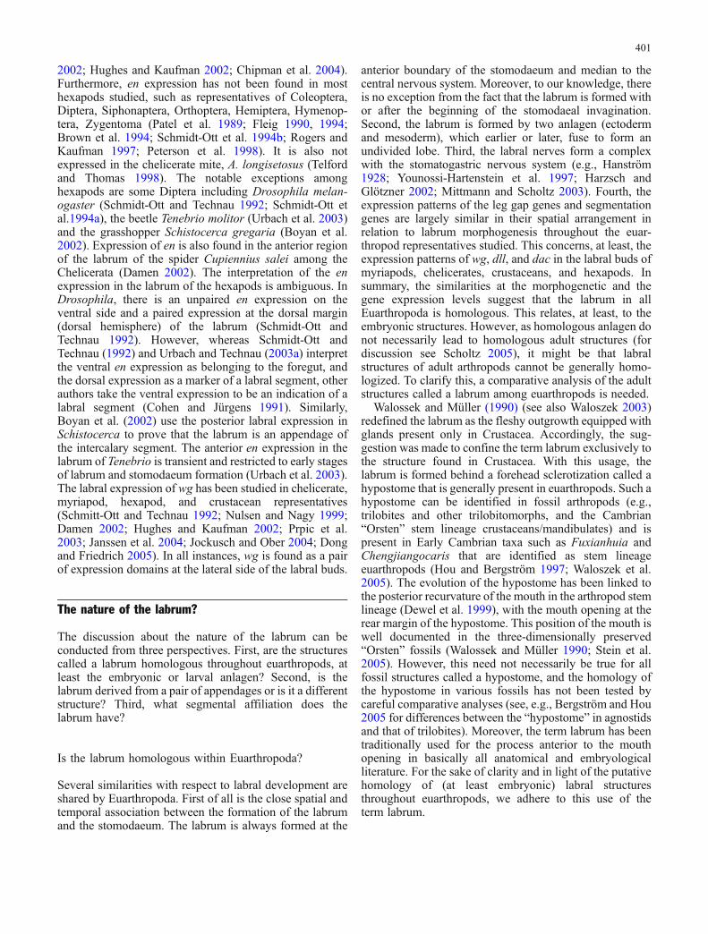
The labrum alwayswas (Weber 1952; Rempel 1975; Scholtz1997, 2001) and still is the most controversial structure ofthe euarthropod head, and even the new molecular andmorphological methods have not led to a conclusive answerabout its nature. The interpretations of the labrum differconcerning its segmental affiliation ranging from theanteriormost segment via a preantennal segment to thetritocerebral/ intercalary segment and the question whetherit represents a derived limb pair, a simple outgrowth (upperlip), a segment, or the anterior body terminus. Even thehomology of the labrum among the Euarthropoda has beenquestioned (Walossek and Müller 1990). In the following,we describe the general characteristics of the labrum interms of morphogenesis and gene expression.
Morphogenesis
The euarthropod labrum is formed at the anterior margin ofthe stomodaeum with or after the beginning of thestomodaeal invagination. In many representatives ofchelicerates, myriapods, crustaceans, and hexapods theearly anlage of the labrum is bilobed to different degrees(e.g., Brauer 1895; Scholl 1963, 1969; Dohle 1964;Ullmann 1964; Bruckmoser 1965; Pross 1966; Hertzel1984; Schoppmeier and Damen 2001; Simonnet et al.2004; Abzhanov and Kaufman 2004; Ungerer and Wolff2005) (see Fig. 4d). Even in pycnogonids with theirextended proboscis, a transient bilobed labral anlage occurswhich grows out to form the dorso-lateral part of theproboscis (Winter 1980). In all arthropods with an earlybilobed labrum anlage, these two lobes fuse to form anunpaired outgrowth anterior to the mouth, later on. Incontrast to this, there are also cases for a single undividedlabrum anlage. Examples are some malacostracan and non-malacostracan crustaceans (Manton 1928; Benesch 1969;Olesen et al. 2001; Alwes and Scholtz 2006; Olesen 2004),hexapods (Rohrschneider 1968), myriapods (Heymons1901; Tiegs 1940), and chelicerates (Scholl 1977; Thomasand Telford 1999).
As the investigations on the expression of the Dll geneshow (see below), in cases such as the mite Archegozeteslongisetosus, and the horseshoe crab Limulus polyphemus,there are two separate early gene expression areas whichlater fuse before the undivided labrum buds out (Thomasand Telford 1999; Mittmann and Scholtz 2001). Further-more, ablation experiments conducted by Haget (1955) inthe coleopteran Leptinotarsa decemlineata, reveal thateven in this species with an undivided labral lobe, this lobeoriginates from two independent anlagen (Haget 1955),results which were confirmed by Wada (1965) in thegrasshopper Tachycines asynamorus. The mesoderm of thelabrum is always formed by a pair of cell massesirrespective of whether the labral bud is bilobed (Ullmann1964) or undivided (Heymons 1901; Tiegs 1940;
Rohrschneider 1968). All this suggests that in general thelabral bud derives from two separated anlagen. Accord-ingly, Scholtz (1997) suggested that a bilobed labrumanlage might be a euarthropod apomorphy.
The mesoderm of the labral region of euarthropods formsthe labral, pharyngeal, and stomodaeal musculature, andsometimes, the anterior aorta (Scholl 1963, 1969, 1977;Dohle 1964; Benesch 1969; Siewing 1969; Anderson1973; de Velasco et al. 2006). Sometimes a pair of transientcoelomic cavities is formed (e.g., Tiegs 1940; Ullmann1964; Rohrschneider 1968; Siewing 1969), but in mostcases, the mesodermal masses stay compact (Scholl 1963,1969; de Velasco et al. 2006). The mesoderm is sometimesrestricted to the labrum itself, but there are also examples ofa large mesoderm area that just shows processes (coelomicor massive) reaching into the labrum (Scholl 1963, 1969,1977; Pross 1966; Rohrschneider 1968; Siewing 1969).
Gene expression
Data are available for the expression of two classes ofgenes in the labrum, appendage gap genes, namely, Distal-less (Dll), dachshund (dac), and extradenticle (exd) andsegment polarity genes, engrailed (en) and wingless (wg).The most conspicuous gene expression in the labrum is thatof Dll, which is found in all chelicerates (Popadic et al.1998; Thomas and Telford 1999; Mittmann and Scholtz2001; Schoppmeier and Damen 2001), myriapods (Scholtzet al. 1998; Prpic and Tautz 2003), crustaceans (e.g.,Panganiban et al. 1995; Scholtz et al. 1998; Shiga et al.2002; Olesen et al. 2001; Abzhanov and Kaufman 2004;Browne et al. 2005), and hexapods investigated, in thisrespect (e.g. Panganiban et al. 1995; Niwa et al. 1997,Scholtz et al. 1998; Prpic et al. 2001; Rogers and Kaufman1997). Throughout euarthropods the overall pattern of Dllin the labrum resembles that of arthropod limbs: it isexpressed in the distal area, and it is always present beforethe morphological buds are visible. As mentioned above, insome cases, it is found in two separate expression sites. dacis the second limb-related gene for which expression hasbeen studied in the early labrum of some arthropodembryos. In hexapods, myriapods, and chelicerates, it isexpressed in the anterior portion of the labral buddescribing a half circle (Prpic et al. 2001, 2003; Prpicand Tautz 2003; Urbach and Technau 2003a). Unfortu-nately, the figures in Abzhanov and Kaufman (2000a) donot reveal whether a crustacean has a labral dac expressioncomparable to that of other arthropods. The third gene inthis context is exd, though we lack sufficient data for abroader comparison. In the grasshopper Schistocercaamericana, exd is expressed in the basal two-thirds of thelabrum in a pattern comparable to that in the antennae andlegs (Dong and Friedrich 2005).
The widely studied segment polarity gene, en, is notexpressed in the labrum of the crustaceans and myriapodsstudied so far (Patel et al. 1989; Scholtz et al. 1994; Scholtz1995; Manzanares et al. 1996; Abzhanov and Kaufman2000b; Browne et al. 2005; Janssen et al. 2004; Damen
400
2002; Hughes and Kaufman 2002; Chipman et al. 2004).Furthermore, en expression has not been found in mosthexapods studied, such as representatives of Coleoptera,Diptera, Siphonaptera, Orthoptera, Hemiptera, Hymenop-tera, Zygentoma (Patel et al. 1989; Fleig 1990, 1994;Brown et al. 1994; Schmidt-Ott et al. 1994b; Rogers andKaufman 1997; Peterson et al. 1998). It is also notexpressed in the chelicerate mite, A. longisetosus (Telfordand Thomas 1998). The notable exceptions amonghexapods are some Diptera including Drosophila melan-ogaster (Schmidt-Ott and Technau 1992; Schmidt-Ott etal.1994a), the beetle Tenebrio molitor (Urbach et al. 2003)and the grasshopper Schistocerca gregaria (Boyan et al.2002). Expression of en is also found in the anterior regionof the labrum of the spider Cupiennius salei among theChelicerata (Damen 2002). The interpretation of the enexpression in the labrum of the hexapods is ambiguous. InDrosophila, there is an unpaired en expression on theventral side and a paired expression at the dorsal margin(dorsal hemisphere) of the labrum (Schmidt-Ott andTechnau 1992). However, whereas Schmidt-Ott andTechnau (1992) and Urbach and Technau (2003a) interpretthe ventral en expression as belonging to the foregut, andthe dorsal expression as a marker of a labral segment, otherauthors take the ventral expression to be an indication of alabral segment (Cohen and Jürgens 1991). Similarly,Boyan et al. (2002) use the posterior labral expression inSchistocerca to prove that the labrum is an appendage ofthe intercalary segment. The anterior en expression in thelabrum of Tenebrio is transient and restricted to early stagesof labrum and stomodaeum formation (Urbach et al. 2003).The labral expression of wg has been studied in chelicerate,myriapod, hexapod, and crustacean representatives(Schmitt-Ott and Technau 1992; Nulsen and Nagy 1999;Damen 2002; Hughes and Kaufman 2002; Prpic et al.2003; Janssen et al. 2004; Jockusch and Ober 2004; Dongand Friedrich 2005). In all instances, wg is found as a pairof expression domains at the lateral side of the labral buds.
The nature of the labrum?
The discussion about the nature of the labrum can beconducted from three perspectives. First, are the structurescalled a labrum homologous throughout euarthropods, atleast the embryonic or larval anlagen? Second, is thelabrum derived from a pair of appendages or is it a differentstructure? Third, what segmental affiliation does thelabrum have?
Is the labrum homologous within Euarthropoda?
Several similarities with respect to labral development areshared by Euarthropoda. First of all is the close spatial andtemporal association between the formation of the labrumand the stomodaeum. The labrum is always formed at the
anterior boundary of the stomodaeum and median to thecentral nervous system. Moreover, to our knowledge, thereis no exception from the fact that the labrum is formed withor after the beginning of the stomodaeal invagination.Second, the labrum is formed by two anlagen (ectodermand mesoderm), which earlier or later, fuse to form anundivided lobe. Third, the labral nerves form a complexwith the stomatogastric nervous system (e.g., Hanström1928; Younossi-Hartenstein et al. 1997; Harzsch andGlötzner 2002; Mittmann and Scholtz 2003). Fourth, theexpression patterns of the leg gap genes and segmentationgenes are largely similar in their spatial arrangement inrelation to labrum morphogenesis throughout the euar-thropod representatives studied. This concerns, at least, theexpression patterns of wg, dll, and dac in the labral buds ofmyriapods, chelicerates, crustaceans, and hexapods. Insummary, the similarities at the morphogenetic and thegene expression levels suggest that the labrum in allEuarthropoda is homologous. This relates, at least, to theembryonic structures. However, as homologous anlagen donot necessarily lead to homologous adult structures (fordiscussion see Scholtz 2005), it might be that labralstructures of adult arthropods cannot be generally homo-logized. To clarify this, a comparative analysis of the adultstructures called a labrum among euarthropods is needed.
Walossek and Müller (1990) (see also Waloszek 2003)redefined the labrum as the fleshy outgrowth equipped withglands present only in Crustacea. Accordingly, the sug-gestion was made to confine the term labrum exclusively tothe structure found in Crustacea. With this usage, thelabrum is formed behind a forehead sclerotization called ahypostome that is generally present in euarthropods. Such ahypostome can be identified in fossil arthropods (e.g.,trilobites and other trilobitomorphs, and the Cambrian“Orsten” stem lineage crustaceans/mandibulates) and ispresent in Early Cambrian taxa such as Fuxianhuia andChengjiangocaris that are identified as stem lineageeuarthropods (Hou and Bergström 1997; Waloszek et al.2005). The evolution of the hypostome has been linked tothe posterior recurvature of the mouth in the arthropod stemlineage (Dewel et al. 1999), with the mouth opening at therear margin of the hypostome. This position of the mouth iswell documented in the three-dimensionally preserved“Orsten” fossils (Walossek and Müller 1990; Stein et al.2005). However, this need not necessarily be true for allfossil structures called a hypostome, and the homology ofthe hypostome in various fossils has not been tested bycareful comparative analyses (see, e.g., Bergström and Hou2005 for differences between the “hypostome” in agnostidsand that of trilobites). Moreover, the term labrum has beentraditionally used for the process anterior to the mouthopening in basically all anatomical and embryologicalliterature. For the sake of clarity and in light of the putativehomology of (at least embryonic) labral structuresthroughout euarthropods, we adhere to this use of theterm labrum.
401

Is the labrum a fused pair of limbs?
The arguments in favor of an appendicular labrum
Even if one accepts the homology of the labrum amongeuarthropods, this is not automatically a clear evidence infavor of its limb nature. Several lines of evidence suggestthat the labrum might be a derived pair of appendages. Forexample, it has a bilobed origin and is equipped withmesoderm which resembles the formation of appendages inthe trunk (e.g. Siewing 1969; Lauterbach 1973; Rempel1975). Furthermore, Boyan et al. (2002, 2003) claim thatthe pattern of nerve cells in the labrum of a grasshoppershows some correspondence to that in limbs and concludethat this supports the idea of the labrum being appendicularin nature. Haas et al. (2001b) discuss the interesting case ofa seemingly homeotic transformation of the labrum inTribolium. Here, the labrum is replaced by structures thatresemble mandibles in several respects. From this, theauthors conclude that the labrum represents the coxalportion of a limb. The strongest support, so far, for the limbnature of the labrum comes from gene expression patterns.This suggestion is mainly based on the patterns of Dll, dac,exd, wg, and en expression which show some resemblancesto corresponding patterns in trunk limbs (see above). Weneed to ask whether this evidence really provides us withunambiguous support for the hypothesis that the labrum isderived from a pair of appendages.
Arguments disputing the limb nature of the labrum
The fact that the labrum is formed by two independentanlagen is not a convincing argument for the limb nature.Other paired structures that resemble early limb buds havebeen shown to have nothing to do with true limbs, i.e.,paired segmental appendages. Examples are the paragnathsfound in several crustaceans (Waloszek 2003; Wolff 2004),the paired lateral horns of the nauplius larvae of barnacles(which, e.g., Darwin 1854 interpreted as second antennae),and some of the paired terminal structures such as the furcaand the anal valves in crustaceans, hexapods, andmyriapods. Furthermore, the labrum is formed and situatedbetween the central nervous system (CNS), whereas, limbbuds have their origin lateral to the CNS. The correspon-dence of the patterns of nerve cells in the labrum andappendages (Boyan et al. 2002, 2003) is not very complex;basically, only a two-branched arrangement of a number ofnerve cell clusters.
The expressions of the dll, dac, exd, wg, and en genes donot show the patterns which are characteristic for limbs.For instance, the dac expression is unlike that of the limbs,in that, it does not describe a full circle around the bud. Thesegment polarity genes, if expressed at all, show aninverted pattern, i.e., wg is expressed posterior to en(Schmidt-Ott and Technau 1992). As the mouth and labralregion undergoes a migration towards the posterior, theseexpressions stem perhaps from different segmental rudi-ments, e.g., the wg expression from the antennal segment
and en from the ocular region. As en, wg, and hh are alsoexpressed in the foregut of insects, the expression in thelabrum might not be segmental at all but related to theorigin of the labrum from the anterior stomodeal region(Inoue et al. 2002). In addition, the segment polarity genesare expressed in segmental structures in general, i.e., evenan outgrowth of a sternite expresses en and wg. Accord-ingly, this does not indicate the limb character of a givenmorphological structure. Moreover,Dll,wg, and en are alsoexpressed in other paired structures, most notably, terminalstructures such as anal valves of myriapods, the furca incrustaceans or the posterior terminus of the chelicerategerm band (e.g., Peterson et al. 1998; Scholtz et al. 1998;Nulsen and Nagy 1999; Mittmann and Scholtz 2001;Schoppmeier and Damen 2001; Damen 2002; Shiga et al.2002; Rogers et al. 2002).
Homeotic changes and the ectopic expression ofstructures, in this case, the formation of mandible-likestructures in the labral area (Haas et al. 2001a,b), have to beinterpreted with caution. Already, Bateson (1894) de-scribed a case of a lobster in which the eye of one side wasreplaced by a first antenna, but this example does not meanthat the first antenna and the eye are homologous.Correspondingly, the impressive experiments by Gehring(2004) on ectopically expressed eyes in Drosophila do notindicate that, e.g., the margin of a wing or the tip of anantenna is homologous with an eye. Interestingly, Haas etal. (2001b) interpret their homeotic data in Tribolium asevidence for the labrum representing basal limb structures,whereas, Schoppmeier and Damen (2001), based on theirRNAi experiments in the spider, Cupiennius, and Prpic etal. (2001), based on dac expression in Tribolium, came tothe conclusion that the labrum can be only the distalmostlimb part. Two aspects of homeotic changes speak againstthe limb character of the labrum. One is the fact that thelabrum of adult Drosophila is not affected by ectopicAntennapedia, as are the other appendages, which aretransformed toward thoracic legs (Schneuwly et al. 1987).The other is the ectopic expression of Ultrabithorax (Ubx)in Drosophila, which leads to the formation of abdominalstructures in every head segment including the ocularregion but not in the labrum (Rogers and Kaufman 1997).
In general, however, it is important to stress thatdifferences in the corresponding patterns in limbs do notdirectly exclude the possibility of homology. Homology isnot disproven if there is only a low degree of similarity, butthe plausibility in favor of homology is low (Scholtz 2005).For instance, the absence of Dll expression in themandibles of hexapods (e.g., Popadic et al. 1998; Scholtzet al. 1998) does not lead to doubts about the limb nature ofthe mandibles. This is also evident when one takes intoaccount the great differences in the gene expression patternsbetween undisputedly homologous limbs (Angelini andKaufman 2005). However, there has to be a distinct degreeof similarity either of the developmental or the adultpattern. Homology can only be convincingly claimed basedon a complex similarity.
In summary, it is questionable whether there is enoughevidence to claim homology between the labrum and a pair
402

of limbs. All characteristics used in favor of the limbhypothesis are not really convincing as they are found inother non-appendiculate structures as well or they are notvery complex. For instance, the close spatial and develop-mental association between the labrum and the stomo-daeum could offer an alternative explanation for theinvolvement of the mentioned genes in labrum formation.The ancestry of the mouth region could be the reason thatsegmental mutants show only little effect on the labralregion. More data are needed to reveal putative complexsimilarities between the labrum and appendages sensustricto. One candidate is the gene, decapentaplegic (dpp),which is known to play a major role in limb bud formationin concert with wg and Dll (Cohen 1993). There are somepromising data, but all are restricted to hexapods and notspecifically interpreted with respect to the labrum problem(Sanchez-Salazar et al. 1996; Friedrich and Benzer 2000;Giorgianni and Patel 2004; Jockusch and Ober 2004;Yamamoto et al. 2004). However, if the labrum should bederived from an appendage which was already specializedbefore segmentation evolved, we might never resolve theproblem convincingly. Perhaps one should consider study-ing the ontogeny and gene expression of the onychophoran“antennae” in comparison to the labrum. As we will seebelow, these might be a corresponding structure with ashared common ancestral appendage.
What is the segmental affiliation of the labrum?
The labrum is not the pair of limbs of the tritocerebral/intercalary segment
The data presented by Haas et al. (2001a,b) and Boyan etal. (2002) in favor of an intercalary segmental origin of thelabrum are not really substantiated. All evidence is basedon the nervous connection between the labrum and thetritocerebrum as had already been argued by Butt (1960).More recently, de Velasco et al. (2006) interpret the partialderivation of the esophageal musculature from the inter-calary segment as additional evidence for the interpretationof the labrum as appendages of the intercalary segment. Incontrast to this view, a number of direct and comparativedata suggest that the labrum may not be the appendage ofthe intercalary segment. It is apparent from all classicalembryological studies that the labral mesoderm stems fromregions anterior to the intercalary/tritocerebral segment(e.g., Tiegs 1940; Ullmann 1964; Rohrschneider 1968;Siewing 1969). Haget (1955) shows that the cells givingrise to the labrum have their origin from the anterior marginof the head lobes far in front of the intercalary/tritocerebralregion. Recent scanning electron microscope (SEM)studies clearly reveal an origin of the labrum far moreanterior to the tritocerebral segment (Ungerer and Wolff2005). Drosophila embryos mutant for empty spiracles(ems) and buttonhead (btd) reveal that even the absence ofthe intercalary segment and the tritocerebrum does notaffect the labrum (Schmidt-Ott et al. 1994b; Younoussi-Hartenstein et al. 1997). The only problem for the labral
nerves is that they find no target in the brain (Younoussi-Hartenstein et al. 1997). In addition, the headless/hunch-back (hb) mutant of the wasp, Nasonia vitripennis, lacks allhead and thoracic segments. Nevertheless, the labralstructures are present (Pultz et al. 1999, 2005). Thestomatogastric and labral nerves in Chelicerata are mainlyconnected to the deutocerebrum, i.e., the pattern with atritocerebral labral innervation in the Mandibulata isderived and cannot indicate the origin of the labrum fromthe tritocerebrum (Mittmann and Scholtz 2003; Harzsch etal. 2005b; Scholtz and Edgecombe 2005). Moreover,Chelicerata and Crustacea possess a labrum and anappendage in the corresponding tritocerebral segment(pedipalp, second antenna), and in some hexapod embryos,transitory limb buds occur in the intercalary segment (e.g.,Tamarelle 1984). Even if one argues that the labrumcomprises only the endites of coxal or basal elements (Haaset al. 2001a,b; Boyan et al. 2002, 2003), this contradictionis not resolved. Both the second antennae of Crustacea andthe pedipalps of chelicerates show strong endites (at leastduring the larval stages).
The labrum is not the appendage of the preantennalsegment
The labrum as the appendage of a preantennal segmentsituated between the ocular region and the antennalsegment (see Siewing 1969; Lauterbach 1973; Rempel1975; Cohen and Jürgens 1991) is also unlikely, as basedon segmental gene expression data, there is no indicationfor an additional segment between the eyes and theantennae (or chelicerae) (see above, Hirth et al. 1995;Scholtz 2001; Damen 2002). This is also evident fromstudies of Drosophila embryos mutant for the head gapgene, ems (Schmidt-Ott et al. 1994b). In these mutants, theocular, antennal, and intercalary regions are reduced orabsent. If there were an additional preantennal segmentbetween the ocular region and the antennal segment, onewould expect that the labrum is also deleted, which is notthe case.
The labrum as part of the ocular/protocerebral regionor as an independent anterior morphological structure
If these two segmental affinities of the labrum can be ruledout, only two possibilities are left. One is that the labrum isan independent morphological unit or segment anterior tothe eye region (Wada 1965; Schmidt-Ott and Technau1992; Urbach and Technau 2003a), the other is that it is partof the ocular/protocerebral region. Both possibilities allowthat the labrum and the mouth occupy the anteriormostposition of the body, and there are several indications thatthis is the case. One is the observation of the early anterioranlage and the posterior migration of labrum and mouth(e.g., Ungerer and Wolff 2005) and the experiments byHaget (1955) and Wada (1965). Furthermore, as mentionedabove, the gene otd/Otx is not expressed in the anteriormost
403

body and brain region, including the labrum (Hirth et al.1995; Li et al. 1996). Functional analyses of head gapgenes by using mutants and RNAi experiments inDrosophila, Tribolium, and Nasonia indicate that thelabrum occupies an anterior position in the head(Schmidt-Ott et al. 1994b; Pultz et al. 1999, 2005; Schröder2003). In these cases, at least the ocular region and theantennal segment (in ems mutants also the intercalarysegments) are suppressed but not the labrum.
Interestingly, Schmidt-Ott et al. (1994b) found that thereare genes which obviously delete or reduce the labral andmouth area alone; this concerns genes that are expressed atthe anterior pole of the embryo such as torso andhuckebein. All this together leaves almost no doubt aboutthe anterior position of the labrum, but it does notautomatically mean that it represents its own segment ashas been suggested by Schmidt Ott and Technau (1992),Schmidt-Ott et al. (1994b), Urbach and Technau (2003a) orperhaps the acron as discussed by Scholtz (2001). The lackof a proper en expression in the labrum of mosteuarthropods speaks against its segmental status. Further-more, if en is expressed in the labral region as inDrosophila and Cupiennius, it is not clearly related to theposterior portion and the expression appears only after themorphogenetic appearance of the labrum; both character-istics are not found in any segment (see Scholtz 1995 fordiscussion). Likewise, the idea that the labrum representsthe acron is problematic given the above discussedexpression of otd/Otx genes in various Metazoa includingarthropods which, instead, supports the hypothesis that theposterior boundary of the ocular region corresponds to theposterior boundary of the ancestral bilaterian brain andthe terminal region. In contrast, the labrum does not containa brain of any sort.
Interestingly, Urbach and Technau (2003a) adopt theclassical subdivision of the protocerebrum into the archice-rebrum (comprising the optical lobes and the mushroombodies) and the prosocerebrum (comprising the centralcomplex) (see Siewing 1969). Siewing (1969) interpretedthe archicerebrum as the anteriormost brain part belonging tothe acron and the prosocerebrum as the neuromere of thepreantennal segment, which also bears the labrum (seeabove). In contrast to this view, Urbach and Technau (2003a)claim that the prosocerebrum and the labrum represent theanteriormost region. However, Hirth et al. (1995) showedthat otd is required for the development of the protocerebralbridge, an important element of the central complex. Basedon his meticulous teratological studies in Tachycines, Wada(1965) suggests that the optical lobes, the corpora pedun-culata, and the central body form a morphological unit.These contradictions can best be resolved by the assumptionof one large anteriormost body unit comprising all elementsof the protocerebrum and the labrum—the ocular/protocer-ebral region.
In summary, it seems highly plausible that the labrum, inconnection to the stomodaeum, occupies the anteriormostregion of the body of arthropods. Furthermore, it seems
sensible to interpret the labrum as part of the first body unitcomprising the protocerebrum with the eyes. The problemwhether the labrum is the highly derived pair of limbsassociated with this ocular/protocerebral region needsfurther clarification.
The chelicerate problem
The traditional text book view that the cheliceral segment inChelicerata corresponds to the second antennal/intercalarysegment of mandibulates was challenged by expression dataof Hox genes and segment polarity genes (Damen et al.1998; Telford and Thomas 1998). If the anterior boundary ofa series of Hox genes is aligned with the anterior boundariesof the corresponding genes in Mandibulata, then thecheliceral segment aligns with the first antennal segmentof crustaceans, the antennal segment of myriapods andhexapods. Moreover, en expression reveals that the ocularstripe, if present, lies directly anterior to the cheliceralexpression (Damen 2002). The conclusions drawn fromthese results have been confirmed by neurogenetic datawhich show that the anlagen of the cheliceral and pedipalpalneuromeres are in the same position on the circumesopha-geal ring as the ganglia of the (first) antennae and secondantennae/intercalary segments in mandibulates (Mittmannand Scholtz 2003) (Fig. 3). As well, the expression data ofseveral neurotransmitters are in accordance with this view(Harzsch et al. 2005b). The post-cheliceral appendagesaccordingly align with the arachnid pedipalps being posi-tionally equivalent to the second antennae/intercalary seg-ment and the first three pairs of walking legs in arachnidsbeing equivalent to the mandible, first and second maxillae,respectively, in mandibulates (Fig. 3).
The idea that the chelicerae are deutocerebral in allChelicerata (s.l.) has not been universally accepted. In arecent paper on the brain in Pycnogonida, Maxmen et al.(2005) interpreted the chelifores of the nymphon larvae ofAnoplodactylus sp. to be innervated by two lobes at theposterior margin of the protocerebrum, the anteriormostbrain part. Accordingly, the authors suggest that thechelifores of Pycnogonida originate from the protocerebralsegment, and are thus not homologous to the chelicerae ofthe Euchelicerata (Xiphosura and Arachnida). This isdifficult to accept, in particular, based on the data presentedfrom only one larval stage. Embryological studies onpycnogonids report a separate origin of the cheliforalneuromere in earlier stages (Meisenheimer 1902; Winter1980). The deutocerebrum in other chelicerates, whichinnervates the chelicerae, is also relatively small and veryclosely attached to the protocerebrum (Babu 1965).Moreover, the anterior expression boundaries of the Hoxgenes labial, proboscipedia, and deformed in pycnogonidsexactly match those in other chelicerates (Jager et al. 2006).In addition, structural resemblances support homology ofchelifores and chelicerae (Dunlop and Arango 2004;Vilpoux and Waloszek 2003).
404

Onychophora
Onychophora are together with the Tardigrada closerelatives of the Euarthropoda. Depending on the positionof the Tardigrada, Onychophora are either alone or togetherwith Tardigrada, the sister group of the Euarthropoda, orthe sister group to Tardigrada plus Euarthropoda (Dewel etal. 1999; Budd 2001; Giribet et al. 2001; Maas andWaloszek 2001; Nielsen 2001). As the heads of Tardigradaare difficult to analyze and need more investigations to gaina conclusive picture, we concentrate our discussion on theOnychophora. The organization of the onychophoran headhas long been interpreted quite controversially. Attemptsby Hanström (1928), Pflugfelder (1948), Manton (1949),and Butt (1960) led to very different results concerningsegment number and arrangement and the relationship toeuarthropod head segmentation. Recent studies by Erikssonand Budd (2000), Eriksson et al. (2003), and Mayer andKoch (2005) clarified some issues. From these studies, it isevident that the so-called antennae of onychophorans areformed and situated anterior to the eyes. Eyes and antennaeare both parts of the anteriormost metameric unit. Based onthe close association with a transitory nephridial structure,Mayer and Koch (2005) homologize the antennae with theonychophoran trunk limbs. Furthermore, Mayer and Koch(2005) suggest that the antennal ocular region correspondsto a true anteriormost segment and does not represent orinclude an acron. In contrast to this, the equipment of theanteriormost region with an annelid-like mushroom bodyconnected to the antennae might indicate the prostomium-like nature of this body part (Scholtz and Edgecombe2005). In any event, apart from these different interpreta-tions (see above), and despite the lack of data of anteriorHox gene expression, the alignment of onychophoran headsegments with those of euarthropods is most convincinglyas depicted in Fig. 3.
“Primary” and “secondary antennae”
The alignment of the onychophoran and the euarthropodheads reveals that the so-called “antennae” of onychopho-rans are not homologous with those of the mandibulatetaxa, namely, myriapods, crustaceans, and hexapods(Fig. 3) (Eriksson and Budd 2000; Scholtz and Edgecombe2005; Mayer and Koch 2005). Onychophoran antennae areappendages of the anteriormost body unit comprising theeyes and the brain. Thus, these antennae are associated withthe protocerebrum. In contrast, the antennae of myriapods,crustaceans, and hexapods are the limbs of the second brainpart, the deutocerebrum. Based on this, Scholtz andEdgecombe (2005) developed the concept of “primary”and “secondary antennae.” The “primary antennae” are theoriginal head sensory organs of the arthropods. These“primary antennae” are lost in the extant/crown groupEuarthropoda. It is not clear whether this loss happenedonce or several times independently. The “primaryantennae” are functionally replaced by the “secondary
Tab
le1
Thistablesummarises
thedifferentview
sof
variou
sauthorsabou
tthealignm
entof
fossilarthropo
dheads
Bud
d20
02CottonandBradd
y20
04Chenet
al.20
04;Waloszeket
al.20
05Scholtz
andEdg
ecom
be20
05/here
Euarthrop
odstem
lineage
Euarthrop
odcrow
ngrou
pEuarthrop
odstem
lineage
Chelicerate
crow
ngrou
pEuarthrop
odstem
lineage
Euarthrop
odcrow
ngrou
pEuarthrop
odstem
lineage
Euarthrop
odcrow
ngrou
p
Protocerebrum
Frontal
(“great”)
append
age
Labrum
XX
XX
“Primaryantenn
a”One
orseverallosses
ortransformation
of“primaryantenn
a”Deutocerebrum
Antenna
(acquiredin
upperpartof
stem
lineage)
Antenna
Antenna
XLim
b-lik
eantenn
aAntenna
(mod
ifiedto
megacheiran
greatappend
age/
chelicerain
cheliceratestem
lineage)
Feeding
great
append
age
Feeding
greatappend
age
(mod
ifiedto
chelicera
incheliceratestem
lineage
andto
“secon
dary
antenn
a”in
mandibu
late
stem
lineage)
Tritocerebrum
Trunk
like
limb
Trunk
like
limb
Megacheiran
greatappend
age
Chelicera
Trunk
like
limb
Trunk
likelim
bTrunk
likelim
bTrunk
likelim
b
405

antennae” of the mandibulate groups, which are connectedto the deutocerebrum.
According to this view, the plesiomorphic condition foreuarthropods is a mouthpart associated with the deutocere-bral segment as is seen in Onychophora and Chelicerata. Ofcourse, this does not necessarily mean that the euarthropodstem species possessed a chelicera. In any case, thisdeutocerebral mouthpart is apomorphically transformedinto a sensory “secondary” antennal structure in themandibulate lineage (Table 1, Fig. 3).
What happened to the “primary antennae?”
There are three possibilities for the fate of the “primaryantennae” in Euarthropoda. One is a total loss without anytraces left. Of course a loss cannot be directly proven.However, there are clear cases among arthropods wheresensory antennae were reduced or entirely lost (see Scholtzand Edgecombe 2005). The most interesting case is theProtura among the Hexapoda. The loss of antennae led to aforward shift of the first pair of thoracic limbs whichbecame sensory antenna-like structures (Fig. 4) and afusion of the first thoracic ganglia with the subesophagealganglion mass (François 1969; Janetschek 1970), a processthat has to be considered as a neuronal cephalization.
The second is that the frontal filaments found in someCrustacea and similar structures such as the little protru-sions in the fossil pycnogonid larva described by Waloszekand Dunlop (2002) are vestigial primary antennae. Thefrontal filaments are enigmatic sensory structures anteriorlyon the head of Remipedia and Cirripedia larvae. Their outerappearance is very much limb-like, including articulation.This led Darwin (1854), for instance, to interpret them asthe first antennae in his Cirripedia monograph. Moreover,
the nerves of the frontal filaments are clearly connected tothe median region of the protocerebrum in Remipedia(Fanenbruck and Harzsch 2005) and Cirripedia (Semmler2005) (Fig. 4).
The third possibility is that the euarthropod labrummight represent transformed primary antennae (Fig. 4) (seealso Budd 2002; Eriksson et al. 2003, see below). Thisimplies that the labrum is, in fact, a highly modified pair ofappendages and not another structure such as a segment oran outgrowth of the anterior stomodeal area. This idea facesthe problem that there is no real structural correspondencebetween the “primary antennae” of onychophorans orfossils or the “great appendages” and the labrum. Never-theless, the idea of the labrum as the transformation productof primary antennae is a testable hypothesis. On the onehand, one would expect that the “antennae” of Onycho-phora are formed in the area anterior to the otd/Otx domain.On the other hand, if peculiarities of gene expression in thelabrum, which are absent in trunk limbs of euarthropods,find their correspondence in that of onychophoran“antennae,” we could have direct evidence for homologybetween these two structures.
Possibilities one and three allow the assumption of asingle loss in the stem lineage of crown group euarthro-pods, whereas, possibility two implies several independentlosses of the “primary antennae.”
Fossil arthropod heads
“Primary” and “secondary antennae” in fossils
Numerous fossil arthropods from the Cambrian havepreserved anterior structures that allow for inferences onhead segmentation. We find a variety of appendages
Fig. 4 The putative fate of “pri-mary antennae.” a Entire lossexemplified by a proturan (Eo-sentomon sp.) showing the loss ofhexapod antennae, which arefunctionally replaced by the firstthoracic appendages (th1). b, cFrontal filaments in the naupliuslarvae of cirripedes (photographsby Henrike Semmler). b SEMimage of a stage 3 nauplius ofBalanus improvisus revealing thelimb-like appearance of the frontalfilaments (ff). c The brain regionof a nauplius larva of B. improvi-sus stained with the anti α-tubulinantibody .The frontal filaments (ff)are connected to the median pro-tocerebrum (pc) via several nervefibers (arrows). d SEM image ofthe head of the embryo of theamphipod crustacean Orchestiacavimana with bilobed labrumanlagen (la) (modified after Un-gerer and Wolff 2005). a1 Firstantennae, a2 second antennae,fh frontal horns, la labrum,md mandibles, pg paragnaths
406

associated with the head and head shields coveringdifferent numbers of head segments. The most intriguingof these head appendages are the antennae and the so-called“great appendage” with putative raptorial or more generalfeeding function. Some of these antennae are attached nearthe anterior margin of the head, whereas, others are situatedin a more ventral position lateral to the hypostome. Scholtzand Edgecombe (2005) take this as evidence for adiscrimination of “primary antennae” (attached near thefrontal margin) and “secondary antennae” (situated lateralto the hypostome). Interestingly, “primary antennae” aresometimes combined with a second pair of raptorial limbs/great appendages, whereas, “secondary antennae” are neverassociated with a great appendage (Fig. 5). The greatappendage is interpreted as the limbs of the deutocerebralsegment, i.e., they correspond to the chelicerae ofChelicerata and the first antennae of the Mandibulata(Fig. 5). The positional correspondence between “short”great appendages [those in the group Megacheira (Hou andBergström 1997), best known from Yohoia, Jiangfengia,Leanchoilia, and Alalcomenaeus] and chelicerae is en-hanced by structural similarities in the appendagesthemselves, which share a basal peduncle and a spine-bearing claw (Chen et al. 2004; Cotton and Braddy 2004).
Controversies about the interpretation of fossil heads
As with the heads of extant arthropods, a lively disputeconcerns the heads of arthropod fossils. Accordingly,several recent hypotheses, summarized in Table 1 competewith our view.
The protocerebral “great appendage” is transformedinto the labrum
Budd (2002) derived the “great appendage” described inseveral Cambrian euarthropods such as Occacaris,Fuxianhuia, and Branchiocaris from the anteriormostappendage of some Cambrian lobopods such as Keryg-machela or Aysheaia. Accordingly, the great appendagein stem lineage euarthropods would also be the anterior-most appendage, the frontal appendage. This is aproblematic homologization because the structural andtopological correspondence between the frontal appen-dages of lobopods and euarthropods is very low. Forinstance, one could alternatively infer an antenniformstructure on the head of Kerygmachela (Budd 1998) to bethe anteriormost appendage. Nevertheless, the Mega-cheira-type great appendage is certainly the anteriormostlimb, and Budd’s resolution of megacheirans low in thearthropod stem lineage was used as an argument on whythis limb is a frontal appendage anatomically anterior toan antenna. The alternative, and in our opinion, bettersupported placement of Megacheira within the euarthro-pod crown group (Chen et al. 2004; Maas et al. 2004;Cotton and Braddy 2004) deflates this argument. The“frontal appendage” hypothesis faces the additionalproblem that in some instances, antennae are placedanterior to the raptorial limb/great appendage. An antennais positioned anterior to an enlarged raptorial appendagein Branchiocaris (Briggs 1976), in Occacaris (Hou 1999;Hou et al. 2004), and in Ovalicephalus (Bergström andHou 2005). Correspondingly, Budd had to suggest aventral movement of the mouth and the great appendageto place the latter at the anterior position. The problem isthat there is no example of a euarthropod stem lineagerepresentative with a terminal mouth. Furthermore, theantennae of Recent Onychophora show that there is nonecessary correlation between a ventral mouth positionand a ventral position of the anteriormost antenna-likeappendage.
The deutocerebral antenna is transformedinto the great appendage which is transformedinto the chelicera
Chen et al. (2004) and Waloszek et al. (2005) suggest atransformation series starting with a reconstructed euarthro-pod stem species with a limb-like antenna posterior to theeyes which in the lineage leading to Chelicerata istransformed into a “short” (Megacheira-type) great append-age which gives rise to the chelicerae of the crown group,Chelicerata. TheMandibulata retain the plesiomorphic limb-like antenna (secondarily modified into a sensorial antennain crown group crustacean, myriapod, and hexapod lineagesfide Chen et al. 2004). Waloszek et al. (2005) reconstructthe attachment of the antennae in the putative stem lineagearthropods, Fuxianhuia, Chengjiangocaris, and Shankouiaas being at the sides of the hypostome and positioned closeto the mouth. This positioning would be consistent with an
Fig. 5 Alignment of structures in the heads of fossil arthropods(see Fig. 3). a Great appendage taxa with “primary antennae” (pa).b Trilobita with “secondary antennae” (sa). The ocular/protocerebralregion (blue), the deutocerebral segment (red), the tritocerebralsegment (green). In a the deutocerebral region bears the greatappendage which is transformed to the “secondary antennae” in thetrilobites (b). The protocerebral primary antennae are lost in trilobites.The structures of the central nervous system (yellow) are inferred, andwe do not know to what degree cephalization included thetritocerebrum. The double line in the posterior region of the trilobitehead marks the posterior margin of the head shield. The mouth isdepicted in blue to show its association with the ocular/protocerebralregion
407
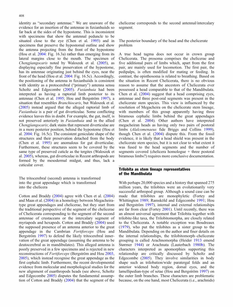
identity as “secondary antennae.” We are unaware of theevidence for an insertion of the antennae in fuxianhuiids sofar back at the sides of the hypostome. This is inconsistentwith specimens that show the antennal peduncle to besituated close to the eye (Chen et al. 1995) and inspecimens that preserve the hypostomal outline and showthe antenna projecting from the front of the hypostome(Hou et al. 2004: Fig. 16.3a) rather than emerging from itslateral margins close to the mouth. The specimen ofChengjiangocaris noted by Waloszek et al. (2005), asdisplaying especially fine preservation of the hypostome,has its antennae originating just behind the eyes, near thefront of the head (Hou et al. 2004: Fig. 16.5c). Accordingly,the positioning of the antenna in fuxianhuiids is consistentwith identity as a protocerebral (“primary”) antenna sensuScholtz and Edgecombe (2005). Fuxianhuia had beeninterpreted as having a raptorial limb posterior to itsantennae (Chen et al. 1995; Hou and Bergström 1997), asituation that resembles Branchiocaris, but Waloszek et al.(2005) instead argued that the alleged raptorial limb ofFuxianhuia is a pair of gut diverticulae. Some conflictingevidence leaves this in doubt. For example, the gut, itself, isnot preserved anteriorly in Fuxianhuia and in the alliedChengjiangocaris; dark stains that represent diverticulae arein a more posterior position, behind the hypostome (Hou etal. 2004: Fig. 16.5c). The consistent geniculate shape of thestructures and their preservation detached from the body(Chen et al. 1995) are anomalous for gut diverticulae.Furthermore, these structures seem to be covered by thesame type of preserved cuticle as the tergites (Waloszek etal. 2005), whereas, gut diverticulae in Recent arthropods areformed by the mesodermal midgut, and thus, lack acuticular cover.
The tritocerebral (second) antenna is transformedinto the great appendage which is transformedinto the chelicera
Cotton and Braddy (2004) agree with Chen et al. (2004)and Maas et al. (2004) in a homology between Megacheira-type great appendages and chelicerae, but they start fromthe traditional perspective of the segment of the cheliceraeof Chelicerata corresponding to the segment of the secondantennae of crustaceans or the intercalary segment ofmyriapods and hexapods. Cotton and Braddy (2004) usedthe supposed presence of an antenna anterior to the greatappendage in the Cambrian Fortiforceps (Hou andBergström 1997) to defend the likely tritocerebral inner-vation of the great appendage (assuming the antenna to bedeutocerebral as in mandibulates). This alleged antenna ispoorly preserved in a few specimens and is rejected in newreconstructions of Fortiforceps (Bergström and Hou 2003,2005), which instead recognise the great appendage as thefirst cephalic limb. Furthermore, the recent developmentalevidence from molecular and morphological studies for thenew alignment of euarthropods heads (see above, Scholtzand Edgecombe 2005) disputes the fundamental assump-tion of Cotton and Braddy (2004) that the segment of the
chelicerae corresponds to the second antennal/intercalarysegment.
The posterior boundary of the head and the chelicerateproblem
A true head tagma does not occur in crown groupChelicerata. The prosoma comprises the chelicerae andfive additional pairs of limbs which, apart from the firstpair, are mainly used for locomotion. The first pair, thepedipalps, is often modified for mating or feeding. Incontrast, the opisthosoma is related to breathing. Based onthe situation in Recent Chelicerata, there is no obviousreason to assume that the ancestors of Chelicerata everpossessed a head comparable to that of the Mandibulata.Chen et al. (2004) suggest that a head comprising eyes,antennae and three post-oral segments was present in thechelicerate stem species. This view is influenced by theresolution of Megacheira on the chelicerate stem lineage,with members of this group apparently having threebiramous cephalic limbs behind the great appendages(Chen et al. 2004). Other authors have interpretedmegacheiran heads as having only two pairs of postorallimbs (Alalcomenaeus fide Briggs and Collins 1999),though Chen et al. (2004) dispute this. From the fossilevidence, it is likely that a head shield was present in thechelicerate stem species, but it is not clear to what extent itwas fused to the head segments and the number ofsegments covered (chelicera homologue + three postoralbiramous limbs?) requires more conclusive documentation.
Trilobita as stem lineage representativesof the Mandibulata
With perhaps 20,000 species and a history that spanned 275million years, the trilobites were an evolutionarily verysuccessful arthropod group. Although a sound case can bemade that trilobites are monophyletic (Fortey andWhittington 1989; Ramsköld and Edgecombe 1991; Houand Bergström 1997), internal and external relationshipsare far from clear (Fortey 2001). Until recently, there wasan almost universal agreement that Trilobita together withtrilobite-like taxa, the Trilobitomorpha, are closely relatedto the Chelicerata. A notable exception was Boudreaux(1979), who put the trilobites as a sister group to theMandibulata. Depending on the author and finer details onthe internal relationships, the trilobitomorph–chelicerategrouping is called Arachnomorpha (Heider 1913 emendStørmer 1944) or Arachnata (Lauterbach 1980b). Thecharacters interpreted as apomorphies supporting thisrelationship are critically discussed by Scholtz andEdgecombe (2005). They involve similarities in bodyshape such as trilobation/wide paratergal folds and ananterior widened body region, dorsal eyes, and thelamellipedian-type of setae (Hou and Bergström 1997) onthe outer limb branches. These characters are problematicbecause, on the one hand, most Chelicerata (i.e., arachnids)
408

do not share them, and on the other hand, we find similarmorphologies in other crown group arthropods, or even inlikely stem lineage arthropods (e.g., trilobation; Waloszeket al. 2005). Homoplasy, in itself of course, does not ruleout the relevance of a character as a potential synapomor-phy, but polarity questions plague the putative arachno-morph synapomorphies. For example, two of the threecharacters that Cotton and Braddy (2004) optimized assynapomorphies of Arachnomorpha involve the absence ofexopod structures that are present only in crustaceans andmarrellomorphs. Given that trilobite–chelicerate affinitiescannot be regarded as beyond question, Scholtz andEdgecombe (2005) drew attention to characters of thehead that trilobites share with mandibulates, and hence, thepossibility of a placement of trilobites on the mandibulatestem lineage. This line of argumentation is followed here.Trilobites possess a head shield that is fused to and coversthe eye region, the antennal segment, and three post-oralsegments. This head structure resembles what has beensuggested on various grounds as the head in the groundpattern of Mandibulata (Lauterbach 1980a; Walossek 1993;Scholtz 1997).
According to the view of the discrimination of “primary”and “secondary antennae” by their position on the head, theTrilobita possess secondary antennae which are situated inan antennal notch at the lateral margin of the hypostome.Trilobites share the “secondary antennae” with theMandibulata (Figs. 3, and 5). As the sensory antennae ofMandibulata are apparently apomorphic within the Euar-thropoda (see above), the occurrence of “secondaryantennae” in Trilobita and Mandibulata is interpreted as asynapomorphy. This places the Trilobita in the stem lineageof the Mandibulata and disputes the close affinity to theChelicerata.
To test whether or not the “secondary antenna” trans-formation series is parsimonious, we incorporated trilobitesand five additional fossil terminals into the morphologicalcharacter set of Giribet et al. (2005). The expanded matrix(available as Electronic Supplementary Material includesnew characters to accommodate the fossils. Trilobita isscored using the Cambrian Olenoides, known from soft-part preservation (Whittington 1975, 1980). Additionalfossil terminals are the stem-crustacean or stem-mandibu-late Martinssonia (Müller and Walossek 1986; Walossekand Müller 1990), Alalcomenaeus (Briggs and Collins1999) as an exemplar of the Megacheira, the chelicerates,Baltoeurypterus (Selden 1981) and Proscorpius (Kjellsvig-Waering 1986), and the Burgess Shale taxon Emeraldella(Bruton and Whittington 1983). Putative apomorphies ofArachnomorpha (e.g., lamellipedian-type outer limbbranch setae) are included.
The result of the analysis is summarized in Fig. 6.Mandibulata is monophyletic, with Trilobita (Olenoides)and other taxa having “secondary antennae” (Martinssonia,Emeraldella) resolved in the mandibulate stem group. Adeutocerebral raptorial appendage maps onto the cladogramas symplesiomorphic relative to a deutocerebral antennabecause Pycnogonida (chelifores), Chelicerata (chelicerae),and Megacheira (Alalcomenaeus: great appendage) are
resolved as a grade rather than a monophyletic group. Thistopology needs testing with the inclusion of additionalfossil terminals, but this analysis demonstrates that thehypothesis that a “secondary antenna” is apomorphic fortrilobites and mandibulates is amenable to parsimonyanalysis.
Conclusion and perspective
From comparisons of Recent and fossil arthropods, twooverlapping trends in the evolution of arthropod cephali-zation emerge. First, the specialization of head regionsprogresses from anterior to posterior, which is perhaps nottoo surprising. In fossils as well as in Recent Chelicerata,plesiomorphically only the anteriormost limbs (chelicerae)are modified and only apomorphically are subsequentappendages transformed to head structures such aspedipalps. This is also true for Mandibulata, in which weplesiomorphically find only one pair of maxillae as isexemplified by cephalocarid crustaceans (Lauterbach1980a) as well as fossil representatives (see above). Thesecond trend is not so obvious and more surprising in that
Fig. 6 Cladogram showing the phylogenetic relationships of fossiland Recent arthropods. Trilobita (here sampled by Olenoides)appear as stem lineage mandibulates. The 356 character, 74-taxonmatrix was rooted between onychophorans and tardigrades andEuarthropoda including Pentastomida (cf. Maas et al. 2004).Heuristic searches with PAUP*4.0b10 using implied weights(Goloboff 1993) yield six shortest cladograms (strict consensusshown) that are stable across a range of concavity values (k=1, 2, 3,4, and 5). The internal resolution of extant Myriapoda, Hexapoda,and Euchelicerata is as in Giribet et al. (2005)
409

the dorsal differentiation precedes that on the ventral side.We always have more dorsal head segments fused thanlimbs differentiated to head structures, such as mouthpartsor sensory organs, on the ventral side. A good example ofthis is the Trilobita, in which we find a head shield fused tothe eye region and four segments, whereas, the post-antennal limbs covered by this head shield have, more orless, the same structure as the trunk limbs. The differen-tiation of these head limbs took place only later in the stemlineage leading to the crown group Mandibulata. TheCrustacea, and in particular, the Decapoda among theMalacostraca reveal that cephalization has not come to anend. The head shield/carapace is fused to all eight thoracicsegments in addition to the head segments. This so-calledcephalothorax can be seen as an extremely elongated headcomprising the ocular region and 13 segments. Accordingto the stated trends, we find ventrally only up to three pairsof anterior thoracic appendages transformed to additionalmouthparts, the maxillipedes (see Richter and Scholtz2001). This condition is correlated with differences in theexpression patterns of the Hox genes Antennapedia,Ultrabithorax, and abdominal-A, which show distinctivelyless overlap in decapod representatives than in othermalacostracan crustaceans such as isopods (Abzhanov andKaufman 2004).
As our review shows, a combination of modern andtraditional approaches reduces the number of viablehypotheses about head segmentation; several of the olderideas are no longer tenable or can be seen in a new light.Nevertheless, the arthropod head remains a contentiousissue, not the least because its study has been so theoryladen. Investigators should be aware of and attempt tominimize the unwarranted assumptions that influence theirviews. Hypotheses about phylogenetic relationships andtransformation series have an impact and the reconstructionof ancestors, assumed starting points, and definitions ofsegments. These problems start with the question over whata head is in the first place. Accordingly, it is not only amatter of better molecular resolution but also of basictheoretical underpinnings that determine whether we makeprogress in interpreting the head as a functionally andevolutionarily important part of the arthropods.
Acknowledgements We thank Jean Deutsch and Michel Veuille,the organizers of the meeting “Development and Phylogeny ofArthropods,” Paris 2005, for the invitation to GS to present ourthoughts on head evolution. Nikola-Michael Prpic made helpfulcomments on an earlier version of the manuscript. We are grateful toHenrike Semmler for her pictures of the innervation of the frontalfilaments in nauplius larvae of cirripedes and Carsten Wolff for thepicture of the embryonic head of Orchestia cavimana (Fig. 4). AlfredPalissa helped with literature about Protura. This article benefitedtremendously from the inspiring discussions with the students in theseminar “Phylogeny of Recent and Fossil Taxa” held at theHumboldt-University.
References
Abzhanov A, Kaufman T (2000a) Homologs of Drosophilaappendage genes in the patterning of arthropod limbs.Dev Biol 227:673–689
Abzhanov A, Kaufman T (2000b) Evolution of distinct expressionpatterns for engrailed paralogues in higher crustaceans(Malacostraca). Dev Genes Evol 210:493–506
Abzhanov A, Kaufman T (2004) Hox genes and tagmatization of thehigher Crustacea. In: Scholtz G (ed) Evolutionary developmentalbiology of crustacea. Balkema, Lisse, pp 43–74
Ackermann C, Dorresteijn A, Fischer A (2005) Clonal domains inpostlarvalPlatynereis dumerilii (Annelida: Polychaeta). JMorph266:258–280
Aguinaldo AMA, Turbeville JM, Linford LS, Rivera MC, Garey JR,Raff RA, Lake JA (1997) Evidence for a clade of nematodes,arthropods and other moulting animals. Nature 387:489–493
Alwes F, Scholtz G (2006) Stages and other aspects of theembryology of the parthenogenetic Marmorkrebs (Decapoda,Reptantia, Astacida). Dev Genes Evol 216(4):169–184
Anderson DT (1973) Embryology and phylogeny in annelids andarthropods. Pergamon, Oxford
Angelini DR, Kaufman TC (2005) Insect appendages andcomparative ontogenetics. Dev Biol 286:57–77
Arendt D, Technau U, Wittbrodt J (2001) Evolution of the bilaterianlarval foregut. Nature 409:81–84
Babu KS (1965) Anatomy of the central nervous system ofarachnids. Zool Jb Anat 82:1–154
Bartolomaeus T, Purschke G, HausenH (2005) Polychaete phylogenybased onmorphological data—a comparison of current attempts.Hydrobiologia 535/536:341–356
Bateson W (1894) Materials for the study of variation. Macmillan,London
Benesch R (1969) Zur Ontogenie und Morphologie von Artemiasalina L. Zool Jb Anat 86:307–458
Bergström J, Hou X-G (2003) Arthropod origins. Bull Geosci CzechGeol Surv 78:323–334
Bergström J, Hou X-G (2005) Early Palaeozoic non-lamellipedianarthropods. In: Koenemann S, Jenner R (eds) Crustacea andarthropod relationships. CRC, Boca Raton, pp 73–93
Boudreaux HB (1979) Arthropod phylogeny—with special referenceto insects. Wiley, New York
Boyan GS, Williams JLD, Posser S, Bräunig P (2002) Morphologicaland molecular data argue for the labrum being non-apical,articulated, and the appendage of the intercalary segment in thelocust. Arthrop Struct Dev 31:65–76
Boyan GS, Bräunig P, Posser S, Williams JLD (2003) Embryonicdevelopment of the sensory innervation of the clypeo–labralcomplex: further support for serially homologous appendagesin the locust. Arthrop Struct Dev 32:289–302
Brauer A (1895) Beiträge zur Kenntnis der Entwicklungsgeschichtedes Skorpions. II. Z Wissensch Zool 59:351–433
Briggs DEG (1976) The arthropod Branchiocaris n. gen., MiddleCambrian, Burgess Shale, British Columbia. Bull Geol SocCanada 264:1–29
Briggs DEG, Collins D (1999) The arthropod Alalcomenaeuscambricus Simonetta, from the Middle Cambrian BurgessShale of British Columbia. Palaeontology 42:953–977
Brown S, Patel NH, Denell RE (1994) Embryonic expression of asingle Tribolium engrailed homolog. Dev Genet 15:7–18
Browne WE, Price AL, Gerberding M, Patel NH (2005) Stages ofembryonic development in the amphipod crustacean, Parhyalehawaiensis. Genesis 42:124–149
Bruce AEE, Shankland M (1998) Expression of the head geneLox22-Otx in the leech Helobdella and the origin of thebilaterian body plan. Dev Biol 201:101–112
410

Bruckmoser P (1965) Embryologische Untersuchungen über denKopfbau der Collembole Orchesella villosa L. Zool Jb Anat82:299–364
Bruton DL, Whittington HB (1983) Emeraldella and Leanchoilia,two arthropods from the Burgess Shale, Middle Cambrian,British Columbia. Phil Trans R Soc Lond B 300:553–585
Budd GE (1998) The morphology and phylogenetic significance ofKerygmachela kierkegaardi (Buen Formation, Lower Cam-brian, N Greenland). Trans R Soc Edinburgh: Earth Sci 89:249–290
Budd GE (2001) Tardigrades as ‘stem-group arthropods’: theevidence from the Cambrian fauna. Zool Anz 240:265–279
Budd GE (2002) A palaeontological solution to the arthropod headproblem. Nature 417:271–275
Butt FH (1960) Head development in the arthropods. Biol Rev35:43–91
Casanova J-P (1996) Gnathophausia childressi, new species, amysid from deep near-bottom waters off California, withremarks on the mouthparts of the genus Gnathophausia.J Crustac Biol 16:192–200
Chaudonneret J (1987) Evolution of the insect brain with specialreference to the so-called tritocerebrum. In: Gupta AP (ed)Arthropod brain. Wiley, New York, pp 3–26
Chen J, Edgecombe GD, Ramsköld L, Zhou G (1995) Headsegmentation in Early Cambrian Fuxianhuia: implications forarthropod evolution. Science 268:1339–1343
Chen J, Waloszek D, Maas A (2004) A new ‘great appendage’arthropod from the Lower Cambrian of China and homology ofchelicerate chelicerae and raptorial antero-ventral appendages.Lethaia 37:3–20
Chipman AD, Arthur W, Akam M (2004) Early development andsegment formation in the centipede, Strigamia maritima(Geophilomorpha). Evol Dev 6:78–89
Cohen SM (1993) Imaginal disc development. In: Martinez-Arias A,Bate M (eds) Drosophila development. Cold Spring Harbor,Cold Spring Harbor, pp 747–841
Cohen S, Jürgens G (1991) Drosophila headlines. Trends Genet7:267–272
Cook CE, SmithML, TelfordMJ, Bastianello A, AkamM (2001)Hoxgenes and the phylogeny of arthropods. Curr Biol 11:759–763
Cotton TJ, Braddy SJ (2004) The phylogeny of arachnomorpharthropods and the origin of the Chelicerata. Trans R SocEdinb: Earth Sci 94:169–193
Dahl E (1956) On the differentiation of the topography of thecrustacean head. Acta Zool 37:123–192
Damen WGM (2002) Parasegmental organization of spider embryoimplies that the parasegment is an evolutionary conserved entityin arthropod embryogenesis. Development 129:1239–1250
Damen WGM, Hausdorf M, Seyfarth E-A, Tautz D (1998) Aconserved mode of head segmentation in arthropods revealedby the expression pattern of Hox genes in a spider. Proc NatlAcad Sci U S A 95:10665–10670
Darwin C (1854) A monograph on the subclass Cirripedia, withfigures of all the species. The Balanidae, the Verrucidae, etc.Ray Society, London
Davis GK, D’Alessio JA, Patel NH (2005) Pax 3/7 genes revealconservation and divergence in the arthropod segmentationhierarchy. Dev Biol 285:169–184
de Velasco B, Mandal L, Mkrtchyan M, Hartenstein V (2006)Subdivision and developmental fate of the head mesoderm inDrosophila melanogaster. Dev Genes Evol 216:39–51
de Rosa R, Prud’homme B, Balavoine G (2005) caudal and even-skipped in the annelid Platynereis dumerilii and the ancestry ofposterior growth. Evol Dev 7:574–587
Dewel RA, Budd GE, Castano DF, Dewel WC (1999) Theorganization of the subesophageal nervous system in tardi-grades: insights into the evolution of the arthropod hypostomeand tritocerebrum. Zool Anz 238:191–203
Dohle W (1964) Die Embryonalentwicklung von Glomeris mar-ginata (Villers) im Vergleich zur Entwicklung andererDiplopoden. Zool Jb Anat 81:241–310
Dong Y, Friedrich M (2005) Comparative analysis of winglesspatterning in the embryonic grasshopper. Dev Genes Evol215:177–197
Dove H, Stollewerk A (2003) Comparative analysis of neurogenesisin the myriapod Glomeris marginata (Diplopoda) suggestsmore similarities to chelicerates than to insects. Development130:2161–2171
Dunlop JA, Arango CP (2004) Pycnogonid affinities: a review. J ZoolSyst Evol Res 43:8–21
Edgecombe GD (2004) Morphological data, extant Myriapoda, andthe myriapod stem-group. Contrib Zool 73:207–252
Edgecombe GD, Wilson GDF, Colgan DJ, Gray MR, Cassis G(2000) Arthropod cladistics: combined analysis of histone H3and U2 snRNA sequences and morphology. Cladistics 16:155–203
Eriksson BJ, Budd GE (2000) Onychophoran cephalic nerves andtheir bearing on our understanding of head segmentation andstem-group evolution of Arthropoda. Arthrop Struct Dev29:197–209
Eriksson BJ, Tait NN, Budd GE (2003) Head development in theonychophoran Euperipatoides kanangrensis with particularreference to the central nervous system. J Morph 255:1–23
Fanenbruck M, Harzsch S (2005) A brain atlas of Godzilliognomusfrondosus Yager, 1989 (Remipedia, Godzilliidae) and compar-ison with the brain of Speleonectes tulumensis Yager, 1987(Remipedia, Speleonectidae): implications for arthropod rela-tionships. Arthrop Struct Dev 34:343–378
Fleig R (1990) Engrailed expression and body segmentation in thehoney bee Apis mellifera. Roux’s Arch Dev Biol 198:467–473
Fleig R (1994) Head segmentation in the embryo of the Coloradobeetle Leptinotarsa decemlineata as seen with the ant-enimmunostaining. Roux’s Arch Dev Biol 203:227–229
Fortey RA (2001) Trilobite systematics: the last 75 years. J Paleontol75:1141–1151
Fortey RA, Whittington HB (1989) The Trilobita as a natural group.Hist Biol 2:125–138
François J (1969) Anatomie et morphologie céphalique desProtoures (Insecta Apterygota). Mém Mus Nation Hist Nat(NS) A (Zoologie) 59:1–144
Friedrich M, Benzer S (2000) Divergent decapentaplegic expressionpatterns in compound eye development and the evolution ofinsect metamorphosis. J Exp Zool (Mol Dev Evol) 288:39–55
Gehring WJ (2004) Historical perspective on the development andevolution of eyes and photoreceptors. Int J Dev Biol 48:707–717
Giorgianni MW, Patel NH (2004) Patterning of the branched headappendages in Schistocercaamericana and Tribolium casta-neum. Evol Dev 6:402–410
Giribet G (2003) Molecules, development and fossils in the study ofmetazoan evolution; Articulata versus Ecdysozoa revisited.Zoology 106:303–326
Giribet G, Edgecombe GD, Wheeler WC (2001) Arthropodphylogeny based on eight molecular loci and morphology.Nature 413:157–161
Giribet G, Richter S, Edgecombe GD, Wheeler WC (2005) Theposition of crustaceans within Arthropoda—evidence from ninemolecular loci and morphology. In: Koenemann S, Jenner R(eds) Crustacea and arthropod relationships. CRC, Boca Raton,pp 307–352
Goloboff PA (1993) Estimating character weights during tree search.Cladistics 9:83–91
Goodrich EJ (1897) On the relation of the arthropod head to theannelid prostomium. Q J Microsc Sci 40:259–268
Haas MS, Brown SJ, Beeman RW (2001a) Pondering theprocephalon: the segmental origin of the labrum. Dev GenesEvol 211:89–95
Haas MS, Brown SJ, Beeman RW (2001b) Homeotic evidence forthe appendicular origin of the labrum in Tribolium castaneum.Dev Genes Evol 211:96–102
Haget A (1955) Expérience mettant en évidence l’origine paire dulabre chez l’embryon du Coléoptère Leptinotarsa. C R Soc Biol149:690–692
411

Hanström B (1928) Vergleichende Anatomie des Nervensystems derwirbellosen Tiere. Springer, Berlin Heidelberg New York
Harada Y, Okai N, Taguchi S, Tagawa K, Humphreys T, Satoh N(2000) Developmental expression of the hemichordate otxortholog. Mech Dev 91:337–339
Harzsch S (2004) The tritocerebrum of Euarthropoda: a “non-drosophilocentric” perspective. Evol Dev 6:303–309
Harzsch S, Glötzner J (2002) An immunohistochemical study onstructure and development of the nervous system in the brineshrimp Artemia salina Linnaeus, 1758 (Branchiopoda, Anos-traca) with remarks on the evolution of the arthropod brain.Arthrop Struct Dev 30:251–270
Harzsch S, Müller CHG, Wolf H (2005a) From variable toconstant cell numbers: cellular characteristics of the arthropodnervous system argue against a sister–group relationships ofChelicerata and “Myriapoda” but favour the Mandibulataconcept. Dev Genes Evol 215:53–68
Harzsch S, Wildt M, Battelle B, Waloszek D (2005b) Immunohis-tochemical localization of neurotransmitters in the nervoussystem of larval Limulus polyphemus (Chelicerata, Xiphosura):evidence for a conserved protocerebral architecture in Euar-thropoda. Arthrop Struct Dev 34:327–342
Hatschek B (1878) Studien über Entwicklungsgeschichte derAnneliden. Arbeit Zool Inst Univ Wien, 57–128
Heider K (1913) Entwicklungsgeschichte und Morphologie derWirbellosen. In: Hinneberg P (ed) Die Kultur der Gegenwart,Teil 3, Abt. 4, Bd. 2. Teubner, Leipzig, 176–332
Hertzel G (1984) Die Segmentation des Keimstreifens vonLithobius forficatus (L.) (Myriapoda, Chilopoda). Zool JbAnat 112:369–386
Heymons R (1901) Die Entwicklungsgeschichte der Scolopender.Zoologica 33:1–244
Hirth F, Therianos S, Loop T, Gehring WJ, Reichert H, Furukubo-Tokunaga K (1995) Developmental defects in brain segmenta-tion caused by mutations of the homeobox genes orthodenticleand empty spiracles in Drosophila. Neuron 15:769–778
Holmgren N (1916) Zur vergleichenden Anatomie des Gehirnsvon Polychaeten, Onychophoren, Xiphosuren, Arachniden,Crustaceen, Myriapoden und Insekten. Vet Akad HandlStockholm 56:1–303
Hou X-G (1999) New rare bivalved arthropods from the LowerCambrian Chengjiang fauna, Yunnan, China. J Paleontol73:102–116
Hou X-G, Bergström J (1997) Arthropods of the Lower CambrianChengjiang fauna, southwest China. Fossils Strata 45:1–116
Hou X-G, Aldridge RJ, Bergström J, Siveter DJ, Siveter DJ, FengX-H (2004) The Cambrian Fossils of Chengjiang, China. Theflowering of early animal life. Blackwell, Oxford
Hughes CL, Kaufman TC (2002) Exploring myriapod segmentation:the expression patterns of even-skipped, engrailed, andwingless in a centipede. Dev Biol 246:47–61
Hwang UW, Friedrich M, Tautz D, Park CJ, Kim W (2001)Mitochondrial protein phylogeny joins myriapods with chelic-erates. Nature 413:154–157
Inoue Y, Niwa N, Mito T, Ohuchi H, Yoshika H, Noji S (2002)Expression patterns of hedgehog, wingless, and decapentaple-gic during gut formation of Gryllus bimaculatus (cricket).Mech Dev 110:245–248
Jager M, Murienne J, Clabaut C, Deutsch J, Le Guyader H, ManuelM (2006) Homology of arthropod anterior appendages revealedby Hox gene expression in a sea spider. Nature 411:506–508
Janetschek H (1970) 3. Protura (Beintastler) In: Helmcke J-G,Starck D, Wermuth H (eds) Handbuch der Zoologie, IV. Band:Arthropoda-2. Hälfte: Insecta, 2. Teil: Spezielles. Parey, Berlin,pp 1–72
Janssen R, Prpic N-M, Damen WGM (2004) Gene expressionsuggests decoupled dorsal and ventral segmentation in themillipede Glomeris marginata (Myriapoda: Diplopoda). DevBiol 268:89–104
Jenner RA, Scholtz G (2005) Playing another round of metazoanphylogenetics: historical epistemology, sensitivity analysis,and the position of Arthropoda within the Metazoa on thebasis of morphology. In: Koenemann S, Jenner R (eds)Crustacea and Arthropod relationships. CRC, Boca Raton,pp 355–385
Jockusch EL, Ober KA (2004) Hypothesis testing in evolu-tionary developmental biology: a case study from insectwings. J Heredity 95:382–396
Kadner D, Stollewerk A (2004) Neurogenesis in the chilopodLithobius forficatus suggests more similarities to cheliceratesthan to insects. Dev Genes Evol 214:367–379
Kjellsvig-Waering EN (1986) A restudy of the fossil Scorpionida ofthe world. Palaeontogr Am 55:1–287
Kuratani S (2003) Evolutionary developmental biology and verte-brate head segmentation: a perspective from developmentalconstraint. Theory Biosci 122:230–251
Kusche K, Hembach A, Hagner-Holler S, Genauer W, Burmester T(2003) Complete subunit sequences, structure and evolution ofthe 6×6-mer hemocyanin from the common house centipede,Scutigera coleoptrata. Eur J Biochem 270:2860–2868
Lauterbach K-E (1973) Schlüsselereignisse in der Evolution derStammgruppe der Euarthropoda. Zool Beitr (NF) 19:251–299
Lauterbach K-E (1980a) Schlüsselereignisse in der Evolution desGrundplans der Mandibulata (Arthropoda). Abh Naturwiss VerHamburg NF 23:105–161
Lauterbach K-E (1980b) Schlüsselereignisse in der Evolution desGrundplans der Arachnata (Arthropoda). Abh Naturwiss VerHamburg NF 23:163–327
Li Y, Brown SJ, Hausdorf B, Tautz D, Denell RE, Finkelstein R(1996) Two orthodenticle-related genes in the short-germ beetleTribolium castaneum. Dev Genes Evol 206:35–45
Maas A, Waloszek D (2001) Cambrian derivatives of the earlyarthropod stem lineage, pentastomids, tardigrades and lobopo-dians—an ‘Orsten’ perspective. Zool Anz 240:451–459
Maas A, Waloszek D, Chen J, Braun A, Wang X, Huang D (2004)Phylogeny and life habits of early arthropods—predation in theEarly Cambrian sea. Prog Nat Sci 14:158–166
Mallatt JM, Garey JR, Shultz JW (2004) Ecdysozoan phylogeny andBayesian inference: first use of nearly complete 28S and 18SrRNA gene sequences to classify the arthropods and their kin.Mol Phylogenet Evol 31:178–191
Manton SM (1928) On the embryology of the crustacean Nebaliabipes. Phil Trans R Soc Lond B 223:163–238
Manton SM (1949) Studies on the Onychophora VII. The earlyembryonic stages of Peripatopsis and some general consider-ations concerning the morphology and phylogeny of theArthropoda. Phil Trans R Soc B 233:483–580
Manzanares M, Williams TA, Marco R, Garesse R (1996) Segmen-tation in the crustacean Artemia: engrailed staining studied withan antibody raised against the Artemia protein. Roux’s ArchDev Biol 205:424–431
Maxmen A, Browne WE, Martindale MQ, Giribet G (2005)Neuroanatomy of sea spiders implies an appendicular originof the protocerebral segment. Nature 437:1144–1148
Mayer G, Koch M (2005) Ultrastructure and fate of the nephridialanlagen in the antennal segment of Epiperipatus biolleyi(Onychophora, Peripatidae) evidence for the onychophoranantennae being modified legs. Arthrop Struct Dev 34:471–480
Meisenheimer J (1902) Beiträge zur Entwicklungsgeschichte derPantopoden. I. Die Entwicklung von Ammothea echinataHodge bis zur Ausbildung derLarvenform. Zeitschr WissZool 72:191–248
Minelli A (2003) The origin and evolution of appendages. Int J DevBiol 47:573–581
Minelli A, Fusco G (2004) Evo–devo perspectives on segmenta-tion: model organisms, and beyond. Trends Ecol Evol19:423–429
412

Mittmann B, Scholtz G (2001) Distal-less expression in embryos ofLimulus polyphemus (Chelicerata, Xiphosura) and Lepismasaccharina (Insecta, Zygentoma) suggests as role in thedevelopment of mechanoreceptors, chemoreceptors, and theCNS. Dev Genes Evol 211:232–243
Mittmann B, Scholtz G (2003) Development of the nervous systemin the “head” of Limulus polyphemus (Chelicerata, Xiphosura):morphological evidence for a correspondence between thesegments of the chelicerae and of the (first) antennae ofMandibulata. Dev Genes Evol 213:9–17
Müller KJ, Walossek D (1986) Martinssonia elongata gen. et sp.n.,a crustacean-like euarthropod from the Upper Cambrian‘Orsten’ of Sweden. Zool Scr 15:73–92
Nederbragt AJ, te Welscher, P, van den Driesche S, van Loon A,Dictus WJAG (2002) Novel and conserved roles for ortho-denticle/otx and orthopedia/otp orthologs in the gastropodmollusc Patella vulgata. Dev Genes Evol 212:330–337
Negrisolo E, Minelli A, Valle G (2004) The mitochondrial genomeof the house centipede Scutigera and the monophyly versusparaphyly of myriapods. Mol Bio Evol 21:770–780
Nielsen C (2001) Animal evolution. Interrelationships of the livingphyla, 2nd edn. Oxford University Press, Oxford
Nielsen C (2005a) Trochophora larvae and adult body regions inannelids: some conclusions. Hydrobiologia 535/536:23–24
Nielsen C (2005b) Larval and adult brains. Evol Dev 7:483–489Niwa N, Saitoh M, Ohuchi H, Yoshioka H, Noji S (1997)
Correlation between Distal-less expression patterns andstructures of appendages in development of the two-spottedcricket, Gryllus bimaculatus. Zool Sci 14:115–125
Northcutt RG (2005) The new head hypothesis revisited. J Exp Zool(Mol Dev Evol) 304B:274–297
Nulsen C, Nagy LM (1999) The role of wingless in thedevelopment of multibranched crustacean limbs. Dev GenesEvol 209:340–348
Oishi S (1959) Studies on the teloblasts in the decapod embryo I.Origin of teloblasts in Heptacarpus rectirostris (Stimpson).Embryol 4:283–309
Olesen J (2004) On the ontogeny of the Branchiopoda (Crustacea):contribution of development to phylogeny and classification.In: Scholtz G (ed) Evolutionary developmental biology ofCrustacea. Balkema, Lisse, pp 217–269
Olesen J, Richter S, Scholtz G (2001) The evolutionary transforma-tion of phyllopodous to stenopodous limbs in the Branchiopoda(Crustacea)—is there a common mechanism for early limbdevelopment in arthropods? Int J Dev Biol 45:869–876
Olsson L, Ericsson, R, Cerny R (2005) Vertebrate head develop-ment: segmentation, novelties, and homology. Theory Biosci124:145–163
Osborne PW, Dearden PK (2005) Expression of Pax group III genesin the honey bee (Apis mellifera). Dev Genes Evol 215:499–508
Page DT (2004) A mode of arthropod brain evolution suggested byDrosophila commissure development. Evol Dev 6:25–31
Panganiban G, Sebring A, Nagy L, Carroll S (1995) The developmentof crustacean limbs and the evolution of arthropods. Science270:1363–1366
Patel NH, Kornberg TB, Goodman CS (1989) Expression ofengrailed during segmentation in grasshopper and crayfish.Development 107:201–212
Paulus H, Weygoldt P (1996) Artrhropoda, Gliederfüßer. In:Westheide W, Rieger R (eds) Spezielle Zoologie, Teil 1:Einzeller und Wirbellose Tiere. Gustav Fischer, Stuttgart,pp 411–419
Peterson MD, Popadic A, Kaufman TC (1998) The expressionof two engrailed-related genes in an apterygote insect anda phylogenetic analysis of insect engrailed-related genes.Dev Genes Evol 208:547–557
Pflugfelder O (1948) Entwicklung von Paraperipatus anboinensisn. sp. Zool Jb Anat 69:443–492
Pisani D, Poling LL, Lyons-Weiler M, Hedges SB (2004) Thecolonization of land by animals: molecular phylogeny anddivergence times among arthropods. BMC Biol 2:1–10
Popadic A, Panganiban G, Rusch D, Shear WA, Kaufman TC(1998) Molecular evidence for the gnathobasic derivation ofarthropod mandibles and for the appendicular origin of thelabrum and other structures. Dev Genes Evol 208:142–150
Pross A (1966) Untersuchungen zur Entwicklungsgeschichte derAraneae (Pardosa hortensis (Thorell)) unter besondererBerücksichtigung des vorderen Prosomaabschnitts. Z MorphÖkol Tiere 58:38–108
Prpic N-M, Tautz D (2003) The expression of the proximodistal axispatterning genes Distal-less and dachshund in the appendagesof Glomeris marginata (Myriapoda: Diplopoda) suggests aspecial role of these genes in patterning the head appendages.Dev Biol 260:97–112
Prpic N-M, Wigand B, Damen WGM, Klingler M (2001) Expressionof dachshund in wild-type and Distal-less mutant Triboliumcorroborates serial homologies in insect appendages. Dev GenesEvol 211:467–477
Prpic N-M, Janssen R, Wigand B, Klingler M, Damen WGM (2003)Gene expression in spider appendages reveals reversal of exd/hthspatial specificity, altered leg gap gene dynamics, and suggestsdivergent distal morphogen signaling. Dev Biol 265:119–140
Prud’homme B, de Rosa R, Arendt D, Julien J-F, Pajaziti R,Dorresteijn AWC, Adoutte A, Wittbrodt J, Balavoine G (2003)Arthropod-like expression patterns of engrailed and wingless inthe annelid Platynereis dumerilii suggest a role in segmentformation. Curr Biol 13:1876–1881
Pultz MA, Pitt JN, Alto NM (1999) Extensive zygotic control ofthe anteroposterior axis in the wasp Nasonia vitripennis.Development 126:701–710
Pultz MA, Westendorf L, Gale SD, Hawkins K, Lynch J, Pitt JN,Reeves NL, Yao JCY, Small S, Desplan C, Leaf DS (2005) Amajor role for zygotic hunchback in patterning the Nasoniaembryo. Development 132:3705–3715
Ramsköld L, Edgecombe GD (1991) Trilobite monophyly revisited.Hist Biol 4:267–283
Rempel JG (1975) The evolution of the insect head: the endlessdispute. Quaest Ent 11:7–25
Richter S, Scholtz G (2001) Phylogenetic analysis of theMalacostraca (Crustacea). J Zool Syst Evol Res 39:113–136
Richter S, Wirkner C (2004) Kontroversen in der phylogenetischenSystematik der Euarthropoda. Sber Ges Naturf Freunde Berlin(NF) 43:73–102
Rogers BT, Kaufman TC (1997) Structure of the insect head inontogeny and phylogeny: a view from Drosophila. Int RevCytol 174:1–84
Rogers BT, Peterson MD, Kaufman TC (2002) The development andevolution of insect mouthparts as revealed by the expressionpatterns of gnathocephalic genes. Evol Dev 4:96–110
Rohrschneider I (1968) Beiträge zur Entwicklung des Vorderkopfesund der Mundregion von Periplaneta americana. Zool Jb Anat85:537–578
Sanchez-Salazar J, Pletcher MT, Bennett RL, Brown SJ, DandamundiTJ, Denell RE, Doctor JS (1996) The Tribolium decapentaplagicgene is similar in sequence, structure, and expression to theDrosophila dpp gene. Dev Genes Evol 206:237–246
Schmidt-Ott U, Technau GM (1992) Expression of en and wg in theembryonic head and brain of Drosophila indicates a refoldedband of seven segment remnants. Development 116:111–125
Schmidt-Ott U, Sander K, Technau GM (1994a) Expression ofengrailed in embryos of a beetle and five dipteran species withspecial reference to the terminal regions. Roux Arch Dev Biol203:298–303
Schmidt-Ott U, González-Gaitán M, Jäckle H, Technau GM (1994b)Number, identity, and sequence of the Drosophila headsegments as revealed by neural elements and their deletionpatterns in mutants. Proc Natl Acad Sci U S A 91:8363–8367
Schmidt-Rhaesa A, Bartolomaeus T, Lemburg C, Ehlers U, GareyJR (1998) The position of the Arthropoda in the phylogeneticsystem. J Morphol 238:263–285
Schneuwly S, Klemenz R, Gehring WJ (1987) Redesigning thebody plan of Drosophila by ectopic expression of the homeoticgene Antennapedia. Nature 325:816–818
413

Scholl G (1963) Embryologische Untersuchungen an Tanaidaceen(Heterotanais oerstedi Kröyer). Zool Jb Anat 80:500–554
Scholl G (1969) Die Embryonalentwicklung des Kopfes undProthorax von Carausius morosus Br. (Insecta, Phasmida).Z Morph Tiere 65:1–142
Scholl G (1977) Beiträge zur Embryonalentwicklung von Limuluspolyphemus L. (Chelicerata, Xiphosura). Zoomorphologie86:99–154
Scholtz G (1995) Head segmentation in Crustacea—an immuno-cytochemical study. Zoology 98:104–114
Scholtz G (1997) Cleavage, germ band formation and head segmen-tation: the ground pattern of the Euarthropoda. In: Fortey RA,Thomas RH (eds) Arthropod relationships. Chapman and Hall,London, pp 317–332
Scholtz G (2001) Evolution of developmental patterns in arthro-pods-the contribution of gene expression to morphology andphylogenetics. Zoology 103:99–111
Scholtz G (2002) The Articulata hypothesis—or what is a segment?Organ Divers Evol 2:197–215
Scholtz G (2003) Is the taxon Articulata obsolete? Arguments infavour of a close relationship between annelids and arthropods.In: Legakis A, Sfenthourakis S, Polymeni R, Thessalou-LegakiM (eds) Proceedings of the 18th International Congress ofZoology, Athens 2000. Penfolds, Sofia, pp 489–501
Scholtz G (2004) Baupläne versus ground patterns, phyla versusmonophyla: aspects of patterns and processes in evolutionarydevelopmental biology. In: Scholtz G (ed) Evolutionarydevelopmental biology of Crustacea. Balkema, Lisse, pp 3–16
Scholtz G (2005) Homology and ontogeny: pattern and processin comparative developmental biology. Theory Biosci124:121–143
Scholtz G, Edgecombe GD (2005) Heads, Hox and the phylogeneticposition of trilobites. In: Koenemann S, Jenner R (eds)Crustacea and arthropod relationships. CRC, Boca Raton, pp139–165
Scholtz G, Patel NH, Dohle W (1994) Serially homologousengrailed stripes are generated via different cell lineages inthe germ band of amphipod crustaceans (Malacostraca,Peracarida). Int J Dev Biol 38:471–478
Scholtz G, Mittman B, Gerberding M (1998) The pattern of Distal-less expression in the mouthparts of crustaceans, myriapods andinsects: new evidence for a gnathobasic mandible and thecommon origin of Mandibulata. Int J Dev Biol 42:801–810
Schoppmeier M, Damen WGM (2001) Double-stranded RNAinterference in the spider Cupiennius salei: the role of Distal-less is evolutionarily conserved in arthropod appendageformation. Dev Genes Evol 211:76–82
Schröder R (2003) The genes orthodenticle and hunchback substitutefor bicoid in the beetle Tribolium. Nature 422:621–625
Seaver EC (2003) Segmentation: mono or polyphyletic? Int J DevBiol 47:583–595
Seaver EC, Kaneshige LM (2006) Expression of “segmentation”genes during larval and juvenile development in the polychaetesCapitella sp. I and H. elegans. Dev Biol 289:179–194
Seaver EC, Thamm K, Hill SD (2005) Growth patterns duringsegmentation in the two polychaete annelids, Capitella sp. I andHydroides elegans: comparisons at distinct life history stages.Evol Dev 7:312–326
Selden PA (1981) Functional morphology of the prosoma ofBaltoeurypterus tetragonophthalmus (Fischer) (Chelicerata:Eurypterida). Trans R Soc Edinburgh: Earth Sci 72:9–48
Semmler H (2005) Immuncytochemische Studien zur larvalenMyo- und Neuroanatomie von Balanus improvisus (Crustacea,Cirripedia, Thecostraca). Diplom Thesis: Humboldt-Universitätzu Berlin
Shiga Y, Yasumoto R, Yamagata H, Hayashi S (2002) Evolvingrole of Antennapedia protein in arthropod limb patterning.Development 129:3555–3561
Siewing R (1963) Das Problem der Arthropodenkopfsegmentierung.Zool Anz 170:429–468
Siewing R (1969) Lehrbuch der vergleichenden Entwicklungs-geschichte der Tiere. Parey, Hamburg
Simonnet F, Deutsch J, Quéinnec E (2004) hedgehog is a segmentpolarity gene in a crustacean and a chelicerate. Dev Genes Evol214:527–545
Snodgrass RE (1960) Facts and theories concerning the insect head.Smithson Misc Collect 142:1–61
Starck D (1963) Die Metamerie des Kopfes der Wirbeltiere. ZoolAnz 170:393–428
Stein M, Waloszek D, Maas A (2005) Oelandicaris oelandica andthe stem lineage of Crustacea. In: Koenemann S, Jenner R (eds)Crustacea and arthropod relationships. CRC, Boca Raton, pp55–71
Størmer L (1944) On the relationships and phylogeny of fossil andrecent Arachnomorpha. Skrift Utgitt Norske Vidensk-akad OsloI Math-Naturvitensk Klasse 5:1–158
Tamarelle M (1984) Transient rudiments of second antennae on the“intercalary” segment of embryos of Anurida maritima Guer.(Collembola: Arthropleona) and Hyphantria cunea Drury(Lepidoptera: Arctiidae) Int J Insect Morphol Embryol13:331–336
Tautz D (2004) Segmentation. Dev Cell 7:301–312Telford MJ, Thomas RH (1998) Expression of homeobox genes
shows chelicerate arthropods retain their deutocerebral segment.Proc Natl Acad Sci U S A 95:10671–10675
Thomas RH, Telford MJ (1999) Appendage development inembryos of the oribatid mite Archegozetes longisetus (Acari,Oribatei, Thrypochthoniidae) Acta Zool 80:193–200
Tiegs OW (1940) The embryology and affinities of the Symphyla,based on a study ofHanseniella agilis. Q JMicrosc Sci 82:1–225
Tomsa JM, Langeland JA (1999) Otx expression during lampreyembryogenesis provides insights into the evolution of thevertebrate head and jaw. Dev Biol 207:26–37
Ullmann SL (1964) The origin and structure of the mesoderm andthe formation of the coelomic sacs in Tenebrio molitor L(Insecta, Coleoptera). Phil Trans R Soc Lond B 747:245–276
Umesono Y, Watanabe K, Agata K (1999) Distinct structuraldomains in the planarian brain defined by the expression ofevolutionarily conserved homeobox genes. Dev Genes Evol209:31–39
Ungerer P,Wolff C (2005) External morphology of limb developmentin the amphipod Orchestia cavimana (Crustacea, Malacostraca,Peracarida). Zoomorphology 124:89–99
Urbach R, Technau G (2003a) Early steps in building the insectbrain: neuroblasts formation and segmental patterning in thedeveloping brain of different insect species. Arthrop Struct Dev32:103–123
Urbach R, Technau G (2003b) Molecular markers for identifiedneuroblasts in the developing brain ofDrosophila. Development130:3621–3637
Urbach R, Technau G, Breidbach O (2003) Spatial and temporalpattern of neuroblasts, proliferation, and engrailed expressionduring early brain development. Arthrop Struct Dev 32:125–140
Vilpoux K, Waloszek D (2003) Larval development and morpho-genesis of the sea spider Pycnogonum litorale (Ström, 1762)and the tagmosis of the body of Pantopoda. Arthrop Struct Dev32:349–383
von Wistinghausen C (1891) Untersuchungen über die Entwicklungvon Nereis dumerilii. Mitt Zool Stat Neapel 10:41–74
Wada S (1965) Analyse der Kopf-Hals-Region von Tachycines(Saltatoria) in morphogenetische Einheiten. II. Mitteilung:Experimentell-teratologische Befunde am Kopfskelett mitBerücksichtigung des zentralen Nervensystems. Zool Jb Anat83:235–326
Wägele J-W, Misof B (2001) On the quality of evidence inphylogeny reconstruction: a reply to Zrzavý’s defence of the‘Ecdysozoa’ hypothesis. J Zool Syst Evol Res 39:165–176
Walossek D (1993) The Upper Cambrian Rehbachiella and thephylogeny of Branchiopoda and Crustacea. Fossils Strata32:3–202
Waloszek D (2003) Cambrian ‘Orsten’-type preserved arthropodsand the phylogeny of Crustacea. In: Legakis A, Sfenthourakis,S, Polymeni R and Thessalou-Legaki M (eds) Proceedings ofthe 18th International Congress of Zoology, Athens 2000:69–87
414

Waloszek D, Dunlop JA (2002) A larval sea spider (Arthropoda:Pycnogonida) from the Upper Cambrian ‘Orsten’ of Sweden,and the phylogenetic position of pycnogonids. Palaeontology45:421–446
Walossek D, Müller KJ (1990) Upper Cambrian stem-lineagecrustaceans and their bearing upon the monophyletic origin ofCrustacea and the position of Agnostus. Lethaia 23:409–427
Waloszek D, Chen J, Maas A, Wang X (2005) Early Cambrianarthropods—new insights into arthropod head and structuralevolution. Arthrop Struct Dev 34:189–205
Weber H (1952) Morphologie, Histologie und Entwicklungs-geschichte der Articulaten II. Die Kopfsegmentierung und dieMorphologie des Kopfes überhaupt. Fortschr Zool 9:18–231
Weygoldt P (1985) Ontogeny of the arachnid central nervoussystem. In: Barth FG (ed) Neurobiology of arachnids. Springer,Berlin Heidelberg New York, pp 20–37
Whittington HB (1975) Trilobites with appendages from the MiddleCambrian Burgess Shale, British Columbia. Fossils Strata4:97–136
Whittington HB (1980) Exoskeleton, moult stage, appendagemorphology and habits of the Middle Cambrian trilobiteOlenoides serratus. Palaeontology 23:171–204
Winter G (1980) Beiträge zur Morphologie und Embryologie desvorderen Körperabschnitts (Cephalosoma) der PantopodaGerstaecker, 1863. Z Zoolog Syst Evol Forsch 18:27–61
Wolff C (2004) Die Beinentwicklung des amphipoden KrebsesOrchestia cavimana (Peracarida, Malacostraca - eine zellgen-ealogische Studie. Doctoral Thesis, Humboldt-Universität zuBerlin
Woltereck R (1905) Zur Kopffrage der Anneliden. Verh Dtsch ZoolGes 15:154–186
Yamamoto DS, Sumitani M, Tojo K, Lee JM, Hatakeyama M(2004) Cloning of a decapentaplegic orthologue from thesawfly, Athalia rosae (Hymenoptera) and its expression in theembryonic appendages. Dev Genes Evol 214:128–133
Younossi-Hartenstein A, Green P, Liaw G-J, Rudolph K, Lengyel J,Hartenstein V (1997) Control of early neurogenesis of theDrosophila brain by the head gap genes tll, otd, ems, and btd.Dev Biol 182:270–283
415