giant colloid cyst of the third ventricle: challenges in
TRANSCRIPT

CASE REPORT Open Access
Giant colloid cyst of the third ventricle:challenges in managementAmit Agrawal1*, Vissa Santhi2 and Reddy V. Umamaheswara3
Abstract
Background: Giant colloid cysts (size > 3 cm) are very rare with only few reported cases in the literature.
Case presentation: We report a case of 44 year female who presented with features of raised intracranial pressure,memory and gait disturbances. CT and MR imaging showed a large colloid cyst at foramen of Monro leading toobstructive hydrocephalus. The patient underwent right interhemispheric transcallosal-transforaminal approach andcomplete excision of the cyst.
Conclusions: For a large size of colloid cyst complete surgical excision is recommended. However deep midlinelocation, proximity to the vital structures and giant size of the lesions make surrounding vital structures vulnerablefor injury.
Keywords: Colloid cyst, Giant, Intracranial tumor
BackgroundIntracranial colloid cysts usually present as incidental find-ings on neuroimaging [1, 2]. Colloid cysts are of tremen-dous interest as they are of benign nature, are surgicallychallenging to manage as these are deeply located, andhave an excellent prognosis when diagnosed early andtotally excised [3]. Usually the size of the colloid cystsrange from range from 0.3–4.0 cm in size (mean 1.5 cm)[4–7]. Giant colloid cysts (size > 3 cm) are very rare withonly few reported cases in the literature [8–12]. Wepresent a case of giant colloid cyst of the anterior thirdventricle which was managed successfully.
Case reportA 44 year female presented with the history off and onmild headache relieved by medication of 3 months dur-ation. She had one episode of low grade fever 15 daysback. She also developed difficulty of speech 15 days, anddifficulty in walking 15 days duration. She also had mem-ory loss and urinary incontinence of similar duration. Hergeneral and systemic examination was unremarkable. Fun-dus showed bilateral papilloedema. Cranial nerves werenormal. Neurological examination showed mild memory
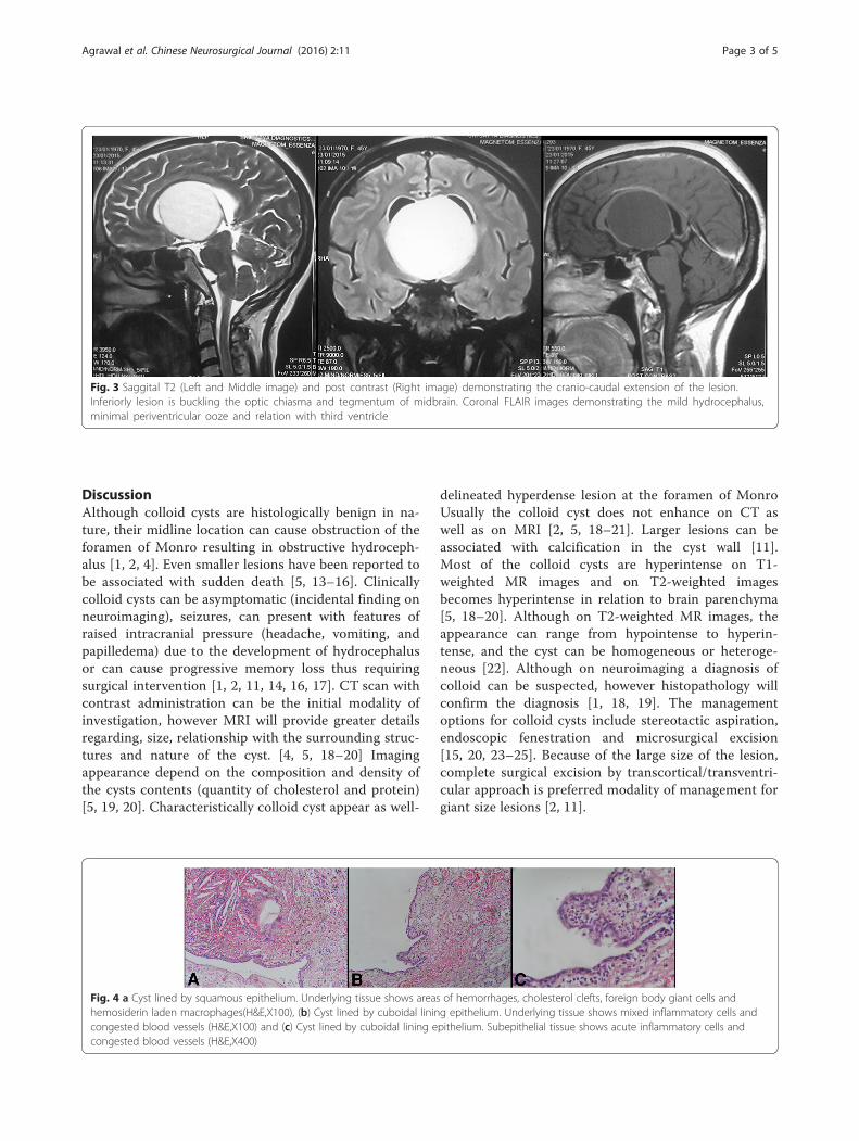
disturbances and gait disturbances. Motor and sensoryexamination was normal. Blood investigations includingcoagulation profile were normal. Axial CT precontrast im-ages showed a well-defined 5 × 5 cm size, hyper-attenuating cystic lesion in the region of foramen ofMonro leading to obstructive hydrocephalus. There wasno enhancement after contrast administration (Fig. 1).Axial MRI images confirmed the cystic nature of the le-sion, diffusion weighted sequence showed no restriction inthe cyst and on post contrast images there was no en-hancement of the cyst (Fig. 2). MRI saggital T2 and postcontrast images showed the cranio-caudal extension ofthe lesion. Inferiorly lesion was buckling the optic chiasmaand tegmentum of midbrain. Coronal FLAIR imagesshowed mild hydrocephalus and minimal periventricularooze (Fig. 3). Based on the imaging findings a diagnosis ofgiant colloid cyst was suspected. The patient underwentright fronto-parietal parasagittal large craniotomy midlineinterhemispheric transcallosal-transforaminal approachand complete excision of the cyst. Histopathology con-firmed the diagnosis of colloid cyst (Fig. 4). Post-operativeCT scan showed complete excision of the cyst with midintraventventricular hemorrhage (Fig. 5). At 1 year thepatient is doing well at follow up except mild memorydisturbances.* Correspondence: [email protected]
1Department of Neurosurgery, Narayana Medical College Hospital,Chinthareddypalem, Nellore, Andhra Pradesh, IndiaFull list of author information is available at the end of the article
CHINESE NEUROSURGICAL SOCIETYCHINESE NEUROSURGICAL SOCIETY CHINESE MEDICAL ASSOCIATION
© 2016 Agrawal et al. Open Access This article is distributed under the terms of the Creative Commons Attribution 4.0International License (http://creativecommons.org/licenses/by/4.0/), which permits unrestricted use, distribution, andreproduction in any medium, provided you give appropriate credit to the original author(s) and the source, provide a link tothe Creative Commons license, and indicate if changes were made. The Creative Commons Public Domain Dedication waiver(http://creativecommons.org/publicdomain/zero/1.0/) applies to the data made available in this article, unless otherwise stated.
Agrawal et al. Chinese Neurosurgical Journal (2016) 2:11 DOI 10.1186/s41016-016-0031-x

Fig. 1 Axial CT images pre-contrast (a) showing well defined hyper-attenuating cystic lesion in the region of foramen of monro and on post contrastimage (b) there is no enhancement. Mild dilatation of frontal horns is also noted
Fig. 2 Axial MRI images (a, b) confirming the cystic nature of the lesion. On diffusion sequence (c) there is no restriction in the cystic lesion. Postcontrast images (d) showing no enhancement within the cystic lesion
Agrawal et al. Chinese Neurosurgical Journal (2016) 2:11 Page 2 of 5
DiscussionAlthough colloid cysts are histologically benign in na-ture, their midline location can cause obstruction of theforamen of Monro resulting in obstructive hydroceph-alus [1, 2, 4]. Even smaller lesions have been reported tobe associated with sudden death [5, 13–16]. Clinicallycolloid cysts can be asymptomatic (incidental finding onneuroimaging), seizures, can present with features ofraised intracranial pressure (headache, vomiting, andpapilledema) due to the development of hydrocephalusor can cause progressive memory loss thus requiringsurgical intervention [1, 2, 11, 14, 16, 17]. CT scan withcontrast administration can be the initial modality ofinvestigation, however MRI will provide greater detailsregarding, size, relationship with the surrounding struc-tures and nature of the cyst. [4, 5, 18–20] Imagingappearance depend on the composition and density ofthe cysts contents (quantity of cholesterol and protein)[5, 19, 20]. Characteristically colloid cyst appear as well-
delineated hyperdense lesion at the foramen of MonroUsually the colloid cyst does not enhance on CT aswell as on MRI [2, 5, 18–21]. Larger lesions can beassociated with calcification in the cyst wall [11].Most of the colloid cysts are hyperintense on T1-weighted MR images and on T2-weighted imagesbecomes hyperintense in relation to brain parenchyma[5, 18–20]. Although on T2-weighted MR images, theappearance can range from hypointense to hyperin-tense, and the cyst can be homogeneous or heteroge-neous [22]. Although on neuroimaging a diagnosis ofcolloid can be suspected, however histopathology willconfirm the diagnosis [1, 18, 19]. The managementoptions for colloid cysts include stereotactic aspiration,endoscopic fenestration and microsurgical excision[15, 20, 23–25]. Because of the large size of the lesion,complete surgical excision by transcortical/transventri-cular approach is preferred modality of management forgiant size lesions [2, 11].
Fig. 3 Saggital T2 (Left and Middle image) and post contrast (Right image) demonstrating the cranio-caudal extension of the lesion.Inferiorly lesion is buckling the optic chiasma and tegmentum of midbrain. Coronal FLAIR images demonstrating the mild hydrocephalus,minimal periventricular ooze and relation with third ventricle
Fig. 4 a Cyst lined by squamous epithelium. Underlying tissue shows areas of hemorrhages, cholesterol clefts, foreign body giant cells andhemosiderin laden macrophages(H&E,X100), (b) Cyst lined by cuboidal lining epithelium. Underlying tissue shows mixed inflammatory cells andcongested blood vessels (H&E,X100) and (c) Cyst lined by cuboidal lining epithelium. Subepithelial tissue shows acute inflammatory cells andcongested blood vessels (H&E,X400)
Agrawal et al. Chinese Neurosurgical Journal (2016) 2:11 Page 3 of 5

ConclusionsIt has been suggested that neuroendoscopy is a safer andmore effective approach (shorter operating time andhospital stay) than transcallosal craniotomy and can be con-sidered as the first line of treatment [26]. However neu-roendscopy can be more challenging and demanding tomanage such a large lesions. Deep midline location, prox-imity to the vital structures and giant size of the lesions canpredispose surrounding vital structures vulnerable forinjury [1, 23, 24, 27, 28]. Injury to the vital structures canlead to transient or permanent memory loss, motor deficits,seizures, hemorrhage, hydrocephalus and infection [23, 24].
ConsentWe obtained written permission from the patient topublish this case report.
AbbreviationsCT: Computerized Tomography; MRI: Magnetic Resonance Imaging;FLAIR: Fluid attenuation inversion recovery.
Competing interestsThe authors declare that they have no competing interests.
Authors’ contributionsAll authors read and approved the final manuscript.
Author details1Department of Neurosurgery, Narayana Medical College Hospital,Chinthareddypalem, Nellore, Andhra Pradesh, India. 2Department of
Pathology, Narayana Medical College Hospital, Chinthareddypalem, Nellore,Andhra Pradesh, India. 3Department of Radiology, Narayana Medical CollegeHospital, Chinthareddypalem, Nellore, Andhra Pradesh, India.
Received: 30 November 2015 Accepted: 18 February 2016
References1. Woodley-Cook J, Martinez JL, Kapadia A, Munoz DG, Bharatha A, Spears J.
Neurosurgical management of a giant colloid cyst with atypical clinical andradiological presentation. J Neurosurg. 2014;121:1185–8.
2. Ravnik J, Bunc G, Grcar A, Zunic M, Velnar T. Colloid cysts of the thirdventricle exhibit various clinical presentation: a review of three cases. Bosn JBasic Med Sci. 2014;14:132–5.
3. Rao BH, Satyavaraprasad K, Rajiv PK, Phaneeswar T. Giant Colloid Cyst ofThird Ventricle: A Rare Case Report. Int J Sci Stud. 2015;3(6):247–250.
4. Mamourian AC, Cromwell LD, Harbaugh RE. Colloid cyst of the thirdventricle: sometimes more conspicuous on CT than MR. AJNR Am JNeuroradiol. 1998;19:875–8.
5. Armao D, Castillo M, Chen H, Kwock L. Colloid cyst of the third ventricle:imaging-pathologic correlation. AJNR Am J Neuroradiol. 2000;21:1470–7.
6. Osborn A. Miscellaneous tumors, cysts, and metastases. Diagnosticneuroradiology. St Louis: Mosby; 1994. p. 631–49.
7. Osborn AG, Maack J. Diagnostic neuroradiology. St. Louis: Mosby; 1994.8. Kasliwal MK, Kiran S, Agrawal D, Sharma BS. Giant colloid cyst in a child.
Pediatr Neurosurg. 2007;43:442–3.9. Yamanaka K, Iwai Y, Nakajima H, Kobayashi Y, Inoue T. Multiple remote brain
hemorrhages after removal of a giant colloid cyst of the third ventricle–casereport. Neurol Med Chir. 1998;38:24–7.
10. Sambasivan M, Padmanabhan S, Sambasivan M. Large colloid cyst of theanterior third ventricle associated with calcification in the cyst wall. NeurolIndia. 2010;58:330–1.
11. Yuceer N, Baskaya M, Gokalp HZ. Huge colloid cyst of the third ventricleassociated with calcification in the cyst wall. Neurosurg Rev. 1996;19:131–3.
12. Hamlat A, Casallo-Quiliano C, Saikali S, Adn M, Brassier G. Huge colloid cyst:case report and review of unusual forms. Acta Neurochir. 2004;146:397–401.discussion 401.
13. Carrasco R, Pascual JM, Medina-López D, Burdaspal-Moratilla A. Acutehemorrhage in a colloid cyst of the third ventricle: A rare cause of suddendeterioration. Surg Neurol Int. 2012;3:24.
14. Roldán-Valadez E, Hernández-Martínez P, Elizalde-Acosta I, Osorio-Peralta S.Colloid cyst of the third ventricle: case description and survey of theliterature. Rev Neurol. 2003;36:833–6.
15. Silva D, Matis G, Chrysou O, et al. Sudden death in a patient with a thirdventricle colloid cyst. Arq Neuropsiquiatr. 2012;70:311.
16. Turillazzi E, Bello S, Neri M, Riezzo I, Fineschi V. Colloid cyst of the thirdventricle, hypothalamus, and heart: a dangerous link for sudden death.Diagn Pathol. 2012;7:144.
17. Pollock BE, Huston J. Natural history of asymptomatic colloid cysts of thethird ventricle. J Neurosurg. 1999;91:364–9.
18. Kimura H, Fukushima T, Ohta T, et al. A case of colloid cyst of the thirdventricle. No Shinkei Geka Neurol Surg. 1988;16:1483–8.
19. Algin O, Ozmen E, Arslan H. Radiologic manifestations of colloid cysts: apictorial essay. Can Assoc Radiol J. 2013;64:56–60.
20. El Khoury C, Brugières P, Decq P, et al. Colloid cysts of the thirdventricle: are MR imaging patterns predictive of difficulty withpercutaneous treatment? AJNR Am J Neuroradiol. 2000;21:489–92.
21. Maeder PP, Holtås SL, Basibüyük LN, Salford LG, Tapper UA, Brun A. Colloidcysts of the third ventricle: correlation of MR and CT findings with histologyand chemical analysis. AJR Am J Roentgenol. 1990;155:135–41.
22. Wilms G, Marchal G, Van Hecke P, et al. Colloid cysts of the third ventricle:MR findings. J Comput Assist Tomogr. 1990;14:527–31.
23. Desai KI, Nadkarni TD, Muzumdar DP, Goel AH. Surgical management ofcolloid cyst of the third ventricle–a study of 105 cases. Surg Neurol.2002;57:295–302. discussion 302.
24. Grondin RT, Hader W, MacRae ME, Hamilton MG. Endoscopic versusmicrosurgical resection of third ventricle colloid cysts. Can J Neurol Sci.2007;34:197–207.
25. Kapu R, Pande A, Vasudevan MC, Ramamurthi R. Giant colloid cyst of thirdventricle with microhemorrhages causing neurological deterioration: a veryrare presentation. Neurol India. 2012;60:557–8.
Fig. 5 Post-operative CT image showing complete excision of thelesion with mild intraventricular hemorrhage
Agrawal et al. Chinese Neurosurgical Journal (2016) 2:11 Page 4 of 5

26. Horn EM, Feiz-Erfan I, Bristol RE, et al. Treatment options for third ventricularcolloid cysts: comparison of open microsurgical versus endoscopicresection. Neurosurgery. 2007;60:613–8. discussion 618-620.
27. Levine NB, Miller MN, Crone KR. Endoscopic resection of colloid cysts:indications, technique, and results during a 13-year period. Minim InvasiveNeurosurg. 2007;50:313–7.
28. de Witt Hamer PC, Verstegen MJT, De Haan RJ, et al. High risk of acutedeterioration in patients harboring symptomatic colloid cysts of the thirdventricle. J Neurosurg. 2002;96:1041–5.
• We accept pre-submission inquiries
• Our selector tool helps you to find the most relevant journal
• We provide round the clock customer support
• Convenient online submission
• Thorough peer review
• Inclusion in PubMed and all major indexing services
• Maximum visibility for your research
Submit your manuscript atwww.biomedcentral.com/submit
Submit your next manuscript to BioMed Central and we will help you at every step:
Agrawal et al. Chinese Neurosurgical Journal (2016) 2:11 Page 5 of 5