giuseppe bigatti - isge · welcome 1 histeroscopy pictures 2 osseous metaplasia interview of the...
TRANSCRIPT

+
Welcome 1
Histeroscopy Pictures 2Osseous metaplasia
Interview of the month 3Bruno van Herendael
Brief Review 5Fluid intra-vasation syndromePathophysiology & Prevention
Conundrums 10What's your opinion aboutthis hysteroscopy?
Original Article 13Intrauterine adhesions (IUAs) & hysteroscopic myomectomy
Hysteroscopy & Fertility 16An Update (II)
Fresh Projects 20Italian School of Endoscopy
www.hysteroscopy.info
1
INSIDE THIS ISSUED
Giuseppe Bigatti
May-Jun 2017 | vol. 3 | issue 3
uring the last few years the world of hysterscopy has revealed to be very active in improving its technique. All the new trends could be summarized in two main directions. The first big innovation, following the indications of Prof. Stefano Bettocchi, has been a reduction in size of all diagnostic and operative hysteroscopes in order to approach all patients in an office set up. In this respect the Trophy scope by Dr. Rudi Campo fulfilled this new trend. The Trophy scope with its small diameter can be used as diagnostic hysteroscope during office procedures and in case of operative necessity an additional operative sheet can be pushed into the uterine cavity allowing this option. Another big attempt in miniaturizing hysteroscopic instruments is the Gubbini resectoscope. Thanks to its small diameter Dr. Giampietro Gubbini has shown the possibility to approach with the resectoscopic technique all major intrauterine pathologies in an office set up. This great innovation still can be listed into the attempt of miniaturization of all hysteroscopic equipment.
The second main revolution in the world of hysteroscopy concerns the possibility to remove the tissue chips during operative procedures at the same time of their resection. This new philosophy in operative hysteroscopy is trying to find an alternative solution to conventional resectosopy in order to reduce all complication related to this technique. In this respect two main instruments has been created by Prof Mark Hans Emanuel and I. Emanuel described a morcellation technique while I speak of the shaver technique. Despite their technical differences both instrument follow the same idea which is to remove the tissue while resecting it allowing always a good visualization and reducing the complication rate. Originally these two techniques were built for operative hysteroscopy but as the technique improves will also be used during office procedures.
After more than 7 years of experience, the first shaver operation was performed in June 2009; it is time for a first review.
I have personally done more than 1000 operations with a very low complication rate. I came to the conclusion that for polypectomy this technique, especially if is performed for infertility problems, should be the first choice procedure in order to minimize side effects on endometrium and to increase the success rate of pregnancy achievement. The shaver technique has proven to be a very fast, easy, precise and safe procedure. In addition, we have approached all types of myomas showing that “it is possible” to remove submucosal myomas with this technique.
The future will bring the possibility to approach most of the pathologies in a office set up with a newly designed optical system and my efforts will be concentrated to approach in a successful way large submucosal myomas. I am personally very optimistic regarding the future applications of the shaver technique. My experience has been shared with more than 90 hysteroscopists coming from all over the world in Italy to learn this new hysteroscopic philosophy. To these colleagues goes all my gratitude and appreciation for the future of Operative hysteroscopy.

TEAM COODINATORSPAIN
L. Alonso
EDITORIAL COMMITTEE
SPAINE. Cayuela
L. Nieto
ITALYG. Gubbini
A. S. Laganà
USAJ. CarugnoL. Bradley
MEXICOJ. Alanis-Fuentes
PORTUGALJ. Metello
ARGENTINA A. M. Gonzalez
VENEZUELAJ. Jimenez
SCIENTIFIC COMMITTEEA. Tinelli (Ita)
O. Shawki (Egy)A. Úbeda (Spa)A. Arias (Ven)
M. Rodrigo (Spa)A. Di Spiezio Sardo (Ita)
E. de la Blanca (Spa)A. Favilli (Ita)
M. Bigozzi (Arg)S. Haimovich (Spa)
R. Lasmar (Bra)A. Garcia (USA)N. Malhotra (Ind)
J. Dotto (Arg)I. Alkatout (Ger)
R. Manchanda (Ind)M. Medvediev (Ukr)M. Elessawy (Ger)
All rights reserved. The responsibility of the signed
contributions is primarily of the authors and does not necessarily reflect the views of the editorial or scientific committees.
HYSTEROSCOPY
PICTURES
www.hysteroscopy.info
2
This type of metaplasia is a rare condition in which there is a transformation of the normal endometrial tissue into bone. It is an uncommon clinical finding with an incidence of 0,3/1000 and most cases occur after miscarriage or abortion. The presence of bone in the endometrium was first described by Virchow who related this condition to a spontaneous differentiation of fibroblasts into osteoblasts. Typically, this type of metaplasia occurs during reproductive years and more than 80% of reported cases ocurr after pregnancy.
There are two main theories to explain the existence of bone fragments in the endometrial tissue. The one by Thaler, who relate this entity to retention of osseous fetal parts after abortion or miscarriage after 12 weeks of pregnancy. This first theory cannot explain cases that occurr in patients without previous pregnancies. The second theory is that of a true endometrial osseous metaplasia, in which there is a osseus transformation of the endometrial stromal cells, this metaplasia is consequence of irritative, toxic or hormonal stimuli. Probably both theories are right, with cases of true metaplasia and cases in which retained bones, causes an endometrial inflammation which leads to a secondary osseous metaplasia.
If you are interested in sharing your cases or have a hysteroscopy image that you consider unique and want to share, send it to [email protected]
Superficial vaginal endometriotic implant
Osseous metaplasia of the endometrium
May-Jun 2017 | vol. 3 | issue 3
Long bone structure as a result of osseous metaplasia

www.hysteroscopy.info
INTERVIEW WITH...Dr. Bruno van Herendael was founder of the European Society of Hysteroscopy in 1985 and promoted the first international congress on hysteroscopy. A man with an advanced mind in contrast to his time.
Bruno Van Herendael
Consultant Endoscopic Gynaecological Surgery at
ZiekenHuis Netwerk Antwerpen (ZNA)
Antwerp Area, Belgium
You founded the European Society of hysteroscopy in 1985. It was something ahead on his time?
When we did found the European Society for Hysteroscopy (ESH) it was something the endoscopic community felt as a need. We had a lightening example in Hans Jochen Lindeman. He was the teacher of my teacher Harry Van der Pas. We the got together with the people of the “Institut Dexeus” in Barcelona more especially Santiago Dexeus and Ramon Labastida. We came to Barcelona and did found the ESH. We were surprised that this society did become an international society in the sense that we did have members from all over the world not only Europe. So it must have be something a lot of colleagues were waiting for.
Do the different gynecological societies pay adequate attention to the hysteroscopy?
Most established societies do have a genuine interest in hysteroscopy. AAGL has pre-congress courses in hysteroscopy, ESGE has sessions on hysteroscopy, ISGE does “Teach the Teachers” sessions on hysteroscopy mainly in Africa and has specific pre-congress courses on hysteroscopy i.e. during our last congress in Montego Bay Jamaica over one full day. ISGE also has one key note speech at its congresses dedicated on hysteroscopy.
What`s the role of the ISGE in the promotion of the hysteroscopy?
As ISGE is a society focused on third world countries we do focus on in office or ambulatory hysteroscopy. Here we do train the local people at our centres of excellence in Yaoundé, Cameroon and Mona Kingston Jamaica. ISGE does join transvaginal laparoscopy and hysteroscopy both diagnostic and operative in the training as both techniques can be used without anaesthesia.
”So it must have be something a lot of colleagues were waiting for”
Just as hysteroscopy has become an essential part of modern gynecologic practice, outpatient procedures in gynecology and gynaecologic surgery with all of the associated benefits to both patient and practice, have increased dramatically in recent years. Ambulatory Hysteroscopy is, first and foremost, a highly practical and rounded manual for the beginner and an essential reference for the experienced practitioner. From a global perspective, this new text addresses all of the practical, diagnostic, operative, administrative, teambuilding, and training issues associated with practicing ambulatory hysteroscopy. Ambulatory Hysteroscopy captures the knowledge and experience of those who have pioneered and refined outpatient procedures. The text is concise, clear, hugely informative and beautifully illustrated. This text is easy to read and easy to use!
3
May-Jun 2017 | vol. 3 | issue 3

SCIENTIFIC COMMITTEEA. Tinelli (Ita)
O. Shawki (Egy)A. Úbeda (Spa)A. Arias (Ven)
M. Rodrigo (Spa)A. Di Spiezio Sardo (Ita)
E. de la Blanca (Spa)A. Favilli (Ita)
M. Bigozzi (Arg)S. Haimovich (Spa)
R. Lasmar (Bra)A. Garcia (USA)N. Malhotra (Ind)
J. Dotto (Arg)I. Alkatout (Ger)
R. Manchanda (Ind)M. Medvediev (Ukr)M. Elessawy (Ger)
All rights reserved. The responsibility of the signed
contributions is primarily of the authors and does not necessarily reflect the views of the editorial or scientific committees.
4
www.hysteroscopy.info
The ISGE is a really international Society. Is that its strength?
The problem os ISGE is that it is a real international society where the Board member do come from the different continents according to the repartition incorporated in the bylaws of the society. This means that we do have board members from America North (USA and Canada) America Central and South, Europe, Africa, Middle East, Asia and Oceania (Australia and New Zealand). The advantage is that we do get impulses from all different continents and religions. The difficulty is that the communication can be difficult at times. Hence we did build a permanent secretariat in Italy as to be able to be in contact with the members and to organise the teleconferences between the Executive Board on monthly bases and more often if necessary and with the organizers of the ISGE conferences.
is it now time to separate between gynecological hysteroscopist and laparoscopists?
I am not qualified to answer that question as I would never have separated hysteroscopy and laparoscopy in my practice. I did perform very advanced laparoscopy and very advanced hysteroscopy and this is now the tradition in the division of gynaecological surgery in our department. During my tenure as professor gynaecological endoscopy first at the university of Pavia and later in Varese at the Università dell’Insubria I did always teach both techniques as being the armamentarium of the gynaecologists. I was the one who together with Professor Maurice Bruhat did bring the ESH within the ESGE. We had several meetings at his home in Clermont Ferrand. We did stay very good friends afterwards and did work together in Africa until he unfortunately passed away. Our main drive was to get the gynaecologist together mastering the two techniques against the interventional radiologists and surgeons. In my opinion both techniques are equally important for gynaecologist. What could be realised is specific units for ambulatory hysteroscopy as these are easy to be realised and can work without anaesthesia. However, when visiting my friends in Jamaica I did realise that they have real operating suites in their private practise combining both. If we would separate laparoscopy and hysteroscopy I would go for the criterion of local or no anaesthesia and combine hysteroscopy with transvaginal laparoscopy as my feeling is that most gynaecologists do want some method or the other to be able to intervene and do some operative gestures, other than intra-uterine pathology.
Do you have any advice for the young physician that is starting out in the world of gynecologic minimally invasive surgery?
A young physician embarking in our speciality I always advise to be aware of the differences in the speciality like there are fertility, oncology etc. and if he or she wants to become pelvic surgeon they have to master all techniques within the spectrum of endoscopy: laparoscopy, hysteroscopy and transvaginal laparoscopy. We do train with virtual reality and I always stress the fact that laparoscopy is different form hysteroscopy and that not all of them will be able to perform hysteroscopy at the highest level as this discipline is more difficult than laparoscopy due to the reduced space and the scarcity of referral points.
” As ISGE is a society focused on third world countries
we do focus on in office or ambulatory hysteroscopy”
” This discipline is more difficult than laparoscopy due to the
reduced space and the scarcity of referral points”
May-Jun 2017 | vol. 3 | issue 3

www.hysteroscopy.info
5
Brief Review
Fluid intra-vasation syndromePathophysiology & Prevention
IVANO RAIMONDO ¹ – GENNARO RAIMONDO ²1- Clinica Ginecologica ed Ostetrica Università degli Studi di Sassari, Italia- 2- Clinica Mediterranea, Napoli. Italia.
The fluid intra-vasation syndrome, also known as Fluid Overload Syndrome, Intravascular Absorption Syndrome of Surgical Hysteroscopy (IAsSH) or Tur(p) Syndrome, is a terrible complication of resectoscopic surgery, which manifests itself acutely after the massive and rapid absorption of the distention medium.
It is a possible complication of any "liquid medium" surgery, and is therefore present not only in urology or gynecology field but also in arthroscopies (Chai C, 1996), rectal tumor interventions, treatment of nephrolithiasis and percutaneous renal ultrasound (Dimberg 1993) and even during cardiac ablation (Di Biase l., 2013)
It is difficult to define the amount of fluid from which the syndrome begins to appear, and its onset depends not only on the intravascular volume but also on the type of fluid, the procedure performed, the general conditions of the patient, age and finally of subjective factors that sometimes are not identified.
Since resectoscopy is performed in two different ways - monopolar and bipolar - and therefore with two different types of distension media - non-conductors and conductors. (2013 AAGL, J Minim Invasive Gynecol.)
The incidence of the syndrome is around 5% of the resectoscopic procedures, including patients who have developed less obvious or subclinical forms. (Shveiky D, 2007)
May-Jun 2017 | vol. 3 | issue 3

www.hysteroscopy.info
6
Pathophysiology
To understand how intravasation is originated, it is necessary to start from the venous sinuses. These, also present in other organs such as the prostate, are very particular vessels because they have no muscular layer and are constituted only of endothelium; Have an extremely low internal pressure of 10-15 mmHg and are present in the myometrium about 5 mm below the endometrium. During the intervention, liquids with an unsuspected high flow (200ml/min.) can be intravased (Hahn RG, 1993).
The uterine cavity is virtual and therefore its distension is mandatory to allow visualization during hysteroscopic procedures, both diagnostic and operative. It is believed that a pressure of about 30 mmHg is required to separate the uterine walls but to obtain adequate vision, a pressure within the cavity of about 50-80 mmHg is indispensable.
Therefore, intravasation occurs if there are liquid entry doors - venous sinuses - and if there is a pressure that introduces and liquid inside them. Inevitably, some resectoscopic procedures such as myomectomy, endometrial ablation, upon reaching the myometrium, cut the venous sinuses and intravasation occurs. If the amount of fluid absorbed is excessive, a series of symptoms (intravasation syndrome), which can be attributed substantially to hypervolemia and hyponatremia, begin to appear if non-conductive liquids are used - monopolar surgery - or only hypervolemia in the case of conductive liquids - bipolar surgery.
Hypervolemia, understood as an increase in the plasma concentration in blood, is generated by any means of distension, electrolytic or not. This situation alters the normal relationship between extracellular fluid (ECF), which accounts for 20% of body weight and intracellular fluid, which accounts for 40% of body weight. The increase in extracellular fluid causes a decrease of the hormone ADH (one of the first symptoms of intravasation is in fact increased diuresis with diluted urine), increased blood pressure and central venous pressure as well as inhibition of the renin-angiotensin system. Consequently, hypoaldosteronism and the activation of atrial natriuretic hormone produce water excretion and natriuria.
By the principle of osmolarity (the intra- and extracellular compartment must be equilibrated) the water from the extracellular compartment goes to the intracellular compartment generating edema (pulmonary, cerebral, subcutaneous)
An increase in blood volume ends up directly affecting cardiac output with possible cardiogenic shock (right or left).
Hyponatremia (hemodilution) is a severe complication that physician performing resectoscopic procedures should know how to manage; There is a scale that correlates the decrease in plasma sodium with the onset and progressive increase of the symptoms attributable to it.
Hyponatremia is diagnosed when the serum sodium falls below 135 mEq / L; above these values the patient does not present significant symptoms. Between 130 and 135 mEq / L there is mainly of gastrointestinal symptoms (vomit, nausea); below 130 mEq / L muscular symptoms (tremors) and neurological symptoms (confusion, disorientation, agitation, stupor); with values lower than 125, severe cardiac arrhythmias and coma are present. It is recognized that treatment of hyponatremia should be initiated when it reaches or falls below 135 mEq / L.
May-Jun 2017 | vol. 3 | issue 3

www.hysteroscopy.info
7
It is generally accepted that every 7-10 mmol / L that decreases of Na corresponds to an absorption of 1000 ml of non-electrolytic solution (Istre O, 1994) and that on average severe hyponatremia (around 125 mEq / L) occurs after absorption of 3-4 L of urological solution. Therefore, in the scale of intravasation syndrome with liquids without electrolytes, it is necessary to consider that the severe damages derived from hyponatremia occur and are evident in after hypervolemia.
Precautionary measures
While it is true that intravasation of liquids by the opening of the venous sinuses in certain resectoscopic procedures is inevitable, it is necessary to implement measures to prevent fluid overload syndrome.The parameters on which it is possible to act are: the fluid pressure, the duration of the procedure, the selection and fluid balance and the caliber of the instruments. In theory, to avoid the passage of fluid to the venous sinuses the distention media inside the uterine cavity should be at a very low pressure, which is unthinkable.
Therefore, it is evident that working with the lowest possible pressure or adjusting it to the Average Mean Arterial Pressure MAP 70-110 mmHg (Mean Arterial Pressure = diastolic pressure value + (the difference between systolic and diastolic pressure / 3) is a useful measure, it should not be considered an absolute measure that if followed will prevent intravasation, nor is there a procedure length limit below which the syndrome does not happens with regard to the duration of the intervention; In fact, both based on personal experience and in published data, there are cases that begin within a few minutes of the beginning of the procedure until 24 hours after the end of the intervention (a peritoneal fluid reabsorption is allowed) ( Mc Swiney, 1995)
Since hypervolemia and hyponatremia are the elements that trigger the process, the way to act in prevention is monitoring both.
The control of fluid balance represents the cardinal point of prevention: the difference between the fluid used and recovered should reflect the amount absorbed. This can be done manually using graduated containers or with the use of a scale that weighs the recovered fluid, but in both cases the estimation may not be accurate.
The errors are based on the loss of fluid not calculated during the procedure or the inaccurate fluid balance.
Hysteroscopy Newsletter
May-Jun 2017 | vol. 3 | issue 3

www.hysteroscopy.info
8
A recent method (commonly used in our practice) is one that provides for the use of 1.5% glycine labeled with 1% ethanol. The principle is based on the determination of the concentration of this alcohol in the air expired by the patient, determined by a device (ethylene): the values obtained allow us to determine the amount of intravasated liquid. The problems often associated with this measuring technique are generally related to the ethylene which from time to time must be calibrated. Obviously, this method is almost exclusive for monopolar surgery. This is determined during the procedure or immediately at the end of the intervention and is probably the safest means to identify the syndrome and its severity.
With respect to the selection of the distention media, this obviously is determined by the type of surgery to be performed: there is no ideal media, all distention media available have some side effects. The size of the instruments deserves a brief mention: there is a rather generalized tendency to increasingly miniaturize its caliber, with the aim of being always less invasive and reducing the flow of fluid to the uterine cavity and therefore intravasation. This is undoubtedly an appreciable reason but it must be considered that the smaller the instrument, the smaller the fragments of tissue that will be obtained. As a result, the use of miniaturized instrument will prolong procedure time than when using a traditional one. It is important to consider that time is a factor that affects the onset of the syndrome.
To conclude with preventive measures, it is important to note that the resectoscopic is a surgical procedure that allows to stop of the procedure at any moment; At the slightest doubt of the onset of the syndrome, the "knowing to stop" at the right time should be part of the hysteroscopist surgeon's culture.
In conclusion, it is important to note:
- If intravasation is inevitable, fluid overload syndrome should be avoided by carrying out prevention measures, working in teams involving the surgeon, the anesthetist and the staff of the surgical room.
- The surgeon should perform major resectoscopic surgery only after formal training
- Fluid Overload Syndrome is not only hyponatraemia but also (and before) hypervolemia.
- Hypervolemia is a common symptom of intravasation resulting from both the use of solutions with electrolytes and without electrolytes. Bipolar surgery does not exempt the surgeon from adopting measures of prevention of intravasation, in fact, "the false sense of security generated using saline solution could create serious dangers for the patient's life" (Glasser, 2005).
- Fluid overload syndrome generated by the use of solutions without electrolytes are more complex and more difficult to treat since hyponatremia is present, but it should be remembered once again that only occurs if the absorption exceeds the recommended the accepted amount (1,000 ml)
Hysteroscopy Newsletter
May-Jun 2017 | vol. 3 | issue 3

www.hysteroscopy.info
9
Hysteroscopy Newsletter
Applications of laser diode in the outpatient
management of endometrial pathology
Sergio Haimovich
During the last decade, we have witnessed changes in hysteroscopy techniques and the development of new and more efficient devices. We moved from a first step diagnostic hysteroscopy followed by a second step surgical hysteroscopy to the “see and treat” concept. Everything is done in a single step.
Over the last 10 years, we have been using diode laser as energy source for in office hysteroscopy. We have developed different surgical techniques for the treatment of endometrial pathologies.
In this book, we illustrate our experience using diode laser in office hysteroscopy setting for the treatment of Endometrial Polyps, Leiomyomas and Uterine Septum. We describe different techniques using laser and their limitations, the success rates and the patient’s tolerance of these procedures.
We hope that you will be able appreciate the advantages of using laser energy in hysteroscopy for the management of endometrial pathology in an office setting without the need of anesthesia.
WHAT'S YOUR DIAGNOSIS?
Sometimes, when performing hysteroscopy, it is important to pay attention to every corner of the uterus, as Vasari stated «cerca trova», «he who
seeks finds»
Answer to the previous issue: Retained products of conception
May-Jun 2017 | vol. 3 | issue 3
www.hysteroscopy.info
10
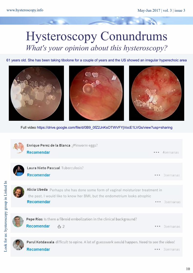
Hysteroscopy ConundrumsWhat's your opinion about this hysteroscopy?
61 years old. She has been taking tibolone for a couple of years and the US showed an irregular hyperechoic area
Loo
k fo
r us
: hys
tero
scop
y gr
oup
in L
inke
d In
A recent method (commonly used in our practice) is one that provides for the use of 1.5% glycine labeled with 1% ethanol. The principle is based on the determination of the concentration of this alcohol in the air expired by the patient, determined by a device (ethylene): the values obtained allow us to determine the amount of intravasated liquid. The problems often associated with this measuring technique are generally related to the ethylene which from time to time must be calibrated. Obviously, this method is almost exclusive for monopolar surgery. This is determined during the procedure or immediately at the end of the intervention and is probably the safest means to identify the syndrome and its severity.
With respect to the selection of the distention media, this obviously is determined by the type of surgery to be performed: there is no ideal media, all distention media available have some side effects. The size of the instruments deserves a brief mention: there is a rather generalized tendency to increasingly miniaturize its caliber, with the aim of being always less invasive and reducing the flow of fluid to the uterine cavity and therefore intravasation. This is undoubtedly an appreciable reason but it must be considered that the smaller the instrument, the smaller the fragments of tissue that will be obtained. As a result, the use of miniaturized instrument will prolong procedure time than when using a traditional one. It is important to consider that time is a factor that affects the onset of the syndrome.
To conclude with preventive measures, it is important to note that the resectoscopic is a surgical procedure that allows to stop of the procedure at any moment; At the slightest doubt of the onset of the syndrome, the "knowing to stop" at the right time should be part of the hysteroscopist surgeon's culture.
In conclusion, it is important to note:
- If intravasation is inevitable, fluid overload syndrome should be avoided by carrying out prevention measures, working in teams involving the surgeon, the anesthetist and the staff of the surgical room.
- The surgeon should perform major resectoscopic surgery only after formal training
- Fluid Overload Syndrome is not only hyponatraemia but also (and before) hypervolemia.
- Hypervolemia is a common symptom of intravasation resulting from both the use of solutions with electrolytes and without electrolytes. Bipolar surgery does not exempt the surgeon from adopting measures of prevention of intravasation, in fact, "the false sense of security generated using saline solution could create serious dangers for the patient's life" (Glasser, 2005).
- Fluid overload syndrome generated by the use of solutions without electrolytes are more complex and more difficult to treat since hyponatremia is present, but it should be remembered once again that only occurs if the absorption exceeds the recommended the accepted amount (1,000 ml)
Full video https://drive.google.com/file/d/0B9_0fZ2JnKsOTWVFYjVocE1LVGs/view?usp=sharing
May-Jun 2017 | vol. 3 | issue 3
Perhaps she has done some form of vaginal moisturizer treatment in
the past. I would like to know her BMI, but the endometrium looks atrophic

www.hysteroscopy.info
11
Loo
k fo
r us
: hys
tero
scop
y gr
oup
in L
inke
d In
May-Jun 2017 | vol. 3 | issue 3

www.hysteroscopy.info
12
May-Jun 2017 | vol. 3 | issue 3

Aim of the study:
To evaluate the prevalence of intrauterine adhesions (IUAs) after myoma resectoscopy.
Type of the study:
Multicenter, Prospective descriptive non-randomized interventional trial
Background and justification of the study:
Intrauterine adhesions (IUA) are fibrous strings at opposing walls of the uterus. The spectrum of IUA formation may vary from minimal IUA to the complete obliteration of the uterine cavity.
IUA formation is the major long-term complication of operative hysteroscopy in women of reproductive age. According to a randomized controlled trial (RCT) on the effectiveness of preoperative treatment before operative hysteroscopy, the incidence of post-surgical IUA at “second look” hysteroscopy is 3.6 % after polyp removal, 6.7 % alter resection of uterine septa, 31.3 % after removal of a single fibroid, and 45.5% after resection of multiple fibroids [3].
www.hysteroscopy.info
13
Original Article Epidemiological evaluation of intrauterine adhesions
(IUAs) after hysteroscopic myomectomy by resectoscopy
Haimovich S¹., Alonso L2, Laganá A3., Costa L4, Moratalla E5, Gica N6, Nappi L7, Stamenov G8, Fung L9, Gonzalez A¹0, Lasmar R¹1.
1- Hospital del Mar, Barcelona Spain; 2- Centro Gutenberg, Malaga, Spain; 3- University of Messina, Italy; 4- Hospital Parc Tauli, Sabadell, Spain; 5- Hospital Universitario Ramón y Cajal, Madrid, Spain; 6- Filantropia Clinical Hospital. Bucharest. Romania; 7- University of Foggia. Italia; 8- Nadezhda
Women's Health Hospital,Sofia, Bulgaria; 9- Prince of Wales Hospital & North District Hospital. Hong Kong; 10- Hospital Naval C.M. Pedro Mallo, Buenos Aires Argentina; 11- Hospital Universitário Antônio Pedro - Rio de Janeiro,Brazil
Hysteroscopy NewsletterHysteroscopy Newsletter
May-Jun 2017 | vol. 3 | issue 3

There is no strong evidence regarding the prevalence of IUA after myomectomy by resectoscopy and this is the first multi-centrical study design in order to determinate this prevalence.
Patients and Methods
Using Hysteroscopy Newsletter as the main tool for communication (http://www.hysteroscopy.info),an invitation for participation was published. 11 centers from 7 countries were included in the study.
Every center passed the local Ethical Committee. 112 patients were recruited, all of them with the diagnostic of at least 1 submucous myoma, and were scheduled for a resectoscopy in surgery room under anesthesia. Age between 25 and 49 (41.9). 44% with one previous delivery and 50% with 2. Indication was Heavy Menstrual Bleeding in 77.9% and infertility in 22.1%.(Fig1)
The myoma sizes (Fig.2): 34.9% between 2-3cm, 25.6% 1-2cm, 23.3% 3-4cm and 14% > 4cm. According to ESGE classificationx(Fig. 3), 53.5% were G1, 24.4% G0 and 22.1% G2. 86% with a single myoma. Between 1 and 3 month after surgery a “second look” hysteroscopy was performed to confirm the presence of IUA. The IUA adhesion were classified according to ESGE classification of Stages 1 to 3.
Results
Complete resección was achieve in 86% of the cases. No adhesions in 90/112 (80%), stage 1 (mild/filmy) 19/112 (16%) and stage 2 in 5/112 (4%)
Conclusions
In our study the prevalence of IUA was of 20%, most of them mild (16%) but high enough to justify performing a 2nd look hysteroscopy, especially in patients with infertility.
14
www.hysteroscopy.info May-Jun 2017 | vol. 3 | issue 3

15
www.hysteroscopy.info
DID YOU KNOW...?
Later studies have shown a correlation of only 65% between findings diagnosed with hysterosalpingogram (HSG) compared
with those diagnosed with hysteroscopy.
Integrity of the uterine cavity is critical to embryonic implantation and it is mandatory evaluate it in the routine investigation of
infertility
May-Jun 2017 | vol. 3 | issue 3

FIBROIDS
Fibroids or myomas are benign, monoclonal tumors of the uterus, mostly composed of smooth muscle cells and extracellular matrix. They are the most common solid tumor of the female pelvis. The prevalence varies widely, according to age, ethnicity, family and might be as high as 80% at age 50. They can be asymptomatic, but around 25% of women might have pain or menorrhagia.Fibroids can be found in up to 10% of infertile women and can be the only abnormal finding in around 2,5%.
Fibroids and infertility
A review by Pritts et al. (2009) concluded that fibroids causing intracavitary distortion result in decreased rates of clinical pregnancy, implantation and livebirth, as well as an increased rate of spontaneous miscarriage.
Although no evidence was found against sub-serosal myomas, concerning intramural fibroids the same author concluded that fibroids with no intracavitary involvement, had also decreased rates of implantation and live birth, and increased rate of spontaneous miscarriage.
Another review by Sunkara (2010) concluded that there was a significant decrease in the live birth in the presence of intramural fibroids that distort the endometrial cavity.
Several explanations have been proposed, mostly related to impairment of the uterine peristalsis, vascular flow as well as disruption of sperm and ovum transportation and embryo implantation.
However other studies are against the conclusion of Pritts, when concerning intra mural fibroids. A cohort study by Somigliana with 238 patients comparing the rate of success of IVF in women with small (less than 50mm) fibroids not encroaching the endometrial cavity in asymptomatic patients selected for IVF, concluded that such fibroids did have an impact on the rate of success of the procedure. Bodzag at all. reached the same conclusion after comparing 61 cases against 444 controls.
In 2015 a Cochrane review addressing this question concluded that probably there might be a trend towards the benefit of removal of submucous fibroids in women with otherwise unexplained subfertility. The odds ratio in a group having regular fertility-oriented intercourse during 12 months for the outcome of clinical pregnancy was 2,44 (95% confidence interval 0.97- 6.17, p=0.06). Concerning miscarriage, there was no evidence of a difference between the groups.
www.hysteroscopy.info
16
Hysteroscopy and Fertility: an Update (II)
José Metello (a,b) José Jiménez (c,d)a Hospital Garica de Orta (Almada, Portugal), b Ginemed-Maloclinics (Lisbon, Portugal),
c Clinica “Leopoldo Aguerrevere” (Caracas, Venezuela), d Unidad de Fertilidad Unifertes (Caracas, Venezuela)
Hysteroscopy Newsletter
Hysteroscopy Newsletter
May-Jun 2017 | vol. 3 | issue 3

www.twitter.com/hysteronews
HYSTEROscopy group
Hysteroscopy newsletter
Hysteroscopy newsletter
www.facebook.com/hysteronews
17
www.hysteroscopy.info
Miomectomy
When the need for surgery has been decided, as always the minimally invasive approach should be chosen as long as this will also be the safer option for the patient. So before a transcervical or hysteroscopic myomectomy is considered at least these factors should be considered:
-Size: generally fibroids up to 4 or 5 cm can be removed histeroscopically. The surgeon should take into
consideration the volume of the myoma as the sfere volume can be calculated: (π x radius)3 x 4/3. So a 1 cm diameter fibroma will have 0,5 ml, while a 2cm 4,2 ml, a 3 cm 14 ml, a 4 cm 33,5 ml and a 5 cm 65ml. Time of the procedure and fluid spent will increase exponentially in the bigger fibroids.
-Number: usually more than three myomas should be considered a limitations, specially if they cover more than 50% of the endometrial surface -Location: usually upper type II myomas, near the uterine cornua will be more difficult and the danger will increase with a closer proximity to the uterine serosa.
-Endometrial preparation: the proliferative phase or during contraceptive pill allow for a thinner endometrium
-Coexisting pelvic disease
-Surgical expertise: As in other surgeries this is probably the most important factor to decide which technique should be used. The Step-W classification by R. Lasmar (LASMAR) might be useful. Five parameters are pontuated from 0 to 2: size, topography, base extension, pernetration and localization on a lateral wall.
-Appropriate equipment: while a forceps might be enough for a very small myoma, a electric device like versapoint® can be used for a 1-2cm fibroid, specially if it is a type 0 or I. A resectoscope or a similar device should be the first choice for bigger myomas.
UTERINE SEPTUM
It is difficult to determine the incidence of congenital uterine malformations in the general population because most affected women do not experience reproductive problems. The incidence has been calculated to be 1 or 2 per 1000 women and as high as 15 per 1000 women. Some studies have reported a 12% incidence (7).
Definition
A uterine septum is believed to develop as a result of failure of resorption of the tissue connecting the two paramesonephric (müllerian) ducts prior to the 20th embryonic week. While the arcuate uterus represents the mildest form of resorption failure, unlike the septum, it is not considered clinically relevant. Septate uteri have a spectrum of configurations including incomplete/partial septate to complete septate uterus.
Initially, uterine septa were believed to be predominantly fibrous tissue. However, biopsy specimens and magnetic resonance imaging (MRI) suggest that septa are composed primarily of muscle fibers and less connective tissue.
Hysteroscopy Newsletter
May-Jun 2017 | vol. 3 | issue 3

Müllerian anomalies in general may be associated with renal anomalies in approximately 11% to 30% of individuals.
However, data do not exist to suggest an association between septate uterus and renal anomalies and, as such, it is not necessary to evaluate the renal system in all patients with a uterine septum.
Uterine septum and fertility
High-quality studies on the clinical impact of the septate uterus in fertile women are lacking but the condition seems to be associated with adverse pregnancy outcomes (increased miscarriage, preterm delivery and breech presentation rates). The effect of a septum on fertility is less clear. The prevalence of the septate uterus in the infertile population is similar compared with a population of patients who underwent investigation of the uterus for other indications (e.g. sterilization, pelvic pain and abnormal bleeding). This suggests that a uterine septum does not play a role in the process of conception as such.
Not only the septate uterus, but also the arcuate uterus is characterized by a certain degree of indentation of the fundus into the uterine cavity. The arcuate uterus is a variation of normal uterine anatomy and is not associated with adverse pregnancy outcomes or infertility. Due to the differences in pregnancy outcome, correct diagnosis of both conditions is essential. In a recent study, however, the agreement on the diagnosis of the septate uterus based on hysteroscopy was demonstrated to be poor with differentiation between a septate and arcuate uterus appearing to be especially difficult. One of the larger studies compared 153 women with all types of uterine anomalies to a control group of 27 women with a normal uterus. In the 33 women diagnosed with a septate uterus there was a higher incidence of infertility compared with controls (21.9% vs 7.7%); however, this difference did not reach statistical significance. One study evaluated infertility in women with müllerian anomalies compared with those with external genital anomalies and a normal uterus. When all other causes had been excluded, infertility was not seen more frequently in the 17 women with a septate uterus. In another study, 33 women were followed prospectively for 24 months after hysteroscopic diagnosis of arcuate and septate/bicornuate uteri. There was no difference in cumulative pregnancy rates or monthly fecundity when compared with those with a normal-shaped cavity. In a more recent study, 92 women with a septate uterus were identified at laparoscopy and hysteroscopy performed for miscarriage or infertility (primary or secondary) and compared with 191 women found to have a normal uterus. Primary infertility was less common in those with a septate uterus compared with controls (43.5%vs 64.9%, P1⁄4.001).
However, in a meta-analysis evaluating the effect of congenital uterine anomalies on reproductive outcomes, septate uterus was the only anomaly that was associated with a significant decrease in the probability of natural conception when compared with controls (relative risk [RR] 0.86, 95% confidence interval [CI] 0.77–0.96).
Treatment
Currently, there are 2 main hysteroscopic treatment options available for a septate uterus: resectoscopic surgery and operative minihysteroscopy with miniaturized instruments.
There are few papers that compared the use of scissor, with recetoscope with unipolar power, and with Versapoint® 5mm electrode bipolar, for the treatment of uterine septum, but in all the reproductive outcomes such as pregnancies, abortions, term deliveries and preterm deliveries were not significantly different between the techniques.
The principal issue of the histeroscopic treatment is to decide where to end the incision to avoid intrauterine complications (i.e. perforation and significant bleeding). Its difficult to know histeroscopically when to stop the septum reception, this can be accomplished using an ultrasound in the procedure or a novel graduate intrauterine palpator. Di Spiezo et al, used a very well define histeroscopic plan based in a 3D-TVS evaluation, and using a calibrate palpator he stop the reception when the length of the wall was 1 cmt, by a previous mathematical calculation defined. (35) This technique allowed an optimal reception in one procedure, 71,5% Vs 41,2% without the palpator.
18
www.hysteroscopy.info
Hysteroscopy Newsletter
May-Jun 2017 | vol. 3 | issue 3

19
www.hysteroscopy.info
Miomectomy
When the need for surgery has been decided, as always the minimally invasive approach should be chosen as long as this will also be the safer option for the patient. So before a transcervical or hysteroscopic myomectomy is considered at least these factors should be considered:
-Size: generally fibroids up to 4 or 5 cm can be removed histeroscopically. The surgeon should take into
consideration the volume of the myoma as the sfere volume can be calculated: (π x radius)3 x 4/3. So a 1 cm diameter fibroma will have 0,5 ml, while a 2cm 4,2 ml, a 3 cm 14 ml, a 4 cm 33,5 ml and a 5 cm 65ml. Time of the procedure and fluid spent will increase exponentially in the bigger fibroids.
-Number: usually more than three myomas should be considered a limitations, specially if they cover more than 50% of the endometrial surface -Location: usually upper type II myomas, near the uterine cornua will be more difficult and the danger will increase with a closer proximity to the uterine serosa.
-Endometrial preparation: the proliferative phase or during contraceptive pill allow for a thinner endometrium
-Coexisting pelvic disease
-Surgical expertise: As in other surgeries this is probably the most important factor to decide which technique should be used. The Step-W classification by R. Lasmar (LASMAR) might be useful. Five parameters are pontuated from 0 to 2: size, topography, base extension, pernetration and localization on a lateral wall.
-Appropriate equipment: while a forceps might be enough for a very small myoma, a electric device like versapoint® can be used for a 1-2cm fibroid, specially if it is a type 0 or I. A resectoscope or a similar device should be the first choice for bigger myomas.
UTERINE SEPTUM
It is difficult to determine the incidence of congenital uterine malformations in the general population because most affected women do not experience reproductive problems. The incidence has been calculated to be 1 or 2 per 1000 women and as high as 15 per 1000 women. Some studies have reported a 12% incidence (7).
Definition
A uterine septum is believed to develop as a result of failure of resorption of the tissue connecting the two paramesonephric (müllerian) ducts prior to the 20th embryonic week. While the arcuate uterus represents the mildest form of resorption failure, unlike the septum, it is not considered clinically relevant. Septate uteri have a spectrum of configurations including incomplete/partial septate to complete septate uterus.
Initially, uterine septa were believed to be predominantly fibrous tissue. However, biopsy specimens and magnetic resonance imaging (MRI) suggest that septa are composed primarily of muscle fibers and less connective tissue.
DEVICESHYSTEROSCOPY
The new compact digital system UBIPACK GYN developed by SOPRO-COMEG represents a true technological gift since it brings together in one device a versatile and economic workstation of high quality videoendoscopy.
The video unit comes with a USB 2.0 connection which allows to connect the UBICAM camera directly to a PC, using the latter as a monitor / recorder, thus eliminating the need for a videoprocessor.
Taking advantage of the quality of the innovative light source UBILED that comes with a very small size state-of-the-art LED lighting system, the video resolution obtained offers very high quality. This system also entails
significant economic savings since it has a guaranteed duration of almost 50,000 hours.
Using the preinstalled software it is possible to capture both images and videos directly from the camera head simply by pressing a button. The acquired images and/or videos are stored in a folder containing the individual patient's information and can be stored on the hard disk, on a USB flash memory stick or DVD, thus creating its
own database.
UBICAM Digital telecamera Mono-CCD with USB 2.0 conexion
UBILEDLED technology light source.
http://www.sopro-comeg.com/
May-Jun 2017 | vol. 3 | issue 3

20
www.hysteroscopy.info May-Jun 2017 | vol. 3 | issue 3
PROJECTSINNOVATION IN MIGS LEARNING PROCESS IN ITALY:
ISE - Italian School of EndoscopyLuigi Fasolino, MD
Founder & CEO www.ise.surgery
Since I was a resident in Ob&Gyn and started focusing on minimally invasive gynaecological surgery, I always appreciated the continuous improvements of this kind of surgery. More precision, less trauma, shorter hospitalization, and in most of the cases the patients were highly satisfied.
Sure, the learning curve for laparoscopy and hysteroscopy can be really tricky for some of our collegues, but as one of my mentors - Dr. Charles Koh - is used to say : “ Adept and successful minimally invasive surgeons are made, not born”. This means that residents and fellows need a good teacher, or at least a good learning algorythm in order to become skilled surgeon.
Well, unfortunately, during the first years of formation I noticed a total lack of standardized teaching methods in Italy, even though there are very good and skilled surgeons in the country, both in laparoscopy and hysteroscopy. What is missing is a standardized and continuous teaching algorythm, in order to always follow a resident or a specialist during the surgical growth.
Obviously, this problem affects also the numbers and the type of our surgery: in 2015 in Italy , less than the half of gynaecological benign procedures have been performed with minimally invasive techniques. And these numbers, considering the age of technological innovation that we’re living in surgery, are terrifying. In addiction to this, we’re living a generational change in our health system: we’re watching the great, old “laparotomic” surgeons retiring and leaving their place to younger surgeons, who must improve their knowledges in order to be always updated with the innovation in medicine and surgery.
So in 2016, with a group of skilled surgeons with revolutionary ideas, ISE - Italian School of Endoscopy has been founded. ISE is both a virtual and social school where all the aspects of minimally invasive gynaecological surgery are treated, studied and teached.
Having base in south of Italy, precisely in Salerno -Amalfi Coast - the intent of ISE is to invite all the residents and specialists in gynaecology to join a 360° learning process, with the possibility to keep each others updated simoultaneously both attending its courses both keeping in touch through social networking.
Free to join, ISE is focused on every aspect of MIGS , from laparoscopy to hysteroscopy, going through ultrasound, ART technologies, robotics, cadaver lab and OR nurses formation. The algorythm for each area, standardized and divided in different levels, let the students become part of a learning circle that starts from the disgnosis of a gynaecological disease ( with simulated cases, imaging and ultrasound) to its best minimally invasive surgical treatment ( lap, hyst, rob) and gives them the possibility to interface to other collegues and share their simulated experience or real cases.
One of the strongest point of ISE is simulation. We wanted to abandon the usual sylicone pads to learn laparoscopic suturing or pig bladder to try to learn hysteroscopy, and give to the students the possibility to simulate a whole surgical case as a first operator in a safe and reproducible environment.
Fresh

21
www.hysteroscopy.infoMay-Jun 2017 | vol. 3 | issue 3
Our models, infact, are composed by insufflated abdomen with pelvic organs 3D printed and reacting to energy in order to simulate an entire laparoscopic/robotic case.
Regarding hysteroscopy and resectoscopy, our uterine models present various pathologies ( polyps, myomas, synechiae, septum, etc) in order to perform an entire hysteroscopic case. Every hysteroscopic station at our center of simulation is fully equipped with both the tower and instruments. After a little briefing, the students will discuss with the faculty different diagnostic strategies, and different surgical methods, performing also ultrasound to identify some of the major intracavitary defects. Once a student is ready to perform his case, a teacher will follow him during the whole surgical process, discussing the strategies and the possible complications. This aspect let ISE participants simulate an “equipe” working, appreciating their synergy during a surgical case.
Some of ISE’s key points are:
- A calendar made of monthly events, divided by area of interest and repeated through the year- Courses more to be intended as “classes”, made of max 9 participants, with 3 faculty, obtaining a
student/teacher ratio of 3:1- An interactive and customizable website- The possibility to always be in touch with other students or teachers through an advanced and highly
performing social area ( FB, YT, IN, TW)- High fidelity simulators, pelvic trainers and organs, in order to reproduce an entire procedure
The ISE project is also proud to be composed by a skilled and adventurous group of multispecialized italian surgeons - Stefano Landi MD, Andrea Fiaccavento MD, Sergio Schettini MD FACOG, Raffaele Tinelli MD, Luca Gianaroli MD FRCOG, Attilio Di Spiezio MD, etc plus some international faculty like Charles Koh MD FACOG FRCOG, who has also been my mentor, Mona Orady MD, Mireille truong MD, etc
Some of our partners include: - SEGI - Italian Society of endoscopic gynaecology, with Mario Malzoni MD as supervisor of ISE- The EGT group- SLS - Society of laparoendoscopic surgeons ( with the active contribution of Phillip Shadduk MD,
Maurice Chung MD FACOG FRCOG, and Paul Wetter MD FACOG)- SRCMIG - Romanian Society of minimally invasive gynaecological surgery ( considering the active role
of the president Marius Craina MD FACOG)
In conclusion, what ISE will try to do is to change the actual teaching panorama in minimally invasive gynaecological surgery, trying to involve in a standardized learning algorythm all the residents and specialists who need to improve their surgical skills, and keep following them through the years.
My thanks go also to the Hysteroscopy Newsletter Team and to the Global Congress of Hysteroscopy members. They put a great effort trying to create a selfgrowing community of minimally invasive surgeons worldwide, and I’m happy to contribute to the success of this team.

HYSTEROscopy group
www.hysteroscopy.info
22
May-Jun 2017 | vol. 3 | issue 3May-Jun 2017 | vol. 3 | issue 3
HYSTEROSCOPY
Editorial teaMHysteroscopy Newsletter is an opened forum to all professionals who want to contribute with their
knowledge and even share their doubts with a word-wide gynecological
community
FIND US ON www.facebook.com/hysteronews
www.twitter.com/hysteronews
Hysteroscopy newsletter
HYSTEROscopy group
Hysteroscopy newsletter
Dear hysteroscopist friends,
The development of hysteroscopy in the past years has been overwhelming, we have witnessed the arrival of new indications, creation of new surgical techniques, development of the last generation of instrumens (decrease of optical size, use of bipolar energy, highly effective tubal occlusion systems for permanent sterilization, developments of morcelators, shavers and prototypes of miniresectoscopes, among other) are some examples of the evolution that is happening in the field of hysteroscopy.
This great advance allows us to perform more procedures in outpatient setting, without anesthesia, with lower patient discomfort, being highly cost - effective.
It is evident that thanks to the support of the industry in technology development that has resulted in the mentioned progress, hysteroscopy has now become an important tool in the diagnostic and therapeutic array of the gynecologist.
We must remember that training is a neverending continuous process, there is always new things to learn from our teachers as well as from our colleagues.
In a few days we will meet in barcelona, it will be a monumental appointment for all the hysteroscopists of the world, since we will be there to discuss and exchange our experience and knowledge.
We will meet with the worldwide leaders in this field, with the history, the present and the future of hysteroscopy. great teachers will be present !!! it is the time to take advantage and ask all your questions!!!!!
Regarding the future of the hysteroscopy, there is not a lot to say... only to wait a few days to find that out in barcelona !!!!!!!!
Miguel A Bigozzi. MD-PhD Hospital. B Rivadavia, Buenos Aires, Argentina.
www.medtube.net