glm0010-23865 fetal heart monitoring - cdhb.health.nz · antenatal electronic fetal monitoring...
TRANSCRIPT

WCH/GLM0010 (23865)
Fetal Heart Monitoring
This document is to be viewed via the CDHB Intranet only. All users must refer to the latest version from the CDHB intranet at all times. Any printed versions, including photocopies, may not reflect the latest version.
Page 1 of 14
August 2017
Maternity Guidelines
WOMEN’S HEALTH SERVICE Christchurch Women’s Hospital
FETAL HEART MONITORING
DEFINITION
The aim of fetal heart monitoring is to prevent adverse perinatal outcomes by identifying fetuses
with metabolic acidosis/cerebral hypoxia at a point when the process is reversible by appropriate
intervention.
Fetal heart rate monitoring can be performed by regular auscultation with a fetoscope, Pinard or
hand-held Doppler (Intermittent Auscultation (IA)) or by continuous electronic fetal monitoring
(EFM) by cardiotocograph (CTG).
ANTENATAL ELECTRONIC FETAL MONITORING
There is no evidence to support the routine antenatal use of EFM for fetal assessment in women with
an uncomplicated pregnancy.1
For women at increased risk of pregnancy complications current evidence has not identified
differences in outcomes with the use of EFM during pregnancy, but more studies are needed.1
There is no evidence to support EFM prior to 28 weeks gestation. Any decision to perform this level
of monitoring at earlier gestation should be discussed with the Obstetric consultant and justification
documented.
Any decision to perform EFM to assess fetal wellbeing between 28-37 weeks of gestation will be
based on clinical indication and should be discussed with the Obstetric team.
EFM is not appropriate in any case of suspected intrauterine fetal demise, ultrasound scan is
recommended as the initial investigation.
Interpretation of antenatal EFM is the same as intrapartum with the added considerations of:
An isolated small variable deceleration is not usually significant on an antenatal CTG if the
remainder of the CTG is normal. However, all decelerations on an antenatal CTG require obstetric
review.
Most decreased baseline variability is due to normal fetal sleep. If decreased variability continues
for more than 40 minutes, in spite of manoeuvres to encourage fetal movements, obstetric
review is required.
During electronic fetal monitoring it is recommended the hand held patient event marker is used
by the woman to clearly determine fetal movements. The automatic fetal movement detector
(FMD or Actogram) is not a reliable method for detecting fetal movement as it can be triggered by
low velocity movement.
WCH/GLM0010 (23865)
Fetal Heart Monitoring
This document is to be viewed via the CDHB Intranet only. All users must refer to the latest version from the CDHB intranet at all times. Any printed versions, including photocopies, may not reflect the latest version.
Page 2 of 14
August 2017
Maternity Guidelines
WOMEN’S HEALTH SERVICE Christchurch Women’s Hospital
USE OF ANTENATAL EFM IN A PRIMARY UNIT (REFER APPENDIX 3)
Primary units offering antenatal EFM for rural women, provide this service for the following:
Reduced fetal movements on first presentation only.
As indicated by the obstetric team following consultation with Christchurch Women’s Maternity
Outpatient Department. Any concerns please fax to Day Assessment Unit (DAU) at 03 364 4471.
USE OF INTRAPARTUM EFM IN A PRIMARY UNIT
This is not recommended or supported.
INTERMITTENT AUSCULTATION
(Refer to algorithm in Appendix 1 for suitability for intermittent auscultation.)
Intermittent auscultation is a listening and counting method and the fetal heart rate should be
documented as a single number (like documentation of maternal pulse rate) instead of a range. The
terminology used around IA is different from that used for CTG’s as there is not a printed trace to
interpret.3
Initial assessment to include:3
Risk factors for increased fetal compromise (refer to Appendix 1)
Abdominal palpation to assess lie, presentation, position, descent, growth and liquor volume,
including plotting fundal height on a customised G.R.O.W. chart
Usual pattern of fetal movements
Assessment of uterine activity – frequency, length, strength, resting tone, uterine irritability and
tenderness
Average fetal heart rate – determined by listening toward the end of a contraction, in the
absence of fetal movements, and counting for 30-60 seconds on several occasions.
Maternal pulse – recorded to distinguish from fetal heart
Fetal heart increases – determined by listening during a fetal movement
Fetal heart decreases – these should not be audible when auscultation is performed immediately
after a contraction for 60 seconds
ONGOING MONITORING USING IA
First stage of labour: Frequency every 15-30 minutes
Timing commence toward the end of a contraction
Duration and count for 30-60 seconds after
Second stage of labour: Frequency at least every 5 minutes or after each contraction
Timing from the end of a contraction
Duration count for 30-60 seconds

WCH/GLM0010 (23865)
Fetal Heart Monitoring
This document is to be viewed via the CDHB Intranet only. All users must refer to the latest version from the CDHB intranet at all times. Any printed versions, including photocopies, may not reflect the latest version.
Page 3 of 14
August 2017
Maternity Guidelines
WOMEN’S HEALTH SERVICE Christchurch Women’s Hospital
IA INTERPRETATION
Normal findings: Fetal heart rate between 110-160 bpm
Fetal heart increases above the average
No fetal heart decreases below the average
Regular rhythm
Abnormal findings: Tachycardia (> 160 bpm)
Bradycardia (< 110 bpm)
Gradual or abrupt decreases in fetal heart
Changes to rhythm (irregular)
CONTINUOUS EFM
A number of antenatal and intrapartum risk factors have been shown to be associated with adverse
perinatal outcomes (see algorithm Appendix 1). In the presence of any of these risk factors,
continuous EFM should be recommended.
Where continuous EFM is required for the substantial part of labour, and if the EFM to date is
considered normal, monitoring may be interrupted for short periods of up to 15 minutes to allow for
personal care (eg. toilet or shower). Such interruptions should be infrequent and not occur following
any intervention that might be expected to alter the fetal heart rate (eg. medication administration,
rupture of membranes). The RANZCOG Intrapartum fetal surveillance guidelines (2014) suggest EFM
may be used continuously or intermittently. The CDHB do not support intermittent EFM.
Intrapartum fetal surveillance and its interpretation is a complex task which requires a sound
understanding of fetal physiological responses to hypoxia, good pattern recognition skills and the
ability to integrate this knowledge with each clinical situation. Health professionals involved in
intrapartum care have a responsibility to access regular training in intrapartum fetal surveillance (see
below for training recommendations). The summary of fetal heart rate patterns provided below is to
be used in addition to, rather than instead of, an understanding of fundamental physiology.
NORMAL CTG
The normal CTG is associated with a low probability of fetal compromise and has the following
features:
Baseline rate 110-160 bpm
Baseline variability of 6-25 bpm
Accelerations 15 bpm for 15 seconds
No decelerations
WCH/GLM0010 (23865)
Fetal Heart Monitoring
This document is to be viewed via the CDHB Intranet only. All users must refer to the latest version from the CDHB intranet at all times. Any printed versions, including photocopies, may not reflect the latest version.
Page 4 of 14
August 2017
Maternity Guidelines
WOMEN’S HEALTH SERVICE Christchurch Women’s Hospital
ABNORMAL CTG
All other CTGs are by this definition abnormal and require further evaluation taking into account the
full clinical picture.
The following features are unlikely to be associated with significant compromise when occurring in
isolation:
Baseline rate 100-109 bpm
Absence of accelerations
Early decelerations
Variable decelerations without complicating features
The following features may be associated with significant fetal compromise and require further
action including consultation (Refer to page 30 RANZCOG (2014) Intrapartum fetal surveillance
Clinical Guidelines – third edition):
Fetal tachycardia > 160 bpm
Reduced baseline variability 3-5 bpm
Rising baseline fetal heart rate
Complicated variable decelerations
Late decelerations
Prolonged decelerations
Rising baseline FHR
The following features are very likely to be associated with significant fetal compromise and require
immediate action, which may include urgent delivery:
Prolonged bradycardia (< 100 bpm for > 5 mins)
Absent baseline variability
Sinusoidal pattern
Complicated variable deceleration with reduced or absent baseline variability
Late decelerations with reduced or absent baseline variability
CORDLESS FETAL TRANSDUCERS
EFM can be performed using cordless transducers via radio wave telemetry giving women freedom
of movement while being monitored. In the event of technical issues with the wireless signal
reception, standard wired monitoring should be resumed.4
The following requirements should be met prior to making the decision for cordless monitoring:
Health professionals using this equipment must be familiar with instructions for use (DVD and
booklet available from Birthing Suite Clinical Coordinators or Midwifery Educators).
Use of cordless monitoring will generally be most appropriate for women birthing after a
caesarean section and will be decided on a case by case basis as discussed with birthing suite
clinical coordinator or obstetric team.
A minimum period of standard wired EFM is required to confirm fetal well-being before
commencement of cordless EFM, including maternal pulse oximetry, to ensure accurate cordless
monitoring.
WCH/GLM0010 (23865)
Fetal Heart Monitoring
This document is to be viewed via the CDHB Intranet only. All users must refer to the latest version from the CDHB intranet at all times. Any printed versions, including photocopies, may not reflect the latest version.
Page 5 of 14
August 2017
Maternity Guidelines
WOMEN’S HEALTH SERVICE Christchurch Women’s Hospital
If a woman is mobilising during EFM, the chance of losing the signal or detecting the maternal
heart rate is higher than for standard wired EFM, and requires extra vigilance from health
professionals around regular checking of maternal heart rate and position of transducers. Ensure
that women stay within range of the base unit, ie. the same corridor as her room.
Cordless transducers may be used while woman is in birthing pools.
Use of the ‘MONICA’ cordless GTG
- Generally most appropriate for women with an increased BMI and for whom continuous fetal
monitoring is difficult due to maternal habitus. Use is decided on a case by case basis.
- Health professionals using the MONICA need to be familiar with its use, including the skin
preparation.
- Extra vigilance required from health professionals around regular checking of maternal heart
rate and position of transducers
- MONICA must NOT be used in water or with multiple pregnancies.
- Changing the ECG electrodes is necessary every 24 hours. Observe for skin irritation.
MANAGEMENT OF ABNORMAL FETAL HEART RATE
In clinical situations where the fetal heart rate pattern is considered abnormal, whether using IA or
continuous EFM, correct action includes:
Checking maternal pulse/attach maternal probe
Checking positioning of CTG transducer
Maternal position change to increase utero-placental perfusion and/or alleviate cord
compression
Continuing or commencing continuous EFM
Identification of any reversible cause of the abnormality and initiation of appropriate action
(eg. correction of maternal hypotension, cessation of oxytocin infusion* and/or acute tocolysis for
excessive uterine activity)
Consideration of fetal blood sampling
Escalation of care
*NOTE: In certain circumstances, oxytocin infusion may be reduced rather than discontinued, in
order to maintain dose sufficient for continuing augmentation of labour but without
hyperstimulation. If CTG is abnormal but unlikely to be associated with fetal compromise, the trace
must be reviewed by the obstetric team prior to decision is made on continuing dose of oxytocin for
augmentation.
FETAL BLOOD SAMPLING
The increased intervention rate associated with EFM can be reduced with the use of fetal blood
sampling (FBS).3
Fetal blood lactate sampling is easier to perform as it requires a smaller sample size. In addition to
testing fetal blood lactate it is recommended that pH is tested if sufficient blood sample is available.
Lactate level gives a more direct measure of metabolic acidosis than pH, as it measures a metabolite

WCH/GLM0010 (23865)
Fetal Heart Monitoring
This document is to be viewed via the CDHB Intranet only. All users must refer to the latest version from the CDHB intranet at all times. Any printed versions, including photocopies, may not reflect the latest version.
Page 6 of 14
August 2017
Maternity Guidelines
WOMEN’S HEALTH SERVICE Christchurch Women’s Hospital
of anaerobic metabolism. The following are recommended actions according to lactate level and pH
level.9
LACTATE pH CLASSIFICATION ACTION
≤ 4.0 ≥ 7.25 Normal Repeat FBS only required if continued concerns about fetal wellbeing (or if CTG does not return to normal).
≥ 4.1-4.7 > 7.21-7.24 Borderline Repeat FBS 20-30 minutes.
≥ 4.8-5.7 7.01-7.20 Indicative of fetal acidaemia Delivery indicated by Category 2 caesarean section unless assisted vaginal birth possible or spontaneous vaginal birth imminent.
≥ 5.8 < 7.0 Abnormal Requires urgent assisted vaginal delivery if possible or a category 1 caesarean section.
As an adjunct to CTG monitoring in the active phase of labour, fetal blood sampling (FBS) for scalp pH
and/or lactate should be considered in all circumstances where the CTG is non-reassuring.
INDICATIONS TO PERFORM FBS INCLUDE
(Refer to page 34 RANZCOG (2014) Intrapartum Fetal Surveillance Clinical Guidelines – third edition)
Abnormal trace with no reassuring features + clinical picture. If normal variability – no need to do
FBS
Persistent variable or late decelerations – if chronic late decelerations do not waste time doing
FBS
Unexplained decrease in variability – look for other reassuring features. Why is there reduced
variability?
Unexplained tachycardia
Sinusoidal pattern – don’t waste time – Category 1 LSCS
Prior to trial of assisted delivery where the CTG is suspicious or pathological (see comments below
regarding fetal blood sampling at full dilatation)
Caution with FBS should be exercised with:
Maternal Pyrexia
Evidence of maternal sepsis
Full dilatation – second stage is naturally accumulative of lactic acids in both mother and fetus
and not necessarily associated with hypoxia.
In the presence of infection fetal condition can change rapidly. Fetal blood sampling may be of less
value in the presence of pyrexia, as it assesses hypoxia/acidaemia and not sepsis. Therefore the
results of fetal blood sampling, if reassuring, should be interpreted with caution.
In general it is reasonable to perform fetal blood sampling in the passive phase of second stage. In
the active phase of the second stage maternal lactate rises by 2mmol/l for every 30 minutes of active
pushing.5 The fetal lactate rises correspondingly, and may be difficult to interpret. FBS may be
appropriate before a ‘trial’ of instrumental delivery, but if delivery is assured at a low station with OA
presentation, then proceeding direct to assisted delivery without FBS may be expedient.
WCH/GLM0010 (23865)
Fetal Heart Monitoring
This document is to be viewed via the CDHB Intranet only. All users must refer to the latest version from the CDHB intranet at all times. Any printed versions, including photocopies, may not reflect the latest version.
Page 7 of 14
August 2017
Maternity Guidelines
WOMEN’S HEALTH SERVICE Christchurch Women’s Hospital
Where more than one sample is obtained, the 1st sample should be tested. If a result is achieved,
discard all other sample(s).
CONTRA-INDICATIONS TO FBS
(Refer to page 34 RANZCOG (2014) Intrapartum Fetal Surveillance Clinical Guidelines – third edition)
Clear evidence of serious fetal compromise e.g. prolonged fetal bradycardia where urgent birth is
required/chronic hypoxic late decelerations
Significant fetal compromise in second stage of labour where assisted vaginal birth is appropriate
Known maternal infection, eg. HIV, hepatitis B&C viruses, active herpes simplex virus or evidence
of intrauterine sepsis. Group B Streptococcus carrier status does not preclude FBS
Prematurity < 34 weeks
Face or uncertain presentation
Bleeding disorder such as suspected haemophilia or known maternal autoimmune
thrombocytopenia
The threshold for FBS should be reduced in the presence of other risk factors such as meconium,
known IUGR or oligohydramnios. Scalp pH/lactate results should be interpreted taking into account
any prior pH/lactate measurement, the rate of progress in labour and any other risk factors.
After a normal FBS result, sampling should be repeated at an interval of 40 to 60 minutes if the CTG
remains non-reassuring or sooner if there are new abnormalities.
After a borderline FBS result, sampling should be repeated at an interval of 20 to 30 minutes if the
CTG remains non-reassuring or sooner if there are new abnormalities.
If the CTG remains unchanged and the FBS result is unchanged at the second test, a further sample
may be deferred unless additional abnormalities develop on the trace.
Where FBS sampling is considered necessary for a third separate occasion, a consultant/specialist
obstetric opinion should be sought prior.
Following any labour where FBS has been performed, paired cord samples should be taken at birth
to confirm acid-base status of the baby.
EXCESSIVE UTERINE ACTIVITY
IN THE ABSENCE OF FETAL HEART RATE ABNORMALITIES
In the presence of excessive uterine activity (defined as either):
Tachysystole (more than five active labour contractions in ten minutes, without fetal heart rate
abnormalities), or
Uterine hypertonus (contractions lasting more than two minutes in duration or contractions
occurring within 60 seconds of each other, without fetal heart rate abnormalities)
Appropriate management of uterine hypertonus or tachysystole should include:
Continuous electronic fetal monitoring;
Consider reducing or ceasing oxytocin infusion;

WCH/GLM0010 (23865)
Fetal Heart Monitoring
This document is to be viewed via the CDHB Intranet only. All users must refer to the latest version from the CDHB intranet at all times. Any printed versions, including photocopies, may not reflect the latest version.
Page 8 of 14
August 2017
Maternity Guidelines
WOMEN’S HEALTH SERVICE Christchurch Women’s Hospital
Maternity staff remaining with woman until normal uterine activity is observed;
Tocolysis may be considered.
IN THE PRESENCE OF FETAL HEART RATE ABNORMALITIES.
Appropriate management of uterine hyperstimulation should include:
Continuous electronic fetal monitoring;
Reducing or ceasing oxytocin infusion;
Maternity staff remaining with the woman until normal uterine activity is observed;
Consideration of tocolysis; before
Consideration of urgent delivery
Maternity care providers should be familiar with and have a protocol for acute tocolysis (relevant to
the level of service) in the event that uterine hyperstimulation occurs.
Tocolytic regimens available may include:
Terbutaline, 250 micrograms subcutaneously or IV (Grade C) – on birthing suite
GTN, 100-200 micrograms IV – in theatre
DOCUMENTATION
Both IA and continuous EFM require careful documentation. All staff asked to review a CTG must
record their findings.
When using IA, the fetal heart rate is documented as a single number, ie. 136 bpm and not as a range
of numbers. The timing and duration are documented as well as the equipment used to listen to the
fetal heart.3
Use of the partogram is recommended during EFM and it may be useful during IA as it may provide
visual clues to changes in the fetal heart rate such as a rising baseline.
WHEN COMMENCING A CTG ALWAYS
Attach the woman’s identification label to the CTG paper
Check the time and date stamp and paper speed and sign as correct on CTG paper
Document in the clinical record the time and date of commencement of CTG
Document the maternal pulse on the CTG paper/ continuous maternal heart rate
WHILE CTG IS IN PROGRESS
Record significant events on the CTG paper, eg. vaginal examinations, insertion of epidural,
episodes of vomiting or hypotension, fetal blood sampling.
Document maternal pulse on the CTG paper if there is a break in recording or if there is a sudden
change in baseline rate. Use continuous maternal pulse rate monitoring where available with
CTG.

WCH/GLM0010 (23865)
Fetal Heart Monitoring
This document is to be viewed via the CDHB Intranet only. All users must refer to the latest version from the CDHB intranet at all times. Any printed versions, including photocopies, may not reflect the latest version.
Page 9 of 14
August 2017
Maternity Guidelines
WOMEN’S HEALTH SERVICE Christchurch Women’s Hospital
Ensure that any member of staff who is asked to provide an opinion signs the trace and
documents in the woman’s clinical record the plan of care along with the date, time and
signature.
A documented systematic assessment to be undertaken every hour or as required.
Where there is a concern about fetal wellbeing all midwifery staff to complete the CTG sticker
tool to assess the CTG features prior to requesting a review by the senior midwife, preferably in
the first instance, or medical staff.
A CTG sticker to be used by medical and midwifery staff when documenting in the clinical record.
(Ref.7329)
When a CTG is reviewed, the reviewer is to also sign the CTG sticker.
NOTE: *CTG sticker to be used in conjunction with this guideline and training programme as
described below, (eg. indications for CTG listed in Appendix 1, action for correction of reversible
causes summarised above in ‘Management of Abnormal Fetal Heart Rate’).
AT COMPLETION OF CTG
Following birth, sign the CTG paper and note the date, time and mode of birth.
Store CTG paper securely with the woman’s clinical record at the end of the monitoring process.
Multiple CTG’s need to be numbered in chronological order.

WCH/GLM0010 (23865)
Fetal Heart Monitoring
This document is to be viewed via the CDHB Intranet only. All users must refer to the latest version from the CDHB intranet at all times. Any printed versions, including photocopies, may not reflect the latest version.
Page 10 of 14
August 2017
Maternity Guidelines
WOMEN’S HEALTH SERVICE Christchurch Women’s Hospital
EDUCATION AND TRAINING
It is acknowledged that these guidelines need to be complemented by a comprehensive and ongoing
education and training programme. CDHB have provided the K2 online fetal monitoring programme
since May 2007 and, from 2012 have also provided access to training as part of the RANZCOG fetal
surveillance education programme (FSEP). The current CDHB version of the K2 fetal monitoring
programme is in line with RANZCOG fetal monitoring recommendations (from January 2013).
Fetal monitoring training is mandatory for all CDHB health professionals undertaking any aspect of
EFM, and is a strong recommendation for all self-employed Lead Maternity Carers (LMC’s).
The training consists of:
Completion of K2 fetal monitoring package or RANZCOG online FSEP programme (contact O&G
education supervisors or midwifery educators for most appropriate option)
RANZCOG FSEP Full day
RANZCOG FSEP Half day refresher
The cycle is repeated every 3 years, in any order but with the full day workshop being completed
prior to the half day refresher. A CDHB staff member with a score below 55% in their FSEP
assessment requires an individual learning/supervision plan developed with their line manager
and/or educator training supervisor within 3 months and re-assessment within 6 months.

WCH/GLM0010 (23865)
Fetal Heart Monitoring
This document is to be viewed via the CDHB Intranet only. All users must refer to the latest version from the CDHB intranet at all times. Any printed versions, including photocopies, may not reflect the latest version.
Page 11 of 14
August 2017
Maternity Guidelines
WOMEN’S HEALTH SERVICE Christchurch Women’s Hospital
REFERENCES
1. Grivell, R.M., Alfirevic, Z., Gyte, G.M.L., & Devane, D. (2010). Antenatal cardiotocography for fetal assessment. Cochrane Database of Systematic Reviews 2010 Issue 1. Art.No: CD007863.
2. Alfirevic, Z., Devane, D. & Gyte, G. (2008) Continuous cardiotocography (CTG) as a form of electronic fetal monitoring (EFM) for fetal assessment in labour
3. RANZCOG (2014) Intrapartum fetal surveillance. Clinical guidelines - Third edition.
4. Phillips (2010) Avalon CTS Cordless Fetal Transducer System “ Instructions for Use”
5. Nordstrom L, Achanna S, Naka K, Arulkumaran S. Fetal and maternal lactate increase during active second stage of labour. Br J Obstet Gynaecol 2001; 108: 262-26.
6. Capital and Coast District Health Board (2012) Fetal Heart Rate Monitoring (intermittent auscultation and electronic) and fetal blood sampling – Intrapartum.
7. Wiberg-Itzel, E., Lipponer, C., Norman, M., Herbst, A., Prebensen, D., Hansson, A., Bryngelsson, A-L., Christoffersson, M., Sennstrom, M.Wennerholm, U-B., & Nordstrom, L. (2008). Determination of pH or lactate in Fetal scalp blood in management of intrapartum fetal distress: randomised conrtolled multicentre trial. British Medical Journal, hppt://www.bmj.co.content/336/7656/1284.full
8. NZCOM (2005). NZCOM consensus statement Foetal monitoring in labour.
9. South Australia Health. South Australian Perinatal Practice Guidelines Assessment of Fetal Acid Base Balance http://www.sahealth.sa.gov.au/wps/wcm/connect/4953cc804ee46e14bcb7bdd150ce4f37/Fetal-acid-base-balance-assess-WCHN-PPG-22112011.pdf?MOD=AJPERES

WCH/GLM0010 (23865)
Fetal Heart Monitoring
This document is to be viewed via the CDHB Intranet only. All users must refer to the latest version from the CDHB intranet at all times. Any printed versions, including photocopies, may not reflect the latest version.
Page 12 of 14
August 2017
Maternity Guidelines
WOMEN’S HEALTH SERVICE Christchurch Women’s Hospital
APPENDIX 1: INTRAPARTUM FETAL HEART MONITORING
WCH/GLM0010 (23865)
Fetal Heart Monitoring
This document is to be viewed via the CDHB Intranet only. All users must refer to the latest version from the CDHB intranet at all times. Any printed versions, including photocopies, may not reflect the latest version.
Page 13 of 14
August 2017
Maternity Guidelines
WOMEN’S HEALTH SERVICE Christchurch Women’s Hospital
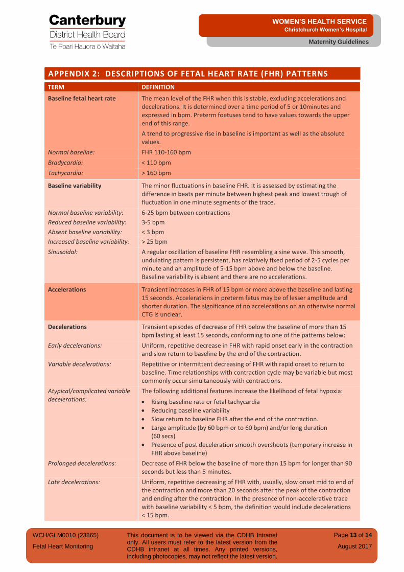
APPENDIX 2: DESCRIPTIONS OF FETAL HEART RATE (FHR) PATTERNS
TERM DEFINITION
Baseline fetal heart rate The mean level of the FHR when this is stable, excluding accelerations and decelerations. It is determined over a time period of 5 or 10minutes and expressed in bpm. Preterm foetuses tend to have values towards the upper end of this range.
A trend to progressive rise in baseline is important as well as the absolute values.
Normal baseline: FHR 110-160 bpm
Bradycardia: < 110 bpm
Tachycardia: > 160 bpm
Baseline variability The minor fluctuations in baseline FHR. It is assessed by estimating the difference in beats per minute between highest peak and lowest trough of fluctuation in one minute segments of the trace.
Normal baseline variability: 6-25 bpm between contractions
Reduced baseline variability: 3-5 bpm
Absent baseline variability: < 3 bpm
Increased baseline variability: > 25 bpm
Sinusoidal: A regular oscillation of baseline FHR resembling a sine wave. This smooth, undulating pattern is persistent, has relatively fixed period of 2-5 cycles per minute and an amplitude of 5-15 bpm above and below the baseline. Baseline variability is absent and there are no accelerations.
Accelerations Transient increases in FHR of 15 bpm or more above the baseline and lasting 15 seconds. Accelerations in preterm fetus may be of lesser amplitude and shorter duration. The significance of no accelerations on an otherwise normal CTG is unclear.
Decelerations Transient episodes of decrease of FHR below the baseline of more than 15 bpm lasting at least 15 seconds, conforming to one of the patterns below:
Early decelerations: Uniform, repetitive decrease in FHR with rapid onset early in the contraction and slow return to baseline by the end of the contraction.
Variable decelerations: Repetitive or intermittent decreasing of FHR with rapid onset to return to baseline. Time relationships with contraction cycle may be variable but most commonly occur simultaneously with contractions.
Atypical/complicated variable decelerations:
The following additional features increase the likelihood of fetal hypoxia:
Rising baseline rate or fetal tachycardia
Reducing baseline variability
Slow return to baseline FHR after the end of the contraction.
Large amplitude (by 60 bpm or to 60 bpm) and/or long duration (60 secs)
Presence of post deceleration smooth overshoots (temporary increase in FHR above baseline)
Prolonged decelerations: Decrease of FHR below the baseline of more than 15 bpm for longer than 90 seconds but less than 5 minutes.
Late decelerations: Uniform, repetitive decreasing of FHR with, usually, slow onset mid to end of the contraction and more than 20 seconds after the peak of the contraction and ending after the contraction. In the presence of non-accelerative trace with baseline variability < 5 bpm, the definition would include decelerations < 15 bpm.
WCH/GLM0010 (23865)
Fetal Heart Monitoring
This document is to be viewed via the CDHB Intranet only. All users must refer to the latest version from the CDHB intranet at all times. Any printed versions, including photocopies, may not reflect the latest version.
Page 14 of 14
August 2017
Maternity Guidelines
WOMEN’S HEALTH SERVICE Christchurch Women’s Hospital
APPENDIX 3: FETAL MONITORING – USING A CTG MACHINE IN A PRIMARY BIRTHING UNIT
The CTG machine is NOT to be used for laboring women in a primary birthing unit. It is only
to be used for the two antenatal situations as described below.
Maternal pulse must be monitored either continuously by way of a pulse oximeter or
manually and documented regularly on the CTG printout.
OCCASIONS FOR USE OF A CTG IN A PRIMARY BIRTHING UNIT
Decreased Fetal Movements (DFM) When a woman reports DFM on the first occasion only, commence a CTG and if there are
concerns at any stage of the CTG call the CWH Birthing Suite Co-ordinator (CCO) on 027 836
4673 to discuss your findings. The CCO will then liaise with the on call Registered Medical
Officer. The CTG is to be faxed to Birthing Suite as required on 03 364 4717.
Follow up CTGS for rural women Following an assessment and consultation with Christchurch Women’s Maternity Outpatient
Department CTGs may be performed as part of the ongoing plan to ensure women do not
have to travel in to CWH. If any concerns during daytime hours please fax to Day
Assessment Unit (DAU) at 03 364 4471.
All instances of CTG use in a primary birthing unit and resultant care plans are to be fully
documented.
Date Issued: April 2017 Fetal Heart Monitoring Review Date: April 2020 Maternity Guidelines Written/Authorised by: Maternity Guidelines Group Christchurch Women’s Hospital Review Team: Maternity Guidelines Group Christchurch New Zealand