global physiology and pathophysiology of cough: part 1
TRANSCRIPT

Global physiology and pathophysiology of cough: Part 1. Coughphenomenology: CHEST Guideline and Expert Panel report
CHEST Expert Cough Panel (2020). Global physiology and pathophysiology of cough: Part 1. Coughphenomenology: CHEST Guideline and Expert Panel report. Chest. https://doi.org/10.1016/j.chest.2020.08.2086
Published in:Chest
Document Version:Peer reviewed version
Queen's University Belfast - Research Portal:Link to publication record in Queen's University Belfast Research Portal
Publisher rightsCopyright 2020 Elsevier.This manuscript is distributed under a Creative Commons Attribution-NonCommercial-NoDerivs License(https://creativecommons.org/licenses/by-nc-nd/4.0/), which permits distribution and reproduction for non-commercial purposes, provided theauthor and source are cited
General rightsCopyright for the publications made accessible via the Queen's University Belfast Research Portal is retained by the author(s) and / or othercopyright owners and it is a condition of accessing these publications that users recognise and abide by the legal requirements associatedwith these rights.
Take down policyThe Research Portal is Queen's institutional repository that provides access to Queen's research output. Every effort has been made toensure that content in the Research Portal does not infringe any person's rights, or applicable UK laws. If you discover content in theResearch Portal that you believe breaches copyright or violates any law, please contact [email protected].
Download date:09. Feb. 2022

Global physiology and pathophysiology of cough: Part 1. Cough phenomenology: CHEST Guideline and Expert Panel report
Kai K. Lee1, Paul W. Davenport2, Jaclyn A Smith3, Richard S. Irwin4, Lorcan McGarvey5*, Stuart B. Mazzone6*, Surinder S. Birring7, on behalf of the CHEST Expert Cough Panel*
Affiliations:
1. School of Immunology and Microbial Sciences, Faculty of Life Sciences & Medicine, King's College London, UK
2. Department of Physiological Sciences, University of Florida, Gainesville, Florida, USA
3. Division of Infection, Immunity and Respiratory Medicine, School of Biological Sciences, Faculty of Biological, Medical and Health Sciences, University of Manchester, Manchester, UK
4. Division of Pulmonary, Allergy, and Critical Care Medicine, Department of Medicine, UMass Memorial Medical Center, Worcester, MA, USA.
5. Centre for Experimental Medicine, Queen's University Belfast, Belfast, Northern Ireland.
6. Department of Anatomy and Neuroscience, The University of Melbourne, Victoria, Australia.
7. Centre for Human & Applied Physiological Sciences, School of Basic & Medical Biosciences, Faculty of Life Sciences & Medicine, King's College London, London, UK.
*Correspondence to:
Lorcan McGarvey, MD, Centre for Experimental Medicine, Queen's University Belfast, Belfast, UK. e-mail: [email protected]
Stuart Mazzone, PhD, Department of Anatomy and Neuroscience, The University of Melbourne, Victoria, 3010 Australia; e-mail: [email protected]
SBM declares personal fees from Merck and NeRRe Therapeutics and grant support from Merck. LM reports personal fees from Chiesi, GSK, Merck, NeRRe Therapeutics, and Shionogi Inc; grant support from Merck; and other support from AstraZeneca, Boehringer Ingelheim, and Chiesi. SB reports personal fees from Merck, Bellus, Bayer, Shionogi, NeRRe, Menlo and Boehringer Ingelheim. JAS reports personal fees from Merck, Bellus, Bayer, Shiongi. Algernon, AstraZeneca, NeRRe, Menlo, Attenua and Boehringer Ingelheim. KKL, RSI and PWD have no conflict of interest to declare.

Disclaimer: American College of Chest Physician guidelines are intended for general information only, are not medical advice, and do not replace professional medical care and physician advice, which always should be sought for any medical condition. The complete disclaimer for this guideline can be accessed at http://chestjournal.chestpubs.org/content/XXX.

ABSTRACT
Background: The purpose of this state-of-the-art review is to update the American College of
Chest Physicians (CHEST) 2006 guideline on global physiology and pathophysiology of
cough.
Methods: A review of the literature was conducted using PubMed and Medline databases from
1951 to 2019 using pre-specified search terms.
Results: We describe the basic phenomenology of cough patterns, behaviors and morphologic
features. We update the understanding of mechanical and physiologic characteristics of cough,
adding a contemporary view of the types of cough and their associated behaviors and
sensations. New information about acoustic characteristics is presented, and recent insights into
cough triggers and the cough hypersensitivity patient phenotype are explored. Lastly, because
the clinical assessment of patients largely focuses on the duration rather than morphologic
features of cough, we review the morphological features of cough that can be measured in the
clinic.
Conclusions: This is the first of a two-part update to the 2006 CHEST Cough Guideline; it
provides a more global consideration of cough phenomenology, beyond simply the mechanical
aspects of a cough. A greater understanding of the typical features of cough, and their
variations, may allow a more informed interpretation of cough measurements and the clinical
relevance for patients.

Information presented was obtained from both animal and human experimental work.
Abbreviations:
CHS (cough hypersensitivity syndrome)
COPD (chronic obstructive pulmonary disease)
CPD (compression phase duration)
EMG (electromyography)
ER (expiratory reflex)
ERS (European Respiratory Society)
FRC (functional residual capacity)
OEP (optoelectronic plethysmography)
TRPV1 (transient receptor potential vanilloid 1)

Introduction
The purpose of this review is to update in two-parts the section on global physiology and
pathophysiology of cough in the 2006 CHEST Cough Guidelines [1]. A review of the literature
was carried out by the authors using PubMed and Medline from 1951 to 2019 using the search
terms shown in Table 1. The terms used to describe cough types in the literature are variable
and inconsistent [2]. . Clinicians categorize cough by its duration and etiology [2-4].
Mechanistic researchers classify cough as, induced cough, voluntary cough and spontaneous
cough when referring to study methodology. Others describe cough as sensitized (hypertussia
and allotussia), typically triggered by heterogeneous stimuli, or desensitized (hypotussia).
Finally, coughs can be defined by physiologic characteristics, sound properties and patterns.
There is no universally accepted way to classify cough types, but an understanding of the
phenomenology (a term used here to describe the patterns, behaviors and morphologic features
of cough) may provide insight into underlying pathophysiologic and neurobiologic
mechanisms. Part 1 of this update will summarize the motor and sensory traits of cough,
presenting typical descriptive characteristics, physiology of mechanics of cough, how cough is
assessed and, where available, how cough characteristics can differ between health and disease.
Part 2 of the update will describe more applied topics of the demographics of cough patients,
the clinical conditions impacting cough mechanics and the relationship between cough and the
role of airway secretions in cough clearance.
Cough and related sensorimotor processes in the clinical setting
Classical cough, expiratory reflexes and the urge-to-cough
Cough can occur reflexively or voluntarily. Cough is commonly induced in the
experimental or clinical setting by way of inhaled challenges using tussive agents, such as

capsaicin from hot chili peppers. Cough challenges are often described as cough reflex testing,
although the true involvement of reflexes versus volitional responses has not been assessed.
Induced cough is often distinguished from spontaneous cough occurring in disease, as although
both are often induced by irritant stimuli, the latter reflects naturally occurring cough in which
the tussive triggers may be endogenous (e.g., mucous, refluxate or inflammation), exogenous
(e.g., cold air, perfume or smoke) or perhaps cognitive (voluntary cough), but unlikely to be
homogeneous for all people. Reflex coughing, like many reflexes, involves neural processes
that are somewhat simpler in organization, integrated at the level of the brainstem. Voluntary
control of coughing, on the other hand, requires more complex neural processing at higher
cortical brain levels and has been described as behavioral regulation of coughing. Indeed,
people can voluntarily produce a cough, with or without accompanying airway stimuli, as well
as regulate cough intensity and even voluntarily suppress cough entirely for periods of time.
These broad types of cough are important to conceptually distinguish. For example, the
study of induced and voluntary cough allows precisely controlled experimental conditions to
be employed for comparisons between cough in disease and healthy volunteers and provides
insights into disease mechanisms and drug target engagement, often difficult when assessing
spontaneous cough. However, one must be cognizant that studies of voluntary and induced
cough do not necessarily predict therapeutic effects on spontaneous coughing or cough severity
in disease. For example, drugs that antagonize the capsaicin receptor (TRPV1) result in
effective attenuation of capsaicin-induced cough responses in healthy volunteers and chronic
cough patients, yet fail to reduce spontaneous coughing in patients with chronic refractory
cough, suggesting this mechanism is not universally relevant to the pathology [5].
Of consideration also, coughs can occur as isolated events or within “bouts” or “epochs”
(Figure 1). Patients with spontaneous cough often complain of these as coughing fits, which
are perceived as contributing to the severity of cough [4, 6]. Although accepted as a series of

expulsive efforts, whether each bout must originate from separate breaths is uncertain and a
variety of definitions have been used in the literature [7]. Studies involving acoustic cough
counting have defined bouts as continuous periods of coughing with less than 2 second pauses
[8, 9]. But most cough frequency data still report the number of coughs as a total number of
events regardless of whether in a bout or not, and in fact the two are well correlated [10].
Where there is glottic closure and expiratory effort but without the preceding inspiration,
the event is termed an expiratory reflex (ER) and this differs from a classical cough (Figure 2)
[7]. It is not uncommon for cough re-accelerations during coughing bouts to be considered
expiratory reflexes [11, 12], but it is equally important to note that expiratory reflexes can be
evoked in isolation using mechanical stimuli around the glottal folds or trachea [13, 14]. The
rationale for recognizing classical cough and ER as separate entities is suggested by the
possible role for ERs in preventing aspiration and pneumonia [7, 14-16]. Another consideration
is that ERs are likely to occur from lower lung volumes than coughs following an inspiration
and therefore will generate lower flows [17]. The counterarguments are that it is at present
difficult to differentiate them in clinical practice because they sound similar and current
monitoring methods (as well as most patients and clinicians) consider all such events as coughs
[18].
Although not well studied, it is possible that in patients with chronic cough, spontaneous
coughing is made up of a mixture of classical coughs, ERs and cough re-accelerations. Some
coughs could be reflexive while others could be under various levels of volitional control. This
again has not been well studied.

Cough triggers and the concept of cough hypersensitivity
Traditionally, clinicians have viewed cough solely as a symptom of an underlying lung
disease or arising as a consequence of an acute inflammatory or infective insult. But the cough
may persist long after the initiating insult has resolved and this chronicity is a source of
considerable morbidity. Clinicians experienced in the management of chronic cough are readily
aware of how troubled their patients are by spasms of cough provoked by everyday activities
including talking or laughing and changes in ambient air temperature or exposure to aerosols
or perfumes [19-21]. Many also describe abnormal sensations such as a persisting itch or tickle
in the throat or the feeling of a ‘lump’ in the back of the throat [22]. These clinical observations
have given rise to the unifying clinical concept of Cough Hypersensitivity Syndrome (CHS)
recently defined by the European Respiratory Society (ERS) Taskforce as a ‘disorder
characterized by troublesome coughing often triggered by low levels of thermal, mechanical
or chemical exposure’ [23]. The triggering of cough by relatively innocuous stimuli suggests
heightened sensitivity of the sensory nerve pathways alluded to above that normally serve to
detect and respond to harmful airway irritants. In these circumstances, the cough should not
be considered as a symptom but rather as a disease entity caused by a disordered nervous
system [24]. The pathologic mechanisms responsible for how such nerves become
pathologically ‘excitable’ is unknown but inflammation-induced injury causing functional
changes of the neural pathways seems possible [25]. The notion that cough triggered by
relatively inoffensive stimuli (allotussia) might be similar to allodynia (pain response from
stimuli that does not normally provoke pain) and excessive coughing in response to a noxious
exposure (hypertussia) could be considered equivalent to hyperalgesia (abnormally increased
sensitivity to pain) supports the view that mechanistic parallels may exist between CHS and
neuropathic pain [26]. The logical extension of this concept has prompted clinical trials
designed to evaluate the efficacy of neuromodulatory drugs more traditionally used to treat

pain such as morphine, gabapentin, pregabalin and amitriptyline [27]. The ERS guidelines on
the diagnosis and treatment of chronic cough have made a conditional recommendation (albeit
on low quality evidence) that a trial of such agents be offered to adult patients with chronic
refractory cough [28]. This clinical phenomenon whereby innocuous sensory stimuli evoke a
strong ‘urge to cough’ or trigger bouts of coughing is in part due to a disorder in the
communication of sensory information from the airway to the brain. As important must be the
cognitive awareness of this information and the processing responsible for the generation of a
cough. The complex role of cognition in cough regulation is discussed below.
Behavioral considerations in the regulation of cough
Cough is cognitively controlled by discriminative and affective cortical neural
mechanisms. The discriminative element provides the patient with an assessment of the cough
stimulus (e.g., what is the intensity, some capacity to localize) and can precede the motor cough
response. Affective neural systems superimpose reward-aversion value judgments onto the
cough response (e.g., how does it make me feel?). Affective mechanisms may therefore
promote suppression or potentiation of the motor cough behavior. Cognitive suppression and/or
modulation of cough is of importance in regulating the cough motor pattern. The cognitive
awareness of a cough stimulus can promote an urge-to-cough, much like thirst promotes an
urge-to-drink. It has been suggested that the urge-to-cough reflects activation of a motivational
neural system in the brain that promotes voluntary cough or other behaviors to help alleviate
the sensations accompanying airway irritation [29]. While these sensory experiences could be
considered the pre-motor phase of cough, they do not precede all coughs, an urge to cough
being reported in 69% of patients with chronic cough, and not all throat irritation/ urge to cough
will evolve into actual coughing, as they can be suppressed or satisfied by other maneuvers
[20, 29]. Functional brain imaging has shown activation of cortical and subcortical regions
during capsaicin induced urge-to-cough that differs between patients suffering from chronic

cough and healthy volunteers [30]. Importantly, the threshold of the urge-to-cough can be
critical for initiating cough, a weak urge-to-cough (high threshold) means the patient will not
voluntarily cough and clear their airways with weak stimuli. The implications of an increased
urge-to-cough threshold are increased risk of aspiration as weak cough and delayed airway
clearance. Investigation of the relationship between high urge-to-cough threshold and
aspiration related lung infection needs to be performed. Cough is additionally subject to strong
placebo/suggestive suppression, mediated by cognitive processes in the higher brain [31-33].
Physiology of the Mechanics of Cough
The major function of cough is to engage high velocity airflow to clear the airways.
Cough airflow is generated by contracting expiratory muscles while the glottis is closed, thus
producing high positive subglottic pressures [34]. When a cough is initiated, the normal cough
motor pattern is characterized [35] by a stereotypic inspiration (inspiratory phase) followed by
complete closure of the glottis allowing compression of the thorax increasing subglottic
pressure (compression phase), followed by rapid opening of the glottis resulting in a high
velocity airflow (peak expiratory airflow phase), a high expiratory airflow rate (plateau phase)
that is sustained for a variable duration with the cough ending by expiratory airflow returning
to baseline (Figure 2A).
This single expulsive effort is the classical definition of a cough [35] that must be
amended to include bouts of expulsive events after a single inspiration [36]. Initiation of a
cough most commonly results in a large inspiration followed by multiple expulsive events
during the decrease in expired volume (Figure 3). An expulsive event following the initial
cough expiratory sequence is characterized by reclosure of the glottis, a compression phase of
equal duration followed by rapid glottis opening and a peak expiratory airflow phase that is
usually less than the initial event but reaccelerates expiratory airflow (Figure 3) [36]. This

pattern of multiple expulsive events for a single inspiration has been termed cough re-
acceleration [36, 37]. The most common coughing pattern is 2 expulsive events for a single
inspiration [37]. Increasing the cough stimulus intensity causes an increased number of
expulsive events, cough re-accelerations, for a single inspiration. Multiple cough re-
accelerations recorded in a flow-volume loop have been reported as cough spiking [38, 39].
The advantage of cough re-acceleration expulsive events following an inspiration is the
repeated shear forces applied to the airway. The initial cough expiratory airflow acceleration
phase produces the shear forces dislodging material from the walls of the airway. The sustained
cough expiratory plateau phase moves the material through the center of the airway airflow
stream. When a cough bout occurs, the second expulsive event occurs while the material moves
toward the exit of the airways. Transient reclosure of the glottis allows the subglottic pressure
to again increase thus generating renewed shear forces when the glottis is reopened. This keeps
the material moving towards the opening of the airways without an interrupting inspiratory
phase, avoiding material reattachment to the airway wall and sustaining clearance. With strong
cough stimuli, the number of cough re-accelerations increases for each inspiratory volume [36,
37]. The critical importance of high airflow velocity on shearing forces dislodging material
from the wall of the airway results in the documented relevance of cough expiratory airflow
volume acceleration and cough peak expiratory airflow velocity measurements for cough
efficacy and strength [39-42].
Functional relevance of the cough motor pattern
The magnitude of the airflow rate during the expiratory peak airflow and plateau phases
of cough is directly related to the initial inspired lung volume [17, 37]. Thus, the inspired
volume primes the thoracic system volume for generating expiratory pressures and airflows.
The greater the cough priming inspiratory volume, the greater the subglottic pressure that is a

combination of volume dependent elastic recoil and expiratory muscle pump force. The
magnitude of the inspiratory volume is also proportional to the number of cough re-
accelerations [37]. Active expiratory muscle contraction and elastic recoil of the thoracic
system against a closed glottis results in a rapidly increasing subglottic pressure [34]. During
this compression phase, the end-inspiratory total respiratory compliance, the magnitude of the
expiratory muscle contraction, the tightness of glottic closure and the duration of the
compression phase determine the peak subglottic pressure. There appears to be a subglottic
pressure threshold for glottis opening although this has received little investigation. The
subglottic pressure determines the magnitude of the cough expiratory airflow acceleration upon
opening of the glottis and is correlated with the clearance of airways.
Cough peak expiratory airflow (Figure 2A) has also been correlated with the successful
clearance of the airway [41-44]. The initial expiratory cough airflow rapidly accelerates,
reaches a peak and then rapidly declines to a sustained cough expiratory airflow plateau. The
peak airflow “spike” (Figure 2A) is of short duration. The subsequent cough expiratory plateau
phase (Figure 2A) is the result of sustained active expiratory muscle contraction and respiratory
system elastic recoil. The cough plateau phase is often extended to sustain expulsive forces
especially when a patient feels they have not cleared their airway. The respiratory system
elastic recoil is dependent on the total thoracic volume and decreases as air is exhaled. The
airflow rate during the plateau phase sustains the airway shear and proximal propulsion of
material from the airways [11, 45]. As lung volume decreases during the plateau phase,
sustaining the airflow rate requires increasing expiratory muscle contraction [11, 36]. The
plateau phase is usually abruptly terminated and expiratory airflow rate returns to baseline.
When a cough bout (multiple expulsive events) occurs with a single inspiration, this return to
baseline becomes a new compression phase with glottis closure (Figure 3). Similar to the initial
pattern, the glottic closure results in respiratory system elastic recoil increasing subglottic

pressure in combination with a resurgence of expiratory muscle contraction [36]. The second
compression phase generally has a similar duration as the initial compression phase that is
again terminated by rapid opening of the glottis, initiating a second cough peak airflow spike
(Figure 3). The second expulsive event often has a diminished cough expiratory airflow
acceleration rate and decreased cough peak expiratory airflow rate primarily due to the lower
lung volume. Each subsequent expulsive event for multiple cough re-accelerations has a lower
initial airflow acceleration, peak expiratory airflow and plateau airflow rate (Figure 3) [11].
Reaccelerated cough expulsive events can occur throughout the expired volume from the end-
inspiratory lung volume to lung volumes below functional residual capacity (FRC) [17, 46].
The expiratory muscle activity required to produce each expulsive event may increase as lung
volume decreases [36].
Physiologic measures of cough
Airflow
Flow is easily measured in voluntary cough performed in the laboratory but challenging
to measure during induced cough or in the ambulatory setting. The classical cough flow pattern
is of an inspiration followed by cessation of flow during glottis closure and then rapid
expiration followed plateau and termination. The peak flow during the expiratory phase is the
most extensively measured physiologic characteristic. Use of peak cough flow has been utilized
to assess respiratory muscle function, airway clearance function and suitability for extubation
from invasive ventilation [47-50]. Peak flows during maximum voluntary cough can reach over
800 L/min and can be greater than flows observed during peak expiratory flow rate maneuvers
[51-53]. Measurement of flow during induced cough is more technically challenging, but is
shown to be significantly lower than maximum voluntary cough flow [50, 54-57]. Flow during
spontaneous cough has seldom been studied and indeed may be altered by the measuring

equipment, but has been reported as less than that of maximum voluntary cough, though higher
than induced cough [55].
Several other flow dynamic characteristics have been described. Compression phase
duration (CPD), the period between cessation of flow after inspiration and onset of the
expiratory flow in the expulsive phase, varies in the literature but estimates in the region of
0.30 seconds are reported in health [55, 58]. CPD cannot be measured via flow for ERs due to
the lack of preceding inspiration but could be studied using other modalities such as chest wall
motion/volume via impedance bands, optoplethysmography (OEP) or electromylography
(EMG). CPD (and cough duration) is shorter for coughs occurring within bouts than single
coughs [17]. Conditions such as laryngectomy predictably result in loss of the compression
phase but reasons for prolonged CPD are more complex and may include reduced motor drive
and impaired laryngeal function [57, 59]. The expiratory rise-time, the time between onset and
peak expiratory flow, has been reported in a small study of healthy adults and ranged from 51
ms to 73 ms, varying with gender and height [53], but can be significantly longer in patients
with amyotrophic lateral sclerosis, diseases with unsafe swallow and in patients with chronic
airways disease [60, 61].
Pressure
Cough gastric pressure is a reflection of intra-abdominal pressure and can be measured
using a balloon catheter and pressure transducer system [62]. Often used to assess expiratory
muscle function, cough gastric pressures can exceed 300 mmHg during maximum voluntary
cough, although values of 214 mmHg and 165 mmHg are reported for normal males and
females respectively [63]. The pattern is typically of a spike in pressure with rapid increase and
decrease coinciding with expiratory flow (Figure 1). Esophageal pressure is a measure of intra-

thoracic pressure and can also be measured with the same balloon catheter system and has a
similar pattern [62]. Esophageal pressure increases during the compression and expulsion
phases of cough and can also reach pressures as high as 300 mmHg [64]. Both pressure
measurement methods are limited by their invasive nature, requirement for assessment in the
laboratory setting, and being subject to effects from body position, state of rest of the abdominal
muscles and external compression [65, 66].
Electromyography
Abdominal muscle EMG has been explored as a measure of cough for over 30 years, but
accessory muscle EMG has also recently been studied [67, 68]. Abdominal EMG correlates
well with flow during voluntary cough and is repeatable during induced cough challenges [51,
69-71]. However, when voluntary coughs are studied from a range of different lung volumes
and with different efforts applied, then unlike flow, the EMG activity is largely independent of
lung volume and is mainly determined by cough effort. The exceptions are coughs performed
from below FRC, such as those occurring at the end of cough bouts; these are associated with
the highest EMG signals [72]. The study of EMG has allowed the observation that voluntary
cough is associated with coordinated sequential activation of the main expiratory and accessory
muscles, whereas induced cough is associated with simultaneous activation of both muscle
groups with greater EMG activity but shorter duration [67].
A limitation of EMG is the inability to compare values between subjects or between
experimental sessions and normalization methods are required for data analysis [67, 69, 72,
73]. Factors such as electrode position, contact between electrode and skin, resting muscle state
and inherent skin resistance can affect the measures and signal contamination from ECG and

limited potential for automation has also made EMG challenging to develop as a clinical
measure [74].
Sound
The characteristic cough sound waveform is comprised of three distinct phases:
explosive, intermediate and voiced (Figure 4). While all coughs contain the first phase, the
voiced phase may be absent in approximately one third of subjects [75-78]. In health, the
duration of a typical cough sound is approximately 410 ms but in disease it can be longer, up
to 600 ms in asthma or bronchitis and up to 1 s in acute viral cough [79-82]. The sound signal
amplitude and power (calculated after transforming the sound signal into its frequency domain)
have been shown to correlate with flow, pressure and with cough EMG during voluntary cough,
although the strength of the relationships has varied among studies [72, 78, 83, 84].
An area of interest is the ability to discern underlying etiology from the cough sound.
Indeed, clinicians are able to discriminate wet from dry cough sounds by ear, but they are less
able to differentiate between causes such as fibrosis, asthma, COPD or bronchiectasis [85].
Analysis of sound properties using signal processing techniques can also differentiate between
wet and dry cough, but have additionally been reported to enable identification of asthma,
COPD, pertussis and pneumonia; however there is currently no validated diagnostic system
available for the clinical setting [78, 86-90]. Although spontaneous and voluntary coughs were
assessed in these studies, there is a lack of data comparing cough sound properties of
spontaneous, induced and voluntary cough directly within subjects. It is not known whether
ERs possess different sound qualities from classical coughs.

Cough Assessment
Assessment of cough requires differentiation between the strength of the individual
cough and the total cough response. The strength of a cough is a function of the individual
cough motor pattern. A high magnitude cough response is characterized by a large number of
expulsive events.
Cough assessment requires the occurrence of a cough when the patient is instrumented
for recording cough sounds, airflow and/or motor patterns. This usually requires that cough is
induced under controlled conditions. The induction of cough without an external tussive
stimulus can be performed by asking the individual to perform a voluntary cough. Reflex cough
can be induced by stimulation of airway cough receptors using capsaicin, citric acid, distilled
water fog or similar airway irritants. Voluntary and reflex cough have similar motor patterns
with the inspiratory phase, compression phase and cough expiratory phases [91]. Induced
cough is useful for evaluating the cough motor pattern. Induced reflex cough is useful for
assessing cough motor pattern, cough stimulus threshold, Urge-to-Cough and cough sensitivity
to a specific stimulus. Voluntary cough is useful in assessing cough motor pattern and voluntary
cough strength. Both voluntary and reflex induced cough reliably generate a cough but do not
allow for the assessment of spontaneous cough. Reflex and voluntary induced cough may be
insensitive to antitussive treatment [92].
Assessment of spontaneous cough has been investigated with ambulatory cough
monitors [93, 94]. Spontaneous cough monitors are effective in assessing cough frequency and
cough sound intensity, as a measure of cough strength. Two systems are commonly used. The
first is the semi-automated Vitalojak cough monitoring system which records sound and
employs a manual analysis to discriminate cough sounds from throat clearing or other
respiratory noises during the cough counting process. The second is the Leicester Cough

Monitor which is a semi-automated system, with user input used to train the detection and
analysis algorithm. Validation results have shown it to be able to differentiate cough sounds
from non-cough sounds such as throat clearing [95]. Parameters derived from ambulatory
cough monitors allow for the assessment of antitussive treatments in more natural
environments and may better reflect treatment efficacy. Whilst cough frequency monitoring is
widely practiced, the uptake of cough sound intensity monitoring in clinical practice has been
limited by the lack of validation data against physiologic measures of cough intensity in the
ambulatory setting [83]. Ambulatory cough monitors usually do not allow for the recording of
cough airflow and cough motor pattern.
Dysfunction and/or disruption of the cough motor pattern that results in reduced cough
effectiveness is defined as dystussia. One dystussic complication of cough motor pattern is
inadequate closure of the glottis (leak) during the compression phase [73, 96]. This results in
reduced subglottic pressure, decreased initial expiratory airflow acceleration, reduced peak
cough expiratory airflow and reduced expulsive forces in the airways; hence, inadequate
clearance of material from the airways. Another complication resulting in dystussia is
decreased expiratory muscle force generating capacity [69]. Reduced expiratory muscle
strength results in decreased subglottic pressure during the compression phase, reduced initial
cough expiratory airflow rate, decreased cough peak expiratory airflow, reduced plateau phase
expiratory airflow and inadequate clearance of material from the airway.
Qualitative patient perspectives highlight both the frequency and intensity of coughs as
determinants of cough severity, in addition to degree of impact or disruption [4]. Cough
monitoring has focused primarily on recording cough frequency, but there is recognition that
the addition of cough intensity monitoring may be valuable.

Cough frequency
Current cough frequency monitoring systems generally work by recording ambient sound
continuously over 24-hour period followed by off-line analysis, either manually or via semi-
automated analysis, to determine cough counts [95, 97]. The monitoring systems count all
recorded events as ‘coughs’ regardless of whether they are classical coughs or ERs because the
current methodology for cough detection is not able to discriminate between them. Cough
frequency is most commonly quantified as the total number of events per hour or day, but the
merits of quantifying in bouts or time spent coughing have also been shown [10]. Cough
frequency data exist for healthy adults (8-30 coughs per day) as well as for various pulmonary
diseases, and cough frequency monitoring has changed the standards by which novel cough
therapies are being evaluated [94, 98].
Cough intensity
Cough intensity is often considered as the harshness or violence of coughing perceived
by patients. However, there is lack of consensus about whether the mechanical properties of
cough events can reflect perceived cough intensity to provide an objective intensity measure,
and as such there is generally little agreement on the best measures of cough intensity. Direct
measures of cough strength assessments are typically made using cough airflow patterns,
including the initial cough expiratory airflow acceleration, the cough peak expiratory airflow
rate and the area under the cough expiratory airflow plateau phase. When surface EMG’s are
recorded, the integrated EMG from abdominal and intercostal areas are correlated with cough
expiratory airflow rate only as a difference within a single subject recording session [51].
The impracticalities of measuring EMG or pressure or flow in the ambulatory setting
have prompted the study of sound as a potential cough intensity monitoring measure but further
studies are needed to determine if this is a clinically useful outcome measure [72, 83, 84, 99].

Other auditory assessments of cough have been used to indirectly assess cough strength. This
is often performed by counting the number of expulsive events elicited by spontaneous,
induced or voluntary cough. There is a direct correlation between the number of expulsive
events, cough frequency recorded from cough sounds and the reported cough strength.
Auditory sound intensity and duration for a single expulsive event are also reported as a
measure of cough strength. Cough strength can be assessed by behavioral magnitude
production tasks. The subject is asked to produce a weak, moderate or strong cough while
simultaneously measuring auditory, airflow and/or EMG outputs. Cognitive cough strength
magnitude production tasks are correlated with peak airflow, cough sound and integrated EMG
area.
Looking to the future, studies are needed to assess the clinical relevance of these cough
patterns. Studies should focus on the relationship between cough phenomenology, the etiology
of cough and the impact on patients (patient reported cough severity and quality of life). The
changes of these morphologic features in response to antitussive treatments in clinical trials
and the potential to monitor patients by utilizing recent advances in technology should also be
assessed.
Conclusion
This update to the 2006 CHEST Cough Guideline reviews the advances in the knowledge
of cough physiology and pathophysiology, specifically describing the features and patterns of
different cough types, the triggers and the regulatory processes with relevance to patients with
chronic cough. The terminology used to describe cough types is varied and consequently it is
important to define the type of cough under assessment and recognize the usual characteristics
in order to support interpretation of findings and, more importantly, clinical relevance. With
the development of improved less invasive and advanced portable technologies, there is major

potential for more detailed assessments of cough to become widespread for remote diagnostics
and monitoring. A better knowledge and understanding of cough phenomenology will surely
support this.

Acknowledgments
Author contributions: All authors participated in conceiving of the content to be covered in
this article, writing specific sections of the first draft, and reading and editing and approving
of the final draft that was prepared by KKL, LM, and SBM.
Other contributions: The authors thank Nancy Harger, MLS, an Education and Clinical
Services Librarian, working in the University of Massachusetts Medical School Library in
Worcester, MA who undertook all the searches for each section in this article.
*Collaborating Authors:

References
1. McCool, F.D., Global Physiology and Pathophysiology of Cough. Chest, 2006. 129(1 suppl): p. 48S‐53S.
2. Chung, K.F., et al., Semantics and types of cough. Pulm Pharmacol Ther, 2009. 22(2): p. 139‐42.
3. Irwin, R.S., et al., Diagnosis and management of cough executive summary: ACCP evidence‐based clinical practice guidelines. Chest, 2006. 129(1 Suppl): p. 1S‐23S.
4. Vernon, M., et al., Measuring cough severity: Perspectives from the literature and from patients with chronic cough. Cough, 2009. 5: p. 5.
5. Khalid, S., et al., Transient receptor potential vanilloid 1 (TRPV1) antagonism in patients with refractory chronic cough: a double‐blind randomized controlled trial. J Allergy Clin Immunol, 2014. 134(1): p. 56‐62.
6. Everett, C.F., et al., Chronic persistent cough in the community: a questionnaire survey. Cough, 2007. 3: p. 5.
7. Fontana, G.A. and J. Widdicombe, What is cough and what should be measured? Pulm Pharmacol Ther, 2007. 20(4): p. 307‐12.
8. Lee, K.K., et al., A Longitudinal Assessment of Acute Cough. American Journal of Respiratory and Critical Care Medicine, 2013. 187(9): p. 991‐997.
9. Morice, A.H., et al., ERS guidelines on the assessment of cough. European Respiratory Journal, 2007. 29(6): p. 1256‐76.
10. Kelsall, A., et al., How to quantify coughing: correlations with quality of life in chronic cough. European Respiratory Journal, 2008. 32(1): p. 175‐9.
11. Davenport, P.W., et al., The effect of codeine on the Urge‐to‐Cough response to inhaled capsaicin. Pulm Pharmacol Ther, 2007. 20(4): p. 338‐46.
12. Hegland, K.W., M.S. Troche, and P.W. Davenport, Cough expired volume and airflow rates during sequential induced cough. Frontiers in Physiology, 2013. 4.
13. Nishino, T., Y. Tagaito, and S. Isono, Cough and other reflexes on irritation of airway mucosa in man. Pulm Pharmacol, 1996. 9(5‐6): p. 285‐92.
14. Tatar, M., J. Hanacek, and J. Widdicombe, The expiration reflex from the trachea and bronchi. European Respiratory Journal, 2008. 31(2): p. 385‐390.
15. Stephens, R.E., et al., Videofluoroscopy of the diaphragm during voluntary and reflex cough in humans. Am J Phys Med Rehabil, 2003. 82(5): p. 384.
16. Widdicombe, J. and G. Fontana, Cough: what's in a name? Eur Respir J, 2006. 28(1): p. 10‐5. 17. Smith, J.A., et al., Chest wall dynamics during voluntary and induced cough in healthy
volunteers. J Physiol, 2012. 590(3): p. 563‐74. 18. Morice, A.H., Rebuttal: cough is an expiratory sound. Lung, 2008. 186 Suppl 1: p. S7‐9. 19. McGarvey, L., et al., Are there clinical features of a sensitized cough reflex? Pulm Pharmacol
Ther, 2009. 22(2): p. 59‐64. 20. Hilton, E., et al., Clinical features of the urge‐to‐cough in patients with chronic cough. Respir
Med, 2015. 109(6): p. 701‐7. 21. Won, H.K., et al., Cough‐Related Laryngeal Sensations and Triggers in Adults With Chronic
Cough: Symptom Profile and Impact. Allergy, asthma & immunology research, 2019. 11(5): p. 622‐631.
22. Vertigan, A.E., S.L. Bone, and P.G. Gibson, Laryngeal sensory dysfunction in laryngeal hypersensitivity syndrome. Respirology, 2013. 18(6): p. 948‐56.
23. Morice, A.H., et al., Expert opinion on the cough hypersensitivity syndrome in respiratory medicine. European Respiratory Journal, 2014. 44(5): p. 1132.
24. McGarvey, L. and P.G. Gibson, What Is Chronic Cough? Terminology. J Allergy Clin Immunol Pract, 2019. 7(6): p. 1711‐1714.

25. McGovern, A.E., et al., Translational review: Neuroimmune mechanisms in cough and emerging therapeutic targets. J Allergy Clin Immunol, 2018. 142(5): p. 1392‐1402.
26. Chung, K.F., L. McGarvey, and S.B. Mazzone, Chronic cough as a neuropathic disorder. Lancet Respir Med, 2013. 1(5): p. 414‐22.
27. Gibson, P., et al., Treatment of Unexplained Chronic Cough: CHEST Guideline and Expert Panel Report. Chest, 2016. 149(1): p. 27‐44.
28. Morice, A.H., et al., ERS guidelines on the diagnosis and treatment of chronic cough in adults and children. European Respiratory Journal, 2020. 55(1): p. 1901136.
29. Davenport, P.W., Urge‐to‐cough: what can it teach us about cough? Lung, 2008. 186 Suppl 1: p. S107‐11.
30. Mazzone, S.B., et al., Representation of capsaicin‐evoked urge‐to‐cough in the human brain using functional magnetic resonance imaging. Am J Respir Crit Care Med, 2007. 176(4): p. 327‐32.
31. Hutchings, H.A., et al., Voluntary suppression of cough induced by inhalation of capsaicin in healthy volunteers. Respiratory Medicine, 1993. 87(5): p. 379‐82.
32. Lee, P.C., C. Cotterill‐Jones, and R. Eccles, Voluntary control of cough. Pulm Pharmacol Ther, 2002. 15(3): p. 317‐20.
33. Eccles, R., The Powerful Placebo in Cough Studies? Pulmonary Pharmacology & Therapeutics, 2002. 15(3): p. 303‐308.
34. Yanagihara, N., H. Von Leden, and E. Werner‐Kukuk, The physical parameters of cough: the larynx in a normal single cough. Acta Otolaryngol, 1966. 61(6): p. 495‐510.
35. Morice, A.H., et al., ERS guidelines on the assessment of cough. Eur Respir J, 2007. 29(6): p. 1256‐76.
36. Vovk, A., et al., Capsaicin exposure elicits complex airway defensive motor patterns in normal humans in a concentration‐dependent manner. Pulm Pharmacol Ther, 2007. 20(4): p. 423‐32.
37. Hegland, K.W., M.S. Troche, and P.W. Davenport, Cough expired volume and airflow rates during sequential induced cough. Front Physiol, 2013. 4: p. 167.
38. Beardsmore, C.S., et al., Maximum voluntary cough: an indication of airway function. Bull Eur Physiopathol Respir, 1987. 23(5): p. 465‐72.
39. Chaudri, M.B., et al., Relationship between supramaximal flow during cough and mortality in motor neurone disease. Eur Respir J, 2002. 19(3): p. 434‐8.
40. Kim, J., P. Davenport, and C. Sapienza, Effect of expiratory muscle strength training on elderly cough function. Arch Gerontol Geriatr, 2009. 48(3): p. 361‐6.
41. Smith Hammond, C.A., et al., Assessment of aspiration risk in stroke patients with quantification of voluntary cough. Neurology, 2001. 56(4): p. 502‐6.
42. Ward, K., et al., Poor cough flow in acute stroke patients is associated with reduced functional residual capacity and low cough inspired volume. BMJ Open Respir Res, 2017. 4(1): p. e000230.
43. Dickey, B.F., What it takes for a cough to expel mucus from the airway. Proc Natl Acad Sci U S A, 2018. 115(49): p. 12340‐12342.
44. Ren, S., et al., Numerical Analysis of Airway Mucus Clearance Effectiveness Using Assisted Coughing Techniques. Sci Rep, 2020. 10(1): p. 2030.
45. Wei, J. and Y. Li, Human Cough as a Two‐Stage Jet and Its Role in Particle Transport. PLoS One, 2017. 12(1): p. e0169235.
46. Vilozni, D., et al., Cough characteristics and FVC maneuver in cystic fibrosis. Respir Care, 2014. 59(12): p. 1912‐7.
47. Szeinberg, A., et al., Cough capacity in patients with muscular dystrophy. Chest, 1988. 94(6): p. 1232‐5.
48. Polkey, M.I., et al., Expiratory muscle function in amyotrophic lateral sclerosis. American Journal of Respiratory and Critical Care Medicine, 1998. 158(3): p. 734‐41.
49. Smina, M., et al., Cough peak flows and extubation outcomes. Chest, 2003. 124(1): p. 262‐8.

50. Kulnik, S.T., et al., Higher cough flow is associated with lower risk of pneumonia in acute stroke. Thorax, 2016. 71(5): p. 474‐5.
51. Fontana, G.A., et al., A noninvasive electromyographic study on threshold and intensity of cough in humans. European Respiratory Journal, 1997. 10(5): p. 983‐9.
52. Ross, B.B., R. Gramiak, and H. Rahn, Physical dynamics of the cough mechanism. Journal of Applied Physiology, 1955. 8(3): p. 264‐8.
53. Feinstein, A.J., et al., Measurement of Cough Aerodynamics in Healthy Adults. Ann Otol Rhinol Laryngol, 2017. 126(5): p. 396‐400.
54. Ward, K., et al., Acute ischaemic hemispheric stroke is associated with impairment of reflex in addition to voluntary cough. Eur Respir J, 2010. 36(6): p. 1383‐90.
55. Lee, K.K., et al., The Intensity of Voluntary, Induced, and Spontaneous Cough. Chest, 2015. 148(5): p. 1259‐1267.
56. Wheeler Hegland, K., et al., Comparison of voluntary and reflex cough effectiveness in Parkinson's disease. Parkinsonism Relat Disord, 2014. 20(11): p. 1226‐30.
57. Lavorini, F., et al., Fog‐induced cough with impaired respiratory sensation in congenital central hypoventilation syndrome. Am J Respir Crit Care Med, 2007. 176(8): p. 825‐32.
58. Yanagihara, N., H. von Leden, and E. Werner‐Kukuk, The Physical Parameters of Cough: The Larynx in A Normal Single Cough. Acta Oto‐Laryngologica, 1966. 61(1‐6): p. 495‐510.
59. Pitts, T., et al., Voluntary cough production and swallow dysfunction in Parkinson's disease. Dysphagia, 2008. 23(3): p. 297‐301.
60. Plowman, E.K., et al., Voluntary Cough Airflow Differentiates Safe Versus Unsafe Swallowing in Amyotrophic Lateral Sclerosis. Dysphagia, 2016. 31(3): p. 383‐90.
61. Piirila, P. and A.R. Sovijarvi, Differences in acoustic and dynamic characteristics of spontaneous cough in pulmonary diseases. Chest, 1989. 96(1): p. 46‐53.
62. American Thoracic Society/European Respiratory Society, ATS/ERS Statement on respiratory muscle testing. American Journal of Respiratory and Critical Care Medicine, 2002. 166(4): p. 518‐624.
63. Man, W.D.‐C., et al., Cough Gastric Pressure and Maximum Expiratory Mouth Pressure in Humans. American Journal of Respiratory and Critical Care Medicine, 2003. 168(6): p. 714‐717.
64. Sharpey‐Schafer, E.P., Effects of coughing on intra‐thoracic pressure, arterial pressure and peripheral blood flow. The Journal of Physiology, 1953. 122(2): p. 351‐357.
65. Hebbard, G.S., et al., Postural changes in proximal gastric volume and pressure measured using a gastric barostat. Neurogastroenterology and Motility, 1995. 7(3): p. 169‐74.
66. Vanderstappen, G. and E.C. Texter, Jr., Response of the physiologic gastroesophageal sphincter to increased intra‐abdominal pressure. Journal of Clinical Investigation, 1964. 43: p. 1856‐68.
67. Lasserson, D., et al., Differences in motor activation of voluntary and reflex cough in humans. Thorax, 2006. 61(8): p. 699‐705.
68. Cox, I.D., et al., An electromyographic method of objectively assessing cough intensity and use of the method to assess effects of codeine on the dose‐response curve to citric acid. Br J Clin Pharmacol, 1984. 18(3): p. 377‐82.
69. Fontana, G.A., et al., Defective motor control of coughing in Parkinson's disease. Am J Respir Crit Care Med, 1998. 158(2): p. 458‐64.
70. Fontana, G.A., et al., Coughing in laryngectomized patients. Am J Respir Crit Care Med, 1999. 160(5 Pt 1): p. 1578‐84.
71. Strohl, K.P., et al., Regional differences in abdominal muscle activity during various maneuvers in humans. Journal of Applied Physiology: Respiratory, Environmental and Exercise Physiology, 1981. 51(6): p. 1471‐6.

72. McGuinness, K., et al., Muscle activation and sound during voluntary single coughs and cough peals in healthy volunteers: Insights into cough intensity. Respir Physiol Neurobiol, 2018. 257: p. 42‐50.
73. Chellini, E., et al., Motor features of voluntary cough following partial laryngectomy for glottal carcinoma. Bratisl Lek Listy, 2011. 112(3): p. 115‐9.
74. Lavorini, F., et al., Respiratory expulsive efforts evoked by maximal lung emptying. Chest, 2011. 140(3): p. 690‐6.
75. Doherty, M.J., et al., The acoustic properties of capsaicin‐induced cough in healthy subjects. Eur Respir J, 1997. 10(1): p. 202‐7.
76. Korpas, J., J. Sadlonova, and M. Vrabec, Analysis of the cough sound: an overview. Pulm Pharmacol, 1996. 9(5‐6): p. 261‐8.
77. Murata, A., et al., Discrimination of productive and non‐productive cough by sound analysis. Intern Med, 1998. 37(9): p. 732‐5.
78. Thorpe, C.W., L.J. Toop, and K.P. Dawson, Towards a quantitative description of asthmatic cough sounds. Eur Respir J, 1992. 5(6): p. 685‐92.
79. Debreczeni, L.A., J. Korpas, and D. Salat, Spectral analysis of cough sounds recorded with and without a nose clip. Bull Eur Physiopathol Respir, 1987. 23 Suppl 10: p. 57s‐61s.
80. Kelemen, S.A., T. Cseri, and I. Marozsan, Information obtained from tussigrams and the possibilities of their application in medical practice. Bulletin Europeen de Physiopathologie Respiratoire, 1987. 23 Suppl 10: p. 51s‐56s.
81. Korpas, J., et al., The origin of cough sounds. Bull Eur Physiopathol Respir, 1987. 23 Suppl 10: p. 47s‐50s.
82. Van Hirtum, A. and D. Berckmans, Assessing the sound of cough towards vocality. Med Eng Phys, 2002. 24(7‐8): p. 535‐40.
83. Lee, K.K., et al., Sound: a non‐invasive measure of cough intensity. BMJ Open Respiratory Research, 2017. 4(1): p. e000178.
84. Pavesi, L., S. Subburaj, and K. Porter‐Shaw, Application and validation of a computerized cough acquisition system for objective monitoring of acute cough: a meta‐analysis. Chest, 2001. 120(4): p. 1121‐8.
85. Smith, J.A., et al., The description of cough sounds by healthcare professionals. Cough, 2006. 2: p. 1.
86. Abeyratne, U.R., et al., Cough sound analysis ‐ a new tool for diagnosing pneumonia. Conf Proc IEEE Eng Med Biol Soc, 2013. 2013: p. 5216‐9.
87. Al‐Khassaweneh, M. and R. Bani Abdelrahman, A signal processing approach for the diagnosis of asthma from cough sounds. J Med Eng Technol, 2013. 37(3): p. 165‐71.
88. Knocikova, J., et al., Wavelet analysis of voluntary cough sound in patients with respiratory diseases. J Physiol Pharmacol, 2008. 59 Suppl 6: p. 331‐40.
89. Pramono, R.X., S.A. Imtiaz, and E. Rodriguez‐Villegas, A Cough‐Based Algorithm for Automatic Diagnosis of Pertussis. PLoS One, 2016. 11(9): p. e0162128.
90. Swarnkar, V., et al., Automated algorithm for Wet/Dry cough sounds classification. Conf Proc IEEE Eng Med Biol Soc, 2012. 2012: p. 3147‐50.
91. Magni, C., et al., Voluntary and reflex cough: similarities and differences. Pulm Pharmacol Ther, 2011. 24(3): p. 308‐11.
92. Dicpinigaitis, P.V., et al., Antitussive drugs‐‐past, present, and future. Pharmacol Rev, 2014. 66(2): p. 468‐512.
93. Ryan, N.M., S.S. Birring, and P.G. Gibson, Gabapentin for refractory chronic cough: a randomised, double‐blind, placebo‐controlled trial. Lancet, 2012. 380(9853): p. 1583‐9.
94. Abdulqawi, R., et al., P2X3 receptor antagonist (AF‐219) in refractory chronic cough: a randomised, double‐blind, placebo‐controlled phase 2 study. The Lancet, 2015. 385(9974): p. 1198‐1205.

95. Birring, S.S., et al., The Leicester Cough Monitor: preliminary validation of an automated cough detection system in chronic cough. Eur Respir J, 2008. 31(5): p. 1013‐8.
96. Vertigan, A.E., et al., Laryngeal Dysfunction in Cough Hypersensitivity Syndrome: A Cross‐Sectional Observational Study. J Allergy Clin Immunol Pract, 2018. 6(6): p. 2087‐2095.
97. Smith, J.A., et al., Effects of a novel sodium channel blocker, GSK2339345, in patients with refractory chronic cough. Int J Clin Pharmacol Ther, 2017. 55(9): p. 712‐719.
98. Yousaf, N., et al., Cough frequency in health and disease. European Respiratory Journal, 2013. 41(1): p. 241‐243.
99. Kerem, E., et al., Ambulatory quantitative waking and sleeping cough assessment in patients with cystic fibrosis. J Cyst Fibros, 2011. 10(3): p. 193‐200.

Table 1. Search terms used for reviewing the literature.
MeSH search terms Cough physiology Cough pathophysiology Cough AND Respiratory
mechanics
Cough AND “reflex/physiology”
Voluntary cough Cough AND (sensation OR reflex)
Urge to cough Cough triggers Laryngeal sensitivity OR (cough and sensory nerve endings)
Hypertussia OR (cough AND sensitized) OR reduced cough
Hypotussia OR (cough AND desensitized)
Allotussia OR chronic refractory cough OR (cough AND sensory neuropathic disorder)
Cough airflow Cough sounds OR (cough AND respiratory sounds) OR (cough AND sound)
Cough monitoring OR (cough AND monitoring) OR (cough AND “Monitoring, ambulatory”)
Cough intensity OR cough strength
Cough pattern
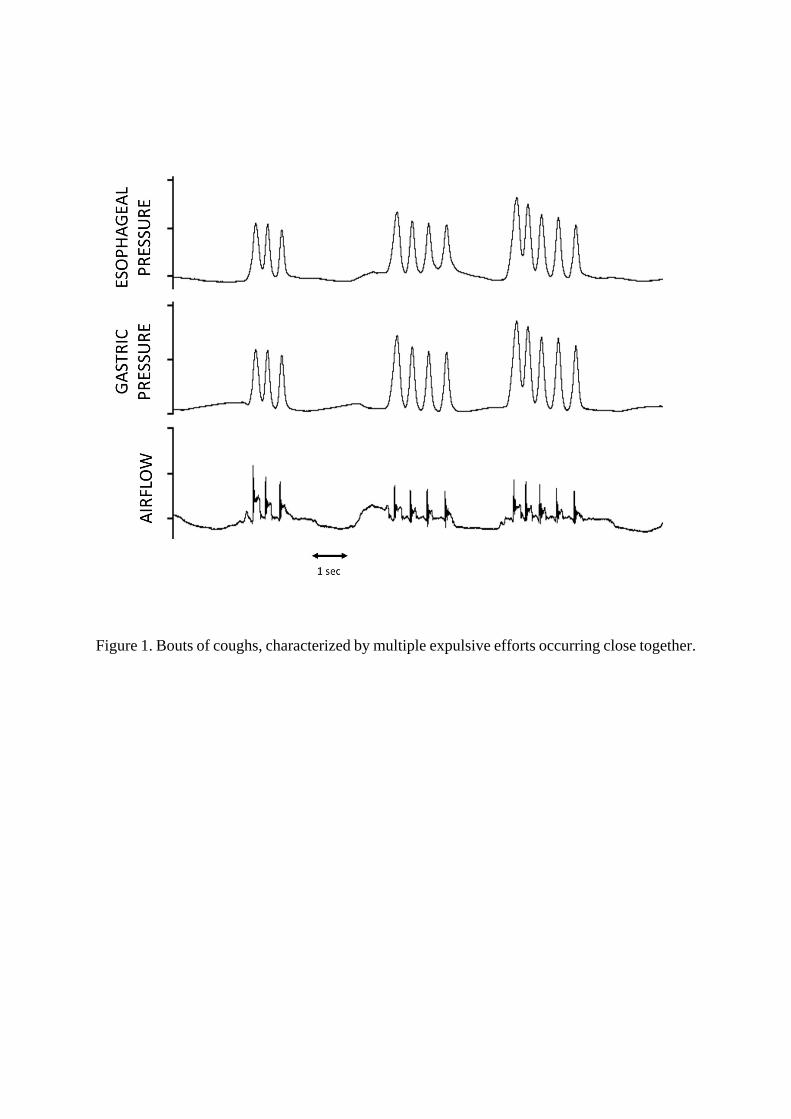
Figure 1. Bouts of coughs, characterized by multiple expulsive efforts occurring close together.

Figure 2. (A) The three-phase flow pattern of a classical cough, characterized by an initial inspiratory phase which is followed by cessation of flow during the compression phase (glottis closure) and then rapid expulsion of air during the expiratory phase (B) Expiratory reflexes and classical cough. Flow trace depicting a series of three expiratory reflexes (characterized by lack of preceding inspiration) followed by a single classical cough and two expiratory reflexes.

Figure 3. Example of diminishing cough strength during cough reaccelerations within a bout. An initial inspiratory effort is followed by multiple expiratory events efforts which have sequentially less flow as the bout progresses.

Figure 4. A typical 3-phase cough sound shown in the time domain. The first phase, explosive, relates to the expulsion of air through the glottis and is followed in most cases by a voiced phase with a gradually diminishing sound signal, and a quiet intermediate phase in between.