globalexpressionanalysisidentifiedapreferentiallynerve ...erk)andap-1proteinactivation...
TRANSCRIPT

Global Expression Analysis Identified a Preferentially NerveGrowth Factor-induced Transcriptional Program Regulatedby Sustained Mitogen-activated Protein Kinase/ExtracellularSignal-regulated Kinase (ERK) and AP-1 Protein Activationduring PC12 Cell Differentiation*□S
Received for publication, June 22, 2011, and in revised form, November 4, 2011 Published, JBC Papers in Press, November 7, 2011, DOI 10.1074/jbc.M111.274076
Steven Mullenbrock, Janki Shah, and Geoffrey M. Cooper1
From the Department of Biology, Boston University, Boston, Massachusetts 02215
Background: Neuronal differentiation of PC12 cells requires sustained ERK signaling induced by NGF.Results: Global expression analysis identified a set of genes that was induced preferentially by NGF-mediated ERK signaling.The NGF-induced genes were targeted by AP-1 and CREB transcription factors.Conclusion: Preferential NGF gene induction is mediated by sustained AP-1 activity.Significance: A transcriptional program specifically induced during neuronal differentiation has been characterized.
Neuronal differentiation of PC12 cells in response toNGF is aprototypical model in which signal duration determines a bio-logical response. Sustained ERK activity induced by NGF, ascompared with transient activity induced by EGF, is critical tothe differentiation of these cells. To characterize the transcrip-tional program activated preferentially by NGF, we comparedglobal gene expression profiles between cells treated with NGFandEGF for 2–4 h, when sustained ERK signaling in response toNGF is most distinct from the transient signal elicited by EGF.This analysis identified 69genes thatwerepreferentially up-reg-ulated in response to NGF. As expected, up-regulation of thesegenes was mediated by sustained ERK signaling. In addition,they were up-regulated in response to other neuritogenic treat-ments (pituitary adenylate cyclase-activating polypeptide and12-O-tetradecanoylphorbol-13-acetate plus dbcAMP) andwereenriched for genes related to neuronal differentiation/function.Computational analysis and chromatin immunoprecipitationidentified binding of CREB and AP-1 family members (Fos,FosB, Fra1, JunB, JunD) upstream of >30 and 50%, respectively,of the preferentially NGF-induced genes. Expression of severalAP-1 family members was induced by both EGF and NGF, buttheir induction was more robust and sustained in response toNGF. The binding of Fos family members to their target geneswas similarly sustained in response to NGF and was reduceduponMEK inhibition, suggesting that AP-1 contributes signifi-cantly to the NGF transcriptional program. Interestingly, Fra1as well as two other NGF-induced AP-1 targets (HB-EGF andmiR-21) function in positive feedback loops thatmay contributeto sustained AP-1 activity.
PC12 rat pheochromocytoma cells are an established cellculture model for studying neuronal differentiation. Inresponse to treatment with nerve growth factor (NGF) andother neuritogenic agents, PC12 cells differentiate into cellsresembling sympathetic neurons, as indicated by cessation ofcell proliferation, neurite outgrowth, electrical excitability, andexpression of neuronal markers. NGF induces differentiationvia the receptor-tyrosine kinase, TrkA, which activates down-stream signaling pathways including phospholipase C�, phos-phatidylinositol 3-kinase, and Ras/Raf/MEK/ERK (1). Notably,treatment of PC12 cells with epidermal growth factor (EGF),which also stimulates a receptor-tyrosine kinase (ErbB), fails toinduce differentiation butmaintains PC12 cell proliferation (2).The distinct biological responses induced by NGF compared
with EGF is intriguing as both growth factors activate receptor-tyrosine kinases that are coupled to a similar set of downstreamsignaling pathways (3). One of these pathways, the MEK/ERKpathway, has been shown to be necessary and sufficient forNGF-induced differentiation, as expression of constitutivelyactive forms of Ras, Raf, MEK, and ERK results in differentia-tion even in the absence of NGF (4–10), whereas functionalinhibition of Ras or MEK blocks NGF-induced differentiation(8, 11–13).However, EGF also results in robust activation of theERK pathway. Comparisons of NGF and EGF signaling haveindicated that the induction of differentiation by NGF, but notby EGF, is the result of sustained activation of ERK in responseto NGF versus transient activation in response to EGF. In par-ticular, ERK activity remains elevated for several hours afterNGF stimulation but returns to base-line levels after 30–60min of treatment with EGF (14–19). The differentiation ofPC12 cells thus provides an importantmodel for understandingthe mechanisms by which the duration of growth factor signal-ing can lead to distinct responses at the cellular level.Whereas the role of sustained ERK activity in NGF-induced
differentiation is well established, the transcriptional programthat is activated by this sustained ERK signaling and is ulti-mately responsible for acquisition of a neuronal phenotype has
* This work was supported, in whole or in part, by National Institutes of HealthGrant RO1 CA18689 (to G. M. C.).
□S The on-line version of this article (available at http://www.jbc.org) containssupplemental Tables 1 and 2.
1 To whom correspondence should be addressed: Boston University, Dept. ofBiology, 5 Cummington St., Boston, MA 02215. Tel.: 617-353-8735; Fax:617-353-8484; E-mail: [email protected].
THE JOURNAL OF BIOLOGICAL CHEMISTRY VOL. 286, NO. 52, pp. 45131–45145, December 30, 2011© 2011 by The American Society for Biochemistry and Molecular Biology, Inc. Printed in the U.S.A.
DECEMBER 30, 2011 • VOLUME 286 • NUMBER 52 JOURNAL OF BIOLOGICAL CHEMISTRY 45131
by guest on June 27, 2018http://w
ww
.jbc.org/D
ownloaded from

not been as well characterized. The initial transcriptionalresponse to growth factor stimulation is the induction of imme-diate-early genes within 30–60 min of growth factor stimula-tion. Nearly all of the immediate-early genes induced by NGFare also induced by EGF (1, 20), so this initial transcriptionalresponse, which occurs before observed differences in NGF-versus EGF-induced ERK signaling, does not distinguish theeffects of NGF and EGF treatment. However, previous studieshave identified a few genes that are preferentially induced byNGF as compared with EGF at later time points (greater than1 h) (21–27). In the present study we have expanded thisapproach by using global expression profiling to identify a set ofgenes that is preferentially induced by NGF compared withEGF at the time points corresponding to NGF-specific sus-tainedERKactivity (2–4h after growth factor stimulation). Thegenes that were up-regulated preferentially by NGF at thesetimes were found to be dependent on sustained ERK activityand to encodemany proteins with established roles in neuronaldifferentiation and/or function. Computational predictionsand experimental analysis identified AP-12 and CREB tran-scription factors as major regulators of this NGF-induced tran-scriptional program. The expression and in vivo DNA bindingactivity of several AP-1 family members was enhanced afterstimulationwithNGF comparedwith EGF, suggesting that sus-tained activation of these factors contributes to the preferentialinduction of a large number of genes in response to NGF. Fur-thermore, several preferentially NGF-induced AP-1 targets,including Fra1 (Fosl1), miR-21, and HB-EGF, participate inpositive feedback regulation of MEK/ERK and AP-1 signalingand thusmay contribute directly to propagating sustainedAP-1activity in response to NGF.
EXPERIMENTAL PROCEDURES
Cell Culture and Treatments—PC12 rat pheochromocytomacells were grown in Dulbecco’s modified Eagle’s medium(DMEM) (Mediatech) containing 10% fetal bovine serum(HyClone) and 5% horse serum (Invitrogen). For gene expres-sion studies, PC12 cells were plated at 7.6 � 105 cells/60-mmplate and 3.5 � 105 cells per 35-mm plate and allowed to growfor 24 h. After 24 h cells were washed once in low serummedia(DMEM with 0.5% horse serum) and then starved for 24 h inthis low serum media. PC12 cells were treated with NGF (50ng/ml; R&D Systems), EGF (25 ng/ml; Calbiochem), PACAP38(100 nM; Phoenix Pharmaceuticals), TPA (20 nM; Sigma),dbcAMP (0.5 mM, Sigma), and U0126 (10 �M; Cell SignalingTechnology).Microarray Analysis—Microarrays were performed on three
independent biological samples. Total RNA for microarrayexperiments was extracted with TRIzol reagent (Invitrogen).After ethanol precipitation, RNAwas applied to an RNeasy col-umn (Qiagen) for further purification as per themanufacturer’sprotocol. The quality of the RNAwas determined using an Agi-lent bioanalyzer before analysis on Affymetrix Rat Gene 1.0ST
microarrays. Microarray sample preparation/labeling, hybrid-ization, scanning, and subsequent data analysis were conductedby the Boston University Microarray Facility. Heatmaps wereconstructed using the programMultiExperiment Viewer (28).Real-time Reverse Transcription-Polymerase Chain Reaction
(RT-PCR)—Total RNA used for real time RT-PCR wasextracted using a TRIzol extraction as per the manufacturer’sprotocol. Real-time RT-PCR was carried out as previouslydescribed (29). Primer sequences are listed in supplementalTable 1.Gene Ontology Analysis—Overrepresentation of Gene
Ontology (GO) terms was determined using the Data Base forAnnotation, Visualization and Integrated Discovery (DAVID)Version 6.7 (30, 31). 65/69 preferentially NGF-induced geneshad an identifier recognized by the DAVID data base and wereused for this analysis. A GO category was considered to beenriched significantly comparedwith the rat genome as awholeif it was associatedwith�10%of the geneswith a p value�0.05.The DAVID functional annotation clustering tool was used toidentify overrepresented clusters of GO terms that co-associatewith one another.siRNA Transfection—PC12 cells were plated at 1 � 105 cells/
35-mm plate in 2 ml of complete medium 1 day before trans-fection with Lipofectamine 2000 (Invitrogen) as per the manu-facturer’s protocol. Amixture containing 5�l of Lipofectamine2000 and the corresponding siRNA in 500 �l of serum-freemedia was incubated at room temperature for 20 min beforeadding it to the cells. Negative control siRNA #1 (Ambion,4390843) and siRNAs targeting Plaur (Ambion, S132350) andPVR (Ambion, S128925) were added at a final concentration of20 nM. Cells were incubatedwith the siRNAs for�7 h, and thenthe medium was changed to low serummedia for another �12h after which the cells were treated with NGF as indicated inFig. 4. To assess the extent of knockdown, expression of thesiRNA-targeted genes after 2 hNGF treatment was determinedby real time RT-PCR.Transcription Factor Binding Site Analysis—Identification of
overrepresented transcription factor binding sites in theupstream sequences of the preferentiallyNGF-induced gene setcompared with a background gene set was conducted as previ-ously described (29). The background gene set consisted of 291genes whose expression levels from the microarray experi-ments did not change in response to treatment (log 2 � 0.1 andlog 2 � �0.1). The regions 5 kb upstream of the transcriptionstart sites in rat and the corresponding human orthologoussequences were analyzed with the MATCH program using theMinSUM threshold for thematrices found in TRANSFACVer-sion 12.1. Sequences and MULTIZ alignments were obtainedfrom the University of California Santa Cruz Genome Browser(Version rn4/Nov 2004, Version hg18/March 2006), whichwere available for 43 of the 69 preferentially NGF-inducedgenes. Binding site matrices that averaged more than 1 pre-dicted binding site/kb of sequence in the background gene setwere excluded from subsequent analysis. Permutation p valueswere FDR-corrected.AP-1 and CREB Chromatin Immunoprecipitation (ChIP)—
ChIP assays were performed as previously described (32) using5 �g of the following antibodies: Fos (Santa Cruz, sc-7202),
2 The abbreviations used are: AP-1, activator protein-1; PACAP, pituitaryadenylate cyclase-activating polypeptide; TPA, 12-O-tetradecanoylphor-bol-13-acetate; dbcAMP, dibutyryl-cyclic AMP; FDR, false discovery rate;GO, gene ontology; CREB, cyclic AMP response element-binding protein.
NGF-induced Transcriptional Program
45132 JOURNAL OF BIOLOGICAL CHEMISTRY VOLUME 286 • NUMBER 52 • DECEMBER 30, 2011
by guest on June 27, 2018http://w
ww
.jbc.org/D
ownloaded from

FosB (Santa Cruz, sc-48), Fra-1 (Santa Cruz, sc-605), Fra-2(Santa Cruz, sc-171), Jun (Santa Cruz, sc-1694X and Abcam,ab31419), JunB (Santa Cruz, sc-73X), JunD (Santa Cruz, sc-74),and CREB-1 (Santa Cruz, sc-186). For ChIP assays using anti-bodies for the AP-1 family members, protein A-agarose beads(Upstate Biotechnology) were washed successively in low saltwash, high salt wash, LiCl wash, and twice in 1� Tris/EDTAbuffer. When using the CREB-1 antibody, protein A-agarosebeads were washed successively in low salt wash three times,once in LiCl wash, and twice in 1� Tris/EDTA buffer. Cross-links were reversed using high salt and heating to 65 °C over-night. Immunoprecipitated DNA was purified using a gelextraction kit (Qiagen) and quantified with real-time PCRusing primers located near the respective predicted tran-scription factor binding sites (within 300 bp, see supplemen-tal Table 1).Immunoblots—PC12 cells were lysed in 2� Laemmli buffer.
Proteins were electrophoresed on SDS-polyacrylamide gels,transferred to nitrocellulose or polyvinylidene difluoridemem-branes, and immunoblotted with antibodies to total p44/42MAPK (ERK1/2) (Cell Signaling #9102), phospho p44/42MAPK (ERK1/2) (Cell Signaling #9101), neurofilament-L (CellSignaling #2837), Fos (Santa Cruz, sc-7202), FosB (Santa Cruz,sc-48), Fra-1 (Santa Cruz, sc-605), JunB (Santa Cruz, sc-73X),and �-actin (Sigma) overnight at 4 °C. Membranes were visual-ized with chemiluminescence or fluorescence after using theappropriate horseradish peroxidase-linked or Cy3-labeled sec-ondary antibody. Phospho-ERK1/2 and ERK1/2 immunoblotswere quantified using ImageJ software. Values obtained forboth p44 and p42 ERK bands were added together, and phos-pho p44/42 values were normalized to total p44/42.
RESULTS
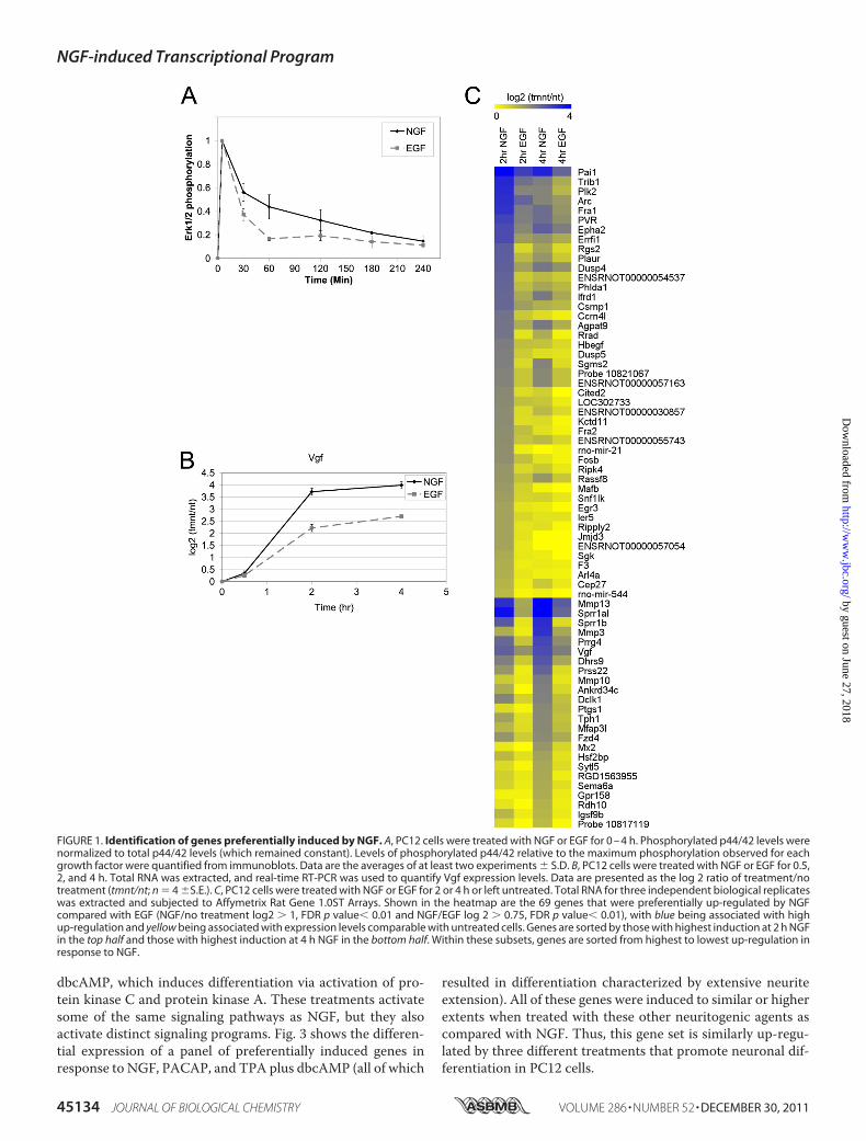
Identification of Genes That Are Preferentially Induced byNGF—Global expression profiling was used to identify changesin gene expression that occurred preferentially in response toNGF after 2 and 4 h of treatment of PC12 cells with NGF orEGF. These time points coincided with the period of sustainedERK signaling induced by NGF (Fig. 1A) as well as with thepreferential induction of Vgf, which has been previously iden-tified as a preferentially NGF-induced gene involved in neuro-nal differentiation (21) (Fig. 1B).The results of microarray analyses (supplemental Table 2)
are summarized in Table 1. Treatment with NGF for 2 or 4 hresulted in a �2-fold up-regulation of 265 genes and down-regulation of 97 genes (p � 0.01, FDR corrected). Treatmentwith EGF resulted in the up-regulation of 85 genes and down-regulation of 20 genes. All of the genes affected by EGF werealso up- or down-regulated in response to NGF, and there wereno genes that were up- or down-regulated to a greater extent byEGF than by NGF.Although larger numbers of genes were up-regulated or
down-regulated byNGF than by EGF, the differences in expres-sion between NGF and EGF treatments for most of these geneswere small. We, therefore, sought to identify a set of geneswhose expression was significantly different between NGF andEGF treatments by comparing the extent of expression changesinduced by NGF versus EGF. A set of preferentially NGF-regu-
lated genes was, therefore, defined by expression changes thatwere 1.7-fold greater (p� 0.01) in response toNGF than to EGF(Table 1). None of the genes was down-regulated �1.7-foldmore by NGF compared with EGF and thus did not meet thesecriteria for preferential NGFdown-regulation at the 2–4 h timepoints.In contrast to the down-regulated genes, 69 genes were up-
regulated preferentially by NGF compared with EGF (NGF/EGF �1.7-fold, p � 0.01) (Table 1). The expression levels ofthese genes after both EGF andNGF treatment are summarizedas a heatmap in Fig. 1C. Of the 69 genes that were preferentiallyinduced by NGF, some demonstrated no appreciable levels ofinduction in response to EGF treatment (i.e. Rgs2, Jmjd3,Kctd11, Rrad), whereas others (i.e. Sprr1al, Mmp13, Mmp3)were induced by EGF but to a lesser extent than induction byNGF. The 69 genes that were preferentially up-regulated byNGF included genes that were previously shown to be morehighly up-regulated by NGF than EGF, such as Plaur,Vgf, Pai1,Arc,Mmp3, andMmp13 (21–24). These genes will be referredto as “preferentiallyNGF-induced genes,” and subsequent stud-ies focused on this gene set.Several different patterns of gene expression were observed
in the microarray experiments. In Fig. 1C, the 69 preferentiallyNGF-induced genes were separated into 2 groups; 1) the upperhalf contains 45 genes that were more highly induced at 2 h ofNGF treatment, and 2) the bottom half contains 24 genes thatweremore highly induced at 4 h ofNGF treatment. The kineticsof gene induction also varied, with the expression of some genes(e.g. Jmjd3, Kctd11, and Ccrn4l) being up-regulated at the ear-lier 2 h time point and subsequently returning to basal levels at4 h of NGF treatment. Other genes were expressed at higherlevels at 4 h (e.g. Mmp3 andMmp10), and there was a variety ofintermediate kinetic profiles where the expression levels at 2and 4 h were roughly the same (Vgf, Dclk1, Epha2, Agpat9).
The differential expression of representative genes preferen-tially induced by NGF was also examined by real time RT-PCRover a period of 0.5–4 h of growth factor treatment (Fig. 2).These experiments confirmed the preferential NGF-mediatedinduction that was observed in the microarray experiments,with genes such as Jmjd3, Kctd11, and Rgs2 showing no signif-icant up-regulation in response to EGF. Sprr1b, which demon-strated the largest differential expression between NGF andEGF treatment in themicroarray experiments, exhibited a sim-ilarly large differential expression (�20-fold) betweenNGF andEGF treatment via real time RT-PCR analysis (Fig. 2). In addi-tion, the different kinetic patterns of gene expression that wereobserved in themicroarray experimentswere confirmed by realtime RT-PCR.Genes Preferentially Induced by NGF Play a Role in Neuronal
Differentiation/Function—PC12 cells can undergo differentia-tion in response to treatments other than NGF, so it was ofinterest to determine if the genes preferentially induced byNGF were also induced by other agents that induce neuronaldifferentiation. We, therefore, investigated the expression ofthese genes in response to treatment of PC12 cells with theneuropeptide PACAP, which binds to G protein-coupledreceptors and activates phospholipase C, cAMP, and MEK/ERK signaling pathways (33), and to treatment with TPA plus
NGF-induced Transcriptional Program
DECEMBER 30, 2011 • VOLUME 286 • NUMBER 52 JOURNAL OF BIOLOGICAL CHEMISTRY 45133
by guest on June 27, 2018http://w
ww
.jbc.org/D
ownloaded from
dbcAMP, which induces differentiation via activation of pro-tein kinase C and protein kinase A. These treatments activatesome of the same signaling pathways as NGF, but they alsoactivate distinct signaling programs. Fig. 3 shows the differen-tial expression of a panel of preferentially induced genes inresponse to NGF, PACAP, and TPA plus dbcAMP (all of which
resulted in differentiation characterized by extensive neuriteextension). All of these genes were induced to similar or higherextents when treated with these other neuritogenic agents ascompared with NGF. Thus, this gene set is similarly up-regu-lated by three different treatments that promote neuronal dif-ferentiation in PC12 cells.
FIGURE 1. Identification of genes preferentially induced by NGF. A, PC12 cells were treated with NGF or EGF for 0 – 4 h. Phosphorylated p44/42 levels werenormalized to total p44/42 levels (which remained constant). Levels of phosphorylated p44/42 relative to the maximum phosphorylation observed for eachgrowth factor were quantified from immunoblots. Data are the averages of at least two experiments � S.D. B, PC12 cells were treated with NGF or EGF for 0.5,2, and 4 h. Total RNA was extracted, and real-time RT-PCR was used to quantify Vgf expression levels. Data are presented as the log 2 ratio of treatment/notreatment (tmnt/nt; n � 4 �S.E.). C, PC12 cells were treated with NGF or EGF for 2 or 4 h or left untreated. Total RNA for three independent biological replicateswas extracted and subjected to Affymetrix Rat Gene 1.0ST Arrays. Shown in the heatmap are the 69 genes that were preferentially up-regulated by NGFcompared with EGF (NGF/no treatment log2 � 1, FDR p value� 0.01 and NGF/EGF log 2 � 0.75, FDR p value� 0.01), with blue being associated with highup-regulation and yellow being associated with expression levels comparable with untreated cells. Genes are sorted by those with highest induction at 2 h NGFin the top half and those with highest induction at 4 h NGF in the bottom half. Within these subsets, genes are sorted from highest to lowest up-regulation inresponse to NGF.
NGF-induced Transcriptional Program
45134 JOURNAL OF BIOLOGICAL CHEMISTRY VOLUME 286 • NUMBER 52 • DECEMBER 30, 2011
by guest on June 27, 2018http://w
ww
.jbc.org/D
ownloaded from

The functions of the genes preferentially induced by NGFwere investigated by GO analysis using the DAVID Bioinfor-matics Data base (30, 31). Consistent with their induction bymultiple factors that induce PC12 differentiation, this gene setwas enriched for GO categories related to development and
differentiation, such as “developmental process” (39% of geneset), “multicellular organismal development” (32% of gene set),“system development” (29% of gene set), “tissue development”(12% of gene set), “nervous system development” (15% of geneset), and “cell differentiation” (19% of gene set) (Fig. 4A). We
TABLE 1Summary of microarray analysis of NGF- and EGF-treated PC12 cellsPC12 cells were treated with NGF or EGF for 2 or 4 h or left untreated. Total RNA for three independent biological replicates was extracted and subjected to Affymetrix RatGene 1.0ST arrays. Shown are the numbers of genes that were up-regulated or down-regulated at 2 or 4 h for each treatment as well as those that were preferentially affectedby either growth factor. 12 rapidly induced immediate-early genes were excluded from these counts and subsequent analyses because they reached maximum inductionmuch earlier (30 min).
Genes affected by NGFNGF/NT > 2-fold (p < 0.01)
Genes affected by EGFEGF/NT > 2-fold (p < 0.01)
Genes preferentially affected by NGFNGF/EGF > 1.7-fold (p < 0.01)
Genes preferentially affected by EGFEGF/NGF > 1.7-fold (p < 0.01)
Up-regulated 265 85 69 0Down-regulated 97 20 0 0
FIGURE 2. Real-time RT-PCR analysis of a panel of genes induced preferentially by NGF compared with EGF. RNA was extracted from PC12 cells treatedwith NGF (solid line) or EGF (dashed line) for 0, 0.5, 2, and 4 h and subjected to real-time RT-PCR analysis. Data are the average (n � 2– 4) log 2 ratio oftreatment/no treatment � S.E.
NGF-induced Transcriptional Program
DECEMBER 30, 2011 • VOLUME 286 • NUMBER 52 JOURNAL OF BIOLOGICAL CHEMISTRY 45135
by guest on June 27, 2018http://w
ww
.jbc.org/D
ownloaded from

next applied the DAVID functional annotation clustering toolto our gene set. This tool finds overrepresented clusters of GOterms that co-associate with one another, thus grouping similarGOannotations together. The top twoGOclusters for the pref-erentially NGF-induced genes were related to development(p � 0.01, enrichment score � 2.22) (Fig. 4B) and peptidase/matrix metalloproteinase activity (p � 0.05, enrichmentscore � 1.4) (not shown). Modification of the extracellularenvironment by matrix metalloproteinases is known to be cru-cial for neurite formation/outgrowth, and many matrix metal-loproteinases (Mmp3, Mmp10, Mmp13) were preferentiallyinduced by NGF. Fig. 4B was generated from the functionalannotation clustering results for those genes that were associ-ated with GO terms related to developmental functions. Twen-ty-nine genes were associated with aGO term in this functionalcluster, which included GO terms such as “nervous systemdevelopment,” and “cell differentiation,” suggesting that thepreferentiallyNGF-induced gene set is enriched in geneswhosefunctions may be critical for PC12 differentiation.The function of preferentially NGF-induced genes in PC12
differentiation was investigated by determining the effect ofsiRNAknockdownon the expression of neurofilament-L, awellcharacterized neuronal marker whose expression is up-regu-lated at later time points upon acquisition of a neuronal pheno-type (34, 35). Neurofilament-L was nearly undetectable inuntreated, undifferentiated PC12 cells (Fig. 4C, first lane) butwas robustly expressed after cells were induced to differentiatefor 2 days in the presence of NGF (Fig. 4C, second lane). EGF, asexpected based on its inability to promote PC12 differentiation,resulted in much lower neurofilament-L expression (Fig. 4C,third lane). siRNA knockdown of the preferentially NGF-in-duced genes, Plaur and PVR, significantly inhibited the NGF-mediated induction of neurofilament-L (Fig. 4C, fifth and sixthlanes), demonstrating a critical role for these genes in PC12differentiation.These results are consistent with previous studies that dem-
onstrated a role for Plaur and PVR in PC12 differentiation (24,36). In addition, several other preferentially NGF-induced
genes (Mmp3, Mmp10, Plk2, and Rgs2) have previously beenshown to be critical for PC12 differentiation through siRNAknockdown or functional inhibition (36–38). Many also haveestablished roles in neuronal or brain differentiation/function(Sgk, Sprr1a, Jmjd3, Kctd11, Ifrd1, Epha2, rno-miR-21, Pai1,Tph1, Sema6a, Hbegf, Egr3, Arc, Vgf, Dclk1) (21, 39–55). Intotal, at least 21 (30%) of the preferentially NGF-induced geneshave previously described roles in neuronal differentiation/function in PC12 cells and other neuronal systems. Combiningthese 21 genes with the 29 genes associated with a GO term inthe development functional cluster (Fig. 4B), roughly 50% (34)of the genes preferentially induced by NGF are implicated inneuronal development.Sustained MEK/ERK Signaling Contributes to Gene Induc-
tion by NGF—Because sustained ERK activation is the key dif-ference between NGF and EGF signaling that leads to neuronaldifferentiation, we investigated the role of MEK/ERK signalingin the up-regulation of the preferentially NGF-induced genes.Pretreatment of PC12 cells with the MEK inhibitor U0126 sig-nificantly reduced the induction of a representative subset ofthese genes in response to NGF (Fig. 5A). Thus, all of the pref-erentially NGF-induced genes tested are downstream of thesignaling pathway critical for PC12 differentiation.To determine the importance of a sustained ERK signal for
the up-regulation of these genes, U0126 was added 45min afterNGF treatment, resulting in a transient ERK signal similar tothat observed in response to EGF treatment. When U0126 wasadded 45 min after NGF treatment, ERK phosphorylation wasnot detectable 15 min later (after a total of 1 h NGF treatment)(Fig. 5, lower panel). Such delayed treatment with U0126 gen-erally reduced the up-regulation of the preferentially NGF-in-duced genes tested at both 2 and 4 h ofNGF treatment (Fig. 5B),indicating that sustained ERK signaling contributes to induc-tion of these genes. It is noteworthy that the effect of delayedU0126 addition was less than that observed upon pretreatmentwith the inhibitor (compare Fig. 5, A and B). This is consistentwith the observed differences in gene expression induced byNGF compared with EGF, as EGF also resulted in a significantbut reduced up-regulation of many of the preferentially NGF-induced genes (see Fig. 1C).AP-1 and CREB Bind to Regulatory Regions of Many Genes
Preferentially Induced by NGF—Because genes that are acti-vated downstream of common stimuli or signaling pathwaysmay be regulated by a similar set of transcription factors (29, 56,57), we identified transcription factor binding sites that wereoverrepresented in the regulatory regions of the genes thatwerepreferentially induced by NGF. In addition, only binding sitesthat were conserved between the rat and human sequence wereconsidered, as binding sites that are conserved across speciesare more likely to be biologically relevant (58).Binding sites were predicted utilizing the TRANSFAC 12.1
data base and the MinSUM threshold in regions spanning 5 kbupstream of the transcription start sites for both the gene setpreferentially induced by NGF treatment as well as a back-ground gene set composed of 291 genes whose expression didnot change in response to growth factor treatment. For thebinding site analysis, 43 of the 69 preferentially NGF-induced
FIGURE 3. Genes preferentially induced by NGF compared with EGF weresimilarly up-regulated by other agents that promote PC12 differentia-tion. Real time RT-PCR analysis was conducted on PC12 cells treated withNGF, PACAP, and TPA�dbcAMP over a time course of 1– 6 h. Plotted are themaximum observed inductions for each of the treatments over the timecourse. Data are the averages from 2–3 independent experiments � S.D.
NGF-induced Transcriptional Program
45136 JOURNAL OF BIOLOGICAL CHEMISTRY VOLUME 286 • NUMBER 52 • DECEMBER 30, 2011
by guest on June 27, 2018http://w
ww
.jbc.org/D
ownloaded from

genes were used, as an upstream sequence could not beobtained for the other 26 genes.Binding sites for AP-1 and CREB were found to be overrep-
resented in the upstream regions of the preferentially NGF-induced gene set (FDR corrected p value� 0.01). Using theV$AP1_Q6_01 matrix, �65% (28/43) of the genes preferen-tially induced by NGF had at least one predicted AP-1 bind-ing site in their upstream regions, suggesting that AP-1 could
regulate a large subset of this gene set. �40% (18/43) of thegenes preferentially induced by NGF had a predicted CREBbinding site in their upstream regions when using theV$CREB_Q2_01 matrix. CREB and AP-1 were not only pre-dicted to target a large fraction of the preferentially NGF-induced genes, but both of these factors have been previouslylinked to PC12 neuronal differentiation (59–66), so theywere investigated further.
FIGURE 4. Genes induced preferentially by NGF are enriched for functions related to differentiation/development. A, gene ontology analysis using theDAVID Bioinformatics Data base was conducted on the gene set that was preferentially induced by NGF. Shown are GO terms that were overrepresented in thisgene set compared with all rat genes (p � 0.05, �10% of genes had to be classified by a GO term). B, functional cluster analysis of this gene set identified acluster of similar GO terms related to development/differentiation (p � 0.01, enrichment score � 2.22). An enrichment score �1.3 is generally consideredsignificant. For this analysis, classification stringency was set to low. The GO terms associated with this cluster are shown on the right, and the preferentiallyNGF-induced genes within this cluster are listed across the top. A black box denotes that a gene was associated with the corresponding GO term. C, Western blotanalysis was used to determine levels of neurofilament-L after NGF treatment in the presence/absence of siRNAs against the preferentially NGF-induced genesPlaur and PVR. PC12 cells were transfected with siRNAs against Plaur, PVR, or a negative control siRNA (Neg) and then treated with NGF for 2 days. Knockdownof both Plaur and PVR was greater than 80%. Results are representative of three independent experiments. NT, not treated.
NGF-induced Transcriptional Program
DECEMBER 30, 2011 • VOLUME 286 • NUMBER 52 JOURNAL OF BIOLOGICAL CHEMISTRY 45137
by guest on June 27, 2018http://w
ww
.jbc.org/D
ownloaded from

ChIP assays were conducted to identify in vivo binding ofCREB to these predicted binding sites (Fig. 6). In untreatedPC12 cells, 14 of the predicted CREB target genes tested hadCREB bound at the predicted sites compared with an IgG con-trol and compared with the upstream region of the negativecontrol Myog (�3-fold compared with both controls). CREBhas previously been shown to bind to its target genes constitu-tively (67), and CREB binding did not change upon NGF treat-ment (data not shown). Thus, CREBwas bound to the upstreamregions of �30% (14/43) of the preferentially NGF-inducedgenes.Putative AP-1 binding sites upstream of 65% (28/43) of the
preferentially NGF-induced genes were identified. The AP-1family of transcription factors consists of the Fos family mem-bers (Fos, FosB, Fra1, Fra2) and Jun family members (Jun, JunB,JunD). These family members bind as dimers in various com-binations, with the Jun family members able to homo- or het-
erodimerize with themselves or with the Fos family members,whereas the Fos familymembersmust heterodimerizewith oneof the Jun family members (68). Because AP-1 can bind in somany different combinations, ChIP assays were conductedfor all AP-1 family members. Fra2 and Jun could not bedetected at the promoter regions of the genes that were pref-erentially induced by NGF treatment. In contrast, NGFtreatment resulted in the recruitment of Fos, FosB, Fra1,JunB, and JunD to predicted sites upstream of 18 of the pref-erentially NGF-induced genes (Fig. 7A). These factors werebound �2-fold higher at the predicted AP-1 target regions inNGF-treated cells compared with an IgG control and to theupstream region of the negative control Myog. Althoughthere was a high level of recruitment of most AP-1 familymembers in response to NGF, JunD was bound at a relativelyhigh level in untreated cells and was recruited to a lesserextent than the other AP-1 family members. In some cases,such as for JunD binding upstream ofAgpat9, Trib1, andArc,there was almost no recruitment in response to NGF treat-ment (Fig. 7A).CREB and AP-1 share a very similar consensus binding site
and have previously been shown to regulate genes through thesame binding site (69–73). To obtain a more comprehensivelist of AP-1 and CREB target genes, we conducted ChIP assaysto detect binding of CREB in regions that contain a predictedAP-1 site and to detect binding of AP-1 in regions that containa predicted CREB site. We did not detect binding of CREB toregions containing any of the AP-1 binding sites identified inFig. 7A. However, we did detect binding (�2-fold comparedwithMyog and IgG controls) ofAP-1 to several regions contain-ing predictedCREB sites aswell as to the region upstreamof thepreferentially NGF-induced microRNA, miR-21 (Fig. 7B). This
FIGURE 5. Genes up-regulated preferentially by NGF are regulated bysustained ERK signaling. Real time RT-PCR analysis was conducted on totalRNA from PC12 cells that were pretreated with DMSO or U0126 1 h beforeNGF treatment (A) or from PC12 cells that were treated with DMSO or U012645 min after NGF (B). Results are presented as the average log2 ratio of treat-ment/no treatment in the form of a heatmap, n � 2. An immunoblot of phos-pho and total ERK after 0 –3 h of NGF treatment when cells were treated withUO126 or vehicle control (DMSO) 45 min after NGF is shown below.
FIGURE 6. Identification of CREB binding sites in the upstream regions ofgenes induced preferentially by NGF. ChIP assays were conducted usingantibody against CREB1 or an IgG control. Immunoprecipitated DNA wasquantified via real time PCR using primers adjacent to/overlapping the pre-dicted CREB binding sites (see supplemental Table 1 for locations). Data areplotted as % input and are the average from three determinations � S.E. Theupstream region of Myog, a muscle-specific gene, served as a negative con-trol. If a gene had several predicted CREB binding sites in its upstream regu-latory sequence, only the region with the highest level of binding is pre-sented. Different CREB binding sites located in the upstream regulatoryregion of the same gene are designated by C1, C2, etc. and can be found insupplemental Table 1. The 14 genes that had CREB bound to their upstreamregions (�3-fold compared with Myog and IgG controls) are denoted by thesolid line in the figure.
NGF-induced Transcriptional Program
45138 JOURNAL OF BIOLOGICAL CHEMISTRY VOLUME 286 • NUMBER 52 • DECEMBER 30, 2011
by guest on June 27, 2018http://w
ww
.jbc.org/D
ownloaded from

increased the total number of preferentially NGF-inducedgenes with upstream AP-1 sites to a total of 24, representingmore than 50% (24/44) of the genes analyzed.AP-1 Recruitment in Response to NGF Is Downstream of
MEK/ERK Signaling—Because up-regulation of the preferen-tially NGF-induced genes was downstream of MEK/ERK sig-naling (see Fig. 5), we investigated the effect of MEK/ERK sig-naling on recruitment of AP-1 transcription factors to the
upstream regions of these genes. ChIP assays were conductedon PC12 cells treated with U0126 before NGF treatment, andthe levels of Fos, FosB, Fra1, and JunB at the upstream regionsof representative AP-1 target genes were determined (Fig. 8),with similar results observed for the other AP-1 target genes.The recruitment of all of these AP-1 family members inresponse to NGF was reduced by inhibition of MEK withU0126, indicating that the MEK/ERK pathway contributes to
FIGURE 7. Identification of AP-1 binding sites in the upstream regions of genes induced preferentially by NGF. A, ChIP assays were conducted in PC12cells that were either left untreated or treated with NGF for 2 h using antibodies for Fos, FosB, Fra1, JunB, and JunD. Immunoprecipitated DNA was quantifiedvia real time PCR using primers adjacent to/overlapping predicted AP-1 binding sites (see supplemental Table 1 for locations) and plotted as % input. Theupstream region of Myog, a muscle-specific gene, served as the negative control. Data are the averages of 2– 6 biological replicates � S.E. Shown are only thoseupstream sequences that were significantly immunoprecipitated by AP-1 antibodies (�2-fold compared with Myog and the IgG control for at least 2 AP-1family members). If a gene had more than 1 predicted AP-1 binding site in its upstream regulatory region, only the region with the highest binding is presented.Different AP-1 binding sites located in the upstream regulatory region of the same gene are designated by S1, S2, etc. and can be found in supplemental Table1. B, AP-1 binding in regions containing predicted CREB sites and in the upstream region of miR-21 was detected by conducting JunB ChIPs in PC12 cells treatedas in A. Immunoprecipitated DNA was quantified via real time PCR using primers adjacent to/overlapping predicted CREB or previously identified AP-1 (formiR-21) (87, 88) binding sites. Data are the average of two determinations � S.E.
NGF-induced Transcriptional Program
DECEMBER 30, 2011 • VOLUME 286 • NUMBER 52 JOURNAL OF BIOLOGICAL CHEMISTRY 45139
by guest on June 27, 2018http://w
ww
.jbc.org/D
ownloaded from

AP-1 activation in the NGF-mediated transcriptional programof PC12 cells.Expression and Recruitment of AP-1 Family Members Are
Increased Preferentially by NGF Compared with EGF—We fur-ther investigated the effects of NGF comparedwith EGF signal-ing on both the expression of AP-1 family members and ontheir recruitment to upstream regions of NGF target genes.Using real time RT-PCR, the mRNA levels for Fos, Fra1, FosB,
JunB, and JunD were examined in response to NGF and EGFtreatment over a time course of 0.5–4 h (Fig. 9A). These AP-1family members were highly up-regulated in response togrowth factor treatment, with the exception of JunD, whoseexpression did not significantly change. These results for JunDare consistent with previous studies (20, 74, 75) as well as withour microarray and ChIP data, which indicated minimalchanges in JunD expression or recruitment in response to NGFtreatment. The up-regulation of Fos, Fra1, FosB, and JunB atearly time points (0.5 h) was similar in response to NGF andEGF. However, their expression levels at later time points werehigher in response to NGF. These results are consistent withthe phospho-ERK levels observed in Fig. 1A, where higher sus-tained phosphorylationwas observed at later time points (0.5–2h ofNGF treatment). In fact, FosB and Fra1 are two of the genesthat were up-regulated preferentially by NGF compared withEGF (see Fig. 1C).These studies also illustrate differences in the kinetics of
induction of theAP-1 familymembers. Fos andFosBweremorehighly up-regulated at 0.5 h, and subsequently their expressionlevels decreased at 2 and 4 h, although the decrease in FosBexpressionwas somewhat slower. Fra1wasmore highly up-reg-ulated at 2 h ofNGF treatment, and this high level of expressionwasmaintained at 4 h, consistentwith previous studies (76) thatdemonstrated more delayed induction kinetics of this AP-1family member. JunB displayed rapid induction (similar to Fos)at 0.5 h and subsequently decreased at 2 and 4 h of NGFtreatment.The protein levels of these AP-1 family members were also
investigated because their expression is not only regulated atthe mRNA level but also at the protein level via post-transla-tional modifications and stability (68). The levels of Fos, FosB,Fra1, and JunB proteins were increased to a much greaterextent in response to NGF as compared with EGF (Fig. 9B). Inaddition, the varying kinetic patterns observed at the mRNAlevel were mirrored at the protein level, with Fos, FosB, andJunB proteins reaching high levels very rapidly (1 h NGF),whereas Fra1 reached high levels at later time points (4 h NGF).These kinetic patterns at both themRNA and protein levels areconsistent with previous studies (20, 76, 77).ChIP assays were conducted to determine binding of Fos,
Fra1, and FosB over a time course (1–3 h) of NGF or EGF treat-ment for a panel of genes that was induced preferentially byNGF (Fig. 10). NGF treatment resulted in a higher level ofrecruitment for Fos, Fra1, and FosB throughout the duration ofthe experiment compared with EGF treatment. In addition, thesame kinetic patterns observed at the mRNA and protein levelswere observed for the recruitment of each of these AP-1 familymembers. Fos binding levels were highest at earlier time points(1 h), whereas Fra1 was recruited at later time points (3 h). FosBexhibited similar levels of binding at 1 and 2h of treatment, thusillustrating a more intermediate binding pattern, as was mir-rored by itsmRNAand protein expression kinetics. Overall, thepreferential recruitment of AP-1 family members in responsetoNGF, as comparedwith EGF, paralleled the increases in theirexpression, suggesting that their sustained expression drivesthe transcriptional response to NGF.
FIGURE 8. AP-1 recruitment to NGF-induced genes is reduced upon MEKinhibition. ChIP assays were conducted on PC12 cells pre-treated with DMSOor U0126 1 h before NGF treatment. Chromatin was immunoprecipitatedusing antibody to Fos, FosB, Fra1, and JunB and quantified via real time PCR asin Fig. 7. Data are plotted as the average % input (n � 2) � S.E.
NGF-induced Transcriptional Program
45140 JOURNAL OF BIOLOGICAL CHEMISTRY VOLUME 286 • NUMBER 52 • DECEMBER 30, 2011
by guest on June 27, 2018http://w
ww
.jbc.org/D
ownloaded from

DISCUSSION
The distinct biological responses elicited by NGF and EGF(differentiation or proliferation) in PC12 cells are primarilydetermined by differences in signal duration. Differentiation isdriven by the sustained ERK activity induced by NGF, as com-pared with the transient activation of ERK induced by EGF. Inthe present study, we sought to characterize the transcriptionalprogram that is activated specifically downstream of sustainedERK signaling and would, therefore, be expected to contributedirectly to the differentiation process.We used microarray analysis to identify the global gene
expression changes that occurred preferentially in response toNGF compared with EGF at 2–4 h after growth factor stimula-tion, when the sustained ERK activity induced by NGF wasmost distinct from the transient activity induced by EGF. Theset of 69 genes that we identified as preferentiallyNGF-inducedby this global analysis included several genes (Vgf,Mmp3, Arc,Mmp13, Pai1, and Plaur) that had previously been identified asbeing preferentially up-regulated byNGF at similar time points
in non-global studies (21–24). Many of the genes we identifiedas being preferentially induced by NGF had also been shown tobe preferentially induced by treatments that induce differenti-ation (NGF and PACAP) compared with a treatment that doesnot induce PC12 differentiation (insulin) (78). We similarlyfound that the preferentially NGF-induced genes were inducedby treatment of PC12 cells with PACAP or with TPA plusdbcAMP, which also induce neuronal differentiation.Inhibition of MEK with a small molecule inhibitor blocked
induction of the preferentially NGF-induced genes, consistentwith the central role of the MEK/ERK pathway downstream ofNGF. In addition, we found that the induction of these geneswas significantly reduced by inhibition of MEK 45 min afterNGF treatment, resulting in a transient ERK signal, mimickingthat induced by EGF. Thus, sustained ERK signaling induced byNGF contributed to up-regulation of the preferentially inducedgenes.Approximately 50% of the genes preferentially induced by
NGF were implicated in neuronal development as determined
FIGURE 9. AP-1 mRNA and protein expression. A, real time RT-PCR analysis was used to quantify mRNA expression of Fos, FosB, Fra1, JunB, and JunD over atime course (0 – 4 h) of NGF or EGF treatment. Data are average log 2 ratios of treatment/no treatment for 3– 4 replicate experiments � S.E. B, Western blotanalysis was used to determine protein levels of Fos, FosB, Fra1, and JunB in response to NGF or EGF treatment over a time course of 0.5– 4 h. FosB is atruncated alternatively spliced form of FosB that is recognized by the FosB antibody. Immunoblot shown is representative of at least two experiments. �-Actinwas used as a loading control.
NGF-induced Transcriptional Program
DECEMBER 30, 2011 • VOLUME 286 • NUMBER 52 JOURNAL OF BIOLOGICAL CHEMISTRY 45141
by guest on June 27, 2018http://w
ww
.jbc.org/D
ownloaded from

by GO functional cluster analysis and previous studies thatcharacterized their roles in neuronal differentiation/function inPC12 cells and other neuronal systems. We further confirmedthat the preferentially NGF-induced genes Plaur and PVRwererequired for PC12 differentiation by siRNA knockdown in thepresent study. A number of these genes (e.g. Arc, Vgf, Mmp3)have been extensively studied with regard to their roles in neu-ronal systems. Others have been implicated in neuronal func-tion, but their roles are less characterized. For example, Sgk1has been shown to play a role in spatial learning in rats and hasbeen shown to promote neuronal dendrite growth (39). Sprr1a,which has the highest level of differential expression betweenNGF and EGF in our study, has previously been shown to beinduced after peripheral axon damage and to localizewith F-ac-tin and play a role in axonal outgrowth (40). The expression ofJmjd3, a histone H3K27 demethylase, was shown to be regu-lated in neural stem cell differentiation in response to retinoicacid. In addition, Jmjd3 regulated a number of neuronal geneswhen overexpressed in neural stem cells (41) and thus couldregulate PC12 differentiation through subsequent gene expres-sion changes. Epha2 receptor activation was shown to be
involved in the differentiation of neural precursor cells via acti-vation of the MEK/ERK pathway and has been implicated inaxon guidance and neural crest cell migration (44, 45). We alsoidentified two microRNAs as being preferentially up-regulatedby NGF (miR-21 andmiR-544), suggesting that theymay selec-tively down-regulate a number of targets at later time points inresponse to NGF. MiR-21, a well studied oncomir, has beenshown to be up-regulated in the hippocampus after traumaticbrain injury (46). In addition, upon MYCN knockdown in SK-N-BE cells, which promotes neuronal differentiation, miR-21was strongly up-regulated (48). These studies suggest that inaddition to its role in cell proliferation and apoptosis, miR-21could be important for neuronal differentiation.Computational analysis identified conserved binding sites
for CREB and AP-1 as being overrepresented in the upstreamregulatory regions of the preferentially NGF-induced genes.ChIP assays subsequently verified binding of AP-1 to predictedsites upstream of more than 50% of the preferentially NGF-induced genes and binding of CREB to more than 30%. Thus, asubstantial fraction of the genes that were preferentiallyinduced by NGF were targeted by these two transcription fac-
FIGURE 10. Kinetics of AP-1 recruitment to preferentially NGF-induced genes in response to NGF and EGF. ChIP assays were conducted on PC12 cellstreated with NGF or EGF for 0 –3 h using antibodies for Fos, FosB, or Fra1. Binding of the AP-1 family members at the regions upstream of the genes Prss22, Ripk4,Dclk1, and Dhrs9 was quantified via real time PCR as in Fig. 7. Plotted is the average % input for two independent time course experiments � S.E.
NGF-induced Transcriptional Program
45142 JOURNAL OF BIOLOGICAL CHEMISTRY VOLUME 286 • NUMBER 52 • DECEMBER 30, 2011
by guest on June 27, 2018http://w
ww
.jbc.org/D
ownloaded from
tors. Because the computational prediction of binding sites waslimited to sequences within 5 kb upstream of the transcriptionstart site, it is likely that additional genes in the preferentiallyNGF-induced gene set are also targeted by CREB and AP-1, atsites located further upstream or downstream of their tran-scription start sites.Both CREB and AP-1 have previously been found to play a
role in PC12 differentiation (59–66). Overexpression of a dom-inant negative CREB mutant inhibited neuritogenesis inresponse to NGF in PC12 cells, indicating that CREB is neces-sary for NGF-mediated differentiation (61). Moreover, an acti-vating phosphorylation of CREB by Rsk at serine 133 has beenshown to be sustained in response to NGF compared with EGF(79, 80), suggesting that CREB could play a role in preferentialgene induction by NGF.AP-1 has been shown to be necessary and sufficient for PC12
differentiation through functional inhibition and overexpres-sion studies (59, 60, 62–66). Overexpression of Jun familymembers induced PC12 differentiation in the absence of NGF(59, 60, 63, 64, 66), and this was further accentuated by overex-pression of Fos (60). Knockdown, functional inhibition, oroverexpression of dominant negative constructs demonstratedthat Fos (60, 62), Jun (60, 62), and JunB (65) are necessary forNGF-induced differentiation.In addition to being critical for NGF-induced differentiation,
a number of studies have demonstrated that AP-1 is regulatedpreferentially by NGF compared with EGF as a result of sus-tained ERK activation. AP-1 expression, phosphorylation, andin vitro DNA binding activity were sustained in PC12 cellstreated with NGF compared with EGF (60, 81–84). Our resultsfurther indicate that the sustained expression and/or activationof Fos, FosB, Fra1, and JunB contribute to the preferentialinduction of a number of genes in response to NGF. The bind-ing of these AP-1 family members to their target genes in vivowas sustained after stimulation with NGF as compared withEGF and was reduced upon inhibition of MEK, indicating thatAP-1 is activated downstream of MEK/ERK signaling. Thesefindings suggest that the sustained expression and/or activationof these AP-1 family members allows them to mediate pro-grams of gene expression that translate differences in ERK sig-nal duration into distinct biological outcomes (differentiationversus proliferation), analogous to what was originally charac-terized in fibroblasts in response to varying ERK signal duration(77, 85). It is noteworthy that distinct kinetics of both expres-sion and DNA binding were observed for different Fos familymembers, with Fos and FosB showing highest expression andDNA binding at early times after NGF stimulation, whereasFra1 expression and DNA binding were maximal after 2–4 h.These differences in induction kinetics for the Fos family areconsistent with previous studies in other cell types (20, 76) andindicate that the composition of AP-1 dimers changes overtime. Interestingly, Fra1, which is induced preferentially byNGF, contains an AP-1 binding site in its upstream regulatoryregion (see Fig. 7). This suggests that preferential regulation ofthe early AP-1 family members Fos and FosB affects the laterexpression of Fra1, consistent with a previous report that dem-onstrated Fra1 regulation by AP-1 in several cell types (86).
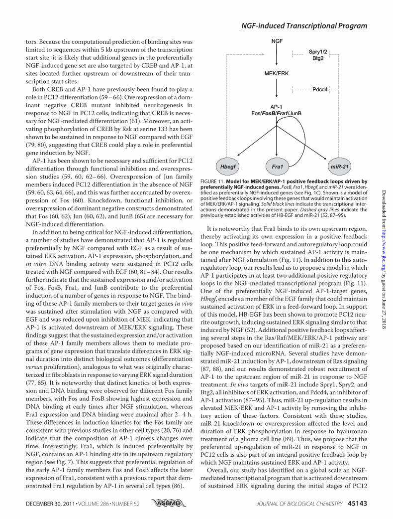
It is noteworthy that Fra1 binds to its own upstream region,thereby activating its own expression in a positive feedbackloop. This positive feed-forward and autoregulatory loop couldbe one mechanism by which sustained AP-1 activity is main-tained after NGF stimulation (Fig. 11). In addition to this auto-regulatory loop, our results lead us to propose amodel in whichAP-1 participates in at least two additional positive regulatoryloops in the NGF-mediated transcriptional program (Fig. 11).One of the preferentially NGF-induced AP-1-target genes,Hbegf, encodes amember of the EGF family that couldmaintainsustained activation of ERK in a feed-forward loop. In supportof this model, HB-EGF has been shown to promote PC12 neu-rite outgrowth, inducing sustainedERK signaling similar to thatinduced byNGF (52). Additional positive feedback loops affect-ing several steps in the Ras/Raf/MEK/ERK/AP-1 pathway areproposed based on our identification of miR-21 as a preferen-tially NGF-induced microRNA. Several studies have demon-stratedmiR-21 induction byAP-1, downstreamof Ras signaling(87, 88), and our results demonstrated robust recruitment ofAP-1 to the upstream region of miR-21 in response to NGFtreatment. In vivo targets of miR-21 include Spry1, Spry2, andBtg2, all inhibitors of ERK activation, and Pdcd4, an inhibitor ofAP-1 activation (87–95). Thus, miR-21 up-regulation results inelevated MEK/ERK and AP-1 activity by removing the inhibi-tory action of these factors. Consistent with these studies,miR-21 knockdown or overexpression affected the level andduration of ERK phosphorylation in response to hyaluronantreatment of a glioma cell line (89). Thus, we propose that thepreferential up-regulation of miR-21 in response to NGF inPC12 cells is also part of an integral positive feedback loop bywhich NGF maintains sustained ERK and AP-1 activity.Overall, our study has identified on a global scale an NGF-
mediated transcriptional program that is activated downstreamof sustained ERK signaling during the initial stages of PC12
FIGURE 11. Model for MEK/ERK/AP-1 positive feedback loops driven bypreferentially NGF-induced genes. FosB, Fra1, Hbegf, and miR-21 were iden-tified as preferentially NGF-induced genes (see Fig. 1C). Shown is a model ofpositive feedback loops involving these genes that would maintain activationof MEK/ERK/AP-1 signaling. Solid black lines indicate the transcriptional inter-actions demonstrated in the present paper. Dashed gray lines indicate thepreviously established activities of HB-EGF and miR-21 (52, 87–95).
NGF-induced Transcriptional Program
DECEMBER 30, 2011 • VOLUME 286 • NUMBER 52 JOURNAL OF BIOLOGICAL CHEMISTRY 45143
by guest on June 27, 2018http://w
ww
.jbc.org/D
ownloaded from

differentiation. Extending previous studies showing sustainedactivation of AP-1 in response to NGF in PC12 cells, we havelinked the sustained expression/activity of AP-1 transcriptionfactors to the preferential NGF-induction of�50% of the genesin this transcriptional program. Many of these genes haveestablished roles in neuronal differentiation, and several mayparticipate in autoregulatory loops that maintain this sustainedAP-1 activity.
Acknowledgment—We are grateful to Ulla Hansen for helpful com-ments and discussion of the manuscript.
REFERENCES1. Segal, R. A., and Greenberg, M. E. (1996) Annu. Rev. Neurosci. 19,
463–4892. Huff, K., End, D., and Guroff, G. (1981) J. Cell Biol. 88, 189–1983. Chao, M. V. (1992) Cell 68, 995–9974. Bar-Sagi, D., and Feramisco, J. R. (1985) Cell 42, 841–8485. Noda,M., Ko,M., Ogura, A., Liu, D. G., Amano, T., Takano, T., and Ikawa,
Y. (1985) Nature 318, 73–756. Troppmair, J., Bruder, J. T., App, H., Cai, H., Liptak, L., Szeberényi, J.,
Cooper, G. M., and Rapp, U. R. (1992) Oncogene 7, 1867–18737. Wood, K. W., Qi, H., D’Arcangelo, G., Armstrong, R. C., Roberts, T. M.,
and Halegoua, S. (1993) Proc. Natl. Acad. Sci. U.S.A. 90, 5016–50208. Cowley, S., Paterson, H., Kemp, P., and Marshall, C. J. (1994) Cell 77,
841–8529. Robinson, M. J., Stippec, S. A., Goldsmith, E., White, M. A., and Cobb,
M. H. (1998) Curr. Biol. 8, 1141–115010. Wood, K. W., Sarnecki, C., Roberts, T. M., and Blenis, J. (1992) Cell 68,
1041–105011. Hagag, N., Halegoua, S., and Viola, M. (1986) Nature 319, 680–68212. Szeberényi, J., Cai, H., and Cooper, G. M. (1990) Mol. Cell. Biol. 10,
5324–533213. Pang, L., Sawada, T., Decker, S. J., and Saltiel, A. R. (1995) J. Biol. Chem.
270, 13585–1358814. Vaudry, D., Stork, P. J., Lazarovici, P., and Eiden, L. E. (2002) Science 296,
1648–164915. Marshall, C. J. (1995) Cell 80, 179–18516. Nguyen, T. T., Scimeca, J. C., Filloux, C., Peraldi, P., Carpentier, J. L., and
Van Obberghen, E. (1993) J. Biol. Chem. 268, 9803–981017. Gotoh, Y., Nishida, E., Yamashita, T., Hoshi,M., Kawakami,M., and Sakai,
H. (1990) Eur. J. Biochem. 193, 661–66918. Traverse, S., Gomez, N., Paterson, H., Marshall, C., and Cohen, P. (1992)
Biochem. J. 288, 351–35519. Qui, M. S., and Green, S. H. (1992) Neuron 9, 705–71720. Herschman, H. R. (1991) Annu. Rev. Biochem. 60, 281–31921. Salton, S. R., Fischberg, D. J., and Dong, K. W. (1991) Mol. Cell. Biol. 11,
2335–234922. Machida, C. M., Rodland, K. D., Matrisian, L., Magun, B. E., and Ciment,
G. (1989) Neuron 2, 1587–159623. Vician, L., Basconcillo, R., and Herschman, H. R. (1997) J. Neurosci. Res.
50, 32–4324. Farias-Eisner, R., Vician, L., Silver, A., Reddy, S., Rabbani, S. A., and Her-
schman, H. R. (2000) J. Neurosci. 20, 230–23925. Vician, L., Silver, A. L., Farias-Eisner, R., and Herschman, H. R. (2001)
J. Neurosci. Res. 64, 108–12026. Töröcsik, B., Angelastro, J. M., and Greene, L. A. (2002) J. Neurosci. 22,
8971–898027. Harada, T., Morooka, T., Ogawa, S., and Nishida, E. (2001) Nat. Cell Biol.
3, 453–45928. Saeed, A. I., Bhagabati, N. K., Braisted, J. C., Liang, W., Sharov, V., Howe,
E. A., Li, J., Thiagarajan, M., White, J. A., and Quackenbush, J. (2006)Methods Enzymol. 411, 134–193
29. Tullai, J. W., Schaffer, M. E., Mullenbrock, S., Kasif, S., and Cooper, G. M.(2004) J. Biol. Chem. 279, 20167–20177
30. Dennis, G., Jr., Sherman, B. T., Hosack, D. A., Yang, J., Gao, W., Lane,H. C., and Lempicki, R. A. (2003) Genome Biol. 4, P3
31. Huang da, W., Sherman, B. T., and Lempicki, R. A. (2009) Nat. Protoc. 4,44–57
32. Tullai, J. W., Chen, J., Schaffer, M. E., Kamenetsky, E., Kasif, S., and Coo-per, G. M. (2007) J. Biol. Chem. 282, 9482–9491
33. Ravni, A., Bourgault, S., Lebon, A., Chan, P., Galas, L., Fournier, A.,Vaudry, H., Gonzalez, B., Eiden, L. E., andVaudry, D. (2006) J. Neurochem.98, 321–329
34. Virtanen, I., Lehto, V. P., Lehtonen, E., Vartio, T., Stenman, S., Kurki, P.,Wager, O., Small, J. V., Dahl, D., and Badley, R. A. (1981) J. Cell Sci. 50,45–63
35. Lee, V., Trojanowski, J. Q., and Schlaepfer, W. W. (1982) Brain Res. 238,169–180
36. Draghetti, C., Salvat, C., Zanoguera, F., Curchod, M. L., Vignaud, C., Pei-xoto, H., Di Cara, A., Fischer, D., Dhanabal, M., Andreas, G., Abderrahim,H., Rommel, C., and Camps, M. (2009) J. Biol. Chem. 284, 32053–32065
37. Nordstrom, L. A., Lochner, J., Yeung,W., andCiment, G. (1995)Mol. Cell.Neurosci. 6, 56–68
38. Heo, K., Ha, S. H., Chae, Y. C., Lee, S., Oh, Y. S., Kim, Y.H., Kim, S.H., Kim,J. H., Mizoguchi, A., Itoh, T. J., Kwon, H. M., Ryu, S. H., and Suh, P. G.(2006) Cell. Signal. 18, 2182–2192
39. Lang, F., Böhmer, C., Palmada, M., Seebohm, G., Strutz-Seebohm, N., andVallon, V. (2006) Physiol. Rev. 86, 1151–1178
40. Bonilla, I. E., Tanabe, K., and Strittmatter, S. M. (2002) J. Neurosci. 22,1303–1315
41. Jepsen, K., Solum, D., Zhou, T., McEvilly, R. J., Kim, H. J., Glass, C. K.,Hermanson, O., and Rosenfeld, M. G. (2007) Nature 450, 415–419
42. Gallo, R., Zazzeroni, F., Alesse, E., Mincione, C., Borello, U., Buanne, P.,D’Eugenio, R., Mackay, A. R., Argenti, B., Gradini, R., Russo, M. A., Mar-oder,M., Cossu,G., Frati, L., Screpanti, I., andGulino, A. (2002) J. Cell Biol.158, 731–740
43. Roth, A., Gill, R., and Certa, U. (2003)Mol. Cell. Neurosci. 22, 353–36444. Aoki, M., Yamashita, T., and Tohyama, M. (2004) J. Biol. Chem. 279,
32643–3265045. Klein, R. (2001) Curr. Opin. Cell Biol. 13, 196–20346. Redell, J. B., Zhao, J., and Dash, P. K. (2011) J. Neurosci. Res. 89, 212–22147. Soeda, S., Koyanagi, S., Kuramoto, Y., Kimura, M., Oda, M., Kozako, T.,
Hayashida, S., and Shimeno, H. (2008) Thromb Haemost 100, 1014–102048. Buechner, J., Henriksen, J. R., Haug, B. H., Tømte, E., Flaegstad, T., and
Einvik, C. (2011) Differentiation 81, 25–3449. Nakamura, K., and Hasegawa, H. (2007)Mol. Neurobiol. 35, 45–5450. Kerjan,G., Dolan, J., Haumaitre, C., Schneider-Maunoury, S., Fujisawa,H.,
Mitchell, K. J., and Chédotal, A. (2005) Nat. Neurosci. 8, 1516–152451. Xu, X. M., Fisher, D. A., Zhou, L., White, F. A., Ng, S., Snider, W. D., and
Luo, Y. (2000) J. Neurosci. 20, 2638–264852. Zhou, Y., and Besner, G. E. (2010) Neurosignals 18, 141–15153. Li, L., Yun, S. H., Keblesh, J., Trommer, B. L., Xiong, H., Radulovic, J., and
Tourtellotte, W. G. (2007)Mol. Cell. Neurosci. 35, 76–8854. Lyford, G. L., Yamagata, K., Kaufmann,W. E., Barnes, C. A., Sanders, L. K.,
Copeland, N. G., Gilbert, D. J., Jenkins, N. A., Lanahan, A. A., andWorley,P. F. (1995) Neuron 14, 433–445
55. Weimer, J. M., and Anton, E. S. (2006) Neuron 49, 3–456. Jürchott, K., Kuban, R. J., Krech, T., Blüthgen, N., Stein, U., Walther, W.,
Friese, C., Kiełbasa, S. M., Ungethüm, U., Lund, P., Knösel, T., Kemmner,W., Morkel, M., Fritzmann, J., Schlag, P. M., Birchmeier, W., Krueger, T.,Sperling, S., Sers, C., Royer, H. D., Herzel, H., and Schäfer, R. (2010) PLoSGenet 6, e1001231
57. Hughes, J. D., Estep, P. W., Tavazoie, S., and Church, G. M. (2000) J. Mol.Biol. 296, 1205–1214
58. Wasserman, W. W., and Sandelin, A. (2004) Nat. Rev. Genet. 5, 276–28759. Leppä, S., Eriksson,M., Saffrich, R., Ansorge,W., and Bohmann, D. (2001)
Mol. Cell. Biol. 21, 4369–437860. Eriksson, M., Taskinen, M., and Leppä, S. (2007) J. Cell. Physiol. 210,
538–54861. Sun, P.,Watanabe,H., Takano, K., Yokoyama, T., Fujisawa, J., and Endo, T.
(2006) Genes Cells 11, 1097–111362. Gil, G. A., Bussolino, D. F., Portal, M. M., Alfonso Pecchio, A., Renner,
NGF-induced Transcriptional Program
45144 JOURNAL OF BIOLOGICAL CHEMISTRY VOLUME 286 • NUMBER 52 • DECEMBER 30, 2011
by guest on June 27, 2018http://w
ww
.jbc.org/D
ownloaded from

M. L., Borioli, G. A., Guido,M. E., and Caputto, B. L. (2004)Mol. Biol. Cell15, 1881–1894
63. Leppä, S., Saffrich, R., Ansorge, W., and Bohmann, D. (1998) EMBO J. 17,4404–4413
64. Heasley, L. E., Storey, B., Fanger, G. R., Butterfield, L., Zamarripa, J., Blum-berg, D., and Maue, R. A. (1996)Mol. Cell. Biol. 16, 648–656
65. Lönn, P., Zaia, K., Israelsson, C., Althini, S., Usoskin, D., Kylberg, A., andEbendal, T. (2005) Neurochem. Res. 30, 753–765
66. Dragunow,M., Xu, R.,Walton,M.,Woodgate, A., Lawlor, P.,MacGibbon,G. A., Young, D., Gibbons, H., Lipski, J., Muravlev, A., Pearson, A., andDuring, M. (2000) Brain Res. Mol. Brain Res. 83, 20–33
67. Conkright, M. D., Guzmán, E., Flechner, L., Su, A. I., Hogenesch, J. B., andMontminy, M. (2003)Mol. Cell 11, 1101–1108
68. Hess, J., Angel, P., and Schorpp-Kistner, M. (2004) J. Cell Sci. 117,5965–5973
69. Masquilier, D., and Sassone-Corsi, P. (1992) J. Biol. Chem. 267,22460–22466
70. van Dam, H., and Castellazzi, M. (2001) Oncogene 20, 2453–246471. Sassone-Corsi, P., Ransone, L. J., and Verma, I. M. (1990) Oncogene 5,
427–43172. Hoeffler, J. P., Deutsch, P. J., Lin, J., and Habener, J. F. (1989) Mol. Endo-
crinol. 3, 868–88073. Manna, P. R., and Stocco, D. M. (2007) J. Mol. Endocrinol. 39, 261–27774. Hirai, S. I., Ryseck, R. P.,Mechta, F., Bravo, R., and Yaniv,M. (1989) EMBO
J. 8, 1433–143975. Ryder, K., Lanahan, A., Perez-Albuerne, E., and Nathans, D. (1989) Proc.
Natl. Acad. Sci. U.S.A. 86, 1500–150376. Cohen, D. R., and Curran, T. (1988)Mol. Cell. Biol. 8, 2063–206977. Murphy, L. O., MacKeigan, J. P., and Blenis, J. (2004) Mol. Cell. Biol. 24,
144–15378. Chung, J., Kubota, H., Ozaki, Y., Uda, S., and Kuroda, S. (2010) PLoS One
5, e901179. Bonni, A., Ginty, D. D., Dudek, H., and Greenberg, M. E. (1995)Mol. Cell.
Neurosci. 6, 168–183
80. Xing, J., Kornhauser, J. M., Xia, Z., Thiele, E. A., and Greenberg, M. E.(1998)Mol. Cell. Biol. 18, 1946–1955
81. Boss, V., Roback, J. D., Young, A. N., Roback, L. J., Weisenhorn, D. M.,Medina-Flores, R., and Wainer, B. H. (2001) J. Neurosci. 21, 18–26
82. Pellegrino, M. J., and Stork, P. J. (2006) J. Neurochem. 99, 1480–149383. Schlingensiepen, K. H., Wollnik, F., Kunst, M., Schlingensiepen, R., Her-
degen, T., and Brysch, W. (1994) Cell. Mol. Neurobiol. 14, 487–50584. Su, F., Kozak, K. R., Herschman, H., Reddy, S. T., and Farias-Eisner, R.
(2007) J. Neurosci. Res. 85, 1952–195885. Murphy, L. O., Smith, S., Chen, R. H., Fingar, D. C., and Blenis, J. (2002)
Nat. Cell Biol. 4, 556–56486. Bergers, G., Graninger, P., Braselmann, S., Wrighton, C., and Busslinger,
M. (1995)Mol. Cell. Biol. 15, 3748–375887. Talotta, F., Cimmino, A., Matarazzo, M. R., Casalino, L., De Vita, G.,
D’Esposito, M., Di Lauro, R., and Verde, P. (2009) Oncogene 28, 73–8488. Fujita, S., Ito, T., Mizutani, T., Minoguchi, S., Yamamichi, N., Sakurai, K.,
and Iba, H. (2008) J. Mol. Biol. 378, 492–50489. Kwak, H. J., Kim, Y. J., Chun, K. R., Woo, Y. M., Park, S. J., Jeong, J. A., Jo,
S. H., Kim, T. H., Min, H. S., Chae, J. S., Choi, E. J., Kim, G., Shin, S. H.,Gwak, H. S., Kim, S. K., Hong, E. K., Lee, G. K., Choi, K. H., Kim, J. H., Yoo,H., Park, J. B., and Lee, S. H. (2011) Oncogene 30, 2433–2442
90. Hatley,M. E., Patrick, D.M., Garcia,M. R., Richardson, J. A., Bassel-Duby,R., van Rooij, E., and Olson, E. N. (2010) Cancer Cell 18, 282–293
91. Frankel, L. B., Christoffersen, N. R., Jacobsen, A., Lindow, M., Krogh, A.,and Lund, A. H. (2008) J. Biol. Chem. 283, 1026–1033
92. Asangani, I. A., Rasheed, S. A., Nikolova, D. A., Leupold, J. H., Colburn,N. H., Post, S., and Allgayer, H. (2008) Oncogene 27, 2128–2136
93. Lu, Z., Liu, M., Stribinskis, V., Klinge, C. M., Ramos, K. S., Colburn, N. H.,and Li, Y. (2008) Oncogene 27, 4373–4379
94. Sayed, D., Rane, S., Lypowy, J., He, M., Chen, I. Y., Vashistha, H., Yan, L.,Malhotra, A., Vatner, D., and Abdellatif, M. (2008) Mol. Biol. Cell 19,3272–3282
95. Liu, M., Wu, H., Liu, T., Li, Y., Wang, F., Wan, H., Li, X., and Tang, H.(2009) Cell Res. 19, 828–837
NGF-induced Transcriptional Program
DECEMBER 30, 2011 • VOLUME 286 • NUMBER 52 JOURNAL OF BIOLOGICAL CHEMISTRY 45145
by guest on June 27, 2018http://w
ww
.jbc.org/D
ownloaded from
Steven Mullenbrock, Janki Shah and Geoffrey M. Cooperand AP-1 Protein Activation during PC12 Cell Differentiation
Mitogen-activated Protein Kinase/Extracellular Signal-regulated Kinase (ERK)Factor-induced Transcriptional Program Regulated by Sustained
Global Expression Analysis Identified a Preferentially Nerve Growth
doi: 10.1074/jbc.M111.274076 originally published online November 7, 20112011, 286:45131-45145.J. Biol. Chem.
10.1074/jbc.M111.274076Access the most updated version of this article at doi:
Alerts:
When a correction for this article is posted•
When this article is cited•
to choose from all of JBC's e-mail alertsClick here
Supplemental material:
http://www.jbc.org/content/suppl/2011/11/07/M111.274076.DC1
http://www.jbc.org/content/286/52/45131.full.html#ref-list-1
This article cites 95 references, 33 of which can be accessed free at
by guest on June 27, 2018http://w
ww
.jbc.org/D
ownloaded from