glucocorticoids delay raf-induced senescence promoted by egr1 · research article glucocorticoids...
TRANSCRIPT

RESEARCH ARTICLE
Glucocorticoids delay RAF-induced senescence promotedby EGR1Cyril Carvalho1, Valentin L’Hote1, Regis Courbeyrette1, Gueorgui Kratassiouk1, Guillaume Pinna1,Jean-Christophe Cintrat2, Cyril Denby-Wilkes1, Celine Derbois3, Robert Olaso3, Jean-François Deleuze3,Carl Mann1,* and Jean-Yves Thuret1,*
ABSTRACTExpression of hyperactive RAF kinases, such as the oncogenicB-RAF-V600E mutant, in normal human cells triggers a proliferativearrest that blocks tumor formation.We discovered that glucocorticoidsdelayed the entry into senescence induced by B-RAF-V600E inhuman fibroblasts, and allowed senescence bypass when thecells were regularly passaged, but that they did not allowproliferation of cells that were already senescent. Transcriptomeand siRNA analyses revealed that the EGR1 gene is one target ofglucocorticoid action. Transcription of the EGR1 gene is activated bythe RAF-MEK-ERK MAPK pathway and acts as a sensor of hyper-mitogenic pathway activity. The EGR1 transcription factor regulatesthe expression of p15 and p21 (encoded by CDKN2B and CDKN1A,respectively) that are redundantly required for the proliferative arrestof BJ fibroblasts upon expression of B-RAF-V600E. Our resultshighlight the need to evaluate the action of glucocorticoid on cancerprogression in melanoma, thyroid and colon carcinoma in whichB-RAF-V600E is a frequent oncogene, and cancers in which evasionfrom senescence has been shown.
KEYWORDS:Oncogene-induced senescence, B-RAF-V600E, EGR1,Glucocorticoid, CDKN1A, CDKN2B
INTRODUCTIONA remarkably high fraction (∼50%) of melanomas express theoncogenic mutation B-RAF-V600E leading to constitutive activationof the B-RAF kinase (Shain et al., 2015). B-RAF-V600E is also afrequent oncogenic mutation in thyroid and colorectal cancers(Cantwell-Dorris et al., 2011). However, expression of this drivermutation in normal human fibroblasts or melanocytes triggerspremature senescence and thus blocks progression to a tumor(Damsky and Bosenberg, 2017; Shain et al., 2015). This findingsuggests that the proliferative arrest induced by the expression ofhyper-active RAF kinases in normal cells acts as a suppressormechanism that must be bypassed if a tumor is to be formed. Themolecular mechanisms responsible for RAF-induced senescence arepoorly defined. Increased expression of the p21, p16 and p15 (alsoknown as CDKN1A, CDKN2A and CDKN2B, respectively) cyclin-
dependent kinase inhibitors have been implicated in this proliferativearrest to different extents depending on the investigatedmodel, but thepathways leading to derepression of the corresponding genes areincompletely understood (Jeanblanc et al., 2012;McNeal et al., 2015;Michaloglou et al., 2005; Woods et al., 1997; Zhu et al., 1998).Senescence induced by the RAS-val12 oncogene has been studied ingreater detail. In response to mitogenic factors binding to cell surfacereceptors, RAS activates several growth-promoting pathwaysincluding the RAF-MEK-ERK, PI3K and Ral GTPase pathways(Fey et al., 2016). Expression ofRAS-val12 has been shown to inducea replicative stress that is amajor factor in triggering proliferative arrest(Di Micco et al., 2006), but DNA damage does not appear to benecessary for senescence induced by RAF (Damsky and Bosenberg,2017; Jeanblanc et al., 2012). Hyperactivation of the RAF-MEK-ERK pathway may trigger proliferative arrest through specificsignaling pathways. We screened a library of clinically approvedmolecules and discovered that glucocorticoids are potent modulatorsof RAF-induced senescence in human fibroblasts. Glucocorticoidsare widely used as anti-inflammatory agents in the clinic, butactivation of the glucocorticoid receptor NR3C1 by binding ofglucocorticoids leads to the rapid activation and repression ofhundreds of genes implicated in diverse pathways in a cell-specificmanner (Sacta et al., 2016; Weikum et al., 2017). Transcriptomicanalysis identified the EGR1 gene as a candidate target ofglucocorticoids. Loss of function experiments for EGR1 and ChIP-seq analyses suggest that the transcription factorEGR1acts as a sensorof MAPK hyperactivity contributing to the triggering of senescence.
RESULTSGlucocorticoids delay or bypass entry into oncogene-induced senescenceWe screened the Prestwick drug repositioning library for moleculesthat could inhibit senescence of WI-38hTert human fibroblastsinduced by the expression of an activated form of the C-RAFkinase that we previously characterized (Jeanblanc et al., 2012).26 different glucocorticoids inhibited RAF-induced senescence inthis screen (see Materials and Methods). In addition, previouswork by Cristofalo and colleagues has shown that glucocorticoidscan delay replicative senescence of human fibroblasts (Mawal-Dewan et al., 2003) and Laberge et al. have recently shownthat glucocorticoids partially inhibit the senescence-associatedsecretory phenotype (SASP) of fibroblasts induced intosenescence by RAS-val12 (Laberge et al., 2012). We therefore setout to describe and analyze effects of glucocorticoids on RAF-induced senescence. To this end, and to study the effects ofglucocorticoids on senescence induced by a mutation relevant forhuman oncogenesis, we developed a new cellular model of BJhTerthuman foreskin fibroblasts where the expression of B-RAF-V600Eis under the control of a doxycycline responsive tet-ON promoter.Received 13 February 2019; Accepted 22 July 2019
1Institute for Integrative Biology of the Cell (I2BC), CEA, CNRS, Univ. Paris-Sud,Universite Paris-Saclay, 91198 Gif-sur-Yvette cedex, France. 2Service de ChimieBio-organique et Marquage (SCBM), CEA, Universite Paris-Saclay, 91191 Gif-sur-Yvette, France. 3Centre National de Recherche en Genomique Humaine (CNRGH),Institut de Biologie François Jacob, CEA, Universite Paris-Saclay, F-91057 Evry,France.
*Authors for correspondence ([email protected]; [email protected])
V.L., 0000-0001-5168-2943; J.-Y.T., 0000-0001-5385-7620
1
© 2019. Published by The Company of Biologists Ltd | Journal of Cell Science (2019) 132, jcs230748. doi:10.1242/jcs.230748
Journal
ofCe
llScience

Oncogene-induced senescence (OIS) is known to occurindependently of hTert expression and allowed us to study thissenescence in the absence of possible confounding contributionsdue to the loss of telomeric sequences in non-immortalizedfibroblasts (Jeanblanc et al., 2012; Wei and Sedivy, 1999). In ourmodel, the concentration of doxycycline controls the expression ofB-RAF-V600E at the mRNA and protein levels (Fig. 1A,B). Theexpression of B-RAF-V600E at low levels of doxycycline (in therange of 5–20 ng ml−1 depending on the experiment) is, on average,similar to the endogenous level of B-RAF (Fig. 1B; Fig. S1G).We then assessed the effect of B-RAF-V600E expression
levels on proliferation. Cell numbers decreased with increasingconcentrations of doxycycline over 5 days of culture (Fig. 1C). Thedoxycycline concentration for which the number of cells startsdeclining is similar to the concentration at which the expression ofB-RAF-V600E is close to endogenous levels. Greater than 80% ofcells lost the ability to go through S-phase within the first 24 h inhigh (1000 ng ml−1) concentrations of doxycycline (Fig. 1D). Theproliferation arrest was slower at lower concentrations ofdoxycycline (100 ng ml−1, orange; 50 ng ml−1, blue). To takeadvantage of the fast proliferative arrest in order to investigatemechanisms, all further experiments were carried out at 1000 ng ml−1
doxycycline unless indicated otherwise. As expected, in addition tocell cycle arrest, the expression of B-RAF-V600E led to increasedsenescence-associated (SA)-β-galactosidase activity (Fig. 1E), theformation of senescence-associated heterochromatic foci (SAHFs,Fig. 1F), and the derepression of cyclin-dependent kinase inhibitors(see below) as typical senescence markers.To assess the effects of glucocorticoids on senescence induction,
we then incubated the cells with clobetasol, a potent glucocorticoid
used in topical applications to treat inflammatory skin diseases.Within the timeframe of the experiment (5 days), low nanomolarlevels of clobetasol strongly inhibited the proliferative arrest of BJcells induced by B-RAF-V600E (Fig. 2A). All further experimentswere carried out at 2 nM clobetasol. We also observed delays inproliferative arrest after induction of an activated C-RAF kinase in BJcells (with cortisol, Fig. S1A, and clobetasol, Fig. S1C) and WI-38cells (with clobetasol, Fig. S1B), and also in IMR-90 cells expressingRAS-val12 (with clobetasol, Fig. S1D). Glucocorticoids can thusinterfere with the proliferative arrest induced by both activated RAFand RAS oncogenes in three different fibroblast cell lines.
Clobetasol treatment allowed BJ cells expressing high levels ofB-RAF-V600E to maintain significant DNA synthesis for at least5 days (Fig. 2B, green). Proliferation was assayed in 96-well plates(see Materials and Methods). In this microwell assay, we cannotdetermine whether the reduced DNA synthesis beginning at 3 daysis due to delayed entry into senescence in the presence of clobetasolor whether it is due to contact inhibition of proliferation as cellsapproach confluence. We followed proliferation in continuouscultures to determine the long-term effects of clobetasol onRAF-induced senescence. Remarkably, incubation of cells withclobetasol at low or high doxycycline concentration allowed fullbypass of senescence when the cells were passaged every 2 days(Fig. 2C). Surprisingly, when cells expressing B-RAF-V600E werenot passaged, the presence of clobetasol delayed, but did not lead toa bypass of the senescence proliferative arrest (Fig. 2D; Fig. S1E).Similar results were observed in clonogenic assays (Fig. S1H). Lowlevels of doxycycline (15 or 30 ng ml−1) blocked the ability of BJcells to form colonies on plates, but colonies were restored in thepresence of clobetasol. However, clobetasol did not allow colony
Fig. 1. Rapid Induction of senescence with inducible B-RAF-V600E. (A) Relative RNA expression as determined by RT-qPCR of B-RAF-V600E cellswith increasing doxycycline concentrations and 24 h incubation normalized to the expression with ethanol (dox=0) (error bars s.d.). A representative experiment fromtwobiological repeatseachwithn=3 is shown. (B)Representative anti-B-RAFwesternblot onwhole-cell extracts ofBJhTert cells (WT)orBJhTert cells transducedwitha pTRIPz-3HA-B-RAF-V600E lentivirus (B-RAF-V600E). Cells were harvested 48 h after inducing with increasing doxycycline concentrations.WT also indicates theendogenous B-RAFprotein and VE indicates the B-RAF-V600E construct that has threeN-terminal tandem hemagglutinin (HA) epitopes andmigrates slightly slowerthan the endogenous protein. A portion of the gel stained with 2,2,2-trichloroethanol (TCE) is shown as a loading control for total protein. (C) Number of cells afterculture with increasing doxycycline concentrations for 5 days. Green line: maximum number of cells obtained by culture with ethanol solvent control. Red line:minimum number of cells obtained by plating with medium containing 20 µM etoposide instead of doxycycline. This level of etoposide rapidly blocks proliferationthrough acute DNA damage. A representative experiment from three biological repeats eachwith n=3 is shown. Error bars show the s.d. (D) Percentage of cells goingthrough S-phase during a 48-h sliding windowat the indicated times after doxycycline addition at the indicated concentrations. A representative experiment from threebiological repeats each with n=3 is shown. (E) Representative SA-β-galactosidase staining of proliferative cells (Untreated) and cells incubated for 20 days with1000 ng ml−1 doxycycline. Scale bars: 400 µm. (F) Left panels, representative images of DAPI-stained nuclei to visualize DNA compaction in proliferating cells(Control) and in cells treated with 1000 ng ml−1 doxycycline for 4 days. Scale bars: 10 µm. Right panel, measure of chromatin compaction {DAPI CV, coefficient ofvariation of DAPI intensity [standard deviation as a percentageof themean (Contrepois et al., 2012)]} in untreated proliferating control cells (Ctl) versus cells incubatedfor 4 days with 1000 ng ml−1 doxycycline (D). A representative experiment from two biological repeats each with n=3 is shown. Error bars show s.d.
2
RESEARCH ARTICLE Journal of Cell Science (2019) 132, jcs230748. doi:10.1242/jcs.230748
Journal
ofCe
llScience

formation when BJ cells were plated in the presence of 1000 ng ml−1
doxycycline. Collectively, these results suggest that an irreversibleproliferative arrest results from the integration of positive(glucocorticoid signaling, frequent cell passing and renewal ofnutrients) and negative proliferative signals (hyperactive RAF kinasesignaling, contact inhibition and stress associated with high celldilution on plates for clonogenicity assays).We conclude from these experiments that B-RAF-V600E
triggers senescence in human BJ fibroblasts when the oncogene isexpressed at levels similar to the endogenous kinase, but the cellcycle arrest is faster at high expression levels of B-RAF-V600E.Glucocorticoids allow the cells to fully bypass senescence, even athigh levels of oncogene expression, when regularly passaged inthe absence of exogenous stress. In contrast, once senescent, BJcells did not resume proliferation when treated with clobetasol(Fig. 2D; Fig. S1E,F). Thus, clobetasol delays or bypasses entry intosenescence, but does not revert the established senescent state. Wetherefore set out to identify the mechanisms involved in senescenceinduced by B-RAF-V600E and modulated by glucocorticoids. In allsubsequent experiments, cells were not passaged after doxycyclineaddition unless indicated.
Identification of glucocorticoid targets in senescenceinductionGlucocorticoids are steroid hormones that function by binding to theglucocorticoid receptor (GR, encoded by the NR3C1 gene), whichthen activates or represses target genes after translocation to the
nucleus (Sacta et al., 2016; Weikum et al., 2017). The GR waslocated in both the cytosol and nucleus of BJ cells in proliferation orafter B-RAF-V600E expression, but treatment with 2 nM clobetasolresulted in a rapid increase in nuclear GR (Fig. S2A,B). The effectsof clobetasol on senescence induction were dependent on the GR, asdepleting NR3C1 mRNA fully abolished the effect of clobetasol onproliferation (Fig. S2C), and RU-486, which antagonizes theactivation of the GR by glucocorticoids (Cadepond et al., 1997),also blocked the effect of clobetasol on proliferation (Fig. S2D).We next sought to determine how activated GR suppressesRAF-induced senescence. Clobetasol treatment did not interferewith the induction of B-RAF-V600E or the phosphorylation ofMEK1 and MEK2 (MEK1/2, also known as MAP2K1 andMAP2K2, respectively) or ERK1 and ERK2 (ERK1/2, also knownas MAPK3 and MAPK1, respectively) (Fig. 2E), so glucocorticoidsmust work downstream of ERK1/2 hyperactivation to suppresssenescence.
GR-mediated antagonism of transcription factors such as NF-κBexplains its anti-inflammatory action, and NF-kB (RelA subunit)-mediated expression of inflammatory factors has been implicatedin contributing to senescence induction in some systems (Chienet al., 2011). We first tested a role for NF-κB in RAF-inducedproliferative arrest by constructing a version of our BJhTert/ptet-ON-B-RAF-V600E cell line that expresses the IκB-SR (superrepressor), a mutant version of IκB that acts as a dominant-negativesuppressor of NF-κB by sequestering RelA in the cytoplasm (Karinand Ben-Neriah, 2000). Treatment of parental cells with TNF led to
Fig. 2. Clobetasol interferes with senescence onsetwithout interfering with MAPK activation. (A) Number ofcells after culture with 1000 ng ml−1 doxycycline andincreasing clobetasol concentrations for 5 days. Green line:culture with ethanol (vehicle for doxycycline) and DMSO(vehicle for clobetasol). Red line: culture with etoposide(20 µM). A representative experiment from three biologicalrepeats each with n=3 is shown. Error bars show s.d. (B)Percentage of cells going through S-phase during a 48-hsliding window at the indicated times after 1000 ng ml−1
doxycycline addition with 2 nM clobetasol (green) or DMSO(black). A representative experiment from three biologicalrepeats each with n=3 is shown. (C) Number of cumulativegenerations. Cells were systematically passaged every2–3 days with the indicated drugs and counted. Arepresentative experiment from two biological repeats eachwith n=3 is shown. Doxycycline (Dox) concentrations inng ml−1, clobetasol (Clob) in nM. (D) Number of cumulativegenerations as in panel C with the difference that cells werenot passaged. Cells counted (then discarded) fromsuccessive plates prepared at day 0 and kept in identicalconditions, except for EtOH-treated cells that had to bepassaged at confluency. A representative experiment fromtwo biological repeats each with n=3 is shown. Theremaining cultures had undergone proliferative arrestbefore confluency. Doxycycline (Dox) concentrations inng ml−1, clobetasol (Clob) in nM. (E) Representativewestern blots (n=4) on whole-cell extracts. 3HA-B-RAF-V600E was detected with an anti-B-RAF antibody. ERK1/2(ERK) and phospho-ERK1/2 (P-ERK) were detected on thesame gel with 700 nm and 800 nm fluorescent secondaryantibodies. ERK2 is the strong band at 40 kDa and ERK1 isthe weaker band that migrates at a slightly higher molecularmass. P-MEK is phospho-MEK1/2. Portions of themembranes stained with the Revert protein stain (Li-Cor)are shown as loading controls.
3
RESEARCH ARTICLE Journal of Cell Science (2019) 132, jcs230748. doi:10.1242/jcs.230748
Journal
ofCe
llScience

a rapid nuclear translocation of RelA, whereas RelA remainedcytosolic after treatment of the IκB-SR-expressing cell line withTNF (Fig. S3A). Notably, expression of B-RAF-V600E triggeredthe proliferative arrest of these IκB-SR-expressing cell lines to asimilar degree to that seen in the parental cell line (Fig. 3A), andclobetasol treatment interfered with this proliferative arrest as in theparental cell line (Fig. 3B). Thus, RelA transcriptional activity is notrequired for the RAF-induced proliferative arrest, or for clobetasolmodulation of this proliferative arrest.We further tested a possible role for the repression of inflammatory
gene expression on the suppression of RAF-induced senescence bycharacterizing the effect of compound A (CpdA). Long-termtreatment with glucocorticoids in the clinic produces adverse sideeffects that are thought to be due to the transcriptional activation ofmultiple genes by the GR. CpdA, a non-steroidal plant-derivedcompound, was identified as a molecule that could block NF-κB-driven gene expression without transcriptionally activating genesthat could be responsible for the unwanted side effects of the GR(Lesovaya et al., 2015). We found no effect of CpdA on senescenceinduced by the expression of B-RAF-V600E (Fig. S3B), despitethe existence of a transcriptional response partially overlappingthe response to clobetasol (Fig. S3C). This result supports ourconclusion that the repression of inflammatory gene expression byglucocorticoids is not required for their action in bypassing RAF-induced senescence.Having excluded inflammatory gene repression in the senescence
bypass by clobetasol, we next used transcriptome analyses toidentify candidate gene targets. Since we had observed that cellcycle arrest was almost complete 24 h after doxycycline addition(1000 ng ml−1), that clobetasol was active during that period of timeand that CpdA had no effect on senescence induction, we reasoned
that good candidate genes would have the following characteristics:(1) be induced or repressed with 24 h of doxycycline treatmentcompared to non-treated cells, (2) be differentially expressed uponclobetasol treatment (opposing the effect of doxycycline), and (3)not be affected by CpdA. Furthermore, as we sought direct targets ofglucocorticoid, we chose a short treatment of 2 h with clobetasol orCpdA after 24 h of incubation with doxycycline. As shown inFig. 3C, we found a limited number of candidate Illuminamicroarray probes with such characteristics [at a false discoveryrate (FDR)<1×10⁻⁴, 17 upregulated and 45 downregulated,corresponding to 14 distinct upregulated genes and 39 distinctdownregulated genes].
In parallel to this study, we also performed an siRNA screen toidentify chromatin-associated or signaling proteins required for RAF-induced senescence. EGR1, which is a candidate glucocorticoid-target gene as defined above (Fig. 3C), was among the genes targetedin this screening. Interestingly, knockdown of EGR1 allowed cells todivide more during the first 4 days of oncogene expression (Fig. S4).The early growth response-1 (EGR1) gene encodes a transcriptionfactor, and the expression of the EGR1 gene is directly controlled bytheMAPKpathway (Bahrami andDrabløs, 2016).We thus decided tofocus on its role in senescence induction and the response toglucocorticoids.
EGR1 and the cell cycle arrest during senescence inductionEGR1 has three homologs in the human genome (EGR2–EGR4).EGR3 (but not EGR2 and EGR4) was also identified as a possibleglucocorticoid target during senescence induction (Fig. 3C;Fig. S5A). As shown in Fig. 4A, treatment of cells with an anti-EGR1 siRNA (different from the siRNA used in Fig. S4), but notwith two anti-EGR3 siRNAs, allowed more proliferation after
Fig. 3. Senescence suppression by clobetasol does notrequire RelA, and transcriptomics analysis identifiescandidate target genes. (A) Number of IκB-SR-expressingcells after culture with increasing doxycyclineconcentrations for 5 days. Green line: culture with ethanol(vehicle for doxycycline). Red line: culture with etoposide(20 µM). A representative experiment from two biologicalrepeats each with n=3 is shown. Error bars show s.d.(B) Number of IκB-SR-expressing cells after culture with1000 ng ml−1 doxycycline and increasing clobetasolconcentrations for 5 days. Green line: culture with ethanoland DMSO (vehicles for doxycycline and clobetasol). Redline: culture with etoposide (20 µM). A representativeexperiment from two biological repeats each with n=3 isshown. Error bars show s.d. (C) Transcriptomics analysisusing Illumina beadarray data. Upper left panel: number ofup- and down-regulated probes (FDR<1×10⁻⁴) betweencells treated with ethanol and DMSO (proliferative) andcells treated with 1000 ng ml−1 doxycycline for 24 h, withsuccessive filters for differential expression after short (2 h)clobetasol treatment and the absence of response to shortcompound A treatment. Note that some genes wererepresented by two different probes so that the 17upregulated probes and 45 downregulated probescorresponded to only 14 distinct upregulated genes and 39distinct downregulated genes. Other panels: for probescorresponding to the indicated genes, the log2(FoldChange) between proliferative cells and cells incubated for24 h with 1000 ng ml−1 doxycycline and 2 h with DMSO,clobetasol (2 nM) or compound A (2 µM, see Fig. S3B) isshown as a color representation, with blue indicatingdownregulation and red upregulation.
4
RESEARCH ARTICLE Journal of Cell Science (2019) 132, jcs230748. doi:10.1242/jcs.230748
Journal
ofCe
llScience

BRAF-V600E induction. We thus excluded EGR3 from furtherconsideration.EGR1 RNA was significantly induced as soon as 6 h after
doxycycline addition (Fig. 4B) and plateaued thereafter. Continuoustreatment with clobetasol partially repressed EGR1 RNA induction at24 h (Fig. 4B), but not 48 h after doxycycline addition. EGR1 proteinlevels increased at 6 h after doxycycline addition, and thesimultaneous addition of doxycycline plus clobetasol had littleeffect on EGR1 protein levels (Fig. 4C). We then estimated the levelsof EGR1 in individual cells through immunofluorescence staining.About a third (37%) of proliferating cells stained positively for EGR1(Fig. S6), but almost all cells stained positively for EGR1 afterincubating with low or high concentrations of doxycycline for 24 h(Fig. S6; Fig. 4D). Additionally, whereas a short treatment (2 h) withclobetasol resulted in a significant decrease in EGR1 levels, acontinuous treatment with clobetasol did not (Fig. 4D). Thus,modulation ofEGR1protein levels by clobetasol is unlikely to explainall of the effects of clobetasol on the proliferative arrest.EGR1 was previously implicated in cyclin-dependent kinase
inhibitor (CKI) expression in different experimental contexts(Besancenot et al., 2010; Krones-Herzig et al., 2003; Salotti et al.,2015), and CKI accumulation is required for the proliferative arrestin senescence (Beausejour et al., 2003; Jeanblanc et al., 2012). Wethus tested whether EGR1 contributes to CKI expression in ourmodel. First, we identified the CKIs relevant to B-RAF-V600E-induced senescence in BJ fibroblasts. As shown in Table S1, at leastone probe for CDKN1A, CDKN2A and CDKN2B showedsignificant induction 24 h after doxycycline addition in Beadarraytranscriptomics experiments. RT-qPCR experiments confirmed that
CDKN1A, CDKN2A and CDKN2B mRNA levels were upregulatedafter doxycycline addition (Fig. 5A), as were the correspondingp15, p16 and p21 protein levels (Fig. 5B). Interestingly, whereasCDKN1A andCDKN2Bwere not differentially expressed after shortclobetasol treatments (2 h, Table S2), and accordingly were notidentified as putative clobetasol targets, they were downregulated atthe mRNA level during continuous 24 and 48 h clobetasoltreatments (Fig. 5A). Levels of p15 and p21 proteins were alsoreduced (Fig. 5B). CDKN2A mRNA and protein levels were notdecreased by short or long clobetasol treatments (Fig. 5A,B;Table S2).
We then assessed the effects of p21, p16 and p15 depletion on theproliferative arrest induced by the expression of B-RAF-V600E.Depletion of p16 had no effect on the proliferative arrest, whereasdepletion of p21, and, to a lesser extent, p15, increased proliferation(Fig. 5C and Fig. S7A,B). Interestingly, combined depletion of p21and p15 allowed more proliferation than either individual depletion,and yielded similar or better effects than the depletion of EGR1(Fig. 5C). We then used siRNA to knockdown EGR1 in BJ cells andobserved decreased expression of CDKN1A and CDKN2B inresponse to B-RAF-V600E expression (Fig. 6). Furthermore, ChIP-seq experiments revealed a significant increase of EGR1 occupancyat consensus EGR1-binding sites within the promoter of theCDKN2B gene and within the first intron of theCDKN1A gene uponexpression of B-RAF-V600E (Fig. 7). From these experiments, weconclude that although p15, p16 and p21 are induced in response toB-RAF-V600E, only p15 and p21 play an important, partiallyredundant, role in the proliferative arrest in this cell line. Moreover,CDKN1A and CDKN2B, but not CDKN2A, are repressed by
Fig. 4. EGR1 is a target of clobetasol rapidly induced after B-RAF-V600E expression, and its knockdown delays proliferation arrest. (A) Number of cellsafter treatment with the indicated siRNAs for 24 h, then incubationwith ethanol (Prolif) or 1000 ng ml−1 doxycycline (Dox1000) for 96 h. A representative experimentfrom three biological repeats each with n=3 is shown. Error bars show s.d. (B) A representative experiment from two biological repeats each with n=3, showing therelative expression of EGR1 determined by RT-qPCR, and normalized to the expression level in P (ethanol) and DMSO at 24 h. D6, D24, D48, doxycycline(1000 ng ml−1) and DMSO for 6, 24 or 48 h; DC24, DC48, doxycycline (1000 ng ml−1) and clobetasol (2 nM) for 24 or 48 h. Error bars show s.d. (C) Representativewestern blot (n=3) of EGR1 expression at indicated times after doxycycline (1000 ng ml−1) and DMSOor clobetasol (2 nM). A portion of themembrane stained withRevert protein stain (Li-Cor) is shown as a loading control. (D) Boxplots showing the integrated nuclear fluorescence (AU, arbitrary units) after anti-EGR1immunostaining, with the median (thick line), first and third quartiles (lower and upper hinges) and whiskers extending to values 1.5 times the interquartile range.E, ethanol, Dox30, Dox1000, doxycycline 30 at 1000 ng ml−1 for 24 h. DMSOor clobetasol were either added at the same time as doxycycline (24 hrs) or 2 h beforefixation with formaldehyde (2 hrs). The red dotted line indicates the intensity threshold for which 37% of cells were determined positive for nuclear EGR1. Theasterisk indicates P<2.2×10⁻¹⁶ in a double-sided Kolmogorov–Smirnov test. A representative experiment from two biological repeats each with n=3 is shown.
5
RESEARCH ARTICLE Journal of Cell Science (2019) 132, jcs230748. doi:10.1242/jcs.230748
Journal
ofCe
llScience
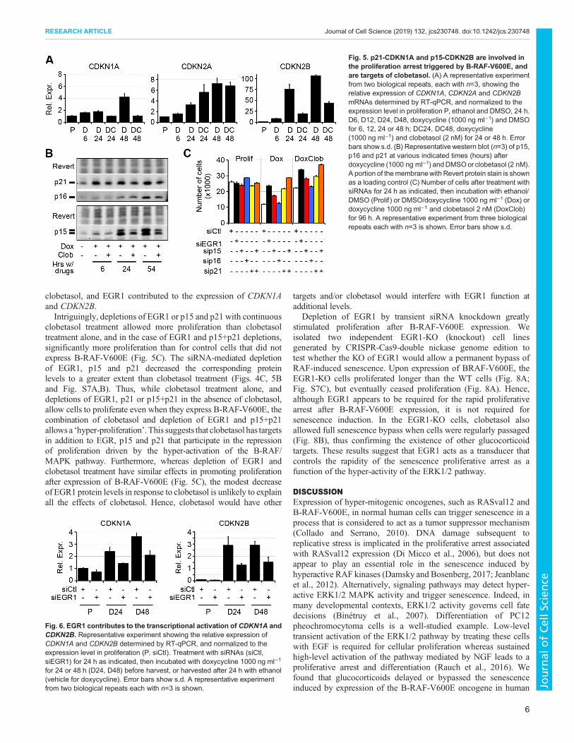
clobetasol, and EGR1 contributed to the expression of CDKN1Aand CDKN2B.Intriguingly, depletions of EGR1 or p15 and p21 with continuous
clobetasol treatment allowed more proliferation than clobetasoltreatment alone, and in the case of EGR1 and p15+p21 depletions,significantly more proliferation than for control cells that did notexpress B-RAF-V600E (Fig. 5C). The siRNA-mediated depletionof EGR1, p15 and p21 decreased the corresponding proteinlevels to a greater extent than clobetasol treatment (Figs. 4C, 5Band Fig. S7A,B). Thus, while clobetasol treatment alone, anddepletions of EGR1, p21 or p15+p21 in the absence of clobetasol,allow cells to proliferate even when they express B-RAF-V600E, thecombination of clobetasol and depletion of EGR1 and p15+p21allows a ‘hyper-proliferation’. This suggests that clobetasol has targetsin addition to EGR, p15 and p21 that participate in the repressionof proliferation driven by the hyper-activation of the B-RAF/MAPK pathway. Furthermore, whereas depletion of EGR1 andclobetasol treatment have similar effects in promoting proliferationafter expression of B-RAF-V600E (Fig. 5C), the modest decreaseof EGR1 protein levels in response to clobetasol is unlikely to explainall the effects of clobetasol. Hence, clobetasol would have other
targets and/or clobetasol would interfere with EGR1 function atadditional levels.
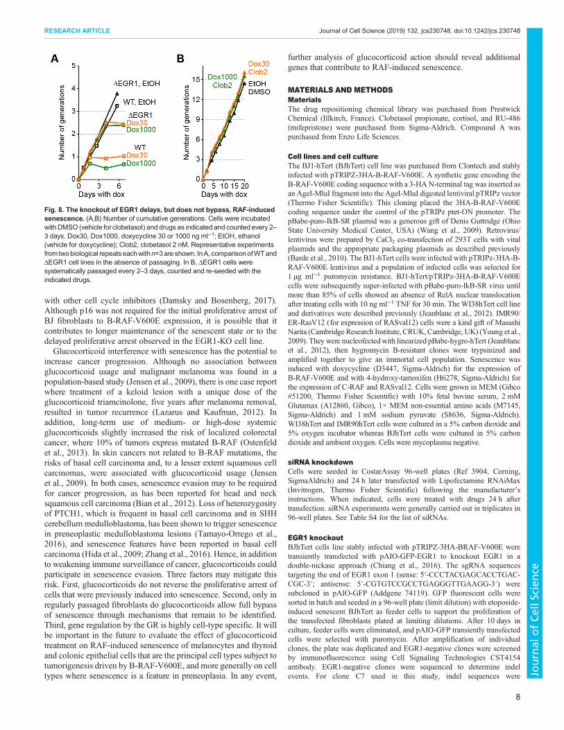
Depletion of EGR1 by transient siRNA knockdown greatlystimulated proliferation after B-RAF-V600E expression. Weisolated two independent EGR1-KO (knockout) cell linesgenerated by CRISPR-Cas9-double nickase genome edition totest whether the KO of EGR1 would allow a permanent bypass ofRAF-induced senescence. Upon expression of BRAF-V600E, theEGR1-KO cells proliferated longer than the WT cells (Fig. 8A;Fig. S7C), but eventually ceased proliferation (Fig. 8A). Hence,although EGR1 appears to be required for the rapid proliferativearrest after B-RAF-V600E expression, it is not required forsenescence induction. In the EGR1-KO cells, clobetasol alsoallowed full senescence bypass when cells were regularly passaged(Fig. 8B), thus confirming the existence of other glucocorticoidtargets. These results suggest that EGR1 acts as a transducer thatcontrols the rapidity of the senescence proliferative arrest as afunction of the hyper-activity of the ERK1/2 pathway.
DISCUSSIONExpression of hyper-mitogenic oncogenes, such as RASval12 andB-RAF-V600E, in normal human cells can trigger senescence in aprocess that is considered to act as a tumor suppressor mechanism(Collado and Serrano, 2010). DNA damage subsequent toreplicative stress is implicated in the proliferative arrest associatedwith RASval12 expression (Di Micco et al., 2006), but does notappear to play an essential role in the senescence induced byhyperactive RAF kinases (Damsky and Bosenberg, 2017; Jeanblancet al., 2012). Alternatively, signaling pathways may detect hyper-active ERK1/2 MAPK activity and trigger senescence. Indeed, inmany developmental contexts, ERK1/2 activity governs cell fatedecisions (Binétruy et al., 2007). Differentiation of PC12pheochromocytoma cells is a well-studied example. Low-leveltransient activation of the ERK1/2 pathway by treating these cellswith EGF is required for cellular proliferation whereas sustainedhigh-level activation of the pathway mediated by NGF leads to aproliferative arrest and differentiation (Rauch et al., 2016). Wefound that glucocorticoids delayed or bypassed the senescenceinduced by expression of the B-RAF-V600E oncogene in human
Fig. 5. p21-CDKN1A and p15-CDKN2B are involved inthe proliferation arrest triggered by B-RAF-V600E, andare targets of clobetasol. (A) A representative experimentfrom two biological repeats, each with n=3, showing therelative expression of CDKN1A, CDKN2A and CDKN2BmRNAs determined by RT-qPCR, and normalized to theexpression level in proliferation P, ethanol and DMSO, 24 h.D6, D12, D24, D48, doxycycline (1000 ng ml−1) and DMSOfor 6, 12, 24 or 48 h; DC24, DC48, doxycycline(1000 ng ml−1) and clobetasol (2 nM) for 24 or 48 h. Errorbars show s.d. (B) Representative western blot (n=3) of p15,p16 and p21 at various indicated times (hours) afterdoxycycline (1000 ng ml−1) andDMSOor clobetasol (2 nM).A portion of themembranewith Revert protein stain is shownas a loading control (C) Number of cells after treatment withsiRNAs for 24 h as indicated, then incubation with ethanol/DMSO (Prolif ) or DMSO/doxycycline 1000 ng ml−1 (Dox) ordoxycycline 1000 ng ml−1 and clobetasol 2 nM (DoxClob)for 96 h. A representative experiment from three biologicalrepeats each with n=3 is shown. Error bars show s.d.
Fig. 6. EGR1 contributes to the transcriptional activation of CDKN1A andCDKN2B. Representative experiment showing the relative expression ofCDKN1A and CDKN2B determined by RT-qPCR, and normalized to theexpression level in proliferation (P, siCtl). Treatment with siRNAs (siCtl,siEGR1) for 24 h as indicated, then incubated with doxycycline 1000 ng ml−1
for 24 or 48 h (D24, D48) before harvest, or harvested after 24 h with ethanol(vehicle for doxycycline). Error bars show s.d. A representative experimentfrom two biological repeats each with n=3 is shown.
6
RESEARCH ARTICLE Journal of Cell Science (2019) 132, jcs230748. doi:10.1242/jcs.230748
Journal
ofCe
llScience

fibroblasts, but did not revert the established senescent state. Weused glucocorticoids as probes to dissect the signaling pathwaysinvolved in triggering RAF-induced senescence. Transcriptomeanalyses and siRNA knockdown experiments allowed us to identifythe EGR1 gene as one target of glucocorticoid action, but epistaticand phenotypic analyses indicated that glucocorticoids must alsotarget other yet to be identified genes. E2F7 may be one such genesince its transcriptional activation by B-RAF-V600E is inhibited byclobetasol (Fig. 3C) and it was previously shown to contribute tosenescence induced by RASval12 (Aksoy et al., 2012).Expression of B-RAF-V600E leads to rapid activation of the
MEK1/2 and ERK1/2 kinases (Fig. 2E) followed by transcriptionalactivation of the EGR1 gene. ERK1/2 activates EGR1 transcriptionby phosphorylating the ELK1 subunit of the SRF transcriptionfactor (Bahrami and Drabløs, 2016). EGR1 expression can thus actas a sensor of ERK1/2 MAPK activity triggering a rapid cell cyclearrest. EGR1 has been previously implicated in the replicativesenescence of murine embryonic fibroblasts through transcriptionalactivation of the TP53 gene (Krones-Herzig et al., 2003), and in asenescent-like cell cycle arrest associated with megakaryocytematuration by activating CDKN1A transcription (Besancenot et al.,2010). In our model, EGR1 does not appear to activate TP53
expression (data not shown and Fig. S5B), but the increased levelsof EGR1 after B-RAF-V600E are associated with increased EGR1genomic occupancy at the CDKN2B and CDKN1A genes andincreased expression of these genes (Figs 6 and 7). In BJ fibroblasts,p15 and p21, but not p16, are required for the initial proliferativearrest induced by B-RAF-V600E expression (Fig. 5C). CDKN2Bwas recently shown to play an important role in RAF-inducedsenescence of melanocytes (McNeal et al., 2015). A role forCDKN1A in the RAF-induced senescence of melanocytes has notbeen directly demonstrated to our knowledge, although it wassuggested that a dominant-negative TP53-R248W mutationfacilitates melanomagenesis by decreasing CDKN1A expression(McNeal et al., 2015). Our work with BJ fibroblasts showed thatEGR1 appears to trigger rapid senescence in response to B-RAF-V600E expression at least in part by binding to the CDKN1A andCDKN2B genes and increasing their expression. AlthoughCDKN2A is a melanoma suppressor gene, it is not required forRAF-induced senescence (McNeal et al., 2015; Michaloglou et al.,2005). Individuals with CDKN2A-inactivating mutations tend toform larger and more numerous nevi, which suggests that p16contributes to the proliferative arrest of melanocytes in response toB-RAF-V600E expression in a non-essential fashion in cooperation
Fig. 7. EGR1 binds the CDKN1A and CDKN2B genes and EGR1-binding peaks encompass EGR1 binding motifs. (A) EGR1 binding to CDKN1A andCDKN2B genomic loci determined by paired-end ChIP-seq and peak-calling with MACS2 (q=FDR<1×10⁻⁵) using ‘Prolif’ as background to identify differentialpeaks with ‘Dox24hrs’ or ‘DoxClob24hrs’. Cells treated for 24 h with ethanol (vehicle for doxycycline) and DMSO (vehicle for clobetasol) (Prolif ), doxycycline1000 ng ml−1 and DMSO (Dox24hrs) or doxycycline 1000 ng ml−1 and clobetasol 2 nM (DoxClob24hrs). Numerical values, number of reads. (B) SeqLogofor motif ‘MA0162.2’ from JASPAR (http://jaspar.genereg.net/; Khan et al., 2018). MA0162.2 is derived from 12256 EGR1-binding sequences from ENCODEChIPseq. (C) Kernel density plot of the distance normalized to the length of the peak, between the maximum of the peak and each EGR1 motif identified atFDR<0.01 by FIMO (Grant et al., 2011). (D) EGR1 peaks identified on CDKN1A and CDKN2B genes encompass EGR1-binding motifs close to the maximum ofthe peak. Box, representation of the peak corresponding to genomic coordinates as indicated; peak summit position and width as a green box. Red boxes,position of EGR1 consensus binding sites (and q-values associated with the sequence vs consensus calculated by FIMO).
7
RESEARCH ARTICLE Journal of Cell Science (2019) 132, jcs230748. doi:10.1242/jcs.230748
Journal
ofCe
llScience
with other cell cycle inhibitors (Damsky and Bosenberg, 2017).Although p16 was not required for the initial proliferative arrest ofBJ fibroblasts to B-RAF-V600E expression, it is possible that itcontributes to longer maintenance of the senescent state or to thedelayed proliferative arrest observed in the EGR1-KO cell line.Glucocorticoid interference with senescence has the potential to
increase cancer progression. Although no association betweenglucocorticoid usage and malignant melanoma was found in apopulation-based study (Jensen et al., 2009), there is one case reportwhere treatment of a keloid lesion with a unique dose of theglucocorticoid triamcinolone, five years after melanoma removal,resulted in tumor recurrence (Lazarus and Kaufman, 2012). Inaddition, long-term use of medium- or high-dose systemicglucocorticoids slightly increased the risk of localized colorectalcancer, where 10% of tumors express mutated B-RAF (Ostenfeldet al., 2013). In skin cancers not related to B-RAF mutations, therisks of basal cell carcinoma and, to a lesser extent squamous cellcarcinomas, were associated with glucocorticoid usage (Jensenet al., 2009). In both cases, senescence evasion may to be requiredfor cancer progression, as has been reported for head and necksquamous cell carcinoma (Bian et al., 2012). Loss of heterozygosityof PTCH1, which is frequent in basal cell carcinoma and in SHHcerebellum medulloblastoma, has been shown to trigger senescencein preneoplastic medulloblastoma lesions (Tamayo-Orrego et al.,2016), and senescence features have been reported in basal cellcarcinoma (Hida et al., 2009; Zhang et al., 2016). Hence, in additionto weakening immune surveillance of cancer, glucocorticoids couldparticipate in senescence evasion. Three factors may mitigate thisrisk. First, glucocorticoids do not reverse the proliferative arrest ofcells that were previously induced into senescence. Second, only inregularly passaged fibroblasts do glucocorticoids allow full bypassof senescence through mechanisms that remain to be identified.Third, gene regulation by the GR is highly cell-type specific. It willbe important in the future to evaluate the effect of glucocorticoidtreatment on RAF-induced senescence of melanocytes and thyroidand colonic epithelial cells that are the principal cell types subject totumorigenesis driven by B-RAF-V600E, and more generally on celltypes where senescence is a feature in preneoplasia. In any event,
further analysis of glucocorticoid action should reveal additionalgenes that contribute to RAF-induced senescence.
MATERIALS AND METHODSMaterialsThe drug repositioning chemical library was purchased from PrestwickChemical (Illkirch, France). Clobetasol propionate, cortisol, and RU-486(mifepristone) were purchased from Sigma-Aldrich. Compound A waspurchased from Enzo Life Sciences.
Cell lines and cell cultureThe BJ1-hTert (BJhTert) cell line was purchased from Clontech and stablyinfected with pTRIPZ-3HA-B-RAF-V600E. A synthetic gene encoding theB-RAF-V600E coding sequence with a 3-HA N-terminal tag was inserted asan AgeI-MluI fragment into the AgeI-MluI digested lentiviral pTRIPz vector(Thermo Fisher Scientific). This cloning placed the 3HA-B-RAF-V600Ecoding sequence under the control of the pTRIPz ptet-ON promoter. ThepBabe-puro-IkB-SR plasmid was a generous gift of Denis Guttridge (OhioState University Medical Center, USA) (Wang et al., 2009). Retrovirus/lentivirus were prepared by CaCl2 co-transfection of 293T cells with viralplasmids and the appropriate packaging plasmids as described previously(Barde et al., 2010). The BJ1-hTert cells were infected with pTRIPz-3HA-B-RAF-V600E lentivirus and a population of infected cells was selected for1 µg ml−1 puromycin resistance. BJ1-hTert/pTRIPz-3HA-B-RAF-V600Ecells were subsequently super-infected with pBabe-puro-IkB-SR virus untilmore than 85% of cells showed an absence of RelA nuclear translocationafter treating cells with 10 ng ml−1 TNF for 30 min. TheWI38hTert cell lineand derivatives were described previously (Jeanblanc et al., 2012). IMR90/ER-RasV12 (for expression of RASval12) cells were a kind gift of MasashiNarita (Cambridge Research Institute, CRUK, Cambridge, UK) (Young et al.,2009). Theywere nucleofectedwith linearized pBabe-hygro-hTert (Jeanblancet al., 2012), then hygromycin B-resistant clones were trypsinized andamplified together to give an immortal cell population. Senescence wasinduced with doxycycline (D3447, Sigma-Aldrich) for the expression ofB-RAF-V600E and with 4-hydroxy-tamoxifen (H6278, Sigma-Aldrich) forthe expression of C-RAF and RASval12. Cells were grown in MEM (Gibco#51200, Thermo Fisher Scientific) with 10% fetal bovine serum, 2 mMGlutamax (A12860, Gibco), 1× MEM non-essential amino acids (M7145,Sigma-Aldrich) and 1 mM sodium pyruvate (S8636, Sigma-Aldrich).WI38hTert and IMR90hTert cells were cultured in a 5% carbon dioxide and5% oxygen incubator whereas BJhTert cells were cultured in 5% carbondioxide and ambient oxygen. Cells were mycoplasma negative.
siRNA knockdownCells were seeded in CostarAssay 96-well plates (Ref 3904, Corning,SigmaAldrich) and 24 h later transfected with Lipofectamine RNAiMax(Invitrogen, Thermo Fisher Scientific) following the manufacturer’sinstructions. When indicated, cells were treated with drugs 24 h aftertransfection. siRNA experiments were generally carried out in triplicates in96-well plates. See Table S4 for the list of siRNAs.
EGR1 knockoutBJhTert cells line stably infected with pTRIPZ-3HA-BRAF-V600E weretransiently transfected with pAIO-GFP-EGR1 to knockout EGR1 in adouble-nickase approach (Chiang et al., 2016). The sgRNA sequencestargeting the end of EGR1 exon 1 (sense: 5′-CCCTACGAGCACCTGAC-CGC-3′; antisense: 5′-CGTGTCCGCCTGAGGGTTGAAGG-3′) weresubcloned in pAIO-GFP (Addgene 74119). GFP fluorescent cells weresorted in batch and seeded in a 96-well plate (limit dilution) with etoposide-induced senescent BJhTert as feeder cells to support the proliferation ofthe transfected fibroblasts plated at limiting dilutions. After 10 days inculture, feeder cells were eliminated, and pAIO-GFP transiently transfectedcells were selected with puromycin. After amplification of individualclones, the plate was duplicated and EGR1-negative clones were screenedby immunofluorescence using Cell Signaling Technologies CST4154antibody. EGR1-negative clones were sequenced to determine indelevents. For clone C7 used in this study, indel sequences were
Fig. 8. The knockout of EGR1 delays, but does not bypass, RAF-inducedsenescence. (A,B) Number of cumulative generations. Cells were incubatedwith DMSO (vehicle for clobetasol) and drugs as indicated and counted every 2–3 days. Dox30, Dox1000, doxycycline 30 or 1000 ng ml−1; EtOH, ethanol(vehicle for doxycycline); Clob2, clobetasol 2 nM. Representative experimentsfrom twobiological repeatseachwithn=3are shown. InA, comparisonofWTandΔEGR1 cell lines in the absence of passaging. In B, ΔEGR1 cells weresystematically passaged every 2–3 days, counted and re-seeded with theindicated drugs.
8
RESEARCH ARTICLE Journal of Cell Science (2019) 132, jcs230748. doi:10.1242/jcs.230748
Journal
ofCe
llScience

CA*ACCCTCAGGCGGACAC---------agggctg--------GAC*C and CA*AC-CCTCAGGCG-------------ag-------------TGAC*C (uppercase: wild-typesequence, *: single-strand break site). For clone A9 only one indel sequencewas obtained: CA*ACCCTCAGGCGGACACG------------t-----------------AGG.
High-content microscopyCell countingCells grown in CostarAssay 96-well plates (Ref 3904, Corning, Sigma-Aldrich) were fixed with 1.6% paraformaldehyde (Sigma-Aldrich) andstained with and bisbenzimide trihydrochloride (Hoechst 33342, Sigma-Aldrich) at 50 µg ml−1 (final concentrations). Following automaticsegmentation, the nuclei were counted on a high-content screeningmicroscope [Operetta from Perkin-Elmer (Courtaboeuf, FR) or CellInsightCX5 from Thermo Fisher Scientific].
Measure of DNA replicationCell ability to replicate DNA over 48-h windows was determined by theincorporation of the nucleotide analogue 5-ethynyl-2′-deoxyuridine (EdU)in neo-synthesized DNA. EdU was subsequently revealed by click-chemistry using Click-iT EdU AlexaFluor imaging kits (Thermo FisherScientific) following the manufacturer’s instructions. Nuclei were alsostained with 50 µg ml−1 Hoechst 33342. Nuclei staining was used forautomatic segmentation and determination of EdU-positive nuclei on ahigh-content screening microscope (Operetta from Perkin-Elmer orCellInsight CX5 from Thermo Fisher Scientific).
SAHF formationCells were fixed as described above and nuclei were stained with 4′,6-diamidino-2-phénylindole (DAPI, 0.5 µg ml−1). Following imaging on theCellInsight CX5 high-content screening microscope (Thermo FisherScientific), nuclei were automatically segmented and the mean andstandard deviation of nuclear DAPI intensity were determined for allnuclei and used to calculate the DAPI CV (s.d. as a percentage of the mean)as previously described (Contrepois et al., 2012).
ImmunofluorescenceCells were fixed as described above and stained with anti-EGR1 antibody(CST4154, lot 3, 1:1000) with permeabilization, washes, and secondary488 nm antibodies as described (Jeanblanc et al., 2012). Following imagingon the CellInsight CX5 high-content screening microscope (Thermo FisherScientific), nuclei were automatically segmented and the integrated intensityfor EGR1 staining recovered for each nucleus for processing in R (scriptavailable upon request).
Screening of the Prestwick chemical libraryWI-38hTert/C-RAF-ER cells where senescence was induced with 20 nM4-hydroxytamoxifen (Jeanblanc et al., 2012) were incubated with 1030molecules from the Prestwick drug repositioning library. The finalconcentration of the drugs was 35 µM in 0.35% DMSO. Cells wereincubated with the drugs for 4 days before the number of cells was evaluatedas described above (Cell counting). The primary screen was carried out induplicate plates. For each plate, the mean number of cells and thecorresponding standard deviation were calculated in eight negative controlwells (DMSO only). From this, we calculated the coefficient of variation forthe negative control (CV, s.d. as a percentage of the mean), and the plate wasconsidered valid only if the CV was less than 5%. For every well, wecalculated a score corresponding to the number of cells in the well minus themean number of cells of the negative controls, divided by the standarddeviation of the negative controls. For each molecule, we thus had twoz-scores, corresponding to the difference in number of cells with the moleculeassayed and with the negative control, expressed as the number of standarddeviations of the negative control. Amoleculewas determined a hit if its scorewas higher than five in both replicates. Of the 1030 molecules assayed, 40(4%) were positive, including all the glucocorticoids of the Prestwick library:fluticasone proprionate (scores: 40,29), triamcinolone (33,32), alclometasonediproprionate (35,25), fluocinonide (25,23), clocortolone pivalate (20,32),mometasone furoate (21,41), betamethasone (19,20), rimexolone (23,20),
diflorasone diacetate (19,34), beclomethasone dipropionate (26,39),fluorometholone (39,26), dexamethasone acetate (23,25), flumethasone(23,20), flunisolide (24,33), clobetasol proprionate (19,17), halcinonide(30,22), flurandrenolide (20,27), hydrocortisone base (15,22), budesonide(12,26), fludrocortisone acetate (26,28), 6-alpha methylprednisolone (25,25),prednisolone (24,23), medrysone (13,11), corticosterone (8,10), prednicarbate(5,6), and cortisone (10,11). Other hits had scores ranging from (5,6) to(22,46). We used clobetasol proprionate for further experiments tocharacterize the mechanism of glucocorticoid action in suppressing RAF-induced senescence.
SA-β-galactosidase stainingSA-β-gal assays were performed by staining formaldehyde (3%)-fixed cellswith 1 mg ml−1 5-bromo-4-chloro-3-indolyl-β-D-galactoside (X-Gal) in40 mM citric acid-sodium phosphate (pH 6.0), 5 mM potassiumferrocyanide/5 mM potassium ferricyanide, 150 mM NaCl and 2 mMMgCl2 buffer overnight at 37°C. Images were acquired on an OlympusSZ61 stereomicroscope at 2.5× magnification with a Moticam 2300 camera(MoticEurope, Barcelona, Spain).
Growth curvesCells were grown in 10-cm dishes with the indicated drugs. Cells weretrypsinised every 2–3 days, unless unnecessary, using trypsin-EDTA (1 g/lporcine trypsin, 0.4 g/l EDTA in PBS, 2′ incubation). Cells in suspension werecounted using an automated cell counter (Scepter, MerkMillipore, Saint-Quentin-en-Yvelines, FR) and 60 µm sensors (#PHCC60050)within the linearrangeof the apparatus.Cellswere seeded at a densityof300,000per 10-cmdish.
SDS-PAGE and western blottingWhole-cell extracts were harvested by scraping cells in PBS at roomtemperature, centrifugation at 500 g and resuspension/lysis in 1× SDS-PAGE sample buffer with protease inhibitors (Complete EDTA-free#11836170001, Roche, Sigma-Aldrich) and phosphatase inhibitorcocktail (B15001-A&B, Biotools, Euromedex, Souffelweyersheim,France). Extracts were incubated at 70°C for 10 min before freezing.After thawing, the extracts were briefly sonicated to reduce viscosity.Samples were first normalized on a Bis-Tris-MOPS SDS-PAGE with in-gelstaining with Instant Blue and quantification on a LiCor NIR scanner(Odyssey Classic or CLx, 700 nm channel, LI-COR Biosciences GmbH,Bad Homburg, Germany). When necessary, sample concentrations werematched using 1× sample buffer as diluent.
After 11% SDS-PAGE, proteins were transferred to nitrocellulosemembranes (Protran 0.2 µm, #10600001, GE Healthcare). Membraneswere blocked with LiCor Odyssey Blocking Buffer (PBS) (#927-4000),diluted 1:1 in PBS and incubated with primary antibody overnight at 4°C.NIR-secondary antibodies were revealed using the Li-Cor Odysseyinfrared imaging system following the manufacturer’s protocols.Antibodies were from Santa Cruz Biotechnologies [anti-B-RAF sc5284,lot I3013, 1:1000 (Fig. 1B), sc-166, lot G2408, 1:1000 (Fig. 2C), anti-EGR1 sc-189X, lot B2315, 1:10000 (Fig. 4C), anti-p15 sc-612, lot E9008,1:1000 (Fig. 5B)], Cell Signaling Technologies [anti-phospho-MEK1/2,CST9121, lot 47, 1:1000 (Fig. 2C), anti-phospho-ERK1/2, CST9106, lot45, 1:1000 (Fig. 2C), anti-ERK1/2, CST4695, lot 5, 1:1000 (Fig. 2C), anti-p21, CST2947, lot 10, 1:1000 (Fig. 5B)] and BD Pharmingen [anti-p16,554079, lot 5044728, 1:250). Unless otherwise indicated, membraneswere stained with the Revert stain (LiCor) which was used forquantification and normalization after imaging on the LiCor Odysseyat 700 nm. Alternatively, 2,2,2-trichloroethanol (TCE) staining oftryptophan residues was used for normalization after UV irradiation(Ladner et al., 2004).
To quantify the level of doxycycline-inducible B-RAF-V600E relative toendogenous B-RAF, we added a 3HA epitope tag to the N-terminus of theB-RAF-V600E protein (see description of cell lines above) so that we coulddistinguish it from the endogenousWT B-RAF protein by its slightly slowermigration on SDS-PAGE gels. This allowed us to estimate the relativeexpression of B-RAF-V600E to endogenous B-RAF by quantifying therelative intensity of the two bands in anti-B-RAF western blots visualizedwith the LiCor Odyssey scanner.
9
RESEARCH ARTICLE Journal of Cell Science (2019) 132, jcs230748. doi:10.1242/jcs.230748
Journal
ofCe
llScience

RNA extractionBJhTert-BRAF-V600E cells were grown in the presence of doxycycline (orethanol as control) and clobetasol (or DMSO as control) for the indicateddurations and at the indicated concentrations. RNA was extracted using theNucleoSpin RNA kits (Macherey Nagel, Hoerdt, FR) following themanufacturer’s instructions.
RT-qPCRThe total RNA yield was determined spectrophotometrically with aNanodrop 2000 (Thermo Fisher Scientific) and reverse transcription wascarried out with random hexamer primers from 500 or 1000 ng RNA withthe Maxima Reverse Transcriptase (Thermo Fisher Scientific). cDNA wasdiluted 1:10 for quantitative (q)PCR which was performed with a LuminarisColor HiGreen qPCRMaster Mix (Thermo Fisher Scientific) and monitoredon an IQ5 apparatus (Bio-Rad) following the manufacturer’s instructions.GAPDH was used for normalization. See Table S3 for the list of primers.
Illumina bead chip array transcriptome analysesBJhTert-BRAF-V600E cells were grown with drugs as indicated, andtriplicates were harvested as described above. The total RNA yield wasdetermined spectrophotometrically using the NanoDrop ND-100 (Labtech,Palaiseau, France). Total RNA profiles were recorded using a Bioanalyzer2100 (Agilent). RNA integrity numbers were determined, and the meanvalue found was 9.94±0.14 (±s.d.). The coefficient of variation (CV) was1.32%. cRNA was synthesized, amplified and purified with theTargetAmp™ Nano Labeling Kit for Illumina® Expression BeadChip®
(Epicentre, Illumina, Evry, France) following the manufacturer’sinstructions. Briefly, 100 ng of RNA were reverse transcribed. Aftersecond strand synthesis, the cDNA was transcribed in vitro and cRNAlabeled with biotin-UTP. Labeled probes were then hybridized with beadsusing Illumina Bead Chip human HT-12 v4 arrays. These bead chipscontain 47,231 unique 50-mer oligonucleotides in total, with hybridizationto each probe assessed at 15 different beads on average. 28,403 probes(60.7%) are targeted at Reference Sequence (RefSeq) transcripts and theremaining 18,543 (39.3%) are for other transcripts, generally less wellcharacterized (including predicted transcripts).
Bead Chips were scanned on the Illumina iScan Reader using IlluminaiScan control software version 3 and Illumina Genome Studio software(version 2011.1) were used for preliminary data analysis. Multiple qualitycontrols were performed and analyzed with the Illumina Genome Studiosoftware, including scatter plots of control RNA samples from differentlabeling runs. The scatter plots compared duplicated control andexperimental samples and were used to calculate correlation coefficients.These comparisons determined whether controls and samples from differentlabeling runs varied in quality. The control summary report generated byGenome Studio evaluates variations in signal intensity, hybridization signal,background signal, and the background/noise ratio for all the samplesanalyzed in that run. The Illumina idat files have been deposited in the GEOdatabase under accession number GSE134747.
Bead chip bioinformatics analysisRaw bead chip array intensity data contained in the proprietary Illumina .idatfiles were analyzed using open-source Bioconductor packages. Data wasretrieved from the .idat files using thebead array package (Dunning et al., 2007).Data were quantile normalized with control probes using the neqc function inthe limmaRpackage (Ritchie et al., 2015).Batch effectswere correctedwith thesva package (Leek et al., 2012). The gene symbols corresponding to Illuminaprobe ids were retrieved with the Illumina HumanHT12v4 annotation data.Differential gene expression analysis was performed with the limma packageand heat maps were constructed with pheatmap. R scripts used for thetranscriptomic analyses are available upon request.
EGR1 ChIP-seq and peak-calling10 million cells were used for each ChIP (5 million fibroblasts/15 cm dish).Cells were fixed in 1% final concentration of EM-grade formaldehyde for10 min and then quenched by the addition of glycine to a final concentrationof 0.2 M for 10 min. Cells were washed once with cold PBS and thenscraped in cold PBS, centrifuged, and washed again with cold PBS. Cells
were then resuspended in 1 ml of cold IP buffer (10 mM Tris-HCl pH 8,100 mM NaCl, 1 mM EDTA, 0.5 mM EGTA, 0.1% sodium deoxycholate,0.5% N-lauroylsarcosine sodium salt) and sonicated in a Covaris S220machine (Brighton, UK) with the following parameters: 150 W, 10%, 200bursts/cycle, 5×2 min. Then 20 µl was removed after sonication to verify thecorrect size of the sonicated DNA. After verification, 250 µl of sonicatedchromatin was diluted with 500 µl of cold IP buffer containing additionallyRoche protease inhibitor cocktail. 12.5 µl (5%) was removed to isolate inputDNA. 6 µg of anti-EGR1 antibody (sc-189X) was then added for each IPand incubated overnight with rotation at 4°C. 30 µl of protein G–Sepharosebeads that had been blocked by incubation with LB3-containing 2% BSAwere then added to the IP solution and incubated for 2 h with rotation at 4°C.The beads were then washed once consecutively with WB1 (20 mM Tris-HCl pH 8, 150 mM NaCl, 2 mM EDTA, 0.1% SDS, 0.25% Triton-X-100),WB1 with 500 mM NaCl, WB2 (10 mM Tris-HCl pH 8, 250 mM LiCl,1 mM EDTA, 0.1% sodium deoxycholate, 1% Igepal CA-630), and TE(10 mM Tris-HCl pH 8, 1 mM EDTA). The washed beads were thenresuspended in 200 µl of TE+1% SDS. 1 µl (20 µg) of proteinase K solutionwas added and the suspension was incubated with shaking overnight at 65°Cin a Thermomixer (Eppendorf, Dutscher) to digest proteins and to reversethe formaldehyde crosslinks. Immunoprecipitated DNA was then purifiedby phenol/chloroform extraction and ethanol precipitation. A DNA libraryfor sequencing was prepared with a Diagenode IP-Star platform(Diagenode, Seraing, Belgium) using their MicroPlex Library Preparationkit v2. Amplified DNA fragments with ligated sequence adapters were sizedto ∼325 bp with AMPure XP beads (A63880, Beckman Coulter). The sizeof the final library fragments was verified on an Agilent Bioanalyzer andDNA concentrations were determined with an Invitrogen Qubit fluorimeter.Sequencing was performed by the NGS platform at the I2BC. Libraries werepooled in equimolar proportions and sequenced on an Illumina NextSeq500instrument, using NextSeq 500/550 High Output 75 cycles kits; 43 (Pairedend) sequencing cycles were performed. Demultiplexing was undertakenwith bcl2fastq2 V2-2.18.12, adapters removed with Cutadapt v1.15, andreads longer than 10 pb were kept for analysis. FastQC v0.11.5 was used forquality control. The final number of reads was between 28 and 68million foreach sample. Three replicates were sequenced per condition. Reads weremapped to the human genome (hg19) using bwa-sampe (0.7.15-r1140), andsamtools (1.3.1, using htslib 1.3.2) was used to sort, index and removeduplicates. FastQ files have been deposited in the GEO database underaccession number GSE134924.
Peak-calling was performed using MACS2 (2.1.1.20160309, https://github.com/taoliu/MACS) using the BAMPE option, collecting all peaksirrespective of the reported q-value. Peaks with q-value lower than 1×10⁻⁵
were selected with R. Peaks were differentially called using three replicatesfor each condition (24 h treatment): doxycycline+DMSO vsethanol+DMSO and doxycycline+clobetasol vs ethanol+DMSO. Thespecificity of the peaks was estimated in two ways. First, 73% of thepeaks called by MACS2 were found to overlap peaks for EGR1 alreadyidentified by ENCODE (48341 peaks concatenated from data from K562,hESC, ECC, HCT116 and MCF7 cells). Second, FIMO (meme-suite.org/tools/fimo) was used to identify occurrences of the EGR1-binding motif(Fig. 7B) in peaks identified in this study. At a FDR<0.01, 80% of the peaksidentified byMACS2 (doxycycline+DMSO vs ethanol+DMSO) had at leastone EGR1-binding motif. Furthermore, the EGR1-binding motif was highlycorrelated with the maximum of the peak (Fig. 7C). 81% of the peaksidentified by MACS2 that had at least one EGR1-binding motif had alreadybeen identified by ENCODE.
AcknowledgementsWe thank Cecile Dulary et Virginie Lavilla for their technical help. This work hasbenefited from the platform and expertise of the High-throughput SequencingPlatform of the I2BC.
Competing interestsThe authors declare no competing or financial interests.
Author contributionsConceptualization: C.M., J.-Y.T.; Validation: G.P., R.O., C.M., J.-Y.T.; Formalanalysis: G.K., J.-Y.T.; Investigation: C.C., V.L., R.C., G.K., C.Derbois, J.-Y.T.;
10
RESEARCH ARTICLE Journal of Cell Science (2019) 132, jcs230748. doi:10.1242/jcs.230748
Journal
ofCe
llScience

Resources: G.P., J.-C.C., C.Denby-Wilkes, R.O., J.-F.D.; Data curation: J.-Y.T.;Writing - original draft: C.M., J.-Y.T.; Writing - review & editing: C.C., V.L., C.M.,J.-Y.T.; Visualization: J.-Y.T.; Supervision: C.M., J.-Y.T.; Project administration:C.M., J.-Y.T.; Funding acquisition: C.M., J.-Y.T.
FundingThis work was supported with funding by the Fondation ARC pour la Recherche surle Cancer, Fondation pour la Recherche Medicale, the GEFLUC Paris Ile-De-France, and the Ligue Contre le Cancer (Comite Val d’Oise).
Data availabilityIllumina bead chip array transcriptome analyses and ChIP-seq results have beendeposited, in the GEO database under accession numbers GSE134747 andGSE134924, respectively.
Supplementary informationSupplementary information available online athttp://jcs.biologists.org/lookup/doi/10.1242/jcs.230748.supplemental
ReferencesAksoy, O., Chicas, A., Zeng, T., Zhao, Z., McCurrach, M., Wang, X. and Lowe,S. W. (2012). The atypical E2F family member E2F7 couples the p53 and RBpathways during cellular senescence. Genes Dev. 26, 1546-1557. doi:10.1101/gad.196238.112
Bahrami, S. and Drabløs, F. (2016). Gene regulation in the immediate-earlyresponse process. Adv. Biol. Regul. 62, 37-49. doi:10.1016/j.jbior.2016.05.001
Barde, I., Salmon, P. and Trono, D. (2010). Production and titration of lentiviralvectors. Curr. Protoc. Neurosci. 53, 4.21.1-4.21.23. doi:10.1002/0471142301.ns0421s53
Beausejour, C. M., Krtolica, A., Galimi, F., Narita, M., Lowe, S. W., Yaswen, P.and Campisi, J. (2003). Reversal of human cellular senescence: roles of the p53and p16 pathways. EMBO J. 22, 4212-4222. doi:10.1093/emboj/cdg417
Besancenot, R., Chaligne, R., Tonetti, C., Pasquier, F., Marty, C., Lecluse, Y.,Vainchenker, W., Constantinescu, S. N. and Giraudier, S. (2010). Asenescence-like cell-cycle arrest occurs during megakaryocytic maturation:Implications for physiological and pathological megakaryocytic proliferation.PLoS Biol. 8, e1000476. doi:10.1371/journal.pbio.1000476
Bian, Y., Hall, B., Sun, Z.-J., Molinolo, A., Chen, W., Gutkind, J. S., Waes, C. V.and Kulkarni, A. B. (2012). Loss of TGF-β signaling and PTEN promotes headand neck squamous cell carcinoma through cellular senescence evasion andcancer-related inflammation. Oncogene 31, 3322-3332. doi:10.1038/onc.2011.494
Binetruy, B., Heasley, L., Bost, F., Caron, L. and Aouadi, M. (2007). Concisereview: regulation of embryonic stem cell lineage commitment by mitogen-activated protein kinases. Stem Cells 25, 1090-1095. doi:10.1634/stemcells.2006-0612
Cadepond, F., Ulmann, A. and Baulieu, E.-E. (1997). RU486 (MIFEPRISTONE):mechanisms of action and clinical uses. Annu. Rev. Med. 48, 129-156. doi:10.1146/annurev.med.48.1.129
Cantwell-Dorris, E. R., O’Leary, J. J. and Sheils, O. M. (2011). BRAFV600E:implications for carcinogenesis and molecular therapy. Mol. Cancer Ther. 10,385-394. doi:10.1158/1535-7163.MCT-10-0799
Chiang, T.-W. W., le Sage, C., Larrieu, D., Demir, M. and Jackson, S. P. (2016).CRISPR-Cas9D10A nickase-based genotypic and phenotypic screening toenhance genome editing. Sci. Rep. 6, 24356. doi:10.1038/srep24356
Chien, Y., Scuoppo, C., Wang, X., Fang, X., Balgley, B., Bolden, J. E.,Premsrirut, P., Luo, W., Chicas, A., Lee, C. S. et al. (2011). Control of thesenescence-associated secretory phenotype by NF-κB promotes senescenceand enhances chemosensitivity. Genes Dev. 25, 2125-2136. doi:10.1101/gad.17276711
Collado, M. and Serrano, M. (2010). Senescence in tumours: evidence from miceand humans. Nat. Rev. Cancer 10, 51-57. doi:10.1038/nrc2772
Contrepois, K., Thuret, J.-Y., Courbeyrette, R., Fenaille, F. and Mann, C. (2012).Deacetylation of H4-K16Ac and heterochromatin assembly in senescence.Epigenet. Chromatin 5, 15. doi:10.1186/1756-8935-5-15
Damsky, W. E. and Bosenberg, M. (2017). Melanocytic nevi and melanoma:unraveling a complex relationship. Oncogene 36, 5771-5792. doi:10.1038/onc.2017.189
Di Micco, R., Fumagalli, M., Cicalese, A., Piccinin, S., Gasparini, P., Luise, C.,Schurra, C., Garre’, M., Nuciforo, P. G., Bensimon, A. et al. (2006). Oncogene-induced senescence is a DNA damage response triggered by DNA hyper-replication. Nature 444, 638-642. doi:10.1038/nature05327
Dunning, M. J., Smith, M. L., Ritchie, M. E. and Tavare, S. (2007). beadarray: Rclasses and methods for Illumina bead-based data. Bioinformatics 23,2183-2184. doi:10.1093/bioinformatics/btm311
Fey, D., Matallanas, D., Rauch, J., Rukhlenko, O. S. and Kholodenko, B. N.(2016). The complexities and versatility of the RAS-to-ERK signalling system in
normal and cancer cells.Semin. Cell Dev. Biol. 58, 96-107. doi:10.1016/j.semcdb.2016.06.011
Grant, C. E., Bailey, T. L. and Noble, W. S. (2011). FIMO: scanning for occurrencesof a given motif. Bioinformatics 27, 1017-1018. doi:10.1093/bioinformatics/btr064
Hida, Y., Kubo, Y. and Arase, S. (2009). Activation of fibroblast growth factorreceptor 3 and oncogene-induced senescence in skin tumours. Br. J. Dermatol.160, 1258-1263. doi:10.1111/j.1365-2133.2009.09068.x
Jeanblanc, M., Ragu, S., Gey, C., Contrepois, K., Courbeyrette, R., Thuret, J.-Y.and Mann, C. (2012). Parallel pathways in RAF-induced senescence andconditions for its reversion. Oncogene 31, 3072-3085. doi:10.1038/onc.2011.481
Jensen, A., Thomsen, H. F., Engebjerg, M. C., Olesen, A. B., Friis, S., Karagas,M. R. and Sørensen, H. T. (2009). Use of oral glucocorticoids and risk of skincancer and non-Hodgkin’s lymphoma: a population-based case–control study.Br. J. Cancer 100, 200-205. doi:10.1038/sj.bjc.6604796
Karin, M. and Ben-Neriah, Y. (2000). Phosphorylation meets ubiquitination: thecontrol of NF-κB activity.Annu. Rev. Immunol. 18, 621-663. doi:10.1146/annurev.immunol.18.1.621
Khan, A., Fornes, O., Stigliani, A., Gheorghe, M., Castro-Mondragon, J. A., vander Lee, R., Bessy, A., Cheneby, J., Kulkarni, S. R., Tan, G. et al. (2018).JASPAR 2018: update of the open-access database of transcription factor bindingprofiles and its web framework. Nucleic Acids Res. 46, D260-D266. doi:10.1093/nar/gkx1126
Krones-Herzig, A., Adamson, E. andMercola, D. (2003). Early growth response 1protein, an upstream gatekeeper of the p53 tumor suppressor, controls replicativesenescence. Proc. Natl. Acad. Sci. USA 100, 3233-3238. doi:10.1073/pnas.2628034100
Laberge, R.-M., Zhou, L., Sarantos, M. R., Rodier, F., Freund, A., de Keizer,P. L. J., Liu, S., Demaria, M., Cong, Y.-S., Kapahi, P. et al. (2012).Glucocorticoids suppress selected components of the senescence-associatedsecretory phenotype. Aging Cell 11, 569-578. doi:10.1111/j.1474-9726.2012.00818.x
Ladner, C. L., Yang, J., Turner, R. J. and Edwards, R. A. (2004). Visiblefluorescent detection of proteins in polyacrylamide gels without staining. Anal.Biochem. 326, 13-20. doi:10.1016/j.ab.2003.10.047
Lazarus, M. and Kaufman, H. (2012). An association between corticosteroid useand melanoma recurrence: a case report and review of the literature.Med. Oncol.29, 2018-2020. doi:10.1007/s12032-011-0064-0
Leek, J. T., Johnson, W. E., Parker, H. S., Jaffe, A. E. and Storey, J. D. (2012).The sva package for removing batch effects and other unwanted variation in high-throughput experiments. Bioinformatics 28, 882-883. doi:10.1093/bioinformatics/bts034
Lesovaya, E., Yemelyanov, A., Swart, A. C., Swart, P., Haegeman, G. andBudunova, I. (2015). Discovery of compound A - a selective activator of theglucocorticoid receptor with anti-inflammatory and anti-cancer activity.Oncotarget6, 30730-30744. doi:10.18632/oncotarget.5078
Mawal-Dewan, M., Frisoni, L., Cristofalo, V. J. and Sell, C. (2003). Extension ofreplicative lifespan in WI-38 human fibroblasts by dexamethasone treatment isaccompanied by suppression of p21Waf1/Cip1/Sdi1 levels. Exp. Cell Res. 285,91-98. doi:10.1016/S0014-4827(03)00013-2
McNeal, A. S., Liu, K., Nakhate, V., Natale, C. A., Duperret, E. K., Capell, B. C.,Dentchev, T., Berger, S. L., Herlyn, M., Seykora, J. T. et al. (2015). CDKN2Bloss promotes progression from benign melanocytic nevus to melanoma. CancerDiscov. 5, 1072-1085. doi:10.1158/2159-8290.CD-15-0196
Michaloglou, C., Vredeveld, L. C.W., Soengas,M. S., Denoyelle, C., Kuilman, T.,van der Horst, C. M. A. M., Majoor, D. M., Shay, J. W., Mooi, W. J. and Peeper,D. S. (2005). BRAFE600-associated senescence-like cell cycle arrest of humannaevi. Nature 436, 720-724. doi:10.1038/nature03890
Ostenfeld, E. B., Erichsen, R., Thorlacius-Ussing, O., Riis, A. H. and Sørensen,H. T. (2013). Use of systemic glucocorticoids and the risk of colorectal cancer.Aliment. Pharmacol. Ther. 37, 146-152. doi:10.1111/apt.12115
Rauch, N., Rukhlenko, O. S., Kolch, W. and Kholodenko, B. N. (2016). MAPKkinase signalling dynamics regulate cell fate decisions and drug resistance. Curr.Opin. Struct. Biol. 41, 151-158. doi:10.1016/j.sbi.2016.07.019
Ritchie, M. E., Phipson, B., Wu, D., Hu, Y., Law, C. W., Shi, W. and Smyth, G. K.(2015). limma powers differential expression analyses for RNA-sequencing andmicroarray studies. Nucleic Acids Res. 43, e47-e47. doi:10.1093/nar/gkv007
Sacta, M. A., Chinenov, Y. and Rogatsky, I. (2016). Glucocorticoid signaling: anupdate from a genomic perspective. Annu. Rev. Physiol. 78, 155-180. doi:10.1146/annurev-physiol-021115-105323
Salotti, J., Sakchaisri, K., Tourtellotte, W. G. and Johnson, P. F. (2015). An Arf-Egr-C/EBPβ pathway linked to Ras-induced senescence and cancer. Mol. Cell.Biol. 35, 866-883. doi:10.1128/MCB.01489-14
Shain, A. H., Yeh, I., Kovalyshyn, I., Sriharan, A., Talevich, E., Gagnon, A.,Dummer, R., North, J., Pincus, L., Ruben, B. et al. (2015). The genetic evolutionof melanoma from precursor lesions. N. Engl. J. Med. 373, 1926-1936. doi:10.1056/NEJMoa1502583
Tamayo-Orrego, L., Wu, C.-L., Bouchard, N., Khedher, A., Swikert, S. M.,Remke, M., Skowron, P., Taylor, M. D. and Charron, F. (2016). Evasion of cellsenescence leads to medulloblastoma progression. Cell Rep. 14, 2925-2937.doi:10.1016/j.celrep.2016.02.061
11
RESEARCH ARTICLE Journal of Cell Science (2019) 132, jcs230748. doi:10.1242/jcs.230748
Journal
ofCe
llScience

Wang, J., Jacob, N. K., Ladner, K. J., Beg, A., Perko, J. D., Tanner, S. M.,Liyanarachchi, S., Fishel, R. and Guttridge, D. C. (2009). RelA/p65 functions tomaintain cellular senescence by regulating genomic stability and DNA repair.EMBO Rep. 10, 1272-1278. doi:10.1038/embor.2009.197
Wei, S. and Sedivy, J. M. (1999). Expression of catalytically active telomerase doesnot prevent premature senescence caused by overexpression of oncogenic Ha-Ras in normal human fibroblasts. Cancer Res. 59, 1539-1543.
Weikum, E. R., Knuesel, M. T., Ortlund, E. A. and Yamamoto, K. R. (2017).Glucocorticoid receptor control of transcription: Precision and plasticity viaallostery. Nat. Rev. Mol. Cell Biol. 18, 159-174. doi:10.1038/nrm.2016.152
Woods, D., Parry, D., Cherwinski, H., Bosch, E., Lees, E. and McMahon, M.(1997). Raf-induced proliferation or cell cycle arrest is determined by the level of
Raf activity with arrest mediated by p21Cip1. Mol. Cell. Biol. 17, 5598-5611.doi:10.1128/MCB.17.9.5598
Young, A. R. J., Narita, M., Ferreira, M., Kirschner, K., Sadaie, M., Darot, J. F. J.,Tavare, S., Arakawa, S., Shimizu, S., Watt, F. M. et al. (2009). Autophagymediates the mitotic senescence transition. Genes Dev. 23, 798-803. doi:10.1101/gad.519709
Zhang, L., Huang, X., Zhu, X., Ge, S., Gilson, E., Jia, R., Ye, J. and Fan, X. (2016).Differential senescence capacities in meibomian gland carcinoma and basal cellcarcinoma. Int. J. Cancer 138, 1442-1452. doi:10.1002/ijc.29882
Zhu, J., Woods, D., McMahon, M. and Bishop, J. M. (1998). Senescence ofhuman fibroblasts induced by oncogenic Raf. Genes Dev. 12, 2997-3007. doi:10.1101/gad.12.19.2997
12
RESEARCH ARTICLE Journal of Cell Science (2019) 132, jcs230748. doi:10.1242/jcs.230748
Journal
ofCe
llScience