gluteal muscle activity and patellofemoral pain...
TRANSCRIPT
Gluteal muscle activity and patellofemoral painsyndrome: a systematic reviewChristian J Barton, Simon Lack, Peter Malliaras, Dylan Morrissey
Centre for Sports and ExerciseMedicine, Queen MaryUniversity of London, London,UK
Correspondence toDr Dylan Morrissey, Centre forSports and Exercise Medicine,Queen Mary University ofLondon Mile End Hospital,Bancroft road, London E14DG, UK;[email protected]
Received 11 January 2012Revised 26 July 2012Accepted 2 August 2012Published Online First3 September 2012
To cite: Barton CJ, Lack S,Malliaras P, et al. Br J SportsMed 2013;47:207–214.
ABSTRACTObjective There is growing evidence to support theassociation of gluteal muscle strength deficits inindividuals with patellofemoral pain syndrome (PFPS)and the effectiveness of gluteal strengthening whentreating PFPS. In additiona, an impressive body of workevaluating gluteal electromyography (EMG) has recentlyemerged, further supporting the importance of glutealmuscle function in PFPS. This systematic reviewsynthesises these EMG findings in order to betterunderstand the role of gluteal muscle activity in theaetiology, presentation and management of PFPS.Methods MEDLINE, EMBASE, CINAHL, Web ofKnowledge and Google Scholar databases were searchedin September 2011 for prospective and case–controlstudies evaluating the association of gluteal EMG withPFPS. Two independent reviewers assessed each paperfor inclusion and quality. Means and SDs were extractedfrom each included study to allow effect size calculationsand comparison of results.Results Ten case–control, but no prospective studieswere identified. Moderate-to-strong evidence indicatesgluteus medius (GMed) activity is delayed and of shorterduration during stair negotiation in PFPS sufferers. Inaddition, limited evidence indicates GMed activity isdelayed and of shorter duration during running, andgluteus maximus (GMax) activity is increased during stairdescent.Conclusions Delayed and shorter duration of GMedEMG may indicate impaired ability to control frontal andtransverse plane hip motion. Further research evaluatingthe value of gluteal muscle activity screening inidentifying individuals most likely to develop PFPS, andthe effectiveness of interventions targeting changes togluteal muscle activation patterns is needed.
INTRODUCTIONPatellofemoral pain syndrome (PFPS) is one of themost common presentations to sports medicinepractitioners. In a large study of 2519 presentationsto a sports medicine clinic, 5.4% were diagnosedwith PFPS, accounting for 25% of all knee injurypresentations.1 In additiona, incidence estimatesrange between 9% and 15% in active populationssuch as athletes and military recruits.2–10 Puttogether, these statistics highlight the importance ofunderstanding aetiology and developing effectivemanagement strategies for PFPS.Despite debate regarding the source of pain,11
consensus that PFPS results due to altered or ele-vated lateral patellofemoral joint (PFJ) stress cur-rently exists.11–14 Multiple factors are thought tolead to altered lateral PFJ stress, with variousextrinsic and intrinsic biomechanical characteristicsthought to be involved. One particular intrinsic
biomechanical factor that has received increasingattention within previous literature is neuromuscu-lar control at the knee and the hip. Traditionally,research has focused on muscle function of thevastii, with imbalance between vastus medialisoblique (VMO) and vastus lateralis (VL) thought toelevate lateral PFJ stress.15 Providing tentative evi-dence to support this theory, a recent systematicreview and meta-analysis reported a delayed onsetof VMO relative to VL may exist in some indivi-duals with PFPS.16 In additional, previous researchindicates reversal of this delay through physiother-apy may be associated with better clinicaloutcomes.17
With developing clarity about the role of musclesacting primarily at the knee, it is an ideal time toconsider neuromuscular control of the hip in moredetail. Recent research and theoretical analyses18
have expanded the neuromuscular control focus toaddress this. It is theorised that impaired glutealmuscle function may result in increased hip jointadduction and internal rotation movement duringactivities such as running, squatting and stair nego-tiation. This excessive hip motion is proposed toincrease lateral PFJ stress, associated with PFPSdevelopment.18 Supporting this theory, glutealmuscle strengthening programmes have been asso-ciated with positive clinical outcomes.19 20 In add-itional, a recent systematic review21 found thatindividuals with PFPS exhibit reduced gluteusmedius (GMed) and gluteus maximus (GMax)muscle strength.Despite the growing evidence to support the effi-
cacy of gluteal muscle strengthening19 20 and indi-cating individuals with PFPS possess impairedgluteal muscle strength,21 one recent prospectivestudy reported PFPS development may not be pre-dicted through gluteal muscle strength testing,22
while another reported greater hip external rotationstrength may be predictive.2 This may indicategluteal muscle weakness develops due to the pres-ence of PFPS rather than being an aetiologicalfactor.22 Regardless of the true relationship, evalu-ating gluteal muscle strength in isolation does notprovide a complete picture of the influence ofgluteal muscle function on PFPS. Indeed, isometricstrength tests may relate only loosely to functionalmuscle activity, kinematics or joint forces.Addressing this gap, an impressive body of workhas recently emerged, utilising electromyography(EMG) measurement of the gluteal muscles duringa range of functional tasks, often reporting differ-ences in onset times, amplitude levels and/or activ-ity durations between symptomatic and controlparticipants. A systematic literature review designedto synthesise recent EMG findings in order to
Editor’s choiceScan to access more
free content
Barton CJ, et al. Br J Sports Med 2013;47:207–214. doi:10.1136/bjsports-2012-090953 1 of 9
Review
on 23 June 2018 by guest. Protected by copyright.
http://bjsm.bm
j.com/
Br J S
ports Med: first published as 10.1136/bjsports-2012-090953 on 3 S
eptember 2012. D
ownloaded from
better understand the role of gluteal muscle activity in the aeti-ology, presentation and management of PFPS was thereforeundertaken. This review aims to provide clinicians with a betterunderstanding of the relationship between gluteal muscle activ-ity and PFPS with the ultimate objective of facilitating improvedpatient management, while also identifying priorities for futureresearch.
METHODSInclusion and exclusion criteriaProspective and case–control studies evaluating gluteal EMGvariables were considered for inclusion. The inclusion criteriarequired participants to be described as having patellofemoralpain, anterior knee pain or chondromalacia patellae. Studiesincluding participants with other knee conditions such as patel-lar tendinopathy or osteoarthritis, where individuals with PFPScould not be separately analysed, were excluded.
Search strategyMEDLINE, EMBASE, CINAHL, Web of Knowledge andGoogle Scholar databases were searched from inception untilSeptember 2011. A search strategy from the Cochrane system-atic review on exercise therapy for PFPS was used for diagnosissearch terms.23 This was then combined with the key termsEMG or muscle; and gluteal or hip or trunk or proximal.Reference lists and citing articles of included papers were alsoscreened and a cited reference search for each included paperwas completed in Google Scholar for additional publications ofinterest. Unpublished research was not sought. Although thismay potentially lead to publication bias,24 it was deemed
impractical to identify all unpublished work on EMG activityassociated with PFPS from all authors and institutions aroundthe world interested in this research area.
Review processTitles and abstracts identified in the search were downloadedinto Endnote V.X4 (Thomson, Reuters, Carlsbad, California,USA), cross referenced and any duplicates deleted. All potentialpublications were assessed by two independent reviewers (CBand SL) for inclusion, with full texts obtained if necessary. Anydiscrepancies were resolved during a consensus meeting, and athird reviewer was available if needed, but was not required.
Study analysisTwo separate scales were used to evaluate methodologicalquality, including a modified version of the Downs and BlackQuality Index25 and the PFPS diagnosis checklist.26 Each scalewas applied by two reviewers (CB and DM), with discrepanciesresolved during a consensus meeting. A third reviewer was avail-able if needed, but was not required. The diagnosis checklist is aseven-item scale summarising the reporting of key inclusion andexclusion criteria for the diagnosis of PFPS, with higher scoresindicating a greater number of desired criteria had beenreported. The modified version of the Downs and Black QualityIndex25 is scored out of 16, with higher scores indicatinghigher-quality studies. Studies with scores of 10 or greater wereconsidered to be ‘high quality’ (HQ) and studies with scoresbelow 10 were considered to be ‘low quality’ (LQ).
Sample sizes, participant demographics, population sources,activities, muscles and variables evaluated were also extracted.
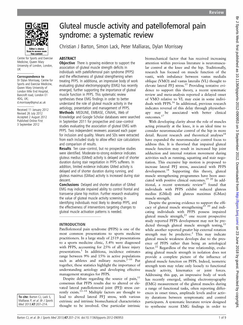
Figure 1 Flow diagram summarisingstudy selection for inclusion.
Table 1 Study details including sample sizes, participant demographics and population sources
Paper
Sample size Gender (F:M) Age range (mean age) Height (m), weight (kg)
PFPS CON PFPS CON PFPS CON PFPS CON
Aminaka et al36 20 20 13 : 7 13 : 7 NR (21±4) NR (21±4) 1.70±0.10, 71±15 1.72±0.09, 70±10Brindle et al39 16 12 12 : 4 7 : 5 18–35 (NR) 18–35 NR, NR NR, NRBoling et al34 14 14 9 : 5 9 : 5 18–42 (24±6) 18–42 (23±2) 1.68±0.10, 72±12 1.71±0.07, 72±16Cowan et al32 10 27 7 : 3 15 : 12 18–40 (26±10) 18–40 (25±6) 1.72±0.04, 63±8 1.90±0.09, 69±9Earl et al33 16 16 13 : 3 13 : 3 NR (22±4) NR (21±6) 1.65±0.10, 62±13 1.66±0.12, 65±14Nakagawa et al38 9 10 9 : 0 10 : 0 18–35 (23±5) 18–35 (23±2) 1.65±0.07, 61±10 1.63±0.06, 56±4Ott et al40 20* 20 NR NR 18–45 (21±NR) 18–45 (23±4) 1.71±0.07, 70±8 1.68±0.07, 77±7Saad et al37 15 15 NR NR NR (23±2) NR (23±2) 1.60±3, 59±4 1.60±3, 53±2Souza and Powers35 21 20 21 : 0 20 : 0 18–45 (27±6) 18–45 (26±5) 1.70±0.06, 65±10 1.70±0.05, 63±7Willson et al31 20 20 20 : 0 20 : 0 18–35 (21±3) 18–35 (22±5) 1.68±0.06, 63±8 1.69±0.09, 62±9
*Comprised of two groups; (1) low pain≤1.4 cm increase in pain visual analogue scale following aerobic exercise, n=9 and (2) high pain≥1.4 cm increase in pain visual analogue scalefollowing aerobic exercise, n=11.CON, control; F, female; M, male; NR, not reported; PFPS, patellofemoral pain syndrome.
2 of 9 Barton CJ, et al. Br J Sports Med 2013;47:207–214. doi:10.1136/bjsports-2012-090953
Review
on 23 June 2018 by guest. Protected by copyright.
http://bjsm.bm
j.com/
Br J S
ports Med: first published as 10.1136/bjsports-2012-090953 on 3 S
eptember 2012. D
ownloaded from
Means and SDs of each variable were extracted or sought fromoriginal authors to allow effect size (ES) calculations. Data werepooled where studies evaluated the same EMG variable andfunctional activity. Calculated individual or pooled ES werecategorised as small (≤0.59), medium (0.60–1.19) or large(≥1.20). The level of statistical heterogeneity for pooled datawas established using the χ2 and I2 statistics (heterogeneitydefined as p<0.05). Definitions for ‘levels of evidence’ wereguided by recommendations made by van Tulder et al.27
Strong evidence = pooled results derived from three or morestudies, including a minimum of two HQ studies, which are stat-istically homogenous (p>0.05)—may be associated with a statis-tically significant or non-significant pooled result.
Moderate evidence = statistically significant pooled resultsderived from multiple studies, including at least one HQ study,which are statistically heterogeneous (p<0.05); or from multipleLQ studies which are statistically homogenous (p>0.05).
Limited evidence = results from multiple LQ studies whichare statistically heterogeneous (p<0.05); or from one HQ study.
Very limited evidence = results from one LQ study.Conflicting evidence = pooled results insignificant and
derived from multiple studies regardless of quality which arestatistically heterogeneous (p<0.05, ie, inconsistent).
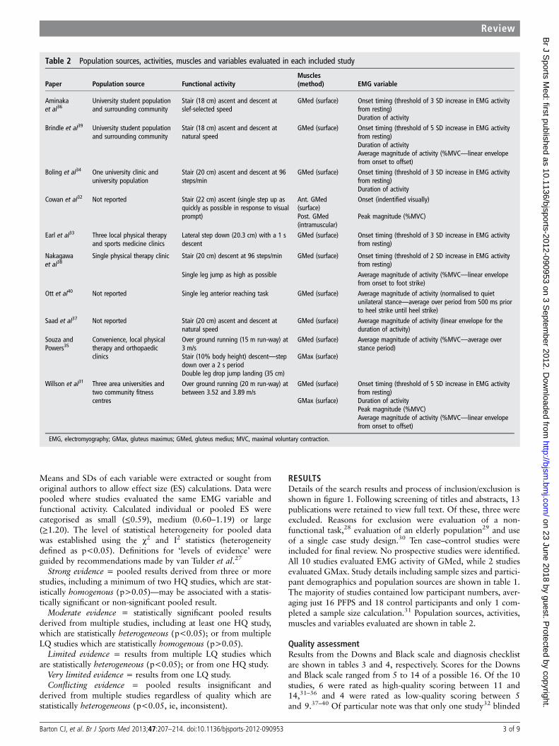
RESULTSDetails of the search results and process of inclusion/exclusion isshown in figure 1. Following screening of titles and abstracts, 13publications were retained to view full text. Of these, three wereexcluded. Reasons for exclusion were evaluation of a non-functional task,28 evaluation of an elderly population29 and useof a single case study design.30 Ten case–control studies wereincluded for final review. No prospective studies were identified.All 10 studies evaluated EMG activity of GMed, while 2 studiesevaluated GMax. Study details including sample sizes and partici-pant demographics and population sources are shown in table 1.The majority of studies contained low participant numbers, aver-aging just 16 PFPS and 18 control participants and only 1 com-pleted a sample size calculation.31 Population sources, activities,muscles and variables evaluated are shown in table 2.
Quality assessmentResults from the Downs and Black scale and diagnosis checklistare shown in tables 3 and 4, respectively. Scores for the Downsand Black scale ranged from 5 to 14 of a possible 16. Of the 10studies, 6 were rated as high-quality scoring between 11 and14,31–36 and 4 were rated as low-quality scoring between 5and 9.37–40 Of particular note was that only one study32 blinded
Table 2 Population sources, activities, muscles and variables evaluated in each included study
Paper Population source Functional activityMuscles(method) EMG variable
Aminakaet al36
University student populationand surrounding community
Stair (18 cm) ascent and descent atslef-selected speed
GMed (surface) Onset timing (threshold of 3 SD increase in EMG activityfrom resting)Duration of activity
Brindle et al39 University student populationand surrounding community
Stair (18 cm) ascent and descent atnatural speed
GMed (surface) Onset timing (threshold of 5 SD increase in EMG activityfrom resting)Duration of activityAverage magnitude of activity (%MVC—linear envelopefrom onset to offset)
Boling et al34 One university clinic anduniversity population
Stair (20 cm) ascent and descent at 96steps/min
GMed (surface) Onset timing (threshold of 3 SD increase in EMG activityfrom resting)Duration of activity
Cowan et al32 Not reported Stair (22 cm) ascent (single step up asquickly as possible in response to visualprompt)
Ant. GMed(surface)
Onset (indentified visually)
Post. GMed(intramuscular)
Peak magnitude (%MVC)
Earl et al33 Three local physical therapyand sports medicine clinics
Lateral step down (20.3 cm) with a 1 sdescent
GMed (surface) Onset timing (threshold of 3 SD increase in EMG activityfrom resting)
Nakagawaet al38
Single physical therapy clinic Stair (20 cm) descent at 96 steps/min GMed (surface) Onset timing (threshold of 2 SD increase in EMG activityfrom resting)
Single leg jump as high as possible Average magnitude of activity (%MVC—linear envelopefrom onset to foot strike)
Ott et al40 Not reported Single leg anterior reaching task GMed (surface) Average magnitude of activity (normalised to quietunilateral stance—average over period from 500 ms priorto heel strike until heel strike)
Saad et al37 Not reported Stair (20 cm) ascent and descent atnatural speed
GMed (surface) Average magnitude of activity (linear envelope for theduration of activity)
Souza andPowers35
Convenience, local physicaltherapy and orthopaedicclinics
Over ground running (15 m run-way) at3 m/s
GMed (surface) Average magnitude of activity (%MVC—average overstance period)
Stair (10% body height) descent—stepdown over a 2 s period
GMax (surface)
Double leg drop jump landing (35 cm)Willson et al31 Three area universities and
two community fitnesscentres
Over ground running (20 m run-way) atbetween 3.52 and 3.89 m/s
GMed (surface) Onset timing (threshold of 5 SD increase in EMG activityfrom resting)
GMax (surface) Duration of activityPeak magnitude (%MVC)Average magnitude of activity (%MVC—linear envelopefrom onset to offset)
EMG, electromyography; GMax, gluteus maximus; GMed, gluteus medius; MVC, maximal voluntary contraction.
Barton CJ, et al. Br J Sports Med 2013;47:207–214. doi:10.1136/bjsports-2012-090953 3 of 9
Review
on 23 June 2018 by guest. Protected by copyright.
http://bjsm.bm
j.com/
Br J S
ports Med: first published as 10.1136/bjsports-2012-090953 on 3 S
eptember 2012. D
ownloaded from
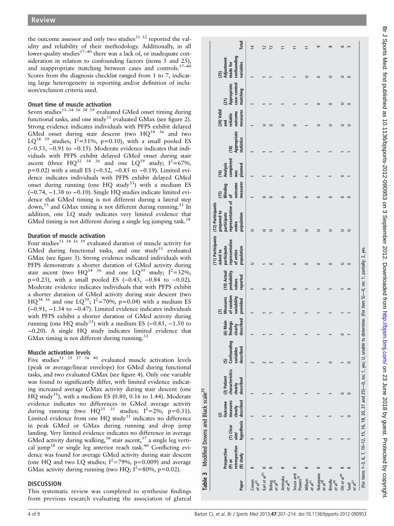
the outcome assessor and only two studies31 32 reported the val-idity and reliability of their methodology. Additionally, in alllower-quality studies37–40 there was a lack of, or inadequate con-sideration in relation to confounding factors (items 5 and 25),and inappropriate matching between cases and controls.37–40
Scores from the diagnosis checklist ranged from 1 to 7, indicat-ing large heterogeneity in reporting and/or definition of inclu-sion/exclusion criteria used.
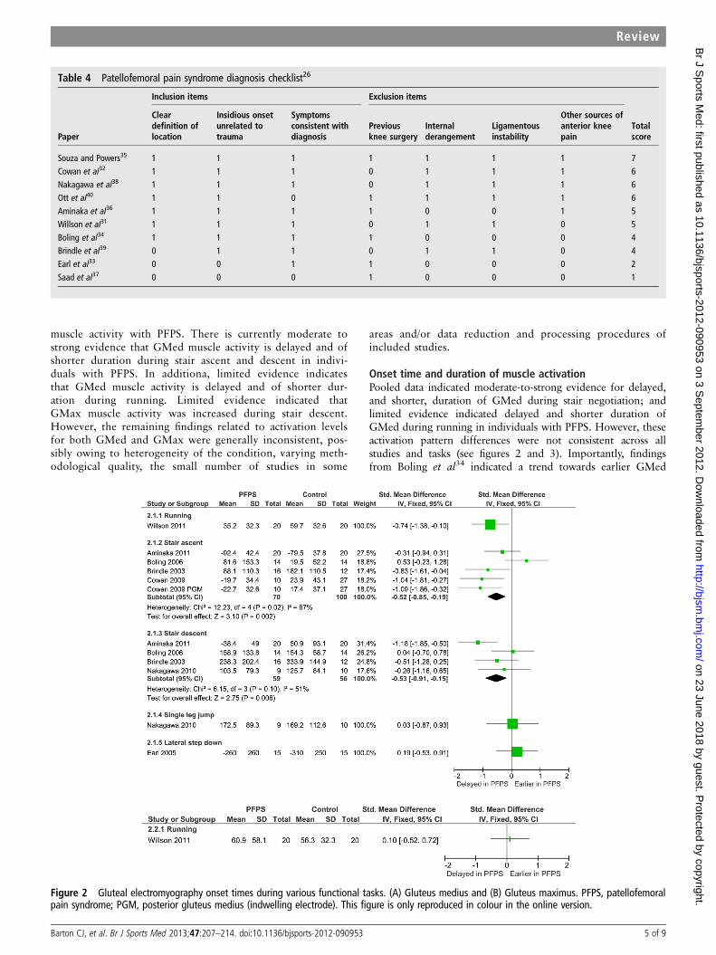
Onset time of muscle activationSeven studies31–34 36 38 39 evaluated GMed onset timing duringfunctional tasks, and one study31 evaluated GMax (see figure 2).Strong evidence indicates individuals with PFPS exhibit delayedGMed onset during stair descent (two HQ34 36 and twoLQ38 39_studies; I2=51%, p=0.10), with a small pooled ES(−0.53, −0.91 to −0.15). Moderate evidence indicates that indi-viduals with PFPS exhibit delayed GMed onset during stairascent (three HQ32 34 36 and one LQ39 study; I2=67%,p=0.02) with a small ES (−0.52, −0.85 to −0.19). Limited evi-dence indicates individuals with PFPS exhibit delayed GMedonset during running (one HQ study31) with a medium ES(−0.74, −1.38 to −0.10). Single HQ studies indicate limited evi-dence that GMed timing is not different during a lateral stepdown,33 and GMax timing is not different during running.31 Inaddition, one LQ study indicates very limited evidence thatGMed timing is not different during a single leg jumping task.38
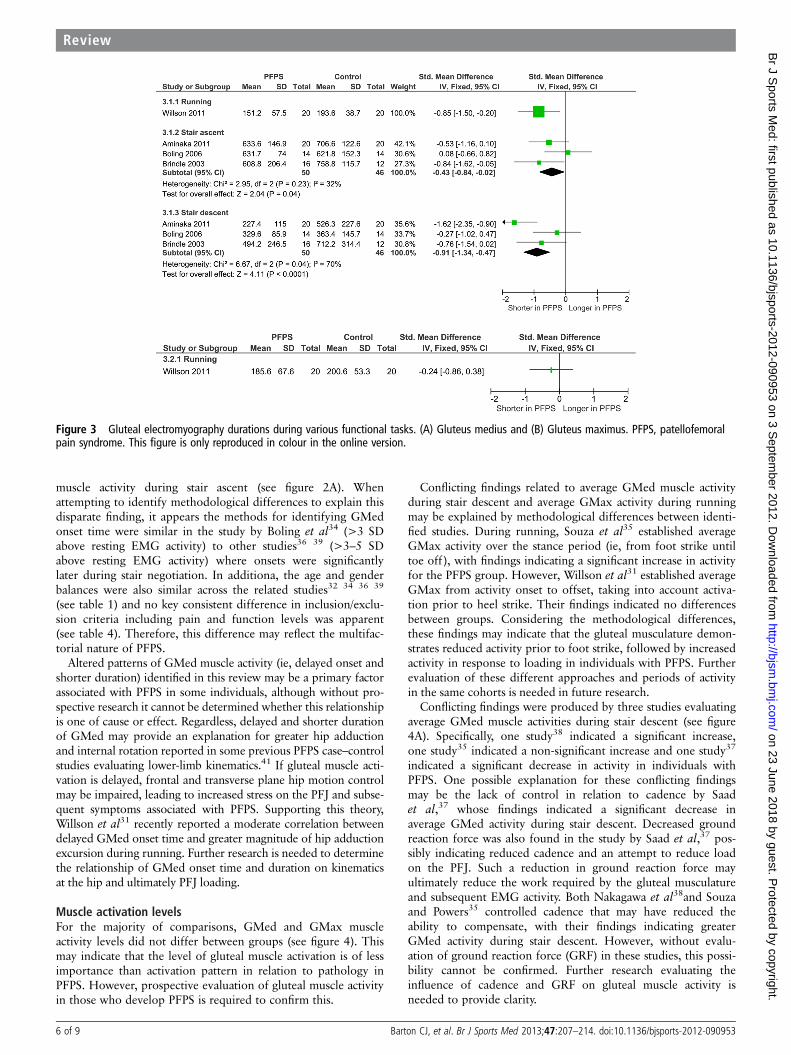
Duration of muscle activationFour studies31 34 36 39 evaluated duration of muscle activity forGMed during functional tasks, and one study31 evaluatedGMax (see figure 3). Strong evidence indicated individuals withPFPS demonstrate a shorter duration of GMed activity duringstair ascent (two HQ34 36 and one LQ39 study; I2=32%,p=0.23), with a small pooled ES (−0.43, −0.84 to −0.02).Moderate evidence indicates individuals that with PFPS exhibita shorter duration of GMed activity during stair descent (twoHQ34 36 and one LQ39; I2=70%, p=0.04) with a medium ES(−0.91, −1.34 to −0.47). Limited evidence indicates individualswith PFPS exhibit a shorter duration of GMed activity duringrunning (one HQ study31) with a medium ES (−0.85, −1.50 to−0.20). A single HQ study indicates limited evidence thatGMax timing is not different during running.31
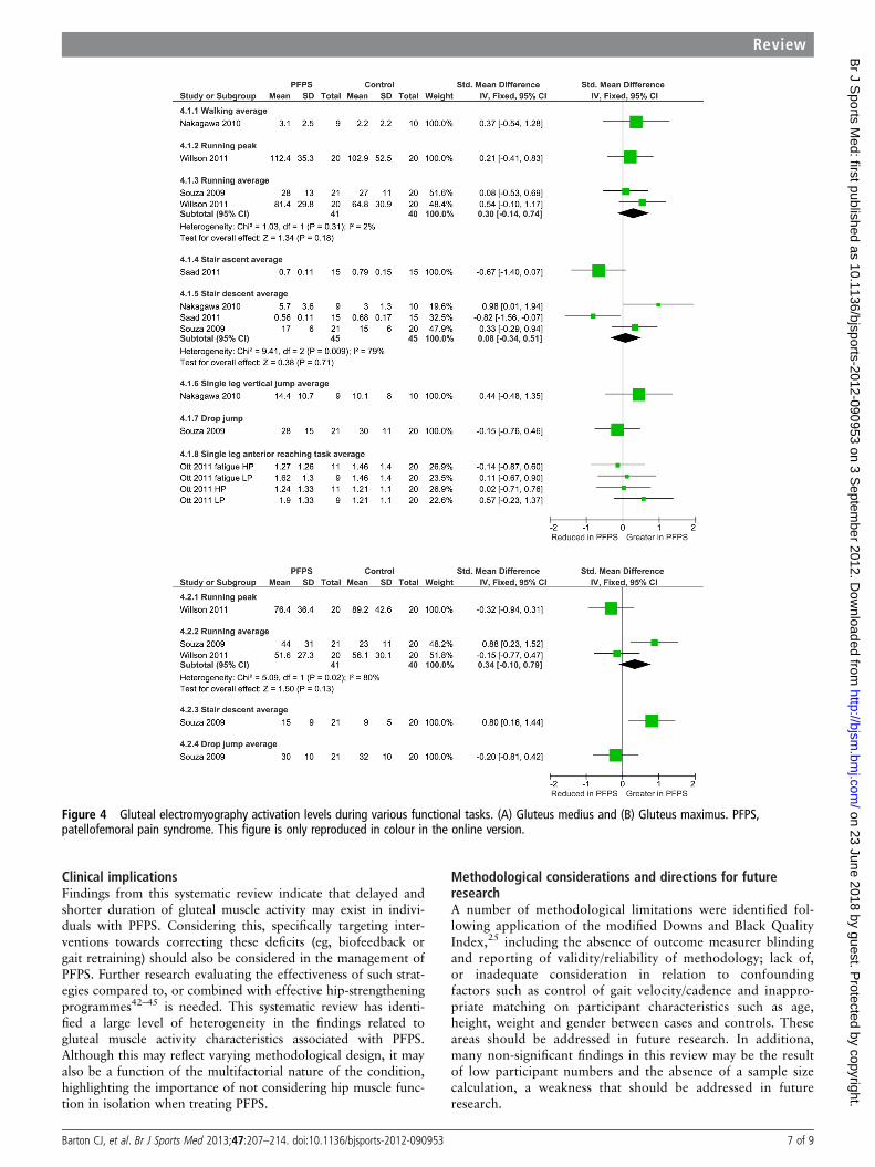
Muscle activation levelsFive studies31 35 37 38 40 evaluated muscle activation levels(peak or average/linear envelope) for GMed during functionaltasks, and two evaluated GMax (see figure 4). Only one variablewas found to significantly differ, with limited evidence indicat-ing increased average GMax activity during stair descent (oneHQ study35), with a medium ES (0.80, 0.16 to 1.44). Moderateevidence indicates no differences in GMed average activityduring running (two HQ31 35 studies; I2=2%, p=0.31).Limited evidence from one HQ study31 indicates no differencein peak GMed or GMax during running and drop jumplanding. Very limited evidence indicates no difference in averageGMed activity during walking,38 stair ascent,37 a single leg verti-cal jump38 or single leg anterior reach task.40 Conflicting evi-dence was found for average GMed activity during stair descent(one HQ and two LQ studies; I2=79%, p=0.009) and averageGMax activity during running (two HQ; I2=80%, p=0.02).
DISCUSSIONThis systematic review was completed to synthesise findingsfrom previous research evaluating the association of gluteal
Table3
Modified
Downs
andBlackscale2
5
Pape
r
Prospe
ctive
(P)or
retrospe
ctive
(R)stud
y
(1)Clea
raim/
hypo
thesis
(2)
Outcome
mea
sures
clea
rlyde
scrib
ed
(3)Patie
ntcharacteristic
sclea
rlyde
scrib
ed
(5)
Confou
nding
varia
bles
describ
ed
(6)Main
finding
sclea
rlyde
scrib
ed
(7)
Mea
sures
ofrand
omvaria
bility
provided
(10)
Actua
lprob
ability
values
repo
rted
(11)
Participan
tsaskedto
participate
representativ
eof
entire
popu
latio
n
(12)
Participan
tsprep
ared
topa
rticipate
represen
tativ
eof
entire
popu
latio
n
(15)
Blinding
of outcom
emea
surer
(16)
Ana
lysis
completed
was
plan
ned
(18)
App
ropriate
statistic
s
(20)
Valid
and
relia
ble
outcom
emeasures
(21)
App
ropriate
case–control
matching
(25)
Adjustm
ent
mad
efor
confou
nding
varia
bles
Total
Cowan
etal32
R1
11
21
11
UU
11
11
11
14
Earlet
al33
R1
11
21
10
1U
01
1U
11
12
Boling
etal34
R1
11
21
11
0U
01
1U
11
12
Aminaka
etal36
R1
11
11
10
1U
01
1U
11
11
Souzaand
Powers35
R1
11
21
10
0U
01
1U
11
11
Willson
etal31
R1
11
11
11
0U
01
11
1U
11
Nakagaw
aet
al38
R1
11
11
11
0U
01
1U
00
9
Brindle
etal39
R1
11
01
11
0U
01
1U
0U
8
Ottet
al40
R1
10
10
11
UU
00
1U
UU
6
Saad
etal37
R1
00
01
10
0U
01
1U
00
5
(For
items1–3,
6,7,
10–12,1
5,16,1
8,20,2
1and25)—
0,no;1
,yes;U
,unableto
determ
ine.(For
item
5)—0,
no:1
;partially:2
,yes.
4 of 9 Barton CJ, et al. Br J Sports Med 2013;47:207–214. doi:10.1136/bjsports-2012-090953
Review
on 23 June 2018 by guest. Protected by copyright.
http://bjsm.bm
j.com/
Br J S
ports Med: first published as 10.1136/bjsports-2012-090953 on 3 S
eptember 2012. D
ownloaded from
muscle activity with PFPS. There is currently moderate tostrong evidence that GMed muscle activity is delayed and ofshorter duration during stair ascent and descent in indivi-duals with PFPS. In additiona, limited evidence indicatesthat GMed muscle activity is delayed and of shorter dur-ation during running. Limited evidence indicated thatGMax muscle activity was increased during stair descent.However, the remaining findings related to activation levelsfor both GMed and GMax were generally inconsistent, pos-sibly owing to heterogeneity of the condition, varying meth-odological quality, the small number of studies in some
areas and/or data reduction and processing procedures ofincluded studies.
Onset time and duration of muscle activationPooled data indicated moderate-to-strong evidence for delayed,and shorter, duration of GMed during stair negotiation; andlimited evidence indicated delayed and shorter duration ofGMed during running in individuals with PFPS. However, theseactivation pattern differences were not consistent across allstudies and tasks (see figures 2 and 3). Importantly, findingsfrom Boling et al34 indicated a trend towards earlier GMed
Figure 2 Gluteal electromyography onset times during various functional tasks. (A) Gluteus medius and (B) Gluteus maximus. PFPS, patellofemoralpain syndrome; PGM, posterior gluteus medius (indwelling electrode). This figure is only reproduced in colour in the online version.
Table 4 Patellofemoral pain syndrome diagnosis checklist26
Inclusion items Exclusion items
TotalscorePaper
Cleardefinition oflocation
Insidious onsetunrelated totrauma
Symptomsconsistent withdiagnosis
Previousknee surgery
Internalderangement
Ligamentousinstability
Other sources ofanterior kneepain
Souza and Powers35 1 1 1 1 1 1 1 7Cowan et al32 1 1 1 0 1 1 1 6Nakagawa et al38 1 1 1 0 1 1 1 6Ott et al40 1 1 0 1 1 1 1 6Aminaka et al36 1 1 1 1 0 0 1 5Willson et al31 1 1 1 0 1 1 0 5Boling et al34 1 1 1 1 0 0 0 4Brindle et al39 0 1 1 0 1 1 0 4Earl et al33 0 0 1 1 0 0 0 2Saad et al37 0 0 0 1 0 0 0 1
Barton CJ, et al. Br J Sports Med 2013;47:207–214. doi:10.1136/bjsports-2012-090953 5 of 9
Review
on 23 June 2018 by guest. Protected by copyright.
http://bjsm.bm
j.com/
Br J S
ports Med: first published as 10.1136/bjsports-2012-090953 on 3 S
eptember 2012. D
ownloaded from
muscle activity during stair ascent (see figure 2A). Whenattempting to identify methodological differences to explain thisdisparate finding, it appears the methods for identifying GMedonset time were similar in the study by Boling et al34 (>3 SDabove resting EMG activity) to other studies36 39 (>3–5 SDabove resting EMG activity) where onsets were significantlylater during stair negotiation. In additiona, the age and genderbalances were also similar across the related studies32 34 36 39
(see table 1) and no key consistent difference in inclusion/exclu-sion criteria including pain and function levels was apparent(see table 4). Therefore, this difference may reflect the multifac-torial nature of PFPS.
Altered patterns of GMed muscle activity (ie, delayed onset andshorter duration) identified in this review may be a primary factorassociated with PFPS in some individuals, although without pro-spective research it cannot be determined whether this relationshipis one of cause or effect. Regardless, delayed and shorter durationof GMed may provide an explanation for greater hip adductionand internal rotation reported in some previous PFPS case–controlstudies evaluating lower-limb kinematics.41 If gluteal muscle acti-vation is delayed, frontal and transverse plane hip motion controlmay be impaired, leading to increased stress on the PFJ and subse-quent symptoms associated with PFPS. Supporting this theory,Willson et al31 recently reported a moderate correlation betweendelayed GMed onset time and greater magnitude of hip adductionexcursion during running. Further research is needed to determinethe relationship of GMed onset time and duration on kinematicsat the hip and ultimately PFJ loading.
Muscle activation levelsFor the majority of comparisons, GMed and GMax muscleactivity levels did not differ between groups (see figure 4). Thismay indicate that the level of gluteal muscle activation is of lessimportance than activation pattern in relation to pathology inPFPS. However, prospective evaluation of gluteal muscle activityin those who develop PFPS is required to confirm this.
Conflicting findings related to average GMed muscle activityduring stair descent and average GMax activity during runningmay be explained by methodological differences between identi-fied studies. During running, Souza et al35 established averageGMax activity over the stance period (ie, from foot strike untiltoe off ), with findings indicating a significant increase in activityfor the PFPS group. However, Willson et al31 established averageGMax from activity onset to offset, taking into account activa-tion prior to heel strike. Their findings indicated no differencesbetween groups. Considering the methodological differences,these findings may indicate that the gluteal musculature demon-strates reduced activity prior to foot strike, followed by increasedactivity in response to loading in individuals with PFPS. Furtherevaluation of these different approaches and periods of activityin the same cohorts is needed in future research.
Conflicting findings were produced by three studies evaluatingaverage GMed muscle activities during stair descent (see figure4A). Specifically, one study38 indicated a significant increase,one study35 indicated a non-significant increase and one study37
indicated a significant decrease in activity in individuals withPFPS. One possible explanation for these conflicting findingsmay be the lack of control in relation to cadence by Saadet al,37 whose findings indicated a significant decrease inaverage GMed activity during stair descent. Decreased groundreaction force was also found in the study by Saad et al,37 pos-sibly indicating reduced cadence and an attempt to reduce loadon the PFJ. Such a reduction in ground reaction force mayultimately reduce the work required by the gluteal musculatureand subsequent EMG activity. Both Nakagawa et al38and Souzaand Powers35 controlled cadence that may have reduced theability to compensate, with their findings indicating greaterGMed activity during stair descent. However, without evalu-ation of ground reaction force (GRF) in these studies, this possi-bility cannot be confirmed. Further research evaluating theinfluence of cadence and GRF on gluteal muscle activity isneeded to provide clarity.
Figure 3 Gluteal electromyography durations during various functional tasks. (A) Gluteus medius and (B) Gluteus maximus. PFPS, patellofemoralpain syndrome. This figure is only reproduced in colour in the online version.
6 of 9 Barton CJ, et al. Br J Sports Med 2013;47:207–214. doi:10.1136/bjsports-2012-090953
Review
on 23 June 2018 by guest. Protected by copyright.
http://bjsm.bm
j.com/
Br J S
ports Med: first published as 10.1136/bjsports-2012-090953 on 3 S
eptember 2012. D
ownloaded from
Clinical implicationsFindings from this systematic review indicate that delayed andshorter duration of gluteal muscle activity may exist in indivi-duals with PFPS. Considering this, specifically targeting inter-ventions towards correcting these deficits (eg, biofeedback orgait retraining) should also be considered in the management ofPFPS. Further research evaluating the effectiveness of such strat-egies compared to, or combined with effective hip-strengtheningprogrammes42–45 is needed. This systematic review has identi-fied a large level of heterogeneity in the findings related togluteal muscle activity characteristics associated with PFPS.Although this may reflect varying methodological design, it mayalso be a function of the multifactorial nature of the condition,highlighting the importance of not considering hip muscle func-tion in isolation when treating PFPS.
Methodological considerations and directions for futureresearchA number of methodological limitations were identified fol-lowing application of the modified Downs and Black QualityIndex,25 including the absence of outcome measurer blindingand reporting of validity/reliability of methodology; lack of,or inadequate consideration in relation to confoundingfactors such as control of gait velocity/cadence and inappro-priate matching on participant characteristics such as age,height, weight and gender between cases and controls. Theseareas should be addressed in future research. In additiona,many non-significant findings in this review may be the resultof low participant numbers and the absence of a sample sizecalculation, a weakness that should be addressed in futureresearch.
Figure 4 Gluteal electromyography activation levels during various functional tasks. (A) Gluteus medius and (B) Gluteus maximus. PFPS,patellofemoral pain syndrome. This figure is only reproduced in colour in the online version.
Barton CJ, et al. Br J Sports Med 2013;47:207–214. doi:10.1136/bjsports-2012-090953 7 of 9
Review
on 23 June 2018 by guest. Protected by copyright.
http://bjsm.bm
j.com/
Br J S
ports Med: first published as 10.1136/bjsports-2012-090953 on 3 S
eptember 2012. D
ownloaded from
The SENIAM guidelines46 provide clear and valid guidanceregarding the preparation and application of electrodes duringthe collection of gluteal EMG. However, the same clear guid-ance is lacking for data collection procedures, reduction andanalysis. As a result, these methodological aspects varied acrossthe included studies (see table 2), possibly explaining some ofthe conflicting findings. Unfortunately, without direct evaluationcomparing outcomes due to varied approaches in the samecohorts, it is difficult to establish the exact nature or size oftheir influence on results. Future studies evaluating gluteal EMGin individuals with PFPS should consider addressing this. In par-ticular, the influence of cadence, method of identifying muscleonset time and method of establishing EMG activity levels onresults needs to be established.
The ability to distinguish between cause and effect in relationto identified differences is impaired by the absence of prospect-ive research. Additional research is needed to determine ifscreening of gluteal muscle activity can successfully identifythose most likely to develop PFPS. Findings from case–controlstudies were inconsistent for all variables evaluated. This may bea function of the large heterogeneity in methodological design,and in particular inconsistent inclusion/exclusion criteria fordiagnosis. It is recommended that future case–control studiesuse inclusion/exclusion criteria checklist26 to guide participantrecruitment which is based on high-quality randomised con-trolled trials evaluating conservative PFPS interventions.47 48
CONCLUSIONCurrent research evaluating the association of gluteal muscleactivity with PFPS is limited by an absence of prospectiveresearch, low sample sizes and heterogeneity in methodologicaldesign including procedures, data reduction and analysis andparticipant inclusion and exclusion criteria. Conflicting findingsmay be a function of these methodological differences and/orthe multifactorial nature of PFPS. Moderate-to-strong evidenceindicates that GMed muscle activity is delayed and of shorterduration during stair ascent and descent in individuals withPFPS. Additionally, limited evidence indicates that GMedmuscle activity is delayed and of shorter duration duringrunning, and GMax muscle activity is increased during stairdescent. Further research evaluating the value of glutealmuscle activity screening in identifying individuals most likely todevelop PFPS is needed. Additionally, evaluating the effective-ness of interventions such as biofeedback and gait retraining tar-geting changes of gluteal muscle activation patterns is needed.
Contributors All authors contributed significantly to the article formulation. SL andCJB had the original idea and led on the writing. PM and DM assisted this process.DM also did the quality assessment.
Competing interests None.
Provenance and peer review Not commissioned; externally peer reviewed.
REFERENCES1 Devereaux MD, Lachmann SM. Patello-femoral arthralgia in athletes attending a
Sports Injury Clinic. Br J Sports Med 1984;18:18–21.2 Boling MC, Padua DA, Marshall SW, et al. A prospective investigation of
biomechanical risk factors for patellofemoral pain syndrome: the Joint Undertakingto Monitor and Prevent ACL Injury ( JUMP-ACL) cohort. Am J Sports Med2009;37:2108–16.
3 Finestone A, Radin EL, Lev B, et al. Treatment of overuse patellofemoral pain.Prospective randomized controlled clinical trial in a military setting. Clin Orthop RelRes 1993;293:208–10.
4 Hetsroni I, Finestone A, Milgrom C, et al. A prospective biomechanical study of theassociation between foot pronation and the incidence of anterior knee pain amongmilitary recruits. J Bone Joint Surg, Br Volume 2006;88:905–8.
5 Milgrom C, Finestone A, Eldad A, et al. Patellofemoral pain caused by overactivity.A prospective study of risk factors in infantry recruits. J Bone Joint Surg, Am Volume1991;73:1041–3.
6 Phillips J, Coetsee MF. Incidnce of non-traumatic anterior knee pain among11–17-year olds. S Afr J Sports Med 2007;19:60–4.
7 Schwellnus MP, Jordaan G, Noakes TD. Prevention of common overuse injuries bythe use of shock absorbing insoles. A prospective study. Am J Sports Med1990;18:636–41.
8 Wills AK, Ramasamy A, Ewins DJ, et al. The incidence and occupational outcome ofoveruse anterior knee pain during army recruit training. J R Army Med Corps2004;150264–9.
9 Witvrouw E, Lysens R, Bellemans J, et al. Intrinsic risk factors for the developmentof anterior knee pain in an athletic population. A two-year prospective study. Am JSports Med 2000;28:480–9.
10 Myer GD, Ford KR, Foss KDB, et al. The incidence and potential pathomechanics ofpatellofemoral pain in female athletes. Clin Biomech 2010;25:700–7.
11 Davis IS, Powers CM. Patellofemoral pain syndrome: proximal, distal, and localfactors, an international retreat, April 30-May 2, 2009, Fells Point, Baltimore, MD. JOrthop Sports Phys Ther 2009;40:A1–16.
12 Feller JA, Amis AA, Andrish JT, et al. Surgical biomechanics of the patellofemoraljoint. Arthroscopy 2007;23:542–53.
13 Heino Brechter J, Powers CM. Patellofemoral joint stress during walking in personswith and without patellofemoral pain. Med Sci Sports Exerc 2002;34:1582–93.
14 Heino Brechter J, Powers CM. Patellofemoral joint stress during stair ascent anddescent in persons with and without patellofemoral pain. Gait Posture2002;16:115–23.
15 McConnell J. Management of patellofemoral problems. Man Ther 1996;1:60–6.16 Chester R, Smith TO, Sweeting D, et al. The relative timing of VMO and VL in the
aetiology of anterior knee pain: a systematic review and meta-analysis. BMCMusculoskelet Disord 2008;9:64.
17 Cowan SM, Bennell KL, Crossley KM, et al. Physical therapy alters recruitment ofthe vasti in patellofemoral pain syndrome. Med Sci Sports Exerc 2002;34:1879–85.
18 Powers CM. The influence of abnormal hip mechanics on knee injury: abiomechanical perspective. J Orthop Sports Phys Ther 2010;40:42–51.
19 Fukuda TY, Rossetto FM, Magalhaes E, et al. Short-term effects of hip abductorsand lateral rotators strengthening in females with patellofemoral pain syndrome: arandomized controlled clinical trial. J Orthop Sports Phys Ther 2010;40:736–42.
20 Mascal CL, Landel R, Powers C. Management of patellofemoral pain targeting hip,pelvis, and trunk muscle function: 2 case reports. J Orthop Sports Phys Ther2003;33:647–60.
21 Prins MR, van der Wurff P. Females with patellofemoral pain syndrome have weakhip muscles: a systematic review. Aust J Physiother 2009;55:9–15.
22 Thijs Y, Pattyn E, Van Tiggelen D, et al. Is hip muscle weakness a predisposingfactor for patellofemoral pain in female novice runners? A prospective study. Am JSports Med 2011;39:1877–82.
23 Heintjes E, Berger MY, Bierma-Zeinstra SM, et al. Exercise therapy for patellofemoralpain syndrome. Cochrane Database Syst Rev 2003:CD003472.
24 Maher C, Sherrington C, Elkins M, et al. Challenges for evidence-based physicaltherapy: accessing and interpreting high quality evidence on therapy. Phys Ther2004;84:644–54.
25 Downs SH, Black N. The feasibility of creating a checklist for the assessment of themethodological quality both of randomised and non-randomised studies of healthcare interventions. J Epidemiol Community Health 1998;52:377–84.
26 Barton CJ, Munteanu SE, Menz HB, et al. The efficacy of foot orthoses in thetreatment of individuals with patellofemoral pain syndrome: a systematic review.Sports Med 2010;40:377–95.
27 van Tulder M, Furlan A, Bombardier C, et al. Updated method guidelines forsystematic reviews in the Cochrane collaboration back review group. Spine2003;28:1290–9.
28 Baldon RM, Nakagawa TH, Muniz TB, et al. Eccentric hip muscle function infemales with and without patellofemoral pain syndrome. J Athl Train2009;44:490–6.
29 Manetta J, Franz LH, Moon C, et al. Comparison of hip and knee muscle momentsin subjects with and without knee pain. Gait Posture 2002;16:249–54.
30 Albisetti W, De Bartolomeo O, Gabbiadini S, et al. Surface EMG evaluation ofpatellofemoral pain syndrome in a professional ballet dancer. Med Probl PerformArtist 2008;23:29–32.
31 Willson JD, Kernozek TW, Arndt RL, et al. Gluteal muscle activation during runningin females with and without patellofemoral pain syndrome. Clin Biomech (Bristol,Avon) 2011;26:735–40.
32 Cowan SM, Crossley KM, Bennell KL. Altered hip and trunk muscle function inindividuals with patellofemoral pain. Br J Sports Med 2009;43:584–8.
33 Earl J, Hertel J, Denegar C. Patterns of dynamic malalignment, muscle activation,joint motion, and patellofemoral-pain syndrome. J Sport Rehabil 2005;14:215–33.
34 Boling MC, Bolgla LA, Mattacola CG, et al. Outcomes of a weight-bearingrehabilitation program for patients diagnosed with patellofemoral pain syndrome.Arch Phys Med Rehabil 2006;87:1428–35.
8 of 9 Barton CJ, et al. Br J Sports Med 2013;47:207–214. doi:10.1136/bjsports-2012-090953
Review
on 23 June 2018 by guest. Protected by copyright.
http://bjsm.bm
j.com/
Br J S
ports Med: first published as 10.1136/bjsports-2012-090953 on 3 S
eptember 2012. D
ownloaded from
35 Souza RB, Powers CM. Differences in hip kinematics, muscle strength, and muscleactivation between subjects with and without patellofemoral pain. J Orthop SportsPhys Ther 2009;39:12–19.
36 Aminaka N, Pietrosimone BG, Armstrong CW, et al. Patellofemoral pain syndromealters neuromuscular control and kinetics during stair ambulation. J ElectromyogrKinesiol 2011;21:645–51.
37 Saad MC, Felicio LR, Masullo CdL, et al. Analysis of the center of pressuredisplacement, ground reaction force and muscular activity during step exercises. JElectromyogr Kinesiol 2011;21:712–18.
38 Nakagawa TH, Muniz TB, Baldon RM, et al. Electromyographic preactivation patternof the gluteus medius during weight-bearing functional tasks in women with andwithout anterior knee pain. Rev Bras Fisioter 2011;15:59–65.
39 Brindle TJ, Mattacola C, McCrory J. Electromyographic changes in the gluteusmedius during stair ascent and descent in subjects with anterior knee pain. KneeSurg Sports Traumatol Arthrosc 2003;11:244–51.
40 Ott B, Cosby NL, Grindstaff TL, et al. Hip and knee muscle function followingaerobic exercise in individuals with patellofemoral pain syndrome. J ElectromyogrKinesiol 2011;21:631–7.
41 Barton C, Levinger P, Menz H, et al. Kinematic gait characteristics associated wtihpatellofemroal pain syndrome: a systematic review. Gait Posture 2009;30:405–16.
42 Dolak KL, Silkman C, McKeon JM, et al. Hip strengthening prior to functionalexercises reduces pain sooner than quadriceps strengthening in females withpatellofemoral pain syndrome: a randomized clinical trial. J Orthop Sports Phys Ther2011;41:560–70.
43 Earl JE, Hoch AZ. A proximal strengthening program improves pain, function, andbiomechanics in women with patellofemoral pain syndrome. Am J Sports Med2011;39:154–63.
44 Fukuda TY, Rossetto FM, Magalhaes E, et al. Short-term effects of hip abductorsand lateral rotators strengthening in females with patellofemoral pain syndrome: arandomized controlled clinical trial. J Orthop Sports Phys Ther 2010;40:736–42.
45 Nakagawa TH, Muniz TB, Baldon RdM, et al. The effect of additional strengtheningof hip abductor and lateral rotator muscles in patellofemoral pain syndrome: arandomized controlled pilot study. Clin Rehabil 2008;22:1051–60.
46 SENIAM. http://www.seniam.org/, (accessed 1 August 2011).47 Collins N, Crossley K, Beller E, et al. Foot orthoses and physiotherapy in the
treatment of patellofemoral pain syndrome: randomised clinical trial. BMJ 208;337:a1735.
48 Crossley K, Bennell K, Green S, et al. Physical therapy for patellofemoral pain: arandomized, double-blinded, placebo-controlled trial. Am J Sports Med2002;30:857–65.
Barton CJ, et al. Br J Sports Med 2013;47:207–214. doi:10.1136/bjsports-2012-090953 9 of 9
Review
on 23 June 2018 by guest. Protected by copyright.
http://bjsm.bm
j.com/
Br J S
ports Med: first published as 10.1136/bjsports-2012-090953 on 3 S
eptember 2012. D
ownloaded from