glycoderivatives: drug candidates molecular tools · glicoderivati di tipo c‐glicosidico, quindi...
TRANSCRIPT

UNIVERSITA’ DEGLI STUDI DI MILANO ‐ BICOCCA
SCUOLA DI DOTTORATO DI SCIENZE
Dipartimento di Biotecnologie e Bioscienze
Dottorato di Ricerca in Biotecnologie Industriali
XXV ciclo
Glycoderivatives: drug candidates
and molecular tools
Giuseppe D’Orazio
Tutor: Dott.ssa Barbara La Ferla


To my family,
my friends
and my beloved Marta


1
Index Preface ................................................................................................ 5
Riassunto ............................................................................................. 7
Chapter 1. Introduction ...................................................................... 13
Carbohydrates – general overview ................................................................... 14
The therapeutic importance of carbohydrates: the development of glycomimetics .... 23 C‐glycosides as glycomimetic scaffolds ........................................................................ 27
Role of carbohydrates and glycomimetics in anti‐inflammatory processes: inflammation and SGLT1 ................................................................................... 32
Inflammation, sepsis and septic shock ......................................................................... 32 SGLT1: function and structure ...................................................................................... 43
Iminosugars: azaglycoderivatives and their widespread biological functions .. 58
Glycosidases inhibition: therapeutic role and biochemical features ............................ 58 Iminosugars as glycosidase inhibitors .......................................................................... 62 Biological activity of iminosugars and their therapeutic applications ......................... 66
Multivalency and glycobiology ......................................................................... 73
References ........................................................................................................ 76
Chapter 2. Development of a library of dansyl‐C‐glycoderivatives as SGLT1 ligand tools: synthesis, biological evaluation and mechanism of action. ................................................................................................ 81
Abstract ............................................................................................................ 82
Introduction ...................................................................................................... 82
Results and discussion ...................................................................................... 84
Conclusions ....................................................................................................... 97
Experimental Section ........................................................................................ 97
References ...................................................................................................... 124
Chapter 3. Synthesis of a labeled SGLT1 ligand for in vitro and in vivo trafficking studies ............................................................................. 127

2
Abstract .......................................................................................................... 128
Introduction .................................................................................................... 128
Results and discussion .................................................................................... 130
Conclusions ..................................................................................................... 133
Experimental Section ...................................................................................... 133
References ...................................................................................................... 142
Chapter 4. Generation of gold nanoparticles decorated with synthetic ligands of co‐transporters SGLT‐1 and B0AT1 for the investigation of the multivalent‐synergistic effect. .......................................................... 145
Abstract .......................................................................................................... 146
Introduction .................................................................................................... 146
Results and Discussion .................................................................................... 149
Conclusions ..................................................................................................... 161
Experimental Section ...................................................................................... 162
References ...................................................................................................... 185
Chapter 5. Iminosugar‐decorated calix[4]arenes: chemical tools for the investigation of multivalent inhibition of glycosidases. ..................... 187
Abstract .......................................................................................................... 188
Introduction .................................................................................................... 188
Results and Discussion .................................................................................... 190
Conclusions ..................................................................................................... 199
Experimental Section ...................................................................................... 200
References ...................................................................................................... 216
Chapter 6. Antiproliferative activity of Arsenical C‐glucoside derivative on neuroblastoma cell line SN‐K‐BE .................................................. 219
Abstract .......................................................................................................... 220
Introduction .................................................................................................... 220

3
Results and Discussion .................................................................................... 222
Experimental Section ...................................................................................... 224
References ...................................................................................................... 227
Chapter 7. Conclusions ..................................................................... 229
Pubblications and communications ................................................... 233
Papers ............................................................................................................. 234
Oral communications ...................................................................................... 234
Other communications ................................................................................... 235
List of abbreviations ......................................................................... 237

4

5
Preface The research work presented in this manuscript is a result of
collaborations between the research group in which I worked,
supervised by Dr. Barbara La Ferla, PhD and Prof. Francesco Nicotra, and
several Italian and European coworkers.
Chapter 2 and chapter 3 describe a research project in collaboration
with the research group of Prof. Cristiano Rumio (University of Milan –
Humanitas Clinical Institute (Milan)). An acknowledgment goes to Dr.
Diego Cardani which performed the biological experiments.
In chapter 4 is described a research work carried out in collaboration
with the research group of Prof. Soledad Penàdes (Laboratory of
Biofunctional Nanomaterials – CicbiomaGUNE, Donostia – San Sebastiàn,
Basque Contry, Spain). In particular, the preparation of the gold
nanoparticles was carried out during an one month research period in
this research center, under the supervision of Dr. Marco Marradi, PhD..
My research period abroad was carried out within the COST Network
project (COST Action CM1102).
Chapter 5 is a result of a collaboration with the research group of Prof.
Alessandro Dondoni (University of Ferrara, Italy) and Prof. Alberto Marra
(University of Montpellier 2, France). In particular, their work was
focused on the preparation of the calixarene structures and their
functionalization with the prepared iminosugars.
Chapter 6 presents a work performed in collaboration with Prof. Marco
Salvetti research group (University of Rome – La Sapienza) and Dr.
Michele Pitaro (Xenuspharma s.r.l, Rome).

6

Riassunto
7
Riassunto
I carboidrati rappresentano la classe di macromolecole biologiche più
abbondante in Natura. La complessità chimica e strutturale che li
caratterizza è alla base delle svariate funzioni svolte nel mondo vivente.
Tale variabilità è dovuta sia dal numero di unità che li compongono, dal
numero di atomi di carbonio che costituiscono ciascuna unità e dal tipo
di funzionalità carbonilica presente. Queste distinzioni comportano una
notevole variabilità in termini di possibili strutture generabili; a ciò va
però aggiunta l’enorme diversità dovuta alle varie combinazioni generate
dalla presenza di più carboni stereogenici in ogni singolo monosaccaride.
La Natura si avvantaggia di tutte queste peculiarità mostrate dai
carboidrati, che sono utilizzati dagli esseri viventi come molecole di
riserva e fonte di energia, come elementi strutturali di piante o come
carrier di informazioni. È senza dubbio quest’ultima funzione quella di
maggiore interesse da un punto di vista biologico e clinico: le cellule del
sistema immunitario, ad esempio, comunicano tra loro grazie
all’interazione tra specifiche proteine di membrana espresse sulla
superficie di talune cellule, e complesse strutture zuccherine costituenti
glicoproteine di membrana di cellule partner. La ricerca e lo studio di
queste complesse catene oligosaccaridiche o di loro analoghi è alla base,
per esempio, dello sviluppo di vaccini, per bloccare o scardinare lo stesso
riconoscimento che avviene tra particella virale e cellula. Oltre a ciò, i
carboidrati rappresentano anche substrati per reazioni enzimatiche che
causano la loro trasformazione, come le idrolisi di oligo‐ e polisaccaridi e
coniugazione a proteine, processi alla base di fondamentali eventi quali
la digestione o il corretto ripiegamento delle proteine. A tali processi
biologici sono inevitabilmente associate numerose patologie, aspetto

Riassunto
8
che rende ancora più importante il ruolo dei carboidrati. Tutto ciò ha
spinto la ricerca scientifica verso lo sviluppo di nuove molecole,
glicomimetici o glicoderivati, in grado di mimare la funzione bioattiva di
queste macromolecole. Inoltre, la generazione di tali derivati permette di
superare alcune intrinseche lacune dei carboidrati in quanto tali, come le
scarse proprietà drug‐like. I carboidrati possono anche essere utilizzati
come scheletro per il design e la generazione di potenziali farmaci,
potendo sfruttare le numerose funzionalità ossidriliche per la
coniugazione di opportuni farmacofori, avvalendosi inoltre delle diverse
combinazioni stereochimiche dei monosaccaridi. Questi aspetti hanno
portato, nel corso dei decenni, ad un incremento nello studio del ruolo di
queste macromolecole nella vita; la glicobiologia è ormai una disciplina il
cui studio risulta cruciale nella comprensione degli eventi cellulari,
fisiologici e patologici: la presenza in commercio di farmaci a base
saccaridica è ormai una realtà consolidata, così come la generazione di
innumerevoli glicoderivati e glicoconiugati come strumenti di studio dei
processi in cui i carboidrati sono coinvolti.
Le potenzialità sopra menzionati rappresentano la base del lavoro svolto
durante il dottorato di ricerca, esposto in questo manoscritto. Abbiamo
posto la nostra attenzione verso l’utilizzo dei carboidrati per la
generazione e la sintesi di molecole bioattive e potenziali NCE (nuove
entità chimiche) e lo sviluppo di glicoderivati e coniugati come tools per
lo studio di importanti processi biologici.
Una parte del lavoro riguarda la generazione di potenziali molecole
antiinfiammatorie a base saccaridica, ligandi di un trasportatore
localizzato a livello intestinale, SGLT1 (Sodium‐Glucose co‐Transporter 1,
co‐trasportatore sodio glucosio 1), per il quale recentemente un
importante ruolo immunologico nella protezione da eventi infiammatori
(sepsi, shock endotossico, infiammazioni croniche e mucositi) è stato

Riassunto
9
individuato. Inizialmente, diversi lavori di letteratura hanno dimostrato
che alte concentrazioni di glucosio in vitro ed in vivo causano ad una
sorta di attivazione di questo trasportatore intestinale, e ciò porta ad
una serie di eventi intracellulari che si traducono nel blocco della
produzione di molecole pro‐infiammatorie, quali citochine e
chemochine. La nostra successiva ricerca di analoghi di glucosio non
metabolizzabili, in grado di agire a concentrazioni farmacologiche, ha
portato alla generazione di un primo lead compound, di natura C‐
glicosidica (Figura 1). Negli ultimi anni, al fine di ampliare la nostra
conoscenza sul meccanismo di azione ‐ ancora non completamente noto
‐ di questa molecola è stata inizialmente sviluppata una piccola libreria di
analoghi (Cap. 2), per studi di relazione struttura attività, e in secondo
luogo è stata messa a punto una via di sintesi per la generazione di un
derivato radiomarcato del lead compound (Cap. 3), per studiarne il
destino fisiologico.
Figura 1 ‐ Struttura del C‐glicoside con potente attività antiinfiammatoria.
Ulteriori lavori scientifici hanno dimostrato come sia SGLT1 sia un co‐
trasportatore sodio‐glutammina (B0AT1), localizzato anch’esso sulla
membrana delle cellule epiteliali intestinali e il cui ligando naturale, la L‐
glutammina, mostra attività antiinfiammatoria, svolgano la loro funzione
sotto forma di oligomeri o organizzandosi sulla membrana come clusters.
Al fine di sfruttare tale comportamento e ipotizzando una possibile
azione antinfiammatoria sinergica tra il C‐glicoside e L‐glutammina, sono
state generate nanoparticelle d’oro decorate con derivati di tali molecole
(Figura 2), quindi strutture polivalenti, come strumenti di indagine e

Riassunto
10
studio di possibili fenomeni di multivalenza associati a tali trasportatori
(Cap. 4).
Figura 2 ‐ Schema generale delle nanoparticelle d'oro funzionalizzate con derivati del
C‐glicoside e L‐glutammina. Il concetto di multivalenza è alla base di una seconda parte del lavoro di
ricerca, in cui sono stati sintetizzati strutture polivalenti (calix[4]areni)
rivestiti con copie multiple di derivati di imminozuccheri, analoghi
saccaridici estremamente importanti come potenziali agenti contro
malattie di tipo metabolico (diabete e obesità) o causate da disordini nel
processamento di glicolipidi e glicoproteine (malattie da accumulo
lisosomiale). I target molecolari di tali composti (inibitori o chaperoni
chimici), sono enzimi chiamati glicosidasi, diffuse e convolte in
moltissimi processi biologici; recentemente, sono state avanzate ipotesi
di comportamento multivalente per gli imminozuccheri verso tali enzimi.
Sono quindi stati preparati calixareni polidecorati con derivati
imminosaccaridici (Figura 3) (Cap. 5), sfruttando approcci di click
chemistry, al fine di generare strumenti di studio della inibizione
multivalente recentemente riscontrata, associata a questi enzimi.

Riassunto
11
Figura 3 ‐ Struttura di un calix[4]arene polisostituito con derivati di imminozuccheri.
Ad ultimo, abbiamo rivolto la nostra attenzione allo sviluppo di
glicoderivati di tipo C‐glicosidico, quindi metabolicamente inerti,
modificati con gruppi contenenti un atomo di arsenico, al fine di
sviluppare potenziali agenti antiproliferativi e citotossici (Cap. 7).
L’ipotesi su cui si fonda questo lavoro è stata quella di coniugare una
entità chimica contenente arsenico, elemento noto fin dall’antichità per
la sua tossicità ma tuttora usato in clinica come agente antileucemico
(come triossido di arsenico) e una unità di glucosio, il cui trasporto e
catabolismo è fortemente incrementato nelle cellule tumorali a causa
del loro fenotipo glicolitico. Il maggior assorbimento porterebbe ad un
accumulo di arsenico intracellulare con conseguente aumento della
citotossicità. Il C‐glucoside recante arsenico come ditioarsenale (AsIII)
(Figura 4) mostra un promettente effetto citotossico verso una linea
cellulare di neuroblastoma, e rappresenta un lead compond per la
generazione di agenti antitumorali.
Figura 4 ‐ Struttura dell'arseno(III)‐C‐glucoside con promettente attività antiproliferativa verso la linea cellulare tumorale di neuroblastoma.

12

Chapter 1
13
Chapter 1. Introduction
Chapter 1
14
Carbohydrates – general overview Carbohydrates are defined currently as polyhydroxyaldehydes and
polyhydroxyketones, or compounds that through acidic hydrolysis can
generate these substances[1]. Among biomacromolecules, carbohydrates
are the most complex and diverse class of biopolymers, compared to
nucleic acid and proteins. A wide array of available monosaccharides,
building blocks of more complex oligo‐ and polysaccharides, as well as
the different stereochemical connections between carbohydrates result
in a huge complexity. Moreover, the chain length of the oligosaccharides
can also vary widely from monosaccharides up to branched
oligosaccharides with more than thirty building blocks, or in the case of
polysaccharides to several thousand building blocks[2]. The nine more
common monosaccharides which can be found in mammalian cells
(Figure 1[2]) diverge for their structural and stereochemical diversity, and
can be combined to generate a huge number of linear or branched
structures, with a higher grade of diversity than the structures
constituted by nucleotides or aminoacids.

Chapter 1
15
Figure 1 ‐ Most common mammalian monosaccharides.
For their variable and complex nature, carbohydrates show different
roles in living organisms. The major part of the carbohydrates exists in
Nature as polysaccharides, constituted by a high number of
monosaccharides connected with glycosidic bridges. The types of
monosaccharidic entities, the chain length, the nature of the glycosidic
bond and the branch degree allow to have polysaccharides with a high
grade of diversity.
These macromolecules exert in Nature several roles. Some of them are
used as tool to store chemical energy, and are involved in energetic
metabolism, some others hold a structural function. The principal
storage polysaccharides are starch, present in plant cells, and glycogen,
in animals. Starch is constituted by D‐glucose linked together by α‐1,4‐O‐
Chapter 1
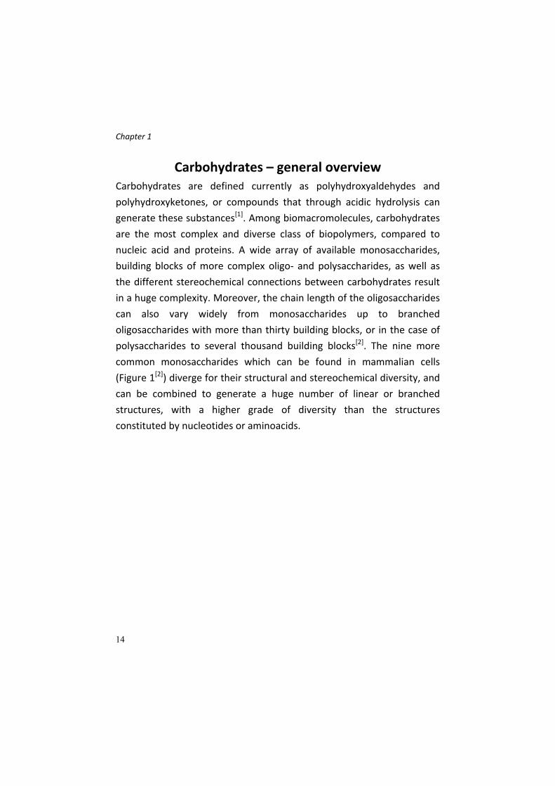
16
glycosidic bonds, which form two types of chain, amylose (a linear
polymer) and amylopectin (a branched D‐glucose polymer with both α‐
1,4 and α‐1,6‐O‐glycosidic bonds). Starch is the principal source of sugar
of human diet. As amylopectin, glycogen is a branched polymer of D‐
glucose, and is the principal storage polysaccharide in animals. Cellulose,
in which D‐glucose is linked by β‐1,4‐O‐glycosidic bonds, is a
polysaccharide with a structural function: is the constituent of plant cell
wall.
Oligosaccharides are constituted by short chains of different sugar
monomers. Oligosaccharides are often present in Nature not as free
entities but connected with other biomacromolecules, constituting
hybrid structures, called glycoconjugates. Glycoproteins, glycolipds,
peptidoglycans and lipopolisaccharides belong to this class of
biomolecules. Glycoconjugates, mainly located on the cell surface, exert
extremely important functions in living organisms, since they are
involved in many biological events and processes, like inflammation, cell‐
cell recognition, immunological response and metastasis formation
(Figure 2[3])[2].

Chapter 1
17
Figure 2 ‐ Role of oligosaccharides in adhesion and cell recognition.
Glycoproteins. Secreted proteins or proteins bound to cell membrane
are often glycosylated. The covalent attachment of an oligo‐ or
monosaccharide on a protein occurs on two possible sites: on the amidic
group of an asparagine (Asn) residue, in the case of N‐glycosilation, or on
the hydroxyl group of serine or threonine sites, for O‐glycosilated
proteins (Figure 3[3]).
Figure 3 ‐ Oligosaccharide linkages in glycoproteins.

Chapter 1
18
The glycosylation is a finely‐regulated cellular event, which involves
many different carbohydrate‐processing enzymes, located mainly in
Endoplasmic Reticulum (ER) and Golgi apparatus of eukaryotic cells.
N‐linked oligosaccharides are different among proteins, but their
biosynthesis is common in the first steps. An oligosaccharidic core made
of fourteen monosaccharide residues is synthesized on a isoprenoid
carrier molecule called dolichol phosphate, which transfers the
oligosaccharide inside the ER lumen, allowing its conjugation on a Asn
residue of a putative peptide. Once in the ER, the 14‐mer undergoes the
so called “trimming” mediated by processing enzymes, which modify the
carbohydrate structure to build different oligosaccharides depending on
the final destination of the protein (“targeting”). In Golgi apparatus, the
O‐glycosilation on Ser or Thr residues, mediated by other glycosidases
and glycosyltransferases, occurs. The conjugation of oligosaccharides on
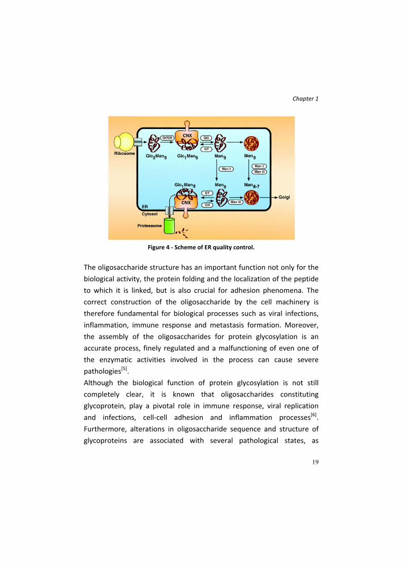
proteins in ER is related to another important biological function: the
protein folding quality control (QC) (Figure 4)[4]. The ER environment
allows proteins to acquire the correct tridimensional structure by the
action, in concert, of several proteins; this process leads to the
elimination of unfolded peptides while the proteins with a correct
structure can continue their path in Golgi apparatus. The proteins
involved in the QC are glycosidases and glycosyltransferases, that add or
remove saccharidic residues of the N‐linked oligosaccharide, and lectins,
which are carbohydrate‐binding proteins, able to recognize if a protein is
correctly folded or not.
Chapter 1
19
Figure 4 ‐ Scheme of ER quality control.
The oligosaccharide structure has an important function not only for the
biological activity, the protein folding and the localization of the peptide
to which it is linked, but is also crucial for adhesion phenomena. The
correct construction of the oligosaccharide by the cell machinery is
therefore fundamental for biological processes such as viral infections,
inflammation, immune response and metastasis formation. Moreover,
the assembly of the oligosaccharides for protein glycosylation is an
accurate process, finely regulated and a malfunctioning of even one of
the enzymatic activities involved in the process can cause severe
pathologies[5].
Although the biological function of protein glycosylation is not still
completely clear, it is known that oligosaccharides constituting
glycoprotein, play a pivotal role in immune response, viral replication
and infections, cell‐cell adhesion and inflammation processes[6].
Furthermore, alterations in oligosaccharide sequence and structure of
glycoproteins are associated with several pathological states, as

Chapter 1
20
rheumatoid arthritis and cancer; in fact, changes in oligosaccharidic
chain on the surface of cells can represent the molecular bases of the
abnormal behavior of tumor cells, allowing the tissue invasion and
metastasis formation[7].
The biological importance of glycoproteins is also proven by the their
role in the inflammation process, in which the extravasation of
leukocytes through body tissues is mediated by the interaction between
a complex oligosaccharide located on the surface of this immune cells,
called Sialyl‐Lewis X, and some proteins able to bind this saccharidic
structure (Lectins), as P‐ and E‐selectins, localized on the membrane of
endothelial cells[8] (Figure 5[9]). In healthy states, this process is finely
regulated and correctly carried out, but when it is out of control, it can
trigger excessive and chronic inflammation states, which generate severe
pathologies, such as arthritis or endotoxic and septic shocks.
Figure 5 ‐ Extravasation process of leukocytes mediated by
carbohydrates‐lectines interactions.
Selectins belong to a big class of proteins called lectins, able to interact
specifically with oligosaccharides located on the surface of almost all
eukaryotic cells; the specific interaction cause several intra‐ and

Chapter 1
21
extracellular events, and for these reasons lectins are involved in many
biological events, like cell adhesion, immune response and apoptosis.
Beside the simpler glycoproteins described above, it is possible to find
more complex structures, made of longer and bigger polysaccharides,
anchored to proteins, on the surface of cells. These conjugates are called
proteoglycans (PGs), and are made of a core protein, and several
polysaccharidic chains O‐ and/or N‐linked. The polysaccharides
constituting proteoglycans are called glycosaminoglycans (GAGs). GAGs
are repetitions of a disaccharide unit, made of a hexouronic acid and a
hexosamine. Based on the backbone structure, it is possible to divide
GAGs into four classes: HSGAGs (heparin/heparan), CSGAGs
(chondroitin/dermatan), keratan and hyaluronic acid. The biosynthesis of
these polysaccharides takes place in Golgi apparatus (for HSGAGs) where
even the O‐ or N‐glycosilation of core proteins of PGs occurs, or directly
by an integral plasma membrane proteins, for hyaluronic acid. Once the
polysaccharide structure is prepared, many successive modifications on
the single monomers constituting the chain occur: events of
epimerization, N‐deacetylation, O‐ and N‐sulphation allow to have a high
particular and modified structure, which is crucial for the biological
activity associated to these biomolecules. The extensive presence of
these glycoconjugates at the cell‐extracellular matrix (ECM) interface is a
proof of their crucial biological function. GAGs exert regulatory functions
in development, angiogenesis, cancer progression, microbial
pathogenesis and is are key factors in anticoagulation process[10].
Glycolipids. Oligosaccharides can be found also linked to lipids; among
glycolipids, the most important are glycosphingolipids, in which an
oligosaccharide (or a monosaccharide) is connected with a hydrophobic

Chapter 1
22
part, called ceramide, constituted by a long‐chain aminoalcohol
(sphingosine) and a fatty acid chain attached on the amino group.
Important glycosphingolipids are gangliosides, cerebrosides and
globosides, in which monosaccharides such as D‐glucose, D‐galactose or
N‐Acetyl‐D‐gluco/galactosamine, or oligosaccharides made of sialic acid
are linked to the ceramide. These glycolipids are localized especially in
brain and nervous tissue. These glycolipids mediate cellular recognition
mechanisms and are exploited by pathogens to enter the cells or to
target their toxins; for example, choleric toxin recognizes and binds
ganglioside GM1 provoking its entrance in the cell. Some other
glycosphingolipids are expressed in particular types of tumors, like GM2
and GD2 gangliosides expressed in breast cancer.
As for glycoproteins, the incorrect trimming of oligosaccharides
constituting the glycolipids can cause their accumulation in cellular
lysosomes, due to defects of the functioning of lysosomal glycosidases,
devoted to the degradation of this class of glycolipids. This represents
the principal cause of Lysosomal Storage Diseases (LSDs), severe
pathologies in which mutations in the aminoacidic sequences of the
carbohydrate‐processing enzymes cause defects in the structure and
function of the protein; the accumulation of glycosphingolipids (GSLs) in
lysosomes lead to important and lethal transformations in the cells. The
degradation pathway of GLSs includes diverse enzymatic activities that, if
altered, lead to different pathologies, like Fabry, Gaucher of Krabbe
disease.
The big number of structural variations which is possible to generate
with carbohydrates, considering both the different stereochemical and
regiochemical combinations, makes carbohydrates “ideal tools” to share
information between cells in living systems. Cells use carbohydrates

Chapter 1
23
located on their surface to transmit and transfer biological information,
allowing communication between different actors of immune systems.
Carbohydrates of glycolipids or glycoproteins represent epitopes
recognized by immune system cells; specific glycosylation patterns
located on the membrane of both organism’s cells and of pathogens
represent the criteria at the basis of the “self” and “non‐self” recognition
processes mediated by immune system. For example, A and B antigens
which represent the blood group determinants are the terminal portion
of glycosphingolipids located on the erythrocytes and vascular
endothelial cells membrane.
The therapeutic importance of carbohydrates: the
development of glycomimetics
The key role of carbohydrates in the life and therefore in the processes
at the basis of relevant physiopathological states, has stimulated the
scientific community towards the research of molecules and entities with
a saccharidic structure that can be used in the treatment of several
diseases, in which the role of carbohydrates is crucial. Compounds which
mimic the carbohydrate shapes and biological functions are called
glycomimetics; these bioactive class of compounds overcomes the
drawbacks of carbohydrates as such (easy degradation in physiological
conditions, difficult synthesis, preparation and purification, low activity
and drug‐like properties)[11]. On the contrary, the advantage in using
carbohydrate scaffolds for the design of potential drugs is due to the
numerous hydroxyl functionalities, that can be exploited for the
conjugation of suitable pharmacophores, taking also advantage of the
different stereochemical combinations.
Chapter 1
24
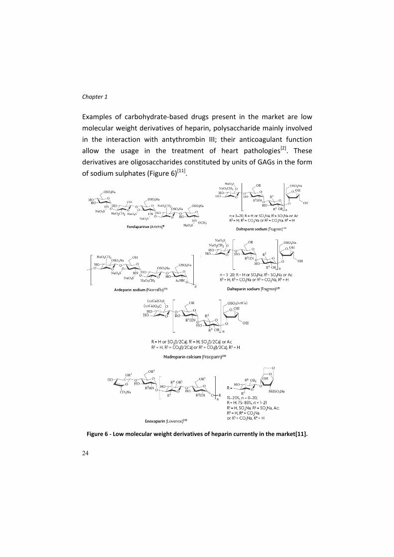
Examples of carbohydrate‐based drugs present in the market are low
molecular weight derivatives of heparin, polysaccharide mainly involved
in the interaction with antythrombin III; their anticoagulant function
allow the usage in the treatment of heart pathologies[2]. These
derivatives are oligosaccharides constituted by units of GAGs in the form
of sodium sulphates (Figure 6)[11].
Figure 6 ‐ Low molecular weight derivatives of heparin currently in the market[11].

Chapter 1
25
Another class of glycomimetics is represented by inhibitors of digestive
α‐glycosidases located on the surface of intestinal epithelium, involved in
the digestion of poly‐ and oligosaccharides, and of glycosidases involved
in the intracellular trimming and targeting of glycolipids and
glycoproteins. Inhibition of digestive α‐glycosidases has an important
role in the treatment of diabetes (as for Voglibose, Miglitol and
Acarbose), while the ability of some “inhibitors” to bind to the enzyme
active site is transformed into a chaperon activity able to rescue
misfolded enzymes in LDSs pathologies such as Gaucher’s disease
(Miglustat), a pathology related to a lysosomal accumulation of
glycosylceramides, caused by a defective form of the β‐glucosidase
enzyme (Figure 7[11]).
Figure 7 ‐ Some commercially available glycosidases inhibitors for diabetes and
Gaucher's disease.
Other carbohydrate‐based drugs available in the market are inhibitors of
viral glycosidases (sialidases), such as Zanamivir (Relenza®) and

Chapter 1
26
Oseltamivir (Tamiflu®), respectively by GlaxoSmithKline and Roche, used
as antivirals for influenza treatment. These drugs are sialidase or
neuraminidase inhibitors, viruses use these enzymes to hydrolyze units
of sialic acid (N‐Acetyl Neuramminic Acid, NANA) located on the surface
of host cells, in order to facilitate the release of new viral nanoparticles.
Zanamivir mimics the transition state of the hydrolytic process and, as
consequence, its structure is very similar to that of sialic acid.
Oseltamivir represents the attempt to make more bioavailable and more
stable this inhibitor, eliminating polar and metabolically susceptible
groups (Figure 8[12]).
Figure 8 ‐ The oxocarbenium ion intermediate of the neuraminidase reaction and two
anti‐flu drugs Zanamivir and Oseltamivir.
Topiramate (Topamax®) and Sodium Hyaluronate (Orthovisc®) represent
carbohydrate‐base drugs acting against epilepsy and osteoarthritis,
respectively (Figure 9[12]).
Figure 9 ‐ Structure of Topiramate and Sodium Hyaluronate.

Chapter 1
27
C‐glycosides as glycomimetic scaffolds
The research of novel carbohydrate‐based drugs has prompted the
attention towards the development of different saccharidic scaffolds,
with drug‐like properties, that can be suitably functionalized, decorated
and modified in order to generate bioactive compounds. There is a high
necessity and interest in the synthesis of new glycoderivatives, due to an
explosion of the glycobiology field in the last decades, potentially useful
for biological, biochemical and pharmacological studies. The field of
organic synthesis on carbohydrates is focused since long time on new
synthetic methods, and in particular on new approaches for the
formation of the glycosidic bond[13]. Despite the numerous elegant
strategies developed for the efficient formation of glycosidic linkages,
the stereoselective synthesis of α‐ and β‐glycosides remains a challenge.
Beside the more famous and well‐known O‐glycosides, a special
attention has been directed on C‐glycosides, whose definition is well
summarized in Figure 10[14]:
Figure 10 ‐ C‐glycoside definition.
C‐glycosides are important biological active compounds. Their
significance is due to the intrinsic metabolic stability, making them ideal
structures for drug candidates development[13]. Synthetic strategies and

Chapter 1
28
approaches for the construction of C‐glycosides are fewer than for the O‐
glycosidic bond; however, current methods provide the preparation of
carbon‐carbon bond via generation of nucleophile, electrophile or
radical species at the anomeric carbon of the sugar ring[13].
By a structural point of view, C‐glycosides do not differ substantially from
O‐glycosides; the substitution of exocyclic oxygen of a glycosidic bond
with a carbon atom has a marked effect on chemical character and on
stability, summarized and listed in Table 1[14]:
Table 1 – Comparison between physical and chemical properties of O‐ and C‐glycosides.
Some physical features (bond length, Van Der Waals radius, bond
rotational barrier) are quite similar between the two class of glycosides.
The most important difference is associated to the chemical reactivity: C‐
glycosides, do not present anomeric effect which, on the contrary,
characterize O‐glycosides, and furthermore C‐glycosides present a better
chemical stability both in acidic environment and towards the action of
enzymes, like glycosidases.
These reasons make C‐glycosides an interesting carbohydrate class,
especially by a chemical point of view, although these molecules are
abundantly present in Nature. It is the case of the C‐glucosidic derivative
of resorcinol or of anthraquinone (Figure 11[14‐15]).

Chapter 1
29
Figure 11 ‐ Natural C‐glycosides: on the left, the resorcinol (1,3‐dihydroxybenzene)
derivative, on the right a C‐glycoside derivative of anthraquinone.
Other C‐glycosides found in Nature possess particular and important
biological activities: examples are pirazomycin, an antiviral, and
showdomycin, an antibacterial and antitumor drug (Figure 12[14]).
Figure 12 ‐ Natural C‐glycosides with biological activity.
The discovery of natural C‐glycosides with associated biological activity
and their intrinsic stability has prompted to a high interest towards the
synthesis of new analogues of this class of carbohydrates, able to
interact and inhibit specific enzymes, to act as agonist or antagonist in
receptor‐ligands interaction phenomena and to induce an immunogenic
response[14]. Among them, we find Urdamycinone B, a carbohydrate‐
anthraquinone hybrid, able to bind to DNA (and thus cytotoxic), C‐
glycosides inhibiting specifically some glycosidases, and glycomimetics of
oligosaccharide Sialil‐Lewis X, whose interaction with Selectins mediates

Chapter 1
30
the starting point of inflammation processes and the generation of
tumor metastasis (Figure 13[14]).
Figure 13 ‐ On the left, a synthetic C‐glycoside, irreversible inhibitors of glycosidases;
on the right, the C‐disaccharide mimetic of Sialil‐Lewis X.
Nowadays, some procedures and methods for the generation of C‐
glycosides exist; Scheme 1 summarizes the strategies and synthons used
for the preparation of desired C‐glycosides, starting from aldoses or
ketoses. The carbonyl group of the open monosaccharide (A), which is in
equilibrium with the emiacetal/emiketal form (B), can undergo
transformation by species bearing nucleophilic carbon atom, or
alternatively to olefination reactions. In the first case, an open reduced
form of the sugar is obtained (C); after, this can be cyclized with the loss
of a water molecule, to get the final C‐glycoside. In the second option,
the olefination step generates an structure bearing an activated double
bond (i.e. a double bond in α‐position to an electron withdrawing group),
which is appropriate for a conjugate 1,4 addition by the free OH group of
D. The emiacetal/emiketal form can be converted into a C‐glycoside by
two strategies. The anomeric position can be activated with the
conversion of its hydroxyl group in a better leaving group (an halogen, an
imidate of a thioderivative), to generate cationic (oxocarbenium ion, F) ,
anionic (G) or radical (H) intermediates, depending on the nature of the
leaving group. The reaction of these intermediates with suitable species
bearing nucleophile, electrophile or radical carbon atoms, will generate
the C‐glycoside. The activation of the anomeric position can be also
achieved by using chemical reagents such as Lewis acids, which cause

Chapter 1
31
the direct activation of the hydroxyl anomeric group, to give the
oxonium intermediate F. The last strategy is represented by the
oxidation of the anomeric portion to the respective lactone I, which
reacting with organometallic species is transformed in the lactol L, easily
transformed in the final C‐glycosides, via the oxocarbenium ion
intermediate M, with reducing reactants.
Scheme 1 – General scheme for C‐glycoside synthesis.

Chapter 1
32
Role of carbohydrates and glycomimetics in anti‐
inflammatory processes: inflammation and SGLT1
Inflammation, sepsis and septic shock
Sepsis describe a complex clinical syndrome, caused by a harmful
response that a host activates as a result of an infection. This response is
characterized by a hyperactivation and deregulation of the mechanisms
which lead the immune system to react against an invasion caused by an
external pathogen[16]. From a clinical point of view, sepsis appears with
fever, mental confusion, transient hypotension, thrombocytopenia and
diminished urine output. If not treated, the patient can undergo to
irreversible damages to the respiratory and renal systems, abnormal
blood coagulation and deep hypotension. One of the main features of
this pathology is the high rate of death, about 30% of the patients, and is
close to 50% in patients which possess the more severe and acute septic
shock. Sepsis is defined as the systemic response to an infections[17], and
should be distinct for other pathological states, as:
SIRS (Systemic Inflammatory Response Syndrome), which involves
the whole body; non necessary it derives from a microbial infections,
but presents similar syndromes to sepsis (body temperature lower
than 36°C or higher 38°C, heart frequency of 90 bpm, increase
respiratory frequency and profound alteration of blood white cells);
Bacteremia and septicemia: presence of bacteria in the blood.
Sepsis and septic shock: are the most severe forms of sepsis,
characterized by a accentuated hypotension, vascular
hypoperfusion, and several body organ failures.
Multi‐organ failure: the most critical state, with seriously altered
body functions which do not permit an internal homeostasis
maintenance without external actions[17].

Chapter 1
33
The most common sites of infection that lead to such types of response
are the respiratory apparatus, the abdominal cavity, the urinary tract
and blood. The diagnosis of microbiological type characterized a half of
the patient cases and in particular is due to Gram negative infections.
For this reason, the study of the structural components of bacteria which
cause sepsis in an important scientific field, to understand the
mechanism responsible of the disease and to identify potential
therapeutic targets. These components, recognized by immune systems,
belong in the so called Pathogen‐Associated Molecular Patterns
(PAMPs)[16]. In Gram negative bacteria, Lipopolysaccharides (LPSs) have a
predominant role. The external membrane of Gram negative bacteria is
structured in an asymmetric manner, and consist of phospholipids in the
internal section, and of glycolipid anchors of LPS in the external side
(Figure 14[18] [19]).
Figure 14 ‐ Structure of Gram negative bacteria envelope (left) and LPS structure (right). Bacteria LPS are constituted by a glycolipids portion, called Lipid A, an oligosaccharidic core and a more external section, the O‐antigen.
Chapter 1
34
In Gram positive bacteria, the structural determinants for sepsis and in
general inflammation states are structures contained in the cell wall, like
peptidoglycans and lipoteichoic acid. Molecules causing inflammation
states which remains anchored to the cell are called endotoxins, while
compounds that are produced and secreted on the external side of cells
are exotoxins, produced especially by Gram positive bacteria. These type
of toxins have a great interest because are able to bind the Class II Major
Histocompatibility Complex (MHC‐II) and to some T‐lymphocyte
receptors, triggering a massive activation of these cells and a strong
release of pro‐inflammatory cytokines[20]. Not only bacterial
glycoproteins or glycolipids show pro‐inflammatory activities: flagellin, a
protein located on the cell wall of flagellated bacteria, or non‐
methylated CpG sequence of naked bacterial DNA, are able to bind
specific host receptors, belonging the Toll‐like receptor family (TLR), as in
the case of LPSs. TLRs play a key role in the recognition of structures of
microbial or pathogenic origin; the interaction between these receptors
and the respective pro‐inflammatory ligands is at the basis both of the
recognition of pathogens which come in contact with host defense
barriers, to produce an adequate immune response (“non‐self”), and of
the recognition of all commensal bacteria agents which constitute the
intestinal flora and belong to the “self”(Figure 15)[19].

Chapter 1
35
Figure 15 ‐ The complexity of interaction between bacteria determinants and immune
system.
TLRs family is constituted by several members, each one responsible for
the recognition of diverse bacterial, viruses and fungi structural
elements. As depicted in Figure 15, the receptor involved in LPSs
recognition is TLR4; TLR2 recognizes structure of Gram positive cells,
TLR5 is the receptor for flagellin and TLR9 binds CpG elements of
bacteria DNA[21]. In these years, several studies allowed a deep
comprehension of the mechanism at the basis of these specific
inflammatory responses, and nowadays it is clear that the process is to
be ascribed not only to TLRs but to a more extended protein complex,
which can be defined as “LPS Receptor”. Several protein actors are
involved, as LPS binding protein, that binds LPS and presents it to other
proteins located on the cell membrane: TLR4, CD14 and MD2. The
association of all these proteins with LPS forms a protein complex which
allows the activation of signal cascades in the cell and the beginning of

Chapter 1
36
the inflammatory response. Cells which express this complex are
members of immune system, like macrophages, dendritic cells but also
intestinal epithelial cells (IECs), which are always involved in the external
environment recognition. The activation of a particular signal cascade
transduction mediated by several intracellular proteins leads definitively
to the nuclear translocation of the transcriptional factor NFkB, event that
increases the expression of pro‐inflammatory cytokines, like IL‐1 or
TNFα[16]. These cytokines are the principal pro‐inflammatory molecules
secreted after the signal cascade transduction, released within 30‐90
minutes after LPS exposure. Successively, they mediate a second level of
inflammatory cascade, in which new cytokines, lipid mediators and
Reactive Oxygen Species (ROS) are synthesized. The intense pro‐
inflammatory response determining the sepsis state is balanced by a
complex of counter‐regulatory molecules which maintain an
immunological equilibrium[16]. These anti‐inflammatory modulators are
antagonist like the TNF soluble receptor, some complement inactivator
proteins or anti‐inflammatory cytokines like IL‐10.
Role of intestinal mucosal cells. Intestinal mucosa represents a body
district where several function are intertwined: digestion of food
ingested with the diet and substances absorption are the activities which
characterize this organ, but a fundamental role is represented by the
first real barrier that the body has against external environment.
Intestinal epithelium is not only a physical barrier against pathogens, but
plays actively in immune and inflammatory response[22]. Intestinal
mucosa has to recognize immediately eventual pathogenic threats
present in the intestinal lumen, and, as a consequence, has to control
the immune response against these agents. But intestine has to maintain
also the hyporesponsivity towards the pool of commensal

Chapter 1
37
microorganisms living in the intestinal lumen. Intestinal epithelial cells
are able to discriminate between the “non‐self” (the pathogens) and
“self” (the own bacterial flora). For intestinal cells, as well as for immune
system actors, the recognition of pathogen structure (PAMPs) is
mediated by the Pattern Recognition Receptors (PRRs), in particular by
TLRs. For this reasons, it is possible to define the intestinal epithelial cells
as “sensors” of external environment, and, if activated, can address the
development of the inflammatory response, not only recruiting the
competent immune cells, but directly influencing their action[22]. TLRs
are widely expressed in different cellular types of gastrointestinal
mucosa (stomach epithelial cells, small intestine, colon, macrophages,
monocytes, intestinal dendritic cells). The discrimination between self
and non‐self, realized by TLRs, probably is due to expression level of
these proteins on the membrane of IECs: in healthy tissues, a low
expression level of TLR2 and 4 is present, in order to minimize the
intestinal commensal bacteria recognition. On the contrary, TLR4 is
highly expressed in the IECs when pathological condition occurs,
determined by chronic inflammation states, like Chron disease or
ulcerative colitis. Several molecular mechanisms allow the tolerance of
IECs towards bacterial flora: a reduced expression of TLRs, an increase
level of TLR4 signal transduction cascade suppressors or production of
external regulators which suppress the TLR4‐mediated pathway. With
such mechanism, enterocytes maintain a hyporesponsivity towards
commensal bacteria, due to a constant basal level of activation of pro‐
inflammatory pathways. The action of the different TLRs expressed on
the surface of intestinal cells is the same shown by other innate immune
system cells; in particular, TLR4 is responsible, also in this apparatus, for
the recognition of LPS associated to Gram negative bacteria present in
the intestinal lumen. The subcellular localization of the different TLRs, in

Chapter 1
38
particular TLR4, has a key role in the creation of the responsiveness of
IECs towards pathogens; in fact, TLRs are strategically localized both on
the cellular surface and in intracellular compartments. TLR2 and TLR4 are
located mainly on apical membrane of enterocytes (that constitutes the
so called brush border membrane, made of numerous evaginations of
villi, fundamental to increase the absorption surface of nutrients)[23]. In
this manner, IECs can monitor bacteria presence in gut lumen. Other
TLRs are localized at intracellular level (as TLR9, which recognize CpG
DNA sequences) or on basolateral membrane of enterocytes (the portion
of plasma membrane faced towards the internal side of intestinal villus,
like TLR5[23]).
When genetic dysfunctions associated with the action of TLRs occur,
inflammatory process can become chronic, leading to inflammatory
intestinal diseases, called IBDs (Inflammatory Bowel Diseases)[24]. These
pathologies are characterized by a constant and deregulated response
towards commensal bacteria. The etiological causes are multiple, but
genetic predisposition is fundamental[24]. In pathological situations, like
IBDs, the surface expression of TLRs is increased, leading to a
hypersensitivity of IECs even towards the bacterial flora.
One of the most important effect which is possible to observe in
inflammation and sepsis states is the LPS‐TLR4 mediated activation of
signal transduction cascades that lead to cell apoptosis of enterocytes.
Apoptosis is a common and physiological phenomenon in the intestine,
and is associated with the high turnover of the IECs, to maintain a
correct balance of cell proliferation and death and then to a correct
homeostasis. The physiological extrusion of enterocytes from intestinal
villi do not compromise the protective function of intestinal mucosa,
which is lower in some circumstances (excessive exposure to pathogens
and cytotoxic agents)[25]. When this exposure occurs, the apoptosis

Chapter 1
39
phenomena increase, thus leading to a diminished mucosal integrity,
with negative consequences on the protective function of this apparatus.
Cells which undergo this process suffer from sensible morphological
changes, as dimension reduction, mitochondrial swelling, plasma
membrane alterations and nuclear fragmentation[26].
Role of glucose in the protection of inflammation events. Recent
works[25‐26] [22] have demonstrated that high exogenous glucose
concentrations, administered in vitro to IECs, are able to protect these
cells from damages caused by pathogens expressing LPSs. These works
have proposed that the mechanism of glucose‐mediated cytoprotection
depends on an increased glucose uptake, by enterocytes, mediated by
Sodium‐Glucose Co‐Transporter 1 (SGLT1). This protein represents the
principal way with which the intestinal epithelium absorbs glucose, and
it’s localized mainly on the brush border membrane. SGLT1 mediates the
unidirectional transport of glucose from intestinal lumen to the internal
side of epithelial cell. Further information and details on the structure
and function of this transporter are described afterwards.
Previously, it has been mentioned that enteric bacteria can be indirectly
considered as responsible for the pathogenesis of the IBDs. However, a
high number of cells of Enterobacteriaceae, Proteobacteria and
Bacteroids genera are often identified in ileal mucose of patients with
Chron disease[26]. An augmented intestinal bacterial load leads inevitably
to a higher exposure of intestinal mucosa towards bacterial pathogenic
structures, in particular LPSs. The consequent enterocyte‐mediated
inflammatory response produce a vast damage to the mucose,
characterized by apoptotic phenomena and injury on thight juctions,
present between mucosa cells, fundamental for the homeostatic
equilibrium and immunological protection. In particular, thight junction

Chapter 1
40
damages lead to a higher mucose permeability, since frequently in
patients affected by Chron disease, LPS or anti‐endotoxin antibodies are
found in blood plasma[26]. For the capacity to generate apoptotic event in
epithelial cells, LPSs are used in studies in which the cytoprotective
function, SGLT1‐mediated, of high glucose concentration is evaluated[25‐
26] [22]. Preliminary studies performed by Buret et al.[26] have allowed to
verify the mechanisms which triggers the apoptotic phenomena in IECs,
is mediated by the interaction of bacterial LPS and TLR4. The results have
demonstrated a participation of the mitochondrial pathway in the
apoptotic mechanism, and that a higher glucose uptake can modify the
apoptotic level cause by LPS. The increased uptake is due to a high
glucose level located on the external of these cells, in which the higher
glucose absorption is mediated by SGLT1. The higher SGLT1‐mediated
glucose uptake provokes profound changes of apoptotic intracellular
signaling, thus leading to a cytoprotective effect against LPSs exposure.
SGLT1 role was also confirmed testing the cytoprotective ability of
glucose on epithelial cells in the presence of phlorizin, a SGLT1 specific
inhibitor[27]. This molecule inhibits the glucose uptake of almost 60%,
thus suppressing the glucose‐mediated cytoprotection. Successive
studies[25] allowed to understand that the change of glucose uptake in
protective effect phenomena leads to changes in the production of
proinflammatory cyto‐ and chemokines. Furthermore, the higher uptake
is directly associated to an increase of specific activity of SGLT1. The
augmented transport activity depends exclusively on Vmax increase and
not on the affinity constant for glucose. This means higher expression
membrane level for this transporter. The higher expression levels are
due to higher translocation level from cytoplasm to the membrane,
mediated by cytoskeleton structures, and not due to increasing
intracellular concentration of SGLT1. The translocation of SGLT1 on

Chapter 1
41
apical membrane of enterocytes is a finely regulated process, in which
growth factors as EGF (epidermal growth factor) and IGF (insulin growth
factor) are involved[28] [29].
Further studies were performed by the research group of Prof. C.
Rumio[22], regarding the protective effect that SGLT1 activation has on
IECs, to which bacterial LPS were administered. In this case the attention
was focused to anti‐inflammatory and not anti‐apoptotic ability of high
glucose concentrations, able to activate SGLT1, blocking the intracellular
signaling that leads to pro‐inflammatory mediators production. The
activation of apoptotic pathway requires higher LPS concentrations (> 10
µg/mL) than those necessary for the pro‐inflammatory response (from
200 ng/mL to 10 µg/mL)[26]. ELISA test performed on HT29 (human
adenocarcinoma cells) insulted with LPS or CpG‐ODN (TRL9 ligand),
revealed that high glucose concentrations (5 g/L) can reduce the IL‐8
production, a marker of inflammatory states (Figure 16[22]).
Figure 16 ‐ IL‐8 production levels of HT29 cells treated with LPS or CpG‐ODN and/or
glucose (5 g/L).
To prove the role of SGLT1 in this phenomenon, its action was blocked
both silencing its gene (with the siRNA technique) or inhibiting (with
phlorizin) the transporter. In both cases, HT29 cells treated with pro‐

Chapter 1
42
inflammatory molecules and exposed to glucose, produce IL‐8 with level
comparable to that secreted in with normal glucose concentrations
(Figure 17[22]).
Figure 17 ‐ Levels of IL‐8 produced by HT29 cells, treated with LPS or Cpg‐ODN, in
presence of phlorizin (left) and silencing SGLT1 (right).
Similar experiments and results were obtained using a non‐
metabolizable glucose derivative, 3‐O‐Me‐D‐glucopyranose (3OMG), in
order to exclude any possible metabolic causes of the higher glucose
uptake. The anti‐inflammatory effect of glucose was observed also in
vivo, treating mice orally with 2,5 g/kg of D‐glucose or 3OMG, and
insulting with LPS or Cpg‐ODN. Serum levels of different chemokines
were lower in the presence of glucose or its derivative, and mice treated
with LPS, D‐Galactosamine (murine model of septick shock) and D‐
glucose showed a complete level of survival, compared to mice which
were not treated with high glucose. Also with in vivo test the SGLT1 role
was proven: only the oral administration of glucose and phlorizin (hence
the presence of these two molecules in the gut lumen) led to the same
results obtained in vitro. With intraperitoneal administration of glucose,

Chapter 1
43
no protective effect was observed, and no blocking of anti‐inflammatory
response was noticed with phlorizin administration. In conclusion,
experimental data clearly indicate that glucose, at high concentration,
administered in vitro or orally in vivo is able to:
guarantee a cytoprotection on IECs from possible apoptotic
events provoked, in patients affected by IBDs, by hypersensitivity
to commensal bacterial agents located normally in the gut, which
cause is principally due to altered level of expression of TLRs on
the enterocyte surface;
reduce or stop the systemic inflammatory response triggered by
LPS or PAMPs which, interacting with TLRs, lead to the expression
of genes codifying for pro‐inflammatory modulators.
In both cases, a pivotal function is carried out by SGLT1, whose activation
due to the high glucose concentration allows the beginning of the
protective phenomena against apoptosis, inflammation and septic shock.
SGLT1 is therefore not only devoted to the nutrient absorption, and in
particular sugars, but it shows also a key role under an immunological
point of view, actively participating to processes at the base of the
protective function of intestinal epithelium against external
environment.
SGLT1: function and structure
Digestion and absorption of carbohydrates assumed with the diet is a
process that involves all the gastrointestinal system of mammalian,
starting from the mouth, the first body district, where saliva amylases
begin the digestion of amylopectin and amylose chains of starch. Then
the digestion, stopped in stomach by gastric juices, starts again in
duodenum by pancreatic amylases which break down starch chains into
maltose and isomaltose, glucose dimers. The last phase of the digestion

Chapter 1
44
is localized at the intestinal level, where maltases secreted by epithelial
cells of microvilli hydrolize the dimeric units in single glucose molecules.
Other disaccharides, like lactose or sucrose, are hydrolyzed in single
monosaccharides (glucose, fructose and galactose) by the action of other
glycosidases (β‐galactosidase o lactase and saccharase or invertase).
Glucose plays a central role in energetic metabolism: an adult man uses
about 250 g of glucose every day to perform all the body functions (half
from the diet, half from glycogen or gluconeogenesis)[30]. The most
difficult task is the maintenance of the sugar concentration in the blood
(80 – 120 mg/dL) to ensure a constant and continue supply of glucose for
the brain[31]. But surprisingly, glucose is not an essential component of
the human diet: this observation is confirmed by cases of individuals
intolerant to this monosaccharide, as for patients affected by the
glucose‐galactose malabsorption disease (GGM), which can live normally
without glucose ingestion.
The carbohydrate absorption occurs in the intestine after a long phase of
digestion; the polarity of these molecules avoids their free diffusion
across the plasma membrane. The absorption of sugars requires the
existence of specific transport proteins localized both on the apical
membrane of enterocytes, which constitute the brush border membrane
(BBM) and on basolateral membrane, structure that separates these
cells from blood flux. The two localization reflects the two steps required
for sugar absorption: at first, there is an accumulation of sugars in the
BBM, then the translocation in the blood occurs[30, 32]. In both processes
specific proteins are required; these transporters are the GLUT proteins
(gene family SLC2) and the sodium‐glucose co‐transporters, SGLTs (gene
family SLC5). These proteins are located mainly in the intestine, but are
also involved in the sugar reuptake in kidneys from glomerular filtrate
and in the glucose uptake across the Blood Brain Barrier (BBB)[33]. GLUT

Chapter 1
45
transporters are devoted to the passive translocation of sugars, without
energy consumption; examples are GLUT1 (in BBB) and GLUT4 (for
insulin‐dependent glucose uptake)[30]. SGLTs protein carry out an active
transport of sugars across the membrane, i.e. with energy consumption.
SGLTs constitute a protein family of 11 members in human, whose genes
are identified by the genes SLC5. SGLTs belong to a bigger class of
transporters, the SSSFs (Sodium Substrate Symporter Family). These
proteins, both in eukaryotic and prokaryotic cells, carry out the transport
of different solutes coupling it with the transport, according to their
electrochemical gradient, of different ions, such as sodium or protons.
The flux of ions is produced by the presence of an electrochemical
gradient across the two sides of the membrane (SMF, Sodium Motive
Force). This allows the accumulation of energy to pump the solutes
inside the cell, acting against their gradient. The sodium gradient is
generated by sodium/potassium‐ATPase pump (Na+/K+‐ATPases) that
continuously translocates sodium at the external side of the cell, across
the basolateral membrane of the cell. For this reason, member of SSSF
family are called active secondary transporters because the translocation
of solutes is not matched directly with the energy consumption (ATP), as
on the contrary happens for Na+/K+‐ATPase pumps. In the co‐transport,
the energy necessary for the solute translocation is produced with other
processes, in the form of an electrochemical gradient[33]. The direction
and the rate of the glucose transport is a function of the direction and
intensity of the sodium gradient across the apical membrane[30].
SSSF transporters are grouped in different subfamilies, according to
sequence similarity[34]. Over two hundred members of this family exist,
both of prokaryotic and eukaryotic origin. From a functional point of
view, we can distinguish some subclasses: transporters of sodium‐
glucose (human SGLTs), sodium‐aminoacids (like PutP of E. coli, a

Chapter 1
46
sodium‐proline transporter), sodium‐vitamins (SMVT of H. sapiens) and
sodium‐ion (NIS, sodium‐iodide contransporter of E. coli).
In human genome, 11 members of the subfamily of SLC have been
identified (Table 2); among them, the most important is certainly SGLT1
(SLC5A1), mainly expressed in BBM of IECs, in a smaller part in proximal
tubules of kidneys, where it is involved in the sugars reuptake from the
glomerular filtrate, work principally carried out by SGLT2. SGLT1 is the
most studied member of this family, because mutations in its gene are
associated with a pathology called Glucose‐Galactose Malabsorption
(GGM)[35].
Table 2 ‐ Members of human genes family SLC5.
Human
gene
Protein
name
Main
substrates Tissue distribution
Gene
locus
SLC5A1 SGLT1 Glucose,
Galactose
Small intestine>>Kidneys,
heart 22q13.1
SLC5A2 SGLT2 Glucose Kidneys, Heart, Liver, Thyroid,
Muscles, Brain
16p12‐
p11
SLC5A3 SMIT Myo‐inositol>>
Glucose Brain, heart, kidneys e lungs 21q22.12
SLC5A4 SGLT3 Sodium (H+)
Small intestine, skeletric
muscles, kydneys
21q22.2‐
q12.3
SLC5A5 NIS I‐(ClO4
‐, SCN
‐,
NO3‐, Br
‐)
Thyroid, colon, ovaries 19p13.2‐
p12
SLC5A6 SMVT Biotin, lipoate,
pantothenate Brain, Heart, Kidneys, Lungs 2p23
SLC5A7 CHT Choline Spinal cord 2q12
SLC5A8 SGLT4 Glucose,
Mannose
Small intestine, Kidneys, Liver,
Lungs and brain 1p32
SLC5A9 SGLT5 ? Kidneys 17p11.2
SLC5A10 SGLT6 Myo‐inositol,
glucose
Small intestine, Brain, Kidneys,
Liver, Heart and Lungs 16p12.1
SLC5A11 AIT Iodine Thyroid 12q23.1

Chapter 1
47
SGLT1 and sugar absorption. SGLT1 mediates the glucose (Glc) and
galactose (Gal) absorption in the intestine, with their translocation
across the brush border membrane of intestinal villi; the required energy
is given by the sodium electrochemical gradient generated by the
Na+/K+‐ATPase pump of the basolateral membrane of the same cells. A
gradient of sodium between the intestinal lumen (external side) and
cytoplasm (internal side) is created and this allows the symport of
sodium and glucose or galactose inside the cell (Figure 18a[3]). This
process shows a precise stoichiometry; for every molecule of Glc or Gal,
two sodium ions are translocated inside the IEC. Experimental evidences
indicate that the co‐transport seems to be completely reversible,
depending exclusively on the direction of the sodium electrochemical
gradient and of the monosaccharides[36]. In Figure 18 the successive
passage of the two sugars into the blood stream, mediated by GLUT
transporters, is depicted. However, some studies suggest an alternative
pathway for this second step, in which glucose is translocated across the
basolateral membrane by an exocytosis mechanism (Figure 18b[33]).
a)

Chapter 1
48
b)
Figure 18 ‐ Glucose absorption in intestinal epithelial cells.
It is well known that glucose entry, mediated by enterocytes, leads to a
high rate of water absorption in the intestine. This is due by the action of
SGLT1, responsible for sodium absorption, which inevitably allows the
translocation of other anions (Cl‐ and HCO3‐) inside the cell. By this point
of view, SGLT1 can be seen as an important vehicle of water absorption
at intestinal level[33]. For each glucose or galactose, two sodium and 260
water molecules enter in the cell. This feature is at the basis of an
important therapy of the 20th century, the Oral Rehydration Therapy
(ORT), fundamental for the treatment of acute diarrhea[37]. The
introduction of this therapy have considerably lowered the number of
death among children affected by acute diarrhea (60% from 1980 to
2000). ORT is simple, cheap and often associated with cholera therapies,
because this pathology leads to a strong loss of liquids, that can be
balanced with the oral administration of salts and glucose solutions. In
this way, the glucose assumption stimulates the SGLT1‐mediated sodium
and water uptake by IECs (for each glucose mole, 2 moles of NaCl and 4 –
6 L of water are co‐transported)[38].

Chapter 1
49
On the contrary, SGLT1 is also the cause of a pathology called GGM
(glucose‐galactose malabsorption, OMIM 182380). A defective form of
SGLT1, or its absence in the intestinal epithelium, due to mutations in
the respective gene, prevents the absorption of the two
monosaccharides. Diarrhea is the principal symptom, that ceases when
the assumption of the two sugars is interrupted. No defects are present
in patients with this pathology in the absorption of fructose, since its
transport in the intestine is mediated by GLUT5, localized in the apical
membrane of enterocytes[33].
Furthermore, SGLT1 represents a target for type II diabetes, since several
studies indicate that its expression levels (together to GLUT5) are three‐
four fold higher in such patients compared to healthy individuals[39]. This
leads inevitably to an increased capacity of glucose and fructose
absorption by intestinal epithelial cells. A plethora of scientific works aim
to the synthesis and development of SGLT1 inhibitors, able to reduce
glucose absorption; in Nature, a SGLT1 inhibitor still exists, phlorizin, a β‐
glucosides present in a variety of fruits (cherries, apples, service tree).
Phlorizin is a member of the class called chalcones, and is constituted by
a hydrophobic unit of two aromatic rings, linked to the glucose by a β‐
glucosidic bond. This molecule has a strong affinity and inhibition
constant for SGLT1[40].
Structural features of SGLT1. The structure of SGLT1 has been the
subject of numerous studies within the last fifteen years, and the
knowledge about it are due in particular to the works of Rolf K. H. Kinne
and Ernst M. Wright. The human isoform of SGLT1 (hSGLT1) (UniProt
entry P13866) is made of 664 aminoacids, with a total molecular weight
of 75 KDa[32] [41]. The tridimensional structure of the protein has not been
resolved yet, but recently the tertiary structure, from X‐ray diffraction

Chapter 1
50
data, of the variant of Vibrio parahemoliticus is available[42]. To date, the
knowledge on the secondary structure of the transporter and on the
presence and localization of ligands bindig sites (monosaccharides,
sodium, or inhibitors like phlorizin) are discordant, since there are
differences between the experimental methods used and, moreover, an
intrinsic difficulty in the structural studies of transmembrane protein
exists. SGLT1 is structured in 14 α‐helix transmembrane domains, as for
all the SSSF members[33, 43]. The N‐terminal hydrophilic terminus is
localized in the extracellular side, whereas for the C‐terminus the
localization is still uncertain, although many studies confirm the
extracellular hypothesis[43]. In the N‐terminus some N‐glycosilation sites
are present, like Asn248[44], probably necessary for the membrane
targeting of the protein; however, the glycosylation is not strictly
required for the transport activity[43]. In the figure below (Figure 19[33]), a
topological scheme of the structure of SGLT1 is present.
Figure 19 ‐ Secondary structure model for SGLT1. The 664 aminacids constituting the transporter are disposed on 14 transmembrane α‐helices. In this model, both N‐ and C‐terminus are located extracellularly. In green the oligosaccharide which represents the glycosylation, on N248 residue. In yellow, residues which sharing the same architecture among SSSF members, which also show a consensus sequence, in violet. In red the consensus sequence between SGLT1 and SMIT (Sodium‐MyoInositol coTransporter) is highlighted.

Chapter 1
51
The presented model is the most accredited, although a first model,
based on the gene structure of SGLT1, provided a cytoplasmic
localization of the C‐terminal extremity, and even a topology of 12
transmembrane α‐helix[45].
Concerning the ligands binding sites (for glucose/galactose, sodium,
phlorizin), it is now clear that the N‐terminus is involved in sodium
binding, where the role of A166 residue seems to be fundamental[46].
The C‐terminal extremity participates in the binding and transport of
sugars and/or inhibitors[47] [48]. The role, structure and localization of the
C‐terminus are the aspect towards which most of the studies have been
focused in the last years. One of the most critical and intriguing aspect is
the localization of the site/s of interactions of the two monosaccharides
and inhibitors. Initially, the hypothesis provided a unique binding site;
recently, several studies clarified the existence of at least two regions,
one involved in initial binding (first step), directed extracellularly, and
the second responsible for the real translocation inside the cell
(intracellular site)[49]. A recent work has allowed to identify a precise
localization for the two binding sites on the C‐terminal portion[50].
Previously, it was possible to reduce the area related to the glucose
binding site: several works established an important role for loop 13, an
unfolded region between the α‐helix TM13 and TM14 (aa 541‐638),
which is involved in the binding with inhibitor phlorizin[27] and
alkylglucosides[51]. It has been hypothesized a direct role for this loop in
the binding with glucose, too; this portion was isolated, immobilized on a
lipid bilayer, in order to study its interaction features[50]. Exploiting SPR
(Surface Plasmon Resonance) analysis, it was understood that loop 13
contains both a non‐specific sugar interaction site, in its early portion (aa
548‐571), probably located intracellularly, and a stereospecific region, in
terminal part (aa 622 – 633), extracellular. This study has proposed
Chapter 1
52
indirectly a double localization for the loop 13, which seems to be free to
cross the plasma membrane. According to author’s hypothesis, the two
regions of loop 13 allow to have a multistep recognition and binding
process; the initial stereospecific interaction, mediated by late loop 13,
discriminates between D‐Glc and other non‐transported
monosaccharides (as L‐Glc), while the intracellular interaction is a second
binding step, which allows the translocation of the sugar inside the cell.
A further work indicates the existence of a sodium‐independent glucose
translocation[52]. This uniport has a low affinity and is not inhibited by
phlorizin, which interaction was confirmed to occur extracellularly[53]. Is
therefore possible to conclude that two binding sites for glucose or
galactose exist: one with high affinity (KD 0,5 mM) and one with low
affinity (KD 3 – 4 mM). Fuorescence studies indicate that sugars bind to
this site with a stacking interaction between their pyranose ring and
indol ring of Trp residue in position 561. This residue is located in the
early portion of loop 13 which localizes intracellularly, associated with
the non‐stereospecific interaction. Therefore, accordingly with all these
results, it is possible to hypothesize that the high affinity binding site is
involved in stereospecific recognition of sugars, whereas the low affinity
site is intracellular and mediates the sodium‐dependent translocation of
Glc or Gal. Recently, new insights for loop 13 localization have been
proposed: it could behave as a re‐entrant mobile loop, during binding
and translocation events[54]. This idea could lead to solve many disputes
about its nature.
Structural determinants for ligands binding. Many works led to
understand which SGLT1 residues interact with functional groups on
transported monosaccharides or inhibitors, like phlorizin. Figure 20[55]
shows a model for the interactions between hydroxyl groups of glucose

Chapter 1
53
and side chains of aminoacids which constitute the binding domain for
sugars. This region is localized, as mentioned, between α‐helix TM X and
XIV. After the binding with two sodium ions, in the N‐terminal region of
the protein, conformational changes occur, thus allowing the exposure
of the glucose binding site towards the extracellular side. A hydrophilic
pocket, constituted by Q457, T460, Q445 and R499 is formed[55]. These
residues form dipolar interactions and hydrogen bonds with OH groups
of sugars.
Figure 20 ‐ Model of sugar binding domain of SGLT1.
Among the residue involved in glucose/galactose binding, the most
important one is certainly Gln457: several mutation experiments of this
aminoacid show a dramatic loss of affinity for glucose binding[56].
Moreover, Q457R is one of the more relevant genetic determinants of
the glucose galactose malabsorption, because this mutation avoids
completely the sugar absorption in the intestine.
Studies carried out with other ligands, as the inhibitor phlorizin, allowed
to have a more precise interaction model of SGLT1. Phlorizin binds to the
same region involved in glucose interaction, with a two‐step process: the
first one represents the rapid formation of a complex, while in the

Chapter 1
54
second step a slow isomerization of the inhibitor occurs, causing the
occlusion of the glucoside inside the binding site[53]. The aromatic
portion of phlorizin (aglycone) binds to a region of loop 13 comprises
between aminoacid 604 and 610, where, accordingly to this model,
stacking interactions between the aromatic rings of the inhibitor and
Phe602 and Phe609 residues occurs (Figure 21)[53].
Figure 21 ‐ Principal interaction between phlorizin and hSGLT1.
Thanks to these studies, it has been possible to understand the
structural determinants for ligand interaction with SGLT1, and in
particular which functional groups of ligands are critical for the binding
with the C‐terminal domain of SGLT1[53] [55]. First, the hydroxyl group in
position C‐2 of the two monosaccharides is crucial for the binding, with a
compulsory equatorial configuration (D‐mannose, epimer for C‐2
position, in neither recognized nor transported by SGLT1). C‐3 hydroxyl
group is important, since SGLT1 has a low affinity for the
monosaccharide with the opposite configuration on this carbon, D‐allose,
but shows a hydrogen bond acceptor rather than donor, since 3‐OMe‐D‐
Glc, a non‐metabolizable analogue, is efficiently transported[22]. Hydroxyl
group in position C‐4 has an important role but its configuration is non‐
critical, since both Glc and Gal, epimers for this position, are
translocated, with a very similar affinity constant. Substituting this OH
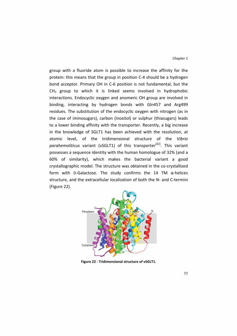
Chapter 1
55
group with a fluoride atom is possible to increase the affinity for the
protein: this means that the group in position C‐4 should be a hydrogen
bond acceptor. Primary OH in C‐6 position is not fundamental, but the
CH2 group to which it is linked seems involved in hydrophobic
interactions. Endocyclic oxygen and anomeric OH group are involved in
binding, interacting by hydrogen bonds with Gln457 and Arg499
residues. The substitution of the endocyclic oxygen with nitrogen (as in
the case of iminosugars), carbon (inositol) or sulphur (thiasugars) leads
to a lower binding affinity with the transporter. Recently, a big increase
in the knowledge of SGLT1 has been achieved with the resolution, at
atomic level, of the tridimensional structure of the Vibrio
parahemoliticus variant (vSGLT1) of this transporter[42]. This variant
possesses a sequence identity with the human homologue of 32% (and a
60% of similarity), which makes the bacterial variant a good
crystallographic model. The structure was obtained in the co‐crystallized
form with D‐Galactose. The study confirms the 14 TM α‐helices
structure, and the extracellular localization of both the N‐ and C‐termini
(Figure 22).
Figure 22 ‐ Tridimensional structure of vSGLT1.

Chapter 1
56
The galactose binding site is comprised between two groups of
hydrophobic residues which form two barriers, one intracellular and one
extracellular. These portions control the ligand transport. The
extracellular barrier is made of tyrosine, metionine and phenylalanine
residues which block the ligand, avoiding its leakage in the extracellular
space. The intracellular gate is represented by a tyrosine residue in
position 263, involved in stacking interactions with the pyranose ring of
the sugar. Analyzing the residues which surround the sugar (Figure 23), it
is possible to observe that there is a high similarity between them and
the groups described for the human variant, according to the available
models in literature.
Figure 23 ‐ Galactose binding site, from the extracellular side, obtained removing the
residues constituting the extracellular gate.
The identification of the sodium binding site has required a comparison
of the vSGLT1 with the model of LeuT, a bacterial transporter for proline.
This site has been localized between α‐helices TM 2 and 9, 10 Å far from
glucose binding site. Furthermore, a functional model for this protein has
been proposed. According with this work, the transporter alternates
between two conformation, one extracellular‐oriented, which allows the
sugar binding, and one directed intracellularly, for the Glc/Gal release.

Chapter 1
57
Transport kinetics of Glucose and Galactose. Transport mechanism of
glucose mediated by SGLT1 has been deeply studied and modeled in the
last years, thanks to the cloning and the expression of the transporter in
Xenopus oocytes and exploiting electrophysiological techniques, which
allow the measurement of the currents associated to the sugar
transport. In fact, SGLT1 is an electrogenic protein, since the sugar
transport is sodium‐mediates in physiological conditions[32]. The co‐
transport is achieved by alternating access mechanism, which provides
conformational changes induced by ligands. The overall mechanism was
simplified in a six‐state model, depicted in Figure 24[31].
Figure 24 ‐ Six‐state kinetic model for SGLT1. First, the protein is negatively charged, with a net valence of ‐2 (C1). The binding of two sedum ions causes a drastic conformational change which allows the extracellular exposure of the sugar binding site (C2). C3 and C4 states represent the translocation process of the sugar inside the cell; once the monosaccharide is released, the SGLT1 affinity for sodium ion decreases and the ion is released (C5). This passage brings back the protein to its default state (C6).

Chapter 1
58
Iminosugars: azaglycoderivatives and their
widespread biological functions
Glycosidases inhibition: therapeutic role and biochemical
features
Glycosidase inhibition has been subjected to extensive interest in the
past decades. The cleavage of the glycosidic bond is a biologically
widespread process and thus inhibitors of this event have many
therapeutic applications[57]. The processes in which glycosidases are
involved are both catabolic and anabolic, as digestion, lysosomal
degradation of glycoconjugates, biosynthesis, trimming and degradation
of glycoproteins. Each mentioned process has prompted towards the
research of new inhibitors: for example, the inhibition of intestinal α‐
glycosidases can be used for diabetes treatment, lowering blood glucose
levels; inhibitors of glycosidases involved in the synthesis and trimming
of oligosaccharides, located on the surface of cells, can be exploited as
possible treatments against influenza virus infections, as in the case of
the neuraminidase inhibitors Oseltamivir and Zanamivir; inhibitors or in
general ligands of glycosidases involved in the degradation of
glycosphingolipids can be used as therapy for lysosomal storage disease
(Gaucher and Fabry disease). Furthermore, glycosidases inhibition can be
used against fungi and insects, and also be used as therapeutic agents
for diabetes, obesity, genetic disorders and tumors[58].
The glycoside bond can undergo both to acidic and enzymatic hydrolysis.
In acidic media, the protonation of the exocyclic oxygen in the glycosidic
bond converts it into a leaving group, so the formation of an
oxocarbenium ion intermediate occurs[57] (Figure 26, A).
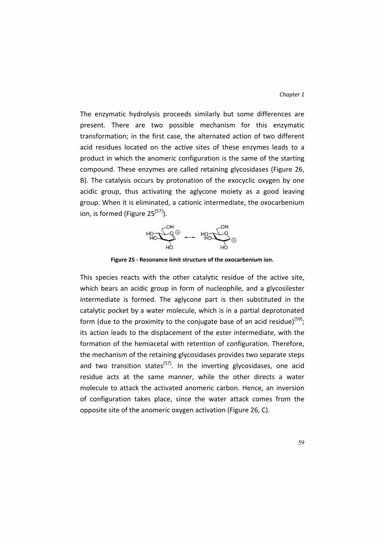
Chapter 1
59
The enzymatic hydrolysis proceeds similarly but some differences are
present. There are two possible mechanism for this enzymatic
transformation; in the first case, the alternated action of two different
acid residues located on the active sites of these enzymes leads to a
product in which the anomeric configuration is the same of the starting
compound. These enzymes are called retaining glycosidases (Figure 26,
B). The catalysis occurs by protonation of the exocyclic oxygen by one
acidic group, thus activating the aglycone moiety as a good leaving
group. When it is eliminated, a cationic intermediate, the oxocarbenium
ion, is formed (Figure 25[57]).
Figure 25 ‐ Resonance limit structure of the oxocarbenium ion.
This species reacts with the other catalytic residue of the active site,
which bears an acidic group in form of nucleophile, and a glycosilester
intermediate is formed. The aglycone part is then substituted in the
catalytic pocket by a water molecule, which is in a partial deprotonated
form (due to the proximity to the conjugate base of an acid residue)[59];
its action leads to the displacement of the ester intermediate, with the
formation of the hemiacetal with retention of configuration. Therefore,
the mechanism of the retaining glycosidases provides two separate steps
and two transition states[57]. In the inverting glycosidases, one acid
residue acts at the same manner, while the other directs a water
molecule to attack the activated anomeric carbon. Hence, an inversion
of configuration takes place, since the water attack comes from the
opposite site of the anomeric oxygen activation (Figure 26, C).

Chapter 1
60
Figure 26 ‐ Acidic and enzymatic reactions for glycosides hydrolysis.
Since the catalytic mechanism for these enzymes has been elucidated, an
attractive approach for the development of inhibitors is the creation of
compounds which structures resemble the transition state (TS)
intermediate of the enzyme‐catalyzed reaction. The rationale of this

Chapter 1
61
strategy is the belief that the TS represent the step of the reaction with
the highest degree of enzymatic stabilization. For glycosidases, the
hypothesis is that the stabilization is very high, with a dissociation
constant between the TS and the enzyme almost of 10‐20 M[57].
Some glycosidase inhibitors are still present on the market, for the
treatment of several disease in which important glycosidases are
involved. Previously, some of these molecule have been mentioned.
Glycosidase inhibitors used for diabetes treatment are Voglibose,
Miglitol and Acarbose. Acarbose, a potent α‐glucosidase inhibitors, was
isolated by a strains of Actinoplanes, present on market since 1990 in
Germany as Glucobay® and in USA since 1996[58]. This molecule is able to
mimic the TS of the enzymatic hydrolysis reaction. Instead, Miglustat is a
glycosidase inhibitors used for the therapy of the lysosomal storage
pathology called Gaucher disease. Oseltamivir and Zanamivir represent
antiviral drugs which inhibit viral glycosidases, used as anti‐influenza
agents. It is known that the first phases of viral infections are based on
the recognition by virus of glycoconjugates on the membrane of host
cells. Therefore it is frequent that antibiotics or antimicrobial drugs, both
of natural and chemical origin, contains or are constituted by structures
which inhibit these interactions[60].
Among antiviral drugs, the most important agents are inhibitors of
neuraminidases, viral enzymes whose scope is the cleavage of glycosidic
bond between sialic acid residues of some glycoconjugates located on
the membrane of infected host cells. In particular two inhibitors,
Zanamivir and Oseltamivir, are in market since 1999 and are active
against several virus strains.

Chapter 1
62
Iminosugars as glycosidase inhibitors
As mentioned, molecules mimicking the oxocarbenium ion TS of the
glycoside cleavage can be potentially exploited as glycosidase inhibitors,
and have been widely investigated[57]. Among these inhibitor molecules,
the most important and studied are the iminosugars or azasugars.
Iminosugars, whether of natural or synthetic origin, are small organic
molecules that mimic carbohydrates or their hydrolysis TS, containing a
nitrogen atom instead of oxygen in the ring system[61]. The first
discovered molecule belonging to this family was nojirimycin, in 1966[62].
It was described as an antibiotic produced in the fermentation culture by
Streptomyces roseochromogenes R‐468 and S. lavendulae SF‐425[62]. It
shows a high inhibitory potency towards α‐ and β‐glucosidases. In the
last decades, there has been an increasing interest towards this class of
molecule, and iminosugars have become versatile tools for medicinal
chemists, especially for their valence as potential therapeutic agents[63].
The research has been focused on iminosugar supply both from natural
sources and by chemical synthesis, providing a plethora of different
nojirimycin analogues.
By a systematic and structural point of view, iminosugars can be divided
into five different classes: piperidines, pyrrolidines, indolizidines,
pyrrolizidines, azepanes and nortropanes[62] (Figure 27[61]).
Figure 27 ‐ Stylized representations of the iminosugar families.
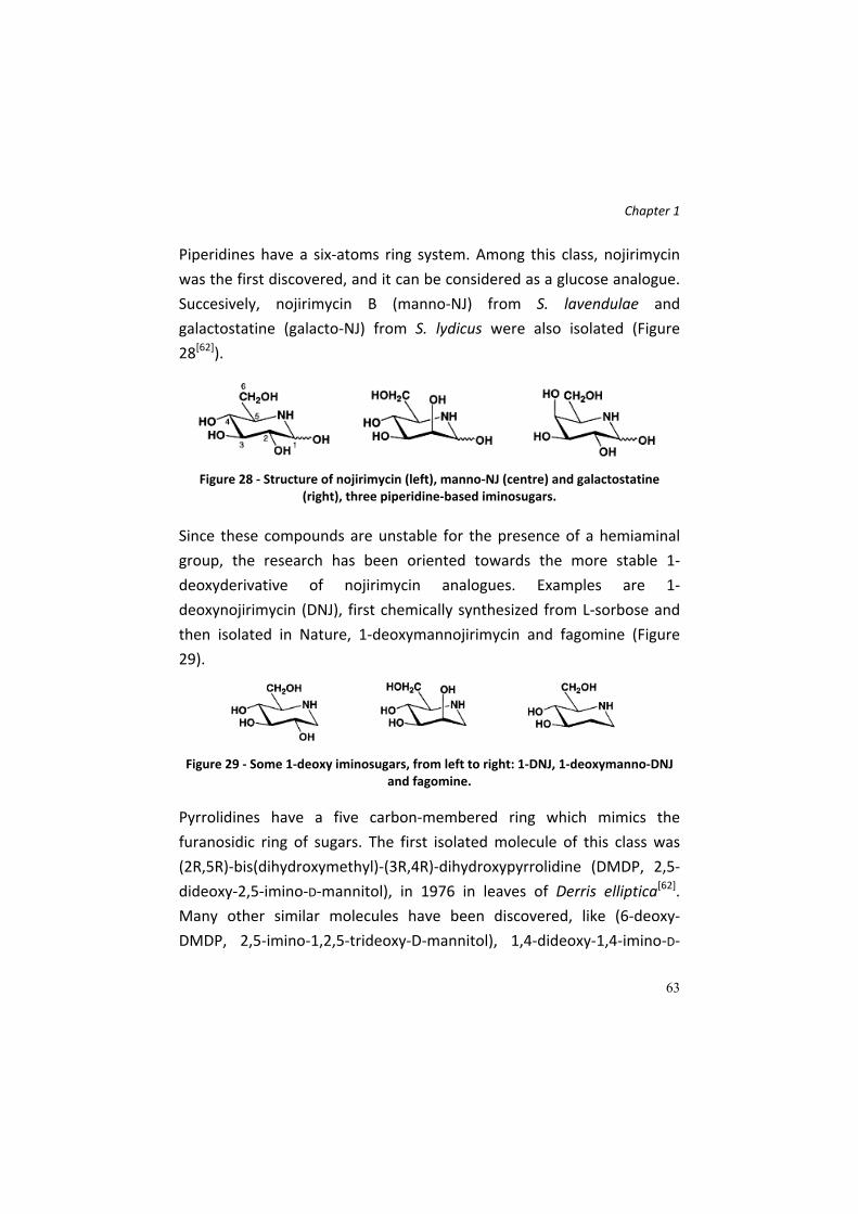
Chapter 1
63
Piperidines have a six‐atoms ring system. Among this class, nojirimycin
was the first discovered, and it can be considered as a glucose analogue.
Succesively, nojirimycin B (manno‐NJ) from S. lavendulae and
galactostatine (galacto‐NJ) from S. lydicus were also isolated (Figure
28[62]).
Figure 28 ‐ Structure of nojirimycin (left), manno‐NJ (centre) and galactostatine
(right), three piperidine‐based iminosugars.
Since these compounds are unstable for the presence of a hemiaminal
group, the research has been oriented towards the more stable 1‐
deoxyderivative of nojirimycin analogues. Examples are 1‐
deoxynojirimycin (DNJ), first chemically synthesized from L‐sorbose and
then isolated in Nature, 1‐deoxymannojirimycin and fagomine (Figure
29).
Figure 29 ‐ Some 1‐deoxy iminosugars, from left to right: 1‐DNJ, 1‐deoxymanno‐DNJ
and fagomine.
Pyrrolidines have a five carbon‐membered ring which mimics the
furanosidic ring of sugars. The first isolated molecule of this class was
(2R,5R)‐bis(dihydroxymethyl)‐(3R,4R)‐dihydroxypyrrolidine (DMDP, 2,5‐
dideoxy‐2,5‐imino‐D‐mannitol), in 1976 in leaves of Derris elliptica[62].
Many other similar molecules have been discovered, like (6‐deoxy‐
DMDP, 2,5‐imino‐1,2,5‐trideoxy‐D‐mannitol), 1,4‐dideoxy‐1,4‐imino‐D‐

Chapter 1
64
arabinitol (D‐AB1), the 2‐epimer of D‐AB1 (1,4‐dideoxy‐1,4‐imino‐D‐
ribitol), the polyhydroxylpyrrolidine nectrisine and 2,5‐dideoxy‐2,5‐
imino‐DL‐glycero‐D‐manno‐heptitol (homoDMDP) (Figure 30).
Figure 30 ‐ Examples of pyrrolidines, from left to right: DMDP, 6‐deoxy‐DMDP, D‐AB1,
2‐epi‐D‐AB1, nectrisine and homo‐DMDP.
Indolizidines (Figure 31) are constituted by a six and a five carbon‐
membered rings fused together. These bicyclic alkaloids show a less
structural relationship to monosaccharides but the hydroxyl
configuration of each ring is similar to those of sugars. The first
discovered molecule are swainsonine, drug of the legumens Swainsona
canescens, and castanospermine, from Castanospermum australe, in
1979 and 1981, respectively. From literature data results that bicylic
compounds are more potent and more selective inhibitors than
piperidines towards glycosidases. These iminosugars could be seen as
the bicycle derivative of DNJ, since it has an ethylene bridge between the
hydroxymethyl group and the ring nitrogen. 6‐epi‐castanospermine
represent the manno‐ analogue, and it shows a good inhibition against
human neutral α‐mannosidase[62].

Chapter 1
65
Figure 31 ‐ Indolizidines: swainsonine, castanospermine and 6‐epi‐castanospermine.
Pyrrolizidines are other iminosugars with two fused five‐membered
cycles (Figure 32). Alexine was the first molecule of this family
discovered, isolated from the legume Alexa leiopetala. Also an epimer in
position C‐7a was found, called 7a‐epi‐alexine or australine, from the
seeds of the plant Castanospermum australe. This molecule can be
considered as the contracted variant of the castanospermine or as a
DMDP derivative, with an ethylene bridge between the hydroxymethyl
group and the endocyclic nitrogen. An interesting analogue is the 7a‐epi‐
alexaflorine, which bears a carboxylic acid in C‐3 position of a
pyrrolizidine ring.
Figure 32 ‐ Pyrrolizidine structures: alexine (on the left), 7a‐epi‐alexine or australine
(centre) and 7a‐epi‐alexaflorine (on the right).
The last discovered family of natural iminosugars is represented by
nortropanes (tropanes which lack of a N‐methyl group), characterized by
hydroxyl groups in different position and stereochemistry and a new
aminoketal functionality, which generates a tertiary hydroxyl group at
the bicyclic ring bridgehead[62]. Among these molecules, the most
famous are the calystegines (Figure 33).
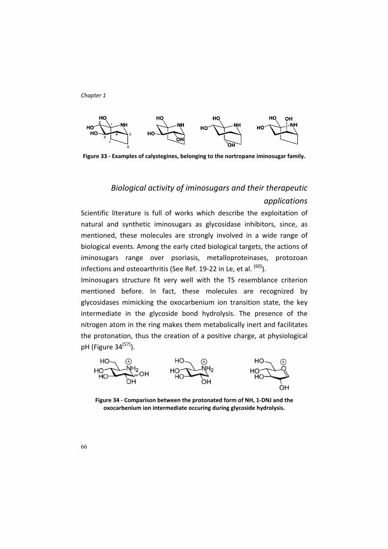
Chapter 1
66
Figure 33 ‐ Examples of calystegines, belonging to the nortropane iminosugar family.
Biological activity of iminosugars and their therapeutic
applications
Scientific literature is full of works which describe the exploitation of
natural and synthetic iminosugars as glycosidase inhibitors, since, as
mentioned, these molecules are strongly involved in a wide range of
biological events. Among the early cited biological targets, the actions of
iminosugars range over psoriasis, metalloproteinases, protozoan
infections and osteoarthritis (See Ref. 19‐22 in Le, et al. [60]).
Iminosugars structure fit very well with the TS resemblance criterion
mentioned before. In fact, these molecules are recognized by
glycosidases mimicking the oxocarbenium ion transition state, the key
intermediate in the glycoside bond hydrolysis. The presence of the
nitrogen atom in the ring makes them metabolically inert and facilitates
the protonation, thus the creation of a positive charge, at physiological
pH (Figure 34[57]).
Figure 34 ‐ Comparison between the protonated form of NH, 1‐DNJ and the
oxocarbenium ion intermediate occuring during glycoside hydrolysis.
Chapter 1
67
Furthermore, the recognition is allowed by the structure similarity of
iminosugars with monosaccharides. All these features suggest that
iminosugars are perfect glycosidases inhibitors; actually, the presence of
the protonated form of the nitrogen atom is not a sufficient requisite for
an ideal oxocarbenium ion TS, since these molecules have chair
conformations instead of the half chair conformation of the TS (due to
the partial sp2 character of anomeric carbon). For the oxocarbenium ion
TS is impossible to have a charge on the ring oxygen and a chair
conformation[57]; thus, NJ and DNJ, for example, are not perfect TS
analogue and this could explain the good, but not surprising, inhibition
constant of these two compounds.
As mentioned, one of the most important direction toward which the
development of iminosugars has been focused is represented by the
inhibition the glycoside hydrolase activity, in particular those regarding
the digestion of dietary carbohydrates. As known, intestinal di‐ and
oligosaccharidases are fixed component of the membrane of cell
constituting the brush border membrane of the small intestine. These
enzymes are involved in the digestion and hydrolysis of dietary
carbohydrates, allowing their successive absorption. Due to the
important physiological function, it is clear that their inhibition can be
exploited for the regulation of carbohydrate absorption, in order to
control several pathological states, as diabetes and obesity[64]. Acarbose,
a commercially available drug (GLYCOBAY™), shows a potent inhibition
activity towards pig intestinal sucrase, with an IC50 of 0,5 µM. It reduces
postprandial blood glucose and increases insulin secretion[64]. Also the
natural iminosugar DNJ showed an inhibitory effect against mammalian
α‐glucosidases in vitro, but the scarce in vivo effect led to the
development of more effective DNJ inhibitors, as Miglitol or Emiglitate

Chapter 1
68
(Figure 35[62]), reported as good agents against the postprandial
elevation of blood glucose[62].
Figure 35 ‐ Structures of miglitol (on the left) and emiglitate (on right).
The biological activity of iminosugars was also detected on processing
glycosidases involved in the trimming of oligosaccharidic chain linked on
Asn residues of glycoproteins. These enzymes are responsible of the
diversity of the sugar chain connected with a N‐linkage to glycoproteins,
which are biosynthesized and correctly modified in the ER. In fact, a
newly synthesized polypeptide is glycosylated on a particular Asn residue
present in a consensus sequence, and successively this sugar chain is
modified by a series of reaction both in ER and Golgi apparatus, by
specific glycosidases and mannosidases (trimming) and glycosil
transferases, which act elongating the chain with fucose, galactose, N‐
acetyl‐glucosamine and sialic acid residues. Iminosugars as
castanospermine and DNJ are potent inhibitors of glucosidase I and II
localized in the ER. Mannosidases I and II are instead blocked by other
iminosugars, such as deoxymannojirimycin, kifunensine (a two‐fused‐
ring iminosugar), 1,4‐deoxy‐1,4‐imino‐D‐mannitol. The inhibitory activity
of iminosugars against processing glycosidases leads to a different
glycosylation pattern of the N‐linked oligosaccharide, thus altering the
correct targeting of the nascent glycoprotein, delaying their
translocation in Golgi, their secretion outside the cell or otherwise
causing their degradation mediated by other cellular systems. However,
the importance of iminosugars in this case is given by their potential
ability to prevent an unfair retention of some glycoproteins in ER, due to

Chapter 1
69
their unfolded state, although they maintain a fairly functional activity. It
represents the molecular explanation of several pathologies, like cystic
fibrosis, familiar hypercholesterolemia or heritable forms of pulmonary
emphysema[64].
Many scientific works are focused on the use of iminosugars as
therapeutic agents against the so called lysosomal storage disease
(LSDs), caused by an accumulation of glycosphingolipids (GSLs) in
lysosomes. These disorders are due to defective enzymes responsible for
the degradation of glycolipids in lysosomes. A glycosphingolipid
accumulation in these organelles occurs, causing lysosomal swelling and
definitively the disease. The defect is represented by mutations in the
genes coding for these enzymes, which are less or not active, thus
leading to partial or absent glycosphingolipid degradation[64]. The
degradation pathway include eight enzymatic transformation, and each
one, if hampered, leads to a different pathology (Figure 36[62]).

Chapter 1
70
Figure 36 ‐ Lysosomal degradation pathway for glycosphingolipids. On each blocked
passage the associated disease is indicated.
Several therapies have been developed to treat these pathologies. In the
enzyme replacement therapy (ERT), a periodic substitution of the
defective enzyme is performed, to ensure a control of GSLs level in
lysosomes. This treatment is not free of heavy side effects; in one of the
most important LSD, Gaucher disease, ERT consists of injection of
recombinant glucocerebrosidases, which helps the regression of the
disease. SRT (Substrate Reduction Therapy) consist of a chemiotherapic
treatment with drugs as Miglustat (N‐butyl‐DNJ), able to inhibit the
glucosylceramide synthase, a glycosyl transferase responsible for the
synthesis of many GSLs. Miglustat is used for the treatment of type I
Gaucher disease, caused by a dysfunction of glucocerebrosidase enzyme
that provokes an intracellular accumulation of glucosylceramide causing

Chapter 1
71
liver and spleen enlargement[61]. The reduction of substrate (GSL) levels
allows a less accumulation of glycolipids in lysosomes. In the last
decades, a new approach has been developed for the treatment of these
pathology, and it consists on the development of molecules able to
restore the correct protein folding of the target defective enzymes. Since
iminosugars are good ligands for these enzymes, some of them have
been developed and tested for this ability. Iminosugars would act as real
“molecular chaperones”, since the interactions that occur between
iminosugar ligands and the binding site of the target enzyme can
stabilize the active and functional conformation of the enzyme. The
correct conformation leads not only to a restored lysosomal activity, but
also to a proper protein trafficking, since in some cases the diminished
enzymatic activity is correlated to a less lysosomal glycohydrolase
content.
The best known LSDs are Fabry and Gaucher diseases. The first
pathology is caused a deficiency of human α‐galactosidase A (α‐GalA)[64].
The substrate of this enzyme, globotriosylceramide, progressively
accumulates within visceral tissues and body fluids[64]. After a first
therapeutic approach based on ERT, recently the use of 1‐
deoxygalactonojirimycin A as molecular chaperone for this enzyme has
been developed[58].
Since, as mentioned, glycosidases are highly involved in the
oligosaccharide biosynthesis pathway and since tumor cells are
decorated with a different glycosylation pattern then healthy cells,
iminosugars show important biological activities against tumor
development. Inhibiting the glycohydrolase activities, it could be possible
to alter the glycosylation structure of tumor cells, thus contrasting the
tumor growth. As example, swainsonine stops the tumor growth and
stimulates the immune response; castanospermine and Me‐DNJ showed

Chapter 1
72
anti‐metastasis ability, inhibiting cell aggregation and cell adhesion of
cancer cell to vascular endothelium. Other glucosidases are often
present at high level in the interstitial fluids of tumor because they
degrade glycoconjugates of extracellular matrix (ECM), allowing the
tumor cell invasion. Iminosugars such as 2‐acetamido‐1,5‐imino‐1,2,5‐
trideoxy‐D‐glucitol and an iminosugar‐like glucuronic analogue are able
to inhibit these glycosidases.
Iminosugars have been developed also as antiviral agents. α‐
Glucosidases I and II, involved in the biosynthesis of glycoprotein of viral
capside during a viral cell cycle of an infection are molecular targets for
the iminosugar N‐butyl‐DNJ and its peracylated prodrug derivative,
which were developed as anti‐HIV drugs.

Chapter 1
73
Multivalency and glycobiology
The surface of a cell is a complex and dynamic environment[65], made of
a huge number of proteins and sugars of different types and shape. The
cell calyx acts both as a sensor of the external environment and as
information carrier, because it permits the communication between cells
(also pathogens). The cell‐cell contact or between cells and biomolecules
is based on specific ligand‐receptor binding events. The rationalization of
these processes, i.e. studying how single ligands bind their specific
receptors and what happens in the cell after these events, allows the
development of strategies, approaches and molecules as potential tools
able to stop or block these events. But the cell‐cell interactions cannot
be fully rationalized in this manner, since, as mentioned, the cell calix is
full of molecules that interact with their receptors on the other cell, and
the biological intracellular effects are multiple. Hence, it is clear that
another way to study this process must be taken into account. In
general, on the cell surface it is possible to find oligosaccharides of
glycoproteins and glycolipids, displayed as multiple copies, and these
structures are called multivalent. Multivalent structures of
carbohydrates of a cell surface are involved in many cell‐cell recognition
events, because they interact with receptors of the opposite cell. For
examples, chemokines interact with glycosaminoglycans displayed in a
multivalent fashion and this recognition leads to intracellular events that
definitively causes leukocytes recruitment[65]. The knowledge about
glycans‐glycan binding proteins/lectins interactions is well structured.
Lectins are carbohydrate‐binding proteins which bind specific moieties of
glycan chains, although this interaction exerts a very low affinity.
However, this feature is compensated in biological systems by
multivalency interaction, both from lectin structure, containing more

Chapter 1
74
than one carbohydrate binding site, and/or by the presentation of the
glycan moieties with a multivalent fashion on the cell surface[66].
Multivalent ligands often have increasing affinity for their target
receptors compared to their monovalent form[65]. The reasons for the
stronger biological effect of presenting and displaying a carbohydrate, or
in general a molecule, in a multivalent manner, i.e. the rationale models
for the multivalency, are multiple (Figure 37[65]).
Figure 37 ‐ Mechanism of cell‐surface receptors – multivalent ligands interactions. A) Chelate effect. B) Cluster effect. C) Subsite binding. D) Increasing of ligand local concentration.

Chapter 1
75
For example, multivalent ligands can bind and be recognized by an
oligomeric receptor on the cell surface of the interacting cell. This
hypothesis is called chelate effect. The high affinity of the multivalent
structure for the oligomeric receptor is due to a facilitated binding of the
monomeric ligands after the first receptor‐multivalent ligand interaction.
This is possible also for receptor not structured as oligomers, since the
membrane bilayer shows a high fluidity, therefore allowing the receptor
clusterization (cluster effect). The clusterization leads to the activation of
intracellular signaling pathways, often associated with an amplification
of the signal, thus explaining the biological effect of multivalency.
Alternatively, with a multivalent‐displayed ligand it is possible to exploit
the subsites often possessed by some membrane proteins, as for
example allosteric sites. Even in the case of only one receptor, it is
possible to rationalize a multivalent effect, because a polyvalent‐
presented ligand can display a higher local concentration of the ligands.
In fact, for multivalency‐based events, statistical effects due to effective
concentrations and rebinding occurrences should be taking into account,
although the multivalency effect was also described as “the
enhancement in the activity of a multivalent ligand beyond what would
be expected due to the increase in sugar local concentration alone”[67].
The study of multivalent interactions has prompted towards the
development and the preparation of multivalent structures and scaffold
decorated with biological interesting ligands, such as carbohydrates or
peptides. The chemical synthesis of multivalent systems requires to take
into account many critical aspects, such as the shape and the nature of
the multivalent structure, the selection of the spacer which divides the
scaffold and the ligand, the stability of the whole structure, its solubility
in aqueous solutions and the toxicity. The types of scaffold used for
multivalent ligand generation are several: glycoliposomes,

Chapter 1
76
glycocalixarenes, fullerenes, glycopolymers, nanoparticles, carbon
nanotubes have been developed for the study of a plethora of biological
events in which the multivalency shows a critical role. The multivalent‐
functionalized structures can exert both inhibitory or agonistic effects[65].
References [1] G. H. Schmid, Organic chemistry, WCB/MacGraw‐Hill, Dubuque,
IA, 1997.
[2] D. B. Werz, P. H. Seeberger, Chemistry‐a European Journal 2005,
11, 3194‐3206.
[3] D. L. Nelson, M. M. Cox, Lehninger principles of biochemistry,
W.H. Freeman and Co., New York City, NY, 2008.
[4] L. Ellgaard, M. Molinari, A. Helenius, Science 1999, 286, 1882‐
1888.
[5] R. G. Spiro, Glycobiology 2002, 12, 43R‐56R.
[6] R. A. Dwek, Chemical Reviews 1996, 96, 683‐720.
[7] S. Hakomori, Cancer Research 1985, 45, 2405‐2414.
[8] J. W. Dennis, M. Granovsky, C. E. Warren, Bioessays 1999, 21,
412‐421.
[9] C. Janeway, Immunobiology : the immune system in health and
disease, Garland Science, New York, 2005.
[10] R. Sasisekharan, R. Raman, V. Prabhakar, in Annual Review of
Biomedical Engineering, Vol. 8, 2006, pp. 181‐231.
[11] B. Ernst, J. L. Magnani, Nat Rev Drug Discov 2009, 8, 661‐677.
[12] F. Nicotra, L. Cipolla, B. La Ferla, C. Airoldi, C. Zona, A. Orsato, N.
Shaikh, L. Russo, Journal of Biotechnology, In Press, Corrected Proof.
[13] M. J. McKay, H. M. Nguyen, Acs Catalysis 2012, 2, 1563‐1595.

Chapter 1
77
[14] F. Nicotra, C. Airoldi, F. Cardona, P. K. Johannis, in Comprehensive
Glycoscience, Elsevier, Oxford, 2007, pp. 647‐683.
[15] D. E. Levy, P. Fügedi, The organic chemistry of sugars, Taylor &
Francis, Boca Raton, 2006.
[16] J. Cohen, Nature 2002, 420, 885‐891.
[17] R. C. Bone, R. A. Balk, F. B. Cerra, R. P. Dellinger, A. M. Fein, W. A.
Knaus, R. M. H. Schein, W. J. Sibbald, Chest 1992, 101, 1644‐1655.
[18] J. A. Yethon, C. Whitfield, Current Drug Targets ‐ Infectious
Disorders 2001, 1, 91‐106.
[19] S. I. Miller, R. K. Ernst, M. W. Bader, Nat. Rev. Microbiol. 2005, 3,
36‐46.
[20] P. M. Lavoie, J. Thibodeau, F. Erard, R. P. Sekaly, Immunol. Rev.
1999, 168, 257‐269.
[21] S. Bauer, C. J. Kirschning, H. Hacker, V. Redecke, S. Hausmann, S.
Akira, H. Wagner, G. B. Lipford, Proceedings of the National Academy of
Sciences of the United States of America 2001, 98, 9237‐9242.
[22] M. Palazzo, S. Gariboldi, L. Zanobbio, S. Selleri, G. F. Dusio, V.
Mauro, A. Rossini, A. Balsari, C. Rumio, Journal of Immunology 2008, 181,
3126‐3136.
[23] E. Cario, Gut 2005, 54, 1182‐1193.
[24] T. Wemer, D. Haller, Mutation Research‐Fundamental and
Molecular Mechanisms of Mutagenesis 2007, 622, 42‐57.
[25] L. C. H. Yu, J. R. Turner, A. G. Buret, Experimental Cell Research
2006, 312, 3276‐3286.
[26] L. C. H. Yu, A. N. Flynn, J. R. Turner, A. G. Buret, Faseb Journal
2005, 19, 1822‐1835.
[27] M. M. Raja, N. K. Tyagi, R. K. H. Kinne, Journal of Biological
Chemistry 2003, 278, 49154‐49163.

Chapter 1
78
[28] B. M. Chung, L. E. Wallace, J. A. Hardin, D. G. Gall, Canadian
Journal of Physiology and Pharmacology 2002, 80, 872‐878.
[29] A. N. Alexander, H. V. Carey, American Journal of Physiology‐
Gastrointestinal and Liver Physiology 2001, 280, G222‐G228.
[30] E. M. Wright, B. A. Hirayama, D. F. Loo, Journal of Internal
Medicine 2007, 261, 32‐43.
[31] E. M. Wright, D. F. Loo, B. A. Hirayama, E. Turk, Chapter 64. Sugar
Absorpition, In: Johnson LR et al. Physiology of the Gastrointestinal Tract,
4th Ed., San Diego : Elsevier/Academic Press 2006.
[32] E. M. Wright, Annu. Rev. Physiol. 1993, 55, 575‐589.
[33] E. M. Wright, D. D. F. Loo, B. A. Hirayama, E. Turk, Physiology
2004, 19, 370‐376.
[34] H. Jung, Febs Letters 2002, 529, 73‐77.
[35] E. M. Wright, E. Turk, M. G. Martin, 2002, pp. 115‐121.
[36] M. Quick, J. Tomasevic, E. M. Wright, Biochemistry 2003, 42,
9147‐9152.
[37] N. Hirschhorn, W. B. Greenough, Scientific American 1991, 264,
50‐56.
[38] aT. Zeuthen, B. Belhage, E. Zeuthen, Journal of Physiology‐London
2006, 570, 485‐499; bD. D. F. Loo, E. M. Wright, T. Zeuthen, Journal of
Physiology‐London 2002, 542, 53‐60.
[39] J. Dyer, I. S. Wood, A. Palejwala, A. Ellis, S. P. Shirazi‐Beechey,
American Journal of Physiology‐Gastrointestinal and Liver Physiology
2002, 282, G241‐G248.
[40] D. F. Diedrich, Archives of Biochemistry and Biophysics 1966, 117,
248‐&.
[41] UniProtKB P13866 (SC5A1_HUMAN)
http://www.uniprot.org/uniprot/P13866.

Chapter 1
79
[42] S. Faham, A. Watanabe, G. M. Besserer, D. Cascio, A. Specht, B. A.
Hirayama, E. M. Wright, J. Abramson, Science 2008, 321, 810‐814.
[43] E. M. Wright, E. Turk, Pflugers Archiv‐European Journal of
Physiology 2004, 447, 510‐518.
[44] E. Turk, C. J. Kerner, M. P. Lostao, E. M. Wright, Journal of
Biological Chemistry 1996, 271, 1925‐1934.
[45] E. Turk, M. G. Martin, E. M. Wright, Journal of Biological
Chemistry 1994, 269, 15204‐15209.
[46] A. K. Meinild, D. D. F. Loo, B. A. Hirayama, E. Gallardo, E. M.
Wright, Biochemistry 2001, 40, 11897‐11904.
[47] M. PanayotovaHeiermann, S. Eskandari, E. Turk, G. A. Zampighi,
E. M. Wright, Journal of Biological Chemistry 1997, 272, 20324‐20327.
[48] M. PanayotovaHeiermann, D. D. F. Loo, C. T. Kong, J. E. Lever, E.
M. Wright, Journal of Biological Chemistry 1996, 271, 10029‐10034.
[49] T. Puntheeranurak, B. Wimmer, F. Castaneda, H. J. Gruber, P.
Hinterdorfer, R. K. H. Kinne, Biochemistry 2007, 46, 2797‐2804.
[50] B. Wimmer, M. Raja, P. Hinterdorfer, H. J. Gruber, R. K. H. Kinne,
Journal of Biological Chemistry 2009, 284, 983‐991.
[51] M. M. Raja, H. Kipp, R. K. H. Kinne, Biochemistry 2004, 43, 10944‐
10951.
[52] A. Kumar, N. K. Tyagi, P. Goyal, D. Pandey, W. Siess, R. K. H. Kinne,
Biochemistry 2007, 46, 2758‐2766.
[53] N. K. Tyagi, A. Kumar, P. Goyal, D. Pandey, W. Siess, R. R. H.
Kinne, Biochemistry 2007, 46, 13616‐13628.
[54] T. Puntheeranurak, M. Kasch, X. B. Xia, P. Hinterdorfer, R. K. H.
Kinne, Journal of Biological Chemistry 2007, 282, 25222‐25230.
[55] B. A. Hirayama, D. D. F. Loo, A. Diez‐Sampedro, D. W. Leung, A. K.
Meinild, M. Lai‐Bing, E. Turk, E. M. Wright, Biochemistry 2007, 46, 13391‐
13406.

Chapter 1
80
[56] A. Diez‐Sampedro, E. M. Wright, B. A. Hirayama, Journal of
Biological Chemistry 2001, 276, 49188‐49194.
[57] V. H. Lillelund, H. H. Jensen, X. F. Liang, M. Bols, Chemical Reviews
2002, 102, 515‐553.
[58] N. Asano, Glycobiology 2003, 13, 93R‐104R.
[59] T. D. Heightman, A. T. Vasella, Angewandte Chemie‐International
Edition 1999, 38, 750‐770.
[60] G. T. Le, G. Abbenante, B. Becker, M. Grathwohl, J. Halliday, G.
Tometzki, J. Zuegg, W. Meutermans, Drug Discovery Today 2003, 8, 701‐
709.
[61] G. Horne, F. X. Wilson, J. Tinsley, D. H. Williams, R. Storer, Drug
discovery today 2011, 16, 107‐118.
[62] N. Asano, R. J. Nash, R. J. Molyneux, G. W. J. Fleet, Tetrahedron‐
Asymmetry 2000, 11, 1645‐1680.
[63] D. D'Alonzo, A. Guaragna, G. Palumbo, Current medicinal
chemistry 2009, 16, 473‐505.
[64] N. Asano, Curr. Top. Med. Chem. 2003, 3, 471‐484.
[65] L. L. Kiessling, J. E. Gestwicki, L. E. Strong, Current Opinion in
Chemical Biology 2000, 4, 696‐703.
[66] M. Mammen, S. K. Choi, G. M. Whitesides, Angewandte Chemie‐
International Edition 1998, 37, 2755‐2794.
[67] Y. C. Lee, R. T. Lee, Neoglycoconjugates : preparation and
applications, Academic Press, San Diego, 1994.

Chapter 2
81
Chapter 2. Development of a
library of dansyl‐C‐glycoderivatives
as SGLT1 ligand tools: synthesis,
biological evaluation and
mechanism of action.

Chapter 2
82
Abstract In this paper we report the generation of a library of analogues of C‐
glucoside 1, an anti‐inflammatory glycomimetic recently developed in
our laboratory, in order to achieve additional information about its
unclear mechanism of action. Preliminary biological experiments
performed on an in vitro model of doxorubicin‐induced mucositis, a
severe intestinal inflammatory state, indicate that the presence of the
aromatic moiety which characterize all the compounds of the library is
important for the biological activity, while less influence seems to be
associated to the sugar part. The results will be exploited for the
generation of new and more potent antiinflammatory compounds and
for the comprehension and rationalization of the antiinflammatory
mechanism of action.
Introduction The sodium glucose co‐transporter 1 (SGLT1) is a high affinity/low
capacity glucose and galactose transporter, mainly expressed on the
apical membrane of intestinal epithelium cells and of cells of S3 segment
of proximal tubule of kidneys[1] [2]. This transporter is responsible for
glucose and galactose absorption in intestinal tissue and for sugar
reuptake in kidneys. Its fundamental physiological function has made
this protein an important molecular target for the development of
compounds able to inhibit the sugar absorption, to be exploited as
potential antidiabetic drugs. Many efforts have been made to clarify the
physiological, functional and structural features of this transporter;
nowadays the knowledge of its functional behavior is profound, although
the overall tridimensional structure of the human variant remains still

Chapter 2
83
unknown. Recently, alongside the known physiological function, a new
immunological function role for this protein has been discovered Several
works indicate its involvement in the D‐glucose‐mediated protection of
the intestinal mucosa against a series of insults and injury, as
lipopolysaccharides (LPSs). High glucose concentrations, tested both in
vitro and orally on mice, are able to protect the intestinal epithelium
from LPS‐induced inflammatory injury in murine models of septic shock,
and data suggest a SGLT1 engagement in this phenomenon. The high
glucose dosage required represents a significant drawback for the usage
of this compound as potential drug, due to the inevitable impact on
metabolism, so we developed a library of glycomimetics which could act
at the same manner but with pharmacological concentration. Among the
synthesized compounds, the most active was the C‐glycoside 1 which
exerts a high protective effect at concentration of several orders of
magnitude lower than D‐glucose. This compound is a C‐glycoside, thus a
non‐metabolizable glycoderivative, important feature to take into
account in the development of carbohydrate‐base drugs.
In these years our efforts have been focused on the study of the SGLT1‐
mediated protective role of 1 on several inflammation states and
injuries, as Chron disease, ulcerative colitis and on doxorubicin and 5‐
fluorouracil‐induced mucositis. The latest pathological states are serious
side effect of doxorubicin and 5‐fluorouracil‐based tumor chemotherapy.
Mucositis are common in patient treated by a chemotherapy regimen for
head and neck cancer, and derive from the direct cytotoxic effect of the
drugs on the mucosal cells. The heavy anticancer agents induce
apoptosis and reduced proliferation on epithelial cells, thus leading to
enhanced permeability and ulceration of the mucosae. For these
reasons, it is necessary to develop new supporting therapies for
chemotherapy‐induced mucositis.

Chapter 2
84
Furthermore, our research has been oriented to the comprehension at
the basis of the protective role exerted by glucose and synthesized
glycomimetics. Alongside to the studies on the possible intracellular
processes, with the construction of the putative signal transduction
pathway which could connect the SGLT1 activation and the blockage of
the inflammatory response, our efforts are directed on the study of how
D‐glucose and C‐glycosides can activate SGLT1, involving it in the cellular
protection events.
After a first library of potential SGLT1 ligands (see La Ferla, et al. [3]),
constituted by glycomimetics which bear mainly differences in the
aromatic moiety, we decided to build a new small library of C‐glucoside 1
analogues, in order to clarify and rationalize the importance of the sugar
region in the activity of these compounds. The developed molecules
have been preliminarily tested for their protective activity on a
doxorubicin‐induced mucositis in in vitro models, to gain preliminary
information on structure‐activity relashionship.
Results and discussion Compound 1 (Figure 38) is a C‐glucoside with a dansyl residue connected
to a glycopyranose ring through an ethyleneamine spacer. The synthetic
approaches developed for this compound were described previously, in
La Ferla, et al. [3]. Biological evaluation of the small glycomimetic library
prepared in that work, in order to identify some structural requirements
correlated with the anti‐inflammatory activity on HT29 cell line treated
with LPS, indicated that C‐galactose analogue is still effective as anti‐
inflammatory agents, at the same manner of compound 1, that the
aromatic entity is crucial for the activity and that the absence of the
dimethylamino group on the naphtyl moiety causes a decrease in the

Chapter 2
85
anti‐inflammatory effect. With the present work we moved our attention
to the biological importance of the saccharidic portion of the molecules,
and we designed and synthesized a new small library of C‐glucoside 1
analogues (Figure 39).
Figure 38 ‐ Structure of C‐glycoside 1.
Figure 39 ‐ Library of compound 1 analogues.
Compound 2 represent the dansyl‐β‐glucoside analogue of 1. We
planned to design and synthesize this compound since, according to our
hypotesis, β‐glucosides should not have anti‐inflammatory activity: this
class of compounds acts as inhibitors of SGLT1, rather than
activators/agonist of this transporter/receptor, (as we suppose
compound 1 is). One example is represented by the most important
SGLT1 inhibitors, phlorizin, which binds to SGLT1 avoiding glucose
absorption mediated by the transporter. Furthermore, phlorizin is able
to stop the glucose‐mediated protective effect against inflammatory and
apoptotic insults[4] [5] [6]. A similar choice was done for compound 3, the
manno‐ analogue: it is well established that D‐mannose is neither

Chapter 2
86
recognized nor transported by SGLT1[7]; hence, hypothesizing that our
bioactive C‐glycosides exert the anti‐inflammatory activity, as SGLT1
agonists, through a binding event to the same interaction site of sugars,
compound 3 should not exert the protective effect. The synthetic
pathways for compound 2 and 3 are depicted in Scheme 2 and Scheme
3. For compound 2, 2,3,4,6‐tetra‐O‐benzyl‐D‐glucopyranose was chosen
as starting material, and converted to the β‐C‐allyl glucoside 10
according to procedure already described[8]. Briefly 2,3,4,6‐tetra‐O‐
benzyl‐D‐glucopyranose was oxidized to protected gluconolactone 8,
followed by C‐allylation upon reaction with suitable Grignard reagents to
afford lactol 9, which was then subjected to reductive deoxygenation
affording C‐Allyl glucoside 10. Its ozone‐mediated oxidation and
successive hydride reduction afforded alcohol 12; the obtained primary
alcohol was converted into an azido group (compound 13) upon
Mitzunobu reaction. The hydrogenation/hydrogenolysis on this
compound afforded unprotected C‐ethyleneamine derivative 14, which
was converted in the final product 2, by reaction with dansyl chloride.
For compound 3, the C‐allylation was performed directly on the
unprotected α‐O‐Me‐D‐mannospyranoside, exploting the higher
stereoselection associated to the anomeric functionalization reaction on
this monosaccharide. The exhaustive benzylation gave intermediate 16,
and the same pathway used for compound 2 was carried out, affording
the final compound 3.
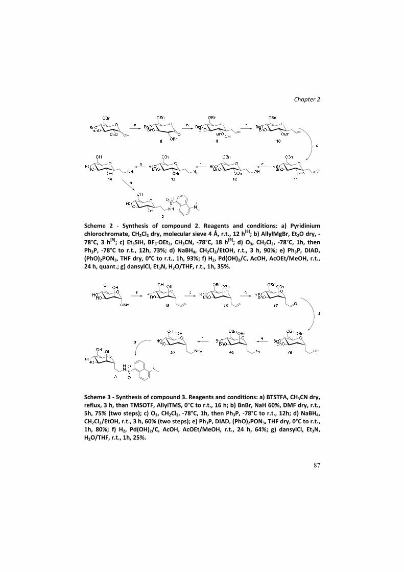
Chapter 2
87
Scheme 2 ‐ Synthesis of compound 2. Reagents and conditions: a) Pyridinium chlorochromate, CH2Cl2 dry, molecular sieve 4 Å, r.t., 12 h
[8]; b) AllylMgBr, Et2O dry, ‐
78°C, 3 h[9]; c) Et3SiH, BF3OEt2, CH3CN, ‐78°C, 18 h
[9]; d) O3, CH2Cl2, ‐78°C, 1h, then
Ph3P, ‐78°C to r.t., 12h, 73%; d) NaBH4, CH2Cl2/EtOH, r.t., 3 h, 90%; e) Ph3P, DIAD, (PhO)2PON3, THF dry, 0°C to r.t., 1h, 93%; f) H2, Pd(OH)2/C, AcOH, AcOEt/MeOH, r.t., 24 h, quant.; g) dansylCl, Et3N, H2O/THF, r.t., 1h, 35%.
Scheme 3 ‐ Synthesis of compound 3. Reagents and conditions: a) BTSTFA, CH3CN dry, reflux, 3 h, than TMSOTF, AllylTMS, 0°C to r.t., 16 h; b) BnBr, NaH 60%, DMF dry, r.t., 5h, 75% (two steps); c) O3, CH2Cl2, ‐78°C, 1h, then Ph3P, ‐78°C to r.t., 12h; d) NaBH4, CH2Cl2/EtOH, r.t., 3 h, 60% (two steps); e) Ph3P, DIAD, (PhO)2PON3, THF dry, 0°C to r.t., 1h, 80%; f) H2, Pd(OH)2/C, AcOH, AcOEt/MeOH, r.t., 24 h, 64%; g) dansylCl, Et3N, H2O/THF, r.t., 1h, 25%.

Chapter 2
88
C‐glycoside 4 is derived from a coupling reaction of the C‐glucosyl
ethanamine 24 with a derivative of cinnamic acid, commercially
available. Compound 4 was synthesized due to the high similarity with 1,
bearing a dimethylamino group attached to a vinyl‐functionalized
aromatic nucleus, structure present in the dansyl entity. The synthesis
(Scheme 4) involves the obtainment of the amine intermediate 24,
prepared by a protecting group strategy, in which the peracetylated C‐
allyl glucopyranose 21 (McGarvey, et al. [9]) is first converted to the
corresponding aldehyde 22; the obtained carbonyl group is transformed
into a tert‐butyl carbamate (23), according to a procedure of Dubé and
Scholte [10]. The carbamate and acetyl esters deprotection afforded
amine 24, which was finally coupled with N,N‐dimethylamino cinnamic
acid and deprotected to afford the desired product.
Scheme 4 ‐ Synthesis of compound 4. Reagents and conditions: a) OsO4, NaIO4, tBuOH/Acetone/H2O, r.t., 5 h
[11]; b) tert‐butylcarbamate, Et3SiH, TFA, CH3CN, r.t., 3h,
71%; c) TFA 50% in CH2Cl2, r.t., 2 h, quant.; d) N,N‐dimethylaminocinnamic acid, HOBt, DCC, iPr2EtN, DMF, r.t., 12 h, 47%; e) MeONa, MeOH, r.t., 2 h, 24%.
Compound 5 (Scheme 5) represents a side product isolated during the
synthesis of C‐glucoside 1, obtained adopting the synthetic strategy
depicted in scheme 3.

Chapter 2
89
Scheme 5 ‐ Synthesis of compound 5. Reagents and conditions: a) tert‐butylcarbamate, Et3SiH, TFA, CH3CN, r.t., 3h; b) TFA 50% in CH2Cl2, r.t., 2 h; c) dansyl chloride, Et3N, MeOH, r.t., 3 h; d) MeONa, MeOH, r.t., 2 h.
Compound 5 is a kind of dimer, in which two C‐ethanamine
glucopyranosyl residues are attached to a single dansyl unit. Since we
were focused on the large‐scale set up of compound 1 preparation, we
performed the synthesis in gram scale. Our most plausible hypothesis for
the origin of this compound is the creation, on a scaled‐up reaction, of a
glucosyl dimer during the carbamate formation on the peracetylated C‐
glucosylethanaldehyde, since this reaction is a kind of reductive
amination, and it is reported that the formation of di‐ and tri‐ alkylated
species during such reactions often occurs.
Compounds 6 and 7 are derivatives of 1, consisting of a C‐glucosidic
bicycle structure, with a terminal amino group which was used for the
dansyl linkage. The preparation of these compound was performed
within the development of C‐glucoside 1 analogues with improved water
solubility, since the latest compound shows a low solubility in aqueous
media (0,5 mg/mL, 1 mM) and aggregation phenomena over time at
higher concentrations. The amphiphilicity of 1 is probably the cause of
this character, thus, in our idea, a bicyclic scaffold could ensure a higher
structural rigidity, which should prevent the formation of aggregates,
allowing a better solubility in water. The exploited approach for the
preparation of the bicyclic compounds has been previously developed by

Chapter 2
90
our group (see ref. of La Ferla, et al. [12a], Mari, et al. [12b]). A
iodocyclization reaction between the hydroxyl group of C‐2 position and
the allylic olefin of α‐C‐allyl‐glucopyranoside, mediated by iodine‐
donating species, such as I2 or N‐iodo‐succinimide (NIS), occurs. A
mixture of two diastereoisomer is generated, depending on the side of
attack of the OH group on the iodonium structure formed. The resulted
byciclic C‐glucoside mixture bears an electrophilic site which can be
exploited for a substitution with, for example, an azido group, easily
convertible in a free amine. The two stereoisomers cannot be separated
with simple silica gel chromatography; initially the diastereoisomeric
mixture of the two iodine‐bearing bicycles, described elsewere[13] was
used for the synthesis, generating a mixture of the two dansyl‐bearing
bicycles, in a ratio of 70:30 (according to 1H‐NMR analysis) (Scheme 6).
Scheme 6 ‐ Synthesis scheme of bicyclic dansyl‐C‐glucosides obtained as mixture of two diastereoisomers. Reagents and conditions: a) BSTFA, CH3CN, reflux, 3 h, then AllylTMS, TMSOTf, 0°C to r.t., 16 h
[12a]; b) NIS, DMF, r.t., 2 h
[12a]; c) NaN3, DMF, 80°C,
24 h[12a]
; d) Pd(OH)2/C, H2 atm., MeOH, r.t., 12 h[12a]
; d) dansyl chloride, Et3N, MeOH, r.t., 3 h
[13].
Actually, we noticed a good water solubility for the mixture, higher than
for compound 1. Furthermore, preliminary biological tests revealed a
considerable anti‐inflammatory effect both in vivo and in vitro: the
change to a bicyclic scaffold does not seem to affect the biological
activity (unpublished data). Therefore, we decided to perform a

Chapter 2
91
synthesis with the aim to obtain the two separate diastereoisomers, in
order to understand if the biological activity depends on only one
stereoisomers, or if the difference in stereochemistry is not crucial for
the effect. Moreover, the diastereoisomeric mixture resolution allows to
assign the correct stereochemistry of the new chiral center. The
synthetic plan is depicted below (Scheme 7).
Scheme 7 ‐ Synthesis of 6 and 7. Reagents and conditions: a) Benzaldehyde dimethylacetal, CSA, dry DMF, 70°C, 12 h, 78% (51% for 31a and 27% for 31b); b) Pd(OH)2/C, H2 atm., AcOH, r.t., 24 h, quant.; c) dansyl chloride, Et3N, MeOH, r.t., 4 h (41% for 6 and 57% for 7).
The separation of the two stereoisomers is performed protecting the
hydroxyl in C4 and C6 positions of the bicyclic C‐glycoside as benzylidene
acetal. This allowed to an easily separation of the two stereoisomers
with classic flash chromatography technique. The diastereoisomers were
then use separately for the synthesis, performing the same
transformations: a reduction of the azide into amino group with
contemporary cleavage of the acetal protecting group and the
attachment of the dansyl moiety. Compound 6 was obtained in higher
amount compared to 7, due to the diastereoisomeric excess of the
iodocyclization reaction. To assign the correct stereochemistry of the
new chiral center, NOESY analysis were performed on the azido
intermediates 31a and 31b (Figure 40). In NOESY spectrum of 31b an
evident crosspeak between H2’and H3 is present, indicating their spatial
proximity, which is possible for the diastereisomer with an (S)

Chapter 2
92
configuration at C2’. This crosspeak is absent in the spectrum of the
other diastereoisomer, as expected for the C2’ R configuration,
corresponding to compound 31a.
Compound 31a (R) Compound 31b (S)
31a

Chapter 2
93
31b
Figure 40 ‐ NOESY experimnents on compounds 31a and 31b. In NOESY spectrum of 31b, a evident crosspeak between H2’and H3 is present, indicating their spatial proximity, which is possible for this diastereisomer and not for 31a.
Diastereoisomers differ from physical‐chemical features: in our case the
definition is perfectly in agreement with what observed. The water
solubility is completely different between the two compound (31a is
completely water‐soluble, 31b is completely water‐unsoluble at
concentration ranges of 0,5 – 5 mg/mL: the diastereoisomeric mixture
previously prepared, containing the two molecules in 7:3 ratio, is soluble
in water between these concentration ranges, since the 70% is
constituted by 31a). Surprisingly, we observed also a great difference in
fluorescence of the two compound bearing the dansyl unit (dansyl
chloride is used commonly as fluorescent derivatization reagent) (Figure
41).

Chapter 2
94
Figure 41 – Fluorescence emission (under a UVA lamp with a excitation wavelength of
365 nm) of compounds 31a (on the left) and 31b (on the right).
Compounds 1‐6, 30 (as mixture) and compounds 34‐37 (Figure 42),
which were previously synthesized and for which a biological assay was
already performed[3], were tested for their protective activity on an in
vitro model of doxorubicin‐induced cytotoxicity, using SGLT1‐
overexpressing Caco‐2 cell line (Figure 43). No test were conducted with
compound 7 due to its insolubility in water.
Figure 42 ‐ Structure of compounds 34,35,36.

Chapter 2
95
Figure 43 ‐ Preliminary viability experiments with synthesized compounds. Neutral red assay for protective effect on Doxorubicin‐induced cytotoxicity on Caco‐2 cells overexpressing SGLT1.
Preliminary results seem to indicate that the presence of the aromatic
moiety (dansyl of dimethylaminocinnamic residue) is important for the
biological activity of the tested compounds, and less influence is
associated to the sugar portion, since for almost all compounds a
cytoprotection is observed at 5 µg/mL. However, with these preliminary
results, the extent of the standard deviations are such as to not allow a
reliable interpretation of the data. This experiment has thus to be
performed again in order to obtain significant data. Meanwhile, these
preliminary results suggested us to extend the members of the library
including few compounds bearing the dansyl moiety and lacking the
sugar entity. We decided to prepared other molecules bearing a dansyl
residue attached to a completely different structure, in order to
delucidate the importance of the different parts of the molecule
involved in the biological effect and with the aim to obtain other
information about the structure‐activity relashionship. We planned the
synthesis of the new compounds taking into account the probable water
0,0000,0500,1000,1500,2000,2500,3000,3500,400
CONTR
DOX 1 2 3 5 4 6
30
34
35
36
37
OD
compounds (tested at 5 µg/mL, 8 ‐ 13 µM )

Chapter 2
96
solubility of the products. In fact, the synthesized molecules containing a
dansyl attached on the amino functionality of glucosamine, on a
hexaethylene glycol chain and on a glycine (Figure 44). The synthesis of
42 and 43 have involved a simple reaction between D‐glucosamine and L‐
glycine, commercially available, and dansyl chloride, wherease the
functionalization with a hexaethyleneglycol chain required the
monomesylation of one of the two hydroxyl group, a following
conversion to azide and its reduction into an amino functionality. These
compounds, all water soluble, and the already synthesized ones will be
tested for their cytoprotective activity.
Figure 44 – Structure and synthesis of new prepared dansyl‐derivative. Reagents and condition: a) Ag2O, MsCl, CH2Cl2, r.t., 12 h, 47%; b) NaN3, DMF, r.t., 36 h, 70%; c) H2 atm., Pd(OH)2/C, r.t., 15 h, quant.; d) dansyl chloride, Et3N, MeOH, r.t., 3 h, 60%; e) dansyl chloride, Et3N, MeOH, r.t., 12 h, 68%; f) dansyl chloride, NaOH 1M, THF, r.t., 3 h[14]
.

Chapter 2
97
Conclusions In this work we have described the design and synthesis of putative
SGLT1 ligands as potential cytoprotective agents and molecular/chemical
tools for studying the SGLT1‐mediated cytoprotection process. The
mechanism at the base of this phenomenon is still unclear, in particular
how C‐glucoside 1, interacting with SGLT1, acts as anti‐
inflammatory/protective agent. Organic synthesis of its analogues can
help this work, by preparing a library of molecules to use in structure‐
activity relationship studies. Testing the biological effect of analogues of
compound 1, it will be possible to build a putative pharmacophore map,
thus identifying which groups are crucial for the binding to SGLT1, hence
for the biological effect. The knowledge of these aspects represents a
fundamental point; a rationalization of how the protective compounds
bind to SGLT1 will be useful for the development of new and more active
molecules.
Experimental Section
Cell cultures and treatments
Cell lines Caco‐2/bbe and Caco‐2/bbe permanently transfected with
SGLT‐1 (kind gift of Prof. Mark Donowitz, MD, Johns Hopkins University
School of Medicine, Baltimore) were cultured in DMEM high‐glucose
medium (4,5g/L) (Euroclone, Pero, Italy) supplemented with 10% FBS
(Euroclone), 1% glutamine (Euroclone), 15 nM sterile HEPES solution
(Euroclone). For Caco‐2/bbe/SGLT1 cells, 250 µg/ml G418 gentamicin
bisulfate salt solution (Sigma‐Aldrich) was added as antibiotic agent; for
normal Caco‐2/bbe penicillin/streptamicin solution (Euroclone) was
added.

Chapter 2
98
For viability assays 1 x 10⁵ Caco‐2 cells were plated on 96 flat‐bottom
well plates and treated after 24 hours with doxorubicin (100 µM) with
the compounds at concentrations of 5 µg/mL (8 – 13 µM). Cell viability
was evaluated after 48 h with Neutral Red assay kit (Sigma Aldrich),
according to the datasheet.
Synthesis of compounds
General remarks. All commercial chemicals were purchased from Sigma‐
Aldrich. All chemicals were used without further purification. All required
anhydrous solvents were dried with molecular sieves for at least 24h
prior to use. Thin layer chromatography (TLC) was performed on silica
gel 60 F254 plates (Merck) with detection under UV light when possible,
or by charring with a solution of (NH4)6Mo7O24 (21g), Ce(SO4)2 (1g),
concentrated H2SO4 (31 mL) in water (500 mL) or with an ethanol
solution of ninhydrin or with Dragendorff’ spray reagent. Flash‐column
chromatography was performed on silica gel 230–400 mesh (Merck). 1H
and 13C NMR spectra were recorded at 25°C, unless otherwise stated,
with a Varian Mercury 400‐MHz instrument. Chemical shift assignments,
reported in parts per million, were referenced to the corresponding
solvent peaks. Mass spectra were recorded on a QTRAP system with ESI
source.

Chapter 2
99
Synthetic procedures
2,3,4,6‐tetra‐O‐benzyl‐D‐gluconolactone 8
The synthesis of 8 is described in Brenna, et al. [8].
3‐hydroxy‐3‐(2,3,4,6‐tetra‐O‐benzyl‐D‐glucopyranosyl)‐propene 9
3‐(2,3,4,6‐tetra‐O‐benzyl‐D‐glucopyranosyl)‐propene 10
The synthesis of 9 and 10 are described in McGarvey, et al. [9].
2‐(2,3,4,6‐tetra‐O‐benzyl‐β‐D‐glucopyranosyl)‐ethanal 11
In a solution of 10 (1,42 g, 2,51 mmol) in CH2Cl2 (50 mL, 0,05 M) at ‐78°C,
O3 was bubbled until a pale blue colour appears, indicating the end of
the reaction. The reaction was followed by TLC (PE/AcOEt 8:2). Then the
excess of O3 was removed by purging the reaction with a stream of
Argon at ‐78°C and then Ph3P (6,15 mmol, 2,45 eq) were added. The

Chapter 2
100
reaction was stirred for 24 h at r.t. The product was purified directly
from the crude reaction by flash chromatography (PE/AcOEt 9:1 –
PE/AcOEt 8,5:1,5) to provide aldehyde 11 (1,032 g, 1,82 mmol, 73%). 1H NMR (400 MHz, CDCl3) δ 9.72 (s, 1H, CHO), 7.40 – 7.03 (m, 20H, CH
Ar), 4.98 – 4.86 (m, 3H, CH2Ph), 4.82 (d, J = 10.7 Hz, 1H, CH2Ph), 4.63 –
4.55 (m, 3H, CH2Ph), 4.51 (d, J = 12.2 Hz, 1H, CH2Ph), 3.83 (td, J = 9.1, 4.5
Hz, 1H, H1), 3.77 – 3.63 (m, 4H, H2, H4, H6a, H6b), 3.52 – 3.44 (m, 1H,
H5), 3.34 (t, J = 9.1 Hz, 1H, H3), 2.77 – 2.67 (m, 1H, H1’a), 2.57 (ddd, J =
16.2, 7.9, 2.4 Hz, 1H, H1’b). 13C NMR (101 MHz, CDCl3) δ 200.44 (C2’ (CHO)), 138.47, 138.06, 137.69
(Cq Ar x 4), 129.79, 128.73, 128.68, 128.64, 128.58, 128.53, 128.34,
128.19, 128.05, 127.98, 127.92, 127.86, 127.83, 127.80 (C Ar x 20),
87.22, 81.25, 79.25, 78.39 (C1, C2, C3, C4, C5), 75.73, 75.21 (CH2Ph x 4),
74.58 (C5), 68.75 (C6), 46.20 (C1’).
C36H38O6; calcd. mass 566,69; ESI‐MS: m/z 567,61 [M+H]+.
2‐(2,3,4,6‐tetra‐O‐benzyl‐β‐D‐glucopyranosyl)‐ethanol 12
Compound 11 (736 mg, 1,3 mmol) was dissolved in a mixture of
CH2Cl2/MeOH 1:1 (6 mL, 0,2 M) at r.t. NaBH4 (5,19 mmol, 4 eq) was
added and the reaction was stirred vigorously at r.t. The reaction was
followed by TLC (PE/AcOEt 7:3). At the end of the reaction, the solvent
was evaporated; the residue was suspended in a volume of a saturated
solution of sodium carbonate and stirred vigorously for 30 min. The
aqueous phase was extracted with AcOEt (3 x). The organic phases were
combined, dried with sodium sulphate and filtrated. The product was

Chapter 2
101
purified from the residue by flash chromatography (PE/AcOEt 7:3), to
provide compound 11 (646 mg, 1,136 mmol, 90%). 1H NMR (400 MHz, CDCl3) δ 7.38 – 7.23 (m, 18H, CH Ar), 7.23 – 7.13 (m,
2H, CH Ar), 4.93 – 4.87 (m, 3H, CH2Ph), 4.83 (d, J = 10.8 Hz, 1H, CH2Ph),
4.64 (d, J = 10.9 Hz, 1H, CH2Ph), 4.59 – 4.49 (m, 3H, CH2Ph), 3.79 (t, J =
5.4 Hz, 2H, H2’a,b), 3.73 – 3.64 (m, 2H, H6a, H3), 3.63 – 3.54 (m, 2H, H6b,
H2), 3.54 – 3.44 (m, 2H, H1, H5), 3.35 (t, J = 9.2 Hz, 1H, H4), 2.12 – 2.00
(m, 1H, H1’a), 1.84 – 1.67 (m, 1H, H1’b). 13C NMR (101 MHz, CDCl3) δ 138.55, 138.02, 137.97, 137.95 (Cq Ar),
128.63, 128.61, 128.58, 128.56, 128.17, 128.11, 128.08, 127.99, 127.96,
127.86, 127.83 (C Ar x 20), 87.11 (C3), 81.85 (C2), 80.00 (C1), 78.72 (C4),
78.57 (C5), 75.75, 75.47, 75.21, 73.60 (CH2Ph), 69.17 (C6), 61.68 (C2’),
33.80 (C1’).
C36H40O6; calcd. mass 568,61; ESI‐MS: m/z 569,64 [M+H]+.
2‐(2,3,4,6‐tetra‐O‐benzyl‐β‐D‐glucopyranosyl)‐1‐azidoethane 13
Compound 12 (631 mg, 1,1 mmol) was dissolved in THF dry (5,5 mL, 0,2
M) and Ph3P (3,3 mmol, 3 eq) was added. The solution was cooled to 0°C
and DIAD (3,3 mmol, 3 eq) was added dropwise. After 10 min, 3,5 mmol
(3.2 eq) of phosphoryldiphenil azide (PhO)2P(O)N3 are added at 0°C and
the reaction was stirred for 5 h at r.t. The reaction was followed by TLC
(PE/AcOEt 8:2) and the product was purified from the crude by flash
chromatography (eluent PE/AcOEt 9:1) to provide compound 13 (621
mg, 1,03 mmol, 93%).
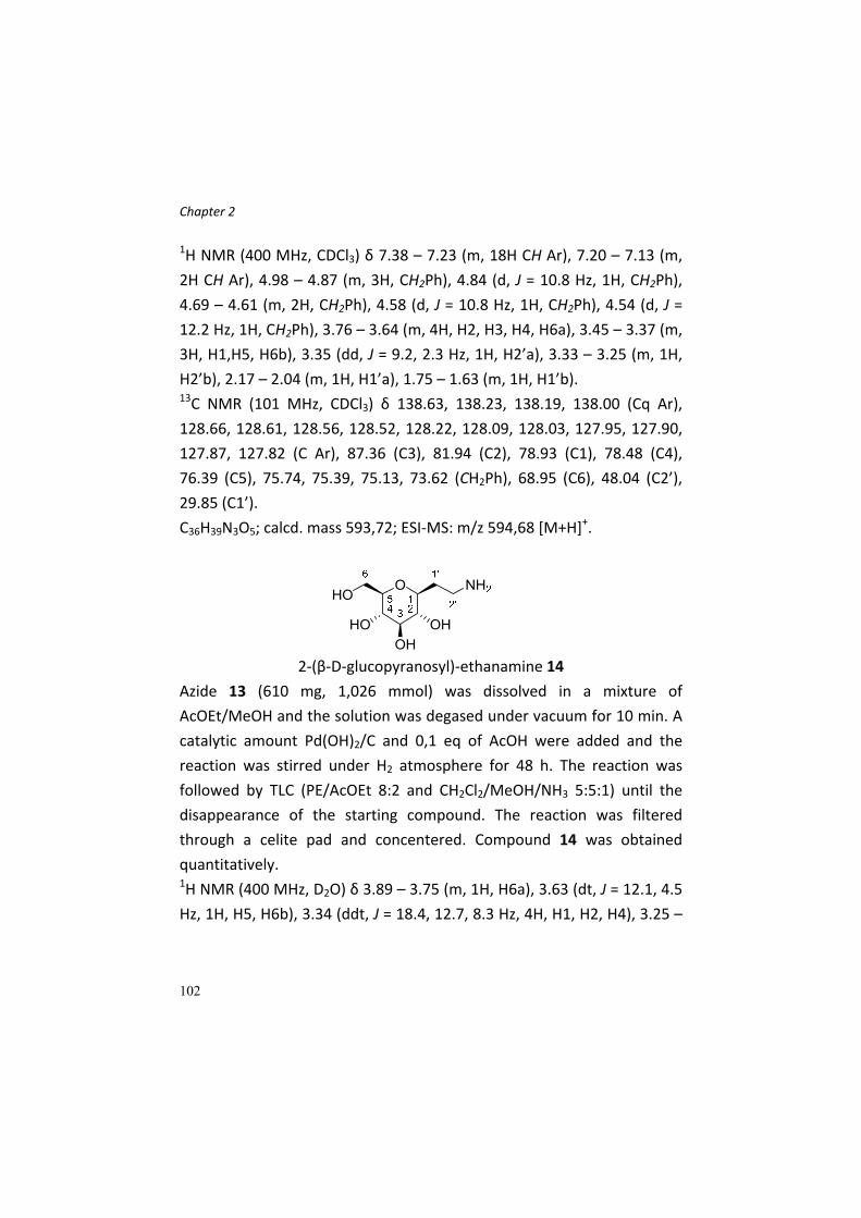
Chapter 2
102
1H NMR (400 MHz, CDCl3) δ 7.38 – 7.23 (m, 18H CH Ar), 7.20 – 7.13 (m,
2H CH Ar), 4.98 – 4.87 (m, 3H, CH2Ph), 4.84 (d, J = 10.8 Hz, 1H, CH2Ph),
4.69 – 4.61 (m, 2H, CH2Ph), 4.58 (d, J = 10.8 Hz, 1H, CH2Ph), 4.54 (d, J =
12.2 Hz, 1H, CH2Ph), 3.76 – 3.64 (m, 4H, H2, H3, H4, H6a), 3.45 – 3.37 (m,
3H, H1,H5, H6b), 3.35 (dd, J = 9.2, 2.3 Hz, 1H, H2’a), 3.33 – 3.25 (m, 1H,
H2’b), 2.17 – 2.04 (m, 1H, H1’a), 1.75 – 1.63 (m, 1H, H1’b). 13C NMR (101 MHz, CDCl3) δ 138.63, 138.23, 138.19, 138.00 (Cq Ar),
128.66, 128.61, 128.56, 128.52, 128.22, 128.09, 128.03, 127.95, 127.90,
127.87, 127.82 (C Ar), 87.36 (C3), 81.94 (C2), 78.93 (C1), 78.48 (C4),
76.39 (C5), 75.74, 75.39, 75.13, 73.62 (CH2Ph), 68.95 (C6), 48.04 (C2’),
29.85 (C1’).
C36H39N3O5; calcd. mass 593,72; ESI‐MS: m/z 594,68 [M+H]+.
2‐(β‐D‐glucopyranosyl)‐ethanamine 14
Azide 13 (610 mg, 1,026 mmol) was dissolved in a mixture of
AcOEt/MeOH and the solution was degased under vacuum for 10 min. A
catalytic amount Pd(OH)2/C and 0,1 eq of AcOH were added and the
reaction was stirred under H2 atmosphere for 48 h. The reaction was
followed by TLC (PE/AcOEt 8:2 and CH2Cl2/MeOH/NH3 5:5:1) until the
disappearance of the starting compound. The reaction was filtered
through a celite pad and concentered. Compound 14 was obtained
quantitatively. 1H NMR (400 MHz, D2O) δ 3.89 – 3.75 (m, 1H, H6a), 3.63 (dt, J = 12.1, 4.5
Hz, 1H, H5, H6b), 3.34 (ddt, J = 18.4, 12.7, 8.3 Hz, 4H, H1, H2, H4), 3.25 –
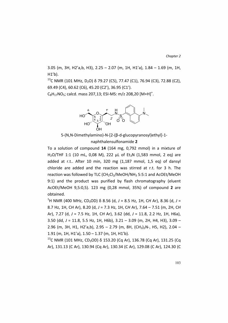
Chapter 2
103
3.05 (m, 3H, H2’a,b, H3), 2.25 – 2.07 (m, 1H, H1’a), 1.84 – 1.69 (m, 1H,
H1’b). 13C NMR (101 MHz, D2O) δ 79.27 (C5), 77.47 (C1), 76.94 (C3), 72.88 (C2),
69.49 (C4), 60.62 (C6), 45.20 (C2’), 36.95 (C1’).
C8H17NO5; calcd. mass 207,13; ESI‐MS: m/z 208,20 [M+H]+.
5‐(N,N‐Dimethylamino)‐N‐[2‐(β‐d‐glucopyranosyl)ethyl]‐1‐
naphthalensulfonamide 2
To a solution of compound 14 (164 mg, 0,792 mmol) in a mixture of
H2O/THF 1:1 (10 mL, 0,08 M), 222 µL of Et3N (1,583 mmol, 2 eq) are
added at r.t.. After 10 min, 320 mg (1,187 mmol, 1,5 eq) of dansyl
chloride are added and the reaction was stirred at r.t. for 3 h. The
reaction was followed by TLC (CH2Cl2/MeOH/NH3 5:5:1 and AcOEt/MeOH
9:1) and the product was purified by flash chromatography (eluent
AcOEt/MeOH 9,5:0,5). 123 mg (0,28 mmol, 35%) of compound 2 are
obtained. 1H NMR (400 MHz, CD3OD) δ 8.56 (d, J = 8.5 Hz, 1H, CH Ar), 8.36 (d, J =
8.7 Hz, 1H, CH Ar), 8.20 (d, J = 7.3 Hz, 1H, CH Ar), 7.64 – 7.51 (m, 2H, CH
Ar), 7.27 (d, J = 7.5 Hz, 1H, CH Ar), 3.62 (dd, J = 11.8, 2.2 Hz, 1H, H6a),
3.50 (dd, J = 11.8, 5.5 Hz, 1H, H6b), 3.21 – 3.09 (m, 2H, H4, H3), 3.09 –
2.96 (m, 3H, H1, H2’a,b), 2.95 – 2.79 (m, 8H, (CH3)2N‐, H5, H2), 2.04 –
1.91 (m, 1H, H1’a), 1.50 – 1.37 (m, 1H, H1’b). 13C NMR (101 MHz, CD3OD) δ 153.20 (Cq Ar), 136.78 (Cq Ar), 131.25 (Cq
Ar), 131.13 (C Ar), 130.94 (Cq Ar), 130.34 (C Ar), 129.08 (C Ar), 124.30 (C

Chapter 2
104
Ar), 120.66 (C Ar), 116.45 (C Ar), 81.18 (H4), 79.64 (H3), 78.08 (C1), 75.26
(C2), 71.62 (C5), 62.78 (C6), 45.83 ((CH3)2N‐ x2), 40.58 (C2’), 32.90 (C1’).
C10H28N2O7S; calcd. mass 440,51; ESI‐MS: m/z 441,6 [M+H]+.
C‐allyl‐α‐D‐mannopyranoside 15
To a suspension of O‐Me‐α‐D‐mannopyranoside (2,024 g, 10,42 mmol) in
CH3CN dry (5 mL) were added 8,3 mL (31,27 mmol, 3 eq) of BSTFA; the
suspension was heated to reflux and stirred for 3 h or otherwise when
the sugar is completely dissolved. The reaction was then cooled to 0°C
and AllylTMS and TMSOTf were added dropwise. The reaction was
stirred at r.t. for 16 h and followed by TLC (AcOEt/MeOH 8:2). At the end
of the reaction, 20 mL of water were added to the solution at 0°C; the
reacxtion was neutralized with the addition of basic resin that was
filtered off. The solution is concentrated and then washed with AcOEt.
The aqueous phase containing the compound is evaporated and the
residue (≈ 4 g) is directly used for the following step.
All spectral data was consistent with that reported in the literature[15].
3‐(2,3,4,6‐tetra‐O‐benzyl‐D‐mannopyranosyl)‐propene 15
Crude compound 15 (≈ 4 g) was dissolved in dry DMF under argon. 2,7 g
(68 mmol, 6,5 eq) of NaH (60% dispersion in mineral oil) are added at
r.t.. After 10 min, benzyl bromide (7,4 mL, 63 mmol, 6 eq) is added

Chapter 2
105
dropwise to the reaction that was stirred at r.t. for 5 h and then heated
for 80°C for 24 h. The reaction is followed by TLC (PE/AcOEt 9:1) until the
disappearance of starting material. The reaction was quenched with the
addition of MeOH. The solution is then diluted with AcOEt and washed
with H2O (3x). The organic phase is then dried with sodium sulphate and
evaporated. The crude is purified by flash chromatography (eluent
PE/AcOEt 10:1), to provide compound 16 (4,37 g, 7,74 mmol, 75% over
two steps). 1H NMR (400 MHz, CDCl3) δ 7.43 – 7.15 (m, 20H, CH Ar), 5.77 (dt, J =
16.4, 6.9 Hz, 1H, H8), 5.08 – 4.99 (m, 2H, H9a,b), 4.72 (d, J = 11.3 Hz, 1H,
CH2Ph), 4.64 – 4.50 (m, 7H, CH2Ph), 4.06 (dd, J = 12.2, 6.3 Hz, 1H, H1),
3.90 – 3.82 (m, 2H, H4, H6a), 3.82 – 3.76 (m, 2H, H3, H5), 3.72 (dd, J =
10.2, 3.4 Hz, 1H, H6b), 3.64 (dd, J = 4.5, 3.1 Hz, 1H, H2), 2.43 – 2.28 (m,
2H, H1’a,b). 13C NMR (101 MHz, CDCl3) δ 138.57, 138.40, 138.39, 138.30 (Cq Ar),
134.45 (C8), 128.50, 128.44, 128.42, 128.14, 128.11, 128.01, 127.85,
127.82, 127.75, 127.59 (C Ar x 20), 117.32 (C9), 77.01 (C3), 75.27 (C2),
75.02 (C4), 73.96 (CH2Ph), 73.84 (C5), 73.42 (CH2Ph), 72.46 (C1), 72.19
(CH2Ph), 71.65 (CH2Ph), 69.29 (C6), 34.80 (C1’).
C37H40O5; calcd. mass 564,72; ESI‐MS: m/z 565,7 [M+H]+.
2‐(2,3,4,6‐tetra‐O‐benzyl‐α‐D‐mannopyranosyl)‐ethanal 16
A solution of compound 16 (2,27 g, 4 mmol) in dry CH2Cl2 (30 mL, ≈ 0,1
M) is cooled to ‐78°C and ozone was bubbled through the solution for
approximately 3 h, until the solution turned blue. The reaction is then

Chapter 2
106
deozonized bubbling argon through the solution and Ph3P (3,3 g, 12,5
mmol, 3 eq) was added at ‐78°C. The solution is slowly warmed to r.t.
and stirred overnight. The reaction was followed by TLC (PE/AcOEt 8:2).
The crude product was directly used for the next reaction without
further purifications. A 1H‐NMR performed on the crude confirmed the
complete transformation of the allyl group into aldehyde function.
2‐(2,3,4,6‐tetra‐O‐benzyl‐α‐D‐mannopyranosyl)‐ethanol 18
Crude aldehyde 17 was dissolved in a mixture of CH2Cl2/MeOH 1:1 (80
mL, ≈ 0,05 M). NaBH4 (605 mg, 16 mmol, 4 eq related to compound 16)
was added at r.t. and the reaction is stirred at r.t. for 2 h. The product
was extracted as for compound 11 and purified by flash chromatography
(PE/AcOEt 6:4). 1363 mg (2,4 mmol, 60% yield over two steps) of alcohol
18 were obtained. 1H NMR (400 MHz, CDCl3) δ 7.39 – 7.23 (m, 18H, CH Ar), 7.23 – 7.16 (m,
2H, CH Ar), 4.63 – 4.45 (m, 8H, CH2Ph), 4.21 – 4.13 (m, 1H, H1), 4.01 –
3.94 (m, 1H, H5), 3.84 – 3.74 (m, 4H, H3, H6a, H2’a,b), 3.71 (t, J = 5.7 Hz,
1H, H4), 3.66 – 3.55 (m, 2H, H2, H6b), 2.71 (s, 1H, OH), 1.93 – 1.81 (m,
1H, H1’a), 1.80 – 1.71 (m, 1H, H1’b). 13C NMR (101 MHz, CDCl3) δ 138.13, 138.11, 137.69, 137.66 (Cq Ar),
129.84, 128.56, 128.53, 128.52, 128.47, 128.11, 128.00, 127.92, 127.91,
127.88, 127.77 (C Ar), 76.29 (C2), 76.00 (C3), 74.91 (C4), 73.63 (C5),
73.40 (CH2Ph), 72.49 (CH2Ph), 72.25 (C1), 71.74 (CH2Ph), 68.68 (C6),
61.56 (C2’), 32.25 (C1’).
C36H40O6; calcd. mass 568,71; ESI‐MS: m/z 569,6 [M+H]+, 591,5 [M+Na]+.
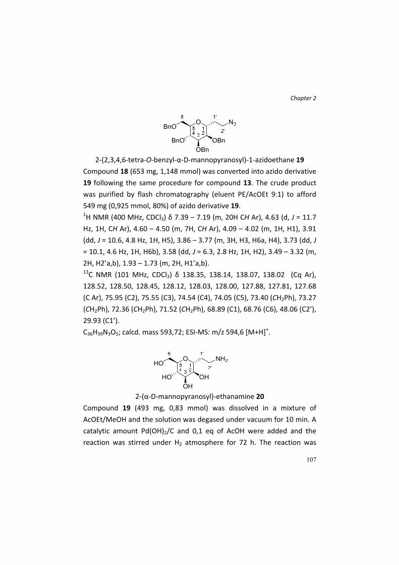
Chapter 2
107
2‐(2,3,4,6‐tetra‐O‐benzyl‐α‐D‐mannopyranosyl)‐1‐azidoethane 19
Compound 18 (653 mg, 1,148 mmol) was converted into azido derivative
19 following the same procedure for compound 13. The crude product
was purified by flash chromatography (eluent PE/AcOEt 9:1) to afford
549 mg (0,925 mmol, 80%) of azido derivative 19. 1H NMR (400 MHz, CDCl3) δ 7.39 – 7.19 (m, 20H CH Ar), 4.63 (d, J = 11.7
Hz, 1H, CH Ar), 4.60 – 4.50 (m, 7H, CH Ar), 4.09 – 4.02 (m, 1H, H1), 3.91
(dd, J = 10.6, 4.8 Hz, 1H, H5), 3.86 – 3.77 (m, 3H, H3, H6a, H4), 3.73 (dd, J
= 10.1, 4.6 Hz, 1H, H6b), 3.58 (dd, J = 6.3, 2.8 Hz, 1H, H2), 3.49 – 3.32 (m,
2H, H2’a,b), 1.93 – 1.73 (m, 2H, H1’a,b). 13C NMR (101 MHz, CDCl3) δ 138.35, 138.14, 138.07, 138.02 (Cq Ar),
128.52, 128.50, 128.45, 128.12, 128.03, 128.00, 127.88, 127.81, 127.68
(C Ar), 75.95 (C2), 75.55 (C3), 74.54 (C4), 74.05 (C5), 73.40 (CH2Ph), 73.27
(CH2Ph), 72.36 (CH2Ph), 71.52 (CH2Ph), 68.89 (C1), 68.76 (C6), 48.06 (C2’),
29.93 (C1’).
C36H39N3O5; calcd. mass 593,72; ESI‐MS: m/z 594,6 [M+H]+.
2‐(α‐D‐mannopyranosyl)‐ethanamine 20
Compound 19 (493 mg, 0,83 mmol) was dissolved in a mixture of
AcOEt/MeOH and the solution was degased under vacuum for 10 min. A
catalytic amount Pd(OH)2/C and 0,1 eq of AcOH were added and the
reaction was stirred under H2 atmosphere for 72 h. The reaction was

Chapter 2
108
followed by TLC (PE/AcOEt 9:1 and AcOEt/MeOH/H2O/AcOH 5:5:1:1)
until the disappearance of the starting compound. The reaction was
filtered through a celite pad and concentered. 110 mg (0,53 mmol, 64%)
of compound 20 were obtained. 1H NMR (400 MHz, D2O) δ 4.04 – 3.91 (m, 1H), 3.89 – 3.81 (m, 1H), 3.81 –
3.74 (m, 2H), 3.74 – 3.67 (m, 1H), 3.60 (t, J = 8.8 Hz, 1H), 3.51 (t, J = 6.7
Hz, 1H), 3.17 – 3.01 (m, 2H), 2.16 (dt, J = 20.1, 11.3 Hz, 1H), 1.96 – 1.83
(m, 1H).
C8H17NO5; calcd. mass 207,23; ESI‐MS: m/z 208,2 [M+H]+.
5‐(N,N‐Dimethylamino)‐N‐[2‐(‐D‐mannopyranosyl)ethyl]‐1‐
naphthalensulfonamide 3
To a solution of compound 20 (86 mg, 0,415 mmol) in MeOH (4 mL, 0,1
M), 116 µL of Et3N (0,83 mmol, 2 eq) are added at r.t.. After 10 min, 168
mg (0,625 mmol, 1,5 eq) of dansyl chloride are added and the reaction
was stirred at r.t. for 3 h. The reaction was followed by TLC
(AcOEt/MeOH/H2O/AcOH 5:5:1:1 and AcOEt/MeOH 9:1) and the product
was purified by flash chromatography (eluent AcOEt/MeOH 8,5:1,5). 41
mg (0,1 mmol, 25%) of compound 3 are obtained. 1H NMR (400 MHz, CD3OD) δ 8.56 (d, J = 8.5 Hz, 1H, CH Ar), 8.34 (d, J =
8.7 Hz, 1H, CH Ar), 8.20 (d, J = 7.2 Hz, 1H, CH Ar), 7.63 – 7.54 (m, 2H, CH
Ar), 7.27 (d, J = 7.5 Hz, 1H, CH Ar), 3.86 – 3.79 (m, 1H, H1), 3.71 – 3.64
(m, 2H, H6a,b), 3.58 – 3.45 (m, 3H, H2, H3, H4), 3.35 (m, 1H, H5), 2.95 (t,
J = 6.9 Hz, 2H, H2’a,b), 2.88 (s, 6H, (CH3)2N‐), 1.86 – 1.73 (m, 1H, H1’a),
1.62 – 1.48 (m, 1H, H1’b).

Chapter 2
109
13C NMR (101 MHz, D2O) δ 150.57 (Cq Ar), 133.01 (Cq Ar), 130.21 (C Ar),
130.07 (C Ar), 128.87 (Cq Ar), 128.75 (Cq Ar), 128.64 (C Ar), 123.95 (C Ar),
119.28 (C Ar), 116.11 (C Ar), 78.57 (C5), 77.03 (C1), 75.35 (C2), 72.86
(C4), 68.96 (C3), 59.92 (C6), 44.86 ((CH3)2N‐), 38.14 (C2’), 30.07 (C1’).
C20H28N2O7S; calcd. mass 440,51; ESI‐MS: m/z 441,6 [M+H]+.
3‐(2,3,4,6‐tetra‐O‐acetyl‐α‐D‐glucopyranosyl)‐propene 21
The synthesis and characterization for this compound are described in
McGarvey, et al. [9]. All NMR data are in agreement with those published.
2‐(2,3,4,6‐tetra‐O‐acetyl‐α‐D‐glucopyranosyl)‐ethanal 22
The synthesis and characterization for this compound are described in
Stepanek, et al. [11] and Abdel‐Rahman, et al. [16]. All NMR results are
consistent with published data.

Chapter 2
110
2‐(2,3,4,6‐tetra‐O‐acetyl‐α‐D‐glucopyranosyl)‐tert‐
butoxycarbonylaminoethane 23
Compound 22 (800 mg, 2,14 mmol) was dissolved in CH3CN dry (10 mL)
under argon atmosphere. Tert‐butyl carbamate (6,42 mmol, 3 eq.),
triethylsilane (6,42 mmol, 3 eq.) and TFA (6,21 mmol, 2,9 eq) were
successively added to the solution, and the reaction, followed by TLC
(PE/AcOEt 7:3), was stirred at r.t.. After 3 h, Et3N was added to neutralize
the reaction and the solvent evaporated. The residue was purified by FC
(eluent PE/AcOEt 7:3), affording compound 23 (1,52 mmol, 71% yield). 1H NMR (400 MHz, CDCl3) δ = 5.51 (m, 1H), 5.18 (dd, J=18.9, 9.9, 1H),
5.00 – 4.92 (m, 1H), 4.92 – 4.84 (m, 1H), 4.27 – 4.12 (m, 2H), 4.01 (dt,
J=12.7, 6.6, 2H), 3.24 – 3.00 (m, 1H), 1.99 (m, 12H), 1.65 (s, 1H), 1.35 (s,
9H), 1.16 (t, J=7.0, 2H). 13C NMR (101 MHz, CDCl3) δ = 170.79 (s), 170.10 (s), 169.66 (s), 155.99
(s), 70.97 (s), 70.20 (s), 70.10 (s), 69.79 (s), 69.40 (s), 68.64 (s), 62.21 (s),
62.03 (s), 37.28 (s), 29.91 – 29.69 (m), 28.89 (s), 28.54 (s), 28.48 (s),
28.41 (s), 25.98 (s), 20.80 (s), 14.33 (s).
C21H33NO11; calcd. mass 475,49; ESI‐MS: m/z 476,5 [M+H]+, 498,5
[M+Na]+.

Chapter 2
111
2‐(2,3,4,6‐tetra‐O‐acetyl‐α‐D‐glucopyranosyl)‐ethaneamine 24
To a solution of 23 (310 mg, 0,65 mmol) in CH2Cl2 (2,5 mL), 2,5 mL of
trifluoroacetic acid were added. The reaction was followed by TLC
(AcOEt/MeOH/H2O/AcOH 7:3:1:1) and after 2 h the solvent was
evaporated. Compound 24 (quant. yield) was obtained directly without
further purifications. 1H NMR (400 MHz, CDCl3) δ = 5.16 (t, J=8.9, 1H), 4.92 (dd, J=9.2, 5.7, 1H),
4.82 (dd, J=15.9, 7.1, 1H), 4.27 – 4.02 (m, 3H), 4.01 – 3.89 (m, 2H), 3.89 –
3.68 (m, 4H), 2.82 – 2.62 (m, 2H), 1.91 (dd, J=14.7, 6.4, 12H), 1.54 (dd,
J=7.3, 4.2, 1H).
C16H25NO9; calcd. mass 375,37; ESI‐MS: m/z 376,5 [M+H]+.
(E)‐3‐(4‐(dimethylamino)phenyl)‐N‐(2,3,4,6‐tetra‐O‐acetyl‐α‐D‐
glucopyranosyl)‐acrylamide 25
255 mg of compound 24 (0,68 mmol) were dissolved in 3 mL of DMF dry
under argon atmosphere. Then N,N‐dimethylaminocinnamic acid, HOBt,
DIPEA were added; the reaction is cooled to 0°C and DIC was added. The
reaction was stirred at r.t. and followed by TLC (PE/AcOEt 5:5). When
TLC indicated the disappereance of the starting material, the crude was
purified by TLC (PE/AcOEt 3:7), affording compound 25 (175 mg, 0,32
mmol, 47%).
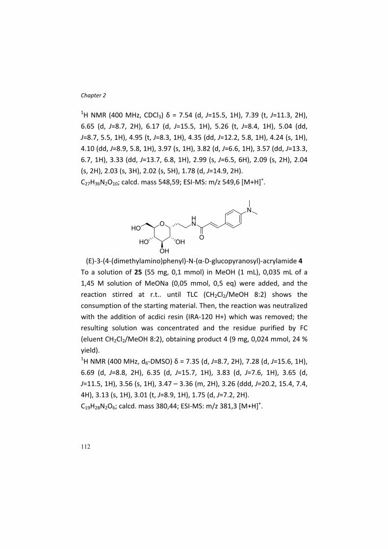
Chapter 2
112
1H NMR (400 MHz, CDCl3) δ = 7.54 (d, J=15.5, 1H), 7.39 (t, J=11.3, 2H),
6.65 (d, J=8.7, 2H), 6.17 (d, J=15.5, 1H), 5.26 (t, J=8.4, 1H), 5.04 (dd,
J=8.7, 5.5, 1H), 4.95 (t, J=8.3, 1H), 4.35 (dd, J=12.2, 5.8, 1H), 4.24 (s, 1H),
4.10 (dd, J=8.9, 5.8, 1H), 3.97 (s, 1H), 3.82 (d, J=6.6, 1H), 3.57 (dd, J=13.3,
6.7, 1H), 3.33 (dd, J=13.7, 6.8, 1H), 2.99 (s, J=6.5, 6H), 2.09 (s, 2H), 2.04
(s, 2H), 2.03 (s, 3H), 2.02 (s, 5H), 1.78 (d, J=14.9, 2H).
C27H36N2O10; calcd. mass 548,59; ESI‐MS: m/z 549,6 [M+H]+.
(E)‐3‐(4‐(dimethylamino)phenyl)‐N‐(α‐D‐glucopyranosyl)‐acrylamide 4
To a solution of 25 (55 mg, 0,1 mmol) in MeOH (1 mL), 0,035 mL of a
1,45 M solution of MeONa (0,05 mmol, 0,5 eq) were added, and the
reaction stirred at r.t.. until TLC (CH2Cl2/MeOH 8:2) shows the
consumption of the starting material. Then, the reaction was neutralized
with the addition of acdici resin (IRA‐120 H+) which was removed; the
resulting solution was concentrated and the residue purified by FC
(eluent CH2Cl2/MeOH 8:2), obtaining product 4 (9 mg, 0,024 mmol, 24 %
yield). 1H NMR (400 MHz, d6‐DMSO) δ = 7.35 (d, J=8.7, 2H), 7.28 (d, J=15.6, 1H),
6.69 (d, J=8.8, 2H), 6.35 (d, J=15.7, 1H), 3.83 (d, J=7.6, 1H), 3.65 (d,
J=11.5, 1H), 3.56 (s, 1H), 3.47 – 3.36 (m, 2H), 3.26 (ddd, J=20.2, 15.4, 7.4,
4H), 3.13 (s, 1H), 3.01 (t, J=8.9, 1H), 1.75 (d, J=7.2, 2H).
C19H28N2O6; calcd. mass 380,44; ESI‐MS: m/z 381,3 [M+H]+.

Chapter 2
113
5‐(N,N‐Dimethylamino)‐N,N‐di‐[2‐(‐D‐glucopyranosyl)ethyl]‐1‐
naphthalensulfonamide 5
Compound 5 was isolated as a by‐product during the synthesis depicted
in Scheme 5 (see Results and Discussion); 2 g of crude amine 24
(containing 24 bis as side product) were dissolved in dry CH2Cl2 (15 mL).
An excess of Et3N and dansyl chloride were then added and the reaction
stirred at r.t.. The reaction was followed by TLC (PE/AcOEt 6:4 and
AcOEt/MeOH/AcOH 6:3:1). After 2 h, the reaction was concentrated and
the residue subjected to FC (PE/AcOEt 6:4). 1,07 g (1,1 mmol) of the
peracetylated dimer and 600 mg (0,99 mmol) of the monomer derivative
(compound 1 in peracetylated form) were obtained, as confirmed by ESI‐
MS analysis (m/z 967,3 [M+H]+ for the dimer and 609,2 [M+H]+ for the
monomer). The dimer was directly deprotected with MeONa (6 mL of 1
M solution in MeOH) in MeOH and CH2Cl2 (10 + 5 mL). After 1 h the pH
was adjusted to neutrality adding IRA‐120 H+ resin. After its removal by
filtration, the filtrate was concentrated, to obtain 5 with quantitative
yield. 1H NMR (400 MHz, CD3OD) δ 8.56 (d, J = 8.4 Hz, 1H, CH Ar), 8.29 (d, J =
8.7 Hz, 1H, CH Ar), 8.21 (d, J = 7.3 Hz, 1H, CH Ar), 7.63 – 7.53 (m, 2H, CH
Ar), 7.25 (d, J = 7.5 Hz, 1H, CH Ar), 3.94 – 3.81 (m, 2H, H1 (x2)), 3.75 (d, J

Chapter 2
114
= 11.7 Hz, 2H, H6a (x2)), 3.65 – 3.47 (m, 6H, H6b (x2), H2 (x2), H2’a (x2)),
3.41 (t, J = 9.0 Hz, 2H, H3 (x2)), 3.37 – 3.27 (m, 4H, H5 (x2), H2’b (x2)),
3.20 (t, J = 9.1 Hz, 2H, H4 (x2)), 2.87 (s, 6H, (CH3)2N‐), 2.03 – 1.75 (m, 4H,
H1’a,b (x2)). 13C NMR (101 MHz, CD3OD) δ 153.15 (Cq Ar), 136.13 (Cq Ar), 131.52 (C
Ar), 131.32 (Cq Ar), 131.25 (Cq Ar), 130.91 (C Ar), 129.18 (C Ar), 124.38 (C
Ar), 120.64 (C Ar), 116.45 (C Ar), 75.16 (C3), 74.88 (C1), 74.53 (C5), 72.62
C2), 72.12 (C4), 62.99 (C6), 45.82 ((CH3)2N‐), 45.71 (C2’), 24.92 (C1’).
C28H42N2O12S; calcd. mass 630,71; ESI‐MS: m/z 631,3 [M+H]+ 653,3
[M+Na]+.
The synthesis of compound 26‐30 was already described in Rumio, et al. [13] and La Ferla, et al. [12a].
(2R,4aR,5aR,7R,8aR,9R,9aS)‐7‐(azidomethyl)‐2‐
phenyloctahydrofuro[2',3':5,6]pyrano[3,2‐d][1,3]dioxin‐9‐ol 31a
(2R,4aR,5aR,7S,8aR,9R,9aS)‐7‐(azidomethyl)‐2‐
phenyloctahydrofuro[2',3':5,6]pyrano[3,2‐d][1,3]dioxin‐9‐ol 31 b
Mixture 28 (1115 mg, 4,54 mmol) was dissolved in DMF dry (5 mL, ≈ 1
M). Benzaldehyde dimethylacetal (5,45 mmol, 1,2 eq) and
camphorsulphonic acid (2,27 mmol, 0,5 eq) are added at r.t.. The
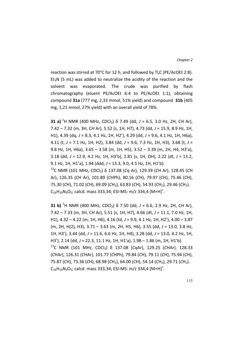
Chapter 2
115
reaction was stirred at 70°C for 12 h, and followed by TLC (PE/AcOEt 2:8).
Et3N (5 mL) was added to neutralize the acidity of the reaction and the
solvent was evaporated. The crude was purified by flash
chromatography (eluent PE/AcOEt 6:4 to PE/AcOEt 1:1), obtaining
compound 31a (777 mg, 2,33 mmol, 51% yield) and compound 31b (405
mg, 1,21 mmol, 27% yield) with an overall yield of 78%.
31 a) 1H NMR (400 MHz, CDCl3) δ 7.49 (dd, J = 6.5, 3.0 Hz, 2H, CH Ar),
7.42 – 7.32 (m, 3H, CH Ar), 5.52 (s, 1H, H7), 4.73 (dd, J = 15.9, 8.9 Hz, 1H,
H1), 4.39 (dq, J = 8.3, 4.1 Hz, 1H, H2’), 4.29 (dd, J = 9.6, 4.1 Hz, 1H, H6a),
4.11 (t, J = 7.1 Hz, 1H, H2), 3.84 (dd, J = 9.6, 7.3 Hz, 1H, H3), 3.68 (t, J =
9.8 Hz, 1H, H6a), 3.65 – 3.58 (m, 1H, H5), 3.52 – 3.39 (m, 2H, H4, H3’a),
3.18 (dd, J = 12.9, 4.2 Hz, 1H, H3’b), 2.81 (s, 1H, OH), 2.22 (dt, J = 13.2,
9.1 Hz, 1H, H1’a), 1.94 (ddd, J = 13.3, 9.0, 4.5 Hz, 1H, H1’b). 13C NMR (101 MHz, CDCl3) δ 137.08 (Cq Ar), 129.39 (CH Ar), 128.45 (CH
Ar), 126.35 (CH Ar), 101.89 (CHPh), 80.16 (CH), 79.97 (CH), 75.46 (CH),
75.30 (CH), 71.02 (CH), 69.09 (CH2), 63.83 (CH), 54.93 (CH2), 29.46 (CH2).
C16H19N3O5; calcd. mass 333,34; ESI‐MS: m/z 334,4 [M+H]+.
31 b) 1H NMR (400 MHz, CDCl3) δ 7.50 (dd, J = 6.6, 2.9 Hz, 2H, CH Ar),
7.42 – 7.33 (m, 3H, CH Ar), 5.51 (s, 1H, H7), 4.66 (dt, J = 11.1, 7.0 Hz, 1H,
H1), 4.32 – 4.22 (m, 1H, H6), 4.16 (td, J = 9.9, 4.1 Hz, 1H, H2’), 4.00 – 3.87
(m, 2H, H(2), H3), 3.71 – 3.63 (m, 2H, H5, H6), 3.55 (dd, J = 13.0, 3.8 Hz,
1H, H3’), 3.44 (dd, J = 11.6, 6.6 Hz, 1H, H4), 3.28 (dd, J = 13.0, 4.2 Hz, 1H,
H3’), 2.14 (dd, J = 22.3, 11.1 Hz, 1H, H1’a), 1.98 – 1.86 (m, 1H, H1’b). 13C NMR (101 MHz, CDCl3) δ 137.08 (CqAr), 129.25 (CHAr), 128.33
(CHAr), 126.31 (CHAr), 101.77 (CHPh), 79.84 (CH), 79.11 (CH), 75.94 (CH),
75.87 (CH), 73.36 (CH), 68.98 (CH2), 64.00 (CH), 54.14 (CH2), 29.71 (CH2).
C16H19N3O5; calcd. mass 333,34; ESI‐MS: m/z 334,4 [M+H]+.

Chapter 2
116
(2R,3aR,5R,6S,7S,7aR)‐2‐(aminomethyl)‐5‐(hydroxymethyl)hexahydro‐
2H‐furo[3,2‐b]pyran‐6,7‐diol 32
To a solution of compound 31b (94 mg, 0,28 mmol) in a mixture of
MeOH/AcOEt (5 + 3 mL) were added few drops of glacial acetic acid and
a catalytic amount of Pd(OH)2/C. The reaction was stirred at r.t. under
Hydrogen atmosphere until TLC (PE/AcOEt 1:1 and
AcOEt/MeOH/H2O/AcOH 6:4:1:1) indicated the absence of starting
material. The reaction is then filtered through a pad of celite and the
solution containing the product was concentrated. 1H‐NMR confirmed
the purity of the deprotected bicyclic sugar that was directly used for the
next reaction without further purification (60 mg, 0,27 mmol, quant.
yield). 1H NMR (400 MHz, CD3OD) δ 4.61 (s, 1H, H1), 4.38 (d, J = 8.7 Hz, 1H, H2’),
3.98 – 3.90 (m, 1H, H2), 3.79 (dd, J = 12.0, 6.8 Hz, 1H, H6a), 3.74 – 3.63
(m, 2H, H3, H4), 3.60 – 3.52 (m, 1H, H5), 3.43 – 3.33 (m, 1H, H6b), 3.13
(dd, J = 13.0, 3.0 Hz, 1H, H3’a), 2.92 (dd, J = 13.1, 8.8 Hz, 1H, H3’b), 2.19
(ddd, J = 13.5, 6.3, 2.6 Hz, 1H, H1’a), 1.86 – 1.75 (m, 1H, H1’b). 13C NMR (101 MHz, D2O) δ 85.34 (CH), 80.54 (CH), 76.40 (CH), 76.09 (CH),
75.35 (CH), 70.23 (CH), 62.68 (CH2), 45.17 (CH2), 36.37 (CH2).
C9H17NO5; calcd. mass 219,24; ESI‐MS: m/z 220,2 [M+H]+.

Chapter 2
117
(2S,3aR,5R,6S,7S,7aR)‐2‐(aminomethyl)‐5‐(hydroxymethyl)hexahydro‐
2H‐furo[3,2‐b]pyran‐6,7‐diol 33
The preparation of compound 33 was performed exactly as described for
compound 32. The catalytic hydrogenolysis (41 mg, 0,123 mmol)
afforded 27 mg (0,123 mmol, quant. yield) of amine 33 that was used
directly for the next reaction. 1H NMR (400 MHz, CD3OD) δ 4.46 (s, 1H, H1), 4.30 (dd, J = 8.7, 4.6 Hz,
1H, H2’), 4.01 (dd, J = 12.1, 8.3 Hz, 1H, H6a), 3.89 – 3.82 (m, 1H, H4), 3.75
(dt, J = 8.1, 3.6 Hz, 2H, H2, H5), 3.62 (dd, J = 12.1, 3.2 Hz, 1H, H6b), 3.54
(t, J = 4.9 Hz, 1H, H3), 3.13 (dd, J = 13.2, 3.6 Hz, 1H, H3’a), 3.05 (dd, J =
13.2, 5.0 Hz, 1H, H3’b), 2.51 – 2.38 (m, 1H, H1’a), 1.96 (s, 1H, H1’b). 13C NMR (101 MHz, D2O) δ 85.24 (CH), 80.52 (CH), 77.03 (CH), 75.40 (CH),
74.99 (CH), 70.02 (CH), 62.32 (CH2), 45.98 (CH2), 36.56 (CH2).
C9H17NO5; calcd. mass 219,24; ESI‐MS: m/z 220,2 [M+H]+.
N‐(((2R,3aR,5R,6S,7S,7aR)‐6,7‐dihydroxy‐5‐(hydroxymethyl)hexahydro‐
2H‐furo[3,2‐b]pyran‐2‐yl)methyl)‐5‐(dimethylamino)naphthalene‐1‐
sulfonamide 6
Biclyclic amine 32 (60 mg, 0,27 mmol) was dissolved in MeOH (5 mL);
Et3N (0,57 mmol, 2,1 eq) and dansyl chloride (0,41 mmol, 1,5 eq) were
added and the reaction was stirred at r.t. until TLC (AcOEt/MeOH 9:1 and
AcOEt/MeOH/H2O/AcOH 6:4:1:1) indicated the end of the reaction. The
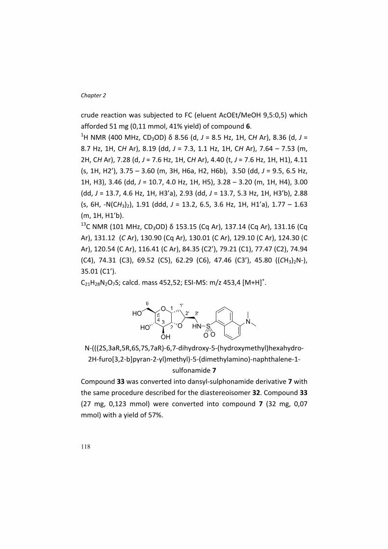
Chapter 2
118
crude reaction was subjected to FC (eluent AcOEt/MeOH 9,5:0,5) which
afforded 51 mg (0,11 mmol, 41% yield) of compound 6. 1H NMR (400 MHz, CD3OD) δ 8.56 (d, J = 8.5 Hz, 1H, CH Ar), 8.36 (d, J =
8.7 Hz, 1H, CH Ar), 8.19 (dd, J = 7.3, 1.1 Hz, 1H, CH Ar), 7.64 – 7.53 (m,
2H, CH Ar), 7.28 (d, J = 7.6 Hz, 1H, CH Ar), 4.40 (t, J = 7.6 Hz, 1H, H1), 4.11
(s, 1H, H2’), 3.75 – 3.60 (m, 3H, H6a, H2, H6b), 3.50 (dd, J = 9.5, 6.5 Hz,
1H, H3), 3.46 (dd, J = 10.7, 4.0 Hz, 1H, H5), 3.28 – 3.20 (m, 1H, H4), 3.00
(dd, J = 13.7, 4.6 Hz, 1H, H3’a), 2.93 (dd, J = 13.7, 5.3 Hz, 1H, H3’b), 2.88
(s, 6H, ‐N(CH3)2), 1.91 (ddd, J = 13.2, 6.5, 3.6 Hz, 1H, H1’a), 1.77 – 1.63
(m, 1H, H1’b). 13C NMR (101 MHz, CD3OD) δ 153.15 (Cq Ar), 137.14 (Cq Ar), 131.16 (Cq
Ar), 131.12 (C Ar), 130.90 (Cq Ar), 130.01 (C Ar), 129.10 (C Ar), 124.30 (C
Ar), 120.54 (C Ar), 116.41 (C Ar), 84.35 (C2’), 79.21 (C1), 77.47 (C2), 74.94
(C4), 74.31 (C3), 69.52 (C5), 62.29 (C6), 47.46 (C3’), 45.80 ((CH3)2N‐),
35.01 (C1’).
C21H28N2O7S; calcd. mass 452,52; ESI‐MS: m/z 453,4 [M+H]+.
N‐(((2S,3aR,5R,6S,7S,7aR)‐6,7‐dihydroxy‐5‐(hydroxymethyl)hexahydro‐
2H‐furo[3,2‐b]pyran‐2‐yl)methyl)‐5‐(dimethylamino)‐naphthalene‐1‐
sulfonamide 7
Compound 33 was converted into dansyl‐sulphonamide derivative 7 with
the same procedure described for the diastereoisomer 32. Compound 33
(27 mg, 0,123 mmol) were converted into compound 7 (32 mg, 0,07
mmol) with a yield of 57%.

Chapter 2
119
1H NMR (400 MHz, CD3OD) δ 8.56 (d, J = 8.5 Hz, 1H, CH Ar), 8.36 (d, J =
8.7 Hz, 1H, CH Ar), 8.20 (dd, J = 7.3, 1.1 Hz, 1H, CH Ar), 7.59 (td, J = 8.8,
7.6 Hz, 2H, CH Ar), 7.28 (d, J = 7.2 Hz, 1H, CH Ar), 4.44 (dd, J = 12.0, 5.3
Hz, 1H, H1), 3.94 – 3.83 (m, 1H, H2’), 3.72 (dd, J = 12.0, 6.4 Hz, 1H, H6a),
3.64 (dd, J = 12.4, 3.2 Hz, 1H, H6b), 3.62 – 3.53 (m, 2H, H2, H3), 3.48 –
3.41 (m, 1H, H5), 3.36 – 3.27 (m, 1H, H4), 3.02 (dd, J = 5.4, 2.4 Hz, 2H,
H3’a, b), 2.88 (s, 6H, ‐N(CH3)2), 2.00 (dt, J = 13.7, 7.0 Hz, 1H, H1’a), 1.71
(ddd, J = 13.1, 7.5, 5.3 Hz, 1H, H1’b). 13C NMR (101 MHz, CD3OD) δ 153.14 (Cq Ar), 136.95 (Cq Ar), 131.16 (Cq
Ar), 131.14 (C Ar), 130.87 (Cq Ar), 130.06 (C Ar), 129.20 (C Ar), 124.28 (C
Ar), 120.49 (C Ar), 116.43 (C Ar), 83.95 (C2’), 78.88 (C1), 78.23 (C2), 75.48
(C4), 74.92 (C3), 69.49 (C5), 62.30 (C6), 47.99 (C3’), 45.79 ((CH3)2N‐),
34.88 (C1’).
C21H28N2O7S; calcd. mass 452,52; ESI‐MS: m/z 453,4 [M+H]+.
Hexaethyleneglycol monomethylsulphonate 38
500 mg (1,77 mmol) of commercially available hexaethyleneglycol and
616 mg of Ag2O (2,7 mmol, 1,5 eq) were dissolved in 18 mL (0,1 M) of
CH2Cl2 under argon atmosphere and MsCl (2,12 mmol, 1,2 eq) was added
to this suspension. The reaction was stirred at r.t. and followed by TLC
(AcOEt/MeOH 9:1). After 12 h, the suspension was filtered through a
celite pad to remove silver oxide and washed with CH2Cl2. The solution
was concentrated and chromatographed on silica gel (eluent
CH2Cl2/MeOH 9,5:0,5). 300 mg of monomesyl hexaethyleneglycol (0,83
mmol) were obtained with a yield of 47%. 1H NMR (400 MHz, CDCl3) δ 4.42 – 4.34 (m, 2H, CH2OMs), 3.78 – 3.73 (m,
2H, CH2OH), 3.73 – 3.69 (m, 2H, CH2CH2OMs), 3.69 – 3.61 (m, 16H, ‐

Chapter 2
120
OCH2CH2O‐), 3.61 – 3.55 (m, 2H, CH2CH2OH), 3.08 (s, 3H, CH3SO2), 2.77
(s, 1H, OH). 13C NMR (101 MHz, CDCl3) δ 72.70, 70.68, 70.65, 70.64, 70.62, 70.59,
70.32, 69.50 (CH2 PEG x 10), 69.09 (CH2OMs), 61.77 (CH2OH), 37.82
(CH3SO2).
C13H28O9S; calcd. mass 360,42; ESI‐MS: m/z 361,2 [M+H]+; 383,2
[M+Na]+; 399,1 [M+K]+
17‐azido‐3,6,9,12,15‐pentaoxaheptadecan‐1‐ol 39
To a solution of compound 38 (283 mg, 0,786 mmol) in DMF, NaN3 (1,97
mmol, 2,5 eq) was added and the reaction was stirred at r.t. for 36 h.
When TLC (AcOEt/MeOH 9,5:0,5) indicated the absence of starting
material and the presence of a new spot with a Rf ≈ 0,4, the reaction was
filtered through a celite pad to remove the excess of sodium azide and
washed with CH2Cl2. The solution was then concentrated and the
product purified by FC (AcOEt/MeOH 9,5:0,5) affording 165 mg (0,53
mmol, 70% yield) of compound 39. 1H NMR (400 MHz, CDCl3) δ 3.75 – 3.69 (m, 2H), 3.69 – 3.62 (m, 18H),
3.62 – 3.56 (m, 2H), 3.38 (t, J = 5.0 Hz, 2H, CH2N3), 2.90 (s, 1H, OH). 13C NMR (101 MHz, CD3OD) δ 73.66, 71.62, 71.58, 71.54, 71.50, 71.38,
71.15 (CH2 PEG x 10), 62.21 (CH2OH), 51.76 (CH2N3).
C12H25N3O9; calcd. mass 307,35; ESI‐MS: m/z 308,3 [M+H]+; 330,3
[M+Na]+ ; 346,3 [M+K]+

Chapter 2
121
17‐amino‐3,6,9,12,15‐pentaoxaheptadecan‐1‐ol 40
Compound 39 (151 mg, 0,49 mmol) was dissolved in MeOH (5 mL, 0,1 M)
and the solution was degased with a stream of argon for 10 min. A
catalytic amount of Pd(OH)2/C was added and the suspension was
vigorously stirred under a hydrogen atmosphere. The hydrogenation was
followed by TLC (CH2Cl2/MeOH 9:1 and CH2Cl2/MeOH 7:3 + 1% NH3 (aq)).
After 15 h, the mixture was filtered on celite and the solvent was
evaporated. The residue, containing the product 40 in a pure form, was
used directly for the next reaction without further purification (quant.
yield). 1H NMR (400 MHz, CDCl3) δ 3.75 – 3.69 (m, 2H, CH2OH), 3.69 – 3.57 (m,
20H, CH2 PEG), 3.32 (s, 3H, OH, NH2), 2.98 – 2.90 (m, 2H, CH2NH2). 13C NMR (101 MHz, CD3OD) δ 73.67 (CH2CH2NH2), 71.59, 71.54, 71.50,
71.39, 71.38, 71.33, 71.22, 71.05, 70.53 (CH2 PEG x 9), 62.07 (CH2OH),
41.37 (CH2NH2).
C12H27NO6; calcd. mass 281,35; ESI‐MS: m/z 282,3 [M+H]+.
5‐(dimethylamino)‐N‐(17‐hydroxy‐3,6,9,12,15‐
pentaoxaheptadecyl)naphthalene‐1‐sulfonamide 41
To a solution of compound 40 (100 mg, 0,356 mmol) in MeOH (4 mL, ≈
0,1 M), Et3N (0,534 mmol, 1,5 eq) and dansyl chloride (0,427 mmol, 1,3
eq) were added. The reaction, followed by TLC (CH2Cl2/MeOH 9:1 and
CH2Cl2/MeOH 7:3 + 1% NH3 (aq)) was stirred at r.t.. After 3 h, the solvent
was removed in vacuo and the residue chromatographed (eluent

Chapter 2
122
CH2Cl2/MeOH 9,5:0,5) affording 110 mg of compound 41 (0,214 mmol,
60%). 1H NMR (400 MHz, CDCl3) δ 8.51 (d, J = 8.5 Hz, 1H, CH Ar), 8.32 (d, J = 8.6
Hz, 1H, CH Ar), 8.23 (dd, J = 7.3, 1.1 Hz, 1H, CH Ar), 7.63 – 7.44 (m, 2H,
CH Ar), 7.17 (d, J = 7.2 Hz, 1H, CH Ar), 5.90 (s, 1H, NHSO2), 3.75 – 3.69 (m,
2H, CH2OH), 3.64 (d, J = 4.8 Hz, 10H, CH2 PEG), 3.62 – 3.56 (m, 4H, CH2
PEG), 3.52 – 3.44 (m, 2H, CH2 PEG), 3.41 – 3.33 (m, 4H, CH2 PEG), 3.11
(dd, J = 10.0, 5.2 Hz, 2H, CH2 PEG), 2.88 (s, 6H, ‐N(CH3)2). 13C NMR (101 MHz, D2O) δ 150.94 (Cq Ar), 134.97 (Cq Ar), 129.67 (C Ar),
129.02 (Cq Ar), 128.95 (Cq Ar), 128.71 (C Ar), 128.28 (C Ar), 123.51 (C Ar),
118.99 (C Ar), 115.31 (C Ar), 71.62 (CH2 PEG), 69.54 (CH2 PEG), 69.45 (CH2
PEG), 69.41 (CH2 PEG), 69.37 (CH2 PEG), 69.27 (CH2 PEG), 69.04 (CH2
PEG), 68.60 (CH2 PEG), 60.23 (CH2NHSO2), 44.70 (‐N(CH3)2).
C24H38N2O8S; calcd. mass 514,63; ESI‐MS: m/z 515,7 [M+H]+.
5‐(dimethylamino)‐N‐(2‐deoxyglucosyl)naphthalene‐1‐sulfonamide 42
To a solution of commercially available D‐glucosamine hydrochloride
(125 mg, 0,58 mmol) in MeOH (2,3 mL, 0,25 M) were added Et3N (1,74
mmol, 3 eq) and dansyl chloride (0,7 mmol, 1.2 eq). The reaction was
stirred at r.t. and followed by TLC (AcOEt/MeOH 9:1 and
AcOEt/MeOH/H2O/AcOH 6:4:1:1). After 12 h, TLC indicated the
disappearance of starting compound, and the solvent was evaporated.
The residue was purified by FC (AcOEt/MeOH 9:1) which afford
compound 42 (163 mg, 0,4 mmol, 68% yield).
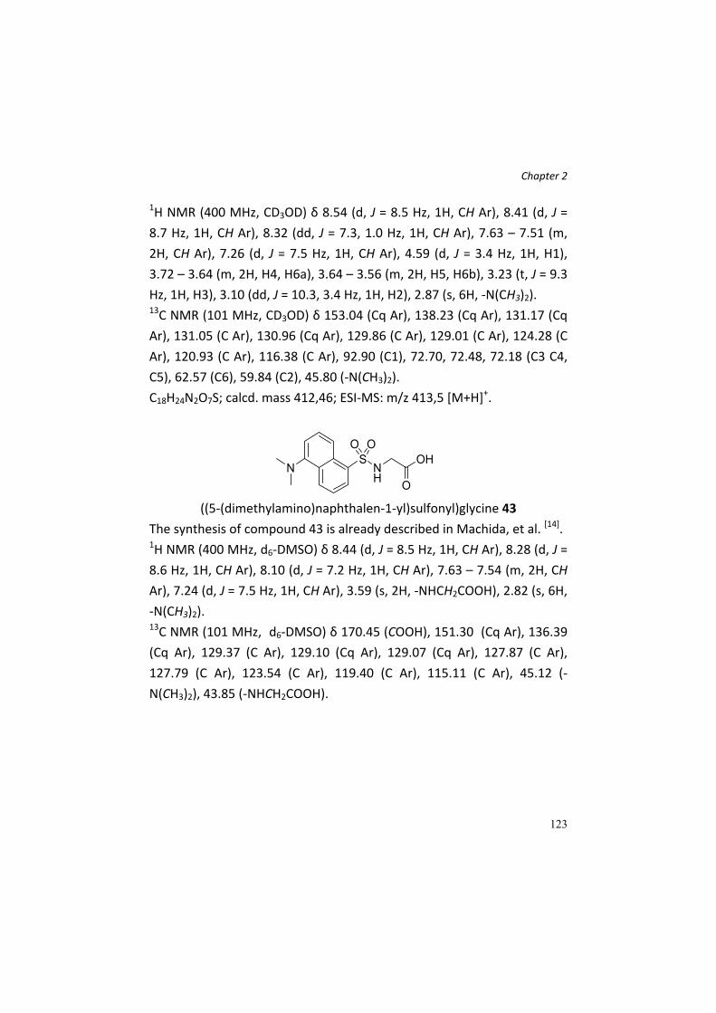
Chapter 2
123
1H NMR (400 MHz, CD3OD) δ 8.54 (d, J = 8.5 Hz, 1H, CH Ar), 8.41 (d, J =
8.7 Hz, 1H, CH Ar), 8.32 (dd, J = 7.3, 1.0 Hz, 1H, CH Ar), 7.63 – 7.51 (m,
2H, CH Ar), 7.26 (d, J = 7.5 Hz, 1H, CH Ar), 4.59 (d, J = 3.4 Hz, 1H, H1),
3.72 – 3.64 (m, 2H, H4, H6a), 3.64 – 3.56 (m, 2H, H5, H6b), 3.23 (t, J = 9.3
Hz, 1H, H3), 3.10 (dd, J = 10.3, 3.4 Hz, 1H, H2), 2.87 (s, 6H, ‐N(CH3)2). 13C NMR (101 MHz, CD3OD) δ 153.04 (Cq Ar), 138.23 (Cq Ar), 131.17 (Cq
Ar), 131.05 (C Ar), 130.96 (Cq Ar), 129.86 (C Ar), 129.01 (C Ar), 124.28 (C
Ar), 120.93 (C Ar), 116.38 (C Ar), 92.90 (C1), 72.70, 72.48, 72.18 (C3 C4,
C5), 62.57 (C6), 59.84 (C2), 45.80 (‐N(CH3)2).
C18H24N2O7S; calcd. mass 412,46; ESI‐MS: m/z 413,5 [M+H]+.
((5‐(dimethylamino)naphthalen‐1‐yl)sulfonyl)glycine 43
The synthesis of compound 43 is already described in Machida, et al. [14]. 1H NMR (400 MHz, d6‐DMSO) δ 8.44 (d, J = 8.5 Hz, 1H, CH Ar), 8.28 (d, J =
8.6 Hz, 1H, CH Ar), 8.10 (d, J = 7.2 Hz, 1H, CH Ar), 7.63 – 7.54 (m, 2H, CH
Ar), 7.24 (d, J = 7.5 Hz, 1H, CH Ar), 3.59 (s, 2H, ‐NHCH2COOH), 2.82 (s, 6H,
‐N(CH3)2). 13C NMR (101 MHz, d6‐DMSO) δ 170.45 (COOH), 151.30 (Cq Ar), 136.39
(Cq Ar), 129.37 (C Ar), 129.10 (Cq Ar), 129.07 (Cq Ar), 127.87 (C Ar),
127.79 (C Ar), 123.54 (C Ar), 119.40 (C Ar), 115.11 (C Ar), 45.12 (‐
N(CH3)2), 43.85 (‐NHCH2COOH).

Chapter 2
124
References [1] S. K. Banerjee, K. R. McGaffin, N. M. Pastor‐Soler, F. Ahmad,
Cardiovascular Research 2009, 84, 111‐118. [2] V. Gorboulev, A. Schuermann, V. Vallon, H. Kipp, A. Jaschke, D.
Klessen, A. Friedrich, S. Scherneck, T. Rieg, R. Cunard, M. Veyhl‐Wichmann, A. Srinivasan, D. Balen, D. Breljak, R. Rexhepaj, H. E. Parker, F. M. Gribble, F. Reimann, F. Lang, S. Wiese, I. Sabolic, M. Sendtner, H. Koepsell, Diabetes 2012, 61, 187‐196.
[3] B. La Ferla, V. Spinosa, G. D'Orazio, M. Palazzo, A. Balsari, A. A. Foppoli, C. Rumio, F. Nicotra, ChemMedChem 2010, 5, 1677‐1680.
[4] M. Palazzo, S. Gariboldi, L. Zanobbio, S. Selleri, G. F. Dusio, V. Mauro, A. Rossini, A. Balsari, C. Rumio, Journal of Immunology 2008, 181, 3126‐3136.
[5] L. C. H. Yu, J. R. Turner, A. G. Buret, Experimental Cell Research 2006, 312, 3276‐3286.
[6] L. C. H. Yu, A. N. Flynn, J. R. Turner, A. G. Buret, Faseb Journal 2005, 19, 1822‐1835.
[7] E. M. Wright, D. D. F. Loo, B. A. Hirayama, Physiological Reviews 2011, 91, 733‐794.
[8] E. Brenna, C. Fuganti, P. Grasselli, S. Serra, S. Zambotti, Chemistry‐a European Journal 2002, 8, 1872‐1878.
[9] G. J. McGarvey, C. A. LeClair, B. A. Schmidtmann, Organic Letters 2008, 10, 4727‐4730.
[10] D. Dubé, A. A. Scholte, Tetrahedron Letters 1999, 40, 2295‐2298. [11] P. Stepanek, O. Vich, L. Kniezo, H. Dvorakova, P. Vojtisek,
Tetrahedron‐Asymmetry 2004, 15, 1033‐1041. [12] aB. La Ferla, F. Cardona, I. Perdiga, F. Nicotra, Synlett 2005, 2641‐
2642; bS. Mari, F. J. Canada, J. Jimenez‐Barbero, A. Bernardi, G. Marcou, I. Motto, I. Velter, F. Nicotra, B. La Ferla, European Journal of Organic Chemistry 2006, 2925‐2933.
[13] C. Rumio, M. Palazzo, A. Balsari, F. Nicotra, B. La Ferla, 2008.

Chapter 2
125
[14] S. Machida, K. Usuba, M. A. Blaskovich, A. Yano, K. Harada, S. M. Sebti, N. Kato, J. Ohkanda, Chemistry – A European Journal 2008, 14, 1392‐1401.
[15] K. C. Nicolaou, C. K. Hwang, M. E. Duggan, Journal of the American Chemical Society 1989, 111, 6682‐6690.
[16] A. A. H. Abdel‐Rahman, E. S. H. El Ashry, R. R. Schmidt, Carbohydr. Res. 1999, 315, 106‐116.

126
Chapter 3
127
Chapter 3. Synthesis of a labeled
SGLT1 ligand for in vitro and in vivo
trafficking studies

Chapter 3
128
Abstract Recently, a novel immunological function was established for the
sodium‐glucose co‐transporter 1 (SGLT1), a protein involved in sugars
absorption in small intestine. High‐glucose dosage and pharmacological
concentrations of C‐glucoside 1 showed a protective role of both in vivo
and in vivo systems treated with several inflammatory insults;
experimental evidences suggest the involvement of SGLT1 in this
process, being this protein “over‐activated” by high glucose and/or C‐
glucoside 1; the mechanism of action at the basis of the protection is still
unclear, and in order to increase our knowledge about the phenomenon
we have developed a synthesis for the preparation of a radiolabelled‐
tritiated analogue of C‐glucoside 1, as radiotracer for cellular – tissue
localization.
Introduction SGLT1 (sodium glucose co‐transporter 1) represents the principal
transport protein for glucose and galactose absorption in mammalian
intestine[1]. It acts translocating two sodium ions and one molecule of
glucose or galactose across the apical membrane of intestinal epithelial
cells, which constitute the brush border membrane of small intestine.
Beyond its crucial physiological function, recently an immunological role
for this transporter has been established[2]. Several works and
experimental evidences suggest its involvement and activation in
glucose‐mediated protection of in vivo and in vitro models of
inflammation states, caused by several insults, like Lipopolysaccharides
(LPS)[2], acetaminophen, D‐glucosamine and alpha‐amanitin[3]. High
glucose concentrations (5 g/L in vitro and 2,5 g/kg in vivo) completely

Chapter 3
129
block the inflammatory response, and this fact seems to be correlated
and related to a hyperactivation of SGLT1. The obvious drawback of this
treatment, that is, its impact in metabolism, has prompted towards the
development of glycomimetics able to act as glucose but at
pharmacological concentration. Among a library of C‐glycosides, we
found that compound 1 showed the best protective and anti‐
inflammatory activity both in vivo and in vitro at dramatic lower
concentrations (5 µg/L in vitro and 25 µg/Kg in vivo), compared to D‐
glucose[4]. C‐glycosides are glycomimetics which do not undergo to
metabolic changes, thus avoiding the side effects associated with a high
glucose concentration therapy.
In these years our work has been focused on the study of the protective
role of compound 1 against several inflammatory states, also in vivo, as
Chron disease, ulcerative colitis and mucositis, trying to understand and
clarify the mechanism of action and the molecular features at the basis
of its function. In particular, our attention has been focused on the role
of SGLT1, since we have hypothesized a completely new biological role
for this protein. To date, we do not possess any information about
putative binding site and interaction data about SGLT1 and the bioactive
C‐glycoside.
Figure 45 ‐ Structure of C‐glycoside 1.
In our opinion, one of the most important aspect is to understand the
nature of the interaction between SGLT1, the molecular target, and C‐
glycoside 1. In order to do this, we decided to synthesize a radiolabeled
Chapter 3
130
C‐glycoside 1 derivative, to exploit in radioactivity experiments. The use
of radiolabeled compounds is a reliable approach for trafficking studies,
widely exploited in medicinal and biological chemistry[5] [6]. This allows to
understand the biological destiny of the molecule, in particular if it is
transported inside the cell and thus absorbed.
Results and discussion C‐glycoside 1 (Figure 45) is a glucose analogue bearing a dansyl residue
connected to a C‐ethyleneamine spacer fixed at the anomeric position.
We developed different synthetic approaches for this compound, both
with and without protecting groups. In order to prepare a radiolabeled
derivative, we decide to modify the molecule introducing a tritium (3[H2])
atom in position C6 of the sugar region, since it is certainly the most
accessible and workable point of the molecule. The synthetic procedure
for this purpose was designed in order to introduce the labeling at the
end of the synthesis so to limit the manipulation of the radiolabelled
entity. The aim was to generate a precursor bearing an aldehyde at C6,
that could be exploited for the introduction of the tritium through a
reduction with tritiated form of sodium borohydride (NaB3[H2]4), a cheap
and commercially available radiolabeled reagent. The synthetic scheme
and reaction conditions were set up using the corresponding deuterated
reagents. For this synthesis a protecting group strategy (Scheme 9) was
exploited. As shown in the retrosynthetic scheme (Scheme 8), the
desired deprotected aldehyde precursore could be obtained from the
corresponding acetal, bearing a C‐ethyleneamine, which can be
chemoselectively derivatized with the dansyl moiety. The latest
intermediate could be easily obtained from the corresponding protected

Chapter 3
131
alcohol. Finally the C6 acetal could be synthesized from the orthogonally
protected C‐allyl glucose.
Scheme 8 – Retrosynthetic strategy.
Starting from 2,3,4,6‐tetra‐O‐benzyl‐α‐C‐allyl‐D‐glucopyranoside 2[7] an
acetolysis reaction was performed, affording the 6‐O‐acetyl derivative 3.
The following Zemplèn deacetylation[8] allowed to obtain the
deprotected primary alcohol 4, which was rapidly oxidized to the
aldehyde 5 with Dess‐Martin periodinane. The product was not purified
and used directly for the next reaction, where it was converted into the
dimethylacetal derivative 6, according to a procedure of Deleuze, et al. [9]. Once obtained the protected aldehyde, the synthetic attention was
moved to the conversion of the allylic group, first oxidizing it into the
aldehydic derivative 7, then reducing it to alcohol 8. This group was
successively converted into the azido derivative 9 by a Mitzunobu
reaction; a catalytic hydrogenation/hydrogenolysis on this compound
allowed the removal of the benzyl ethers and the azide reduction,
affording deprotected amine 10. This was reacted with dansyl chloride to
generate the dansyl derivative 11, whose dimethylacetal function was
subjected to acidic cleavage, to restore the aldehyde group, directly

Chapter 3
132
reacted with sodium borodeuteride to give the 2[H]2‐derivative 12 of C‐
glycoside 1. This passage was carried out in order to set up the chemical
condition for the Tritium atom insertion. Consequently, intermediate 11
can be used at the same manner to generate, with NaBT4, the tritiated
analogue 13.
Scheme 9 ‐ Reagents and conditions: a) Ac2O/TFA 4:1, 0°C, 1,5 h, 88%; b) MeONa, MeOH, r.t., 1 h, quant.; c) Dess‐Martin periodinane, CH2Cl2, r.t., 1,5 h; d) CSA, MeOH, 50°C, 2h, 62% (two steps); e) OsO4, NaIO4, H2O/THF/Acetone, r.t., 24 h; f) NaBH4, CH2Cl2/EtOH, r.t., 2,5 h, 65% (two steps); g) Ph3P, DIAD, (PhO)2PON3, THF dry, 0°C to r.t., 2 h, 60%; h) H2 atm., Pd(OH)2/C, MeOH/AcOEt, r.t., 48 h; i) dansylCl, Et3N, H2O/THF, r.t., 2 h, 22% (two steps); l) HCl, H2O, 2h then NaBD4.

Chapter 3
133
Conclusions The synthesis of the deuterated derivative of compound 1 was
successfully carried out. In order to generate the corresponding
radiolabelled compound, for in vivo and in vitro trafficking studies, the
last reaction will be performed employing the corresponding tritiated
reagent. These studies will allow to understand if the protective C‐
glucoside, which seems to be a SGLT1 agonist, simply binds to the
transporter or if it is also translocated inside cells. This represents a
fundamental information to add to our still incomplete knowledge map
about the novel established biological role of SGLT1.
Experimental Section General remarks.
All commercial chemicals were purchased from Sigma‐Aldrich. All
chemicals were used without further purification. All required anhydrous
solvents were dried with molecular sieves for at least 24h prior to use.
Thin layer chromatography (TLC) was performed on silica gel 60 F254
plates (Merck) with detection under UV light when possible, or by
charring with a solution of (NH4)6Mo7O24 (21g), Ce(SO4)2 (1g),
concentrated H2SO4 (31 mL) in water (500 mL) or with an ethanol
solution of ninhydrin or with Dragendorff’ spray reagent. Flash‐column
chromatography was performed on silica gel 230–400 mesh (Merck). 1H
and 13C NMR spectra were recorded at 25°C, unless otherwise stated,
with a Varian Mercury 400‐MHz instrument. Chemical shift assignments,
reported in parts per million, were referenced to the corresponding
solvent peaks. Mass spectra were recorded on a QTRAP system with ESI
source.

Chapter 3
134
Synthetic procedures
2,3,4‐tri‐O‐benzyl‐6‐O‐acetyl‐α‐C‐allyl‐glucopyranoside 3
A mixture of Ac2O/TFA 4:1 (70 mL), prepared at 0°C under argon
atmosphere was added via a double tip needle to a round bottom flask
containing 2 g (3,55 mmol) of 2,3,4,6‐tetra‐O‐benzyl‐α‐D‐glucopyranose.
The solution was stirred vigorously at 0°C and the reaction followed by
TLC (PE/AcOEt 8,5:1,5). After 1,5 h, no more starting compound is
present and the solution is poured into ice‐water and stirred for 10 min.
The aqueous solution was extracted with AcOEt (3x) and the organic
phase was then washed once with a sodium hydrogen carbonate
saturated solution and twice with distilled water. After anhydrification,
filtration and concentration of the remaining organic layer, the crude
was purified by FC (PE/AcOEt 9:1), affording compound 3 (1,61 g, 3,12
mmol, 88% yield). 1H NMR (400 MHz, CDCl3) δ 7.43 – 7.24 (m, 15H, CH Ar), 5.79 (ddt, J =
17.2, 10.2, 6.9 Hz, 1H, H2’), 5.18 – 5.05 (m, 2H, H3’a,b), 4.98 (d, J = 10.8
Hz, 1H, CH2Ph), 4.89 (d, J = 10.7 Hz, 1H, CH2Ph), 4.83 (d, J = 10.8 Hz, 1H,
CH2Ph), 4.72 (d, J = 11.6 Hz, 1H, CH2Ph), 4.65 (d, J = 11.6 Hz, 1H, CH2Ph),
4.57 (d, J = 10.8 Hz, 1H, CH2Ph), 4.24 (d, J = 3.5 Hz, 2H, H6a,b), 4.11 (dt, J
= 9.3, 5.9 Hz, 1H, H1, H1), 3.85 (t, J = 9.0 Hz, 1H, H3), 3.76 (dd, J = 9.4, 5.8
Hz, 1H, H2), 3.70 (dt, J = 9.8, 3.5 Hz, 1H, H5), 3.48 (dd, J = 9.8, 8.7 Hz, 1H,
H4), 2.50 (t, J = 8.2 Hz, 2H, H1’a,b), 2.04 (s, 3H, CH3CO). 13C NMR (101 MHz, CDCl3) δ 170.92 (CH3CO), 138.59 (Cq Ar), 138.15 (Cq
Ar), 137.83 (Cq Ar), 134.40 (C2’), 128.80, 128.65, 128.59, 128.58, 128.49,
128.29, 128.11, 128.05, 128.00, 127.95, 127.85 (C Ar x 15), 117.24 (C3’),
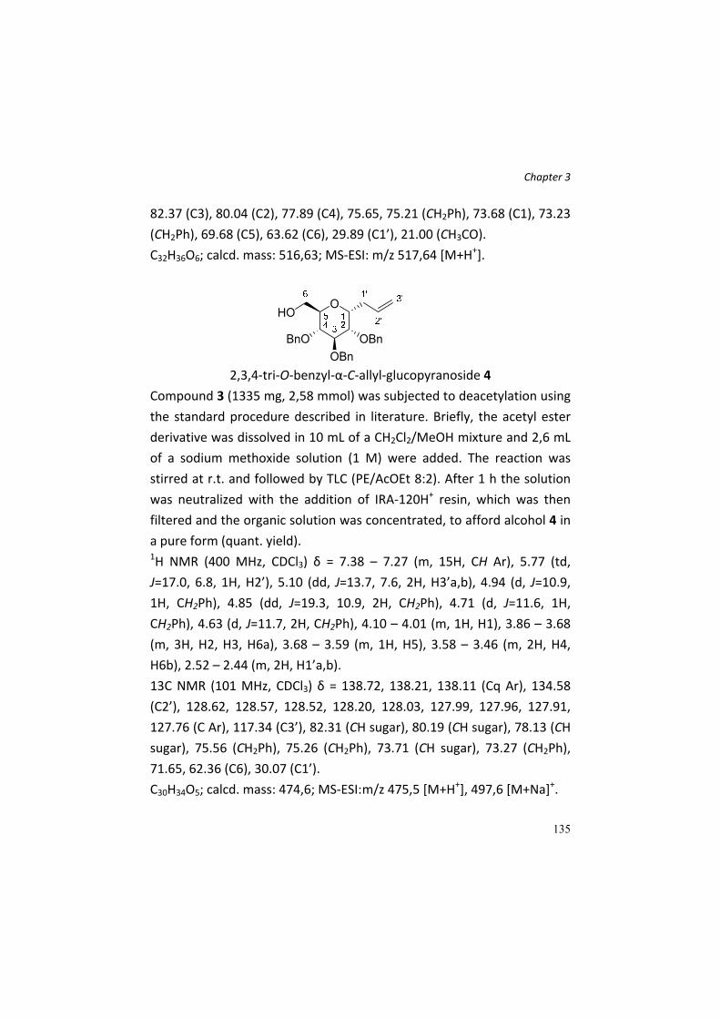
Chapter 3
135
82.37 (C3), 80.04 (C2), 77.89 (C4), 75.65, 75.21 (CH2Ph), 73.68 (C1), 73.23
(CH2Ph), 69.68 (C5), 63.62 (C6), 29.89 (C1’), 21.00 (CH3CO).
C32H36O6; calcd. mass: 516,63; MS‐ESI: m/z 517,64 [M+H+].
2,3,4‐tri‐O‐benzyl‐α‐C‐allyl‐glucopyranoside 4
Compound 3 (1335 mg, 2,58 mmol) was subjected to deacetylation using
the standard procedure described in literature. Briefly, the acetyl ester
derivative was dissolved in 10 mL of a CH2Cl2/MeOH mixture and 2,6 mL
of a sodium methoxide solution (1 M) were added. The reaction was
stirred at r.t. and followed by TLC (PE/AcOEt 8:2). After 1 h the solution
was neutralized with the addition of IRA‐120H+ resin, which was then
filtered and the organic solution was concentrated, to afford alcohol 4 in
a pure form (quant. yield). 1H NMR (400 MHz, CDCl3) δ = 7.38 – 7.27 (m, 15H, CH Ar), 5.77 (td,
J=17.0, 6.8, 1H, H2’), 5.10 (dd, J=13.7, 7.6, 2H, H3’a,b), 4.94 (d, J=10.9,
1H, CH2Ph), 4.85 (dd, J=19.3, 10.9, 2H, CH2Ph), 4.71 (d, J=11.6, 1H,
CH2Ph), 4.63 (d, J=11.7, 2H, CH2Ph), 4.10 – 4.01 (m, 1H, H1), 3.86 – 3.68
(m, 3H, H2, H3, H6a), 3.68 – 3.59 (m, 1H, H5), 3.58 – 3.46 (m, 2H, H4,
H6b), 2.52 – 2.44 (m, 2H, H1’a,b).
13C NMR (101 MHz, CDCl3) δ = 138.72, 138.21, 138.11 (Cq Ar), 134.58
(C2’), 128.62, 128.57, 128.52, 128.20, 128.03, 127.99, 127.96, 127.91,
127.76 (C Ar), 117.34 (C3’), 82.31 (CH sugar), 80.19 (CH sugar), 78.13 (CH
sugar), 75.56 (CH2Ph), 75.26 (CH2Ph), 73.71 (CH sugar), 73.27 (CH2Ph),
71.65, 62.36 (C6), 30.07 (C1’).
C30H34O5; calcd. mass: 474,6; MS‐ESI: m/z 475,5 [M+H+], 497,6 [M+Na]+.

Chapter 3
136
2,3,4‐tri‐O‐benzyl‐6‐oxo‐α‐C‐allyl‐glucopyranoside 5
Alcohol 4 (830 mg, 1,75 mmol) was dissolved in CH2Cl2 (15 mL) and 2,6
mmol (1,5 eq) of Dess‐Martin periodinane were added. The reaction was
stirred at r.t. and followed by TLC (PE/AcOEt 8:2). After 1,5 h, TLC
indicated the absence of the starting material and the formation of a
new spot with a higher Rf, just above those of the starting alcohol. The
reaction is then quenched by the addition of 15 mL of satd. solution of
NaHCO3 and 15 mL of 10% aqueous sodium thiosulphate, and the
reaction was vigorously stirred for 10 min. The mixture was partitioned
between aqueous and organic phases, and the first was extracted (3x)
with CH2Cl2. The combined organic phases were dried over sodium
sulphate and concentrated. The crude residue was analyzed by 1H‐NMR
(δ 9.73 ppm, CDCl3) to check the presence of the aldehydic signal and
immediately used for the next reaction.
2,3,4‐tri‐O‐benzyl‐6,6‐dimethoxy‐α‐C‐allyl‐glucopyranoside 6
The crude aldehyde 5 (912 mg) was dissolved in dry MeOH (20 mL) and
405 mg of camphor sulphonic acid (1,75 mmol, 1 eq relative to alcohol 4)
were added. The reaction was heated and stirred at 50°C until TLC
(PE/AcOEt 8:2) showed no more starting aldehyde. After 2 h, the
reaction was cooled to r.t. and 10 mL of NaHCO3 satd. solution and 20

Chapter 3
137
mL of water were added. The solution was then extracted with AcOEt
(3x). The organic layer was separated, dried and the product purified by
FC (PE/AcOEt 9:1 to 8,5:1,5). 564 mg of dimethylacetal derivative 6 (1,09
mmol, 62% yield over two steps) were obtained. 1H NMR (400 MHz, CDCl3) δ 7.40 – 7.22 (m, 15H, CH Ar), 5.90 – 5.77 (m,
1H, H8), 5.20 – 5.05 (m, 2H, H9a,b), 4.88 (d, J = 11.1 Hz, 1H, CH2Ph), 4.82
(d, J = 11.0 Hz, 1H, CH2Ph), 4.77 (d, J = 11.1 Hz, 1H, CH2Ph), 4.68 (d, J =
11.6 Hz, 1H, CH2Ph), 4.64 (d, J = 11.2 Hz, 1H, CH2Ph), 4.60 (d, J = 11.7 Hz,
1H, CH2Ph), 4.53 (d, J = 2.3 Hz, 1H, H6), 4.14 (dt, J = 10.1, 4.9 Hz, 1H, H1),
3.81 (t, J = 8.4 Hz, 1H, H3), 3.75 – 3.66 (m, 2H, H2, H5), 3.62 (t, J = 8.5 Hz,
1H, H4), 3.40 (s, J = 10.9 Hz, 3H, CH3O‐), 3.38 (s, 3H, CH3O‐), 2.60 – 2.40
(m, 2H, H7a,b). 13C NMR (101 MHz, CDCl3) δ 138.73 (Cq Ar), 138.35 (Cq Ar), 138.28 (Cq
Ar), 134.87 (C8), 128.58, 128.52, 128.51, 128.23, 128.00, 127.95, 127.90,
127.74 (C Ar x 15), 117.14 (C9), 102.28 (C6), 81.39 (C3), 79.29 (C2), 77.97
(C4), 75.20, 74.79 (CH2Ph), 73.58 (C1), 73.08 (CH2Ph), 71.85 (C5), 55.32
(CH3O‐), 55.05 (CH3O‐), 30.51 (C7).
C32H38O6; calcd. mass: 518,65; MS‐ESI: m/z 519,70 [M+H+].
2‐(2,3,4‐tri‐O‐benzyl‐6,6‐dimethoxy‐α‐glucopyranosyl)‐ethanal 7
Compound 6 (560 mg, 1,08 mmol) were dissolved in a H2O/THF/Acetone
solution (4,5:4,5:3 mL). NaIO4 (5,4 mmol, 5 eq) were added and the
suspension was stirred at r.t. for 30 min. Then 0,054 eq of OsO4 (solution
in tBuOH) were dropped in the reaction that was vigorously stirred for at
r.t., following the formation of the product by TLC (PE/AcOEt 6:4). After

Chapter 3
138
24 h, the reaction is concentrated and the aqueous residue was
extracted with AcOEt (3x) and the organic phase back‐extracted twice
with water and brine. The organic solution was dried over Na2SO4 and
concentrated in vacuo, affording crude aldehyde 7 (601 mg) which was
used directly for the next reaction without purification. 1H‐NMR of a
sample of the crude revealed the absence of the allylic bond and the
formation of the aldehyde group (δ 9,71 ppm).
2‐(2,3,4‐tri‐O‐benzyl‐6,6‐dimethoxy‐α‐glucopyranosyl)‐ethanol 8
To a solution of crude compound 7 in CH2Cl2/EtOH (10 + 5 mL), 4,32
mmol (4 eq, relative to compound 6) of NaBH4 were added. The reaction
was stirred at r.t. and followed by TLC (PE/AcOEt 7:3). After 2,5 h, the
solvent was evaporated, the residue resuspended in a solution of
saturated Na2CO3 and stirred for 20 min. The product was extracted
from the aqueous phase with AcOEt (3x), the organic phase was dried,
concentrated and purified by FC (eluent PE/AcOEt 5:5). 365 mg (0,698
mmol) of compound 8 were obtained (65% yield over two steps). 1H NMR (400 MHz, CDCl3) δ 7.41 – 7.24 (m, 15H, CH Ar), 4.83 – 4.75 (m,
2H, CH2Ph), 4.72 – 4.66 (m, 2H, CH2Ph), 4.66 – 4.59 (m, 2H, CH2Ph, C6),
4.59 – 4.52 (m, 1H, CH2Ph), 4.23 – 4.12 (m, 1H, H1), 3.90 – 3.72 (m, 4H,
H3, H4, H5, H8a), 3.63 – 3.53 (m, 2H, H2, H8b), 3.42 (s, 3H, CH3O‐), 3.38
(s, 3H, CH3O‐), 2.16 – 2.03 (m, 1H, H7a), 1.79 – 1.65 (m, 1H, H7b). 13C NMR (101 MHz, CDCl3) δ 138.41, 138.31, 138.08 (Cq Ar), 128.56,
128.55, 128.54, 128.18, 128.12, 128.02, 127.99, 127.95, 127.92, 127.89,
127.85 (C Ar), 102.14 (C6), 79.97 (C3), 78.42 (C2), 76.80 (C4), 74.71

Chapter 3
139
(CH2Ph), 74.19 (CH2Ph), 73.48 (C1), 73.16 (CH2Ph), 72.25 (C5), 61.48 (C8),
56.67 (CH3O‐), 54.54 (CH3O‐), 28.72 (C7).
C31H38O7; calcd. mass: 522,64; MS‐ESI: m/z 523,66 [M+H+].
2‐(2’‐azidoethyl)‐2,3,4‐tri‐O‐benzyl‐6,6‐deoxy‐dimethoxy‐α‐D‐
glucopyranoside 9
Ph3P (1,957 mmol, 3 eq) was added to a solution of alcohol 8 (341 mg,
0,652 mmol) in dry THF (3,23 mL). The solution was cooled to 0°C and
DIAD (1,957 mmol, 3 eq) was added dropwise. After the formation of a
white precipitate, (PhO)2PON3 (2,088 mmol, 3,2 eq) was added and the
reaction was stirred at r.t., following the disappearance of the starting
material by TLC (PE/AcOEt 7:3). After 2 h, the solvent was evaporated
and the crude was loaded on silica gel, performing a FC (eluent PE/AcOEt
9:1 to 6:4) which afforded the desired product 9 (209 mg, 0,38 mmol)
with 60% yield. 1H NMR (400 MHz, CDCl3) δ 7.37 – 7.24 (m, 15H, CH Ar), 4.82 – 4.75 (m,
2H, CH2Ph), 4.74 – 4.61 (m, 3H, CH2Ph), 4.60 – 4.52 (m, 2H, CH2Ph, C6),
4.13 – 4.04 (m, 1H, H1), 3.75 (t, J = 7.7 Hz, 1H, H3), 3.70 – 3.56 (m, 3H,
H2, H4, H5), 3.50 – 3.30 (m, 8H, CH3O‐ x 6, H8a,b), 2.09 – 1.94 (m, 1H,
H7a), 1.91 – 1.79 (m, 1H, H7b). 13C NMR (101 MHz, CDCl3) δ 138.49, 138.33, 138.07 (Cq Ar), 128.59,
128.57, 128.56, 128.15, 128.03, 127.94, 127.91, 127.84 (C Ar), 102.11
(C6), 80.27 (C3), 78.43 (C2), 77.01 (C4), 74.86 (CH2Ph), 74.40 (CH2Ph),
73.12 (CH2Ph), 72.58 (C1), 70.57 (C5), 55.74 (CH3O‐), 54.97 (CH3O‐), 48.06
(C8), 29.84 (C7).

Chapter 3
140
C31H37N3O6; calcd. mass: 547,65; MS‐ESI: m/z 548,66 [M+H+].
α‐C‐(1’‐ethylamino)‐6,6‐dimethoxy‐D‐glucopyranoside 10
Compound 9 (209 mg, 0,38 mmol) was dissolved in a mixture of
MeOH/AcOEt (5 + 5 mL) and the solution was degased under vacuum. A
catalytic amount of Pd(OH)2/C was added and the reaction was stirred
vigorously at r.t. under hydrogen atmosphere. After 48 h, the catalyst
was removed by filtration through a pad of celite and the filtrate was
concentrated. 1H‐NMR analysis was performed on the crude to confirm
the complete debenzylation of the molecule. The crude amine was used
directly for the next reaction.
α‐C‐[(ethylen‐2’‐(N,N‐Dimethylamino)‐N‐naphthalensulfonamidyl]‐6,6‐
dimethoxy‐D‐glucopyranoside 11
Crude amine 10 (69 mg) was dissolved in a mixture of water/THF. Et3N
(0,76 mmol, 2 eq related to compound 9) and 0,57 mmol (1,5 eq) of
dansyl chloride were added to the reaction that was stirred at r.t. and
followed by TLC (CH2Cl2/MeOH/NH3 (aq) 5:5:1 and AcOEt/MeOH 9:1).
After 2 h, the solvent was removed and the product purified by FC
(eluent AcOEt/MeOH 9,5:0,5) obtaining 39,7 mg (0,082 mmol, 22% yield
over two steps) of a mixture of compound 11 and the hydrated form of

Chapter 3
141
the aldehyde, in a ratio, as determined by 1H‐NMR, of ≈ 70:30
(dimethylacetal:hydrated aldehyde).
1H NMR (400 MHz, CD3OD) δ 8.56 (t, J = 7.6 Hz, 1H), 8.34 (d, J = 8.7 Hz,
1H), 8.20 (dd, J = 7.3, 1.0 Hz, 1H), 7.63 – 7.51 (m, 2H), 7.25 (d, J = 7.5 Hz,
1H), 4.64 (d, J = 3.7 Hz, 1H), 3.82 – 3.73 (m, 1H), 3.55 – 3.35 (m, 10H),
2.94 (t, J = 6.9 Hz, 2H), 2.87 (s, 6H), 1.79 – 1.60 (m, 2H).
Signals of the aldehyde in hydrated form: δ 8.15 (dd, J = 7.3, 0.9 Hz, 1H,
CH Ar), 4.62 (d, J = 3.7 Hz, 1H, H6), 3.91 – 3.83 (m, 1H, H6), 3.15 – 3.05
(m, 2H, H2’a,b), 1.98 – 1.81 (m, 2H, H1’a,b). 13C NMR (101 MHz, CD3OD, mixture of signals of 11 and its hydrated
aldehyde as contaminant) δ 153.17, 153.11, 145.10, 145.09, 136.76,
135.75, 135.58 (Cq Ar), 131.41, 131.17 (C Ar), 130.91 (Cq Ar), 130.74,
130.33, 129.12, 129.06, 124.31, 120.86, 120.43, 116.41 (C Ar), 104.69,
104.16 (C6), 74.61, 74.46, 74.44, 74.16, 73.93, 72.24, 72.16, 71.72, 71.46
(CH sugar), 56.40, 56.32, 55.65, 55.32 (CH3OCH‐), 45.79 ((CH3)2N‐), 41.59
(C2’), 26.98, 25.51 (C1’).
C22H32N2O8S; calcd. mass: 484,56; ESI‐MS: m/z 485,3 [M+H]+, 507,4
[M+Na]+, 479,3 [M+H]+ (aldehyde in hydrated form).
[2H2]‐α‐C‐[(ethylen‐2’‐(N,N‐Dimethylamino)‐N‐naphthalensulfonamidyl]‐
D‐glucopyranoside 12
Compound 11 (162) (1 mg) was dissolved in 0,5 mL of distilled water;
0,05 mL of HCl 2 M were added to the solution, stirred at r.t. for 24 h.
The reaction was followed by TLC (CH2Cl2/MeOH), which indicated the
formation of a new product with a minor Rf respect to starting

Chapter 3
142
compound. ESI‐MS spectrum of the crude revealed a m/z signal of 439,3
[M+H]+, confirming the conversion of the dimethylacetal into aldehyde
group. The pH of the solution was then adjusted to 6 adding drops of
NaOH 4 M. NaBD4 (0,009 mmol, 4 eq) was added to the solution, stirred
at r.t. for 1 h. TLC (CH2Cl2/MeOH 8:2) indicated the complete
transformation of the aldehydic intermediate into a more polar
compound, with a Rf identical to that of the H6‐analogue of the
compound (compound 1). A Mass Spectrometry analysis of the solution
revealed the presence of a molecule with a m/z of 442,4 (M + 1).
C20H27DN2O7S; calcd. mass: 441,52 ESI‐MS: m/z 442,4 [M+H]+, 464,4
[M+Na]+.
References [1] E. M. Wright, B. A. Hirayama, D. F. Loo, Journal of Internal
Medicine 2007, 261, 32‐43.
[2] M. Palazzo, S. Gariboldi, L. Zanobbio, S. Selleri, G. F. Dusio, V.
Mauro, A. Rossini, A. Balsari, C. Rumio, Journal of Immunology
2008, 181, 3126‐3136.
[3] L. Zanobbio, M. Palazzo, S. Gariboldi, G. F. Dusio, D. Cardani, V.
Mauro, F. Marcucci, A. Balsari, C. Rumio, Am. J. Pathol. 2009, 175,
1066‐1076.
[4] B. La Ferla, V. Spinosa, G. D'Orazio, M. Palazzo, A. Balsari, A. A.
Foppoli, C. Rumio, F. Nicotra, ChemMedChem 2010, 5, 1677‐
1680.
[5] C. Zona, B. La Ferla, Journal of Labelled Compounds &
Radiopharmaceuticals 2011, 54, 629‐632.
[6] Y. Kuang, N. Salem, D. J. Corn, B. Erokwu, H. Tian, F. Wang, Z. Lee,
Molecular Pharmaceutics 2010, 7, 2077‐2092.

Chapter 3
143
[7] G. J. McGarvey, C. A. LeClair, B. A. Schmidtmann, Organic Letters
2008, 10, 4727‐4730.
[8] Z. Wang, in Comprehensive Organic Name Reactions and
Reagents, John Wiley & Sons, Inc., 2010.
[9] A. Deleuze, M. Sollogoub, Y. Bleriot, J. Marrot, P. Sinay, European
Journal of Organic Chemistry 2003, 2678‐2683.

144
Chapter 4
145
Chapter 4. Generation of gold
nanoparticles decorated with
synthetic ligands of co‐transporters
SGLT‐1 and B0AT1 for the
investigation of the multivalent‐
synergistic effect.

Chapter 4
146
Abstract D‐Glucose, C‐glucoside 1 and L‐glutamine have been derivatized with
suitable thiol ending spacers‐linkers and have been used for the surface
functionalization of gold nanoparticles (AuNPs). In particular C‐glucoside
1, D‐glucose and L‐glutamine decorated nanoparticles have been
generated as molecular tools to investigate the existence of a possible
multivalency effects towards the co‐transporters, respectively the
sodium glucose co‐transporter 1 (SGLT1) and sodium glutamine co‐
transporter (B0AT1). Moreover, gold nanoparticles decorated with a
combination of the three ligands have been generated as tool study
potential synergistic effects. In vitro biological evaluations are ongoing
on Caco 2 cell line expressing both SGLT1 and B0AT1 transporters.
Introduction Recent papers outline the protective effect of sodium‐glucose
cotransporter (SGLT1) engagement with high oral doses of D‐glucose and
non metabolizable 3‐O‐methyl‐D‐glucopyranose (3OMG) on damages
induced by Toll‐Like Receptors (TLRs) ligands in intestinal epithelial cells,
in a murine model of septic shock and in LPS‐induced liver injury, as well
as liver injury and death induced by an overdose of acetaminophen[1].
This was found being due to glucose‐induced down‐regulation of
systemic production of inflammatory cytokines and enhanced
production of anti‐inflammatory cytokines. However, the high amounts
of D‐glucose and 3OMG necessary to induce anti‐inflammatory effects
represent a serious limitation for a hypothetical therapeutic application.
In a previous work we identified a glucose derivative, C‐glucoside 1
(Figure 46), exerting anti‐inflammatory activity at a molar concentration
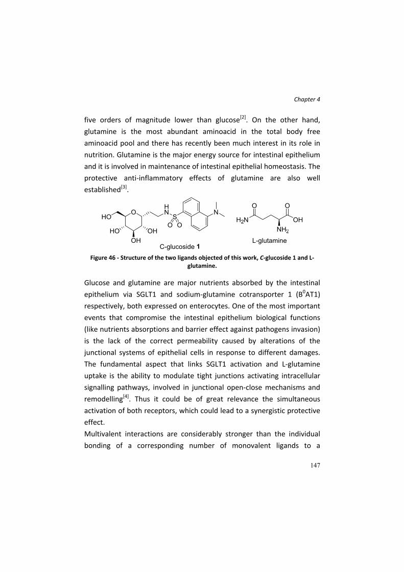
Chapter 4
147
five orders of magnitude lower than glucose[2]. On the other hand,
glutamine is the most abundant aminoacid in the total body free
aminoacid pool and there has recently been much interest in its role in
nutrition. Glutamine is the major energy source for intestinal epithelium
and it is involved in maintenance of intestinal epithelial homeostasis. The
protective anti‐inflammatory effects of glutamine are also well
established[3].
Figure 46 ‐ Structure of the two ligands objected of this work, C‐glucoside 1 and L‐
glutamine.
Glucose and glutamine are major nutrients absorbed by the intestinal
epithelium via SGLT1 and sodium‐glutamine cotransporter 1 (B0AT1)
respectively, both expressed on enterocytes. One of the most important
events that compromise the intestinal epithelium biological functions
(like nutrients absorptions and barrier effect against pathogens invasion)
is the lack of the correct permeability caused by alterations of the
junctional systems of epithelial cells in response to different damages.
The fundamental aspect that links SGLT1 activation and L‐glutamine
uptake is the ability to modulate tight junctions activating intracellular
signalling pathways, involved in junctional open‐close mechanisms and
remodelling[4]. Thus it could be of great relevance the simultaneous
activation of both receptors, which could lead to a synergistic protective
effect.
Multivalent interactions are considerably stronger than the individual
bonding of a corresponding number of monovalent ligands to a

Chapter 4
148
multivalent receptor and are often used in biological systems[5],
particularly for cellular recognition processes and in signal transduction.
Receptor dimerization/clustering is a well‐known and ubiquitous
phenomenon. Several reports have suggested that functional SGLT1 is an
oligomeric protein, resulting by homodimerization of two identical
subunits[6]. Evidence also suggest that the B0AT1 transporter is mainly
assembled as a cluster[7], and, moreover, that the B0AT1 transporter
localized in the small intestine displays a low affinity constant for the
best aminoacid substrate[7]. These findings suggest that the study of
multivalent effects of the single receptor ligand C‐glycoside 1 or
glutamine could lead to a significant increase in the observed activity.
With these preliminary data in hand, and with the above mentioned
rational, our research topic consists in the study and evaluation of
possible multivalent and synergistic effects operated by glucose‐like
ligands (C‐glucoside 1 and other derivatives) in combination with
glutamine and/or glutamine‐like compounds in the preservation and/or
recovery of the intestinal epithelium damaged by inflammation (induced
by LPS and/or its chemotherapeutics agents). To reach our goal we
selected gold nanoparticles as multivalent ligand presenting scaffold, for
straightforward decoration with molecules bearing a suitable thiol
functionalized spacer‐linker. Moreover, the possibility to simultaniously
introduce different thiol ligands, makes them suitable for the generation
of molecular tools for the study of potential synergistic effects, exploiting
the already prepared ligands without additional orthogonal functional
group derivatization.

Chapter 4
149
Results and Discussion In the first phase of the work, our intent was to prepare a set of different
surface‐functionalized gold nanoparticles in order to study both the
multivalent and the synergistic effect of 1 and L‐Gln. We designed and
prepared a set of thiol‐functionalized derivatives of both the ligands
(Figure 47) in order to have the suitable compounds for the preparation
of the corresponding gold nanoparticles (AuNP) (Figure 48)[8]. We
designed the synthesis of the derivatives taking into account the
attachment of a suitable spacer / linker bearing a terminal thiol group.
This linker ensures a correct spatial separation between the core of the
gold nanoparticle and the ligand, which is necessary for the interaction
of the bioactive molecules with the receptor of interest. The linker we
chose is made of a triethyleneglycol (TEG) chain and a pentenyl (C5)
spacer. Polyethylene glycol is bio‐friendly polymer, it is extensively used
in pharma industries, for its low toxicity, excellent solubility in aqueous
media, and seems to be neither immunogenic nor allergenic[9] [10].
Moreover, PEG represents an inert polymer (is a polyether structure)
and is not metabolized. PEGylation is an important and emerging aspect
of the drug delivery field; the conjugation of PEG to drugs leads to an
increase of pharmacokinetics and pharmacodynamics properties of the
molecule to which it is conjugated. The hydrophobicity of the C5 chain is
necessary to allow a good attachment of the ligand to the core of the Au‐
NP.
Concerning compound 1 derivative, we decided to connect the whole
linker to the hydroxyl group on C‐6 position of the pyranose ring, for two
main reasons: firstly, this position bears a primary hydroxyl group, more
easily functionalizable rather than the other secondary OH groups of the
ring; secondly, several works suggest that the C6 OH group of D‐glucose
is not crucial for the binding to SGLT1 interaction and translocation site.

Chapter 4
150
In our opinion, a presence of a linear chain should not affect the
biological activity of the molecule. For L‐glutamine ligands, we designed
two different derivatives, bearing respectively the TEG‐C5 chain on the
nitrogen atom of the amide of lateral chain or connecting it directly to
the Cα position of the aminoacids, with a new amide function. The choice
to synthesized two L‐Gln derivatives with the TEG chains on two different
positions of the aminoacidic structure was made because we have no
information about how L‐Gln interacts with B0AT1 (i.e. which groups are
involved in the binding). The synthesis of both C‐glucoside 1 and L‐Gln
derivative proceeded with a protecting group strategy, starting from
commercially available O‐Me‐α‐D‐glucopyranoside and protected L‐
glutamine compounds. The presence of the ending thiol group was
ensured and achieved by transforming a terminal olefinic appendage, on
the C5 chain, into a thioester group. This was obtained exploiting the so
called “Thiol‐ene coupling” (TEC) approach[11]. Many synthetic
procedures allow the conversion of a terminal alkene into a thioether
linkage, including the reaction of the thiol‐bearing compound and the
alkenyl group in presence of a radical initiator, as AIBN, often heating the
reaction to reflux, under argon atmosphere and with degased solvents.
We decided to exploit the use of a radical photoinitiatior, called 2,2‐
dimethoxy‐2‐acetophenone (DPAP) which allows the thiol‐ene reaction
in a faster and more simple manner. It permits the use of not‐degased
solvents and water, the presence of air, and the proceeding the reaction
at room temperature; only a short (generally 30 min – 1 hour) UV‐light
exposure at 365 nm (using a simple UV‐light lamp), thus close to visible
wavelengths, is required.

Chapter 4
151
Figure 47 – Thiol‐functionalized ligands for the preparation of the Au‐NPs.
During the second phase, the objective consisted in the preparation,
purification and structural characterization of gold nanoparticles (Figure
48) loaded with:
Compound 1 derivative;
L‐glutamine derivatives;
both compound 1 and L‐glutamine derivatives.
Figure 48 ‐ General structure of the designed nanoparticles.

Chapter 4
152
Preparation of ligands
Concerning the synthesis of compound 1 derivative, the following
synthetic steps have been carried out (Scheme 10). O‐Me‐α‐D‐
glucopyranoside was subjected to a C‐allylation according to the
procedure described in Cardona and La Ferla [12] (compound 2). Then the
respective perbenzylated compound 3 was obtained (McGarvey, et al. [13]), and an acetolysis reaction in presence of trifluoroacetic acid and
acetic anhydride allowed the removal of benzyl protecting group from
the hydroxyl group in C‐6 position and the contemporary transformation
into an acetylester group (compound 4). Then the allylic appendage was
oxidized to aldehyde 5, reduced to alcohol 6 and converted to azidoethyl
derivative 7. The acetyl group was successively removed from the OH in
C6 position (8), which was alkylated with the electrophilic PEG‐C5 chain
bearing a methyl‐p‐toluen‐sulphonate function as leaving group. The
spacer was rapidly synthesized starting from TEG, then monoalkylated
with a 4‐pentenyl chain (9). The reaction with tosyl chloride afforded 10.
In our first planned synthesis, we subjected this compound to BCl3‐
promoted benzyl deprotection, but we observed also the cleavage of the
polyethyleneglycol chain. Hence, we decided first of all to subject the
TEGylated sugar derivative to a radical‐mediated reaction. Upon UV
exposure at 365 nm of wavelength and adding a radical photoinitiator, as
DPAP (2,2‐dimethoxyphenyl‐2‐acetophenone), the allyl group was
converted into the thioester over 1 h of reaction, to afford compound
12. Successively, we tried both catalytic hyrogenolysis and Birch
conditions for the azide reduction and benzyl cleavage, but, surprisingly,
in both cases a complete removal of the thioester group, generating a
pentyl ending chain, occurred. By the way, the desulfurization of
thioester by SET (single electron transfer) processes is described and
rationalized in literature[14]. Therefore, we changed again the strategy,

Chapter 4
153
by deprotecting the thioester in basic conditions and promoting the
disulphide bridge formation, in order to obtain a sort of “protected” thiol
(compound 13). Birch reaction on this compound led to the desired fully
deprotected dimer 14, of which the two amine functionalities were
transformed into dansylsulphonamides, affording the final dimer 15. This
compound was used directly for the generation of the nanoparticles: it
is possible to use both thiol‐free and disulphide bearing molecules, since
the addition of an excess of hydride reagents (often NaBH4) leads to the
disulphide bridge reduction.
Scheme 10 ‐ Reagents and conditions: a) i. BSTFA, CH3CN, reflux, 3 h; ii. AllylTMS, TMSOTf, 0°C to r.t., 12 h
[12]; b) BnBr, NaH, DMF dry, r.t., 3 h
[13]; c) Ac2O, TFA, 0°C, 1 h,
88%; d) i. O3, CH2Cl2, ‐78°C, 1 h; ii. Ph3P, CH2Cl2, ‐78°C to r.t. 24 h, 82%; e) NaBH4, MeOH, 0°C, 2 h, 95%; f) DIAD, Ph3P, (PhO)2PON3, THF dry, 0°C to r.t., 2 h, 90%; g) MeONa, MeOH dry, r.t., 1 h, 95%; h) 5‐Bromo‐1‐pentene, NaOH, neat, 100°C, 14 h, 74%; i) TsCl, Et3N, DIPEA, CH2Cl2, r.t., 3 h, 72%; l) NaH, DMF dry, r.t., 12 h, 83%; m) AcSH, DPAP, CH2Cl2, r.t., UV light (365 nm), 1 h, 85%; b) MeONa, MeOH, r.t., 16 h; c) Na, NH3(l), THF dry, ‐78°C, 15 min, then NH4Cl, r.t., 65%; d) dansyl chloride, Et3N, MeOH, r.t., 5 h, 39%.

Chapter 4
154
The synthesis of the L‐glutamine derivatives proceeded with the
following steps (Scheme 11 and Scheme 12). The tosyl group on the C5‐
TEG chain was converted into the azide function, which was reduced to
amine (17). A coupling between this compound and Fmoc‐L‐Gln‐Trt‐OH
afforded the derivative 18, which was subjected to protecting group
cleavage both in basic and acid conditions (compound 19). The thiol‐ene
coupling with thioacetic acid gave thioester 20 rapidly cleaved in basic
condition to afford dimer 21 (as resulted from NMR analysis), directly
used for the nanoparticle preparation. The coupling of the amine 17 with
Boc‐L‐Glu‐O‐tBu (Boc‐L‐glutamic acid 1‐tert‐butyl ester) gave
intermediate 22, which was deprotected by acidic conditions. The
successive TEC with thiolacetic acid afforded compound 24. In this case
the thioester deprotection was not preformed since it requires not a
simple methanolysis, but the use of other mild conditions, in order to
avoid the Cα‐racemization of the glutamine. This process did not occur
for the first aminoacidic derivative (21) since after the coupling, the
racemizable carbon atom of the glutamine is located on the α position to
the new amide formed, and not to a carboxylic acid group.
Scheme 11 ‐ Reagents and conditions: a) NaN3, DMF dry, 80°C, 48 h, 78%; b) i. Ph3P, THF, reflux, 3h; ii. H2O, r.t., 12 h, 93%; c) EDC, HOBt, Et3N, DMF dry, 0°C to r.t., 12 h,

Chapter 4
155
79%; d) i. CH2Cl2, TFA, r.t., 2 h; ii. piperidine, DMF, r.t., 12 h, 96%; e) AcSH, DPAP, MeOH, r.t., UV light (365 nm), 1 h, 47%; f) MeONa, MeOH, r.t., 2 h.
Scheme 12 ‐ Reagents and conditions: a) EDC, HOBt, Et3N, DMF dry, 0°C to r.t., 12 h, 95%; b) CH2Cl2, TFA, r.t., 2 h, 30%; b) AcSH, DPAP, MeOH, r.t., UV light (365 nm), 1 h, 53%.
Preparation of the Au Nanoparticles
After the completion of the synthesis of the ligands of interest, the work
was focused on the preparation of various types of gold nanoparticles,
using the prepared ligands and a glucose derivative, O‐(5’‐thiopentyl)‐β‐
D‐glucopyranoside (Glc‐C5‐SH), synthesized according to Buskas, et al. [15]
and Barrientos, et al. [16]
We used this glycoderivative to modulate and improve the water
solubility of the nanoparticles, since compound 1 shows a low solubility
in water (approximately 0,5 mg/mL). Furthermore, Glc‐C5‐SH should
improve the multivalent effect of these derivatives, rather than interfere
with their biological effects, since SGLT1‐mediated anti‐inflammatory
effect is observed with huge amount of orally administered D‐glucose.
The preparation of the gold nanoparticles (Au‐NPs) involved a one‐pot
process, in which the thiol‐ending ligands was dissolved in MeOH with a
gold (III) compound, hydrogen tetrachloro aurate (HAuCl4), to generate a
kind of Au(I) polymer, on which thiol compounds are grafted. The
successive addition of a reducing agent, as NaBH4, provokes the passage

Chapter 4
156
from Au(I) to Au0, with the generation of the gold nanostructure, made
of tens/hundreds of Au atoms and decorated, on the external side, with
the ligands of interest[16] [8b]. Experimental details and procedures on
gold nanoparticle preparation are described in the experimental section.
The prepared Au‐NPs are the follow:
General structure
R = C‐glycoside 1 or
L‐glutamine
derivatives
AuNP1 AuNP2
Au‐NPs with Glc‐C5‐SH
Au‐NPs with Glc‐C5‐SH and 15

Chapter 4
157
AuNP3 AuNP4
Au‐NPs with Glc‐C5‐SH and 21
Au‐NPs with Glc‐C5‐SH, 15 and 21
The obtained nanoparticles, in a black‐powder form, have been
characterized according to the common experimental techniques, as 1H
NMR (with or without water signal suppression), TEM (Transmission
Electron Microscopy) analysis, UV analysis. The NMR analysis is made
dissolving the powder constituted by nanoparticles in deuterium oxide;
the obtained spectrum shows broad typical signals of molecules grafted
onto a solid structure/colloidal structure. If the ligands attached are one
of different types, a preliminary 1H‐NMR analysis of the ligands‐
containing mixture has to be carried out, in order to check the real
ligands ratio with which they are then loaded on the nanostructure.
Comparing the nanoparticle spectrum with the spectrum of the ligand
mixture before the nanoparticle formation it is possible to understand, in
a qualitatively manner, the correct loading of the ligands the NP.
However, the attachment of a particular ligand on a NP depends on
several factors, including the chemical nature of the compound, its
dimension, the spacer length, therefore the ligand loading must be
always verified. For a quantitative data, a proton spectrum of the

Chapter 4
158
washing solution after nanoparticle formation (see procedure in
experimental section) must be performed; comparing the resulting ratio
of this mixture with the initial one is possible to determine in which ratio
the ligands have been loaded.
Nanoparticle characterization 1H‐NMR of the solution of GlcC5‐SH and C‐glycoside 1 derivative (in
CD3OD) before the AuNP2 preparation. Some diagnostic signals are
depicted. From the integration of the signal the ratio between the two
ligands is 80:20 (Glc‐C5‐SH and C‐glycoside 1 derivative).

Chapter 4
159
1H‐NMR spectrum (with water suppression signal sequence) of the final
AuNP2.
UV spectra of the AuNP2 dissolved in water; dilution 1:2 (1 mg in 2 mL of
water, in red) and 1:4 (in green). Blue line is referred to water (blank).

Chapter 4
160
TEM analysis – transmission electron micrographs. On the left,
micrograph of AuNP1. A double distribution of size (approximately 90%
of ≈2 nm size and 10% of ≈5 nm size) can be seen. On the right, high
magnification which shows the crystalline structure of the bigger gold
nanoclusters. Below, the TEM analysis for AuNP 2,3,4 are presented.
AuNP1 AuNP1
AuNP2

Chapter 4
161
AuNP3 AuNP4
Further characterizations for the obtained nanoparticles will be
performed.
Conclusions In this work we have presented the preparation and synthesis of
different ligands that are derivatives of C‐glycoside 1 and L‐glutamine,
both protective agents against inflammatory insults in vitro and in vivo,
as results from several literature data. These compounds were
functionalized with a triethyleneglycol chain and a five‐carbon spacer
with a terminal thiol group in order to ensure the attachment on the
gold nanoparticle, exploiting the affinity between the gold atom and
thiol group, and to provide an adequate distance of these molecule from
the core of the gold nanoparticle, allowing the binding of the ligands to
their respective receptors. These ligands were used for the preparation
of gold nanoparticle. A glucose derivative, with a C5 chain bearing a
terminal thiol group is also used in the preparation of the nanoparticle to
ensure the water solubility of the NPs. The Au‐NPs will be used for
biological studies in order to determine the multivalent/synergic activity

Chapter 4
162
of the ligands attached to the nano‐structure. The anti‐inflammatory and
protective role of the nanoparticles will be verified, in order to observe if
a nano‐structure with ligands attached in a multivalent fashion can
increase the anti‐inflammatory activity of the bioactive molecules.
Furthermore, if a multivalent effect will be observed, decorated
nanoparticles will be used as chemical tools in order to understand the
nature of the multivalent phenomenon. On the basis of the obtained
results, new nanoparticles could be designed and prepared, to enhance
or modulate the anti‐inflammatory activity. The nanoparticles could be
tested against different kind of inflammation in which the protective role
of SGLT1 and B0AT1 is confirmed, as Chron’ disease or IBDs (Intestinal
Bowel Diseases).
Experimental Section
General remarks.
All commercial chemicals were purchased from Sigma‐Aldrich and Alfa
Aesar. All chemicals were used without further purification. Thin layer
chromatography (TLC) was performed on silica gel 60 F254 plates (Merck)
with detection under UV light when possible, or by charring with a
solution of (NH4)6Mo7O24 (21g), Ce(SO4)2 (1g), concentrated H2SO4 (31
mL) in water (500 mL) or with an ethanol solution of ninhydrin. Flash‐
column chromatography was performed on silica gel 230–400 mesh
(Merck) or on Isolera Four Flash Chromatography System (Biotage™). 1H
and 13C NMR spectra were recorded at 25°C, unless otherwise stated,
with a Varian Mercury 400‐MHz instrument and on a Bruker DRX‐300
spectrometer. Chemical shift assignments, reported in parts per million,
were referenced to the corresponding solvent peaks. Mass spectra were
recorded on a QTRAP system with ESI source. TEM examination was

Chapter 4
163
carried out at 200 KeV with Philips CM200 microscope. UV spectra were
obtained with a UV/vis Perkin‐Elmer Lambda 12 spectrophotometer.
Synthesis of ligands
Synthesis of C‐glucoside derivative 15
For compounds 2 and 3, the synthesis and all characterization are
already described in Cardona and La Ferla [12] and McGarvey, et al. [13].
2,3,4‐tri‐O‐benzyl‐6‐O‐acetyl‐α‐C‐allyl‐glucopyranoside 4
A mixture of Ac2O/TFA 4:1 (70 mL), prepared at 0°C under argon
atmosphere was added via a double tip needle to a round bottom flask
containing 2 g (3,55 mmol) of 2,3,4,6‐tetra‐O‐benzyl‐α‐D‐glucopyranose.
The solution was stirred vigorously at 0°C and the reaction followed by
TLC (PE/AcOEt 8,5:1,5). After 1,5 h, no more starting compound is
present and the solution is poured into ice‐water and stirred for 10 min.
The aqueous solution was extracted with AcOEt (3x) and the organic
phase was then washed once with a sodium hydrogen carbonate
saturated solution and twice with distilled water. After anhydrification,
filtration and concentration of the remaining organic layer, the crude
was purified by FC (PE/AcOEt 9:1), affording compound 4 (1,61 g, 3,12
mmol, 88% yield). 1H NMR (400 MHz, CDCl3) δ 7.43 – 7.24 (m, 15H, CH Ar), 5.79 (ddt, J =
17.2, 10.2, 6.9 Hz, 1H, H2’), 5.18 – 5.05 (m, 2H, H3’a,b), 4.98 (d, J = 10.8
Hz, 1H, CH2Ph), 4.89 (d, J = 10.7 Hz, 1H, CH2Ph), 4.83 (d, J = 10.8 Hz, 1H,
CH2Ph), 4.72 (d, J = 11.6 Hz, 1H, CH2Ph), 4.65 (d, J = 11.6 Hz, 1H, CH2Ph),

Chapter 4
164
4.57 (d, J = 10.8 Hz, 1H, CH2Ph), 4.24 (d, J = 3.5 Hz, 2H, H6a,b), 4.11 (dt, J
= 9.3, 5.9 Hz, 1H, H1, H1), 3.85 (t, J = 9.0 Hz, 1H, H3), 3.76 (dd, J = 9.4, 5.8
Hz, 1H, H2), 3.70 (dt, J = 9.8, 3.5 Hz, 1H, H5), 3.48 (dd, J = 9.8, 8.7 Hz, 1H,
H4), 2.50 (t, J = 8.2 Hz, 2H, H1’a,b), 2.04 (s, 3H, CH3CO). 13C NMR (101 MHz, CDCl3) δ 170.92 (CH3CO), 138.59 (Cq Ar), 138.15 (Cq
Ar), 137.83 (Cq Ar), 134.40 (C2’), 128.80, 128.65, 128.59, 128.58, 128.49,
128.29, 128.11, 128.05, 128.00, 127.95, 127.85 (C Ar x 15), 117.24 (C3’),
82.37 (C3), 80.04 (C2), 77.89 (C4), 75.65, 75.21 (CH2Ph), 73.68 (C1), 73.23
(CH2Ph), 69.68 (C5), 63.62 (C6), 29.89 (C1’), 21.00 (CH3CO).
C32H36O6; calcd. mass: 516,63; MS‐ESI: m/z 517,6 [M+H+].
2‐(2,3,4‐tri‐O‐benzyl‐6‐acetyl‐α‐D‐glucopyranosyl)‐ethanal 5
In a solution of 4 (3,53 g, 6,84 mmol) in CH2Cl2 (100 mL, 0,07 M) at ‐78°C,
O3 was bubbled until a pale blue colour appears, indicating the end of
the reaction. The reaction was followed by TLC (PE/AcOEt 8:2). Then the
excess of O3 was removed by purging the reaction with a stream of
Argon at ‐78°C and then Ph3P (17,1 mmol, 2,5 eq) were added. The
reaction was stirred for 24 h at r.t. The product was purified directly
from the crude reaction by flash chromatography (PE/AcOEt 8:2 –
PE/AcOEt 7:3) to afford aldehyde 5 (2,92 g, 5,63 mmol, 82% yield). 1H NMR (400 MHz, CDCl3) δ 9.72 (s, 1H, H2’ (CHO)), 7.43 – 7.24 (m, 15H,
CH Ar), 4.93 (d, J = 10.9 Hz, 1H, CH2Ph), 4.87 (d, J = 10.8 Hz, 1H, CH2Ph),
4.83 (d, J = 10.9 Hz, 1H, CH2Ph), 4.75 – 4.66 (m, 2H, CH2Ph, H1), 4.63 (d, J
= 11.5 Hz, 1H, CH2Ph), 4.58 (d, J = 10.8 Hz, 1H, CH2Ph), 4.30 – 4.18 (m,
2H, H6), 3.82 – 3.74 (m, 2H, H2, H3), 3.74 – 3.63 (m, 1H, H5), 3.53 – 3.43

Chapter 4
165
(m, 1H, H4), 2.89 (dd, J = 15.8, 5.0 Hz, 1H, H1’a), 2.74 (ddd, J = 16.2, 8.6,
2.8 Hz, 1H, H1’b), 2.05 (s, 3H, CH3CO). 13C NMR (101 MHz, CDCl3) δ 199.49 (C2’ (CHO)), 170.83 (CH3CO), 138.29,
137.67, 137.65 (Cq Ar), 128.66, 128.64, 128.61, 128.20, 128.18, 128.14,
127.99, 127.94 (C Ar), 81.90 (C2), 78.88 (C3), 77.30 (C4), 75.54 (CH2Ph),
75.07 (CH2Ph), 73.67 (CH2Ph), 70.95 (C5), 69.58 (C1), 63.20 (C6), 41.26
(C1’), 20.95 (CH3CO).
C31H34O7; calcd. mass: 516,61; MS‐ESI: m/z 519,6 [M+H+].
2‐(2,3,4‐tri‐O‐benzyl‐6‐acetyl‐α‐D‐glucopyranosyl)‐ethanol 6
Compound 5 (2800 mg, 5,39 mmol) was dissolved in MeOH dry under
argon. The solution was cooled to 0°C and NaBH4 (2,164 mmol, 0,4 eq)
was added in three portions. The reaction was stirred at 0°C and
followed by TLC (PE/AcOEt 6:4). After 4 h, glacial acetic acid was added
at 0°C until the pH is 3. Then the reaction is concentrated and the
residue was redissolved in dichloromethane and washed with a HCl 1 N
(1x) and brine (2x). The organic phase was dried, filtrated and
evaporated. The crude was purified by flash chromatography (eluent
PE/AcOEt 5,5:4,5 + 1% MeOH) to afford compound 6 (2672 mg, 5,14
mmol, 95% yield). 1H NMR (400 MHz, CDCl3) δ 7.40 – 7.26 (m, 15H, m, CH Ar), 4.95 (d, J =
10.9 Hz, 1H, CH2Ph), 4.88 (d, J = 10.9 Hz, 1H, CH2Ph), 4.81 (d, J = 10.9 Hz,
1H, CH2Ph), 4.73 (d, J = 11.6 Hz, 1H, CH2Ph), 4.64 (d, J = 11.6 Hz, 1H,
CH2Ph), 4.59 (d, J = 10.9 Hz, 1H, CH2Ph), 4.29 (dd, J = 11.7, 2.0 Hz, 1H,
H6a), 4.26 – 4.14 (m, 2H, H1, H2), 3.87 – 3.75 (m, 4H, H2’a,b, H3, H5),

Chapter 4
166
3.72 (dd, J = 9.2, 5.8 Hz, 1H, H6b), 3.43 (t, J = 9.0 Hz, 1H, H4), 2.44 (s, 1H,
OH), 2.10 – 1.98 (m, 4H, CH3CO, H1’a), 1.98 – 1.86 (m, 1H, H1’b). 13C NMR (101 MHz, CDCl3) δ 170.86 (CH3CO), 138.41, 137.92, 137.74 (Cq
Ar), 128.59, 128.57, 128.55, 128.13, 128.06, 128.03, 128.00, 127.98,
127.85 (C Ar), 81.87 (CH sugar), 79.46 (CH sugar), 77.82 (CH sugar), 75.49
(CH2Ph), 75.00 (CH2Ph), 73.37 (CH sugar), 73.33 (CH2Ph), 70.30 (CH
sugar), 63.77 (C6), 60.81 (C2’), 27.88 (C1’), 20.91 (CH3CO).
C31H36O7; calcd. mass: 520,62; MS‐ESI: m/z 521,4 [M+H+], 543,5 [M+Na+].
2‐(2,3,4‐tri‐O‐benzyl‐6‐acetyl‐α‐D‐glucopyranosyl)‐1‐azidoethane 7
Alcohol 6 ( 2598 mg, 5 mmol) was dissolved in THF dry under argon. Ph3P
(15 mmol, 3 eq) was added and the solution was cooled to 0°C. DIAD
and, after 10 min, (PhO)2PON3 were added dropwise to the reaction that
was stirred at r.t.. The disappearance of the starting compound was
followed by TLC (PE/AcOEt 7:3) and after 2 h the crude reaction was
purified by flash chromatography (eluent PE/AcOEt 9:1). 2446 mg (4,48
mmol, 90% yield) of azide 7 were obtained. 1H NMR (400 MHz, CDCl3) δ 7.38 – 7.26 (m, 15H, CH Ar), 4.94 (d, J = 10.9
Hz, 1H, CH2Ph), 4.86 (d, J = 10.8 Hz, 1H, CH2Ph), 4.80 (d, J = 10.8 Hz, 1H,
CH2Ph), 4.71 (d, J = 11.6 Hz, 1H, CH2Ph), 4.62 (d, J = 11.6 Hz, 1H, CH2Ph),
4.56 (d, J = 10.9 Hz, 1H, CH2Ph), 4.23 (d, J = 3.4 Hz, 2H, H6a,b), 4.16 –
4.07 (m, 1H, H1), 3.82 – 3.70 (m, 2H, H2, H3), 3.68 – 3.60 (m, 1H, H5),
3.50 – 3.27 (m, 3H, H4, H2’a,b), 2.05 (s, 3H, CH3CO), 1.96 (dd, J = 13.7,
6.5 Hz, 2H, H1’a,b).

Chapter 4
167
13C NMR (101 MHz, CDCl3) δ 170.90 (CH3CO), 138.45, 137.95, 137.76 (Cq
Ar), 128.66, 128.61, 128.23, 128.13, 128.06, 127.92 (C Ar), 82.07 (C3),
79.48 (C2), 77.71 (C4), 75.60 (CH2Ph), 75.17 (CH2Ph), 73.38 (CH2Ph),
71.34 (C1), 70.19 (C5), 63.64 (C6), 47.96 (C2’), 24.64 (C1’), 21.93 (CH3CO).
C31H35N3O6; calcd. mass: 545,64; MS‐ESI: m/z 546,5 [M+H+].
2‐(2,3,4‐tri‐O‐benzyl‐α‐D‐glucopyranosyl)‐1‐azidoethane 8
Pure azido derivative 7 (2420 mg, 4,44 mmol) was dissolved in MeOH (25
mL, ≈ 0,2 M) and ≈ 2 mL of a solution of 2,2 M sodium methoxide (2,2 M)
in MeOH were added. The reaction was stirred at r.t. for 12 h. After the
complete absence of starting compound checked by TLC (PE/AcOEt 7:3),
an amount of acidic resin (IRA‐120 H+) was added the the reaction to
neutralize the basicity of the solution. Then the resin was filtered off and
the solution was concentrated, affording compound 8 (2116 mg, 4,21
mmol, 95% yield) which did not require further purification. 1H NMR (400 MHz, CDCl3) δ 7.41 – 7.27 (m, 15H, CH Ar), 4.94 (d, J = 10.9
Hz, 1H, CH2Ph), 4.88 (d, J = 10.9 Hz, 1H, CH2Ph), 4.82 (d, J = 10.9 Hz, 1H,
CH2Ph), 4.72 (d, J = 11.6 Hz, 1H, CH2Ph), 4.67 – 4.60 (m, 2H, CH2Ph), 4.11
(dd, J = 13.2, 7.2 Hz, 1H, H1), 3.83 – 3.74 (m, 2H, H6a, H3), 3.74 – 3.64
(m, 2H, H2, H6b), 3.56 – 3.45 (m, 2H, H4, H5), 3.45 – 3.37 (m, 1H, H2’a),
3.37 – 3.27 (m, 1H, H2’b), 1.98 (dd, J = 13.8, 7.0 Hz, 2H, H1’a,b), 1.87 (s,
1H, OH). 13C NMR (101 MHz, CDCl3) δ 138.60, 138.05, 138.02 (Cq Ar), 128.65,
128.57, 128.17, 128.09, 128.04, 128.01, 127.84 (C Ar), 82.11 (C3), 79.69

Chapter 4
168
(C2), 77.85 (C4), 75.55 (CH2Ph), 75.22 (CH2Ph), 73.44 (CH2Ph), 72.14 (C5),
71.38 (C1), 62.34 (C6), 48.04 (C2’), 24.71 (C1’).
C29H33N3O5; calcd. mass: 503,60; MS‐ESI: m/z 504,4 [M+H+], 526,3
[M+H+].
2‐(2‐(2‐(pent‐4‐en‐1‐yloxy)ethoxy)ethoxy)ethan‐1‐ol 9
Compound 9 was prepared according to a procedure described in
Svarovsky, et al. [17].
A solution of 1,006 mL of NaOH 50% (12,6 mmol) was added to 10 g of
triethyleneglycol at 100°C. The reaction was stirred for 30 min at 100°C
and 5‐bromo‐1‐pentene (12,6 mmol, 0,19 eq) was added and the
reaction was stirred at 100°C for 24 h. The reaction was followed by TLC
(CH2Cl2/MeOH 9:1). After 24 h, the reaction was diluted with water and
extracted six times with AcOEt. The organic phases were combined,
dried over Na2SO4, evaporated and the crude was purified by FC (eluent
CH2Cl2/MeOH 11:1). 2,039 g (9,34 mmol, 74% yield) of
monopentenylated triethylenglycol were obtained. 1H NMR (400 MHz, CDCl3) δ 5.75 (ddt, J = 16.9, 10.1, 6.6 Hz, 1H, H4), 4.96
(dd, J = 17.2, 1.6 Hz, 1H, H5a), 4.90 (d, J = 10.2 Hz, 1H, H5b), 4.08 (t, J =
5.1 Hz, 1H, OH), 3.66 (d, J = 3.7 Hz, 2H, CH2 PEG), 3.63 – 3.57 (m, 6H, CH2
PEG), 3.57 – 3.50 (m, 4H, CH2 PEG), 3.42 (t, J = 6.7 Hz, 2H, H1a,b), 2.05 (q,
J = 7.3 Hz, 2H, H3a,b), 1.68 – 1.57 (m, 2H, H2a,b). 13C NMR (101 MHz, CDCl3) δ 138.15 (C4), 114.84 (C5), 72.73, 70.80,
70.53, 70.44, 70.12, 69.94 (CH2 PEG), 61.49 (C1), 30.15 (C3), 28.61 (C2).
C11H22O4; calcd. mass: 218,29; MS‐ESI: m/z 219,2 [M+H+].

Chapter 4
169
2‐(2‐(2‐(pent‐4‐en‐1‐yloxy)ethoxy)ethoxy)ethyl‐4‐
methylbenzenesulfonate 10
To a solution of compound 9 (1,62 g, 7,421 mmol) and DMAP (0,148
mmol, 0,02 M) in CH2Cl2 (40 mL, ≈ 0,2 M), p‐toluensulphonyl chloride
(14,84 mmol, 2 eq) and Et3N (18,55 mmol, 2,5 eq) were added. The
reaction was stirred at r.t. and followed by TLC (CH2Cl2/MeOH 9,5:0,5).
After 10 h, TLC indicated the disappearance of the starting material, and
40 mL of water were added. The organic phase was separated and the
aqueous phase was washed three times with CH2Cl2. The combined
organic phases were combined, dried and concentrated. The crude was
purified by FC (CH2Cl2/MeOH 9,9:0,1), affording tosylate 10 (1,976 g, 5,3
mmol, 72 % yield). 1H NMR (400 MHz, CDCl3) δ 7.79 (d, J = 8.2 Hz, 2H, CH Ar), 7.33 (d, J = 8.0
Hz, 2H, CH Ar), 5.80 (ddt, J = 16.8, 10.1, 6.6 Hz, 1H, H4), 5.00 (dd, J = 17.1,
1.5 Hz, 1H, H5a), 4.94 (d, J = 9.4 Hz, 1H, H5b), 4.20 – 4.09 (m, 2H,
CH2CH2OTs), 3.72 – 3.64 (m, 2H, CH2CH2OTs), 3.64 – 3.52 (m, 8H, ‐
OCH2CH2O‐), 3.45 (t, J = 6.7 Hz, 2H, H1a,b), 2.44 (s, 3H, CH3PhSO3), 2.10
(q, J = 7.2 Hz, 2H, H3a,b), 1.72 – 1.60 (m, 2H, H2a,b). 13C NMR (101 MHz, CDCl3) δ 144.90 (Cq Ar), 138.36 (C4), 133.04 (Cq Ar),
129.92, 128.08 (C Ar), 114.82 (C5), 70.86, 70.81, 70.74, 70.64, 70.17,
69.36, 68.77 (CH2 PEG), 30.32 (C3), 28.84 (C2), 21.77 (CH3PhSO3).
C18H28O6S; calcd. mass: 372,48; MS‐ESI: m/z 373,4 [M+H+].

Chapter 4
170
2‐(2,3,4‐tri‐O‐benzyl‐6‐(2‐(2‐(2‐(pent‐4‐en‐1‐yloxy)ethoxy)ethoxy)ethyl)‐
α‐D‐glucopyranosyl)‐1‐azidoethane 11
Compound 8 (1024 mg, 2,036 mmol) was dissolved in dry DMF (20 mL,
0,1 M) under argon at r.t.. 3,054 mmol (1,5 eq) of NaH (60% dispersion in
mineral oil) were added and after 10 min a solution of tosylate 10 (3,9
mmol, 1,9 eq) in 5 mL of dry DMF was added dropwise. The reaction was
stirred at r.t. under argon atmosphere and followed by TLC (PE/AcOEt
7:3). After 12 h, some drops of MeOH are added to the reaction to
quench NaH and the solvent is evaporated. The residue was diluted with
water and extracted three times with AcOEt. The organic phase was
dried, filtered, evaporated and the crude purified by FC (eluent PE/AcOEt
7:3). 1,065 g (1,7 mmol, 83% yield) of compound 11 were obtained. 1H NMR (400 MHz, CDCl3) δ 7.35 – 7.22 (m, 15H, CH Ar), 5.79 (ddt, J =
16.9, 10.1, 6.6 Hz, 1H, H10), 4.99 (dd, J = 17.1, 1.6 Hz, 1H, H11a), 4.93 (d,
J = 12.0 Hz, 1H, H11b), 4.90 (d, J = 11.1 Hz, 1H, CH2Ph), 4.84 (d, J = 10.9
Hz, 1H, CH2Ph), 4.79 (d, J = 10.9 Hz, 1H, CH2Ph), 4.69 (d, J = 11.6 Hz, 1H,
CH2Ph), 4.63 (d, J = 11.0 Hz, 1H, CH2Ph), 4.59 (d, J = 11.6 Hz, 1H, CH2Ph),
4.15 – 4.05 (m, 1H, H1), 3.75 – 3.70 (m, 2H, CH sugar), 3.68 – 3.54 (m,
13H, CH sugar, CH2 PEG), 3.54 – 3.47 (m, 3H, CH sugar), 3.42 (t, J = 6.7 Hz,
2H, H7a,b), 3.39 – 3.25 (m, 2H, H2’a,b), 2.08 (dd, J = 14.2, 7.3 Hz, 2H,
H9a,b), 1.99 – 1.89 (m, 2H, H1’a,b), 1.70 – 1.60 (m, 2H, H8a,b). 13C NMR (101 MHz, CDCl3) δ 138.70 (Cq Ar), 138.39 (C10), 138.34 (Cq Ar),
138.09 (Cq Ar), 128.58, 128.55, 128.50, 128.04, 128.01, 127.98, 127.88,
127.74 (C Ar), 114.80 (C11), 82.31 (CH sugar), 79.63 (CH sugar), 77.98 (CH
sugar), 75.59 (CH2Ph), 75.16 (CH2Ph), 73.37 (CH2Ph), 71.67 (CH sugar),

Chapter 4
171
71.43 (CH sugar), 71.10, 70.78, 70.73, 70.67, 70.59, 70.29, 70.15 (CH‐CH2
sugar, CH2 PEG), 48.02 (C2’), 30.32 (C9), 28.84 (C8), 24.47 (C1’).
C40H53N3O8; calcd. mass: 703,88; MS‐ESI: m/z 704,7 [M+H+].
2‐(2,3,4‐tri‐O‐benzyl‐6‐(1‐thioacetylpentyl‐(5‐(2‐(2‐(2‐
hydroxyethoxy)ethoxy)ethyl)))‐α‐D‐glucopyranosyl)‐1‐azidoethane 12
To a solution of compound 11 (384 mg, 0,545 mmol) in CH2Cl2 (5,4 mL,
0,1 M) contained in a sealed glass vial, thioacetic acid (0,656 mmol, 1,2
eq) and DPAP (0,055 mmol, 0,1 eq) were added. The reaction was stirred
at r.t. under UV light explosion at 365 nm (using an UVA lamp of 4 W
located 2 cm away from the glass vial). The reaction was followed by TLC
(PE/AcOEt 7:3). After 2 h, the TLC indicated the absence of the starting
material and the product was purified from the crude reaction by FC
(eluent PE/AcOEt 7:3), affording thioester 12 (357 mg, 0,46 mmol, 85%
yield). 1H NMR (400 MHz, CDCl3) δ 7.36 – 7.27 (m, 15H, CH Ar), 4.91 (d, J = 10.9
Hz, 1H, CH2Ph), 4.85 (d, J = 10.9 Hz, 1H, CH2Ph), 4.81 (d, J = 10.9 Hz, 1H,
CH2Ph), 4.70 (d, J = 11.6 Hz, 1H, CH2Ph), 4.67 – 4.58 (m, 2H, CH2Ph), 4.16
– 4.07 (m, 1H, H1), 3.75 – 3.71 (m, 2H, CH sugar), 3.69 – 3.47 (m, 16H, CH
sugar, CH2 PEG), 3.44 – 3.27 (m, 4H, H7a,b, H2’a,b), 2.85 (t, J = 7.3 Hz, 2H,
H11a,b), 2.31 (s, 3H, CH3COS), 2.00 – 1.90 (m, 2H, H1’a,b), 1.62 – 1.52
(m, 4H, H10a,b, H8a,b), 1.46 – 1.34 (m, 2H, H9a,b). 13C NMR (101 MHz, CDCl3) δ 196.10 (CH3COS), 138.73, 138.37, 138.12
(Cq Ar), 128.61, 128.58, 128.52, 128.07, 128.03, 128.01, 127.91, 127.76
(C Ar), 82.34 (CH sugar), 79.67 (CH sugar), 78.02 (CH sugar), 75.61

Chapter 4
172
(CH2Ph), 75.19 (CH2Ph), 73.40 (CH2Ph), 71.71 (CH sugar), 71.46 (CH
sugar), 71.22, 71.12, 70.75, 70.70, 70.61, 70.34, 70.18 (CH2 sugar, CH2
PEG), 48.06 (C2’), 30.79 (CH3COS), 29.49 (C8), 29.23 (C10), 29.16 (C11),
25.49 (C9), 24.51 (C1’).
C42H57N3O9S; calcd. mass: 779,99; MS‐ESI: m/z 780,6 [M+H+], 752,6 [M‐
N2+H]+, 802,6 [M+Na]+, 818,6 [M+K]+.
2‐(2,3,4‐tri‐O‐benzyl‐6‐(1‐thio‐(5‐(2‐(2‐(2‐hydroxyethoxy)ethoxy)ethyl)))‐
α‐D‐glucopyranosyl)‐1‐azidoethane (dimer) 13
Pure thioester 12 (38 mg, 0,049 mmol) was dissolved in MeOH (1 mL,
0,05 M) and 1,3 mg (0,024 mmol, 0,5 eq) of solid sodium methylate were
added. The reaction is stirred at r.t., free to air exposure to promote the
oxidation of the thiol group into disulfide bridge. After 24 h, TLC
(PE/AcOEt 6,5:3,5) indicated the end of the reaction, with the complete
absence of starting compound. The reaction is concentrated to dryness
and 1H‐NMR indicated the complete removal of acetyl group and the
formation of a disulfide bond (triplet signal at 2.65 ppm). The residue
was re‐suspended in CH2Cl2 and the undissolved material was filtered off
and discarded. The crude was used directly for the next reaction. 1H NMR (500 MHz, CDCl3) δ 7.37 – 7.24 (m, 15H, CH Ar), 4.94 – 4.90 (m,
1H, CH2Ph), 4.85 (d, J = 10.9 Hz, 1H, CH2Ph), 4.81 (d, J = 10.9 Hz, 1H,
CH2Ph), 4.72 – 4.68 (m, 1H, CH2Ph), 4.65 – 4.59 (m, 2H, CH2Ph), 4.12 (dt, J
= 9.8, 5.0 Hz, 1H, H1), 3.73 (dd, J = 8.5, 6.0 Hz, 2H, H2, H6a), 3.70 – 3.49
(m, 16H, CH2 PEG x 12, H3, H4, H5, H6b, H6b), 3.44 – 3.29 (m, 4H, H7a,b,
H2’a,b), 2.71 – 2.62 (m, 2H, H11a,b), 2.01 – 1.89 (m, 2H, H1’a,b), 1.68 (dt,

Chapter 4
173
J = 15.0, 7.5 Hz, 2H, H10a,b), 1.62 – 1.52 (m, 2H, H8a,b), 1.47 – 1.37 (m,
2H, H9a,b).
2‐(6‐(1‐thio‐(5‐(2‐(2‐(2‐hydroxyethoxy)ethoxy)ethyl)))‐α‐D‐
glucopyranosyl)‐1‐azidoethane (dimer) 14
Compound 14 was prepared by a Birch reduction of protected derivative
13, according to a procedure described by Dettmann and Ziegler [18],
slightly modified. Some drops of liquid ammonia were collected in a two‐
necked round bottomed flask cooled to – 78°C and a small piece of
sodium (approx. 20 – 30 mg) was added. The solution turned blue
immediately and after 10 minutes, a THF solution (1,4 mL) of crude
compound 13 as monomer) was added. The reaction was stirred at ‐78°C
for 15 min, then NH4Cl (≈ 100 mg) was added and the reaction was
warmed to r.t.. The solvent was evaporated and the crude was purified
by Reverse‐Phase C18 Cartridge, and 14 mg (0,032 mmol as monomer,
65% yield over two steps) of 14 were obtained. 1H NMR (500 MHz, MeOD) δ 4.11 (ddd, J = 11.6, 5.1, 2.7 Hz, 1H, H1), 3.79
– 3.58 (m, 18H, CH sugar, CH2 PEG), 3.51 (t, J = 6.6 Hz, 2H, H7a,b), 3.23 –
3.18 (m, 1H, H2’a), 3.17 – 3.10 (m, 1H, H2’b), 2.74 – 2.67 (m, 2H, H11a,b),
2.15 – 2.05 (m, 1H, H1’a), 1.99 – 1.90 (m, 1H, H1’b), 1.76 – 1.68 (m, 2H,
H10a,b), 1.66 – 1.57 (m, 2H, H8a,b), 1.51 – 1.43 (m, 2H, H9a,b).
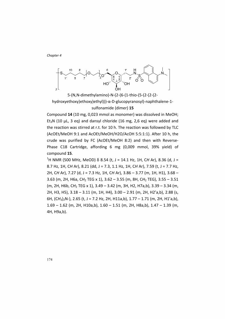
Chapter 4
174
5‐(N,N‐dimethylamino)‐N‐(2‐(6‐(1‐thio‐(5‐(2‐(2‐(2‐
hydroxyethoxy)ethoxy)ethyl)))‐α‐D‐glucopyranosyl)‐naphthalene‐1‐
sulfonamide (dimer) 15
Compound 14 (10 mg, 0,023 mmol as monomer) was dissolved in MeOH;
Et3N (10 µL, 3 eq) and dansyl chloride (16 mg, 2,6 eq) were added and
the reaction was stirred at r.t. for 10 h. The reaction was followed by TLC
(AcOEt/MeOH 9:1 and AcOEt/MeOH/H2O/AcOH 5:5:1:1). After 10 h, the
crude was purified by FC (AcOEt/MeOH 8:2) and then with Reverse‐
Phase C18 Cartridge, affording 6 mg (0,009 mmol, 39% yield) of
compound 15. 1H NMR (500 MHz, MeOD) δ 8.54 (t, J = 14.1 Hz, 1H, CH Ar), 8.36 (d, J =
8.7 Hz, 1H, CH Ar), 8.21 (dd, J = 7.3, 1.1 Hz, 1H, CH Ar), 7.59 (t, J = 7.7 Hz,
2H, CH Ar), 7.27 (d, J = 7.3 Hz, 1H, CH Ar), 3.86 – 3.77 (m, 1H, H1), 3.68 –
3.63 (m, 2H, H6a, CH2 TEG x 1), 3.62 – 3.55 (m, 8H, CH2 TEG), 3.55 – 3.51
(m, 2H, H6b, CH2 TEG x 1), 3.49 – 3.42 (m, 3H, H2, H7a,b), 3.39 – 3.34 (m,
2H, H3, H5), 3.18 – 3.11 (m, 1H, H4), 3.00 – 2.91 (m, 2H, H2’a,b), 2.88 (s,
6H, (CH3)2N‐), 2.65 (t, J = 7.2 Hz, 2H, H11a,b), 1.77 – 1.71 (m, 2H, H1’a,b),
1.69 – 1.62 (m, 2H, H10a,b), 1.60 – 1.51 (m, 2H, H8a,b), 1.47 – 1.39 (m,
4H, H9a,b).

Chapter 4
175
Synthesis of L‐glutamine derivative 21 and 24
5‐(2‐(2‐(2‐azidoethoxy)ethoxy)ethoxy)pent‐1‐ene 16
To a solution of Tosylate 10 (1976 mg, 5,3 mmol) in dry DMF (35 mL),
15,9 mmol (3 eq) of NaN3 were added. The reaction was stirred at r.t. for
2 h and then at 80°C for 10 h. The reaction was followed by TLC
(PE/AcOEt 8:2). At the end of the reaction, the suspension was diluted
with water and extracted with AcOEt (3x). The organic phases were
combined, dried over sodium sulphate and evaporated. The residue was
purified by FC (PE/AcOEt 8:2) affording azide 16 (1012 mg, 4,16 mmol,
78% yield). 1H NMR (400 MHz, CDCl3) δ 5.80 (ddt, J = 16.9, 10.2, 6.7 Hz, 1H, H4), 5.01
(dd, J = 17.1, 1.6 Hz, 1H, H5a), 4.95 (d, J = 10.2 Hz, 1H, H5b), 3.72 – 3.61
(m, 8H, ‐OCH2CH2‐), 3.61 – 3.54 (m, 2H, ‐OCH2CH2‐), 3.46 (t, J = 6.7 Hz,
2H, H1a,b), 3.38 (t, J = 5.0 Hz, 2H, ‐OCH2CH2N3), 2.10 (q, J = 7.3 Hz, 2H,
H3a,b), 1.73 – 1.61 (m, 2H, H2a,b). 13C NMR (101 MHz, CDCl3) δ 138.40 (C4), 114.80 (C5), 70.82, 70.81,
70.77, 70.21, 70.15 (CH2 PEG), 50.78 (CH2N3), 30.34 (C3), 28.86 (C2).
C11H21N3O3; calcd. mass: 243,32; MS‐ESI: m/z 244,2 [M+H+].
2‐(2‐(2‐(pent‐4‐en‐1‐yloxy)ethoxy)ethoxy)ethan‐1‐amine 17
Compound 16 (1153 mg, 4,73 mmol) was dissolved in THF (30 mL) and
Ph3P (5,68 mmol, 1,2 eq) was added. The reaction was stirred at r.t. for 2
h, then 5,67 mmol (1,2 eq) of distilled water were added and the
reaction was heated to 60°C and stirred for 30 min, following the

Chapter 4
176
disappearance of the starting material by TLC (PE/AcOEt 8:2 and
CH2Cl2/MeOH 9:1). The solvent was evaporated and the crude purified
by FC (CH2Cl2/MeOH 9:1 to CH2Cl2/MeOH/NH3 (aq) 7:3:1), affording
amine 17 (0,951 g, 4,4 mmol, 93% yield). 1H NMR (400 MHz, CD3OD) δ 5.83 (ddt, J = 13.5, 10.1, 6.7 Hz, 1H, H4),
5.02 (dd, J = 17.1, 1.1 Hz, 1H, H5a), 4.95 (d, J = 10.1 Hz, 1H, H5b), 3.69 –
3.53 (m, 12H, CH2 PEG), 3.52 – 3.45 (m, 2H, CH2NH2), 2.12 (dd, J = 14.4,
7.2 Hz, 2H, H3a,b), 1.72 – 1.58 (m, 2H, H2a,b). 13C NMR (101 MHz, CD3OD) δ 139.44 (C4), 115.23 (C5), 73.67, 71.56,
71.50, 71.26, 71.12, 70.39 (C1, CH2 PEG), 41.42 (CH2NH2), 31.33 (C3),
30.03 (C2).
C11H23NO3; calcd. mass: 217,31; MS‐ESI: m/z 218,3 [M+H+].
(9H‐fluoren‐9‐yl)methyl‐(S)‐(3,7‐dioxo‐1,1,1‐triphenyl‐11,14,17‐trioxa‐
2,8‐diazadocos‐21‐en‐6‐yl) carbamate 18
Compound 17 (133 mg, 0,612 mmol) was dissolved in dry CH2Cl2 (6 mL,
0,1 M) under argon atmosphere. The solution was cooled to 0°C and
Fmoc‐L‐Gln(Trt)‐OH (0,64 mmol, 1,04 eq), HOBt (0,918 mmol, 1.5 eq) and
EDC (0,918 mmol, 1,5 eq) were added. The reaction was warmed to r.t.
and stirred under argon. After 15 h, TLC (CH2Cl2/MeOH 9,5:0,5 and
CH2Cl2/MeOH/NH3 (aq) 9:1:0,1) indicated the absence of starting
compound 17; the reaction was diluted with CH2Cl2 and washed with
water (3x). The organic phase was gathered, dried over sodium sulphate,
filtrated and evaporated. The crude was purified by FC on Biotage
System (gradient CH2Cl2 100% – CH2Cl2/MeOH 9,3:0,7). 390 mg (0,48
mmol) of compound 18 were obtained (79% yield).

Chapter 4
177
1H NMR (500 MHz, CDCl3) δ 7.80 (d, J = 7.5 Hz, 2H, CH Ar Fmoc), 7.63 (d, J
= 7.4 Hz, 2H CH Ar Fmoc), 7.43 (t, J = 7.4 Hz, 2H CH Ar Fmoc), 7.38 – 7.20
(m, 17H, CH Ar Fmoc, CH Ar Trt), 7.11 (s, 1H, CONHTrt), 6.84 (s, 1H,
CONH PEG), 6.04 (d, J = 6.7 Hz, 1H, NH Fmoc), 5.83 (ddt, J = 16.9, 10.2,
6.6 Hz, 1H, H15), 5.04 (ddd, J = 17.1, 3.3, 1.5 Hz, 1H, H16a), 5.01 – 4.96
(m, 1H, H16b), 4.41 (d, J = 7.1 Hz, 2H, CH2 Fmoc), 4.24 (t, J = 7.0 Hz, 1H,
CH Fmoc), 4.19 – 4.10 (m, 1H, H4), 3.64 – 3.51 (m, 10H, CH2 PEG), 3.49 –
3.40 (m, 4H, H12a,b, H6a,b), 2.58 – 2.49 (m, 1H, H2a), 2.49 – 2.39 (m, 1H,
H2b), 2.18 – 2.07 (m, 3H, H14a,b, H3a), 2.03 – 1.95 (m, 1H, H3b), 1.73 –
1.65 (m, 2H, H13a,b). 13C NMR (101 MHz, CDCl3) δ 171.80 (C5), 171.32 (C1), 156.23 (OCONH
Fmoc), 144.48 (Cq Ar Trt), 143.80 (Cq Ar Fmoc), 143.68 (Cq Ar Fmoc),
141.17 (Cq Ar Fmoc), 141.14 (Cq Ar Fmoc), 138.11 (C15), 128.64 (C Ar
Trt, C Ar Fmoc), 127.82 (C Ar Trt, C Ar Fmoc), 127.62 (C Ar Trt, C Ar
Fmoc), 127.01 (C Ar Trt, C Ar Fmoc), 126.88 (C Ar Trt, C Ar Fmoc), 125.10
(C Ar Fmoc), 119.87 (C Ar Fmoc), 114.73 (C16), 70.56, 70.48, 70.37,
70.34, 70.07, 69.90 (CH2 PEG, Cq Trt), 69.30 (C12), 66.76 (CH2 Fmoc),
54.06 (C4), 47.06 (CH Fmoc), 39.20 (C6), 33.17 (C2), 30.11 (C14), 29.33
(C3), 28.59 (C13).
C50H55N3O7; calcd. mass: 810,00; MS‐ESI: m/z 810,9 [M+H+].
tert‐butyl (S)‐2‐((tert‐butoxycarbonyl)amino)‐5‐oxo‐9,12,15‐trioxa‐6‐
azaicos‐19‐enoate 22
Boc‐L‐glutamic acid 1‐tert‐butyl ester (149 mg, 0,49 mmol) and
compound 17 (0,58 mmol, 1,2 eq) were dissolved in CH2Cl2 (4,9 mL, 0,1
M) under argon and 0,98 mmol (2 eq) of Et3N were added and the

Chapter 4
178
solution was stirred at r.t. for 15 min. Then the temperature was set to
0°C and HOBt (0,73 mmol, 1,5 eq) and DCC (0,73 mmol, 1,5 eq) were
added. The reaction was stirred at r.t. until TLC (CH2Cl2/MeOH 9,5:0,5)
indicated the end of the coupling reaction. The reaction was recovered
as described for compound 18 and the product was purified by FC
(eluent CH2Cl2/MeOH 9,8:0,2) affording compound 22 (207 mg, 0,47
mmol, 95% yield) 1H NMR (400 MHz, CDCl3) δ 6.44 (s, 1H, CONH‐PEG), 5.80 (td, J = 16.9,
6.7 Hz, 1H, H15), 5.26 (d, J = 7.6 Hz, 1H, NHBoc), 5.01 (d, J = 17.2 Hz, 1H,
H16a), 4.95 (d, J = 10.2 Hz, 1H, H16b), 4.14 (s, 1H, H2), 3.67 – 3.52 (m,
10H, CH2 PEG), 3.51 – 3.36 (m, 4H, H12a,b, H6a,b), 2.30 – 2.20 (m, 2H,
H4a,b), 2.18 – 2.00 (m, 3H, H14a,b, H3a), 1.95 – 1.81 (m, 1H, H3b), 1.74 –
1.62 (m, 2H, H13a,b), 1.50 – 1.37 (m, 18H, (CH3)3 Boc, tBu). 13C NMR (101 MHz, CDCl3) δ 172.20 (C5), 171.57 (C1), 155.81 (OCONH
Boc), 138.25 (C15), 114.87 (C16), 82.14 (Cq OtBu), 79.78 (Cq Boc), 70.77,
70.59, 70.33, 70.12, 69.87 (C7‐C12), 53.71 (C2), 39.37 (C6), 32.70 (C4),
30.27 (C14), 29.14 (C3), 28.78 (C13), 28.40, 28.05 ((CH3)3 OtBu, Boc).
C25H46N2O8; calcd. mass: 502,65; MS‐ESI: m/z 503,4 [M+H+].
(S)‐2‐amino‐5‐oxo‐9,12,15‐trioxa‐6‐azaicos‐19‐enoic acid 23
Protected L‐glutamine derivative 22 (246 mg, 0,49 mmol) was dissolved
in a mixture of CH2Cl2/TFA (12 + 3 mL) and stirred at r.t.. The reaction
was followed by TLC (CH2Cl2/MeOH/NH3 (aq) 7:3:0,3) and after 2 h the
solution was neutralized with NH3 (aq) 25%. The crude was concentrated
and the product purified by FC (CH2Cl2/MeOH/NH3 (aq) 7:3:0,2)
obtaining 52 mg (0,15 mmol, 30% yield) of compound 23.
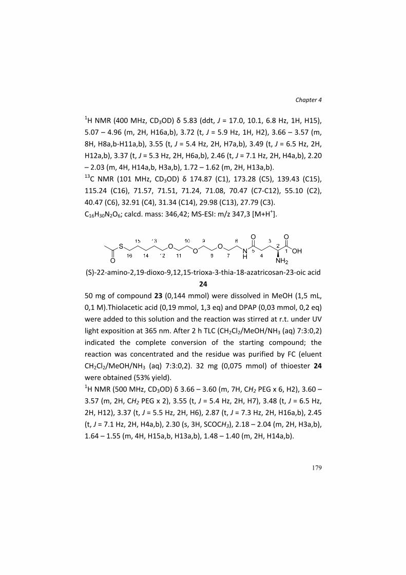
Chapter 4
179
1H NMR (400 MHz, CD3OD) δ 5.83 (ddt, J = 17.0, 10.1, 6.8 Hz, 1H, H15),
5.07 – 4.96 (m, 2H, H16a,b), 3.72 (t, J = 5.9 Hz, 1H, H2), 3.66 – 3.57 (m,
8H, H8a,b‐H11a,b), 3.55 (t, J = 5.4 Hz, 2H, H7a,b), 3.49 (t, J = 6.5 Hz, 2H,
H12a,b), 3.37 (t, J = 5.3 Hz, 2H, H6a,b), 2.46 (t, J = 7.1 Hz, 2H, H4a,b), 2.20
– 2.03 (m, 4H, H14a,b, H3a,b), 1.72 – 1.62 (m, 2H, H13a,b). 13C NMR (101 MHz, CD3OD) δ 174.87 (C1), 173.28 (C5), 139.43 (C15),
115.24 (C16), 71.57, 71.51, 71.24, 71.08, 70.47 (C7‐C12), 55.10 (C2),
40.47 (C6), 32.91 (C4), 31.34 (C14), 29.98 (C13), 27.79 (C3).
C16H30N2O6; calcd. mass: 346,42; MS‐ESI: m/z 347,3 [M+H+].
(S)‐22‐amino‐2,19‐dioxo‐9,12,15‐trioxa‐3‐thia‐18‐azatricosan‐23‐oic acid
24
50 mg of compound 23 (0,144 mmol) were dissolved in MeOH (1,5 mL,
0,1 M).Thiolacetic acid (0,19 mmol, 1,3 eq) and DPAP (0,03 mmol, 0,2 eq)
were added to this solution and the reaction was stirred at r.t. under UV
light exposition at 365 nm. After 2 h TLC (CH2Cl2/MeOH/NH3 (aq) 7:3:0,2)
indicated the complete conversion of the starting compound; the
reaction was concentrated and the residue was purified by FC (eluent
CH2Cl2/MeOH/NH3 (aq) 7:3:0,2). 32 mg (0,075 mmol) of thioester 24
were obtained (53% yield). 1H NMR (500 MHz, CD3OD) δ 3.66 – 3.60 (m, 7H, CH2 PEG x 6, H2), 3.60 –
3.57 (m, 2H, CH2 PEG x 2), 3.55 (t, J = 5.4 Hz, 2H, H7), 3.48 (t, J = 6.5 Hz,
2H, H12), 3.37 (t, J = 5.5 Hz, 2H, H6), 2.87 (t, J = 7.3 Hz, 2H, H16a,b), 2.45
(t, J = 7.1 Hz, 2H, H4a,b), 2.30 (s, 3H, SCOCH3), 2.18 – 2.04 (m, 2H, H3a,b),
1.64 – 1.55 (m, 4H, H15a,b, H13a,b), 1.48 – 1.40 (m, 2H, H14a,b).

Chapter 4
180
13C NMR (101 MHz, CD3OD) δ 197.51 (CH3COS), 190.57, 175.09 (C1),
173.52 (C5), 72.02, 71.57, 71.52, 71.25, 71.09, 70.50 (CH2 PEG), 55.63
(C2) 40.47 (C6), 33.07 (C4), 30.60 (C13), 30.53 (SCOCH3), 30.14 (C15),
29.76 (C16), 27.97 (C3), 26.36 (C14).
C18H34N7O7S; calcd. mass: 422,54; MS‐ESI: m/z 423,3 [M+H+].
(S)‐2‐amino‐N1‐(2‐(2‐(2‐(pent‐4‐en‐1‐
yloxy)ethoxy)ethoxy)ethyl)pentanediamide 19
Compound 18 (148 mg, 0,18 mmol) was dissolved in 1,8 mL of CH2Cl2
(0,1 M) and 0,4 mL of TFA were added. After 6 h, TLC (CH2Cl2/MeOH
9,5:0,5) showed no more starting compound (Trt deprotection), and the
reaction was neutralized to pH 7 adding NH3 25%. Then the solvent was
evaporated and the residue was redissolved in DMF (8 mL). Piperidine
was added (2 mL) and the reaction was stirred at r.t.. After 12 h TLC
confirmed the Fmoc deprotection; the reaction was concentrated and
the residue purified by FC (eluent CH2Cl2/MeOH/NH3 (aq) 8,5:1,5:0,1). 60
mg (0,17 mmol) of deprotected compound 19 were gathered (96%
yield). 1H NMR (500 MHz, CD3OD) δ 5.83 (ddt, J = 17.0, 10.2, 6.7 Hz, 1H, H15),
5.02 (ddd, J = 17.1, 3.5, 1.6 Hz, 1H, H16a), 4.95 (ddt, J = 10.2, 2.2, 1.2 Hz,
1H, H16b), 3.67 – 3.60 (m, 6H, CH2 PEG), 3.60 – 3.54 (m, 4H, CH2 PEG),
3.48 (t, J = 6.5 Hz, 2H, H12a,b), 3.46 – 3.33 (m, 3H, H4, H6a,b), 2.28 (t, J =
7.8 Hz, 2H, H2a,b), 2.15 – 2.09 (m, 2H, H14a,b), 1.99 – 1.90 (m, 1H, H3a),
1.88 – 1.79 (m, 1H, H3b), 1.70 – 1.62 (m, 2H, H13a,b).
C16H31N3O5; calcd. mass: 345,44; MS‐ESI: m/z 346,4 [M+H+], 368,4
[M+Na]+.

Chapter 4
181
(S)‐S‐(1,4‐diamino‐1,5‐dioxo‐9,12,15‐trioxa‐6‐azaicosan‐20‐yl)‐
ethanethioate 20
Compound 19 (60 mg, 0,174 mmol) was converted in the thioester
derivative 20 as described for compound 24. After FC of the crude
reaction (eluent CH2Cl2/MeOH/NH3 (aq) 8,5:1,5:0,1) 29 mg of compound
20 (0,07 mmol, 47% yield) were obtained. The 1H‐NMR analysis suggests
that the product is as thioacetate salt. 1H NMR (500 MHz, MeOD) δ 4.32 (dd, J = 8.4, 5.7 Hz, 1H, H4), 3.66 – 3.60
(m, 6H, CH2 PEG), 3.60 – 3.53 (m, 4H, CH2 PEG), 3.47 (t, J = 6.5 Hz, 2H,
H12a,b), 3.40 (t, J = 5.4 Hz, 1H, H6a), 3.36 (t, J = 5.5 Hz, 1H, H6b), 2.87 (t,
J = 7.3 Hz, 2H, H16a,b), 2.34 – 2.26 (m, 5H, CH3COSCH2, H2a,b), 2.11 –
2.01 (m, 1H, H3a), 1.99 (s, 3H, CH3COS‐), 1.95 – 1.86 (m, 1H, H3b), 1.63 –
1.55 (m, 4H, H13a,b, H15a,b), 1.47 – 1.39 (m, 2H, H14a,b). 13C NMR (101 MHz, CD3OD) δ 197.53 (CH3COSCH2), 177.69 (CH3COS
‐),
173.91 (C5), 173.30 (C1), 72.04, 71.62, 71.56, 71.28, 71.14, 70.44 (CH2
PEG), 54.38 (C2), 40.39 (C6), 32.56 (C4), 30.62 (C13), 30.52 (SCOCH3),
30.17 (C15), 29.77 (C16), 29.10 (C3), 26.39 (C14), 22.52 (CH3COS‐).
structure proposed from spectra

Chapter 4
182
(S)‐2‐amino‐N1‐(2‐(2‐(2‐((5‐mercaptopentyl)oxy)ethoxy)ethoxy)ethyl)
pentanediamide (dimer) 21
Compound 20 (10 mg, 0,0237 mmol) was dissolved in 0,6 mL of MeOH
(previously degased under a stream of argon) and 0,06 mmol (2,5 eq) of
sodium methoxide were added. The reaction was stirred at r.t. and after
24 h a sample of the crude was analysed by 1H‐NMR to check the
complete thioester deprotection. The spectrum clearly indicated the
disappereance of the triplet at 2,9 ppm (‐CH2‐SAc) and of the singlet at
2,3 ppm (CH3COS‐). A new triplet at 2,65 ppm was present, indicating the
formation of a disulfide bond. The reaction was then acidified with HCl 1
N (final pH = 6) and the solvent evaporated. The crude was directly used
for the preparation of the nanoparticle. The 1H‐NMR analysis suggests
that the product is as thioacetate salt. 1H NMR (500 MHz, MeOD) δ 4.31 (dd, J = 8.6, 5.6 Hz, 1H, H4), 3.68 – 3.58
(m, 8H, CH2 PEG), 3.56 (t, J = 5.5 Hz, 2H, CH2 PEG), 3.49 (t, J = 6.5 Hz, 2H,
H12a,b), 3.44 – 3.33 (m, 2H, H6a,b), 2.70 (t, J = 7.2 Hz, 2H, H16a,b), 2.30
(t, J = 7.6 Hz, 2H, H2a,b), 2.11 – 2.03 (m, 1H, H3a), 2.00 (s, 3H, CH3COS‐),
1.96 – 1.90 (m, 1H, 3b), 1.71 (dt, J = 14.7, 7.3 Hz, 2H, H15a,b), 1.65 – 1.56
(m, 2H, H13a,b), 1.54 – 1.43 (m, 2H, H14a,b). 13C NMR (101 MHz, CDCl3) δ 177.65 (CH3COS
‐), 174.01 (C5), 173.33 (C1),
72.13, 71.50, 71.47, 71.24, 71.08, 70.55 (CH2 PEG), 54.49 (C4), 40.33 (C6),
39.54 (C16), 32.55 (C2), 30.26 (C13), 30.00 (C15), 29.01 (C3), 26.03 (C14),
22.58 (CH3COS‐).

Chapter 4
183
structure proposed from spectra
General procedure for gold nanoparticle preparation
The general procedure for
the preparation of the
gold nanoparticles (Au‐
NPs), loaded with the
desired ligands, is the
follow.
The ligand(s) (3 eq) to
load on the Au‐NPs are
dissolved in MeOH, at a
final concentration of 0,012 M. If the ligands are more than one, the final
concentration must be always 0,012 M. The ratio between the ligands is
checked with a 1H NMR spectra, dissolving the ligands in deuterated
methanol. To this solution, 1 eq of a solution of HAuCl4 (hydrogen
tetrachloroaurate) in water is addes. The concentration of this solution
must be 0,025 M. After the addition, the reaction is stirred for few
minutes. In this step, Au(I) polymers are formed and the reaction
becomes turbid, with a pale yellow colour. A solution of NaBH4 (27‐30
eq) in water (1 M) is added in four portion; between the addition, the
vessel containing the reaction is vigorously shaken. After the addition of
NaBH4, a black precipitate is formed, since gold nanoparticles are
1. ligands 0,012 M3 equivalents
2. HAuCl4 0,025 M1 equivalent
3. NaBH4 1M27-30 equivalents
4. stirring 2 h, R.T.
5. washing with MeOH
6. dissolution in water
7. dialysis (2-3 days)
8. liophylization

Chapter 4
184
forming. The reaction is left for 2 h at room temperature with 180 r.p.m.
shaking. At the end of the 2 hours, the black precipitate (Au‐NPs) is
washed several times with MeOH, with cycles of vortexing and
centrifugation, removing the surnatant and adding fresh MeOH. In this
step, the excess of ligands which are still dissolved and were not
attached to the NPs are removed. The washing solutions are gathered
and combined, in order to verifiy, with 1H NMR analysis, the quantity of
the ligands attached to Au‐NPs and the ratio (if more ligands are used in
the initial MeOH solution. The final washed precipitate is dissolved in
water. The water‐dissolved nanoparticles are then purified with a dialysis
process, for 2 – 3 days. This process allows the removal of boron salts
and any other impurity. The final water dispersion of the GNPs is freeze‐
dried, to obtain a black powder.

Chapter 4
185
References [1] a)M. Palazzo, S. Gariboldi, L. Zanobbio, S. Selleri, G. F. Dusio, V.
Mauro, A. Rossini, A. Balsari, C. Rumio, Journal of Immunology 2008,
181, 3126‐3136; b)L. Zanobbio, M. Palazzo, S. Gariboldi, G. F. Dusio, D.
Cardani, V. Mauro, F. Marcucci, A. Balsari, C. Rumio, Am. J. Pathol. 2009,
175, 1066‐1076.
[2] B. La Ferla, V. Spinosa, G. D'Orazio, M. Palazzo, A. Balsari, A. A.
Foppoli, C. Rumio, F. Nicotra, ChemMedChem 2010, 5, 1677‐1680.
[3] a)H. Fillmann, N. A. Kretzmann, B. San‐Miguel, S. Llesuy, N.
Marroni, J. Gonzalez‐Gallego, M. J. Tunon, Toxicology 2007, 236, 217‐
226; b)Y. Huang, N. Li, K. Liboni, J. Neu, Cytokine 2003, 22, 77‐83.
[4] a)X. H. Weng, K. W. Beyenbach, A. Quaroni, American Journal of
Physiology‐Gastrointestinal and Liver Physiology 2005, 288, G705‐G717;
b)A. Ikari, M. Nakano, Y. Suketa, H. Harada, K. Takagi, Journal of Cellular
Physiology 2005, 203, 471‐478; c)J. R. Turner, B. K. Rill, S. L. Carlson, D.
Carnes, R. Kerner, R. J. Mrsny, J. L. Madara, The American journal of
physiology 1997, 273, C1378‐1385; d)N. Li, J. Neu, Journal of Nutrition
2009, 139, 710‐714; e)N. Li, P. Lewis, D. Samuelson, K. Liboni, J. Neu,
American Journal of Physiology‐Gastrointestinal and Liver Physiology
2004, 287, G726‐G733.
[5] M. Mammen, S. K. Choi, G. M. Whitesides, Angewandte Chemie‐
International Edition 1998, 37, 2755‐2794.
[6] a)J. Giudicelli, M. F. Bertrand, S. Bilski, T. T. Tran, J. C. Poiree,
Biochemical Journal 1998, 330, 733‐736; b)B. R. Stevens, A. Fernandez, B.
Hirayama, E. M. Wright, E. S. Kempner, Proceedings of the National
Academy of Sciences of the United States of America 1990, 87, 1456‐
1460; c)M. Takahashi, P. Malathi, H. Preiser, C. Y. Jung, Journal of
Biological Chemistry 1985, 260, 551‐556; d)R. J. Turner, E. S. Kempner,
Journal of Biological Chemistry 1982, 257, 794‐797.

Chapter 4
186
[7] E. Romeo, M. H. Dave, D. Bacic, Z. Ristic, S. M. R. Camargo, J.
Loffing, C. A. Wagner, F. Verrey, American Journal of Physiology‐Renal
Physiology 2006, 290, F376‐F383.
[8] a)J. M. de la Fuente, A. G. Barrientos, T. C. Rojas, J. Rojo, J.
Canada, A. Fernandez, S. Penades, Angewandte Chemie‐International
Edition 2001, 40, 2258‐+; b)O. Martinez‐Avila, K. Hijazi, M. Marradi, C.
Clavel, C. Campion, C. Kelly, S. Penades, Chemistry‐a European Journal
2009, 15, 9874‐9888.
[9] S. Zalipsky, Advanced Drug Delivery Reviews 1995, 16, 157‐182.
[10] F. M. Veronese, G. Pasut, Drug Discovery Today 2005, 10, 1451‐
1458.
[11] A. Dondoni, Angewandte Chemie International Edition 2008, 47,
8995‐8997.
[12] F. Cardona, B. La Ferla, Journal of Carbohydrate Chemistry 2008,
27, 203‐213.
[13] G. J. McGarvey, C. A. LeClair, B. A. Schmidtmann, Organic Letters
2008, 10, 4727‐4730.
[14] A. Wang, J. Hendel, F.‐I. Auzanneau, Beilstein Journal of Organic
Chemistry 2010, 6.
[15] T. Buskas, E. Söderberg, P. Konradsson, B. Fraser‐Reid, The
Journal of Organic Chemistry 2000, 65, 958‐963.
[16] Á. G. Barrientos, J. M. de la Fuente, T. C. Rojas, A. Fernández, S.
Penadés, Chemistry – A European Journal 2003, 9, 1909‐1921.
[17] S. A. Svarovsky, Z. Szekely, J. J. Barchi, Tetrahedron: Asymmetry
2005, 16, 587‐598.
[18] R. Dettmann, T. Ziegler, Carbohydr. Res. 2011, 346, 2348‐2361.
Chapter 5
187
Chapter 5. Iminosugar‐decorated
calix[4]arenes: chemical tools for
the investigation of multivalent
inhibition of glycosidases.

Chapter 5
188
Abstract In this paper report the synthesis of a multivalent calix[4]arenes scaffold
decorated with iminosugars, in particular N‐derivatives of
deoxynojirimycin and of L‐idonojirimycin. The decoration of the
polyvalent structure was performed exploiting a click chemistry
approach, by the copper(I) catalyzed azide alkyne cycloaddition, which
provides fast and clean reactions. The generated calix[4]arene will serve
as molecular probes for the study of the multivalency effect associated
with glycosidase, recently observed.
Introduction Multivalency represents a fundamental principle used by Nature to
achieve strong, but reversible, interactions between biological entities,
such as biomacromolecules, which possess low affinity[1]. The presence
of multiple copies of a biomacromolecule on the surface of a cell, for
example, allows to increase dramatically the binding strength of the
ligand with its receptor (or a cluster of them), compared to a single
ligand‐protein interaction. These events occur in a plethora of biological
processes, in particular in cell‐cell communications, as in the immune
system. The mechanisms and the rationale of the multivalency principle
in biology are multiple (see Introduction – Multivalency and
Glycobiology): if a ligand interacts with its target protein, it is possible to
observe an enhancement in the binding of a second one, due to
particular structural reinforcement in the same protein. The multivalency
effect can be observed also on the side of the biological effects that can
result when a cluster of ligands, disposed close together, binds a
particular molecular target or a set of them: in this case it is possible to

Chapter 5
189
obtain an improvement of the biological response. This is partially
explained by allosteric effect on the same protein, or by events of
dimerization/oligomerization of the target protein, causing an
enhancement of the intracellular biological signal. These kind of
phenomena are well‐documented between carbohydrate ligands and
lectins, and in general in cellular recognition events, but less for
enzymes, where other mechanisms could be taken into account, as
statistical rebinding and an effective increase of local concentration of
the ligand loaded in a multivalent structure[2]. Although the multivalency
effect of a polyvalently‐decorated skeleton is difficult to rationalize in
the case of enzymes, especially those bearing a single binding and
catalytic site, an increase number of scientific works describe the
development of multivalent scaffolds decorated with suitable ligands or
inhibitors, that show a high degree of multivalency effect against the
respective enzymes[2] [3] [4] [5].
In the last decades, many research group have focused their attention in
the preparation of synthetic polyvalent structure, decorated with
glycoderivatives or glycoconjugates, as tools to study the multivalency
effect exerted by a carbohydrate‐based ligand toward their respective
binding proteins (lectins) or processing enzymes.
Some of these papers outline the multivalent properties of particular
sugar (or glycoderivative)‐decorated structures against a specific class of
hydrolytic enzymes, called glycosidases. These enzymes cleave the O‐
glycosidic bond between two sugar units constituting di‐, oligo‐ and
polysaccharides, and their inhibitors show a great clinical relevance, for
many pathologies, as diabetes or obesity. The developed polyvalent
structures, dendrimers[2], fullerenes[3], and cyclodextrins[4], grafted with
iminosugars, known glycosidase inhibitors, display a moderate to

Chapter 5
190
remarkable effects onthe enhancement of the inhibition activity, that
could not simply be compared to the sum of the single inhibitors.
Starting from these literature data, our work is focused on the
development of new structures presenting multiple copies of
iminosugars derivatives, to achieve new and complementary information
about the multivalency effect exerted by this class of inhibitors against
glycosidases. As multivalent scaffold we chose to use a calix[4]arene, in
order to prepare a polydecorated structure made of multiple copies of 1‐
deoxynojirimicin (1‐DNJ) derivatives . Calixarenes are structures made of
several aromatic rings linked together by methylene bridges, and have
been extensively used in the research field of host‐guest chemistry and,
more recently, as multivalent scaffold for biological relevant molecules
and macromolecules. The use of glyco‐decorated calixarenes has already
been reported to shows enhanced binding affinities towards sugar
lectins [6] [7]. We prepared a set of calix[4]arenes functionalized with 1‐
DNJ derivatives, in which the iminosugar entity is connected to the
calixarene core with a spacer bearing a triazole group, as consequence of
the copper catalyzed azide‐alkyne cycloaddition (CuAAC)[8] approach
used for the calixarene functionalization. The obtained polyvalent
calixarenes will be tested for their multivalency inhibitory effect against
a panel of commercially available glycosidases.
Results and Discussion Initially, we decided to follow two different approach for the generation
of the iminosugar‐decorated calix[4]arenes, which were chosen among
the family of this scaffold for their easier synthesis and their
straightforward functionalization. Starting from a panel of already
synthesized calix[4]arenes [6, 8‐9], we planned the synthesis of suitably

Chapter 5
191
functionalized 1‐DNJ derivatives in order to attach them to the skeleton
by the thiol‐ene coupling (TEC) approach[6], and the CuAAC method
(Figure 49).
Azide –
Alkyne
cyclo
addition
Thiol –
ene
reaction
Figure 49 ‐ The two “click reaction” approaches for the chemical functionalization of the calixarene scaffold.
We designed a pool of iminosugars, derivatives of 1‐DNJ and L‐ido‐1‐DNJ,
bearing a N‐alkyl chain with a terminal triple bond, to exploit in the
CuAAC strategy, or with a terminal thiol group, to be used for a thiol‐ene
coupling reaction. These derivatives will then be anchored to n the
calix[4]arene bearing respectively an azido or allylic appendages (Table
3).

Chapter 5
192
Table 3 – Set of designed and prepared iminosugars derivatives for the calix[4]arenes
decoration. R = H or Protecting groups (Ac or Bn).
Alkynyl chain Thiol chain
4‐
carbon
chain
NRO
ROORRO
SH
5‐
carbon
chain
CuAAC Thiol‐ene
As previously said, the thiol derivative was to be prepared from the
alkene precursor, so we decided to prepare a small library containing
both iminosugars functionalized with a 4‐ and with a 5‐carbon chain,
bearing a double or triple bond exploiting a common synthetic strategy,
and the N‐allyl derivatives were synthesized, for the generation of the 3
carbon chain derivatives. The overall synthetic plan used is presented
below:

Chapter 5
193
Scheme 13 ‐ Synthesis of the library of iminosugars. Reagents and conditions: a) NaBH4, CH2Cl2, EtOH, r.t., 24 h
[10]; b) See Ref.
[10] [11]
; c) Allylamine (neat), reflux, 36 h; d) i. (COCl)2, DMSO, CH2Cl2, ‐78°C, 2 h; ii. Et3N, CH2Cl2, ‐78°C to r.t., 2 h; e) allylamine, molecular sieves 3 Å, NaCNBH3, MeOH, 0°C to r.t., 12 h; f) NH4
+HCOO
‐, molecular
sieves 3 Å, NaCNBH3, MeOH, 0°C to r.t.., 12 h; g) TsCl, DMAP, CH2Cl2, Et3N, r.t., 24 h; h,I,l,m) R‐OTs, CH3CN dry, K2CO3, reflux, 48 h; n) BCl3 1M in CH2Cl2, CH2Cl2, ‐60°C to 0°C, 4 h; o) Ac2O, Py, r.t., 24 h.
Commercially available 2,3,4,6‐tetra‐O‐benzyl‐D‐glucopyranose was
reduced with NaBH4 affording glucitol 1, which was used as starting
material for the preparation of all compounds. A double mesylation (2)
and the reaction with allylamine led to the obtainment of N‐allyl‐
iminosugar with an inversion of configuration at C‐5 position, thus giving

Chapter 5
194
the L‐ido configuration. For the synthesis of N‐allyl‐1‐deoxynojirimicyn
(5) glucitol 1 was oxidized through a Swern reaction into the instable
intermediate 4[10] and subjected to a double reductive amination using
allylamine. The same protocol was repeated with ammonium formate
affording the tetra‐O‐benzyl‐1‐DNJ (6), that was used for the preparation
of derivatives 11‐14, obtained by reaction with tosylates (7‐10) .The
successive debenzylation with BCl3 led to the obtainment of the
deprotected iminosugars (15‐20), which were acetylated without further
purification steps, to generate the final compounds 21‐25. The L‐ido
derivative 16 was not subjected to acetylation and used after a
purification step.
Alongside the preparation of the library, our work focused on the
synthesis of the calix[4]arene scaffold. We prepared various
calix[4]arenes which differs for the type and the position of the inserted
functionalizations (Figure 50). The first ones (27‐30) were prepared
according to published procedures[6] and bear from four to eight allyl
groups, on each side of the structure or only on one. The presence of the
double bonds permit the exploitation of the TEC strategy for the
decoration of the calixarene. The second ones, already known and
synthesized structures[6, 8], present four azido functionalities which can
be exploited for the conjugation of compounds with a terminal alkynyl
group by the CuAAC method.

Chapter 5
195
Figure 50 ‐ Structures of the different calix[4]arenes synthesized.
Initially, our efforts focused on the transformation of the alkenyl
appendage on the iminosugars 16 and 21‐25 into a thioester
functionality by a thiol‐ene reaction with thiolacetic acid, in presence of
the radical photoinitiator DPAP upon UV‐light exposure (365 nm), but
unfortunately no satisfying results were obtained with this approach
(Scheme 14). The reaction conditions were identical to those reported in
Fiore, et al. [6] and Fiore, et al. [12].
Scheme 14 – Thiol‐ene coupling strategy for the decoration of calix[4]arene with N‐
alkenyl iminosugars.

Chapter 5
196
We decide to move our attention for the development of the desired
calix[4]arenes using the Cu(I)‐azide alkyne cycloaddition (CuAAC), using
the scaffolds functionalized with azido groups and iminosugars
substituted with an alkynyl chain. In this case we exploited the reaction
conditions and procedures of CuAAC already drescribed in Dondoni and
Marra [8], Marra, et al. [9]. We were able to obtain three different
iminosugar‐decorated calix[4]arenes (34, 36 and 38), performing the
cycloaddition between the N‐alkynyl iminosugar 23 with the three azido‐
scaffolds 30, 31 and 32 (Scheme 15). Calix[4]arene 40 contains a
propylene azido chain on the lower rim, and the performed
cycloaddition with 23 generated the decorated scaffold 33, containing
four 1‐DNJ units linked to the tetra‐aromatic core by a triazole‐bearing
tether, which is directly connected to the calixarene by a propilenoxy
chain and to the iminosugar by an ethylene bridge. The complete
deacetylation with ammonia in methanol afforded the final compound
34. Starting calixarene 31 bears propyl appendages on the phenolic
oxygen atoms located on the lower rim, while propylene azido function
on the upper rim, which were transformed into triazole entities by the
CuAAC with 23. The last calixarene was generated by the starting
compound 32, in which the azido groups are separated from the
aromatic core of the scaffold by a single methylene bridge. Finally
calix[4]arenes 37 and 38 show a similarity to 35 and 36, but the spacer
between the iminosugar and the aromatic ring is shorter.

Chapter 5
197
Scheme 15 – Preparation of the three iminosugar‐decorated calix[4]arenes by the CuAAC approach. Reagents and conditions: a) Cu(I), iPr2EtN, Toluene, 80°C, 2 h; b) NH3, MeOH, r.t., 18 h.
Furthermore, a cycloaddition reaction between the same iminosugar and
benzyl azide afforded compound 40 (Scheme 16), which represents the
ligand constituting the calixarene tetramer. The monomer will be used as
a control during the enzymatic assays to determine the multivalency
effect of the calixarene polyvalent structures.

Chapter 5
198
Scheme 16 – Synthesis of 40. Reagents and conditions: a) Benzyl azide, Cu(I), Toluene,
80°C, 20 h; b) NH3, MeOH, r.t., 18 h.
The obtained final calixarenes 34, 36 and 38, and the monovalent ligand
40 will be tested for their inhibitory activity against a panel of
commercially available glycosidases, exploting a simple and fast
colorimetric assay. Suitable p‐ or o‐nitrophenol glycosides will be used as
chromogenic substrates of several glycosidases, which catalytic activity
allows the cleavage of the O‐glycosidic bond, thus liberating the
respective p‐ or o‐nitrophenolate. The inhibition activity exerted by the
tested compound will cause a decrease of absorbance of the reaction
solution, due to less amount of cleaved chromogenic susbtrate. Using
different concentration of tested compounds and performing the
reaction with several substrate concentration it will be possible to
determine the inhibition constant for each compound, thus allowing a
comparison of the multivalency efficacy among the prepared calixarenes
and against the monovalent ligand.
Chapter 5
199
Conclusions Recent encouraging results on glycosidases inhibition using polyvalent
structure decorated with iminosugars have prompted us in the
development of a new class of structures bearing multiple copies of of 1‐
deoxy‐L‐idonojirimycin and 1‐DNJ derivatives, for which a remarkable
inhibitory activity against several glycosidases is reported. Among the big
number of available scaffolds with multivalent functionalities, we chose
the calixarene skeleton for several reasons. Firstly, calixarenes possess a
precise shape and geometry which can be exploited for the multiple
functionalization with many bioorganic compounds, as carbohydrates or
peptides. The calixarene scaffold is relatively simple, made of phenolic
rings connected together in a conical structure by methylene bridges:
the acidic oxygen can be easily alkylated, allowing the insertion of many
appendages bearing different chemical functionalities. It permits not
only a chemical modulation of the calixarene reactivity, but even the
exploitation of chemoselective methods for the decoration of such
scaffolds. In our case, we selected a calix[4]arene skeleton bearing allylic
or azidoalkyl appendage, thus allowing the use of two chemical
approach: the thiol‐ene coupling and the copper(I)‐catalyzed azide
alkyne cycloaddition. While the first strategy did not give the expected
results, the CuAAC reactions allowed the generation of three
calix[4]arenes with different geometry and shape. The same reaction
performed using benzyl azide and iminosugar 23 afforded a monovalent
ligand, compound 40, which can be used as monovalent control during
the enzymatic assays for the inhibitory activity of the synthesized
compounds. The execution of the enzymatic test will allow to
understand if the obtained calix[4]arenes show a multivalent inhibitory
effect against a pool of commercial glycosidases. The results can be used
as base for the generation of a second library of structures both with a

Chapter 5
200
better multivalency or, moreover, for the creation of hybrid structures:
the typical geometries and shapes of the calixarenes, made generally of
two sides or rims, can be exploited for a a dual ligand type
functionalization, such as iminosugars and fluorescent probes, to achieve
also trafficking studies performing in vivo experiments. The synthesis of
these types of calix[4]arenes and the execution of these experiments will
allow to gather new and more detailed information about the
multivalent effect recently observed on glycosidases.
Experimental Section
General remarks
All commercial chemicals were purchased from Sigma‐Aldrich and used
without further purification. All moisture‐sensitive reactions were
performed under a nitrogen atmosphere using oven‐dried glassware.
Anhydrous solvents were dried over standard drying agents and freshly
distilled prior to use or dried with molecular sieves for at least 24h prior
to use. Thin layer chromatography (TLC) was performed on silica gel 60
F254 plates (Merck) with detection under UV light when possible, or by
charring with a solution of (NH4)6Mo7O24 (21g), Ce(SO4)2 (1g),
concentrated H2SO4 (31 mL) in water (500 mL) or with an ethanol
solution of ninhydrin or with Dragendorff’ spray reagent. Flash‐column
chromatography was performed on silica gel 230–400 mesh (Merck). 1H
and 13C NMR spectra were recorded at 25°C, unless otherwise stated,
with a Varian Mercury 400‐MHz instrument. Chemical shift assignments,
reported in parts per million, were referenced to the corresponding
solvent peaks. Mass spectra were recorded on a QTRAP system with ESI
source, while HRMS were registered on a QSTAR elite system with a
nanospray ion source.

Chapter 5
201
Synthesis of iminosugars
Synthesis of compound 1, intermediate 4 and tetra‐O‐benzyl‐1‐DNJ 6
were already described in literature. All characterizations are consistent
to those reported in literature[10] [11].
2,3,4,6‐tetra‐O‐benzylglucitol dimesylate 2
The synthesis if compound 2 was carried out according to a procedure
described in Jiang, et al. [13] and Rao and Perlin [14]. All NMR data
obtained were consistent to those reported in literature. 1H NMR (400 MHz, CDCl3) δ 7.40 – 7.16 (m, 20H, CH Ar), 5.03 (dt, J = 7.7,
2.7 Hz, 1H, H5), 4.76 (d, J = 11.1 Hz, 1H, CH2Ph), 4.67 (d, J = 11.6 Hz, 1H,
CH2Ph), 4.62 (d, J = 11.1 Hz, 1H, CH2Ph), 4.60 – 4.54 (m, 3H, CH2Ph), 4.50
– 4.44 (m, 3H, CH2Ph, H1a), 4.25 (dd, J = 11.2, 7.2 Hz, 1H, H1b), 4.05 (dd,
J = 4.8, 2.8 Hz, 1H, H4), 3.95 – 3.89 (m, 1H, H2), 3.86 (dd, J = 11.1, 2.6 Hz,
1H, H6a), 3.77 (dd, J = 11.1, 7.9 Hz, 1H, H6b), 3.71 (t, J = 5.2 Hz, 1H, H3),
2.95 (s, 3H, OSO2CH3), 2.83 (s, 3H, OSO2CH3).
2,3,4,6‐tetra‐O‐benzyl‐L‐ido‐N‐allyl‐1‐deoxynojirimycin 3
Dimesylate 2 (2042 mg, 2,926 mmol) was dissolved in allylamine (15 mL)
under argon atmosphere. The reaction was heated and stirred at reflux.
After 36 h, TLC (PE/AcOEt 8:2) showed no more starting compound, and
the solvent was evaporated. The residue was purified by FC (eluent

Chapter 5
202
PE/AcOEt 9:1 to 8:2), affording compound 3 (1,238 g, 2,196 mmol, 75%
yield). 1H NMR (400 MHz, CDCl3) δ 7.44 – 7.28 (m, 20H, CH Ar), 5.91 – 5.74 (m,
1H, H8), 5.18 (dd, J = 23.5, 5.9 Hz, 2H, H9a,b), 4.90 (d, J = 10.9 Hz, 1H,
CH2Ph), 4.85 (d, J = 10.9 Hz, 1H, CH2Ph), 4.75 (d, J = 11.5 Hz, 1H, CH2Ph),
4.72 – 4.64 (m, 3H, CH2Ph), 4.62 – 4.53 (m, 2H, CH2Ph), 3.90 (dd, J = 10.2,
6.8 Hz, 1H, H6a), 3.81 – 3.69 (m, 2H, H6b,H4), 3.67 – 3.54 (m, 2H, H3,H2),
3.51 – 3.39 (m, 2H, H5, H7a), 3.24 (dd, J = 14.0, 6.9 Hz, 1H, H7b), 2.99
(dd, J = 11.8, 4.8 Hz, 1H, H1a), 2.59 (dd, J = 11.4, 10.2 Hz, 1H, H1b). 13C NMR (101 MHz, CDCl3) δ 139.18, 138.69, 138.64, 138.55 (Cq Ar),
136.30 (C8), 128.46, 128.44, 128.40, 128.36, 128.07, 127.86, 127.85,
127.69, 127.62, 127.59, 127.52 (C Ar), 117.28 (C9), 83.01 (C3), 80.17 (C4),
78.85 (C2), 75.49, 73.37, 73.04, 72.72 (CH2Ph), 64.61 (C6), 60.09 (C5),
58.09 (C7), 49.05 (C1), 29.82.
C37H41NO4;; calcd. mass 563,74; MS‐ESI: m/z 564,5 [M+H]+.
2,3,4,6‐tetra‐O‐benzyl‐N‐allyl‐1‐deoxynojirimycin 5
The synthesis of 5 was carried out according to the procedures described
in Matos, et al. [10] and Wennekes, et al. [11], with some modifications.
DMSO (1,3 mL, 18,52 mmol, 10 eq.) was added at ‐78°C to a round‐
bottom flask containing 6 mL of dry CH2Cl2, under argon atmosphere. A
solution of trifluoroacetic anhydride (1,3 mL, 9,26 mmol, 5 eq.) in dry
CH2Cl2 was successively added dropwise to the flask and the reaction
was stirred for 30 min at ‐78°C. Then, a CH2Cl2 solution (8 mL) of tetra‐O‐
benzyl‐glucitol 1 (1 g, 1,852 mmol, 1 eq.) in dry CH2Cl2 was added
dropwise; the reaction was stirred at ‐78°C. After 2 h, a solution of Et3N

Chapter 5
203
(1,65 mL) in CH2Cl2 (6 mL) was added to the reaction that was
successively warmed to r.t. and concentrated. The residue, containing
the intermediate 4, was diluted in MeOH (12 mL) under argon
atmosphere; allylamine (0,29 mL, 3,9 mmol, 2,1 eq. respect to starting
material 1), molecular sieves 3 Å and NaCNBH3 (4,63 mmol, 2,5 eq.) were
added. The reaction was stirred at r.t. and followed by TLC
(Toluene/AcOEt 9:1). After 24 h, the reaction was filtered on a celite pad
to remove molecular sieves; the filtrate was evaporated, the residue was
dissolved in AcOEt and washed three times with NaHCO3 satd. solution.
The organic phase was filtered, dried over Na2SO4 and concentrated. The
crude was subjected to flash chromatography (eluent Toluene/AcOEt
9:1) affording 232 mg (0,416 mmol) of 5, with a yield of 22%. The L‐ido
epimer was also isolated (30 mg, 0,053 mmol, 3% yield). 1H NMR (400 MHz, CDCl3) δ 7.42 – 7.04 (m, 20H), 5.95 – 5.75 (m, 1H),
5.11 (dd, J = 25.3, 13.6 Hz, 2H), 4.95 (d, J = 11.0 Hz, 1H), 4.87 (d, J = 10.8
Hz, 1H), 4.81 (d, J = 11.0 Hz, 1H), 4.72 – 4.60 (m, 2H), 4.48 (s, 2H), 4.38 (d,
J = 10.8 Hz, 1H), 3.72 – 3.53 (m, 4H), 3.45 (t, J = 9.1 Hz, 1H), 3.38 (dd, J =
14.3, 5.5 Hz, 1H), 3.22 – 3.06 (m, 2H), 2.29 (d, J = 9.6 Hz, 1H), 2.19 (t, J =
10.9 Hz, 1H).
C37H41NO4; calcd. mass 563,74; MS‐ESI: m/z 564,6 [M+H]+.
General procedure for the synthesis of compounds 7‐10
Alkenyl or alkynyl alcohol (1 eq) and DMAP (cat.) were dissolved in
CH2Cl2 under argon atmosphere. TsCl (1,1 eq) and Et3N (1,1 eq) were
added to the solution. The reaction was stirred at r.t. and followed by
TLC (PE/AcOEt 9:1). After 24 h, water was added to the reaction, the
phases was separated and the aqueous layer was extracted with CH2Cl2
(3x). The combined organic phases were combined, dried over sodium

Chapter 5
204
sulphate, filtrated and concentrated. The crude was purified by FC
(eluent PE/AcOEt 9,5:0,5).
but‐3‐yn‐1‐yl‐p‐toluensulfonate 7
300 mg of but‐3‐in‐1‐ol (4,28 mmol) were converted into the tosylate 7
(3,81 mmol) with a yield of 89%. 1H NMR (400 MHz, CDCl3) δ 7.79 (d, J = 8.3 Hz, 2H, H Ar), 7.35 (d, J = 8.5
Hz, 2H, H Ar), 4.09 (t, J = 7.0 Hz, 2H, H1a,b), 2.55 (td, J = 7.0, 2.7 Hz, 2H,
H2a,b), 2.45 (s, 3H, CH3Ph), 1.97 (t, J = 2.7 Hz, 1H, H4).
13C NMR (101 MHz, CDCl3) δ 145.15, 132.79 (Cq Ar), 130.01, 128.06 (C
Ar), 78.48 (C3), 70.88 (C4), 67.55 (C1), 21.77 (CH3Ph), 19.52 (C2).
C11H12O3S; calcd. mass 224,27; MS‐ESI: m/z 225,3 [M+H]+.
pent‐4‐yn‐1‐yl‐p‐toluensulphonate 8
Pent‐4‐in‐1‐ol (300 mg, 3,57 mmol) were converted into compound 8
(3,3 mmol) with a yield of 93%. 1H NMR (400 MHz, CDCl3) δ 7.78 (d, J = 8.3 Hz, 2H, CH Ar), 7.34 (d, J = 8.0
Hz, 2H, CH Ar), 4.13 (t, J = 6.1 Hz, 2H, H1a,b), 2.44 (s, 3H, CH3Ph), 2.25
(td, J = 6.9, 2.6 Hz, 2H, H3a,b), 1.88 (t, J = 2.6 Hz, 1H, H5), 1.87 – 1.80 (m,
2H, H2a,b). 13C NMR (101 MHz, CDCl3) δ 144.93, 132.91 (Cq Ar), 129.96, 128.01 (C
Ar), 82.19 (C4), 69.55 (C5), 68.83 (C1), 27.76 (C3), 21.74 (CH3Ph), 14.77
(C2).
C12H14O3S; calcd. mass 238,30; MS‐ESI: m/z 239,3 [M+H]+.

Chapter 5
205
pent‐4‐en‐1‐yl‐p‐toluensulfonate 9
Pent‐4‐en‐1‐ol (300 mg, 3,48 mmol) was transformed into the respective
tosylate 9 with a yield of 93% (3,23 mmol). 1H NMR (400 MHz, CDCl3) δ 7.77 (d, J = 8.3 Hz, 2H, CH Ar), 7.34 (d, J = 8.0
Hz, 2H, CH Ar), 5.68 (ddt, J = 16.4, 9.6, 6.7 Hz, 1H, H4), 5.03 – 4.85 (m,
2H, H5a,b), 4.02 (t, J = 6.4 Hz, 2H, H1a,b), 2.44 (s, 3H, CH3Ph), 2.14 – 1.94
(m, 2H, H3a,b), 1.81 – 1.60 (m, 2H, H2a,b). 13C NMR (101 MHz, CDCl3) δ 144.84 (Cq Ar), 136.69 (C4), 133.11 (Cq Ar),
129.93 (C Ar), 127.96 (C Ar), 115.94 (C5), 69.91 (C1), 29.46 (C3), 28.03
(C2), 21.74 (CH3Ph).
C12H16O3S; calcd. mass 240,32; MS‐ESI: m/z 241,2 [M+H]+.
but‐3‐en‐1‐yl‐p‐toluensulfonate 10
But‐3‐en‐1‐ol (202 mg, 2,8 mmol) was transformed into the respective
tosylate 10 with a yield of 93% (2,52 mmol). 1H NMR (400 MHz, CDCl3) δ 7.78 (d, J = 8.3 Hz, 2H, H Ar), 7.34 (d, J = 8.0
Hz, 2H, H Ar), 5.66 (ddt, J = 17.1, 10.4, 6.7 Hz, 1H, H3), 5.07 (ddd, J = 10.3,
2.7, 1.4 Hz, 2H, H4), 4.05 (t, J = 6.7 Hz, 2H, H1), 2.45 (s, 3H, ‐CH3Ph), 2.43
– 2.34 (m, 2H, H2). 13C NMR (101 MHz, CDCl3) δ 144.88, 133.07 (Cq Ar), 132.48 (C3), 129.92,
127.97 (C Ar), 118.30 (C4), 69.52 (C1), 33.21 (C2), 21.74 (CH3Ph).
C11H14O3S; calcd. mass 226,29; MS‐ESI: m/z 227,3 [M+H]+.

Chapter 5
206
General procedure for the synthesis of N‐substituted 1‐DNJ 11‐14
2,3,4,6‐tetra‐O‐benzyl‐1‐deoxynojirimycin (1 DNJ‐OBn) (1 eq) and
tosylate 7‐10 (2 eq) were dissolved in dry CH3CN under argon
atmosphere. K2CO3 (3 eq) was added to the solution and the reaction
was heated and stirred at reflux for 24 h. The reaction was followed by
TLC (PE/AcOEt 8:2). At the end of the reaction, the reaction was
concentrated under vacuum; the residue was resuspended with water
and chloroform and stirred at r.t. for 10 min. The two phases were
separated and the aqueous layer washed three time with chloroform.
The combined organic phases were dried, filtrated and concentrated.
The product was purified from the crude by FC (PE/AcOEt 9:1) to afford
compounds 11‐14.
2,3,4,6‐tetra‐O‐benzyl‐N‐(but‐3‐yn‐1‐yl)‐1‐deoxynojirimycin 11
From 569 mg (1,09 mmol) of 1‐DNJ‐OBn, 572 mg (0,99 mmol) of
compound 11 were obtained, with a yield of 91%. 1H NMR (400 MHz, CDCl3) δ 7.47 – 7.19 (m, 18H, CH Ar), 7.16 – 7.06 (m,
2H, CH Ar), 4.96 (d, J = 11.0 Hz, 1H, CH2Ph), 4.87 (d, J = 10.8 Hz, 1H,
CH2Ph), 4.81 (d, J = 11.0 Hz, 1H, CH2Ph), 4.70 (d, J = 11.6 Hz, 1H, CH2Ph),
4.65 (d, J = 11.6 Hz, 1H, CH2Ph), 4.54 (d, J = 12.2 Hz, 1H, CH2Ph), 4.46 (d, J
= 12.2 Hz, 1H, CH2Ph), 4.37 (d, J = 10.7 Hz, 1H, CH2Ph), 3.74 – 3.58 (m,
3H, H2, H6a,b), 3.55 (t, J = 9.2 Hz, 1H, H4), 3.46 (t, J = 9.0 Hz, 1H, H3),
3.08 (dd, J = 11.1, 4.8 Hz, 1H, H1a), 3.05 – 2.91 (m, 2H, H1b, H7a), 2.49 –
2.35 (m, 2H, H7b, H5), 2.34 – 2.24 (m, 2H, H8a,b), 1.97 (t, J = 2.6 Hz, 1H,
H10).

Chapter 5
207
13C NMR (101 MHz, CDCl3) δ 139.00, 138.50, 138.49, 137.75 (Cq Ar),
128.65 – 127.55 (C Ar x 20), 87.31 (C3), 82.72 (C6), 78.52 (C4), 78.41 (C2),
75.52 (CH2Ph), 75.35 (CH2Ph), 73.62 (CH2Ph), 72.91 (CH2Ph), 69.63 (C9),
65.68 (C10), 62.98 (C5), 54.51 (C7), 51.19 (C1), 13.94 (C8).
C38H41NO4; calcd. mass 575,75; MS‐ESI: m/z 576,8 [M+H]+.
2,3,4,6‐tetra‐O‐benzyl‐N‐(pent‐4‐yn‐1‐yl)‐1‐deoxynojirimycin 12
From 590 mg (1,128 mmol) of 1‐DNJ‐OBn, 580 mg (0,97 mmol) of
compound 12 were obtained, with a yield of 86%. 1H NMR (400 MHz, CDCl3) δ 7.43 – 7.05 (m, 20H, CH Ar), 4.97 (d, J = 11.0
Hz, 1H, CH2Ph), 4.88 (d, J = 10.8 Hz, 1H, CH2Ph), 4.83 (d, J = 11.1 Hz, 1H,
CH2Ph), 4.74 – 4.62 (m, 2H, CH2Ph), 4.54 – 4.45 (m, 2H, CH2Ph), 4.42 (d, J
= 10.8 Hz, 1H, CH2Ph), 3.73 – 3.64 (m, 2H, H2, H6a), 3.64 – 3.55 (m, 2H,
H4, H6b), 3.47 (t, J = 9.0 Hz, 1H, H3), 3.09 (dd, J = 11.2, 4.8 Hz, 1H, H1a),
2.89 – 2.76 (m, 1H, H7a), 2.76 – 2.65 (m, 1H, H7b), 2.38 – 2.30 (m, 1H,
H5), 2.25 (t, J = 10.8 Hz, 1H, H1b), 2.20 – 2.02 (m, 2H, H9a,b), 1.93 (t, J =
2.6 Hz, 1H, H11), 1.73 – 1.54 (m, 2H, H8a,b). 13C NMR (101 MHz, CDCl3) δ 139.07, 138.57, 137.88 (Cq Ar x 4), 128.53,
128.49, 128.43, 127.98, 127.94, 127.76, 127.65, 127.55 (C Ar x 20), 87.40
(C3), 84.06 (C10), 78.66 (C4), 78.53 (C2), 75.44, 75.32, 73.54, 72.87
(CH2Ph), 68.69 (C11), 65.63 (C6), 63.87 (C5), 54.66 (C1), 51.27 (C7), 23.00
(C8), 16.40 (C9).
C39H43NO4; calcd. mass 589,78; MS‐ESI: m/z 590,9 [M+H]+.

Chapter 5
208
2,3,4,6‐tetra‐O‐benzyl‐N‐(pent‐4‐en‐1‐yl)‐1‐deoxynojirimycin 13
Compound 13 was synthesized according to the general procedure. 597
mg (1,141 mmol) of 1‐DNJ‐OBn were converted into 586 mg (0,99 mmol)
of compound 13, with a yield of 87%. 1H NMR (400 MHz, CDCl3) δ 7.44 – 7.04 (m, 20H, CH Ar), 5.76 (ddt, J =
16.9, 10.2, 6.6 Hz, 1H, H10), 5.04 – 4.92 (m, 3H, H11a,b, CH2Ph), 4.89 (d, J
= 10.8 Hz, 1H, CH2Ph), 4.83 (d, J = 11.1 Hz, 1H, CH2Ph), 4.74 – 4.63 (m,
2H, CH2Ph), 4.55 – 4.45 (m, 2H, CH2Ph), 4.43 (d, J = 10.8 Hz, 1H, CH2Ph),
3.72 – 3.64 (m, 2H, H2, H6a), 3.61 (t, J = 9.3 Hz, 1H, H4), 3.55 (dd, J =
10.4, 2.1 Hz, 1H, H6b), 3.47 (t, J = 9.1 Hz, 1H, H3), 3.10 (dd, J = 11.1, 4.8
Hz, 1H, H1a), 2.77 – 2.65 (m, 1H, H7a), 2.65 – 2.55 (m, 1H, H7b), 2.32 (d, J
= 9.5 Hz, 1H, H5), 2.26 (t, J = 10.8 Hz, 1H, H1b), 2.04 – 1.85 (m, 2H,
H9a,b), 1.61 – 1.39 (m, 2H, H8a,b). 13C NMR (101 MHz, CDCl3) δ 139.11, 138.63, 138.62 (Cq Ar), 138.32
(C10), 137.86 Cq Ar), 128.60 ‐ 127.55 (C Ar x 20), 114.92 (C11), 87.47
(C3), 78.67 (C4), 78.63 (C2), 75.45 (CH2Ph), 75.32 (CH2Ph), 73.57 (CH2Ph),
72.86 (CH2Ph), 65.31 (C6), 63.70 (C5), 54.60 (C7), 51.90 (C1), 31.67 (C9),
22.87 (C8).
C39H45NO4; calcd. mass 591,79; MS‐ESI: m/z 592,8 [M+H]+.
2,3,4,6‐tetra‐O‐benzyl‐N‐(but‐3‐en‐1‐yl)‐1‐deoxynojirimycin 14
The general procedure applied on 667 mg (1,275 mmol) of tetra‐O‐
benzyl‐1‐DNJ afforded 653 mg (1,13 mmol, 89% yield) of compound 14.
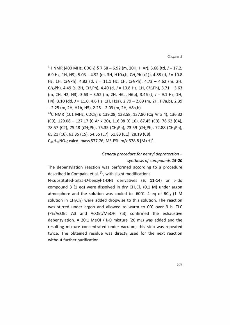
Chapter 5
209
1H NMR (400 MHz, CDCl3) δ 7.58 – 6.92 (m, 20H, H Ar), 5.68 (td, J = 17.2,
6.9 Hz, 1H, H9), 5.03 – 4.92 (m, 3H, H10a,b, CH2Ph (x1)), 4.88 (d, J = 10.8
Hz, 1H, CH2Ph), 4.82 (d, J = 11.1 Hz, 1H, CH2Ph), 4.73 – 4.62 (m, 2H,
CH2Ph), 4.49 (s, 2H, CH2Ph), 4.40 (d, J = 10.8 Hz, 1H, CH2Ph), 3.71 – 3.63
(m, 2H, H2, H3), 3.63 – 3.52 (m, 2H, H6a, H6b), 3.46 (t, J = 9.1 Hz, 1H,
H4), 3.10 (dd, J = 11.0, 4.6 Hz, 1H, H1a), 2.79 – 2.69 (m, 2H, H7a,b), 2.39
– 2.25 (m, 2H, H1b, H5), 2.25 – 2.03 (m, 2H, H8a,b).
13C NMR (101 MHz, CDCl3) δ 139.08, 138.58, 137.80 (Cq Ar x 4), 136.32
(C9), 129.08 – 127.17 (C Ar x 20), 116.08 (C 10), 87.45 (C3), 78.62 (C4),
78.57 (C2), 75.48 (CH2Ph), 75.35 (CH2Ph), 73.59 (CH2Ph), 72.88 (CH2Ph),
65.21 (C6), 63.35 (C5), 54.55 (C7), 51.83 (C1), 28.19 (C8).
C38H43NO4; calcd. mass 577,76; MS‐ESI: m/z 578,8 [M+H]+.
General procedure for benzyl deprotection –
synthesis of compounds 15‐20
The debenzylation reaction was performed according to a procedure
described in Compain, et al. [3], with slight modifications.
N‐substituted‐tetra‐O‐benzyl‐1‐DNJ derivatives (5, 11‐14) or L‐ido
compound 3 (1 eq) were dissolved in dry CH2Cl2 (0,1 M) under argon
atmosphere and the solution was cooled to ‐60°C. 4 eq of BCl3 (1 M
solution in CH2Cl2) were added dropwise to this solution. The reaction
was stirred under argon and allowed to warm to 0°C over 3 h. TLC
(PE/AcOEt 7:3 and AcOEt/MeOH 7:3) confirmed the exhaustive
debenzylation. A 20:1 MeOH/H2O mixture (20 mL) was added and the
resulting mixture concentrated under vacuum; this step was repeated
twice. The obtained residue was directy used for the next reaction
without further purification.

Chapter 5
210
L‐ido‐N‐allyl‐1‐deoxynojirimycin 15
Compound 3 (1196 mg, 2,12 mmol) was subjected to complete
debenzylation according to the general procedure. The obtain crude
residue was used for the successive acetylation without further
purification. C9H17NO4; calcd. mass 203,24; MS‐ESI: m/z 204,1 [M+H]+.
N‐allyl‐1‐deoxynojirimycin 16
Compound 5 (134 mg, 0,238 mmol) was subjected to complete
debenzylation according to the general procedure. The crude was
purified by FC (eluent AcOEt/MeOH 8,5:1,5) to afford compound 16 (34
mg, 0,17 mmol, 70% yield). 1H NMR (400 MHz, cd3od) δ 5.94 (td, J = 17.2,
8.0 Hz, 1H, H8), 5.29 – 5.18 (m, 2H, H9a,b), 3.89 (qd, J = 12.0, 2.5 Hz, 2H,
H6a,b), 3.58 – 3.41 (m, 2H, H7a, H2), 3.37 (t, J = 9.3 Hz, 1H, H4), 3.22 –
3.08 (m, 2H, H7b, H3), 3.00 (dd, J = 11.3, 4.9 Hz, 1H, H1a), 2.19 – 2.06 (m,
2H, H5, H1b). 13C NMR (101 MHz, cdcl3) δ 134.70 (C8), 119.43 (C9), 80.51
(C3), 71.76 (C4), 70.59 (C2), 67.53 (C5), 58.94 (C7), 57.65 (C6), 56.88 (C1).
C9H17NO4; calcd. mass 203,24; MS‐ESI: m/z 204,2 [M+H]+.
N‐(but‐3‐yn‐1‐yl)‐1‐deoxynojirimycin 17
C10H17NO4; calcd. mass 215,25; MS‐ESI: m/z 216,2 [M+H]+.
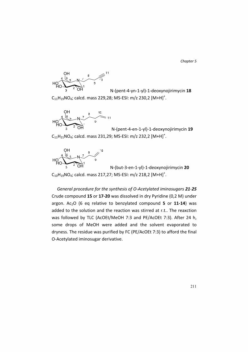
Chapter 5
211
N‐(pent‐4‐yn‐1‐yl)‐1‐deoxynojirimycin 18
C11H19NO4; calcd. mass 229,28; MS‐ESI: m/z 230,2 [M+H]+.
N‐(pent‐4‐en‐1‐yl)‐1‐deoxynojirimycin 19
C11H21NO4; calcd. mass 231,29; MS‐ESI: m/z 232,2 [M+H]+.
N‐(but‐3‐en‐1‐yl)‐1‐deoxynojirimycin 20
C10H19NO4; calcd. mass 217,27; MS‐ESI: m/z 218,2 [M+H]+.
General procedure for the synthesis of O‐Acetylated iminosugars 21‐25
Crude compound 15 or 17‐20 was dissolved in dry Pyridine (0,2 M) under
argon. Ac2O (6 eq relative to benzylated compound 5 or 11‐14) was
added to the solution and the reaction was stirred at r.t.. The reaxction
was followed by TLC (AcOEt/MeOH 7:3 and PE/AcOEt 7:3). After 24 h,
some drops of MeOH were added and the solvent evaporated to
dryness. The residue was purified by FC (PE/AcOEt 7:3) to afford the final
O‐Acetylated iminosugar derivative.

Chapter 5
212
2,3,4,6‐tetra‐O‐acetyl‐L‐ido‐N‐allyl‐1‐deoxynojirimycin 21
485 mg (1,3 mmol, 45% yield over two steps) of compound 21 were
obtained. 1H NMR (400 MHz, CDCl3) δ 5.74 (ddt, J = 16.8, 10.1, 6.3 Hz, 1H, H8), 5.28
– 5.10 (m, 3H, H9a,b, H3), 5.04 (dd, J = 10.2, 6.0 Hz, 1H, H4), 4.93 (td, J =
10.0, 5.8 Hz, 1H, H2), 4.40 (dd, J = 12.1, 6.6 Hz, 1H, H6a), 4.25 (dd, J =
12.1, 3.4 Hz, 1H, H6b), 3.51 – 3.41 (m, 1H, H5), 3.35 (dd, J = 13.9, 5.8 Hz,
1H, H7a), 3.22 (dd, J = 13.9, 6.7 Hz, 1H, H7b), 3.00 (dd, J = 12.3, 5.8 Hz,
1H, H1a), 2.62 (dd, J = 12.0, 10.5 Hz, 1H, H1b), 2.07 (s, 3H), 2.04 (s, 3H,
CH3CO), 2.02 – 1.98 (m, 6H, CH3CO). 13C NMR (101 MHz, CDCl3) δ 170.67, 170.29, 170.23, 170.12 (COCH3),
135.12 (C8), 118.33 (C9), 71.16 (C3), 70.57 (C4), 69.98 (C2), 58.81 (C6),
57.69 (C5), 57.48 (C7), 47.51 (C1), 21.17, 21.14, 21.02, 20.99, 20.96,
20.93, 20.90.
C17H25NO8; calcd. mass 371,39; MS‐ESI: m/z 372,2 [M+H]+.
2,3,4,6‐tetra‐O‐acetyl‐N‐(but‐3‐yn‐1‐yl)‐1‐deoxynojirimycin 22
Crude 17 was converted in peracetylated iminosugars 22, obtaining 168
mg (0,44 mmol) of compound, with a yield of 46% over two steps. 1H NMR (400 MHz, CDCl3) δ 5.08 – 5.00 (m, 2H, H3,4), 5.00 – 4.90 (m, 1H,
H2), 4.25 – 4.14 (m, 2H, H6a,b), 3.19 (dd, J = 11.4, 5.1 Hz, 1H, H1a), 2.97
(t, J = 7.4 Hz, 2H, H7a,b), 2.80 (dd, J = 6.0, 2.6 Hz, 1H, H5), 2.48 (dd, J =

Chapter 5
213
11.3, 10.3 Hz, 1H, H1b), 2.32 (dd, J = 8.6, 5.9 Hz, 2H, H8a,b), 2.09 (s, 3H,
CH3CO), 2.04 – 2.00 (m, 9H, CH3CO), 1.99 (t, J = 2.6 Hz, 1H, H10). 13C NMR (101 MHz, CDCl3) δ 170.97, 170.48, 170.16, 169.87 (COCH3),
81.99 (C9), 74.59 (C10), 70.16 (C3), 69.46 (C4), 69.33 (C2), 60.78 (C5),
60.00 (C6), 53.03 (C1), 50.52 (C7), 21.02, 20.98, 20.90, 20.83 (COCH3),
15.31 (C8).
C18H25NO8; calcd. mass 383,40; MS‐ESI: m/z 384,2 [M+H]+.
2,3,4,6‐tetra‐O‐acetyl‐N‐(pent‐4‐yn‐1‐yl)‐1‐deoxynojirimycin 23
Crude 18 was converted in peracetylated iminosugars 23 (128 mg, 0,322
mmol, 36% yield over two steps). 1H NMR (400 MHz, CDCl3) δ 5.06 – 4.95 (m, 2H, H3, H4), 4.95 – 4.85 (m,
1H, H2), 4.19 – 4.06 (m, 2H, H6a,b), 3.15 (dd, J = 11.5, 5.0 Hz, 1H, H1a),
2.93 – 2.79 (m, 1H, H7b), 2.66 – 2.48 (m, 2H, H5, H7a), 2.27 (t, J = 10.8
Hz, 1H, H1b), 2.17 (td, J = 6.8, 2.4 Hz, 2H, H9a,b), 2.03 (s, 3H, CH3CO),
2.01 – 1.93 (m, 9H, CH3CO), 1.91 (t, J = 2.4 Hz, 1H, H11), 1.71 – 1.49 (m,
2H, H8a,b). 13C NMR (101 MHz, CDCl3) δ 170.92, 170.37, 170.05, 169.77 (COCH3),
83.54 (C10), 74.59 (C3), 69.42 (C4), 69.25 (C2), 68.99 (C11), 61.70 (C5),
59.66 (C6), 52.93 (C1), 50.14 (C7), 24.30 (C8), 20.80, 20.78, 20.74, 20.72
(COCH3), 15.91 (C9).
C19H27NO8; calcd. mass 397,42; MS‐ESI: m/z 398,3 [M+H]+.

Chapter 5
214
2,3,4,6‐tetra‐O‐acetyl‐N‐(pent‐4‐en‐1‐yl)‐1‐deoxynojirimycin 24
Crude 19 was converted in peracetylated iminosugars 24 (149 mg, 0,37
mmol, 40% yield over two steps). 1H NMR (400 MHz, CDCl3) δ 5.77 (ddt, J = 16.9, 10.2, 6.6 Hz, 1H, H10),
5.16 – 4.84 (m, 5H, H11a,b, H2,H3,H4), 4.15 (d, J = 2.4 Hz, 2H, H6a,b),
3.19 (dd, J = 11.4, 5.0 Hz, 1H, H1a), 2.81 – 2.70 (m, 1H, H7a), 2.64 (d, J =
9.0 Hz, 1H, H5), 2.62 – 2.52 (m, 1H, H7b), 2.33 (t, J = 10.7 Hz, 1H, H1b),
2.07 (s, 3H, CH3CO), 2.06 – 1.97 (m, 11H, CH3CO x 9, H9a,b), 1.61 – 1.42
(m, 2H, H8a,b). 13C NMR (101 MHz, CDCl3) δ 171.07, 170.52, 170.17, 169.88 (COCH3),
137.92 (C10), 115.30 (C11), 74.75 (C2), 69.56 (C3), 69.49 (C4), 61.53 (C5),
59.63 (C6), 53.03 (C1), 51.28 (C7), 31.28 (C9), 24.01 (C8), 21.02, 20.98,
20.90, 20.83 (COCH3).
C19H29NO8; calcd. mass 399,44; MS‐ESI: m/z 400,4 [M+H]+.
2,3,4,6‐tetra‐O‐acetyl‐N‐(but‐3‐en‐1‐yl)‐1‐deoxynojirimycin 25
326 mg (0,85 mmol) of compound 25 were obtained, with a yield of 56%
yield over two steps. 1H NMR (400 MHz, CDCl3) δ 5.72 (ddt, J = 17.0, 10.1, 6.8 Hz, 1H, H9), 5.11
– 4.99 (m, 4H, H10a,b, H3,4), 4.95 (td, J = 9.8, 5.1 Hz, 1H, H2), 4.16 (d, J =
2.6 Hz, 2H, H6a,b), 3.20 (dd, J = 11.4, 5.0 Hz, 1H, H1a), 2.89 – 2.75 (m, 1H,
H7a), 2.75 – 2.61 (m, 2H, H7b, H5), 2.39 (t, J = 10.9 Hz, 1H, H1b), 2.28 –
2.12 (m, 2H, H8a,b), 2.07 (s, 3H, CH3CO), 2.05 – 1.96 (m, 9H, CH3CO).

Chapter 5
215
13C NMR (101 MHz, CDCl3) δ 171.06, 170.52, 170.16, 169.87 (COCH3),
135.61 (C9), 116.66 (C10), 74.71 (C2), 69.53 (C3), 69.48 (C4), 61.17 (C5),
59.62 (C6), 52.99 (C1), 51.30 (C7), 29.26 (C8), 21.02, 20.98, 20.90, 20.83
(COCH3).
C18H27NO8; calcd. mass 385,41; MS‐ESI: m/z 386,2 [M+H]+.
Synthesis of calix[4]arenes structures 27‐32
The synthesis, purification and characterization of the presented
calix[4]arenes are already described in literature in Dondoni and Marra [8], Marra, et al. [9], Fiore, et al. [6]. All characterization data are consistent
to those published.
Synthesis of calix[4]arenes 33‐34, 35‐36, 37‐38 and
monovalent ligands 39‐40.
The synthesis of the final calix[4]arene was performed according to the
same procedure described in the works mentioned above. NMR and
mass characterization has to be completed.

Chapter 5
216
References [1] C. Fasting, C. A. Schalley, M. Weber, O. Seitz, S. Hecht, B. Koksch,
J. Dernedde, C. Graf, E.‐W. Knapp, R. Haag, Angewandte Chemie‐
International Edition 2012, 51, 10472‐10498.
[2] J. Diot, M. I. Garcia‐Moreno, S. G. Gouin, C. O. Mellet, K. Haupt, J.
Kovensky, Organic & Biomolecular Chemistry 2009, 7, 357‐363.
[3] P. Compain, C. Decroocq, J. Iehl, M. Holler, D. Hazelard, T. Mena
Barragan, C. Ortiz Mellet, J.‐F. Nierengarten, Angewandte
Chemie‐International Edition 2010, 49, 5753‐5756.
[4] C. Decroocq, D. Rodriguez‐Lucena, V. Russo, T. Mena Barragan, C.
Ortiz Mellet, P. Compain, Chemistry‐a European Journal 2011, 17,
13825‐13831.
[5] S. Cecioni, O.‐A. Argintaru, T. Docsa, P. Gergely, J.‐P. Praly, S.
Vidal, New Journal of Chemistry 2009, 33, 148‐156.
[6] M. Fiore, A. Chambery, A. Marra, A. Dondoni, Organic &
Biomolecular Chemistry 2009, 7, 3910‐3913.
[7] M. Marradi, S. Cicchi, F. Sansone, A. Casnati, A. Goti, Beilstein
Journal of Organic Chemistry 2012, 8, 951‐957.
[8] A. Dondoni, A. Marra, Journal of Organic Chemistry 2006, 71,
7546‐7557.
[9] A. Marra, L. Moni, D. Pazzi, A. Corallini, D. Bridi, A. Dondoni,
Organic & Biomolecular Chemistry 2008, 6, 1396‐1409.
[10] C. R. R. Matos, R. S. C. Lopes, C. C. Lopes, Synthesis‐Stuttgart
1999, 571‐573.
[11] T. Wennekes, R. J. B. H. N. van den Berg, W. Donker, G. A. van der
Marel, A. Strijland, J. M. F. G. Aerts, H. S. Overkleeft, The Journal
of Organic Chemistry 2007, 72, 1088‐1097.
[12] M. Fiore, M. Lo Conte, S. Pacifico, A. Marra, A. Dondoni,
Tetrahedron Letters 2011, 52, 444‐447.

Chapter 5
217
[13] Y. Jiang, Z. Fang, Q. Zheng, H. Jia, J. Cheng, B. Zheng, Synthesis‐
Stuttgart 2009, 2756‐2760.
[14] V. S. Rao, A. S. Perlin, Canadian Journal of Chemistry‐Revue
Canadienne De Chimie 1984, 62, 886‐890.

218

Chapter 6
219
Chapter 6. Antiproliferative activity
of Arsenical C‐glucoside derivative
on neuroblastoma cell line SN‐K‐BE
Chapter 6
220
Abstract C‐glucoside derivatives covalently linked to the arsenic atom in different
oxidation states have been synthesized. While compound 5, bearing an
AsV atom, did not show any significant biological activity, compound 6, in
which the glucose moiety is linked to the arsenic atom as dithioarsenal
(AsIII), showed promising antiproliferative activity on cell line of
neuroblastoma (SK‐N‐BE). In conclusion, compound 6, synthesised
avoiding protection‐deprotection steps, represents a promising lead
compound for the development of selective antitumor agents.
Introduction Since Hippocrates and then through Chinese alchemy, Fowler’s solution,
Erlich’s Salvarsan, until the recent use of arsenic trioxide as a therapy for
promyelocytic leukemia, arsenic has represented a Janus bifrons of
medicine, being remedy and poison, healer and killer. This double‐edged
sword properties stem for a large part from arsenic physicochemical
similarity with phosphorus as metabolic pathways intended for PO43‐
may not distinguish it from AsO43‐[1]. This means that arsenic is a serious
threat for health but also an ideal bullet to be delivered to a therapeutic
target in a masked way, provided it is conjugated with a highly selective
carrier. Aiming at the design of an arsenic‐based anti‐cancer drug, we
envision to exploit the preferential uptake of glucose by cancer cells,
known as Warburg effect. This effect starts in the early stages of
carcinogenesis and seems to increase with tumor aggressiveness[2].
Despite this process being functional for tumor growth, it is absolutely
unfavorable from an energetic point of view, thus forcing tumor cells to
import a great quantity of glucose. This results in an increase in the

Chapter 6
221
expression of membrane proteins devoted to the uptake of glucose
molecules, in particular the GLUT transporters[3]. The use of 2‐(18F)‐
fluoro‐2‐deoxy‐D‐glucose (FDG) as tracer in the positron emission
tomography (PET) confirms the in vivo accumulation of glucose inside
tumor cells, and represents the main application of the Warburg effect.
The success in this diagnostic application has suggested the therapeutic
exploitation of the preferential uptake of glucose, through its
conjugation to cytotoxic drugs, such as cyclophosphamide.
Unfortunately these bulky conjugates do not penetrate inside the cells
through the GLUT transporters, but through the co‐trasporter sodium‐
glucose (SAAT1)[4], thus showing a reduced selectivity towards tumor
cells. Being devoid of significant steric hindrance, arsenic is in principle
an ideal conjugate for glucose. Furthermore, by matching the selectivity
of glucose to the widespread biological impact of arsenic, we meant to
obtain compounds able to specifically deliver to cancer cells a
pharmacological agent capable of simultaneously targeting different
metabolic pathways. This approach may be particularly useful in cancer
since it may limit the development of resistance by targeting different
key points of tumor metabolism.
In this work we generated arsenical C‐glucoside derivatives, in which the
arsenic group is covalently linked to the sugar moiety through the alkyl
appendage at the anomeric centre, thus maintaining the pyranosidic
structure of the glucose moiety whilst generating a metabolically stable
derivative. The arsenal compound should therefore accumulate and
exert its toxic effect inside tumor cell.

Chapter 6
222
Results and Discussion We designed and synthesized two C‐glucoside derivatives covalently
linked to the arsenic atom in different oxidation states as arsenic acid
(AsV) and dithioarsenal (AsIII) (5 and 6, respectively) and carried out
preliminary in vitro biological evaluation on a tumoral cell line of human
neuroblastoma (SK‐N‐BE).
Scheme 1: Reagents and conditions: a) Ref.
[6]; b) MeOH/H2O, NaCNBH3, 3 or 4, yield 5
(35%) and 6 (44 %).
The synthesis (scheme 1) was designed in order to avoid the use of
protecting groups and to insert the arsenic containing moiety at the end
of the synthetic route. As arsenic moiety we chose the two readily
available phenyl arsine derivatives 3 and 4, that can be chemoselectively
attached to the hemiacetalic group of the glucose derivative 2 by
reductive amination. Thus the key C‐glycosyl intermediate 2 was
generated by stereoselective C‐allylation[5] of methyl ‐D‐glucopyranoside, followed by ozonolysis and reductive amination of the
obtained aldehyde (present as hemiacetal 2)[6].
The effects of the two compounds were tested on human
neuroblastoma cells (SK‐N‐BE) and compared with that of As2O3. While
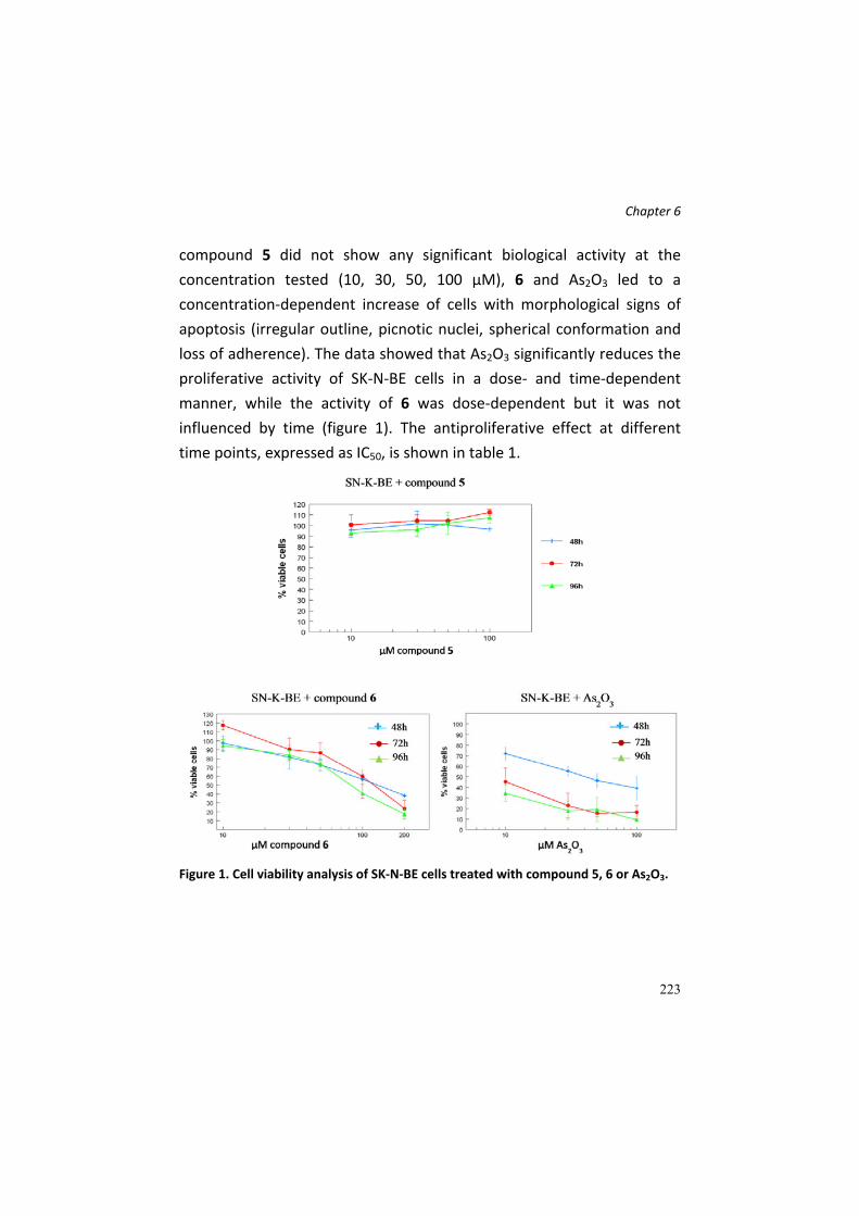
Chapter 6
223
compound 5 did not show any significant biological activity at the
concentration tested (10, 30, 50, 100 μM), 6 and As2O3 led to a
concentration‐dependent increase of cells with morphological signs of
apoptosis (irregular outline, picnotic nuclei, spherical conformation and
loss of adherence). The data showed that As2O3 significantly reduces the
proliferative activity of SK‐N‐BE cells in a dose‐ and time‐dependent
manner, while the activity of 6 was dose‐dependent but it was not
influenced by time (figure 1). The antiproliferative effect at different
time points, expressed as IC50, is shown in table 1.
Figure 1. Cell viability analysis of SK‐N‐BE cells treated with compound 5, 6 or As2O3.

Chapter 6
224
Table 1: The in vitro antiproliferative activity of As2O3 and compound 6.
Test compound IC50 (48h) IC50 (72h) IC50 (96h)
As2O3 28.18 μM 7.10 μM 3.31 μM
Compound 6 110.5 μM 91.93 μM 88.43 μM
In conclusion, we have demonstrated the antiproliferative effect of an
AsIII derivative covalently linked to glucose, that, combined with the
Warburg effect, opens the way to new selective anticancer agents.
Interestingly, the antiproliferative effect of the arsenal C‐glucoside is not
time‐dependent like that of As2O3. Compound 6, synthesized avoiding
protection‐deprotection steps, represents therefore a potential lead
compound for the development of a “magic bullet” aiming at cancer
cells.
Experimental Section General Remarks. All solvents, when necessary, were dried with
molecular sieves for at least 24 h prior to use. Thin layer chromatography
(TLC) was performed on silica gel 60 F254 plates (Merck) with detection
using UV light when possible, or by charring with a solution of concd.
H2SO4/EtOH/H2O (5:45:45) or a solution of (NH4)6Mo7O24 (21 g), Ce(SO4)2
(1 g), concd. H2SO4 (31 mL) in water (500 mL). Flash column
chromatography was performed on silica gel 230‐400 mesh (Merck). 1H
and 13C NMR spectra were recorded at 25 °C unless otherwise stated,
with a Varian Mercury 400 MHz instrument. Chemical shift assignments,
reported in ppm, are referenced to the corresponding solvent peaks.
HRMS were recorded on a ABSciex 2000 QTRAP LC/MS/MS System with
an ESI source. Optical rotations were measured at room temperature
Chapter 6
225
using an Atago Polax‐2L polarimeter and are reported in units of 10‐1
deg∙cm3∙g‐1.
4‐[2‐(α‐D‐glucopyranosyl)ethylamino]phenyl)arsonic acid 5: crude
compound 2 (1.46 mmol, 1 equiv.) was treated with an HCl 0.01 M
solution (7 mL) for 1 h at 70 °C. The pH of the solution was adjusted to
pH > 7 and compound 3 (1.75 mmol, 1.2 equiv.), suspended in
MeOH/H2O (30 mL), was added. Then, the pH of the solution was
decreased to pH = 4 with AcOH; after 20 min, NaCNBH3 (2.04 mmol, 1.4
equiv.) was added. After 30 min the solvent was removed under reduced
pressure and the crude is purified by flash chromatography
(AcOEt/MeOH/H2O/AcOH 70/30/5/5) affording compound 5 with an
overall yield of 35% from 2.: [α]D25 = + 61.7 (c = 1, H2O), 1H NMR (400
MHz, D2O) δ 7.53 (d, J = 8.4 Hz, 2H), 6.84 (d, J = 8.4 Hz, 2H), 4.18 – 4.03
(m, 1H), 3.90 – 3.74 (m, 1H), 3.73 – 3.61 (m, 2H), 3.61 – 3.54 (m, 1H),
3.54 – 3.48 (m, 1H), 3.40 – 3.13 (m, 3H), 2.05 – 1.89 (m, 2H). 13C NMR
(100 MHz, D2O) δ ppm 154.6, 134.1, 123.7, 116.3, 76.83, 75.93, 75.45,
73.664, 72.99, 63.74, 42.32, 25.64; MS calcd for C14H22AsNO8 [M + H]+
408.1: found 408.1.
4‐(1,3,2‐Dithiarsolan‐2‐yl)‐N‐[2‐(α‐D‐glucopyranosyl)ethyl]aniline 6:
crude compound 2 (0.97 mmol, 1 equiv.) was treated with an HCl 0.01 M
solution (7 mL) for 1 h at 70 °C. The pH of the solution was adjusted to
pH > 7 and compound 4 (1.16 mmol, 1.2 equiv.), suspended in
CH2Cl2/MeOH 3:4 (70 mL), was added. Then, the pH of the solution was
decreased to pH = 4 with AcOH; after 20 min, NaCNBH3 (1.36 mmol, 1.4
equiv.) was added. After 30 min the solvent is removed under reduced
pressure and the crude was purified by flash chromatography
(AcOEt/MeOH 95/5) affording compound 6 with an overall yield of 44 %

Chapter 6
226
from 2.: [α] D25 = + 31.7 (c = 0.5, DMSO); 1H NMR (400 MHz, CD3OD) ppm
7.29‐7.21 (m, 1H), 7.04‐6.94 (m, 1H), 6.60‐6.46 (m, 2H), 4.00‐3.90 (m,
1H), 3.75 (bd, J = 11.78 Hz, 1H), 3.56‐3.47 (m, 2H), 3.44‐3.35 (m, 2H),
3.23‐3.06 (m, 7H), 1.90‐1.81 (m, 2H). 13C NMR (100 MHz, CD3OD) 166.7,
153.0, 135.8, 132.8, 117.0, 116.2, 78.79, 78.03, 77.60, 75.67, 75.27,
66.08, 44.94, 44.80, 27.77; MS calcd for C16H24AsNO5S2 [M + H]+ 450:
found 450.
Drugs and chemicals: Arsenic trioxide (As2O3) was purchased from Sigma
Aldrich Company (MI). Arsenic trioxide was dissolved in 0,1 M NaOH and
kept as a stock of 1 mM As2O3 dissolved in distilled water. Growth
medium DMEM High glucose, fetal bovine serum (FBS), antibiotics
(penicillin and streptomycin), L‐Glutamine and L‐phosphate buffered
saline (PBS) were obtained from Euroclone. 3‐(4, 5‐dimethylthiasol‐2‐yl)‐
2, 4,‐diphenyltetrazolium bromide (MTT) solution and Dimethyl sulfoxide
(DMSO) were purchased from Sigma Aldrich Company (MI).
Cell lines and culture conditions. Human neuroblastoma cell line SK‐N‐BE
was cultured in DMEM High glucose supplemented with 10% fetal bovine
serum (FBS), 1% (w/v) penicillin/streptomycin and 2 mM L‐glutamine in a
humidified incubator containing 95% air + 5% CO2 atmosphere.
MTT assay: Cell proliferation was evaluated by MTT metabolism as
previously described[7]. Briefly, SK‐N‐BE were plated in 96‐well
polystyrene tissue culture plates (Falcon) at a density of 5×10³/well in
fresh medium. After 48h of incubation As2O3 or compound 6 solutions
(range 10‐200 µM) were added to each well. Cells incubated in culture
medium alone served as untreated control. Cells were cultured for 48, 72
and 96 hours respectively. After incubation, 20 µl aliquots of MTT

Chapter 6
227
solution (5 mg/mL in PBS) were added to each well and re‐incubated for
4 hours at 37°C. Then, the supernatant culture medium were carefully
aspirated and 200 µl aliquots of dimethylsulfoxide (DMSO) were added
to each well to dissolve the formazan crystals, followed by incubation for
10 minutes to dissolve air bubbles. The culture plate was placed on a
micro‐plate reader and the absorbance was measured at 540 nm. The
amount of color produced is directly proportional to the number of
viable cell. All assays were performed in six replicates for each
concentration and means ± SD values were used to estimate the cell
viability. Cell proliferation rate was calculated as the percentage of MTT
absorption as follows: % survival = (mean experimental
absorbance/mean control absorbance) × 100. Morphological changes
were followed by phase contrast microscopy.
Statistical analysis: Student t test was used for all comparison, all tests
were two‐tailed. The IC50 value were calculated using GraphPad Prism 5.
References [1] Rosen, B. P. FEBS Lett., 2002, 529(1), 86‐92.
[2] Cairns, R. A.; Harris, I. S.; Mak, T. W. Nat Rev Cancer, 2011, 11(2),
85‐95.
[3] Zhao, F. Q.; Keating, A. F. Current Genomics, 2007, 8, 113‐128.
[4] Veyhl, M.; Wagner, K.; Volk, C.; Gorboulev, V.; Baumgarten, K.;
Weber, W. M.; Schaper, M.; Bertram, B.; Wiessler, M.; Koepsell,
H. Proc Natl Acad Sci USA,1998, 17;95(6), 2914‐9.
[5] Bennek, J. A.; Gray, G. A. J. Org. Chem., 1987, 52, 892‐897.
[6] Cardona, F.; La Ferla, B. J. Carbohydr. Chem., 2008, 27(4), 203‐
213.
[7] Mosmann, T. J Immunol Methods, 1983, 65(1‐2), 55‐63.

228

Chapter 7
229
Chapter 7. Conclusions

Chapter 7
230
Carbohydrates represent not only an important class of macromolecules,
widely spread in Nature and involved in practically all the biological
process of living organism, but also tools that chemists and biochemists
can use for the generation of potential drug candidates, and conjugates
or derivatives, for the investigation and the study of the processes in
which carbohydrates are involved.
In the first case, we started from an anti‐inflammatory effect observed
with high glucose doses administered to in vitro and in vivo inflammatory
models, that suggested the search of glucose derivatives bearing the
same function. This prompted us to generate a library of metabolically
inert C‐glycoside derivatives; these bioactive molecules represent both
promising drug candidates, for the treatment of severe inflammatory
disease, as sepsis, endotoxic shock, mucositis or Chron disease, and
chemical tools for the comprehension of the mechanism at the base of
this observed phenomenon. The use of these carbohydrate‐based
structure allowed to make more evident a new biological role, a sort of
immunological protection, for a transport protein (SGLT1), for which the
physiological function has been well established.
The metabolic behavior of sugars, and in particular of glucose, in tumor
cells, represent the fundament of our idea of the development of new
carbohydrate‐based antiproliferative agents, containing the cytotoxic
element Arsenic. The synthesized molecule represents a lead compound,
a starting point for the development of more and more selective and
effective anticancer agents, for which there is a high demand.
Iminosugars are a class of glycoderivatives widely studied both by
chemists and biochemists; their inhibitory and chaperon‐like activity on
glycosidases prompted the researchers towards the synthesis of more
and more analogues, with increasing efficacy; nowadays, two
iminosugars are commercially available for the treatment of diabetes

Chapter 7
231
mellitus type II (Miglitol) and for Gaucher disease (Miglustat). Beside
their clinical importance, iminosugars have been used and developed as
models and tools for the study of phenomena, like multivalent inhibiton
of glycosidases, which could lead to new details and knowledge about
how these critical enzymes work in living organisms.

232

233
Pubblications and communications

234
Papers
La Ferla, B., Spinosa, V., D'Orazio, G., Palazzo, M., Balsari, A., Foppoli, A.
A., Rumio, C. and Nicotra, F. (2010), “Dansyl C‐Glucoside as a Novel
Agent Against Endotoxic Shock”. ChemMedChem, 5: 1677–1680.
Barbara La Ferla, Cristina Airoldi, Cristiano Zona, Alexandre Orsato,
Francisco Cardona, Silvia Merlo, Erika Sironi, Giuseppe D'Orazio and
Francesco Nicotra, Natural glycoconjugates with antitumor activity,
Natural Product Report, 2011, 28, 630‐648
Oral communications
G. D’Orazio, B. La Ferla, M. Coppola, C. Magni, F. Nicotra, “Sodium‐
Glucose Cotransporter 1 (SGLT1) target receptor for carbohydrate based
anti‐inflammatory agents”. Italian‐Spanish Joint Workshop CARIPLO
project 2008‐3175, Chemical tools for molecular recognition studies:
Synthesis and NMR characterization of bioactive molecules, Università
degli Studi di Milano – Bicocca, 22/4/2010.
Giuseppe D’Orazio, Diego Cardani, Cristiano Rumio, Francesco Nicotra,
Barbara La Ferla, “SGLT1‐MEDIATED ANTIINFLAMMATORY AND
PROTECTIVE EFFECT OF DANSYL‐GLYCODERIVATIVES. SYNTHESIS,
BIOLOGICAL EVALUATION AND STUDIES ON THE MECHANISM OF
ACTION”, XIII Convegno‐Scuola sulla Chimica dei Carboidrati, Pontignano
(Si), June 24th – 27th, 2012.

235
Other communications
G. D'Orazio, B. La Ferla, M. Coppola, C. Rumio, F. Nicotra, “Design and
synthesis of 2nd generation carbohydrate‐based antiinflammatory
agents” ‐ XXXV Corso Estivo “A. Corbella ‐ Seminari di Chimica Organica”
della Società Chimica Italiana, Gargnano (Palazzo Feltrinelli), 14 –
18/06/2010.
Giuseppe D’Orazio, Barbara La Ferla, Marta Coppola, Cristiano Rumio,
Francesco Nicotra “DANSYLATED C‐GLYCOSIDES: DRUG CANDIDATES AS
ANTIIFLAMMATORY AGENTS AND MOLECULAR TOOLS FOR BIOLOGICAL
STUDIES”, 16th European Carbohydrate Symposium (Eurocarb 16) –
Sorrento, 03‐07/07/2011.
D’Orazio, G.; Zona, C.; Parisi, G.; Policano, C.; Mechelli, R.; Codacci‐
Pisanelli, G.; Pitaro, M.; Ristori, G.; Salvetti, M.; Nicotra, F.; La Ferla, B.;
“ARSENIC C‐GLYCOSIDES: APPLYING THE PAUL ERLICH’ “MAGIC BULLET”
CONCEPT IN THE TREATMENT OF CANCER”, 26th International
Carbohydrate Symposium (ICS2012), Madrid, July 22nd to 27th, 2012.
Giuseppe D’Orazio, Diego Cardani, Cristiano Rumio, Francesco Nicotra,
Barbara La Ferla, “SGLT1‐MEDIATED ANTIINFLAMMATORY AND
PROTECTIVE EFFECT OF DANSYL‐GLYCODERIVATIVES. SYNTHESIS,
BIOLOGICAL EVALUATION AND STUDIES ON THE MECHANISM OF
ACTION”, XIII Convegno‐Scuola sulla Chimica dei Carboidrati,
Pontignano (Si), June 24th – 27th, 2012.
Barbara La Ferla, Cristiano Zona, Giuseppe D’Orazio, Francisco Cardona,
Crisitina Airoldi, Alexandre Orsato, Francesco Nicotra, “Glycomimetics

236
conjugated Nanoparticles with therapeutic activity and glycidic scaffold
based targeting agents”, MultiGlycoNano ‐ COST‐Meeting 2012, Berne
(Switzerland), 2nd‐4th February 2012.

237
List of abbreviations
aa Aminoacid
Ac Acetyl
Ac2O Acetic anhydride
AcOEt Ethyl acetate
AcOH Acetic acid
Ag2O Silver oxide
AllylMgBr Allylmagnesium bromide
AllylTMS Allyltrimethylsilane
Asn Asparagine
ATP Adenosine triphosphate
B0AT1 Sodium‐glutamine
cotransporter 1
BBM Brush border membrane
BCl3 Boron trichloride
BF3OEt2 Boron trifluoride diethyl
etherate
Bn Benzyl
BnBr Benzyl bromide
BSTFA N,O‐Bis(trimethylsilyl)
trifluoroacetamide
Caco‐2 Human epithelial
colorectal adenocarcinoma
cell
CD14 Cluster of differentiation
14
CH2Cl2 Dichloromethane
CH3CN Acetonitrile
(COCl)2 Oxalyl chloride
CpG‐ODN CpG‐Oligodeoxynucleotide
CSA Camphorsulphonic acid
CSGAGs Chondroitin/dermatan
CuAAC Copper(I)‐catalyzed azide
alkyne cycloaddition
DansylCl Dansyl chloride
DCC N‐N’‐dicyclohexyl
carbodiimide
DIAD Diisopropyl
azodicarboxylate
DIPEA Diisopropylethylamine
DMAP Dimethylaminopyridine
DMEM Dulbecco’s Modified Eagle
Medium
DMF Dimethylformamide
DMSO Dimethylsulfoxide
DNA Deoxy‐ribonucleic acid
1‐DNJ 1‐Deoxynojirimycin
DPAP 2,2‐Dimethoxy‐2‐
acetophenone
ECM Extracellular matrix
EDC N‐(3‐Dimethylamino
propyl)‐N′‐ethyl
carbodiimide
hydrochloride
EGF Epidermal Growth Factor
ELISA Enzyme‐Linked
ImmunoSorbed Assay
ER Endoplasmic Reticulum
ESI‐MS ElectroSpray Ionization –
Mass Spectroscopy
Et2O Diethylether
EtOH Ethanol
Et3N Triethylamine
Et3SiH Triethylsilane

238
FBS Fetal bovine serum
FC Flash chromatography
FDG 2‐(18F)‐fluoro‐2‐deoxy‐
D‐glucose
GAGs Glycosaminoglycans
Gal Galactose
GGM Glucose‐galactose
malabsorption
Glc Glucose
L‐Gln L‐Glutamine
GLUT Glucose transporter
GSLs Glycosphingolipids
HCl Hydrochloric acid
H2O Water
HOBt Hydroxybenzotriazole
HSGAGs Heparin/Heparan
HT29 Human colon
adenocarcinoma
cell line
IBDs Intestinal Bowel Disease
IC50 Half maximal inhibitory
concentration
IECs Intestinal Epithelial Cells
IGF Insulin Growith Factor
IL Interleukin
iPr2EtN Diisopropylethylamine
K2CO3 Potassium carbonate
LPS Lipopolysaccharide
LSDs Lysosomal storage disease
MD2 Lymphocyte antigen 96
MeOH Methanol
MeONa Sodium methylate
MHC‐II Class II Major
Histocompatibility
Complex
MsCl methanesulfonyl chloride
MTT 3‐(4,5‐dimethylthiasol‐
2‐yl)‐2,4‐Diphenyl
tetrazolium bromide
Na Sodium
NaBD4 Sodium borodeuteride
NaBH4 sodium borohydride
NaBT4 Sodium borotritide
NaCNBH3 Sodium
cyanoborohydride
NaH Sodium hydride
NaIO4 Sodium periodate
Na+/K
+‐ATPase Sodium‐potassium ATP‐
synthase
NaN3 Sodium azide
NANA N‐Acetyl Neuraminic Acid
(Sialic Acid)
NaOH Sodium hydroxide
NH3 Ammonia
NH4Cl Ammonium chloride
NH4+HCOO
‐ Ammonium formate
NIS N‐iodo‐succinimide
NMR Nuclear Magnetic
Resonance
NOESY Nuclear Overhauser Effect
SpectroscopY
3OMG 3‐O‐Methyl‐D‐
glucopyranose
OMIM Online Mendelian
Inheritance in Man
ORT Oral Rehydration Therapy
OsO4 Osmium tetroxide
PAMPs Pathogen‐Associated
Molecular Patterns
PBS Phosphate Buffer

239
Saline
Pd(OH)2/C Palladium hydroxide on
activated charcoal
PE Petroleum ether
PEG Polyethyleneglycol
PGs Proteoglycans
(PhO)2PON3 Diphenyl phosphoryl azide
Ph3P Triphenylphosphine
PRRs Pattern Recognition
Receptors
Py Pyridine
QC Quality Control
ROS Reactive Oxygen Species
Satd. Saturated
Ser Serine
SGLT1 Sodium Glucose
coTransporter 1
SIRS Systemic Inflammatory
Response Syndrome
SK‐N‐BE Human neuroblastoma
cell line
SMF Sodium Motive Force
SPR Surface Plasmon
Resonance
SSSFs Sodium substrate
symporter family
tBuOH Tert‐butanol
TEC Thiol‐ene coupling
TEG Triethyleneglycol
TEM Transmission Electron
Microscopy
TFA Trifluoroacetic acid
THF Tetrahydrofuran
Thr Threonine
TLC Thin Layer
Chromatography
TLR Toll‐Like Receptor
TM Transmembrane
TMSOTF Trimethylsilil
trifluoromethane
sulphonate
TNFα Tumor Necrosis
Factor α
TS Transition State
TsCl Toluensulfonyl chloride

240