go molecular!
TRANSCRIPT
Go molecular!
Revised and updated 2nd edition (2021)
A clinical reference guide to molecular allergy Part 1: The basics
2021edition with
information on novel components
rSes i 1 and rPru p 7
2
Molecular allergens have been described in scientific literature for well over a decade now, but it has only been in recent years that they have been used more routinely in the allergy clinic.
New technology can be challenging, and it often requires a period of adjustment and adaptation. There are many allergen components covering many different sources and their clinical relevance is continually emerging year on year. This can make it difficult to remember their relevance. Many physicians have commented to me that they could do with a simplified ‘all in one guide’ so I have tried to simplify molecular allergology based on the allergen components Thermo Fisher Scientific is supplying (manufacturer is Phadia AB).
The intention of part 1 in this guidebook series is to give a basic introduction to molecular allergology focusing on plant food allergy, although other molecular sources such as venoms and aeroallergens are also discussed. This guide gives an introductory
Preface
Disclaimer: The content of this book is intended as an aid to the physician to interpret allergen specific IgE antibody test results. It is not intended as medical advice on an individual level. A definitive clinical diagnosis of IgE mediated allergic disorders should only be made by the physician based on the clinical history for the individual patient after all clinical and laboratory findings have been evaluated. It should not be based on the results of any single diagnostic method.
overview of the important themes within molecular allergology, especially protein families, their clinical relevance and nomenclature. If there is one important aspect to learn in molecular allergology it is the scientific relevance of protein families, as they are the key to understanding clinical molecular allergology.
A straightforward summary of the main allergen components, what ImmunoCAP™ products are available and an aid to interpret test results can be found in part 2 of this series – ‘The Allergen Components’.
I hope you find this guidebook series useful.
Neal Bradshaw Portfolio Manager - Allergy Author of the Go Molecular! booksImmunodiagnosticsThermo Fisher Scientific
172050.AL.EU, INT, JP1.EN.v1.21
3
Foreword 4
Introduction: Molecular allergens tell us more 5
Protein families 7
Allergen component nomenclature 9
Specific and cross-reactive allergens 10
Other clinical considerations 11
Food allergy 13
Plant components 17
Interpreting results from cross-reactive protein families 19
Summary of plant food components 21
Plant allergen components in some common foods 24
Other allergen components 26
Immunotherapeutics – Aeroallergens and venoms 36
Common questions regarding molecular components 37
Glossary 39
Educational resources 40
Using ImmunoCAP Allergen Component tests 41
ImmunoCAP Allergen Component list 43
ImmunoCAP ISAC112i Chip Allergen Components 47
Contents

4
With the advent of allergen components, allergy has got much more complicated. However whole allergen diagnostics, with skin prick testing or serum specific IgE, commonly don’t allow us to unravel the complexity that some of our allergy patients exhibit. Using allergen components to understand the molecular allergology of these complex patients has a real potential to improve our clinical decision-making. The use of component resolved diagnostics may optimize our investigation plans and improve our diagnoses, management plans and the advice we give to our allergy patients. All this though relies on clinicians acquiring an understanding of molecular diagnostics. This is a rapidly evolving area with, for example, the whole peanut allergen suddenly been replaced by more than 10 individual components with different clinical impacts.
Foreword
This edition of this book is very welcome with its updated information about each of the various allergen components. Importantly, their clinical implications are explained allowing us to use information about allergic sensitization to each individual component to improve the management of our patients.
Professor Graham RobertsProfessor of Paediatric Allergy and Respiratory MedicineUniversity of Southampton

5
The diagnosis of IgE mediated allergies is made by the physician based on clinical history for the individual patient in conjunction with clinical findings and test results, e.g. specific IgE sensitization tests such as skin prick and/or blood tests and sometimes allergen provocations. Until recently the sensitization tests in use were based on extracts of allergen sources, but in the past years the use of component resolved diagnosis has become increasingly common in clinical practice. Molecular allergology brings a new level of understanding to physicians who seek to improve on existing diagnostic technologies1-3.
While traditional extract-based IgE blood tests measure the “sum” of sensitization to all protein components in whole allergens, e.g. peanut, molecular allergology makes it possible to investigate important individual proteins within a peanut for specific IgE sensitization. IgE antibody profiles to these molecules vary significantly from patient to patient and they also differ geographically, due to local differences of exposure1-3.
Molecular diagnostics reveals more factual information about what a patient is allergic to, as individual proteins and profiles can indicate different clinical characteristics1-3.
Introduction: Molecular allergology tells us more
Figure 1: Illustration of the common misconception that there is one IgE antibody produced by the human body for a whole peanut allergen.

6
Allergen component diagnostics measures IgE to specific allergen components, uncovering additional information about an underlying allergy. Not only do they indicate specific allergen reactivity in the way that whole extracts do but they are also indicators for:
1. Understanding patient risk for allergic reactions – adding confidence to your assessment1-3.
2. Aiding the selection of the proper treatment extract of Allergen Specific
Immunotherapy (AIT) – useful for example in venom and aero-allergy patient selection1-3.
3. Understanding cross-reactions between species – helping to understand multiple sensitizations e.g. in pollen food syndrome1-3.
The intention of this first guidebook is to give the physician, dietician or scientist a background to molecular allergology. A straightforward summary of allergen components and an aid to interpretation of results can be found in part 2 of this series.
Figure 2: Illustration of the reality that there are lots of different IgE antibodies produced which bind to individual proteins in peanut, like Ara h1, Ara h 2 and Ara h 8.
Ara h 2/Ara h 6 are the proteins that seems to have the highest allergenic potential of all proteins in peanut. Antibodies produced by patients in response to specific allergen proteins can be measured using single or multiplex allergen component tests, indicating the patients’ immunological response in their current allergy status. High levels of IgE to Ara h 2/Ara h 6 will often indicate a patient at high risk of systemic symptoms if peanuts are eaten1-3.
Clinical relevance
The IgE indicated in yellow is directed against peanut Ara h 2*. This can be measured using ImmunoCAP Allergen Components.
Ara h 2 is the most allergenic protein in peanuts. Patients positive to it are at high risk of systemic reactions1-3
Other specific IgE antibodies are directed against lower risk peanut proteins such as Ara h 8 and Ara h 5. Proteins closely related to these are also found in tree and grass pollen.
*ImmunoCAP Allergen f423, Allergen component rAra h 2 Peanut
7
Much of the clinical value of testing with allergen components up to now has been demonstrated within food allergy, especially with plant foods such as nuts, fruits and legume seeds. The majority of information in this reference guide therefore focuses on food allergen components, although an overview of other allergen components which provide clinical value, such as those in pollen, furry animals, mites, latex and insect venoms, is also included.
References
1. Matricardi PM et al. EAACI Molecular Allergology User’s Guide. Pediatric allergy and immunology: official publication of the European Society of Pediatric Allergy and Immunology. 2016;27 Suppl 23:1-250.
2. Kleine-Tebbe J and Jakob T Editors: Molecular Allergy Diagnostics. Innovation for a Better Patient Management. Springer International Publishing Switzerland 2017. ISBN 978-3-319-42498-9 ISBN 978-3-319-42499-6 (eBook),DOI 10.1007/978-3-319-42499-6.
3. Canonica GW et.al. A WAO - ARIA - GA²LEN consensus document on molecular-based allergy diagnostics. World Allergy Organ J. 2013 Oct 3;6(1):17.
Introducing testing with molecular allergens into daily clinical practice will help improve diagnosis of allergy based on allergenic proteins and protein families.
Protein families referred to in this guide are families with similar functions and structures found in many allergen sources1-8. For example, plant seeds contain storage proteins such as vicilins, transport proteins such as lipid transfer proteins and defense proteins such as PR-10s (pathogenesis-
Protein familiesrelated family number 10 proteins). Lipocalins and serum albumins are examples of protein families found in mammalian allergen sources1-8.
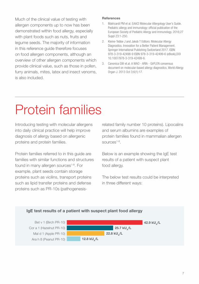
Below is an example showing the IgE test results of a patient with suspect plant food allergy.
The below test results could be interpreted in three different ways:
Bet v 1 (Birch PR-10)
Cor a 1 (Hazelnut PR-10)
Mal d 1 (Apple PR-10)
Ara h 8 (Peanut PR-10)
42.9 kUA/L
25.7 kUA/L
22.8 kUA/L
12.8 kUA/L
IgE test results of a patient with suspect plant food allergy
8
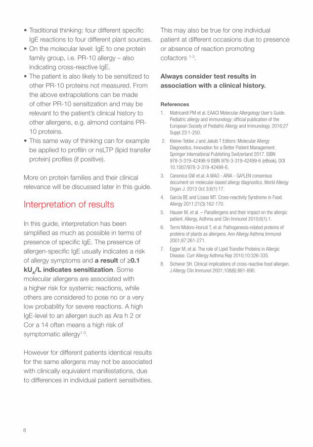
• Traditional thinking: four different specific IgE reactions to four different plant sources.
• On the molecular level: IgE to one protein family group, i.e. PR-10 allergy – also indicating cross-reactive IgE.
• The patient is also likely to be sensitized to other PR-10 proteins not measured. From the above extrapolations can be made of other PR-10 sensitization and may be relevant to the patient’s clinical history to other allergens, e.g. almond contains PR-10 proteins.
• This same way of thinking can for example be applied to profilin or nsLTP (lipid transfer protein) profiles (if positive).
More on protein families and their clinical relevance will be discussed later in this guide.
Interpretation of results
In this guide, interpretation has been simplified as much as possible in terms of presence of specific IgE. The presence of allergen-specific IgE usually indicates a risk of allergy symptoms and a result of ≥0.1 kUA/L indicates sensitization. Some molecular allergens are associated with a higher risk for systemic reactions, while others are considered to pose no or a very low probability for severe reactions. A high IgE-level to an allergen such as Ara h 2 or Cor a 14 often means a high risk of symptomatic allergy1-3.
However for different patients identical results for the same allergens may not be associated with clinically equivalent manifestations, due to differences in individual patient sensitivities.
This may also be true for one individual patient at different occasions due to presence or absence of reaction promoting cofactors 1-3.
Always consider test results in association with a clinical history.
References
1. Matricardi PM et al. EAACI Molecular Allergology User’s Guide. Pediatric allergy and immunology: official publication of the European Society of Pediatric Allergy and Immunology. 2016;27 Suppl 23:1-250.
2. Kleine-Tebbe J and Jakob T Editors: Molecular Allergy Diagnostics. Innovation for a Better Patient Management. Springer International Publishing Switzerland 2017. ISBN 978-3-319-42498-9 ISBN 978-3-319-42499-6 (eBook), DOI 10.1007/978-3-319-42499-6.
3. Canonica GW et.al. A WAO - ARIA - GA²LEN consensus document on molecular-based allergy diagnostics. World Allergy Organ J. 2013 Oct 3;6(1):17.
4. Garcia BE and Lizaso MT. Cross-reactivity Syndrome in Food. Allergy 2011;21(3):162-170.
5. Hauser M, et al. – Panallergens and their impact on the allergic patient. Allergy, Asthma and Clin Immunol 2010;6(1):1.
6. Termi Midoro-Horiuti T, et al. Pathogenesis-related proteins of proteins of plants as allergens. Ann Allergy Asthma Immunol 2001;87:261-271.
7. Egger M, et al. The role of Lipid Transfer Proteins in Allergic Disease. Curr Allergy Asthma Rep 2010;10:326-335.
8. Sicherer SH. Clinical implications of cross-reactive food allergen. J Allergy Clin Immunol 2001;108(6):881-890.
9
Allergen and allergen components are identified and categorized by a joint partnership of The World Health Organization (WHO) and The International Union of Immunological Sciences (IUIS). The WHO/IUIS Allergen Nomenclature Sub-committee is responsible for maintaining and developing a unique, unambiguous and systematic nomenclature for allergenic proteins. The systematic nomenclature is based on the Linnaean system and is applied to all allergens1. For further information check the IUIS allergen nomenclature website at: allergen.org.
Allergen components are given a name based on an abbreviation of the Latin name of the allergen source (the first three letters of the first word and first letter of the second). The allergen protein is also given a number based on the order of discovery (when registered/approved by the IUIS committee)1. An example of peanut allergen component nomenclature:
Peanut – Arachis hypogaea – Ara h 2
Thermo Fisher Scientific, the leading supplier (Phadia AB is the manufacturer) of allergen components, also gives the test a prefix ‘n’ for native sourced allergen proteins or an ‘r’ for recombinant sourced allergen proteins that are used in the IgE tests.
You can look up all WHO IUIS recognized allergens at allergen.org.
References
1. Radauer C et.al. Update of the WHO/IUIS Allergen Nomenclature Database based on analysis of allergen sequences. Allergy 2014; 69: 413–419.
The WHO/IUIS Committee
Allergen component nomenclature
10
Specific and cross-reactive allergens
Dog
Hazelnut
Mould
WheatSoyPeanut
Horse Birch MiteGrass WeedCat
Egg Fish Milk
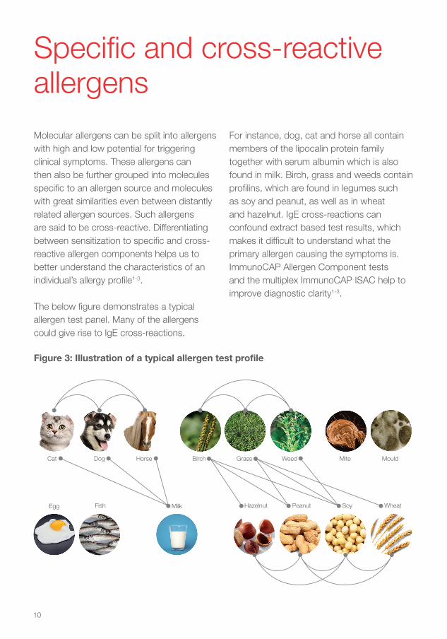
Figure 3: Illustration of a typical allergen test profile
Molecular allergens can be split into allergens with high and low potential for triggering clinical symptoms. These allergens can then also be further grouped into molecules specific to an allergen source and molecules with great similarities even between distantly related allergen sources. Such allergens are said to be cross-reactive. Differentiating between sensitization to specific and cross-reactive allergen components helps us to better understand the characteristics of an individual’s allergy profile1-3.
The below figure demonstrates a typical allergen test panel. Many of the allergens could give rise to IgE cross-reactions.
For instance, dog, cat and horse all contain members of the lipocalin protein family together with serum albumin which is also found in milk. Birch, grass and weeds contain profilins, which are found in legumes such as soy and peanut, as well as in wheat and hazelnut. IgE cross-reactions can confound extract based test results, which makes it difficult to understand what the primary allergen causing the symptoms is. ImmunoCAP Allergen Component tests and the multiplex ImmunoCAP ISAC help to improve diagnostic clarity1-3.
11
Allergen loadThe patient’s clinical history remains the most important part of allergy diagnosis. Component testing will reveal crucial information but as with any IgE test it should be used in support of the clinical history. Only the clinical history can reveal how much of each food allergen the patient has ingested. For example, when consuming large amounts of allergens at the same time, such as when drinking a soy milk drink can affect the symptom outcome1-3.
Low risk allergens such as PR-10 proteins found in soy milk, when consumed in great amounts can provoke more serious allergy symptoms in some patients (such as drinking soy milk1-2).
You can learn more about the significance of these types of allergens at: allergyai.com.
This website contains an educational course which describes the basics of molecular allergy and includes patient case examples.
References
1. Matricardi PM et al. EAACI Molecular Allergology User’s Guide. Pediatric allergy and immunology: official publication of the European Society of Pediatric Allergy and Immunology. 2016;27 Suppl 23:1-250.
2. Kleine-Tebbe J and Jakob T Editors: Molecular Allergy Diagnostics. Innovation for a Better Patient Management. Springer International Publishing Switzerland 2017. ISBN 978-3-319-42498-9 ISBN 978-3-319-42499-6 (eBook), DOI 10.1007/978-3-319-42499-6.
3. Canonica GW et.al. A WAO - ARIA - GA²LEN consensus document on molecular-based allergy diagnostics. World Allergy Organ J. 2013 Oct 3;6(1):17.
Other clinical considerationsA patient sensitized to several allergen sources will often be sensitized to several allergen components. This will contribute to the overall allergen load. For example, if a patient is positive to multiple peanut storage components such as Ara h 1, Ara h 2 and Ara h 3, he or she is likely to have a higher IgE load and therefore possibly be at more risk for severe reactions than someone who is mono-sensitized1-5.
Diagnostic performanceExtract-based tests (whole allergens) contain a mix of many different proteins from an allergen source (e.g. peanut) and measure the sum of IgE antibodies to these, which gives high sensitivity, but sometimes can create difficulties in interpretation of results1-5.

12
ImmunoCAP Allergen Component-based tests, both singleplex and multiplex, contain pure proteins, measure only specific IgE to single molecules and give results with high diagnostic specificity1-5.
Allergen component tests therefore have technical diagnostic superiority at measuring IgE to important individual proteins of interest, such as to Ara h 2 in peanut or Cor a 14 in hazelnut. They simply measure IgE specific to one protein and offer reliable results in terms of minimal variation – like all ImmunoCAP products. However, it must be remembered that a test with allergen components only measures one type of specific IgE and that most patients will have IgE antibodies to several molecules contained in the allergen source1-5.
Presence of allergen specific IgE implies a risk of allergic disease and its significance must be evaluated within the clinical context. Generally the higher the level of IgE antibodies the higher the probability of a clinically manifest allergic reaction1-5.
However, for different patients identical results for the same allergens may not be associated with clinically equivalent manifestations, due to differences in individual patient sensitivities. This may also be true for one individual patient at different occasions due to presence or absence of reaction promoting cofactors1-5.
Absence of detectable allergen specific IgE antibodies does not necessarily exclude the potential for an allergy-like reaction1-2.
For example in food allergy, circulating IgE antibodies may remain undetectable despite a convincing clinical history. The antibodies may be directed towards allergens that are revealed or altered during industrial processing, cooking or digestion and therefore do not exist in the original food for which the patient is tested1-2.
Limitations of ImmunoCAP products test results:
Samples with results below limit of quantitation obtained with ImmunoCAP Allergen Components are recommended to be tested with the corresponding extract based ImmunoCAP Allergen and/or additional relevant ImmunoCAP Allergen Components, if not already performed and a clinical indication is present. The extract based testing can cover additional allergen components present in the allergen source material to which the patient may be sensitized, but which are not presently available as ImmunoCAP Allergen Components or in ImmunoCAP ISAC.
A result below limit of quantitation obtained with an extract based ImmunoCAP Allergen never excludes the possibility of obtaining measurable concentrations of specific IgE when testing with ImmunoCAP Allergen Components from the same allergen source. This is due to the fact that some components may be present in very low amounts in the natural extract.
13
In most cases it is recommended that testing starts with whole allergens to achieve high sensitivity to be followed up with allergen component tests for further specificity and as an aid in risk assessment if the test for the whole allergen is positive1-5.
Further information at allergyai.com.
References
1. Matricardi PM et al. EAACI Molecular Allergology User’s Guide. Pediatric allergy and immunology: official publication of the European Society of Pediatric Allergy and Immunology. 2016;27 Suppl 23:1-250.
2. Kleine-Tebbe J and Jakob T Editors: Molecular Allergy Diagnostics. Innovation for a Better Patient Management. Springer International Publishing Switzerland 2017. ISBN
Food is made up of complex matrices of natural constituents such as proteins, fats and carbohydrates. The way that the human body processes food creates by-products of the original food structure. The natural state of proteins can be changed even before we eat them, most obviously by cooking but also by storage and processing e.g. liquidizing or concentrating (as for fruit juices)1-2.
There are many different metabolic processes that occur as soon as food enters the digestive system. Enzymatic digestion starts straight away in the mouth; acidic pH and gastric juices play a role as food enters the stomach and further digestion takes place in the gut until the food is absorbed as smaller nutrients1-2.
978-3-319-42498-9 ISBN 978-3-319-42499-6 (eBook), DOI 10.1007/978-3-319-42499-6.
3. Canonica GW et.al. A WAO - ARIA - GA²LEN consensus document on molecular-based allergy diagnostics. World Allergy Organ J. 2013 Oct 3;6(1):17.
4. Wickman M. When allergies complicate allergies. Allergy 2005;60(S79):14-18.
5. Van Hage M et.al. ImmunoCAP assays: Pros and cons in allergology. J Allergy Clin Immunol 2017;140:974-7.
Food allergyFats are metabolized into fatty acids; carbohydrates are eventually broken down into small sugar molecules, while proteins are digested into their constituents - amino acids. Most allergens are proteins, made up of amino acid chains, and within these structures are regions called epitopes. It is these recognition sites that specific IgE molecules bind to. This can lead to the release of histamine and other mediators, resulting in allergy symptoms1-2.
14
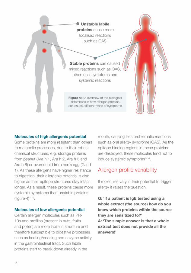
Molecules of high allergenic potentialSome proteins are more resistant than others to metabolic processes, due to their robust chemical structures; e.g. storage proteins from peanut (Ara h 1, Ara h 2, Ara h 3 and Ara h 6) or ovomucoid from hen’s egg (Gal d 1). As these allergens have higher resistance to digestion, their allergenic potential is also higher as their epitope structures stay intact longer. As a result, these proteins cause more systemic symptoms than unstable proteins (figure 4)1-10.
Molecules of low allergenic potentialCertain allergen molecules such as PR-10s and profilins (present in nuts, fruits and pollen) are more labile in structure and therefore susceptible to digestive processes such as heating/cooking and enzyme activity in the gastrointestinal tract. Such labile proteins start to break down already in the
Figure 4: An overview of the biological differences in how allergen proteins
can cause different types of symptoms
mouth, causing less problematic reactions such as oral allergy syndrome (OAS). As the epitope binding regions in these proteins are destroyed, these molecules tend not to induce systemic symptoms1-10.
Allergen profile variability
If molecules vary in their potential to trigger allergy it raises the question:
Q: ‘If a patient is IgE tested using a whole extract (the source) how do you know which proteins within the source they are sensitized to?’A: ‘The simple answer is that a whole extract test does not provide all the answers!’
Unstable labile proteins cause more
localised reactions such as OAS
Stable proteins can caused mixed reactions such as OAS,
other local symptoms and systemic reactions
15
The above question and answer is quite thought-provoking. A whole extract IgE test (the source) is a mixture of lots of individual proteins. It would be impossible to tell which proteins a patient is IgE positive to unless they were separated individually – as they are when using ImmunoCAP Allergen Component tests. Also, all patients vary in which components they are sensitized to1-10.
ImmunoCAP offers a large portfolio of different allergen components, enabling the mapping of individual patient profiles and improving diagnostic clarity. ImmunoCAP ISAC is a multiplexing test that measures IgE to a total of 112 allergen components and through cross-reactivity one can extrapolate sensitizations to other clinically relevant allergen sources6.
Patients testing positive to a whole extract (e.g. positive skin prick test to peanut or serum IgE to peanut) can be sensitized to either allergen proteins of high allergenic potential or of low/no allergenic potential. By using molecular diagnostics it is possible to better differentiate between them, i.e. classify patients into low- and high-risk groups. There are of course also cases where the patient is sensitized to both high-risk and low-risk allergens and can display symptoms such as OAS together with systemic symptoms1-10.
Furthermore, in a given situation other factors such as amount of allergen, stress, ongoing infections etc have an impact on the actual clinical reaction1-2.
Specific allergens and primary food allergyIdentifying IgE to specific molecules often indicates the cause of allergy symptoms. In food allergy, the allergens that initially trigger the immune system to produce specific IgE antibodies are mostly food proteins more resistant to digestion. Such primary sensitization to stable proteins is therefore often associated with systemic allergy symptoms1-10.
Cross-reactive sensitization and pollen-food syndromeAllergen components that have highly similar structures in several different species can give rise to extensive cross-reactivity, these are referred to as “pan-allergens”. Pan-allergens are commonly found in plants and plant derived foods and they can be found even in distantly related species such as celery and birch trees1-10.
Sensitization to pan-allergens may be both asymptomatic and symptomatic, but the symptoms elicited are often of a milder form, such as OAS. In pollen-allergic patients for instance, IgE antibodies primarily targeted towards proteins in pollen (e.g. birch Bet v 1) readily cross-react with similar proteins in food, causing a broad sensitization profile which can be considered “secondary” to the pollen sensitization. In clinical allergy, this is often referred to as pollen-food syndrome and in the context of latex, the latex-fruit syndrome1-10.
Cross-reactive allergens exist also in other sources, such as venoms of stinging insects, fish, mites and shrimp. For example, dust
16
mite and shrimp share a cross-reactive protein called tropomyosin1-5.
When sensitization to cross-reactive allergens is detected, the primary sensitizer should always be sought after in order to understand what is driving the patients’ allergy. Using a range of specific and cross-reactive allergen component tests it is in most cases possible to differentiate primary and secondary reactions1-10.
References
1. Matricardi PM et al. EAACI Molecular Allergology User’s Guide. Pediatric allergy and immunology: official publication of the European Society of Pediatric Allergy and Immunology. 2016;27 Suppl 23:1-250.
2. Kleine-Tebbe J and Jakob T Editors: Molecular Allergy Diagnostics. Innovation for a Better Patient Management. Springer International Publishing Switzerland 2017. ISBN 978-3-319-42498-9 ISBN 978-3-319-42499-6 (eBook), DOI 10.1007/978-3-319-42499-6.
3. Canonica GW et.al. A WAO - ARIA - GA²LEN consensus document on molecular-based allergy diagnostics. World Allergy Organ J. 2013 Oct 3;6(1):17.
4. Sastre J. Molecular diagnosis in allergy. Clin Exp Allergy 2010;40(10):1442-1460.
5. Treudler R. and Simon JC. Overview of component resolved diagnostics. Curr Allergy Asthma Rep 2013;13(1):110-117.
6. Van Hage M et.al. ImmunoCAP assays: Pros and cons in allergology. J Allergy Clin Immunol 2017;140:974-7.7. Garcia BE and Lizaso MT. Cross-reactivity Syndromes in Food Allergy. J Investig Allergol Clin Immunol 2011;21(3):162-170.
8. Zuidmeer L and van Ree R. Lipid transfer protein allergy; primary allergy/food syndrome in some cases. Curr Opinion Clin Immunol 2007;7:269-273.
9. Fernándes-Rivas M, et al. Allergies to fruits and vegetables. Pediatr Allergy Immunol 2008;19:675-681.
10. Santos A and van Ree R. Profilins: Mimickers of Allergy or Relevant Allergens? Int Arch Allergy Immunol 2011;155:191-204.
17
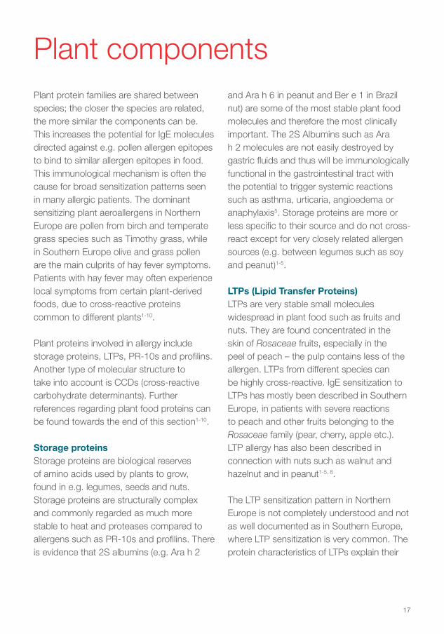
Plant protein families are shared between species; the closer the species are related, the more similar the components can be. This increases the potential for IgE molecules directed against e.g. pollen allergen epitopes to bind to similar allergen epitopes in food. This immunological mechanism is often the cause for broad sensitization patterns seen in many allergic patients. The dominant sensitizing plant aeroallergens in Northern Europe are pollen from birch and temperate grass species such as Timothy grass, while in Southern Europe olive and grass pollen are the main culprits of hay fever symptoms. Patients with hay fever may often experience local symptoms from certain plant-derived foods, due to cross-reactive proteins common to different plants1-10.
Plant proteins involved in allergy include storage proteins, LTPs, PR-10s and profilins. Another type of molecular structure to take into account is CCDs (cross-reactive carbohydrate determinants). Further references regarding plant food proteins can be found towards the end of this section1-10.
Storage proteinsStorage proteins are biological reserves of amino acids used by plants to grow, found in e.g. legumes, seeds and nuts. Storage proteins are structurally complex and commonly regarded as much more stable to heat and proteases compared to allergens such as PR-10s and profilins. There is evidence that 2S albumins (e.g. Ara h 2
and Ara h 6 in peanut and Ber e 1 in Brazil nut) are some of the most stable plant food molecules and therefore the most clinically important. The 2S Albumins such as Ara h 2 molecules are not easily destroyed by gastric fluids and thus will be immunologically functional in the gastrointestinal tract with the potential to trigger systemic reactions such as asthma, urticaria, angioedema or anaphylaxis5. Storage proteins are more or less specific to their source and do not cross-react except for very closely related allergen sources (e.g. between legumes such as soy and peanut)1-5.
LTPs (Lipid Transfer Proteins)LTPs are very stable small molecules widespread in plant food such as fruits and nuts. They are found concentrated in the skin of Rosaceae fruits, especially in the peel of peach – the pulp contains less of the allergen. LTPs from different species can be highly cross-reactive. IgE sensitization to LTPs has mostly been described in Southern Europe, in patients with severe reactions to peach and other fruits belonging to the Rosaceae family (pear, cherry, apple etc.). LTP allergy has also been described in connection with nuts such as walnut and hazelnut and in peanut1-5, 8.
The LTP sensitization pattern in Northern Europe is not completely understood and not as well documented as in Southern Europe, where LTP sensitization is very common. The protein characteristics of LTPs explain their
Plant components
18
clinical relevance due to their high resistance to heat and protease digestion. However, LTP sensitization is also associated with local reactions including OAS1-5, 8.
PR-10 (Pathogenesis-Related family number 10) proteinsThe plant defense proteins of the PR-10 family are present in pollen of Fagales tree species (e.g. birch, hazel, alder and beech) and can also be found in the pulp of fruit. Bet v 1 is the major allergen in birch pollen and is highly similar to other PR-10 proteins in plant foods such as Rosaceae fruits (peach, apple and cherry etc.), as well as to PR-10s in nuts and legumes.
In a typical birch allergy scenario, birch pollen causes a primary sensitization to PR-10 proteins. This can cause typical hay fever-like symptoms such as an itchy/blocked nose, runny eyes etc.As a further consequence, patients who ingest PR-10 proteins found in nuts or fruit can react due to IgE cross-reactions. Food allergy caused via cross-reactivity is sometimes referred to as secondary food allergy. Again this is likely to result in local symptoms such as OAS, but depending on the amount of the cross-reactive protein more severe reactions may also occur (e.g. Gly m 4 induced soy milk reactions)1-5, 9.
Profilin proteinsProfilin proteins occur in many different plant species and cause broad sensitization patterns. They are found for example in pollen (e.g. birch or grass), fruit (e.g. apple, cherry, melon and banana) and vegetables, nuts and latex. It has been proposed that just one profilin from one plant species is
enough for testing IgE sensitization to profilin, due to the close similarity and extensive cross-reactivity of this protein group. Profilins from birch (Bet v 2) and/or Timothy grass (Phl p 12) are often used in measuring IgE to profilin. Profilins are sensitive to heat and proteases and will thus primarily give rise to OAS as the clinical manifestation of food allergy. It is widely accepted that profilins have less clinical relevance than PR-10 proteins, although in some cases profilin sensitization may cause severe reactions1-5, 10.
CCDs (Cross-reactive Carbohydrate Determinants)Some molecular structures such as CCDs are shared between many species and can be found in insect venoms, pollen and plant foods. CCDs are not proteins but specific parts of carbohydrate chains attached to proteins. The clinical impact of specific IgE to CCDs is considered very low although positive IgE test results are frequent1-7.
CCDs help us to understand poly-sensitization to multiple plant foods and latex or double positivity between bee and wasp venoms. It is also worth noting that natural plant allergen extract preparations contain CCD molecules while recombinant sources typically are CCD-free and hence more specific1-7.
19
References
1. Matricardi PM et al. EAACI Molecular Allergology User’s Guide. Pediatric allergy and immunology: official publication of the European Society of Pediatric Allergy and Immunology. 2016;27 Suppl 23:1-250.
2. Kleine-Tebbe J and Jakob T Editors: Molecular Allergy Diagnostics. Innovation for a Better Patient Management. Springer International Publishing Switzerland 2017. ISBN 978-3-319-42498-9 ISBN 978-3-319-42499-6 (eBook), DOI 10.1007/978-3-319-42499-6.
3. Canonica GW et.al. A WAO - ARIA - GA²LEN consensus document on molecular-based allergy diagnostics. World Allergy Organ J. 2013 Oct 3;6(1):17.
4. Sastre J. Molecular diagnosis in allergy. Clin Exp Allergy 2010;40(10):1442-1460.
5. Treudler R. and Simon JC. Overview of component resolved diagnostics. Curr Allergy Asthma Rep 2013;13(1):110-117.
6. Van Hage M et.al. ImmunoCAP assays: Pros and cons in allergology. J Allergy Clin Immunol 2017;140:974-7.
Example 1A variety of allergen component tests could be used when resolving a patient’s birch-food allergy. Is it a primary food allergy? Bet v 1 is a dominating primary sensitizing allergen in a birch allergic patient and could produce cross-reactions between other plant food species.
The example below demonstrates a patient profile of PR-10 sensitization with a suspected case of IgE-mediated peanut allergy. In this example, all other risk allergens such as Ara h 2 in peanut or Cor a 14 from hazelnut were IgE-negative.
Interpreting results from cross-reactive protein families
Like all ImmunoCAP Specific IgE tests, ImmunoCAP Allergen Components give results in kUA/L (ImmunoCAP ISAC gives semi-quantitative results in ISU-E). Primary sensitizing allergens from within the same protein family (in this example PR-10) will normally give the highest specific IgE level. Other secondary IgE sensitizations will give similar specific IgE readings but normally lower levels than the primary sensitizing allergen due to reduced protein homology (and therefore reduced IgE binding)1-6.
7. Garcia BE and Lizaso MT. Cross-reactivity Syndromes in Food Allergy. J Investig Allergol Clin Immunol 2011;21(3):162-170.
8. Zuidmeer L and van Ree R. Lipid transfer protein allergy; primary allergy/food syndrome in some cases. Curr Opinion Clin Immunol 2007;7:269-273.
9. Fernándes-Rivas M, et al. Allergies to fruits and vegetables. Pediatr Allergy Immunol 2008;19:675-681.
10. Santos A and van Ree R. Profilins:Mimickers of Allergy or Relevant Allergens? Int Arch Allergy Immunol 2011;155:191-204.
20
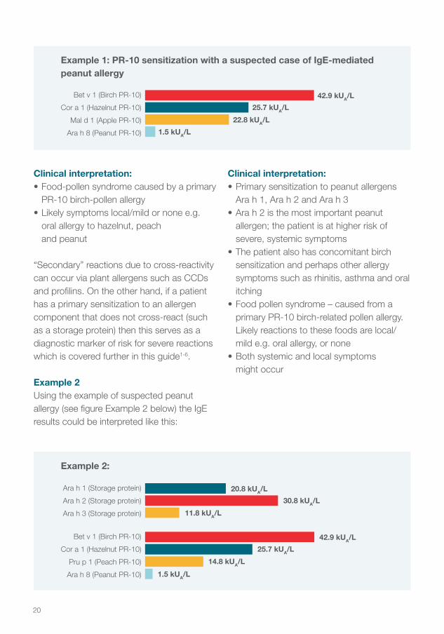
Clinical interpretation:• Food-pollen syndrome caused by a primary
PR-10 birch-pollen allergy• Likely symptoms local/mild or none e.g.
oral allergy to hazelnut, peach and peanut
“Secondary” reactions due to cross-reactivity can occur via plant allergens such as CCDs and profilins. On the other hand, if a patient has a primary sensitization to an allergen component that does not cross-react (such as a storage protein) then this serves as a diagnostic marker of risk for severe reactions which is covered further in this guide1-6.
Example 2Using the example of suspected peanut allergy (see figure Example 2 below) the IgE results could be interpreted like this:
Bet v 1 (Birch PR-10)
Cor a 1 (Hazelnut PR-10)
Mal d 1 (Apple PR-10)
Ara h 8 (Peanut PR-10)
42.9 kUA/L
25.7 kUA/L
22.8 kUA/L
1.5 kUA/L
Example 1: PR-10 sensitization with a suspected case of IgE-mediated peanut allergy
Ara h 1 (Storage protein)
Ara h 2 (Storage protein)
Ara h 3 (Storage protein)
20.8 kUA/L30.8 kUA/L
11.8 kUA/L
Bet v 1 (Birch PR-10)
Cor a 1 (Hazelnut PR-10)
Pru p 1 (Peach PR-10)
Ara h 8 (Peanut PR-10)
42.9 kUA/L25.7 kUA/L
14.8 kUA/L1.5 kUA/L
Example 2:
Clinical interpretation:• Primary sensitization to peanut allergens
Ara h 1, Ara h 2 and Ara h 3• Ara h 2 is the most important peanut
allergen; the patient is at higher risk of severe, systemic symptoms
• The patient also has concomitant birch sensitization and perhaps other allergy symptoms such as rhinitis, asthma and oral itching
• Food pollen syndrome – caused from a primary PR-10 birch-related pollen allergy. Likely reactions to these foods are local/mild e.g. oral allergy, or none
• Both systemic and local symptoms might occur
21
Always use the test results in combination with a clinical history. The presence of specific IgE is not always associated with clinical symptoms but represents a risk of allergic reactions on allergen exposure.
References
1. Matricardi PM et al. EAACI Molecular Allergology User’s Guide. Pediatric allergy and immunology: official publication of the European Society of Pediatric Allergy and Immunology. 2016;27 Suppl 23:1-250.
2. Kleine-Tebbe J and Jakob T Editors: Molecular Allergy Diagnostics. Innovation for a Better Patient Management. Springer International Publishing Switzerland 2017. ISBN 978-3-319-42498-9 ISBN 978-3-319-42499-6 (eBook), DOI 10.1007/978-3-319-42499-6.
3. Canonica GW et.al. A WAO - ARIA - GA²LEN consensus document on molecular-based allergy diagnostics. World Allergy Organ J. 2013 Oct 3;6(1):17.
4. Sastre J. Molecular diagnosis in allergy. Clin Exp Allergy 2010;40(10):1442-1460.
5. Treudler R. and Simon JC. Overview of component resolved diagnostics. Curr Allergy Asthma Rep 2013;13(1):110-117.
6. Van Hage M et.al. ImmunoCAP assays: Pros and cons in allergology. J Allergy Clin Immunol 2017;140:974-7.
Summary of plant food components
Plant protein families are common to many species and the closer the species are related the more similar the proteins can be. But also in distantly related species there are proteins that are very similar which can give rise to cross-reactivity. Thus, IgE molecules directed against pollen allergen epitopes can bind to similar allergen epitopes in foods such as peanuts, tree nuts, fruits and vegetables1-5.
The majority of food allergen components in plants belong to four main protein groups.
These are storage proteins, LTP, PR-10 and profilin proteins. In addition, CCDs (Cross-reactive Carbohydrate Determinants) are allergenic structures found in pollen and plant food, as well as in insects and venoms1-5.
22
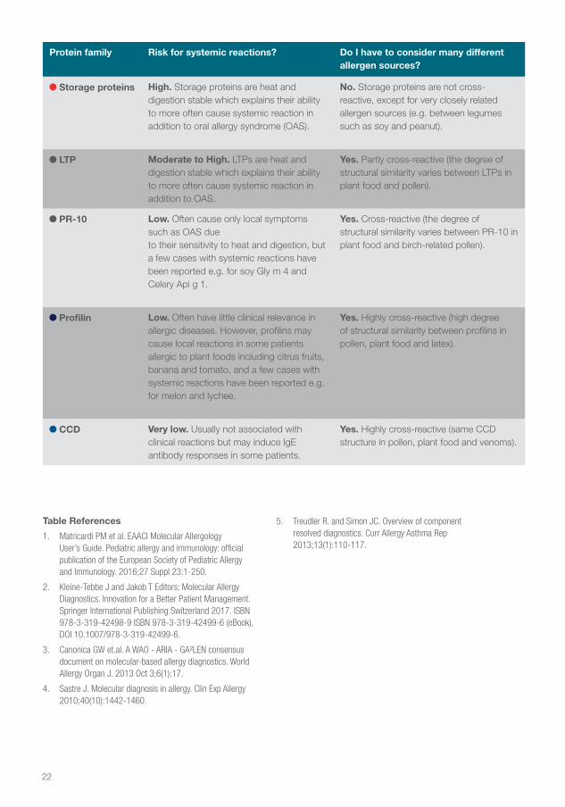
Protein family Risk for systemic reactions? Do I have to consider many different allergen sources?
Storage proteins High. Storage proteins are heat and digestion stable which explains their ability to more often cause systemic reaction in addition to oral allergy syndrome (OAS).
No. Storage proteins are not cross-reactive, except for very closely related allergen sources (e.g. between legumes such as soy and peanut).
LTP Moderate to High. LTPs are heat and digestion stable which explains their ability to more often cause systemic reaction in addition to OAS.
Yes. Partly cross-reactive (the degree of structural similarity varies between LTPs in plant food and pollen).
PR-10 Low. Often cause only local symptoms such as OAS dueto their sensitivity to heat and digestion, but a few cases with systemic reactions have been reported e.g. for soy Gly m 4 and Celery Api g 1.
Yes. Cross-reactive (the degree of structural similarity varies between PR-10 in plant food and birch-related pollen).
Profilin Low. Often have little clinical relevance in allergic diseases. However, profilins may cause local reactions in some patients allergic to plant foods including citrus fruits, banana and tomato, and a few cases with systemic reactions have been reported e.g. for melon and lychee.
Yes. Highly cross-reactive (high degree of structural similarity between profilins in pollen, plant food and latex).
CCD Very low. Usually not associated with clinical reactions but may induce IgE antibody responses in some patients.
Yes. Highly cross-reactive (same CCD structure in pollen, plant food and venoms).
Table References
1. Matricardi PM et al. EAACI Molecular Allergology User’s Guide. Pediatric allergy and immunology: official publication of the European Society of Pediatric Allergy and Immunology. 2016;27 Suppl 23:1-250.
2. Kleine-Tebbe J and Jakob T Editors: Molecular Allergy Diagnostics. Innovation for a Better Patient Management. Springer International Publishing Switzerland 2017. ISBN 978-3-319-42498-9 ISBN 978-3-319-42499-6 (eBook), DOI 10.1007/978-3-319-42499-6.
3. Canonica GW et.al. A WAO - ARIA - GA²LEN consensus document on molecular-based allergy diagnostics. World Allergy Organ J. 2013 Oct 3;6(1):17.
4. Sastre J. Molecular diagnosis in allergy. Clin Exp Allergy 2010;40(10):1442-1460.
5. Treudler R. and Simon JC. Overview of component resolved diagnostics. Curr Allergy Asthma Rep 2013;13(1):110-117.
23
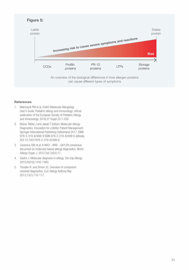
Stableprotein
Labileprotein
Increasing risk to cause severe symptoms and reactions
Risk
Profilin proteins
PR-10proteins
LTPsStorageproteinsCCDs
Figure 5:
An overview of the biological differences in how allergen proteins can cause different types of symptoms
References
1. Matricardi PM et al. EAACI Molecular Allergology User’s Guide. Pediatric allergy and immunology: official publication of the European Society of Pediatric Allergy and Immunology. 2016;27 Suppl 23:1-250.
2. Kleine-Tebbe J and Jakob T Editors: Molecular Allergy Diagnostics. Innovation for a Better Patient Management. Springer International Publishing Switzerland 2017. ISBN 978-3-319-42498-9 ISBN 978-3-319-42499-6 (eBook), DOI 10.1007/978-3-319-42499-6.
3. Canonica GW et.al. A WAO - ARIA - GA²LEN consensus document on molecular-based allergy diagnostics. World Allergy Organ J. 2013 Oct 3;6(1):17.
4. Sastre J. Molecular diagnosis in allergy. Clin Exp Allergy 2010;40(10):1442-1460.
5. Treudler R. and Simon JC. Overview of component resolved diagnostics. Curr Allergy Asthma Rep 2013;13(1):110-117.
24
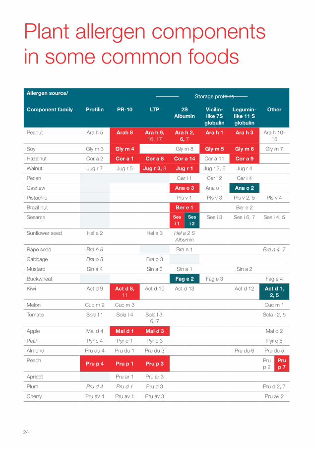
Allergen source/Storage proteins
Component family Profilin PR-10 LTP 2S Albumin
Vicilin-like 7S
globulin
Legumin-like 11 S globulin
Other
Peanut Ara h 5 Arah 8 Ara h 9, 16, 17
Ara h 2, 6, 7
Ara h 1 Ara h 3 Ara h 10-15
Soy Gly m 3 Gly m 4 Gly m 8 Gly m 5 Gly m 6 Gly m 7
Hazelnut Cor a 2 Cor a 1 Cor a 8 Cor a 14 Cor a 11 Cor a 9
Walnut Jug r 7 Jug r 5 Jug r 3, 8 Jug r 1 Jug r 2, 6 Jug r 4
Pecan Car i 1 Car i 2 Car i 4
Cashew Ana o 3 Ana o 1 Ana o 2
Pistachio Pls v 1 Pls v 3 Pls v 2, 5 Pls v 4
Brazil nut Ber e 1 Ber e 2
Sesame Ses i 1
Ses i 2
Ses i 3 Ses i 6, 7 Ses i 4, 5
Sunflower seed Hel a 2 Hel a 3 Hel a 2 S Albumin
Rape seed Bra n 8 Bra n 1 Bra n 4, 7
Cabbage Bra o 8 Bra o 3
Mustard Sin a 4 Sin a 3 Sin a 1 Sin a 2
Buckwheat Fag e 2 Fag e 3 Fag e 4
Kiwi Act d 9 Act d 8, 11
Act d 10 Act d 13 Act d 12 Act d 1, 2, 5
Melon Cuc m 2 Cuc m 3 Cuc m 1
Tomato Sola l 1 Sola l 4 Sola l 3, 6, 7
Sola l 2, 5
Apple Mal d 4 Mal d 1 Mal d 3 Mal d 2
Pear Pyr c 4 Pyr c 1 Pyr c 3 Pyr c 5
Almond Pru du 4 Pru du 1 Pru du 3 Pru du 6 Pru du 5
PeachPru p 4 Pru p 1 Pru p 3
Pru p 2
Pru p 7
Apricot Pru ar 1 Pru ar 3
Plum Pru d 4 Pru d 1 Pru d 3 Pru d 2, 7
Cherry Pru av 4 Pru av 1 Pru av 3 Pru av 2
Plant allergen components in some common foods
Headline as displayed across two lines maximum
25
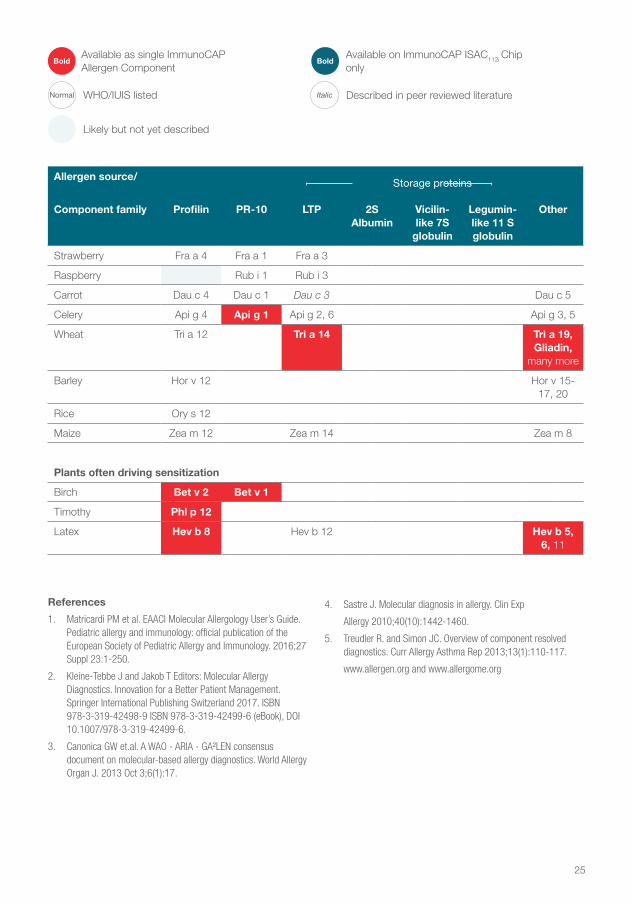
Allergen source/Storage proteins
Component family Profilin PR-10 LTP 2S Albumin
Vicilin-like 7S
globulin
Legumin-like 11 S globulin
Other
Strawberry Fra a 4 Fra a 1 Fra a 3
Raspberry Rub i 1 Rub i 3
Carrot Dau c 4 Dau c 1 Dau c 3 Dau c 5
Celery Api g 4 Api g 1 Api g 2, 6 Api g 3, 5
Wheat Tri a 12 Tri a 14 Tri a 19, Gliadin,
many more
Barley Hor v 12 Hor v 15-17, 20
Rice Ory s 12
Maize Zea m 12 Zea m 14 Zea m 8
Plants often driving sensitization
Birch Bet v 2 Bet v 1
Timothy Phl p 12
Latex Hev b 8 Hev b 12 Hev b 5, 6, 11
References
1. Matricardi PM et al. EAACI Molecular Allergology User’s Guide. Pediatric allergy and immunology: official publication of the European Society of Pediatric Allergy and Immunology. 2016;27 Suppl 23:1-250.
2. Kleine-Tebbe J and Jakob T Editors: Molecular Allergy Diagnostics. Innovation for a Better Patient Management. Springer International Publishing Switzerland 2017. ISBN 978-3-319-42498-9 ISBN 978-3-319-42499-6 (eBook), DOI 10.1007/978-3-319-42499-6.
3. Canonica GW et.al. A WAO - ARIA - GA²LEN consensus document on molecular-based allergy diagnostics. World Allergy Organ J. 2013 Oct 3;6(1):17.
4. Sastre J. Molecular diagnosis in allergy. Clin Exp
Allergy 2010;40(10):1442-1460.
5. Treudler R. and Simon JC. Overview of component resolved diagnostics. Curr Allergy Asthma Rep 2013;13(1):110-117.
www.allergen.org and www.allergome.org
Available on ImmunoCAP ISAC112i Chip only
BoldAvailable as single ImmunoCAP Allergen Component
Bold
WHO/IUIS listedNormal Described in peer reviewed literatureItalic
Likely but not yet described
Headline as displayed across two lines maximum
26
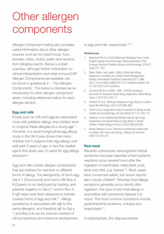
Other allergen componentsAllergen Component testing also provides useful information about other allergen sources such as non-plant foods, furry animals, mites, molds, pollen and venoms from stinging insects. Below is a brief overview, although further information on clinical interpretation and what ImmunoCAP Allergen Components are available can be found in guidebook 2 – ‘The Allergen Components’. The below is intended as an introduction to other allergen component areas, including references below for each allergen section.
Egg and milkFoods such as milk and egg are associated more with pediatric allergy, and children tend to outgrow these allergies at a young age1-7. However, in a recent longitudinal egg allergy study in the UK it was shown that many children don’t outgrow their egg allergy until well past 5 years of age, in fact the median age in this study was 10 years for egg allergy resolution5.
Egg and milk contain allergen components that are markers for reactivity to different forms of allergy. The allergenicity of hen’s egg Gal d 1 (Ovomucoid) and cow’s milk Bos d 8 (Casein) is not destroyed by heating, and patients negative to Gal d 1 and/or Bos d 8 IgE tests have been observed to tolerate cooked forms of egg and milk1-7. Allergy persistency is associated with IgE to the same allergens, and therefore IgE to Gal d 1 and Bos d 8 can be used as markers of clinical reactions and tolerance development
to egg and milk, respectively1,2,5-7.
References
1. Matricardi PM et al. EAACI Molecular Allergology User’s Guide. Pediatric allergy and immunology: official publication of the European Society of Pediatric Allergy and Immunology. 2016;27 Suppl 23:1-250.
2. Kleine-Tebbe J and Jakob J Editors: Molecular Allergy Diagnostics. Innovation for a Better Patient Management. Springer International Publishing Switzerland 2017. ISBN 978-3-319-42498-9 ISBN 978-3-319-42499-6 (eBook), DOI 10.1007/978-3-319-42499-6.
3. Canonica GW et.al. A WAO - ARIA - GA²LEN consensus document on molecular-based allergy diagnostics. World Allergy Organ J. 2013 Oct 3;6(1):17.
4. Chokshi NY et al. Molecular diagnosis of egg allergy: an update. Expert Rev Mol Diagn. 2015;15(7):895-906
5. Clark A et al. A longitudinal study of resolution of allergy to well-cooked and uncooked egg. Clin Exp Allergy 2011;41:706-712.
6. Gradman J et al. Relationship between specific IgE to egg components and natural history of egg allergy in Danish children. Pediatr Allergy Immunol. 2016;27(8):825-830.
7. Nowak-Wegrzyn A et al. Tolerance to extensively heated milk in children with cow’s milk allergy. J Allergy Clin Immunol 2008;122:342-347.
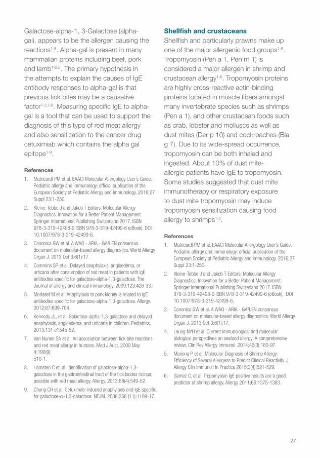
Red meatRecently a previously unrecognized clinical syndrome has been reported where systemic reactions occur several hours after the ingestion of mammalian meat (beef, pork, lamb and offal, e.g. kidney)1-8. Most cases have concerned adults, but recent reports also include children6. Whereas food allergy symptoms generally occur shortly after ingestion, this type of red meat allergy is associated with symptoms delayed 3-6 hours. The most common symptoms include gastrointestinal problems, urticaria and anaphylaxis1-8.
A carbohydrate, the oligosaccharide
27
Galactose-alpha-1, 3-Galactose (alpha-gal), appears to be the allergen causing the reactions1-8. Alpha-gal is present in many mammalian proteins including beef, pork and lamb1-2,5. The primary hypothesis in the attempts to explain the causes of IgE antibody responses to alpha-gal is that previous tick bites may be a causative factor1-2,7,8. Measuring specific IgE to alpha-gal is a tool that can be used to support the diagnosis of this type of red meat allergy and also sensitization to the cancer drug cetuximiab which contains the alpha gal epitope1-9.
References
1. Matricardi PM et al. EAACI Molecular Allergology User’s Guide. Pediatric allergy and immunology: official publication of the European Society of Pediatric Allergy and Immunology. 2016;27 Suppl 23:1-250.
2. Kleine-Tebbe J and Jakob T Editors: Molecular Allergy Diagnostics. Innovation for a Better Patient Management. Springer International Publishing Switzerland 2017. ISBN 978-3-319-42498-9 ISBN 978-3-319-42499-6 (eBook), DOI 10.1007/978-3-319-42499-6.
3. Canonica GW et.al. A WAO - ARIA - GA²LEN consensus document on molecular-based allergy diagnostics. World Allergy Organ J. 2013 Oct 3;6(1):17.
4. Commins SP et al. Delayed anaphylaxis, angioedema, or urticaria after consumption of red meat in patients with IgE antibodies specific for galactose-alpha-1,3-galactose. The Journal of allergy and clinical immunology. 2009;123:426-33.
5. Morisset M et al. Anaphylaxis to pork kidney is related to IgE antibodies specific for galactose-alpha-1,3-galactose. Allergy. 2012;67:699-704.
6. Kennedy JL, et al. Galactose-alpha-1,3-galactose and delayed anaphylaxis, angioedema, and urticaria in children. Pediatrics. 2013;131:e1545-52.
7. Van Nunen SA et al. An association between tick bite reactions and red meat allergy in humans. Med J Aust. 2009 May 4;190(9): 510-1.
8. Hamsten C et. al. Identification of galactose-alpha-1,3-galactose in the gastrointestinal tract of the tick Ixodes ricinus; possible with red meat allergy. Allergy. 2013;68(4):549-52.
9. Chung CH et al. Cetuximab-induced anaphylaxis and IgE specific for galactose-α-1,3-galactose. NEJM. 2008;358 (11):1109-17.
Shellfish and crustaceansShellfish and particularly prawns make up one of the major allergenic food groups1-5. Tropomyosin (Pen a 1, Pen m 1) is considered a major allergen in shrimp and crustacean allergy1-6. Tropomyosin proteins are highly cross-reactive actin-binding proteins located in muscle fibers amongst many invertebrate species such as shrimps (Pen a 1), and other crustacean foods such as crab, lobster and molluscs as well as dust mites (Der p 10) and cockroaches (Bla g 7). Due to its wide-spread occurrence, tropomyosin can be both inhaled and ingested. About 10% of dust mite-allergic patients have IgE to tropomyosin. Some studies suggested that dust mite immunotherapy or respiratory exposure to dust mite tropomyosin may induce tropomyosin sensitization causing food allergy to shrimps1-3.
References
1. Matricardi PM et al. EAACI Molecular Allergology User’s Guide. Pediatric allergy and immunology: official publication of the European Society of Pediatric Allergy and Immunology. 2016;27 Suppl 23:1-250.
2. Kleine-Tebbe J and Jakob T Editors: Molecular Allergy Diagnostics. Innovation for a Better Patient Management. Springer International Publishing Switzerland 2017. ISBN 978-3-319-42498-9 ISBN 978-3-319-42499-6 (eBook), DOI 10.1007/978-3-319-42499-6.
3. Canonica GW et.al. A WAO - ARIA - GA²LEN consensus document on molecular-based allergy diagnostics. World Allergy Organ J. 2013 Oct 3;6(1):17.
4. Leung NYH et al. Current immunological and molecular biological perspectives on seafood allergy: A comprehensive review. Clin Rev Allergy Immunol. 2014;46(3):180-97.
5. Mariona P et al. Molecular Diagnosis of Shrimp Allergy: Efficiency of Several Allergens to Predict Clinical Reactivity. J Allergy Clin Immunol: In Practice 2015;3(4):521-529.
6. Gamez C, et al. Tropomyosin IgE positive results are a good predictor of shrimp allergy. Allergy 2011;66:1375-1383.

28
FishParvalbumins such as Gad c 1 (cod, white fish) and Cyp c 1 (carp) are major fish allergen components and markers of fish sensitization1-6. Fish-allergic patients can sometimes tolerate certain fish species while reacting to others. However, as parvalbumins from different fish species are structurally closely related and highly cross-reactive, analysis of IgE antibody binding to them is generally not informative in regard to discriminating between allergies to different species of fish. A positive test result to either of Gad c 1 and Cyp c 1 nevertheless indicates a risk of severe reactions to fish1-
6. Parvalbumins are expressed in lower amounts in certain fish species such as tuna, swordfish and some mackerels. This perhaps explains why some fish-allergic patients can tolerate these species4-6.
References
1. Matricardi PM et al. EAACI Molecular Allergology User’s Guide. Pediatric allergy and immunology: official publication of the European Society of Pediatric Allergy and Immunology. 2016;27 Suppl 23:1-250.
2. Kleine-Tebbe J and Jakob T Editors: Molecular Allergy Diagnostics. Innovation for a Better Patient Management. Springer International Publishing Switzerland 2017. ISBN 978-3-319-42498-9 ISBN 978-3-319-42499-6 (eBook), DOI 10.1007/978-3-319-42499-6.
3. Canonica GW et.al. A WAO - ARIA - GA²LEN consensus document on molecular-based allergy diagnostics. World Allergy Organ J. 2013 Oct 3;6(1):17.
4. Sharp MF and Lopata AL. Fish Allergy in Review. Clin Rev Allerg Immunol 2014;46:258-271.
5. Griesmeier U et al. Expression levels of parvalbumins determine allergenicity of fishspecies. Allergy 2010;65:191-198.
6. Kuehn A. Fish Allergens at a Glance: Variable Allergenicity of Parvalbumins, the Major Fish Allergens. Front Immunol. 2014; 5:179.
Furry AnimalsFurry animals such as dogs, cats and horses produce some of the most prevalent allergens in our environment and are released into the surroundings through animal saliva, dander and urine. Like many other allergen sources furry animals contain both specific and cross-reactive allergen components1-3.
Clinically uteroglobin and lipocalins have been identified as the most important major allergen components from cat, dog and horse1-7. Serum albumins are often considered to have less clinical relevance in allergy to furry animals, they are minor allergens which cause multiple positivity due to cross-reactivity when using extract tests1-3. However serum albumins are important food allergens in meat1-3,8.
Children with problematic severe asthma often have higher levels of IgE antibodies towards cat, dog and horse compared with children with controlled asthma5-6. Revealing the primary allergen source driving the allergy could help improve allergy management such as allergen reduction/avoidance strategies, and be an aid to select the proper Allergen Specific Immunotherapy (AIT). AIT success is more likely if sensitization to specific components is identified and appropriate therapy administered3,9-10.
References
1. Matricardi PM et al. EAACI Molecular Allergology User’s Guide. Pediatric allergy and immunology: official publication of the European Society of Pediatric Allergy and Immunology. 2016;27 Suppl 23:1-250.
2. Kleine-Tebbe J and Jakob T Editors: Molecular Allergy Diagnostics. Innovation for a Better Patient Management. Springer International Publishing Switzerland 2017. ISBN 978-3-319-42498-9 ISBN 978-3-319-42499-6 (eBook), DOI 10.1007/978-3-319-42499-6.

29
60-70% of pet sensitized patients are sensitized to serveral pet extracts - specific or due to cross-reactivity sensitization11
Typical allergens12
Fel d 2 Equ c 3Can f 3Fel d 1 Fel d 1Fel d 1
Fel d 1
Fel d 1 Equ c 1Can f 4
Can f 1
Fel d 4 Fel d 7 Can f 6
Can f 2
Can f 5
Specific components in squares
Serum albuminsUteroglobin Lipocalin family Kallikrein
Cat HorseDog
Protein family Summary Clinical Importance
Uteroglobin Uteroglobin, a steroid-inducible cytokine-like molecule with anti-inflammatory and immunomodulatory properties.
High. Fel d 1 the major cat allergen belongs to this family.
Lipocalin family Small, specific molecules. Although highly conserved they display limited sequence identity of between 20 – 30%.
High. Lipocalins are often major allergens and constitute an important primary allergen.
Kallikrein Kallikreins are peptidases. Prostate specific antigen (PSA) is a kallikrein which liquefies semen and allows sperm to swim freely.
High. Associated with male dogs (Can f 5). A major allergen.
Serum albumin Large globular proteins present in dander, saliva, meat and milk.
High. Highly cross reactivity, minor allergen and is seldom of clinical importance.
30
3. Canonica GW et.al. A WAO - ARIA - GA²LEN consensus document on molecular-based allergy diagnostics. World Allergy Organ J. 2013 Oct 3;6(1):17.
4. Hilger C et al. Animal Lipocalin allergens, Curr Allergy Asthma Rep 2012;12 438 – 447
5. Konradsen JR et al. Severe childhood asthma and allergy to furry aninmals: Refined assessment and using molecular based allergy diagnostics. Pediatr Allergy Immunol 2014: 25: 187 - 192.
6. Nordlund B et al. IgE antibodies to animal-derived lipocalin, kallikrein and secretoglobin are markers of bronchial inflammation in severe childhood asthma. Allergy 2012;67:661-669.
7. Cosme-Blanco W et.al Anaphylaxis to Horses and Epinephrine Use: Increasing Awareness Among Pediatric Patients and Families. Pediatr Allergy Immunol 2017 ;28(6):608-610.
8. Werfel SJ. Clinical reactivity to beef in children allergic to cow’s milk. J Allergy Clin Immunol 1997 99(3):293-300.
9. Asero R. Component-resolved diagnosis-assisted prescription of allergen-specific immunotherapy: a practical guide Eur Ann Allergy Clin Immunol. 2012;44(5):183-7.
10. Schmid-Grendelmeier et al. Recombinant allergens – routine diagnostics or still only science? Der Hautarzt 2010;61(11):946-953
11. Borres MP et al. Use of allergen components begins a new era in pediatric allergolog. Pediatr Allergy Immunol. 2011;22:454-61.
12. Konradsen W, et al. Allergy to furry animals: New insights, diagnostic approaches, and challenges. J Allergy Clin Immunol. 2015;135:616-25.
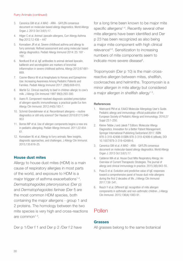
House dust mites Allergy to house dust mites (HDM) is a main cause of respiratory allergies in most parts of the world, and exposure to HDM is a major trigger of asthma exacerbations1-4. Dermatophagoides pteronyssinus (Der p) and Dermatophagoides farinae (Der f) are the most common HDM species, both containing the major allergens - group 1 and 2 proteins. The homology between the two mite species is very high and cross-reactions are common1-3.
Der p 1/Der f 1 and Der p 2 /Der f 2 have
for a long time been known to be major mite specific allergens1-4. Recently several other mite allergens have been identified and Der p 23 has been recognized as also being a major mite component with high clinical relevance5-6. Sensitization to increasing numbers of mite components seem to indicate more severe disease6.
Tropomyosin (Der p 10) is the main cross-reactive allergen between mites, shellfish, cockroaches and helminths. Tropomyosin is a minor allergen in mite allergy but considered a major allergen in shellfish allergy1-3.
References
1. Matricardi PM et al. EAACI Molecular Allergology User’s Guide. Pediatric allergy and immunology: official publication of the European Society of Pediatric Allergy and Immunology. 2016;27 Suppl 23:1-250.
2. Kleine-Tebbe J and Jakob T Editors: Molecular Allergy Diagnostics. Innovation for a Better Patient Management. Springer International Publishing Switzerland 2017. ISBN 978-3-319-42498-9 ISBN 978-3-319-42499-6 (eBook), DOI 10.1007/978-3-319-42499-6.
3. Canonica GW et.al. A WAO - ARIA - GA²LEN consensus document on molecular-based allergy diagnostics. World Allergy Organ J. 2013 Oct 3;6(1):17.
4. Calderon MA et al. House Dust Mite Respiratory Allergy: An Overview of Current Therapeutic Strategies. The journal of allergy and clinical immunology In practice. 2015;3(6):843-55.
5. Posa D et al. Evolution and predictive value of IgE responses toward a comprehensive panel of house dust mite allergens during the first 2 decades of life. J Allergy Clin Immunol 2017;139 :541.
6. Resch Y et.al. Different IgE recognition of mite allergen components in asthmatic and non-asthmatic children. J Allergy Clin Immunol. 2015;136(4):1083-91.
Pollen
Grasses All grasses belong to the same botanical
Furry Animals (continued)
31
family, Poaceae, therefore cross-reactivity between different species is common and the closer the relations (e.g. within subfamilies), the higher the degree of cross reactivity1-4. Grass pollen allergy is common worldwide, and many atopic patients show sensitization to grass pollen1-6. Grass pollen season overlaps with weed pollen such as mugwort and ragweed in most parts of Europe and with tree pollen (olive, plane) in Southern Europe 1-3,5. Group 1 and group 5 allergens (e.g. Phl p 1 and Phl p 5 from Timothy) are dominating grass pollen allergens and markers of primary sensitization in a majority of patients1-6. Sensitization to Phl p 1 usually precedes other grass pollen component sensitizations in the development of hay fever symptoms6. In warmer areas, other grass species such as Bermuda grass are common and they also contain group 1 allergens e.g. Cyn d 11-5. Sensitization to cross-reactive allergens such as profilin (Phl p 12) and polcalcin (Phl p 7) is usually not frequent but several grass allergens carry CCD which can cause cross reactivity in extract based testing1-7.
When no specific grass sensitization is detected other pollen or food specific components should be investigated1-3,5.
References
1. Matricardi PM et al. EAACI Molecular Allergology User’s Guide. Pediatric allergy and immunology: official publication of the European Society of Pediatric Allergy and Immunology. 2016;27 Suppl 23:1-250.
2. Kleine-Tebbe J and Jakob T Editors: Molecular Allergy Diagnostics. Innovation for a Better Patient Management. Springer International Publishing Switzerland 2017. ISBN 978-3-319-42498-9 ISBN 978-3-319-42499-6 (eBook), DOI 10.1007/978-3-319-42499-6.
3. Canonica GW et.al. A WAO - ARIA - GA²LEN consensus
document on molecular-based allergy diagnostics. World Allergy Organ J. 2013 Oct 3;6(1):17.
4. Andersson K. et al. Characteristics and immunobiology of grass pollen allergens. International Archives of Allergy & Immunology. 2003; 130(2): 87–107.
5. Barber D. et al. Understanding patient sensitization profiles in complex pollen areas: a molecular epidemiological study. Allergy. 2008 Nov;63(11):1550–8.
6. Hatzler L et al. Molecular spreading and predictive value of preclinical IgE response to Phleum pratense in children with hay fever. J Allergy Clin Immunol. 2012 Oct;130(4):894–901 e5.
7. Hauser M et al. Panallergens and their impact on the allergic patient. Allergy Asthma Clin Immunol. 2010; 6(1):1.
Trees Opposed to grasses, trees belong to several different botanical families, often even to different orders, and there is less cross-reactivity between specific tree allergens. However all tree pollen contain profilin and most contain polcalcins and CCDs, giving rise to possible cross-reactivity on the extract level1-8.
Due to Bet v 1 sensitization (the major birch allergen) many birch pollen allergic patients react to several pollen, such as the closely related alder, hazel, beech and oak1-3,6. In addition, many of these patients have concomitant pollen-related food allergies due to PR-10 cross-reactivity (Bet v 1) and may react to various fruits, nuts and vegetables (e.g. apple, pear, cherry or hazelnut)1-3. In most cases, symptoms are restricted to oral reactions and the food is often tolerated when cooked since PR-10 allergens are heat labile1-3.
Olive and ash are botanically very closely related (Oleaceae family) and there is extensive cross-reactivity between these
32
species1-5,7. Olive tree pollen allergy is quite common and is one of the most important causes of seasonal respiratory allergy in the Mediterranean area5,7. Ole e 1 is the major marker for primary olive pollen sensitization1-5,7. The European ash (Fraxinus excelsior) is common in most of Europe but ash tree pollen may often be overlooked as a cause of pollinosis1-2,5. Ole e 1 serves as a very good marker allergen for ash due to the high cross reactivity1-5,7.
Plane trees are known as “street trees” and are found planted practically anywhere in the world1-2. Recombinant Pla a 1 is a specific marker allergen discriminating between genuine plane tree pollen sensitization and cross-reactivity1-5. Pla a 3 is a LTP which cross-reacts with other LTPs in e.g. fruits1-3. Pla a 3 is presently not available as a single ImmunoCAP Allergen Component. However, Pla a 3, as well as the plane-tree specific and major allergen Pla a 1 are available on the ImmunoCAP ISAC112i Chip.
Cypresses and cedars are common ornamental trees1-5. There are several species of cypress and cedars and since they are closely related cross-sensitization is extensive. Cypress trees bloom in the winter and may cause winter respiratory allergy which is often misdiagnosed since symptoms are occurring during winter and are very similar to perennial allergies like dust mite allergy1-3,8,9. Cup a 1 is a specific marker for primary sensitization to Cupressaceae pollen. The Cup a 1 allergen is very similar to major allergens of Mediterranean cypress (Cup s 1) Mountain cedar (Jun a 1) Japanese cypress
Cha o 1) and Japanese cedar (Cry j 1) and there is an extensive cross-reactivity between species1-4,8,9.
References
1. Matricardi PM et al. EAACI Molecular Allergology User’s Guide. Pediatric allergy and immunology: official publication of the European Society of Pediatric Allergy and Immunology. 2016;27 Suppl 23:1-250.
2. Kleine-Tebbe J and Jakob T Editors: Molecular Allergy Diagnostics. Innovation for a Better Patient Management. Springer International Publishing Switzerland 2017. ISBN 978-3-319-42498-9 ISBN 978-3-319-42499-6 (eBook), DOI 10.1007/978-3-319-42499-6.
3. Canonica GW et.al. A WAO - ARIA - GA²LEN consensus document on molecular-based allergy diagnostics. World Allergy Organ J. 2013 Oct 3;6(1):17.
4. Asam C et al. Tree pollen allergens - an update from a molecular perspective. Allergy 2015:70:1201-1211.
5. Barber D et al. Understanding patient sensitization profiles in complex pollen areas: a molecular epidemiological study. Allergy. 2008 Nov; 63(11): 1550–8.
6. Hauser M et.al. Bet v 1-like pollen allergens of multiple Fagales species can sensitize atopic individuals. Clinical & Exp Allergy. 2011; 41:1804–181.
7. Rodríguez R, et al. Olive pollen recombinant allergens: value in diagnosis and immunotherapy. J Investig Allergol Clin Immunol 2007;17 Suppl 1:4–10.
8. Charpin D et al .Cypress Pollinosis: from Tree to Clinic. Clin Rev Allergy Immunol. 2017 Apr 11
9. Douladiris N et al. A molecular diagnostic algorithm to guide pollen immunotherapy in Southern Europe: towards component resolved management of allergic diseases. Int Arch Allergy Immunol 2013;162;163-172.
Weeds Weed allergy diagnosis can be unclear and difficult to make due to frequent poly-sensitizations and inconclusive anamnesis because of overlapping flowering seasons with other pollen such as birch and grass1-3. Cross reactions are expected between different weed species when botanically closely related, however many weeds belong to unrelated botanical families and therefore specific marker allergens are available,
33
e.g. Amb a 1 from Ragweed, Art v 1 from Mugwort, Par j 1 from Parietaria, Pla l 1 from English plantain and Sal k 1 from Saltwort1-5. Saltwort is a weed common in dry, semi-arid areas and is becoming more and more common in southern parts of Europe due to climate change.
Apart from profilin and CCDs, mugwort and ragweed pollen contain some other cross-reactive allergens. Cross-reactive IgE antibodies can lead to clinically significant allergic reactions5.
Pollen-food syndromes driven by weed pollen are mainly generated by mugwort and ragweed pollen. In addition to Oral Allergy Syndrome (OAS) more severe allergy is reported such as the celery-mugwort-spice syndrome1-2,6.
References
1. Matricardi PM et al. EAACI Molecular Allergology User’s Guide. Pediatric allergy and immunology: official publication of the European Society of Pediatric Allergy and Immunology. 2016;27 Suppl 23:1-250.
2. Kleine-Tebbe J and Jakob T Editors: Molecular Allergy Diagnostics. Innovation for a Better Patient Management. Springer International Publishing Switzerland 2017. ISBN 978-3-319-42498-9 ISBN 978-3-319-42499-6 (eBook), DOI 10.1007/978-3-319-42499-6.
3. Canonica GW et.al. A WAO - ARIA - GA²LEN consensus document on molecular-based allergy diagnostics. World Allergy Organ J. 2013 Oct 3;6(1):17.
4. Gadermaier G et al. Allergens of weed pollen: An overview on recombinant and natural molecules. Methods 2014;66;55-66.
5. Asero R et al. Concomitant sensitization to ragweed and mugwort pollen: who is who in clinical allergy? Ann Allergy Asthma Immunol 2014;113:307-313.
6. Egger M et al. Pollen food syndromes associated with weed pollinosis: an update from the molecular point of view. Allergy 2006;61:461-476.
Molds There is current evidence to demonstrate a close association between fungal sensitization and asthma severity1-5. Many airborne fungi are involved such as Alternaria, Aspergillus, Cladosporium and Penicillium, and exposure may be indoors, outdoors or both. Fungal sensitization is common in asthmatic patients and the term “severe asthma with fungal sensitization” (SAFS) has been proposed1-5. However, it is recognised that enhanced and precise definition of fungal sensitization will require improvements in diagnostic testing and this can be facilitated by component testing1-9.
Alternaria alternata is a major outdoor as well as indoor aeroallergen in many parts of the world. Sensitivity to Alternaria has been increasingly recognized as a risk factor for the development and persistence of asthma, asthma severity, and potentially fatal asthma exacerbations2-6. Alt a 1 is the major Alternaria allergen. Alt a 1 is considered a specific marker of primary sensitization to Alternaria Alternata and useful in asthma diagnostics2-6.
Aspergillus fumigatus is an opportunistic fungus causing allergic and invasive aspergillosis in humans and animals1-4,7-9 Genuine A. fumigatus sensitization is not always easily identifiable and IgE sensitization tests are used as part of routine workup for diagnosing Allergic Bronchopulmonary Aspergillosis (ABPA). The use of allergen components for A. fumigatus can aid the identification of primary A. fumigatus sensitization1-4,7-9.
34
Asp f 1, Asp f 2 and Asp f 4 are species specific allergens while Asp f 3 and Asp f 6 are described as cross-reactive allergens1,2,7-9. Recent studies investigating ABPA demonstrated that ImmunoCAP Allergen Components could differentiate ABPA from asthma and sensitised patients. ABPA has been particularly linked to Asp f 4 and Asp p 67-9 – see book 2 for further details. References
1. Matricardi PM et al. EAACI Molecular Allergology User’s Guide. Pediatric allergy and immunology: official publication of the European Society of Pediatric Allergy and Immunology. 2016;27 Suppl 23:1-250.
2. Kleine-Tebbe J and Jakob T Editors: Molecular Allergy Diagnostics. Innovation for a Better Patient Management. Springer International Publishing Switzerland 2017. ISBN 978-3-319-42498-9 ISBN 978-3-319-42499-6 (eBook), DOI 10.1007/978-3-319-42499-6.
3. Canonica GW et.al. A WAO - ARIA - GA²LEN consensus document on molecular-based allergy diagnostics. World Allergy Organ J. 2013 Oct 3;6(1):17.
4. Sastre J. Molecular diagnosis in allergy. Clin Exp Allergy 2010;40(10):1442-1460.
5. Medrek SK et.al. Fungal sensitization is associated with increased risk of life-threatening asthma. J Allergy Clin Immunol Pract. 2007;5:1025-31.
6 Kustrzeba-Wójcicka I et.al Alternaria alternata and its allergens: a comprehensive review. Clin Rev Allergy Immunol. 2014 Dec;47(3):354-65.
7. Fukutomi Y et al. Serological diagnosis of allergic bronchopulmonary mycosis: Progress and challenges. Allergology International 65 (2016) 30e36.
8. Bowyer P et al. Relative reactivity of Aspergillus allergens used in serological tests Medical Mycology September 2006;44, S23 -S28.
9. Tanimoto H et al. Molecular-based allergy diagnosis of allergic bronchopulmonary aspergillosis in Aspergillus fumigatus-sensitized Japanese patients Clin Exp Allergy. 2015 2015;45,1790–1800 + erratum Clin Exp Allergy 2016;46(2):381.
VenomsMany patients with suspected honey bee and/or common wasp specific IgE test positive to both species when using extract testing. True double allergic reactivity to both bee and wasp is not clinically common. In many cases double venom IgE positivity can be caused by cross-reactions to CCDs1-5. Recombinant venom components do not carry CCD and therefore provide greater diagnostic specificity, useful when making decisions such as to start AIT1-7. Ves v 1 and Ves v 5 are major allergens from common wasp and Pol d 5 is a marker for sensitization to paper wasp. The picture for honey bee sensitivity seems more complex than for wasp and can involve more varied sensitization patterns to major components. Api m 1, Api m 2, Api m 3, Api m 5 and Api m 10 are all major allergens within bee venom allergy. It has recently been shown that using an increasing number of bee components can improve diagnostic sensitivity1-7.Low level specific IgE below 0.35 kUA/l can be relevant when using components and may be indicative of venom allergy, so measuring down to 0.1 kUA/l can be important1,6.
Patients with suspected venom allergy should also be tested with ImmunoCAP Tryptase5,8-9. Patients with high basal levels of tryptase should be investigated for mastocytosis since these patients have higher risk for severe reactions during venom immunotherapy5,8-9.
References
1. Matricardi PM et al. EAACI Molecular Allergology User’s Guide. Pediatric allergy and immunology: official publication of the European Society of Pediatric Allergy and Immunology. 2016;27 Suppl 23:1-250.

35
2. Kleine-Tebbe J and Jakob T Editors: Molecular Allergy Diagnostics. Innovation for a Better Patient Management. Springer International Publishing Switzerland 2017. ISBN 978-3-319-42498-9 ISBN 978-3-319-42499-6 (eBook), DOI 10.1007/978-3-319-42499-6
3. Canonica GW et.al. A WAO - ARIA - GA²LEN consensus document on molecular-based allergy diagnostics. World Allergy Organ J. 2013 Oct 3;6(1):17.
4. Spillner E. et al., Hymenoptera allergens: from venom to ”venome”. Frontiers in immunology 2014;5:1-7.
5. Biló B, et al. EAACI Interest Group on Insect Venom Hypersensitivity. Diagnosis of Hymenoptera venom allergy. Allergy 2005;60:1339-1349.
6. Kohler et al. Component resolution reveals additional major allergens in patient’s with honeybee venom allergy. J Allergy Clin Immunol 2014;133:1383-9.
7. Korosec P et al. High sensitivity of CAP-FEIA rVes v 5 and rVes v 1 for diagnosis of Vespula venom allergy. J Allergy Clin Immunol. 2012 May;129(5):1406-8.
8. Bonifazi F et al. and EAACI Interest Group on Insect Venom Hypersensitivity. Prevention and treatment of hymenoptera venom allergy: guidelines for clinical practice. Allergy 2005;60:1459-1470.
9. Bonadonna P. et al., Clonal mast cell disorders in patients with systemic reactions to Hymenoptera stings and increased serum tryptase levels. J Allergy Clin Immunol 2009;123:680-6.
LatexPrimary latex allergy should be identified using specific allergen components as many positive extract test results arise as a result of cross-reactive pollen sensitization involving profilin, CCDs or PR-10 allergens1-6. The association of latex allergy and allergy to plant-derived foods is referred to as the latex-fruit syndrome and includes a large number of plant foods such as avocado, banana, chestnut, kiwi, peach, tomato, potato and bell pepper. The latex allergen Hev b 6 is considered the main culprit in this syndrome1-2.
IgE antibodies to Hev b 1, Hev b 3, Hev b 5 and Hev b 6 are markers for primary latex
allergy1-6. Sensitization to these components is frequent in surgery-related latex-allergy, especially among children having undergone multiple and extensive surgery, such as those with spina bifida. Sensitization to Hev b 5 and Hev b 6 is associated with occupational exposure to latex e.g. in healthcare workers and food-handling personnel using latex gloves1-5. Latex allergens Hev b 8 (profilin) and Hev b 6 can be used for examining cross-reactivity to pollen and plant foods, respectively1-4. If an exclusive sensitization to the profilin Hev b 8 is seen, allergic symptoms to latex are hardly to be expected5,6.
References
1. Matricardi PM et al. EAACI Molecular Allergology User’s Guide. Pediatric allergy and immunology: official publication of the European Society of Pediatric Allergy and Immunology. 2016;27 Suppl 23:1-250.
2. Kleine-Tebbe J and Jakob T Editors: Molecular Allergy Diagnostics. Innovation for a Better Patient Management. Springer International Publishing Switzerland 2017. ISBN 978-3-319-42498-9 ISBN 978-3-319-42499-6 (eBook), DOI 10.1007/978-3-319-42499-6.
3. Canonica GW et.al. A WAO - ARIA - GA²LEN consensus document on molecular-based allergy diagnostics. World Allergy Organ J. 2013 Oct 3;6(1):17.
4. Garnier L et al. Molecular allergens in the diagnosis of latex allergy. Eur Ann Allergy Clin Immunol 2012;44(2):73–79.
5. Ebo DG, et al. Component-resolved diagnosis from latex allergy by micro-array. Clin Exp Allergy 2010;40(2):348-358.
6. Schuler S, et al. Microarray-based component-resolved diagnosis of latex allergy: isolated IgE-mediated sensitization to Latex profilin Hev b 8 may act as confounder. Clin Transl Allergy 2013;3:11.
36
Immunotherapeutics – Aeroallergens and venomsUnderstanding cross-sensitization and identifying the right allergen sourceClinically it is obviously important to correctly identify the allergen source causing symptoms before starting immunotherapy and this is not always easy1-6. Patients can be cross-sensitized to several allergen species; therefore sometimes it is not clear what the disease-eliciting source is. This applies to pollen allergies as well as to animal, dust mite and venom allergy. Tests with molecular allergens can help streamline the identification process1-6. Specific molecules from, for example, grass can differentiate and identify true grass-allergic patients.
Determining a patient’s molecular profile will also help to indicate if they are likely to respond satisfactorily to immunotherapy3,
6-7. Immunotherapy products vary from manufacturer to manufacturer; they contain molecules from the allergen source. But which ones and in what quantity? Most products contain larger quantities of the major allergens such as Bet v 1 in birch or Phl p 1 and Phl p 5 in timothy grass and Der p 1 in mite extracts, while sometimes much lower quantities of other allergens are included7. Patients who are positive only to the minor allergens are therefore less likely to respond satisfactorily1-7,10-11.
Many patients with suspected venom allergy can be positive for both bee and wasp whole allergens. Double positivity can be caused by CCDs rather than true double sensitization1-3,8-11. ImmunoCAP recombinant venom components are CCD-free, which enables the physician to distinguish between positivity from cross-reactions and true venom allergy before selecting the right immunotherapeutic solution. Api m 3 and Api m 10 can be absent or underrepresented in VIT extracts and venom AIT in patients sensitized to these components may be less efficient10-11.
References
1. Matricardi PM et al. EAACI Molecular Allergology User’s Guide. Pediatric allergy and immunology: official publication of the European Society of Pediatric Allergy and Immunology. 2016;27 Suppl 23:1-250.
2. Kleine-Tebbe J and Jakob T Editors: Molecular Allergy Diagnostics. Innovation for a Better Patient Management. Springer International Publishing Switzerland 2017. ISBN 978-3-319-42498-9 ISBN 978-3-319-42499-6 (eBook), DOI 10.1007/978-3-319-42499-6
3. Canonica GW et al. A WAO - ARIA - GA²LEN consensus document on molecular-based allergy diagnostics. World Allergy Organ J. 2013 Oct 3;6(1):17.
4. Focke M et al. Heterogeneity of commercial timothy grass pollen extracts. Clin Exp Allergy 2008;38(8):1400-1408.
5. Asero R. Component-resolved diagnosis-assisted prescription of allergen-specific immunotherapy: a practical guide Eur Ann Allergy Clin Immunol. 2012;44(5):183-7.
6. Schmid-Grendelmeier P. Recombinant Allergens – Routine diagnostics or still only science? Der Hautarzt 2010;61(11):946-953.
7. Focke M et al. Molecular composition and biological activity
37
of commercial birch pollen allergen extracts. Eur J Clin Invest 2009;39(5):429-436.
8. Mittermann I et al. Recombinant allergen-based IgE testing to distinguish bee and wasp allergy. J Allergy Clin Immunol 2010;125(6):1300-1307.
9. Muller U et al. IgE to recombinant allergens Api m 1, Ves v 1 and Ves v 5 distinguish double sensitization from cross-reaction in venom allergy. Allergy 2012;67:1069-1073.
10. Grunwald T et al., Molecular cloning and expression in insect cells of honeybee venom allergen acid phosphatase (Api m 3). J Allergy Clin Immunol 2006; 117:848-54.
11. Blank S et al., Api m 10, a genuine A. mellifera venom allergen, is clinically relevant but underrepresented in therapeutic extracts. Allergy 2011; 66:1322-1329.
What is a molecular allergen-specific IgE test and does it differ technically from normal specific IgE tests that I request from my laboratory?Technically they work in the same way and give results in kUA /L, the same as normal whole extract sources such as cat, peanut etc.
How many ImmunoCAP Allergen Components are available?There are currently over 100 tests with allergen components in the product range. A list is included in this guide, however availability may differ between countries.
For each allergen source how do I know that the ImmunoCAP Allergen Components represent the whole allergen extract?
Common questions regarding allergen components
Generally speaking they don’t. For example, there are over 30 proteins reported in the peanut extract, many not clinically relevant or with unknown relevance. The field of molecular allergology is ever-expanding as we gain further scientific information and knowledge. Thermo Fisher Scientificsupplies (Phadia AB is manufacturer) between 2-4 new components every year. Since all components are not available as single tests it is suggested to use the available components together with the whole extract to cover the spectrum of patients’ sensitizations.
What is ImmunoCAP ISAC?ImmunoCAP ISAC is a microarray chip which tests for IgE to 112 allergen components simultaneously. It is a multiplex test giving semi-quantitative results on a patient’s

38
allergen component sensitization profile. It has been found useful for the diagnosis of the following, but this list is not exhaustive: complex allergy, OAS, and cases of multi-sensitization, idiopathic anaphylaxis and high total IgE patients. More information on ImmunoCAP ISAC is available in Go Molecular, Book 2.
Where can I get access to ImmunoCAP ISAC?Your local immunology laboratory may have ImmunoCAP ISAC in-house, otherwise the lab should be able to refer your sample for testing. Therefore contact your local lab to find out what is possible.
Is it possible to have an ImmunoCAP Whole Allergen test result that is negative and an ImmunoCAP Allergen Component test result that is positive (from the same source)?This is possible in some cases. The whole allergen contains a mix of proteins, representing the natural composition at the source, while the allergen component is one single pure protein. Overall the component tests give higher specificity and sometimes even more sensitivity. Using a combination of both whole extract and components (where possible) is currently considered the best strategy for diagnosis.

39
GlossaryAllergen component – single immunogenic protein from an allergen source e.g. Ara h 2 from peanut.
Cross-reactivity/Cross-sensitization – IgE antibodies directed against one allergen may cross-react to structurally related allergens from other allergen sources. Cross-reactive antibodies can cause a variety of different clinical outcomes.
Epitope – Defined substructure of a protein to which an antibody binds.
ImmunoCAP – an in vitro test for the measurement of specific IgE antibodies. ImmunoCAP is one of the market leaders and has been established for several decades. ImmunoCAP is also available for testing for other immunoglobulins (e.g. IgG4/ IgG).
Pan-allergen – evolutionarily conserved and widely distributed allergen, ubiquitous component of several sources of allergens. IgE antibodies to a pan-allergen may cross-react with homologous allergens and thus also give rise to symptoms to many different allergens in a patient.
Primary sensitizing allergen – an allergen originally triggering the immune system to produce specific IgE antibodies. For example
Bet v 1 from birch or Ara h 2 from peanut.
Minor and major allergens – Major allergen components are those to which at least 50% of relevant patients are sensitized. Minor allergens are often less prevalent in triggering allergy. For instance, in birch allergy the major allergen is Bet v 1 (PR-10), whilst a minor allergen is Bet v 2 (profilin).
Secondary sensitization – IgE antibodies directed to a primary sensitizer cross-react due to the similarity of the proteins/allergens and give rise to cross-reactive sensitization. This occurs in food-pollen syndrome for example, when an individual is sensitized to birch PR-10 (Bet v1) and the IgE antibodies then cross-react to peanut PR-10 (Ara h 8).
Allergen extract – refers to the crude mixture of proteins that is obtained by extraction of an allergen source (e.g. birch pollen or peanut).
40
Educational resources allergyai.com – Home Page of Immunodiagnostics, Thermo Fisher Scientific
allergen.org – International Union for Immunological Sciences/WHO Allergen Database
Matricardi PM et al. EAACI Molecular Allergology User’s Guide. Pediatric allergy and immunology: official publication of the European Society of Pediatric Allergy and Immunology. 2016;27 Suppl 23:1-250.
Kleine-Tebbe J and Jakob T Editors: Molecular Allergy Diagnostics. Innovation for a Better Patient Management. Springer International Publishing Switzerland 2017. ISBN 978-3-319-42498-9 ISBN 978-3-319-42499-6 (eBook), DOI 10.1007/978-3-319-42499-6
Canonica GW et al. A WAO – ARIA – GA2LEN consensus document on molecular-based allergy diagnostics. World Allergy Organ J 2013;6(1):17.
41
Using ImmunoCAP Allergen Component tests
The term ‘ImmunoCAP Allergen Component’ is used for singleplex ImmunoCAP products based on molecular allergens purified from either their natural source (native) or biotechnologically produced as recombinant proteins bound to the conventional ImmunoCAP solid phase.
By using tests for single allergen components as a complement to more traditional specific IgE antibody tests, further clinically relevant and quantitative information can be gained as an aid to the physician in making a diagnosis of allergy.
Allergen components are also available in a multiplex microarray format, ImmunoCAP ISAC. Here each test contains 112 components giving 112 semi-quantitative specific IgE results, thus presenting a snapshot of the patient’s complete sensitization profile in one test.
More information on ImmunoCAP ISAC is available in Book 2.
The interpretation of sensitizations to allergen components that is described in this book, (as well as in book 2) is the same for both single and multiplex formats.
ImmunoCAP Allergen Components, singleplex as well as multiplex, are useful tools for the physician when investigating and explaining allergic reactions in more detail and to determine if cross-reacting IgE antibodies or primary sensitization causes them. However as all test results they must be evaluated by the physician together with the clinical history of the individual patient.
Presence of allergen specific IgE implies a risk of allergic disease and generally the higher the level of IgE antibodies the higher the probability of a clinically manifest allergic reaction1-5. However, due to differences in individual patient sensitivities identical results for the same allergens may not be associated with clinically equivalent manifestations. This may also be true for one individual patient at different occasions due to presence or absence of reaction promoting cofactors1-5.
Absence of detectable allergen specific IgE antibodies does not necessarily exclude the potential for an allergy-like reaction1,2.
Limitations of ImmunoCAP products test results:Samples with results below limit of quantitation obtained with ImmunoCAP Allergen Components are recommended
42
to be tested with the corresponding extract based ImmunoCAP Allergen and/or additional relevant ImmunoCAP Allergen Components, if not already performed and a clinical indication is present. The extract based testing can cover additional allergen components present in the allergen source material to which the patient may be sensitized, but which are not presently available as ImmunoCAP Allergen Components or in ImmunoCAP ISAC.
A result below limit of quantitation obtained with an extract based ImmunoCAP Allergen never excludes the possibility of obtaining measurable concentrations of specific IgE when testing with ImmunoCAP Allergen Components from the same allergen source. This is due to the fact that some components may be present in very low amounts in the natural extract.
References
1. Matricardi PM et al. EAACI Molecular Allergology User’s Guide. Pediatric allergy and immunology: official publication of the European Society of Pediatric Allergy and Immunology. 2016;27 Suppl 23:1-250.
2. Kleine-Tebbe J and Jakob T Editors: Molecular Allergy Diagnostics. Innovation for a Better Patient Management. Springer International Publishing Switzerland 2017. ISBN 978-3-319-42498-9 ISBN 978-3-319-42499-6 (eBook), DOI 10.1007/978-3-319-42499-6.
3. Canonica GW et.al. A WAO - ARIA - GA²LEN consensus document on molecular-based allergy diagnostics. World Allergy Organ J. 2013 Oct 3;6(1):17.
4. Wickman M. When allergies complicate allergies. Allergy 2005;60(S79):14-18.
5. Van Hage M et.al. ImmunoCAP assays: Pros and cons in allergology. J Allergy Clin Immunol 2017;140:974-7.
Disclaimer: The content of this book is intended as an aid to the physician to interpret allergen specific IgE antibody test results. It is not intended as medical advice on an individual level. A definitive clinical diagnosis of IgE mediated allergic disorders should only be made by the physician based on the clinical history for the individual patient, after all clinical and laboratory findings have been evaluated. It should not be based on the results of any single diagnostic method.
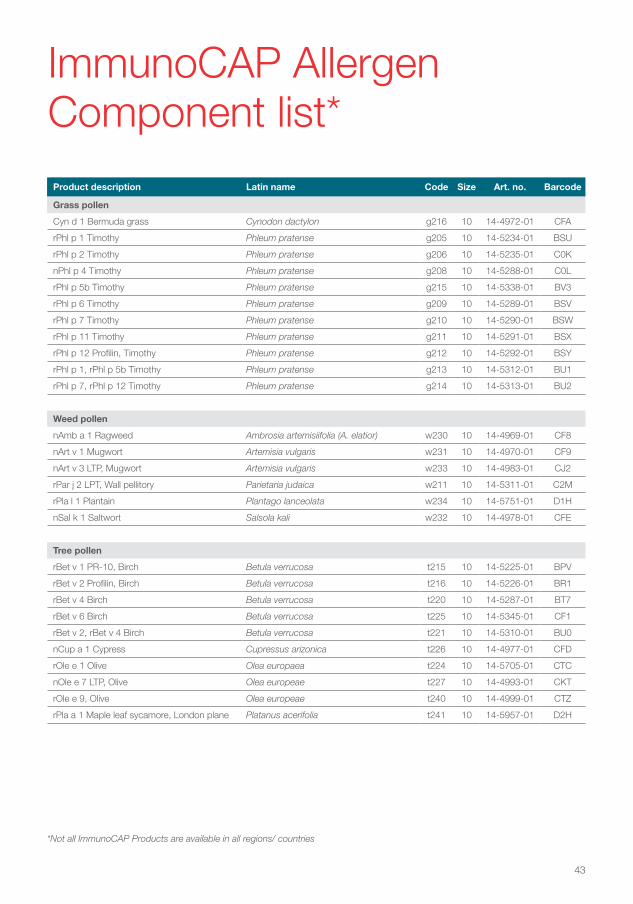
43
Product description Latin name Code Size Art. no. Barcode
Grass pollen
Cyn d 1 Bermuda grass Cynodon dactylon g216 10 14-4972-01 CFA
rPhl p 1 Timothy Phleum pratense g205 10 14-5234-01 BSU
rPhl p 2 Timothy Phleum pratense g206 10 14-5235-01 C0K
nPhl p 4 Timothy Phleum pratense g208 10 14-5288-01 C0L
rPhl p 5b Timothy Phleum pratense g215 10 14-5338-01 BV3
rPhl p 6 Timothy Phleum pratense g209 10 14-5289-01 BSV
rPhl p 7 Timothy Phleum pratense g210 10 14-5290-01 BSW
rPhl p 11 Timothy Phleum pratense g211 10 14-5291-01 BSX
rPhl p 12 Profilin, Timothy Phleum pratense g212 10 14-5292-01 BSY
rPhl p 1, rPhl p 5b Timothy Phleum pratense g213 10 14-5312-01 BU1
rPhl p 7, rPhl p 12 Timothy Phleum pratense g214 10 14-5313-01 BU2
Weed pollen
nAmb a 1 Ragweed Ambrosia artemisiifolia (A. elatior) w230 10 14-4969-01 CF8
nArt v 1 Mugwort Artemisia vulgaris w231 10 14-4970-01 CF9
nArt v 3 LTP, Mugwort Artemisia vulgaris w233 10 14-4983-01 CJ2
rPar j 2 LPT, Wall pellitory Parietaria judaica w211 10 14-5311-01 C2M
rPla l 1 Plantain Plantago lanceolata w234 10 14-5751-01 D1H
nSal k 1 Saltwort Salsola kali w232 10 14-4978-01 CFE
Tree pollen
rBet v 1 PR-10, Birch Betula verrucosa t215 10 14-5225-01 BPV
rBet v 2 Profilin, Birch Betula verrucosa t216 10 14-5226-01 BR1
rBet v 4 Birch Betula verrucosa t220 10 14-5287-01 BT7
rBet v 6 Birch Betula verrucosa t225 10 14-5345-01 CF1
rBet v 2, rBet v 4 Birch Betula verrucosa t221 10 14-5310-01 BU0
nCup a 1 Cypress Cupressus arizonica t226 10 14-4977-01 CFD
rOle e 1 Olive Olea europaea t224 10 14-5705-01 CTC
nOle e 7 LTP, Olive Olea europeae t227 10 14-4993-01 CKT
rOle e 9, Olive Olea europeae t240 10 14-4999-01 CTZ
rPla a 1 Maple leaf sycamore, London plane Platanus acerifolia t241 10 14-5957-01 D2H
ImmunoCAP Allergen Component list*
*Not all ImmunoCAP Products are available in all regions/ countries
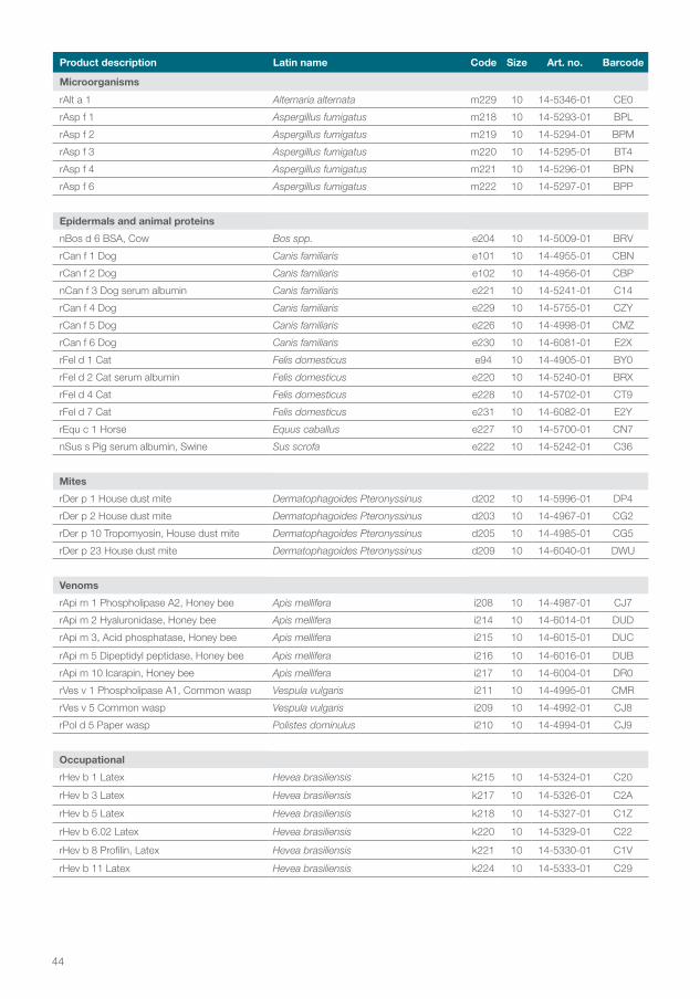
44
Product description Latin name Code Size Art. no. Barcode
Microorganisms
rAlt a 1 Alternaria alternata m229 10 14-5346-01 CE0
rAsp f 1 Aspergillus fumigatus m218 10 14-5293-01 BPL
rAsp f 2 Aspergillus fumigatus m219 10 14-5294-01 BPM
rAsp f 3 Aspergillus fumigatus m220 10 14-5295-01 BT4
rAsp f 4 Aspergillus fumigatus m221 10 14-5296-01 BPN
rAsp f 6 Aspergillus fumigatus m222 10 14-5297-01 BPP
Epidermals and animal proteins
nBos d 6 BSA, Cow Bos spp. e204 10 14-5009-01 BRV
rCan f 1 Dog Canis familiaris e101 10 14-4955-01 CBN
rCan f 2 Dog Canis familiaris e102 10 14-4956-01 CBP
nCan f 3 Dog serum albumin Canis familiaris e221 10 14-5241-01 C14
rCan f 4 Dog Canis familiaris e229 10 14-5755-01 CZY
rCan f 5 Dog Canis familiaris e226 10 14-4998-01 CMZ
rCan f 6 Dog Canis familiaris e230 10 14-6081-01 E2X
rFel d 1 Cat Felis domesticus e94 10 14-4905-01 BY0
rFel d 2 Cat serum albumin Felis domesticus e220 10 14-5240-01 BRX
rFel d 4 Cat Felis domesticus e228 10 14-5702-01 CT9
rFel d 7 Cat Felis domesticus e231 10 14-6082-01 E2Y
rEqu c 1 Horse Equus caballus e227 10 14-5700-01 CN7
nSus s Pig serum albumin, Swine Sus scrofa e222 10 14-5242-01 C36
Mites
rDer p 1 House dust mite Dermatophagoides Pteronyssinus d202 10 14-5996-01 DP4
rDer p 2 House dust mite Dermatophagoides Pteronyssinus d203 10 14-4967-01 CG2
rDer p 10 Tropomyosin, House dust mite Dermatophagoides Pteronyssinus d205 10 14-4985-01 CG5
rDer p 23 House dust mite Dermatophagoides Pteronyssinus d209 10 14-6040-01 DWU
Venoms
rApi m 1 Phospholipase A2, Honey bee Apis mellifera i208 10 14-4987-01 CJ7
rApi m 2 Hyaluronidase, Honey bee Apis mellifera i214 10 14-6014-01 DUD
rApi m 3, Acid phosphatase, Honey bee Apis mellifera i215 10 14-6015-01 DUC
rApi m 5 Dipeptidyl peptidase, Honey bee Apis mellifera i216 10 14-6016-01 DUB
rApi m 10 Icarapin, Honey bee Apis mellifera i217 10 14-6004-01 DR0
rVes v 1 Phospholipase A1, Common wasp Vespula vulgaris i211 10 14-4995-01 CMR
rVes v 5 Common wasp Vespula vulgaris i209 10 14-4992-01 CJ8
rPol d 5 Paper wasp Polistes dominulus i210 10 14-4994-01 CJ9
Occupational
rHev b 1 Latex Hevea brasiliensis k215 10 14-5324-01 C20
rHev b 3 Latex Hevea brasiliensis k217 10 14-5326-01 C2A
rHev b 5 Latex Hevea brasiliensis k218 10 14-5327-01 C1Z
rHev b 6.02 Latex Hevea brasiliensis k220 10 14-5329-01 C22
rHev b 8 Profilin, Latex Hevea brasiliensis k221 10 14-5330-01 C1V
rHev b 11 Latex Hevea brasiliensis k224 10 14-5333-01 C29

45
Product description Latin name Code Size Art. no. Barcode
Occupational / Enzymes
Alkalase Alkalase k205 10 14-5126-01 C1F
nAna c 2 Bromelain, Pineapple nAna c 2 Bromelain, Pineapple k202 10 14-5127-01 BT1
nAsp o 21 alpha-amylase nAsp o 21 alpha-amylase k87 10 14-4370-01 595
nCar p 1 Papain, Papaya nCar p 1 Papain, Papaya k201 10 14-5130-01 BT0
nGal d 4 Lysozyme, Egg nGal d 4 Lysozyme, Egg k208 10 14-5128-01 C0T
Maxatase Maxatase k204 10 14-5129-01 C2F
Savinase Savinase k206 10 14-5132-01 C2R
nSus s Pepsin, Swine nSus s Pepsin, Swine k213 10 14-5258-01 C3B
Foods
rAct d 8 PR-10, Kiwi Actinidia deliciosa f430 10 14-4984-01 CG7
rAna o 3 Cashew nut Anacardium occidentale f443 10 14-5760-01 D0W
rApi g 1.01 PR-10, Celery Apium graveolens f417 10 14-4957-01 CBR
rAra h 1 Peanut Arachis hypogaea f422 10 14-4963-01 CDF
rAra h 2 Peanut Arachis hypogaea f423 10 14-4964-01 CDG
rAra h 3 Peanut Arachis hypogaea f424 10 14-4965-01 CDH
rAra h 6 Peanut Arachis hypogaea f447 10 14-6041-01 DYU
rAra h 8 PR-10, Peanut Arachis hypogaea f352 10 14-5341-01 CEZ
rAra h 9 LTP, Peanut Arachis hypogaea f427 10 14-4980-01 CFC
rBer e 1 Brazil nut Bertholletia excelsa f354 10 14-5343-01 CDS
rSes i 1, Sesame seed Sesamum indicum f449 10 14-6109-01 E7M
nBos d 4 alpha-lactalbumin, Milk Bos spp. f76 10 14-4522-01 CTP
nBos d 5 beta-lactoglobulin, Milk Bos spp. f77 10 14-4523-01 CTR
nBos d 8 Casein, Milk Bos spp. f78 10 14-4524-01 CTS
rCor a 1 PR-10, Hazel nut Corylus avellana f428 10 14-4981-01 CFB
rCor a 8 LTP, Hazel nut Corylus avellana f425 10 14-4968-01 CDP
nCor a 9, Hazel nut Corylus avellana f440 10 14-5758-01 D0M
rCor a 14, Hazel nut Corylus avellana f439 10 14-5754-01 CZP
rCyp c 1 Carp Cyprinus carpio f355 10 14-5344-01 CF0
rGad c 1 Cod Gadus morhua f426 10 14-4971-01 CEY
nGal d 1 Ovomucoid, Egg Gallus spp. f233 10 14-4805-01 904
nGal d 2 Ovalbumin, Egg Gallus spp. f232 10 14-4804-01 903
nGal d 3 Conalbumin, Egg Gallus spp. f323 10 14-5222-01 C18
rGly m 4 PR-10, Soy Glycine max f353 10 14-5340-01 CDR
nGly m 5 beta-conglycinin, Soy Glycine max f431 10 14-4990-01 CLV
nGly m 6 Glycinin Glycine max f432 10 14-4991-01 CLU
rJug r 1 Walnut Juglans regia f441 10 14-5762-01 D0T
rJug r 3 LTP, Walnut Juglans regia f442 10 14-5954-01 D11
rMal d 1 PR-10, Apple Malus domestica f434 10 14-5703-01 CWR
rMal d 3 LTP, Apple Malus domestica f435 10 14-5704-01 CWS
rPen a 1 Tropomyosin, Shrimp Penaeus aztecus f351 10 14-5335-01 C11
rPru p 1 PR-10, Peach Prunus persica f419 10 14-4960-01 CBV
*Not all ImmunoCAP Products are available in all regions/ countries
ImmunoCAP Allergen Component list continued*
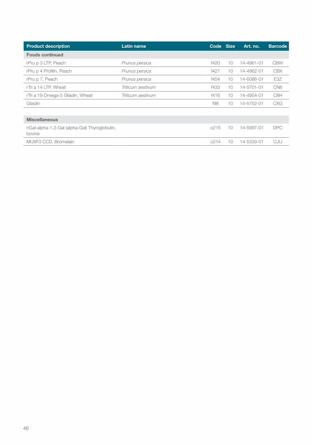
46
Product description Latin name Code Size Art. no. Barcode
Foods continued
rPru p 3 LTP, Peach Prunus persica f420 10 14-4961-01 CBW
rPru p 4 Profilin, Peach Prunus persica f421 10 14-4962-01 CBX
rPru p 7, Peach Prunus persica f454 10 14-6086-01 E3Z
rTri a 14 LTP, Wheat Triticum aestivum f433 10 14-5701-01 CN6
rTri a 19 Omega-5 Gliadin, Wheat Triticum aestivum f416 10 14-4954-01 C8H
Gliadin f98 10 14-5752-01 CXG
Miscellaneous
nGal-alpha-1,3-Gal (alpha-Gal) Thyroglobulin, bovine
o215 10 14-5997-01 DPC
MUXF3 CCD, Bromelain o214 10 14-5339-01 CJU
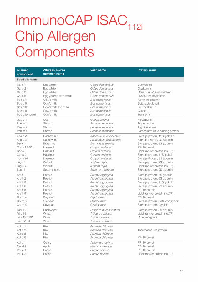
47
Allergen component
Allergen source common name
Latin name Protein group
Food allergens
Gal d 1Gal d 2Gal d 3Gal d 5Bos d 4Bos d 5Bos d 6Bos d 8Bos d lactoferrin
Egg whiteEgg whiteEgg whiteEgg yolk/chicken meatCow’s milkCow’s milkCow’s milk and meatCow’s milkCow’s milk
Gallus domesticusGallus domesticusGallus domesticusGallus domesticusBos domesticusBos domesticusBos domesticusBos domesticusBos domesticus
OvomucoidOvalbuminConalbumin/OvotransferrinLivetin/Serum albuminAlpha-lactalbuminBeta-lactoglobulinSerum albuminCaseinTransferrin
Gad c 1Pen m 1Pen m 2Pen m 4
CodShrimpShrimpShrimp
Gadus callariasPenaeus monodonPenaeus monodonPenaeus monodon
ParvalbuminTropomyosinArginine kinaseSarcoplasmic Ca-binding protein
Ana o 2Ana 0 3Ber e 1Cor a 1.0401Cor a 8Cor a 9Cor a 14Jug r 1Jug r 3Ses i 1
Cashew nutCashew nutBrazil nutHazelnutHazelnutHazelnutHazelnutWalnutWalnutSesame seed
Anacardium occidentaleAnacardium occidentale Bertholletia excelsaCorylus avellanaCorylus avellanaCorylus avellanaCorylus avellanaJuglans regiaJuglans regiaSesamum indicum
Storage protein, 11S globulinStorage Protein, 2S albuminStorage protein, 2S albuminPR-10 proteinLipid transfer protein (nsLTP)Storage protein, 11S globulinStorage Protein, 2S albuminStorage protein, 2S albuminLipid transfer protein (nsLTP)Storage protein, 2S albumin
Ara h 1Ara h 2Ara h 3Ara h 6Ara h 8Ara h 9Gly m 4Gly m 5Gly m 6
PeanutPeanutPeanutPeanutPeanutPeanutSoybeanSoybeanSoybean
Arachis hypogaeaArachis hypogaeaArachis hypogaeaArachis hypogaeaArachis hypogaeaArachis hypogaeaGlycine maxGlycine maxGlycine max
Storage protein ,7S globulinStorage protein, 2S albuminStorage protein, 11S globulinStorage protein, 2S albuminPR-10 proteinLipid transfer protein (nsLTP)PR-10 proteinStorage protein, Beta-conglycininStorage protein, Glycinin
Fag e 2Tri a 14Tri a 19.0101Tri a aA_TI
BuckwheatWheatWheatWheat
Fagopyrum esculentumTriticum aestivumTriticum aestivumTriticum aestivum
Storage protein, 2S albuminLipid transfer protein (nsLTP)Omega-5 gliadin
Act d 1Act d 2Act d 5Act d 8
KiwiKiwiKiwiKiwi
Actinidia deliciosaActinidia deliciosaActinidia deliciosaActinidia deliciosa
Thaumatine-like protein
PR-10 protein
Api g 1Mal d 1Pru p 1Pru p 3
CeleryApplePeachPeach
Apium graveolensMalus domesticaPrunus persicaPrunus persica
PR-10 proteinPR-10 proteinPR-10 proteinLipid transfer protein (nsLTP)
ImmunoCAP ISAC112i Chip Allergen Components
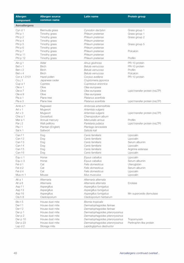
48
Allergen component
Allergen source common name
Latin name Protein group
Aeroallergens
Cyn d 1Phl p 1Phl p 2Phl p 4Phl p 5Phl p 6Phl p 7Phl p 11Phl p 12
Bermuda grassTimothy grassTimothy grassTimothy grassTimothy grassTimothy grassTimothy grassTimothy grassTimothy grass
Cynodon dactylonPhleum pratensePhleum pratensePhleum pratensePhleum pratensePhleum pratensePhleum pratensePhleum pratensePhleum pratense
Grass group 1Grass group 1Grass group 2
Grass group 5
Polcalcin
Profilin
Aln g 1Bet v 1Bet v 2Bet v 4Cor a 1.0101Cry j 1Cup a 1Ole e 1Ole e 7Ole e 9Pla a 1Pla a 3
AlderBirchBirchBirchHazel pollenJapanese cederCypressOliveOliveOlivePlane treePlane tree
Alnus glutinosaBetula verrucosaBetula verrucosaBetula verrucosaCorylus avellanaCryptomeria japonicaCupressus arizonicaOlea europaeaOlea europaeaOlea europaeaPlatanus acerifoliaPlatanus acerifolia
PR-10 proteinPR-10 proteinProfilinPolcalcinPR-10 protein
Lipid transfer protein (nsLTP)
Lipid transfer protein (nsLTP)
Amb a 1Art v 1Art v 3Che a 1Mer a 1Par j 2Pla l 1Sal k 1
RagweedMugwortMugwortGoosefootAnnual mercuryWall pellitoryPlantain (English)Saltwort
Ambrosia artemisiifoliaArtemisia vulgarisArtemisia vulgarisChenopodium albumMercurialis annuaParietaria judaicaPlantago lanceolataSalsola kali
Lipid transfer protein (nsLTP)
ProfilinLipid transfer protein (nsLTP)
Can f 1Can f 2Can f 3 Can f 4Can f 5 Can f 6
DogDogDog DogDog Dog
Canis familiarisCanis familiarisCanis familiaris Canis familiarisCanis familiaris Canis familiaris
LipocalinLipocalinSerum albumin LipocalinArginine esterase Lipocalin
Equ c 1Equ c 3Fel d 1Fel d 2Fel d 4Mus m 1
HorseHorseCatCatCatMouse
Equus caballusEquus caballusFelis domesticusFelis domesticusFelis domesticusMus musculus
LipocalinSerum albuminUteroglobinSerum albuminLipocalinLipocalin
Alt a 1Alt a 6Asp f 1Asp f 3Asp f 6Cla h 8
AlternariaAlternariaAspergillusAspergillusAspergillusCladosporium
Alternaria alternataAlternaria alternataAspergillus fumigatusAspergillus fumigatusAspergillus fumigatusCladosporium herbarum
Enolase
Mn superoxide dismutase
Blo t 5Der f 1Der f 2Der p 1Der p 2Der p 10 Der p 23Lep d 2
House dust miteHouse dust miteHouse dust miteHouse dust miteHouse dust miteHouse dust mite House dust miteStorage mite
Blomia tropicalisDermatophagoides farinaeDermatophagoides farinaeDermatophagoides pteronyssinusDermatophagoides pteronyssinusDermatophagoides pteronyssinus Dermatophagoides pteronyssinusLepidoglyphus destructor
Tropomyosin Peritrophin-like protein
Aeroallergens continued overleaf...
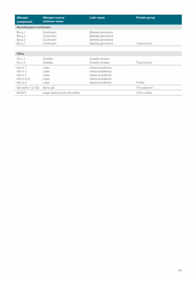
49
Allergen component
Allergen source common name
Latin name Protein group
Aeroallergens continued
Bla g 1Bla g 2Bla g 5Bla g 7
CockroachCockroachCockroachCockroach
Blattella germanicaBlattella germanicaBlattella germanicaBlattella germanica Tropomyosin
Other
Ani s 1Ani s 3
AnisakisAnisakis
Anisakis simplexAnisakis simplex Tropomyosin
Hev b 1Hev b 3Hev b 5Hev b 6.01Hev b 8
LatexLatexLatexLatexLatex
Hevea brasiliensisHevea brasiliensisHevea brasiliensisHevea brasiliensisHevea brasiliensis Profilin
Gal-alpha-1,3-Gal Alpha gal Thryoglobulin
MUXF3 Sugar epitope from Bromelain CCD-marker

50
Notes

51
Notes
Head office Sweden +46 18 16 50 00Austria +43 1 270 20 20Belgium (FR) +329 272 5780 (NL) +329 272 5780Brazil +55 0800 0551 535China +86 800 810 5118Czech Republic +420 7250 84047Denmark +45 70 23 33 06Finland +358 10 3292 110France +33 1 61 37 34 30Germany +49 761 47 8050Hong Kong +852 3107 7600India +91 11 4937 5400Ireland +44 1800 615 167
Italy +39 039 838 91 Japan +81 3 6872 6200Korea +82 2 6196 5556~9Norway +47 21 67 32 80Portugal +351 21 423 5350South Africa +27 11 792 6790Spain +34 935 765 800Sweden +46 18 16 60 60Switzerland +41 43 343 40 50Taiwan +886 2 8751 6655The Netherlands +31 30 602 37 00United Kingdom +44 1908 769110USA +1 800 346 4364Other countries +46 18 16 50 00
thermofisher.com/phadia
© 2021 Thermo Fisher Scientific Inc. All rights reserved. All trademarks are the property of Thermo Fisher Scientific Inc. and its subsidiaries. Manufacturer; Phadia AB, Uppsala Sweden.
75163.AL.GB1.EN.v1.19