golden retreiver lifetime study biopsy kit: collection ... · using the vacutainer holder and...
TRANSCRIPT

Biopsy Kit: Collection & Shipping Instructions
Golden Retreiver Lifetime Study
Morris AnimalF O U N D AT I O NGolden Retriever Lifetime Study

If you have any questions, please contact us at 855.4GR.DOGS (855.447.3647) | 3MAF – BVSKIB-v2
• We recommend having a team of three clinic staff members perform the blood draw – one for gentle control, one for managing the venipuncture, and one to handle the numerous blood tubes required.
• As soon as possible after the annual study examination and sample collection, log on to caninelifetimehealth.org to complete the veterinary questionnaire for your patient. (You must be on the Canine Lifetime Health Project website and not the general Morris Animal Foundation website.)
• Write your username and password in each study patient’s chart where it is easy to find. Your username is your email address and is case sensitive. Your password never needs to be changed.
• For the annual questionnaire you will report on health events, medications and vaccines given throughout the previous 12 months. If your software system does not show problem lists, medications and vaccines separately, consider maintaining an annual list for each category within the patient chart for easy review. You will need the vaccine manufacturer and lot number.
• A hard copy of the veterinary questionnaire is included in each kit. You can complete it as you perform the exam or dictate to a staff member.
• A knowledgeable staff member can enter the annual exam information online from the completed hard copy questionnaire or from your medical records.
• Please schedule extra time the day of a study examination for you or a trusted staff member to complete the online questionnaire.
• You can skip a question by using the table of contents on the right side of the screen (instead of clicking next, which requires the current page to be complete).
Thank you for your participation in this groundbreaking study. We understand study visits involve extra time and effort. Please review the following list of tips that may help streamline the process for you and your staff. If you have any other ideas, please feel free to share, them with the study team or on the Golden Retriever Lifetime Study Veterinarians Facebook page. If you have questions or need assistance, the customer care team is available 8 a.m. — 6 p.m. EST at 855-4GR-DOGS, or [email protected].
Tips for Participating Veterinarians

4 | If you have any questions, please contact us at 855.4GR.DOGS (855.447.3647) MAF – BVSKIB-v2
Collection Process: 1 Place a representative sample of the tumor into formalin at a standard ratio of 1 part sample to
10 parts buffered formalin. Store at room temperature.
2 Place a tumor sample less than 5 mm3 (size of a pencil eraser) in one vial of RNAlater. Circle ‘DISEASED’ on the label. Store at room temperature.
3 Place a sample of healthy tissue adjacent to the tumor less than 5 mm3 (size of a pencil eraser) in a separate vial of RNAlater. Circle ‘HEALTHY’ on the label. Store at room temperature.
Please refer to the following sections for instructions on:1 Biopsy Sample Fixation 2 Clinical Pathology Sample Collection 3 Sample Packaging and Shipping 4 Sample Reporting
Refer to Appendices for guidance on:Appendix 1 – Fixation and Shipping of Small or Large Specimens Appendix 2 – Identification of Surgical Margins Appendix 3 – Tissue Coding
The biopsy kit contents should be kept together in the labeled biopsy kit box or envelope and stored at room temperature. (Please check expiration dates on formalin jars prior to biopsy col-lection.)
Please contact the study team at 855.4GR.DOGS (855.447.3647) for advice or assistance with any sample submission or to request replacement or additional supplies.
Read all instructions before sample collection.
Biopsy Sample Collection Overview1

If you have any questions, please contact us at 855.4GR.DOGS (855.447.3647) | 5MAF – BVSKIB-v2
Biopsy Sample Collection Requirements
1 □ Excise tissue 2 □ Separate tissue into appropriate sections 3 □ Mark tissue as necessary 4 □ Place tissue into respective jars 5 □ Label jars appropriately 6 □ Document necessary information 7 □ Package shipment(s)
8 □ Wait for histopathology results to post to dog’s online account*
9 □ Complete online study Additional Veterinary Visit form
10 □ Discuss the finding with your client
* You will not receive any results from the RNAlater samples.
ANTECH DiagnosticsMajority of the tumor in the formalin for immediate biopsy analysis.
Fisher BioServicesPortion of the tissue (labeled diseased) and adjacent tissue (labeled healthy) in RNAlater for long-term storage.
1

6 | If you have any questions, please contact us at 855.4GR.DOGS (855.447.3647) MAF – BVSKIB-v2
Three Biopsy Specimens Total are Requested 1 Place a representative sample of the tumor in formalin at a standard ratio of 1 part
sample to 10 parts buffered formalin. Store at room temperature.
2 Place a tumor sample less than 5 mm3 (size of a pencil eraser) in one vial of RNAlater. Circle ‘DISEASED’ on the label. Store at room temperature.
3 Place a sample of adjacent healthy tissue less than 5 mm3 (size of a pencil eraser) in a separate vial of RNAlater. Circle ‘HEATLHY’ on the label. Store at room temperature.
Formalin Tumor specimens should be placed into formalin immediately after surgical excision unless tissue marking with dye or suture is necessary (see Appendices 1 and 2). Samples should be placed in enough formalin to achieve a final ratio of 1 part sample to 10 parts formalin. Please contact our study team at 855.447.3647 if you need formalin jars. Formalin jars should be stored at room temperature and shipped the same day of collection (unless collected Friday through Sunday).
RNAlaterThe tumor sample to be placed into the provided tube of RNAlater should be a piece of tissue no larger than 5 mm on each of the three sides (5 mm3 or size of a pencil eraser). After surgical excision, immediately immerse the sample into the tube of RNAlater and replace the cap securely. Circle 'DISEASED' on the label.
The sample of adjacent healthy tissue to be placed into a separate tube of RNAlater also should be a piece of tissue no larger than 5 mm on each of the three sides (5 mm3 or size of a pencil eraser). After surgical excision of the healthy tissue, immediately immerse the sample into a separate tube of RNAlater and replace the cap securely. Circle 'HEALTHY' on the label.
When surgical margins are important, select a non-marginal area of the tumor for the sample intended for RNAlater. RNAlater vials should be stored at room temperature and shipped the same day of collection (unless collected Friday through Sunday).
If the entire tumor sample is <1 cm on each side, place half into formalin and half into RNAlater. If surgical margins are important, then margins and proper sampling for histopathological diagnosis take priority. Please be sure to collect the sample of adjacent healthy tissue and place in a separate vial of RNAlater and circle 'HEALTHY' on the label.
Biopsy Sample Fixation1
If you have any questions, please contact us at 855.4GR.DOGS (855.447.3647) | 7MAF – BVSKIB-v2
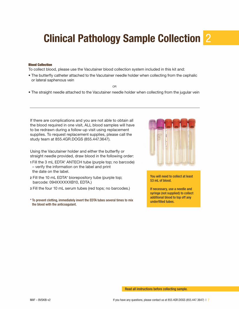
Clinical Pathology Sample Collection
Blood CollectionTo collect blood, please use the Vacutainer blood collection system included in this kit and:
• The butterfly catheter attached to the Vacutainer needle holder when collecting from the cephalic or lateral saphenous vein
or
• The straight needle attached to the Vacutainer needle holder when collecting from the jugular vein
You will need to collect at least 53 mL of blood.
If necessary, use a needle and syringe (not supplied) to collect additional blood to top off any underfilled tubes.
If there are complications and you are not able to obtain all the blood required in one visit, ALL blood samples will have to be redrawn during a follow-up visit using replacement supplies. To request replacement supplies, please call the study team at 855.4GR.DOGS (855.447.3647).
Using the Vacutainer holder and either the butterfly or straight needle provided, draw blood in the following order:
1 Fill the 3 mL EDTA* ANTECH tube (purple top; no barcode) – verify the information on the label and print the date on the label.
2 Fill the 10 mL EDTA* biorepository tube (purple top; barcode: 094XXXXXXB10, EDTA.)
3 Fill the four 10 mL serum tubes (red tops; no barcodes.)
* To prevent clotting, immediately invert the EDTA tubes several times to mix the blood with the anticoagulant.
Read all instructions before collecting sample.
2

8 | If you have any questions, please contact us at 855.4GR.DOGS (855.447.3647) MAF – BVSKIB-v2
Serum Isolation Allow the four serum tubes (red tops) to clot for at least 45 minutes and then centrifuge per your clinic’s normal protocol (we recommend 1,100–1,300 RCF for 10 minutes).
After centrifugation, remove the Vacutainer cap from each of the four serum tubes (red tops) and using the 3 mL transfer pipette provided (DO NOT POUR), transfer the serum as follows:
• Place 3 mL of serum into the 3 mL ANTECH serum tube. The black line on the left side of the tube label indicates the appropriate fill volume. (white top; no barcode.) – Verify the information on the label and print the date
on the label.
• Place 10 mL of serum into the 10 mL biorepository serum transport vial (red screw top; bar code: 94XXXXXXB20, SERUM.)
If additional serum is needed to achieve the required volumes, try re-centrifuging the blood remaining in the tubes.
Urine Collection Collect a minimum of 10 mL of urine via free catch or cystocentesis.
A transfer pipette is provided for your convenience to:
• Place a minimum of 5 mL of urine into the ANTECH urine tube (green top; no barcode.) – Verify the information on the label and print the date on the
label.
• Place a minimum of 5 mL of urine into the biorepository urine tube (green top; bar code: 094XXXXXXB30, URINE.)
Clinical Pathology Sample Collection (Cont.)2

If you have any questions, please contact us at 855.4GR.DOGS (855.447.3647) | 9MAF – BVSKIB-v2
Fecal Collection If the owner did not bring the dog’s fecal sample to the visit, please obtain a sample from the patient during the visit.
Transfer a marble-sized sample (approximately 1 g) of feces into each of the following:
• 30 mL ANTECH fecal collection tube, (white top; no barcode) – Verify the information on the label and print the date on
the label.
• Biorepository fecal collection tube, (brown top; bar code: 094XXXXXXB60, FECAL.)
Hair Collection Cut a lock of hair ≈1/4” in diameter (about the diameter of a wooden pencil) and a minimum of 2” long as close to the root as possible.*
• Place the lock of hair into the 3.5 mL biorepository tube, (orange screw top; bar code: 094XXXXXXB40, HAIR.)
Proceed to shipping instructions on the next page.* Please ask the owner for the preferred collection site.
Toenail Trimmings Collection Collect 5–10 toenail trimmings from the dog.
• Place the toenail trimmings into the biorepository 2 mL plastic cryogenic tube, (blue screw top; bar code: 094XXXXXXB50, NAIL.)
2Clinical Pathology Sample Collection (Cont.)

10 | If you have any questions, please contact us at 855.4GR.DOGS (855.447.3647) MAF – BVSKIB-v2
Sample Packaging & Shipping3
Placing A Service Call For FedEx Shipment(s)Place a service call to FedEx directly at 800.463.3339. Let them know you have packages for pickup and tell them that the shipping cost is being billed to ANTECH Diagnostics and Fisher BioServices. If your clinic is inadvertently charged any fees, please contact the study team at 855.4GR.DOGS (855.447.3647). You will use the provided FedEx labels for shipments.
Please contact the study team at 855.4GR.DOGS (855.447.3647) for advice or assistance with any sample submission or to request replacement or additional supplies.
Samples can ONLY be shipped Monday through Thursday.

If you have any questions, please contact us at 855.4GR.DOGS (855.447.3647) | 11MAF – BVSKIB-v2
ANTECH1 Clinical Pathology • Verify all four tubes (no bar codes) are properly filled and labeled,
including the date. Place the fecal (white top), 3 mL serum (white top), 3 mL EDTA (purple top) and 5 mL urine (green top) tubes in the ANTECH zip-closure bag.
• In the top header of the ANTECH Diagnostics requisition form, provide the following information: your clinic name, address, email, phone and fax number.
• Complete the information (doctor, date, client name, pet name, species, breed and age) in the upper right corner of the form.
• All laboratory tests are ordered with code 87007; this is a no-charge study code, so you must use this requisition form and verify that the study code is preprinted on the form. Failure to do so may result in charges to your clinic. No additional tests may be run with this order.
• If you fail to use the provided requisition form, your clinic will not receive the lab results.
• Retain the bottom copy of the form for your records.
• Place the completed top copy of the ANTECH Diagnostics requisition form into the ANTECH zip-closure bag (which should already contain the necessary samples).
• Place the zip-closure bag inside the ANTECH Diagnostics shipping box and seal the box with one of the provided tape strips (from the front of the plastic sleeve).
2 Histopathology • Ensure all formalin container lids are tightly sealed.
• Tape lids to bottles.
• Handwrite the date and tissue code (see Appendix 3) on the label of all formalin jars.
• Place the labeled formalin jar(s)*, together with 1 absorbing sheet per formalin jar, into the 5”x8” zip-closure bag and seal the bag.
• Place the sealed bag containing the labeled formalin jar(s) plus the completed ANTECH Diagnostics histopathology requisition form along with any other documents or photos into an ANTECH Diagnostics zip-closure bag and seal the bag.
*If using supplies you have on hand at the clinic, please label the jars with the required information.
Acct: 22725 Dog ID: 094-XXXXXX Owner: Last Name, Dog
Name
Date: Tissue Type: Tissue Code: __ __
Sample Packaging & Shipping (Cont.) 3

12 | If you have any questions, please contact us at 855.4GR.DOGS (855.447.3647) MAF – BVSKIB-v2
Sample Packaging & Shipping (Cont.)3
3 Packaging see figure 1 • Place the sealed double bag of formalin jars and documents, and the box containing clinical
pathology samples, into the FedEx Biological Substance Pak. Close the envelope by removing the clear strip to expose the adhesive seal.
• Complete your return address on the right side of the provided FedEx Expanded Billable Stamp labeled for ANTECH Diagnostics, affix it to the indicated location on the FedEx Biological Substance Pak.
• Retain the left half of the label for your records and tracking.
• Call FedEx at 800.463.3339 to arrange for a pickup.
ANTECH Samples: Histopath and clinical pathology report will be shared at no charge.
Figure 1: ANTECH Shipment

If you have any questions, please contact us at 855.4GR.DOGS (855.447.3647) | 13MAF – BVSKIB-v2
Biorepository 1 Clinical Pathology• Fill out the Shipment Inventory Form (Fisher BioServices)—Golden
Retriever Lifetime Study and include the patient information, collection date, collection times, sample shipment inventory, and any optional comments.
• Place the following three samples into the Styrofoam shipping container:*- 5 mL of urine—10 mL green-top tube- 10 mL of EDTA blood—purple-top tube- 10 mL of serum—red-top tube
• Close the Styrofoam shipping container and place it inside a zip-closure Lab-Loc Biohazard bag. Also place the following fi ve items inside the zip-closure Lab-Loc Biohazard bag:- Fecal sample tube—brown-top tube- Nails cryogenic tube—blue-top tube- Hair cryogenic tube—orange-top tube- Absorbent paper towel- Shipping inventory form
2 RNAlaterThere should usually be two vials of RNAlater shipped together - one labeled ‘DISEASED’ and one labeled ‘HEALTHY.’
• Fill out the Tissue Submission Form for Tissues in RNAlater (Fisher BioServices)—Golden Retriever Lifetime Study.
• Ensure you have handwritten the date and tissue code (see Appendix 3) on the label of all RNAlater tubes and circled ‘DISEASED’ or ‘HEALTHY’ on the appropriate vials.
• Place both labeled RNAlater tubes, together with one absorbing sheet, into the padded envelope and seal the envelope.
• Place the sealed padded envelope and Submission Form for Tissues in RNAlater into a Fisher Lab-loc biohazard zip-closure bag and seal the bag.
* NOTE: All tubes for the biorepository shipment are barcoded.
3Sample Packaging & Shipping (Cont.)

14 | If you have any questions, please contact us at 855.4GR.DOGS (855.447.3647) MAF – BVSKIB-v2
Sample Packaging & Shipping (Cont.)3
3 Packaging see figure 2
• Place the Fisher Bioservices Lab-loc biohazard zip-closure bag containing the biopsy samples and the zip-lock bag containing the clinical pathology samples into the FedEx Biological Substance Pak and seal the envelope by removing the clear seal to expose the adhesive strip.
• On the provided FedEx air shipment bill labeled “FedEx Express US Airbill,” complete the required “From” section. Retain the top copy for your records. Place the shipment bill inside of the provided clear FedEx mailer pouch and seal the pouch. Peel the backing from the pouch and affix the pouch to the indicated location on the FedEx Biological Substance Pak.
• Call FedEx at 800.463.3339 to arrange for a pickup.
Fisher BioServices Samples: Healthy and diseased tissue and whole blood, serum, urine, feces, hair and toenails for long-term storage.
Figure 2: Fisher Bioservices Shipment

If you have any questions, please contact us at 855.4GR.DOGS (855.447.3647) | 15MAF – BVSKIB-v2
Results will upload to your patient’s online study account.
Sample Reporting
You will receive the test results from the samples you ship to ANTECH Diagnostics within three to five business days on your patient’s record through your online study account at caninelifetimehealth.org. The results are posted under the tab labeled ‘Lab Results.’ The results will come as two separate reports: one report for the biopsy and a second report for the clinical pathology.
If malignant histopathology results are received, log on at caninelifetimehealth.org/secure to complete an Additional Veterinary Visit (AVV) form for your patient. You will have the option to print a hard copy of this form. You can access the AVV form by selecting the appropriate patient from your ‘My Patients’ page. You will see a red-labeled button on the dog’s Golden Retriever Lifetime Study tab. If you have any questions, please do not hesitate to email the study team at [email protected] or call toll-free at 855.4GR.DOGS (855.447.3647). We are here to help!
Figure 3: Additional Veterinary Visit Form
Dog InformationName: FidoDog ID: 000000Breed: Golden RetireverSex: Intact MaleBirth Date: 01/01/2013
Owner InformationName: Jane DoeEmail: [email protected]
Visit Date InformationVisit Date: 01/01/2016 (Scheduled)
My Patient - Fido
4

16 | If you have any questions, please contact us at 855.4GR.DOGS (855.447.3647) MAF – BVSKIB-v2
Fixation and Shipping of Small or Large SpecimensSpecimen shipping can be complicated when dealing with very small or very large tissues that cannot be routinely processed.
• Samples too large to fix as a whole (e.g., spleen) should be sectioned into portions and submitted in separate, appropriately labeled formalin jars. An annotated digital photograph or sketch of the original specimen to depict sectioning and orientation along with area(s) of gross lesions should accompany the samples. Dye margins before sectioning if appropriate.
• For an amputated limb, remove as much of the noncancerous tissue as possible, and then immerse the tumor into an appropriate-size container of formalin.
• Very small samples, such as endoscopic or punch biopsies, should be placed into the provided tissue cassette, the Dog Study ID# labeled with a #2 pencil, and then put into a formalin container for shipping. Do not use gauze sponges or cardboard because tissue may become compromised upon retrieval. Very small samples may, at times, yield insufficient artifact-free section for diagnosis; therefore, submission of multiple specimens from a single lesion is preferable.
• For luminal organs (e.g., intestine, uterus, large vessels), gently flush the intact lumen with formalin. For long sections of luminal tissues, make a partial longitudinal incision, leaving areas of interest (e.g., resection sites, mass) intact, or submit three labeled sections (e.g., proximal, middle and distal).
• Thin, flat samples (e.g., urinary bladder, stomach, diaphragm) should be placed directly into a formalin container. Larger flat samples can be sutured onto a flat piece of cardboard presoaked in formalin or water before immersion into a formalin container.
• Regional or draining lymph nodes associated with the primary tumor, including those that result in limb amputation, warrant microscopic examination. To ensure proper evaluation, consider dissection or biopsy of the regional lymph node during surgery. Submit the lymph node in a separate, appropriately labeled container.
Appendix: 15

If you have any questions, please contact us at 855.4GR.DOGS (855.447.3647) | 17MAF – BVSKIB-v2
Identification of Surgical Margins The primary means of indicating surgical margins is by marking with tissue dye and/or placement of sutures. It is imperative that a corresponding written description of these tissue demarcations be provided. The best practice is to have the surgeon dye the margins before submission to the laboratory as the surgeon can best identify these areas.
Guidelines for the Appropriate Marking of Tissues: • Marking of tissue margins should occur as soon as possible post-excision.
• The tissue should be blotted dry prior to marking.
• Marking should be done using the provided dye.
• Place dye only on regions that are of specific interest or are true surgical margins. Complete submersion of the sample in dye is not recommended.
• To prevent excess dye from washing off or coating insignificant areas, allow the dye to dry completely (approximately 5 to 10 minutes) before placing the specimen in formalin. Some discoloration of the formalin with dye is expected.
• For larger specimens that require “bread loafing” (i.e., incomplete parallel cuts approximately 1 cm apart), dye the tissue and allow dye to dry prior to cutting.
5Appendix: 2

18 | If you have any questions, please contact us at 855.4GR.DOGS (855.447.3647) MAF – BVSKIB-v2
Code Description Additional Indications
70 Other tissue, source Tissue: Diseased | Healthy
71 Adrenal Gland Left | Right | Both
72 Bone None
73 Bone Marrow None
74 Brain None
75 Colon None
76 Duodenum None
77 Esophagus None
78 Eye Left | Right | Both
79 Gonads Left | Right | Both
80 Heart None
81 Ileocecocolic Junction None
82 Ileum None
83 Jejunum None
84 Kidney Left | Right | Both
85 Liver None
86 Lung Specify Lobe:
87 Lymph Node Left | Right : : Axillary | Mesenteric | Prescapular | Mandibular | Popliteal | Other:
88 Oral Cavity None
89 Pancreas None
90 Parathyroid Gland None
91 Prostate None
92 Rectum None
93 Skeletal Muscle None
94 Skin None
95 Spinal Cord None
96 Spleen None
97 Stomach None
98 Thyroid None
99 Urinary Bladder None
Tissue Coding
Please contact the study team at 855.447.3647 for advice or assistance with any sample submission, or to request replacement or additional supplies.
Appendix: 35

If you have any questions, please contact us at 855.4GR.DOGS (855.447.3647) | 19MAF – BVSKIB-v2

20 | If you have any questions, please contact us at 855.4GR.DOGS (855.447.3647) MAF – BVSKIB-v2
About Morris Animal Foundation
Please contact the study team at 855.447.3647 for advice or assistance with any sample submission or to request replacement or additional supplies.
Thank You to Our PartnersFOUNDING PARTNER
The Mark & Bette Morris Family Foundation
PLATINUM PARTNERS
GOLD SPONSORS
Golden Retriever Foundation Hadley and Marion Stuart Foundation
Zoetis
GOLDEN CHAMPIONS
Mars Veterinary
6
Morris Animal Foundation is a nonprofit organization that invests in science to advance animal health. The foundation is a global leader in funding scientific studies for companion animals, horses and wildlife. Since its founding in 1948, Morris Animal Foundation has invested in studies that have led to significant breakthroughs in diagnostics, treatments, preventions and cures to benefit animals worldwide. Learn more at morrisanimalfoundation.org.