gp96, an endoplasmic reticulum master chaperone for integrins and
TRANSCRIPT

HEMATOPOIESIS AND STEM CELLS
gp96, an endoplasmic reticulum master chaperone for integrins and Toll-likereceptors, selectively regulates early T and B lymphopoiesisMatthew Staron,1 Yi Yang,1 Bei Liu,1 Janet Li,1 Yuankai Shen,1 Juan Carlos Zuniga-Pflucker,2 Hector L. Aguila,1
Irving Goldschneider,1 and Zihai Li1
1Department of Immunology, University of Connecticut School of Medicine, Farmington; and 2Department of Immunology, University of Toronto, SunnybrookResearch Institute, Toronto, ON
Integrins contribute to lymphopoiesis,whereas Toll-like receptors (TLRs) facili-tate the myeloid replenishment duringinflammation. The combined role of TLRsand integrin on hematopoiesis remainsunclear. gp96 (grp94, HSP90b1) is an en-doplasmic reticulum master chaperonefor multiple TLRs. We report herein thatgp96 is also essential for expression of14 hematopoietic system-specific inte-grins. Genetic deletion of gp96 thus en-
ables us to determine the collective rolesof gp96, integrins, and TLRs in hematopoi-esis. We found that gp96-null hematopoi-etic stem cells could support long-termmyelopoiesis. B- and T-cell development,however, was severely compromised withtransitional block from pro-B to pre-B cells and the inability of thymocytes todevelop beyond the CD4�CD8� stage.These defects were cell-intrinsic andcould be recapitulated on bone marrow
stromal cell culture. Furthermore, defec-tive lymphopoiesis correlated stronglywith failure of hematopoietic progenitorsto form close contact with stromal cellniche and was not the result of the defectin the assembly of antigen receptor orinterleukin-7 signaling. These findings de-fine gp96 as the only known molecularchaperone to specifically regulate T- andB-cell development. (Blood. 2010;115:2380-2390)
Introduction
Integrins are a family of 24 �� heterodimers in vertebrates formednoncovalently by 18 � and 8 � integrins, of which 17 integrins areexpressed in the hematopoietic system.1,2 Known best for theiradhesion properties, integrins also orchestrate signals betweenextracellular matrix and intracellular cytoskeletons in regulatingdiverse functions of cells, including proliferation and differentia-tion. However, despite the expression of integrins on hematopoieticstem cells (HSCs) and the role of integrins in HSC homing to thebone marrow (BM) niche, their function in hematopoiesis remainscontroversial. For example, although �4 integrin has been impli-cated in both T and B lymphopoiesis from fetal HSCs,3,4 it appearsto play a less significant role in adult hematopoiesis.5,6 Further-more, combined deletion of both �1 and �7 integrins, which are theonly known partners of �4 integrin, causes no defect in eitherlymphopoiesis or myelopoiesis.7 Genetic �2 integrin deficiencycauses myeloid hyperplasia, including profound granulocytosis andsplenomegaly, but no significant problems in hematopoiesis.8
Clearly, both �4 and �2 integrins are involved in homing of HSCsin the BM and recruitment of leukocytes to sites of inflamma-tion.5,9,10 Although pan-integrin deficient system is now available,11
no resolution of the roles of integrin in hematopoiesis has emerged.Toll-like receptors (TLRs) are pattern recognition receptors that
play important roles in sensing pathogen-associated molecularpatterns from microbes, which are critical for host immuneresponse.12 More than 10 TLRs have been described in vertebrates,recognizing a spectrum of microbial moieties, such as endotoxin,flagellin, dsRNA, and DNA. In the steady state, TLRs do notcontribute significantly to hematopoiesis, although TLRs on HSCs
have been implicated in the replenishment/recruitment of myeloidcells in response to inflammation.13,14 TLRs and integrins do notshare significant structural homology. Nevertheless, the folding andproper expression of many TLR and integrin family members aredependent on gp96, the heat shock protein 90 (HSP90) paraloguein the endoplasmic reticulum (ER). Deletion of gp96 leads toposttranslational loss of multiple TLRs (TLR1, TLR2, TLR4,TLR5, TLR6, TLR7, and TLR9) and several integrins (�2, �4, and�V integrins),15-17 although no study has probed the entire hemato-poietic system-specific integrins for their dependence on gp96.
As a major ER luminal protein whose expression can be furtherinduced by accumulation of misfolded proteins, gp96 is alsothought to participate in the ER-unfolded protein response (UPR)18
and ER-associated protein degradation,19 and has been implicatedto play a major “housekeeping” function to maintain proteinhomeostasis in the secretory pathway.20 The discovery that gp96seems to selectively fold TLRs and integrins15-17 was unexpected,which raises the intriguing possibility that gp96 is evolved to playmore specialized function in the multicellular organism.
In this study, we used tamoxifen (TAM)–inducible gp96knockout (KO) mice to further map the clientele of gp96 and tostudy the roles of gp96 in hematopoiesis. Hematopoiesis is anattractive model system to elucidate the function of gp96, not onlybecause of the possible roles of integrins and TLRs in the process,but also because of the importance of UPR in regulating severalaspects of the immune system, such as B-cell development,21
plasma cell differentiation,22,23 and dendritic-cell (DC) develop-ment and function.24 We demonstrated that gp96 is essential for the
Submitted July 13, 2009; accepted October 26, 2009. Prepublished online as BloodFirst Edition paper, November 13, 2009; DOI 10.1182/blood-2009-07-233031.
An Inside Blood analysis of this article appears at the front of this issue.
The online version of this article contains a data supplement.
The publication costs of this article were defrayed in part by page chargepayment. Therefore, and solely to indicate this fact, this article is herebymarked ‘‘advertisement’’ in accordance with 18 USC section 1734.
© 2010 by The American Society of Hematology
2380 BLOOD, 25 MARCH 2010 � VOLUME 115, NUMBER 12

expression of a majority of hematopoietic system-specific inte-grins. Unexpectedly, we found that gp96 null HSCs were able toengraft and initiate long-term hematopoiesis. We further revealedthat deletion of gp96 led to a selective and stage-specific loss inboth T and B lymphopoiesis, but not NK-cell development ormyelopoiesis, underscoring that gp96 plays a specialized ratherthan a general role in hematopoiesis. Furthermore, because of thecomplete loss of multiple TLRs and integrins in gp96 null cells, wealso conclude that the functional requirement of TLRs and integrinsin hematopoieis is highly lineage-specific.
Methods
Mice
C57BL/6 (CD45.2) mice were obtained from The Jackson Laboratory.C57BL/6 (CD45.1) mice were purchased from NCI-Frederick. hsp90b1floxed mice were crossed to R26R-creERT2 mice (kindly provided byJames Y. H. Li, University of Connecticut Health Center [UCHC]) andfurther backcrossed to C57BL/6 background for 6 to 10 generations.Control mice were hsp90b1flox/flox-creER� littermates (designated as wild-type [WT]) of hsp90b1flox/flox-creER� KO mice. All mice were maintained by theCenter for Laboratory Animal Care of UCHC (Farmington, CT) on anInstitutional Animal Care and Use Committee–approved animal care protocol.
Cell lines
WT and gp96 mutant 70Z/3 pre-B cells were a gift from Brian Seed(Harvard University),15 which were cultured in RPMI medium (Sigma-Aldrich) supplemented with 10% heat-inactivated fetal calf serum (AtlasBiologicals), 55�M 2-mercaptoethanol (Invitrogen), and penicillin-streptomycin (Invitrogen). OP9 and OP9-DL1 cells were cultured in�-minimum essential medium containing L-glutamine and ribonucleotides(Invitrogen) supplemented with 20% fetal calf serum, 1mM sodliumpyruvate (Invitrogen), 10mM N-2-hydroxyethylpiperazine-N�-2-ethanesul-fonic acid (Invitrogen), 55�M 2-mercaptoenthanol, and penicillin-streptomycin. All cells were cultured in 5% CO2 incubator.
Tamoxifen-inducible gp96 deletion
Tamoxifen (Sigma-Aldrich; 100 �g/20 g body weight) in peanut oil(Sigma-Aldrich) was administered by intraperitoneal injection daily for12 to 14 consecutive days to both experimental and control mice, anddeletion of gp96 was monitored by loss of CD11b on Gr1� cells in theperipheral blood leukocytes. Deletion of gp96 was also confirmed byimmunoblot and by intracellular stain for gp96.
Flow cytometry
After Fc-receptor blocking, cells were stained for surface lineage markers.For Lin�Scal-1�c-Kit� (LSK) population, lineage-negative (Lin�) cells(CD3, CD4, CD8, CD5, B220, NK1.1, Gr1, F4/80, and Ter119) wereanalyzed for expression of Sca-1 and c-Kit. For gp96 intracellular staining,cells were stained for surface markers, then fixed in 4% formalin/phosphate-buffered saline, permeabilized using ice-cold MeOH. Nonspecific bindingwas blocked with 10% goat serum, and gp96 was stained intracellularlywith rabbit IgG isotype control or SPA-851 polyclonal gp96 antibody(Stressgen), followed by anti–rabbit secondary antibody. All antibodieswere purchased from eBioscience, except where indicated. Fluorescence-activated cell sorter data were analyzed on FlowJo software (TreeStar)before being imported to ACD Systems Canvas.
Immunoblot
Single-cell suspension of BM cells, thymocytes, and splenocytes was lysedin RIPA buffer supplemented with proteinase inhibitor cocktail (Sigma-Aldrich) as described.17 For all other organs, tissue was minced in lysisbuffer and briefly sonicated. Bradford assay was used to quantitate protein
concentration, and equal amounts of lysate were resolved by sodiumdodecyl sulfate–polyacrylamide gel electrophoresis. gp96 was detected using9G-10 primary antibody (Stressgen) followed by horseradish peroxidase–conjugated anti–rat secondary antibody (Sigma-Aldrich). �-Actin was blotted(AC-74; Sigma-Aldrich) as a loading control.
BM transplantation
BM cells were isolated from both tibia and femur using aseptic technique.Red blood cells were lysed using sterile ammonium chloride lysis buffer,and BM cells were resuspended in sterile phosphate-buffered saline,pH 7.2, at 10 million cells/mL. A total of 2 million BM cells were injectedvia tail vein to irradiated C57BL/6 mice (550 cGy �2, 4 hours apart, 1 daybefore BM transplantation). Recipients were monitored for hematopoieticreconstitution in peripheral blood leukocytes using CD3, B220, Gr1, andCD11b markers. Similarly, for 1:1 transplantation experiments, 2 millionWT (CD45.1/CD45.2) and KO (CD45.2) BM was injected intravenously tolethally irradiated WT C57BL/6 (CD45.1) recipients. Six weeks aftertransplantation, TAM was administered intraperitoneally to induce gp96deletion. For 20:1 transplantation experiments, 7 months post-TAM dele-tion (PTD), 10 million gp96 null BM cells (CD45.2) and 0.5 million WTC57BL/6 (CD45.1) BM cells were injected to lethally irradiated WTC57BL/6 (CD45.1) mice.
Enrichment of Lin�c-Kit� progenitors
BM cells from WT and KO chimeras were isolated, stained usingbiotinylated lineage cocktail, and Lin� cells were depleted using antibiotinMACs beads (Miltenyi Biotec). Subsequently, Lin� cells were stained withc-Kit–phycoerythrin (PE) antibody followed by positive selection usinganti-PE MACS beads. Enrichment of progenitors was routinely verified byflow cytometry after staining with biotinylated lineage-specific antibodycocktail followed by streptavidin-allophycocyanin and Sca-1–fluoresceinisothiocyanate. c-Kit–PE staining was maintained after positive selection.Purity was more than 85%.
OP9 and OP9-DL1 coculture
A total of 10 000 OP9 and OP9-DL1 were plated the day before to a 6-wellplate. When the cells reached approximately 70% confluence, 1 � 105
Lin�c-Kit� progenitors were seeded on top in media supplemented withinterleukin-7 (IL-7; 5 ng/mL; Biovision) and FLT3L (5 ng/mL; Biovision).Cocultures were refed every third day and split to a fresh stromal layerweekly. A small aliquot of cells was reserved for analysis of differentiationby flow cytometry.
Statistics
Numbers in all bar graphs represent mean plus or minus SD. The Studentt test was used to determine whether the difference between 2 groups isstatistically significant (P � .05).
Results
gp96 is essential for cell-surface expression of mosthematopoietic system-specific integrins
Loss of gp96 is embryonic lethal (e5.5).25 To study the roles ofgp96 in hematopoiesis, we generated TAM-inducible gp96 KOmice by crossing hsp90b1flox/flox mice with Rosa26-creERT2 mice.After 14 days of daily injection with TAM, Cre-mediated deletionof gp96 from all adult tissues of KO mice was accomplished(Figure 1A; and data not shown). As expected, loss of gp96 led toabrogation of surface integrins CD11b, �4, and TLR2, but not anonclient CD16/32 on hematopoietic cells (Figure 1B), consistentwith the known chaperone functions of gp96.15-17 Next, wesystematically examined and compared the expression of allintegrins in the hematopoietic system of KO mice and control
ROLES OF gp96 AND ITS CLIENTELE IN HEMATOPOIESIS 2381BLOOD, 25 MARCH 2010 � VOLUME 115, NUMBER 12

littermates by flow cytometry. We found that gp96 is essential forcell surface expression of 14 of 17 integrin pairs (summarized inFigure 1C). KO cells failed to express a majority of integrins except�5�1, �6�1, and �IIb�3 integrins.
General lymphopoiesis, but not myelopoiesis, is dependent ongp96
gp96 is ubiquitously expressed in all tissues. To study thehematopoietic system-autonomous roles of gp96 in hematopoiesis,we used a BM chimera approach to generate chimeric mice thatwere deficient of gp96 only in the hematopoietic system (Figure2A). WT recipient mice were lethally irradiated, followed bytransplantation with either KO or WT BM cells. Chimeric micewere allowed to first establish multilineage reconstitution (6-8 weeks)before TAM treatment and deletion of gp96.
One month PTD, we confirmed the gp96 knockdown byimmunoblot (Figure 2B) and intracellular stain (Figure 2C). Therewas a general leukocytosis at this point in the peripheral blood(Figure 2D). Red blood cell count was normal, although there was aevidence for thrombocytopenia (Figure 2D); the etiology of thelatter is unclear and is being pursued in a separate study (M.S. andJ.L., unpublished observation, May 2007). Noticeably, KO chime-ras had markedly decreased cellularities in thymus and peripherallymph nodes with modestly increased cellularities in the spleen andBM (Figure 2E). Although the absolute numbers of myeloid cellswere well maintained over time in both the BM and spleen (Figure2F-G), there were severe T- and B-cell developmental blocks(discussed in Figure 5) that contributed to a significant reduction inmature peripheral B and T cells 7 months PTD (Figure 2G; anddata not shown). Gr1� cells and other myeloid cells were increased(5- to 10-fold) at all time points in KO mice (Figure 2F-G). Inaddition, at 2 months PTD, immunofluorescent staining of KOspleen sections confirmed a notable increase in Gr1� cells in thered pulp, and the reduced size/organization of T-cell zone andB-cell follicles (supplemental Figure 1, available on the Blood
website; see the Supplemental Materials link at the top of the on-line article).
We also examined the impact of gp96 loss on the developmentof DCs.26 By costaining BM cells with c-Kit and c-FMS, we foundthat gp96 loss did not significantly alter the macrophage-DCprogenitors in the BM (supplemental Figure 2A).27 Because of lossof CD11c, we used lineage gating to enumerate mature DCs. Wenoted that the splenic B220�CD3�CD5�NK1.1�F4/80�MHCII�
(Lin�MHCII�) population that contains myeloid DCs ( 70%CD11c�) was similar between KO and controls (data not shown);likewise, the number and percentage of Lin�MHCII�PDCA-1�
plasmacytoid DCs were comparable (data not shown). Further-more, both macrophage and DCs could be differentiated success-fully in vitro from KO progenitors (supplemental Figure 2B-C),although KO macrophage clearly failed to adhere to the culture dish(supplemental Figure 2D). Taken together, we conclude that myeloidlineage differentiation is surprisingly robust despite the disappearance ofalmost all integrins, multiple TLRs, and gp96 itself.
Hematopoietic progenitors persist in the absence of gp96,integrins, and TLRs
Normal myelopoiesis in the absence of gp96 suggests strongly thatHSCs are able to self-renew and differentiate in a manner that isindependent of gp96. This point was further validated experimen-tally. We first confirmed the loss of gp96 in the LSK population,which contains HSCs (long-term and short-term) and early lym-phoid progenitors, and Lin�Sca-1�c-Kit� progenitors (PROG),which include mostly myeloid progenitors.28 Loss of gp96 wasobserved in both these populations and was concomitant with theloss of �4 (CD49d), �L (CD11a) integrin and TLR2 expression(Figure 3A-C). We observed a significant expansion in bothpercentage and numbers of LSK and, to a lesser extent, PROG inKO BM (Figure 3D). Consistent with the robust myelopoiesis ofKO cells in vivo, colony-forming unit cultures in semisolidmethylcellulose demonstrated no differences in overall myeloid/
Figure 1. Inducible deletion of gp96 reveals its critical role forexpression of multiple integrins. WT or gp96 KO mice were injectedwith tamoxifen (TAM) intraperitoneally for 14 days followed by analysis.(A) Immunoblot for gp96 and �-actin (a loading control) from multipleorgans: Li indicates liver; Lu, lung; Ki, kidney; Spl, spleen; Il, ileum; and Co,colon. (B) Flow cytometric analysis of BM Gr1� cells for cell-surfaceexpression of indicated molecules. Dotted histogram represents KO cells;solid line indicates WT cells. (C) Summary of gp96-dependent andindependent integrins on the hematopoietic system. These data are basedon numerous experiments.
2382 STARON et al BLOOD, 25 MARCH 2010 � VOLUME 115, NUMBER 12

erythroid differentiation potential between WT and KO BM cells(data not shown).
We next sought to more closely examine the effects of loss ofgp96 on progenitor populations in the BM and thymus. Wefocused primarily on the early lymphoid progenitor (Lin�Sca-1�
c-Kit�FLT3�) and early thymic progenitor populations(Lin�CD25�c-Kit�) that are derived from FLT3�LSK.29-33 Wefound that LSK/PROG was increased in the peripheral bloodand spleen of gp96 KO chimeras (Figure 3E), which weremaintained upwards 7 months PTD (Figure 3D; and data notshown). Interestingly, the increase in both LSK percentage andnumber in KO chimeras was predominately of FLT3�/lo thatcontains HSCs and multipotent progenitors (Figure 3F).30
However, although the percentage of BM FLT3�LSK wasreduced, the absolute numbers of FLT3�LSK were comparablebecause of an increase in the overall numbers of BM KO LSK.In addition, the percentage of BM common lymphoid progeni-tors (CLPs; Lin�Sca-1�c-KitloIL-7R��) was also comparablebetween WT and KO 1 month PTD (supplemental Figure 3A).
Consistent with the persistence and function of BM CLPs, wefound that NK cell developed normally in the absence of gp96(supplemental Figure 3B-C).
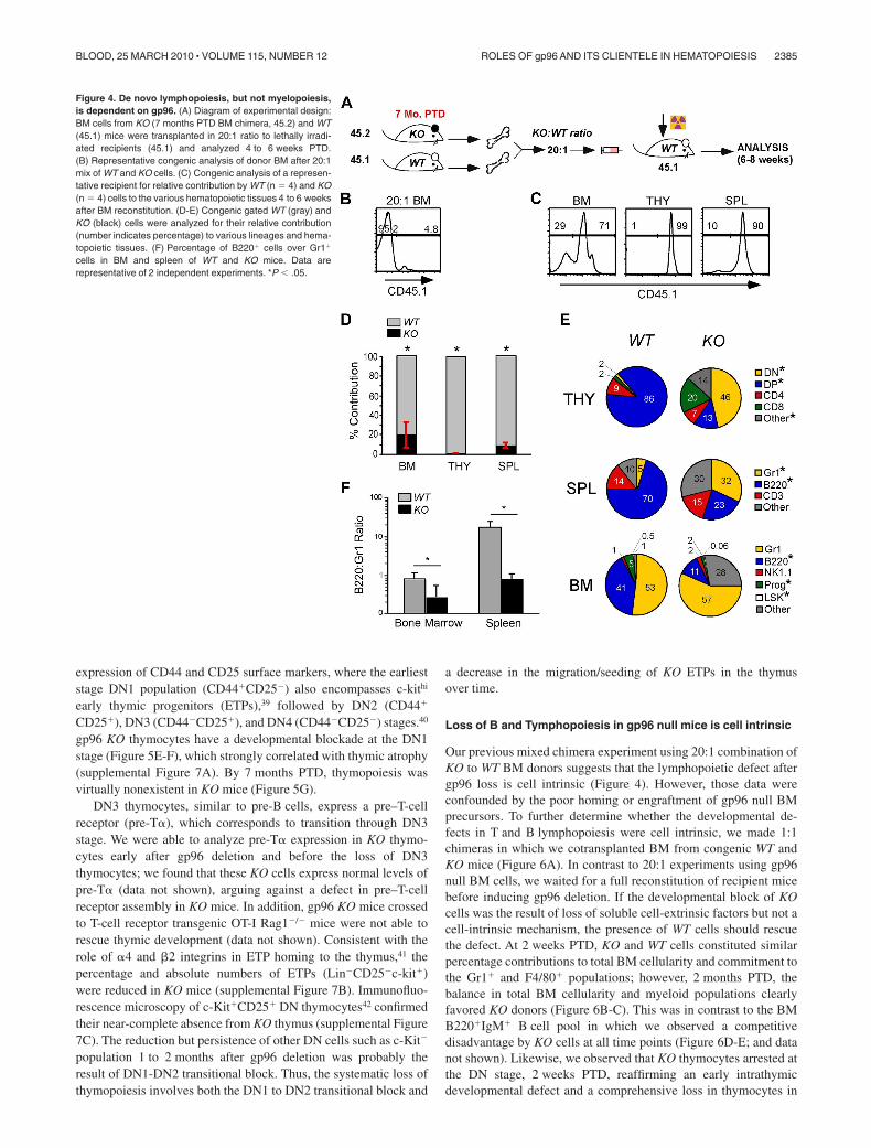
gp96 KO BM cells were unable to rescue lethally irradiatedmice (supplemental Figure 4), which could be the result of poorengraftment. To circumvent this problem and to further address theimpact of gp96 loss on long-term hematopoiesis, we performedsecondary BM transplantation with a mixture of KO and WT BMsin a 20:1 ratio (Figure 4A). WT (CD45.1) and KO (CD45.2) donorcells were congenically marked for easy distinction (Figure 4B).We found that gp96 KO BM cells were able to engraft, albeit not asefficiently as WT BM cells ( 80-fold less efficient based on BMcellularity; Figure 4C-F). Consistent with single donor chimeraexperiments, KO cells were restricted in their hematopoieticpotential and favored myelopoiesis over lymphopoiesis (Figure4E-F). Cells in the “other” category in KO BM and spleen wereprobably of myeloid origin, such as F4/80� cells, which developnormally in the absence of gp96 (Figure 2F). We thereforeconclude that the maintenance or survival of HSCs in vivo is
Figure 2. Global consequence of gp96 deletion on hematopoiesis. (A) Experimental scheme. (B) Immunoblot of various hematopoietic tissues from WT and KO mice12 days post-TAM deletion (PTD). Thy indicates thymus. �-Actin serves as loading control. (C) Intracellular staining for gp96 in various hematopoietic tissues from WT (solidline) or KO (dotted line) bone marrow (BM) chimeras using gp96 antibody (open) or isotype control (shaded). (D) Peripheral blood cell counts of WT (n � 3, �) and KO (n � 4,f) BM chimeras 1 month PTD. (E) Organ cellularity of WT (n � 3, �) and KO (n � 4, f) BM chimeras 1 month PTD. Ing indicates inguinal; Cer, cervical; and Mes, mesenteric.(F-G) Absolute cell numbers of indicated lineages in BM and spleen of WT (n � 5, �) and KO (n � 6, f) BM chimeras 3 to 4 weeks PTD (F) and 7 months PTD (G). Data arerepresentative of at least 3 independent experiments. WBC indicates white blood cell; LY, lymphocyte; MO, monocyte; GR, granulocyte; RBC, red blood cell; and PLT, platelet.*P � .05.
ROLES OF gp96 AND ITS CLIENTELE IN HEMATOPOIESIS 2383BLOOD, 25 MARCH 2010 � VOLUME 115, NUMBER 12

independent of gp96 and its client network including integrinsand TLRs.
gp96 is critical for pro-pre–B-cell transition
We next focused our attention on defining the critical developmen-tal stages of B and T lymphocytes that were controlled by gp96.gp96 KO chimeras were found to have a profound block inB lymphopoiesis at B220loIgM� stage, as evidenced by a markedreduction in the percentage and number of B220loIgM� cells and acomplete absence of B220hiIgM� recirculating B cells at 1 monthand 7 months PTD (Figure 5A-B). More specifically, we noted asignificant developmental blockade at the B220�CD43�CD24lo
stage at the transition from c-Kit� to CD25� corresponding to adefect in late pro-B to early pre–B-cell transition (Figure 5C;supplemental Figure 5A).34 Of note, splenic B-cell numbers weremaintained in gp96 KO BM chimeras early PTD, which indicatedthat once B cells matured, they no longer require gp96 formaintaining homeostasis, which was in line with our previousstudy.17 However, B-cell number did decline significantly by7 months PTD (Figure 2G). Consistent with earlier work,17,35 KOmice had additional loss of the splenic marginal zone B cellsresulting from integrin-mediated retention defect (supplementalFigure 5B-C).
Before acquisition of B-cell receptor (BCR), developing B cellsexpress a pre-BCR, composed of Vpre-B and �5 that comprise thesurrogate light chain on a �H backbone.36 Expression of pre-BCRfurther defines the transition between pro– and pre–B-cell stagesand has been shown to be an important checkpoint in pre-Bproliferation and differentiation.37,38 �5 expression on a gp96 nullpre-B cell line was, however, equivalent to WT cells (Figure 5D).Comparable expression of �5 was also confirmed on B220loIgM�
pre-B cells in WT and KO chimeras (data not shown), thusexcluding a role for gp96 in pre-BCR assembly.
Given the important role of IL-7R in B- and T-cell development,we next examined the role of gp96 on IL-7R� expression andsignaling. IL-7R� was uncompromised on gp96 null pre-B cells(Figure 5D). Furthermore, IL-7 stimulation of gp96 null andcontrol pre-B cells led to equal phosphorylation of Stat-5 (supple-mental Figure 6). In addition, IL-7R� expression on gp96 nullleukocytes and on CLPs was normal (data not shown).
gp96 is essential for effective thymopoiesis
Analogous to B-cell development blockade with gp96 KO chime-ras, at 1 month PTD, we observed a marked reduction of thymiccellularity (Figure 2D). Thymopoiesis begins at the CD4�CD8�
double-negative (DN) stage that can be defined by differences in
Figure 3. Multipotent hematopoietic progenitor persists in the absence of gp96. (A top) Dump gating of BM cells from WT and KO BM chimeras 1 month PTD.(Bottom) Gating of LSK (L) and PROG (P) populations. Numbers represent percentage of cells in the gated population. (B) Intracellular staining of gp96 on BM LSK andPROG populations from WT (solid line) and KO (dotted line) BM chimeras 2 months PTD using polyclonal gp96 antibody or isotype control (shaded). (C) CD49d,CD11a, and TLR2 expression on BM LSK populations from WT (solid line) and KO (dotted line) BM chimeras 1 month PTD. (D) Absolute numbers of BM LSK and PROGfrom WT (n � 3, �) and KO (n � 4, f) BM chimeras 1 month and 7 months PTD. *P � .05. (E) LSK and PROG analysis from WT and KO BM chimeras 2 months PTD inthe indicated tissues. (F) FLT3 expression on LSK from WT and KO BM chimeras 2 months PTD in the indicated tissues. Data are representative of at least2 independent experiments.
2384 STARON et al BLOOD, 25 MARCH 2010 � VOLUME 115, NUMBER 12
expression of CD44 and CD25 surface markers, where the earlieststage DN1 population (CD44�CD25�) also encompasses c-kithi
early thymic progenitors (ETPs),39 followed by DN2 (CD44�
CD25�), DN3 (CD44�CD25�), and DN4 (CD44�CD25�) stages.40
gp96 KO thymocytes have a developmental blockade at the DN1stage (Figure 5E-F), which strongly correlated with thymic atrophy(supplemental Figure 7A). By 7 months PTD, thymopoiesis wasvirtually nonexistent in KO mice (Figure 5G).
DN3 thymocytes, similar to pre-B cells, express a pre–T-cellreceptor (pre-T�), which corresponds to transition through DN3stage. We were able to analyze pre-T� expression in KO thymo-cytes early after gp96 deletion and before the loss of DN3thymocytes; we found that these KO cells express normal levels ofpre-T� (data not shown), arguing against a defect in pre–T-cellreceptor assembly in KO mice. In addition, gp96 KO mice crossedto T-cell receptor transgenic OT-I Rag1�/� mice were not able torescue thymic development (data not shown). Consistent with therole of �4 and �2 integrins in ETP homing to the thymus,41 thepercentage and absolute numbers of ETPs (Lin�CD25�c-kit�)were reduced in KO mice (supplemental Figure 7B). Immunofluo-rescence microscopy of c-Kit�CD25� DN thymocytes42 confirmedtheir near-complete absence from KO thymus (supplemental Figure7C). The reduction but persistence of other DN cells such as c-Kit�
population 1 to 2 months after gp96 deletion was probably theresult of DN1-DN2 transitional block. Thus, the systematic loss ofthymopoiesis involves both the DN1 to DN2 transitional block and
a decrease in the migration/seeding of KO ETPs in the thymusover time.
Loss of B and Tymphopoiesis in gp96 null mice is cell intrinsic
Our previous mixed chimera experiment using 20:1 combination ofKO to WT BM donors suggests that the lymphopoietic defect aftergp96 loss is cell intrinsic (Figure 4). However, those data wereconfounded by the poor homing or engraftment of gp96 null BMprecursors. To further determine whether the developmental de-fects in T and B lymphopoiesis were cell intrinsic, we made 1:1chimeras in which we cotransplanted BM from congenic WT andKO mice (Figure 6A). In contrast to 20:1 experiments using gp96null BM cells, we waited for a full reconstitution of recipient micebefore inducing gp96 deletion. If the developmental block of KOcells was the result of loss of soluble cell-extrinsic factors but not acell-intrinsic mechanism, the presence of WT cells should rescuethe defect. At 2 weeks PTD, KO and WT cells constituted similarpercentage contributions to total BM cellularity and commitment tothe Gr1� and F4/80� populations; however, 2 months PTD, thebalance in total BM cellularity and myeloid populations clearlyfavored KO donors (Figure 6B-C). This was in contrast to the BMB220�IgM� B cell pool in which we observed a competitivedisadvantage by KO cells at all time points (Figure 6D-E; and datanot shown). Likewise, we observed that KO thymocytes arrested atthe DN stage, 2 weeks PTD, reaffirming an early intrathymicdevelopmental defect and a comprehensive loss in thymocytes in
Figure 4. De novo lymphopoiesis, but not myelopoiesis,is dependent on gp96. (A) Diagram of experimental design:BM cells from KO (7 months PTD BM chimera, 45.2) and WT(45.1) mice were transplanted in 20:1 ratio to lethally irradi-ated recipients (45.1) and analyzed 4 to 6 weeks PTD.(B) Representative congenic analysis of donor BM after 20:1mix of WT and KO cells. (C) Congenic analysis of a represen-tative recipient for relative contribution by WT (n � 4) and KO(n � 4) cells to the various hematopoietic tissues 4 to 6 weeksafter BM reconstitution. (D-E) Congenic gated WT (gray) andKO (black) cells were analyzed for their relative contribution(number indicates percentage) to various lineages and hema-topoietic tissues. (F) Percentage of B220� cells over Gr1�
cells in BM and spleen of WT and KO mice. Data arerepresentative of 2 independent experiments. *P � .05.
ROLES OF gp96 AND ITS CLIENTELE IN HEMATOPOIESIS 2385BLOOD, 25 MARCH 2010 � VOLUME 115, NUMBER 12

KO mice over time (Figure 6F-G). Total DN thymocytes of KOorigin also progressively declined to less than 5% of the entire DNthymocytes 4 months PTD (data not shown), which was consistentwith the additional homing defect of KO ETPs. Thus, gp96regulates B and T lymphopoiesis in a cell-intrinsic fashion. Inaddition, despite the presence of a WT competitor, gp96 LSK andprogenitors were well sustained in the BM, and their percentageand number were consistently increased. Moreover, gp96 LSK andPROG were capable of long-term myelopoiesis upward 9 monthsPTD even in this competitive setting (data not shown).
gp96 null hematopoietic progenitors proliferate poorly and failto differentiate into B and T cells on stromal cells in vitro
The failure of gp96 KO cells to differentiate along T- and B-celllineages in vivo could be the result of a defect in the intrinsicdevelopmental program or to the inability of progenitors to migrateinto the proper niche, or both. To examine these possibilities, weperformed an in vitro differentiation assay using a well-describedB- and T-cell developmental coculture system. The stromal cell lineOP9, and OP9-DL1, which expresses notch ligand Delta-like
1 (DL1), have been used to effectively drive B- and T-celldifferentiation, respectively.43 We purified Lin�c-Kit� progenitorsfrom WT and KO BM (supplemental Figure 8) and plated equalnumbers of cells onto OP-9 and OP9-DL1 in culture. Cells werethen enumerated and analyzed by flow cytometry at weeklyintervals. We found that KO cells proliferated poorly on both OP-9and OP9-DL1 (Figure 7A-B), with little evidence of B- and T-celldifferentiation (Figure 7C-D). Moreover, gp96 KO progenitorswere unable to transmigrate beneath stromal cells to form prototypi-cal “cobblestone” colonies (Figure 7E), a sign of an early differen-tiation program.4 The inability of KO cells to transmigrate throughBM stromal layers was also recapitulated quantitatively using agp96 mutant pre–B-cell line (supplemental Figure 9).
Discussion
gp96 is a ubiquitously expressed and evolutionarily conservedER-resident molecular chaperone belonging to the HSP90 family.20
Genetic and biochemical approaches have demonstrated the pivotal
Figure 5. B- and T-cell lymphopoiesis isdependent on gp96 at critical transitionalstages. (A) B220/IgM surface staining ofBM cells from WT and KO BM chimeras1 month PTD. (B) Absolute numbers of BMB cells in various fractions indicated in panelA from WT (n � 5, �) and KO (n � 6, f) BMchimeras 3 to 4 weeks PTD. Data are pooledfrom 2 independent experiments. (C) CD43/CD24 surface staining on B220� cells in BM.(D) Expression of �5 pre-BCR and IL-7R�on the surface of WT (solid line) and KO(dotted line) pre–B-cell line. Shaded histo-grams represent isotype control. (E) CD4/CD8 (top) surface staining and surface ex-pression of CD49d (bottom) on thymocytesfrom WT and KO BM chimeras 1 monthPTD. (F) DN gating (top) and CD44/CD25surface staining on DN thymocytes from WTand KO BM chimeras 1 month PTD. (G) Ab-solute cell numbers of thymocytes from theindicated stages from WT (n � 3, �) andKO (n � 4, f) BM chimeras 1 month and7 months PTD. Data are representative of atleast 2 independent experiments. *P � .05.
2386 STARON et al BLOOD, 25 MARCH 2010 � VOLUME 115, NUMBER 12

role of gp96 in folding and maturation of TLRs and integrins.15-17
In addition, as a canonical member of the HSP90 family, gp96participates in a variety of molecular and biochemical processes tomaintain protein homeostasis in the ER.19 Thus, the functionalimportance of gp96 in hematopoiesis could be at least 2-fold,insomuch as it participates in the folding/maturation of 2 veryimportant classes of immunologically relevant molecules, and italso serves a second perhaps more general housekeeping role in ERprotein homeostasis.
Full appreciation of the function of gp96 requires knowledge ofgp96 client proteins, whose expression is dependent on gp96. Inthis study, we significantly expanded on the gp96 client network bydetermining the differential expression of integrins by WT andgp96 KO hematopoietic cells. We found that gp96 is essential forthe expression of a majority of integrins, including �1, �2, �4, �D,�E, �L, �M, �V, and �X of the hematopoietic system. Because �integrin must heterodimerize with its appropriate � integrin partnerfor cell surface expression, we conclude that gp96 is crucial for theexpression of 14 of 17 integrin pairs expressed by the hematopoi-etic system. The requirement for gp96 in folding these integrins isabsolute and cannot be compensated by any other ER chaperones.Although the structural basis for selective dependence on gp96 byintegrins is unclear, it is intriguing to point out that the loss of all
� integrins can be explained by the loss of their corresponding� partners, but not the converse. By deduction, we postulate thatgp96 is critical for chaperoning �, but not �, subunits of integrins.
The loss of multiple TLRs and integrins in gp96 null micecreated an unprecedented opportunity to address their combina-tional roles in hematopoiesis. Using a TAM-inducible gp96 dele-tion system coupled with competitive BM reconstitution, we foundthat gp96 is surprisingly not required by HSCs to self-renew and toinitiate long-term hematopoiesis along the myeloid lineage. gp96is, however, required at defined critical stages of early T- and B-celldevelopment, which correlated with the inability of gp96 KOprogenitors to transmigrate BM stromal cell layers.
The underlying mechanism by which gp96 selectively regulateslymphopoiesis is not entirely clear. TLRs have been suggested tofunction in replenishment of the myeloid compartment afterbacterial infection.13 Furthermore, although TLRs were not directlyexamined, inflammation can affect lymphoid versus myeloiddistribution in favor of the latter.44 However, the role of TLRs inhematopoiesis in the steady state is limited, evidenced by uncom-promised B-cell development in MyD88 and Trif double KO mice,which lack all known TLR downstream signaling.45 It is thusunlikely that the loss of TLRs contributes significantly to thelymphopoietic defect in gp96 KO mice.
Figure 6. Competitive BM reconstitution reveals cell-intrinsic role for gp96 in B and T lymphopoiesis, butnot myelopoiesis. (A) Diagram of experimental design.(B,D,F) Surface staining of the indicated lineages ongated KO (45.2) and WT (45.1/45.2) BM cells from 1:1BM chimeras 2 weeks and 2 months PTD. (C,E,G) Per-centage of KO and WT contribution to total BM myeloidcells (C), BM B-cell subsets (E), and thymocytes (G) at2 weeks and 2 months PTD (DN: CD4�CD8�; double-positive: CD4�CD8�; 4: CD4�CD8�; 8: CD4�CD8�).Data are representative of 3 mice per group. *P � .05.
ROLES OF gp96 AND ITS CLIENTELE IN HEMATOPOIESIS 2387BLOOD, 25 MARCH 2010 � VOLUME 115, NUMBER 12

In contrast, the role for integrins in hematopoiesis is extensive,particularly related to the function of �4 and �2 integrins, both ofwhich are bona fide clientele of gp96. Like gp96, �4 integrin isessential for embryogenesis and is also required for both earlyB- and T-cell development in fetal liver chimeric mice.3,4 It isrequired for stromal cell adhesion by both B- and T-cell progeni-tors,4,46-48 correlating with its probable role in lymphopoiesis.However, transplantation of adult �4 null BM cells to irradiatedRag2�/� mice revealed only partial defect in thymopoiesis and adecrease in certain subsets of BM B cells.6 Conditional ablation of�4 or �1/�7 integrin from adult mice had little infringement onlymphopoiesis.5,7,49 These studies highlight that the roles of gp96 inearly lymphopoiesis probably extend beyond chaperoning �4 and�2 integrins, and implicate that other members of integrins or/andyet unidentified gp96 clients also play a role in regulatinglymphopoiesis.
In gp96 KO BM chimeras, B- and T-cell differentiation isarrested at developmentally analogous stages, pro-B–pre-B andDN1-DN2 transitions, respectively. Two obvious possibilities havebeen ruled out: the roles of gp96 in the assembly/function ofantigen receptor and IL-7R. In addition, annexin V and propidiumiodide staining of DN thymocytes subsets did not reveal anysignificant increase in apoptosis of gp96 null leukocytes (data notshown). Rather, KO cells appear to lose their tight interaction withdevelopmental niche in the BM stromal cells, a scenario that can beexplained by the role of gp96 in chaperoning integrins. Consistentwith this idea, we observed an increase in LSKs, progenitors, andpro-B cells (B220�CD43�) in the peripheral blood and spleen ofgp96 KO chimeras. However, despite increased BM precursors inthe periphery, we found no evidence of significant extramedullaryhematopoiesis in KO chimeras.
Finally, our BM stromal cell cultures allowed us to concludethat the lymphopoietic defect of gp96 KO HSCs is not simply theresult of poor seeding of progenitors in the BM and the thymus.
However, gp96 KO progenitors clearly had difficulty in transmigrat-ing the stromal cell layer, arguing strongly that KO hematopoieticprecursors might be unable to form an optimal niche locally toinitiate lymphopoiesis at an early stage, despite appropriate differ-entiation cytokines and other cues. More work is necessary todefine the critical downstream signals that are missing in gp96 nullcells at the pre–B- and pre–T-cell stages and the coordinatefunction of integrins in these processes, although it is tempting tospeculate that stromal contact and signals in general are aprerequisite for development of lymphoid but not myeloid compart-ments; such a claim warrants further investigation.
There have been increased interests in the roles of proteinfolding chaperones in hematopoiesis, a highly regulated processwith simultaneous proliferation and multilineage differentiationthat are probably dependent on robust protein chaperone machin-ery. However, deletion of individual molecular chaperone has notresulted in catastrophic loss of hematopoiesis. For example, inmice with loss of critical UPR regulator XBP1 or ATF6, hematopoi-esis proceeds normally. Only plasma differentiation was affectedwith loss of XBP-1 and ATF6,21-23 which may be related in part tothe importance of downstream effector chaperone GRP78 inimmunologlobulin assembly.50 Loss of heat shock factor-1, a majortranscription factor for HSPs, resulted in ablation of induction ofHSPs51 but no noticeable compromise of hematopoiesis.52 Deletionof calreticulin causes heightened immune response resulting fromaberrant activation of peripheral T cells.53 Although genetic dele-tion of GRP78 and calnexin has been done, and it has revealed thecritical importance of these 2 molecules in embryogenesis54 andpostnatal survival,55 no specific roles of either molecule onhematopoiesis have been reported. Likewise, cytosolic HSP70 alsoappeared to be dispensable for hematopoiesis.56 Thus, with theexception of lymphopoiesis defect in gp96 null mice, hematopoi-esis and in particular myelopoiesis does not subject to tightregulation by the protein chaperone machinery, underscoring the
Figure 7. gp96 null hematopoietic progenitors fail to prolifer-ate and differentiate in BM stromal cell cultures. Purified WTand gp96 KO BM Lin�c-kit� progenitor cells were cultured on BMstromal cell OP9 or OP9-DL1 for 3 weeks and analyzed for cellgrowth and differentiation. (A-B) Kinetic analysis of cell prolifera-tion. (C-D) Flow cytometric analysis of T- and B-cell developmentafter 3 weeks of coculture. (E) Bright field image of day 5 cocul-ture of WT or KO Lin�c-kit� progenitors on OP9 (original magnifi-cation �100). Dotted circle denotes “cobblestone” colony. A fewKO progenitors were indicated by œ. Three experiments wereperformed with similar results.
2388 STARON et al BLOOD, 25 MARCH 2010 � VOLUME 115, NUMBER 12

possibility that myelopoiesis is vital to the survival of highvertebrates and is hardwired to withstand environmental andintracellular stress. These arguments also once again highlight theuniqueness of gp96 in hematopoiesis.
In conclusion, we have demonstrated that the expression of amajority of the hematopoietic system-specific integrins is con-trolled by gp96; thus, gp96 is a master chaperone for both TLRsand integrins. Yet, gp96 and its client network are dispensable forself-renewal and differentiation of HSCs. We further uncovered thedramatic difference between lymphopoiesis and myelopoiesis intheir requirement for gp96. Our study thus illustrates that thedevelopmental requirement for gp96 and its client network inhematopoiesis is both lineage- and stage-specific, highlighting thatgp96 plays more specialized rather than general roles in regulatingprotein homeostasis in the ER and the secretory pathway. Highresolution mapping of the gp96 client network is the essential nextstep to understand the function/mechanism of gp96 in hematopoi-esis and to identify key downstream molecules in regulatinglineage decision of lymphopoiesis versus myelopoiesis.
Acknowledgments
The authors thank past and present members of the laboratory ofZ.L. for helpful discussions, Sierra Root for assistance with themethylcellulose assay, and Drs Lynn Puddington, Pramod Srivas-tava, and Leo Lefrancois for their invaluable input and support.
This work was supported by the National Institutes of Health(grants AI070603, AI077283, and RC1HL100556; Z.L.). M.S. issupported in part by the National Institutes of Health (training grant5T32AI007080).
Z.L. is a Clinical Scholar of the Leukemia & LymphomaSociety of the United States.
Authorship
Contribution: M.S. conceived the idea, designed the research,performed all the experiments except Figure 1C, and wrote themanuscript; Y.Y. produced the hsp90b1flox/flox-Rosa26creER� mouseline; B.L., J.L., and Y.S. generated Figure 1C; J.C.Z.-P. generatedFigure 7 and critically read the manuscript; H.L.A. generatedsupplemental Figure 3; I.G. critically read the manuscript; Z.L.conceived the idea, designed the research, and wrote the manu-script; and all authors contributed to the analysis of the data.
Conflict-of-interest disclosure: The authors declare no compet-ing financial interests.
The current address of Y.Y. is Kimmel Center for Biology andMedicine of the Skirball Institute, New York University, New York,NY. The current address of J.L. is Massachusetts Institute ofTechnology, Cambridge, MA.
Correspondence: Zihai Li, MC 1601, University of ConnecticutSchool of Medicine, 263 Farmington Ave, Farmington, CT 06030-1601; e-mail: [email protected].
References
1. Miranti CK, Brugge JS. Sensing the environment:a historical perspective on integrin signal trans-duction. Nat Cell Biol. 2002;4(4):E83-E90.
2. Luo BH, Carman CV, Springer TA. Structural ba-sis of integrin regulation and signaling. Annu RevImmunol. 2007;25:619-647.
3. Arroyo AG, Yang JT, Rayburn H, Hynes RO. Dif-ferential requirements for alpha4 integrins duringfetal and adult hematopoiesis. Cell. 1996;85(7):997-1008.
4. Arroyo AG, Yang JT, Rayburn H, Hynes RO. Al-pha4 integrins regulate the proliferation/differen-tiation balance of multilineage hematopoietic pro-genitors in vivo. Immunity. 1999;11(5):555-566.
5. Scott LM, Priestley GV, Papayannopoulou T. De-letion of alpha4 integrins from adult hematopoi-etic cells reveals roles in homeostasis, regenera-tion, and homing. Mol Cell Biol. 2003;23(24):9349-9360.
6. Banerjee ER, Latchman YE, Jiang Y, PriestleyGV, Papayannopoulou T. Distinct changes inadult lymphopoiesis in Rag2�/� mice fully recon-stituted by alpha4-deficient adult bone marrowcells. Exp Hematol. 2008;36(8):1004-1013.
7. Bungartz G, Stiller S, Bauer M, et al. Adult murinehematopoiesis can proceed without beta1 andbeta7 integrins. Blood. 2006;108(6):1857-1864.
8. Horwitz BH, Mizgerd JP, Scott ML, DoerschukCM. Mechanisms of granulocytosis in the ab-sence of CD18. Blood. 2001;97(6):1578-1583.
9. Papayannopoulou T, Priestley GV, Nakamoto B,Zafiropoulos V, Scott LM. Molecular pathways inbone marrow homing: dominant role ofalpha(4)beta(1) over beta(2)-integrins and selec-tins. Blood. 2001;98(8):2403-2411.
10. Ulyanova T, Priestley GV, Banerjee ER,Papayannopoulou T. Unique and redundant rolesof alpha4 and beta2 integrins in kinetics of recruit-ment of lymphoid vs myeloid cell subsets to theinflamed peritoneum revealed by studies of ge-netically deficient mice. Exp Hematol. 2007;35(8):1256-1265.
11. Lammermann T, Bader BL, Monkley SJ,et al. Rapid leukocyte migration by integrin-independent flowing and squeezing. Nature.2008;453(7191):51-55.
12. Medzhitov R. Recognition of microorganisms andactivation of the immune response. Nature. 2007;449(7164):819-826.
13. Nagai Y, Garrett KP, Ohta S, et al. Toll-like recep-tors on hematopoietic progenitor cells stimulateinnate immune system replenishment. Immunity.2006;24(6):801-812.
14. Massberg S, Schaerli P, Knezevic-Maramica I, etal. Immunosurveillance by hematopoietic pro-genitor cells trafficking through blood, lymph, andperipheral tissues. Cell. 2007;131(5):994-1008.
15. Randow F, Seed B. Endoplasmic reticulum chap-erone gp96 is required for innate immunity but notcell viability. Nat Cell Biol. 2001;3(10):891-896.
16. Yang Y, Liu B, Dai J, et al. Heat shock proteingp96 is a master chaperone for toll-like receptorsand is important in the innate function of macro-phages. Immunity. 2007;26(2):215-226.
17. Liu B, Li Z. Endoplasmic reticulum HSP90b1(gp96, grp94) optimizes B-cell function via chap-eroning integrin and TLR but not immunoglobulin.Blood. 2008;112(4):1223-1230.
18. Malhotra JD, Kaufman RJ. The endoplasmic re-ticulum and the unfolded protein response. SeminCell Dev Biol. 2007;18(6):716-731.
19. Christianson JC, Shaler TA, Tyler RE, Kopito RR.OS-9 and GRP94 deliver mutant alpha1-antitrypsin to the Hrd1-SEL1L ubiquitin ligasecomplex for ERAD. Nat Cell Biol. 2008;10(3):272-282.
20. Yang Y, Li Z. Roles of heat shock protein gp96 inthe ER quality control: redundant or unique func-tion? Mol Cells. 2005;20(2):173-182.
21. Zhang K, Wong HN, Song B, Miller CN, ScheunerD, Kaufman RJ. The unfolded protein responsesensor IRE1alpha is required at 2 distinct steps inB cell lymphopoiesis. J Clin Invest. 2005;115(2):268-281.
22. Iwakoshi NN, Lee AH, Vallabhajosyula P, OtipobyKL, Rajewsky K, Glimcher LH. Plasma cell differ-entiation and the unfolded protein response inter-sect at the transcription factor XBP-1. Nat Immu-nol. 2003;4(4):321-329.
23. Reimold AM, Iwakoshi NN, Manis J, et al. Plasmacell differentiation requires the transcription factorXBP-1. Nature. 2001;412(6844):300-307.
24. Iwakoshi NN, Pypaert M, Glimcher LH. The tran-scription factor XBP-1 is essential for the devel-opment and survival of dendritic cells. J Exp Med.2007;204(10):2267-2275.
25. Wanderling S, Simen BB, Ostrovsky O, et al.GRP94 is essential for mesoderm induction andmuscle development because it regulates insulin-like growth factor secretion. Mol Biol Cell. 2007;18(10):3764-3775.
26. Wu L, Liu YJ. Development of dendritic-cell lin-eages. Immunity. 2007;26(6):741-750.
27. Waskow C, Liu K, Darrasse-Jeze G, et al. Thereceptor tyrosine kinase Flt3 is required for den-dritic cell development in peripheral lymphoid tis-sues. Nat Immunol. 2008;9(6):676-683.
28. Bhandoola A, von Boehmer H, Petrie HT,Zuniga-Pflucker JC. Commitment and develop-mental potential of extrathymic and intrathymicT cell precursors: plenty to choose from. Immu-nity. 2007;26(6):678-689.
29. Christensen JL, Weissman IL. Flk-2 is a marker inhematopoietic stem cell differentiation: a simplemethod to isolate long-term stem cells. Proc NatlAcad Sci U S A. 2001;98(25):14541-14546.
30. Adolfsson J, Borge OJ, Bryder D, et al. Upregula-tion of Flt3 expression within the bone marrowLin(-)Sca1(�)c-kit(�) stem cell compartment isaccompanied by loss of self-renewal capacity.Immunity. 2001;15(4):659-669.
31. Sitnicka E, Bryder D, Theilgaard-Monch K,Buza-Vidas N, Adolfsson J, Jacobsen SE. Keyrole of flt3 ligand in regulation of the common lym-phoid progenitor but not in maintenance of thehematopoietic stem cell pool. Immunity. 2002;17(4):463-472.
ROLES OF gp96 AND ITS CLIENTELE IN HEMATOPOIESIS 2389BLOOD, 25 MARCH 2010 � VOLUME 115, NUMBER 12

32. Benz C, Bleul CC. A multipotent precursor in thethymus maps to the branching point of the T ver-sus B lineage decision. J Exp Med. 2005;202(1):21-31.
33. Benz C, Martins VC, Radtke F, Bleul CC. Thestream of precursors that colonizes the thymusproceeds selectively through the early T lineageprecursor stage of T cell development. J ExpMed. 2008;205(5):1187-1199.
34. Rolink A, Grawunder U, Winkler TH, KarasuyamaH, Melchers F. IL-2 receptor alpha chain (CD25,TAC) expression defines a crucial stage in pre-B cell development. Int Immunol. 1994;6(8):1257-1264.
35. Lu TT, Cyster JG. Integrin-mediated long-termB cell retention in the splenic marginal zone. Sci-ence. 2002;297(5580):409-412.
36. Karasuyama H, Rolink A, Shinkai Y, Young F, AltFW, Melchers F. The expression of Vpre-B/lambda 5 surrogate light chain in early bone mar-row precursor B cells of normal and B cell-defi-cient mutant mice. Cell. 1994;77(1):133-143.
37. Hess J, Werner A, Wirth T, Melchers F, Jack HM,Winkler TH. Induction of pre-B cell proliferationafter de novo synthesis of the pre-B cell receptor.Proc Natl Acad Sci U S A. 2001;98(4):1745-1750.
38. Kitamura D, Kudo A, Schaal S, Muller W,Melchers F, Rajewsky K. A critical role of lambda5 protein in B cell development. Cell. 1992;69(5):823-831.
39. Porritt HE, Rumfelt LL, Tabrizifard S, Schmitt TM,Zuniga-Pflucker JC, Petrie HT. Heterogeneityamong DN1 prothymocytes reveals multiple pro-genitors with different capacities to generateT cell and non-T cell lineages. Immunity. 2004;20(6):735-745.
40. Ceredig R, Rolink T. A positive look at double-nega-tive thymocytes. Nat Rev Immun. 2002;2(11):888-897.
41. Scimone ML, Alfantis I, Apostolou I, von BoehmerH, von Andrian UH. A multistep adhesion cascadefor lymphoid progenitor cell homing to the thy-mus. Proc Natl Acad Sci U S A. 2006;103(18):7006-7011.
42. Lind E, Prockop S, Porrit H, Petrie H. Mappingprecursor movement through the postnatal thy-mus reveals specific microenvironments support-ing defined stages of early lymphoid develop-ment. J Exp Med. 2001;194(2):127-134.
43. de Pooter R, Zuniga-Pflucker JC. T-cell potentialand development in vitro: the OP9-DL1 approach.Curr Opin Immunol. 2007;19(2):163-168.
44. Ueda Y, Kondo M, Kelsoe G. Inflammation andthe reciprocal production of granulocytes andlymphocytes in bone marrow. J Exp Med. 2005;201(11):1771-1780.
45. Gavin AL, Hoebe K, Duong B, et al. Adjuvant-enhanced antibody responses in the absence oftoll-like receptor signaling. Science. 2006;314(5807):1936-1938.
46. Ryan DH, Nuccie BL, Abboud CN, Winslow JM.Vascular cell adhesion molecule-1 and the inte-grin VLA-4 mediate adhesion of human B cellprecursors to cultured bone marrow adherentcells. J Clin Invest. 1991;88(3):995-1004.
47. Miyake K, Weissman IL, Greenberger JS,Kincade PW. Evidence for a role of the integrinVLA-4 in lympho-hemopoiesis. J Exp Med. 1991;173(3):599-607.
48. Crisa L, Cirulli V, Ellisman MH, Ishii JK, ElicesMJ, Salomon DR. Cell adhesion and migration
are regulated at distinct stages of thymic T celldevelopment: the roles of fibronectin, VLA4, andVLA5. J Exp Med. 1996;184(1):215-228.
49. Gribi R, Hook L, Ure J, Medvinsky A. The differ-entiation program of embryonic definitive hemato-poietic stem cells is largely alpha4 integrin inde-pendent. Blood. 2006;108(2):501-509.
50. Haas IG. BiP (GRP78), an essential hsp70 resi-dent protein in the endoplasmic reticulum. Experi-entia. 1994;50(11):1012-1020.
51. Xiao X, Zuo X, Davis AA, et al. HSF1 is requiredfor extra-embryonic development, postnatalgrowth and protection during inflammatory re-sponses in mice. EMBO J. 1999;18(21):5943-5952.
52. Zheng H, Li Z. Cutting edge: cross-presentationof cell-associated antigens to MHC class I mole-cule is regulated by a major transcription factorfor heat shock proteins. J Immunol. 2004;173(10):5929-5933.
53. Porcellini S, Traggiai E, Schenk U, et al. Regula-tion of peripheral T cell activation by calreticulin.J Exp Med. 2006;203(2):461-471.
54. Luo S, Mao C, Lee B, Lee AS. GRP78/BiP is re-quired for cell proliferation and protecting the in-ner cell mass from apoptosis during early mouseembryonic development. Mol Cell Biol. 2006;26(15):5688-5697.
55. Denzel A, Molinari M, Trigueros C, et al. Earlypostnatal death and motor disorders in mice con-genitally deficient in calnexin expression. Mol CellBiol. 2002;22(21):7398-7404.
56. Van Molle W, Wielockx B, Mahieu T, et al. HSP70protects against TNF-induced lethal inflammatoryshock. Immunity. 2002;16(5):685-695.
2390 STARON et al BLOOD, 25 MARCH 2010 � VOLUME 115, NUMBER 12

a very high risk for imminent heart failure.1
Cardiac T2* studies have also dispelled thenotion of a consistently close relationshipbetween liver and cardiac iron,2 and haveestablished direct measurement of cardiaciron as a (if not the) critical outcome in as-sessing the efficacy of iron chelationtherapy.
As a result, we are now gaining increas-ing clarity about the effects on the heart ofthe 3 iron chelators that are available in dif-ferent parts of the world. In randomized,controlled clinical trials in patients withthalassemia and mild to moderate cardiaciron overload, deferiprone, still unlicensedin North America but widely available else-where, has proven more effective than defer-oxamine in improving cardiac T2* and ejec-tion fraction,3 and the combination ofdeferiprone and deferoxamine has provenmore effective than deferoxamine alone inimproving cardiac T2* and ejection frac-tion.4 Combined therapy with deferiproneand deferoxamine has been similarly effec-tive in the particularly vulnerable patientswith severe cardiac iron overload and lowejection fractions.5 These studies supportimportant earlier observations about theeffectiveness of deferiprone in preventingcardiac death in patients with thalassemia.6
The present multicenter, prospectivestudy of the newest iron chelator, defera-sirox, by Pennell et al evaluates changes incardiac T2* and ejection fraction over12 months in a large cohort of patients withthalassemia and ejection fractions of 56% orhigher.7 In patients with cardiac iron over-load (T2* 5-20 ms), the mean T2* improvedfrom 11.2 ms to 12.9 ms; increases occurredin 70% of patients. In patients without sig-nificant cardiac iron overload (baseline T2*values � 20 ms), the mean T2* did not de-teriorate, and no patient fell below 20 ms atthe end of the study. In contrast to the ear-lier studies of deferiprone, the mean ejectionfraction did not improve in patients withcardiac iron overload treated with defera-sirox, although improvement occurred inpatients with normal cardiac iron.
The dosing of deferasirox in this trialdeserves particular attention. Novartis rec-ommends a starting dose of 20 mg/kg/day.However, most patients with cardiac ironoverload began the study at a deferasiroxdose of 30 mg/kg/day. By the end of thestudy, 63% required 40 mg/kg/day, the
highest currently approved dose, to reducecardiac iron. Most patients without in-creased cardiac iron entered the study at adose of 20 mg/kg/day, but almost one-halfrequired 40 mg/kg/day of deferasirox tomaintain a normal cardiac T2*. These doserequirements support the experience of cli-nicians that patients with thalassemia fre-quently need a higher dose of deferasiroxand excellent compliance to remove exces-sive iron or to prevent further iron accumu-lation. Fortunately, the safety profile of de-ferasirox at a dose of 40/mg/kg/dayresembles the profile at lower doses. How-ever, the cost of the higher doses approaches$80 000 per year for an adult and dramati-cally widens the gap between the cost of de-ferasirox and other chelators.8 Compliancemust be watched very carefully, as it is gen-erally poorer outside of clinical trials.
What additional studies will help clini-cians and patients navigate the historicallystormy waters of iron chelation therapy?The authors point out that a study compar-ing deferasirox and deferoxamine is cur-rently under way. Although deferoxamine asa daily subcutaneous infusion has proven tobe a very effective chelator, the widespreadadoption of orally active chelators suggeststhat the days of deferoxamine as mono-therapy are largely over. With 2 orally activechelators now available and with tools fornoninvasive measurement of cardiac ironnow accessible, studies of new combinationsof chelators and a direct comparison of theeffectiveness of deferiprone and deferasirox
in addressing cardiac iron and improvingsurvival will likely guide the future manage-ment of transfusional iron overload.
Conflict-of-interest disclosure: A.R.C. serveson a DSMB for ApoPharma for which he re-ceives reimbursement for meeting-relatedexpenses. ■
REFERENCES1. Kirk P, Roughton M, Porter JB, et al. Cardiac T2*magnetic resonance for prediction of cardiac complica-tions in thalassemia major. Circulation.2009;120(20):1961-1968.
2. Anderson LJ, Holden S, Davis B, et al. Cardiovascu-lar T2-star (T2*) magnetic resonance for the early diag-nosis of myocardial iron overload. Eur Heart J.2001;22(23):2171-2179.
3. Pennell DJ, Berdoukas V, Karagiorga M, et al. Random-ized controlled trial of deferiprone or deferoxamine in beta-thalassemia major patients with asymptomatic myocardialsiderosis. Blood. 2006;107(9):3738-3744.
4. Tanner MA, Galanello R, Dessi C, et al. A random-ized, placebo-controlled, double-blind trial of the effectof combined therapy with deferoxamine and deferiproneon myocardial iron in thalassemia major using cardiovas-cular magnetic resonance. Circulation.2007;115(14):1876-1884.
5. Tanner MA, Galanello R, Dessi C, et al. Combined che-lation therapy in thalassemia major for the treatment of se-vere myocardial siderosis with left ventricular dysfunction.J Cardiovasc Magn Reson. 2008;10(1):12.
6. Borgna-Pignatti C, Cappellini MD, De Stefano P, et al.Cardiac morbidity and mortality in deferoxamine- ordeferiprone-treated patients with thalassemia major. Blood.2006;107(9):3733-3737.
7. Pennell DJ, Porter JB, Cappellini DM, et al. Efficacyof deferasirox in reducing and preventing cardiac ironoverload in beta-thalassemia. Blood.2010;115(12):2364-2371.
8. Neufeld EJ. Oral chelators deferasirox and de-feriprone for transfusional iron overload in thalassemiamajor: new data, new questions. Blood.2006;107(9):3436-3441.
● ● ● HEMATOPOIESIS & STEM CELLS
Comment on Staron et al, page 2380
Chaperoning the lympho-stromal dance----------------------------------------------------------------------------------------------------------------
Melinda S. Merchant NATIONAL INSTITUTES OF HEALTH
In this issue of Blood, Staron and colleagues reveal an unexpected and nonredun-dant role for gp96 in the early development of B and T cells that may help to betterdefine the critical role of integrins in lymphopoiesis.1
Initially identified as a glucose-regulatedprotein, gp96 (also referred to as grp94 or
tumor rejection antigen 1) is an abundantendoplasmic reticulum protein with mul-tiple biologic functions.2 As a chaperonemolecule during protein folding, gp96 isup-regulated during cell stress and plays a
key role in unfolded protein responses(UPR). When released into the extracellularmilieu after necrosis, soluble gp96 can func-tion as an adjuvant for antitumor immuneresponses and can activate macrophagesthrough Toll-like receptor 2 (TLR2). Inaddition, gp96 can bind short peptides and
2334 2 5 M A R C H 2 0 1 0 I V O L U M E 1 1 5 , N U M B E R 1 2 blood

elicit immune responses. Staron and col-leagues use an inducible cre-lox gp96KOmouse to overcome embryonic lethality andreveal new roles for gp96 in mediating thepre-B to pro-B and DN1-DN2 transitionsduring lymphopoiesis (see figure).
As a chaperone protein, gp96’s nonre-dundant role during lymphopoiesis could becaused by aberrant expression of its clientproteins. An essential role of gp96 as a chap-erone for TLRs and integrins is conserved indrosophila3 and appears to be dependent ongp96 binding of ATP.4 Elegant work byRandow and Seed showed that gp96 defi-ciency resulted in the inability of TLR4 totraffic to the cell surface,4 and a previousstudy by Liu and Li demonstrated that ma-ture B cells lacking gp96 were unable to ex-press cell-surface �4 and �2 integrins.5 Theloss of these integrins rendered cells unableto traffic to lymph nodes, peritoneal cavity,or splenic marginal zone even though theyexpressed appropriate chemokine receptors.
Cell adhesion and migration are integrin-mediated functions that are also essential formany steps during lymphopoiesis. The ear-lier Liu and Li study used a CD19 cre-lox toassess gp96’s role in the late pro-B- to ma-ture B-cell stages. In the current study,Staron and colleagues extend these studiesusing ERT2-Cre mice crossed to floxedgp96 mice, which reveal a striking loss of all� integrins and most � integrins in hemato-poietic cells. The loss of gp96 and its part-ners arrests B-cell development at the pro-B-cell stage, transmigration to the thymus,and DN1 to DN2 differentiation (see fig-
ure). One might hypothesize that gp96 cli-ent integrins are especially critical for trans-migration steps such as the egress of T-cellprecursors from bone marrow to thymusbecause there are fewer thymic progenitorsidentified in these mice. Both early B-celland early T-cell development require cell-to-cell contact with specialized stromalcells for differentiation.6,7 gp96-lacking pre-cursors were unable to progress throughthese crucial steps, likely implicating inte-grins in the tightly choreographed lympho-stromal dance.
Integrins signal both outside-in and in-side-out and such cross-talk appears to becritical for the function of hematopoieticstem cells (HSCs), because gp96KO HSCswere able to self-renew but could not facili-tate engraftment on their own. Chimerictransplants were required for engraftmentof gp96KO HSCs, suggesting that wild-type, gp96-, and integrin-expressing HSCswere necessary to signal host stromal cells tosupport lymphopoiesis. Myelopoiesis ap-pears to proceed normally without gp96;however, loss of gp96 does cause thrombo-cytopenia, likely reflecting the critical role ofintegrins in platelet production and/orfunction. It is not entirely clear whether thedefects in lymphopoiesis observed are en-tirely caused by the lack of integrin expres-sion, although similar pro-B and DN1 de-velopmental arrest is seen in �4 integrinknockout mice. gp96 has been identified atthe cell surface of immature thymocytes8
and thus it remains possible that surface
gp96 has other functional roles than just aschaperone.
Is gp96 a specialized immune chaperone?Perhaps yes, as other chaperones can medi-ate the UPR, but gp96 is nonredundant forB- and T-cell development, especially at thestages requiring transmigration. In contrast,gp96-specific loss in Purkinje cells of thecerebellum does not effect function, whereaslack of the related chaperone grp78 leads toaccelerated cerebellar degeneration.9 Suchfindings challenge a passive view of chaper-ones as a generic protective mechanism. In-stead, these studies shed light on chaperonesas subspecialists who carefully choose theirdance card partners and are critical to spe-cific lineagesor tissues.
In summary, blocks in T- and B-cell devel-opment at stages that require interaction withstromal cells reveal a previously unappreciatedrole for gp96 as a specialized chaperone, nonre-dundant for immune development, and high-light what is likely to be essential roles for inte-grins in the specialized microenvironments ofprimary lymphoid organs.
Conflict-of-interest disclosure: The authordeclares no competing financial interests. ■
REFERENCES1. Staron M, Yang Y, Liu B, et al. gp96, an endoplasmicreticulum master chaperone for integrins and Toll-like re-ceptors, selectively regulates early T and B lymphopoiesis.Blood. 2010;115(12):2380-2390.
2. Lee AS. The glucose-regulated proteins: stress induc-tion and clinical applications. Trends Biochem Sci. 2001;26(8):504-510.
3. Morales C, Wu S, Yang Y, Hao B, Li Z. Drosophila gly-coprotein 93 Is an ortholog of mammalian heat shock pro-tein gp96 (grp94, HSP90b1, HSPC4) and retains disulfidebond-independent chaperone function for TLRs and inte-grins. J Immunol. 2009;183(8):5121-5128.
4. Randow F, Seed B. Endoplasmic reticulum chaperonegp96 is required for innate immunity but not cell viability.Nat Cell Biol. 2001;3(10):891-896.
5. Liu B, Li Z. Endoplasmic reticulum HSP90b1 (gp96,grp94) optimizes B-cell function via chaperoning integrin andTLR but not immunoglobulin. Blood. 2008;112(4):1223-1230.
6. Miyake K, Weissman IL, Greenberger JS, Kincade PW.Evidence for a role of the integrin VLA-4 in lympho-hemo-poiesis. J Exp Med. 1991;173(3):599-607.
7. Prockop SE, Palencia S, Ryan CM, Gordon K, Gray D,Petrie HT. Stromal cells provide the matrix for migration ofearly lymphoid progenitors through the thymic cortex.J Immunol. 2002;169(8):4354-4361.
8. Wiest DL, Bhandoola A, Punt J, Kreibich G, McKeanD, Singer A. Incomplete endoplasmic reticulum (ER) re-tention in immature thymocytes as revealed by surface ex-pression of “ER-resident” molecular chaperones. Proc NatlAcad Sci U S A. 1997;94(5):1884-1889.
9. WangM,YeR,BarronE,etal.Essential roleof theunfoldedproteinresponseregulatorGRP78/BiPinprotectionfromneuro-nalapoptosis.CellDeathDiffer.2010;17(3):488-498.
Staron and colleagues demonstrate that loss of gp96 leads to block in early B- and T-cell development (large X)and decreased transmigration of lymphoid precursors to the thymus (�). HSC indicates hematopoietic stemcell; ELP, early lymphocyte progenitor; and CLP, common lymphocyte progenitor.
blood 2 5 M A R C H 2 0 1 0 I V O L U M E 1 1 5 , N U M B E R 1 2 2335