graphene oxide-decorated plga/collagen hybrid fiber sheets for application to tissue ...1)...
TRANSCRIPT
Biomaterials
Research
C The Korean Society for Biomaterials
Biomater. Res. (2014) 18(1) : 18-24
18
Graphene Oxide-decorated PLGA/Collagen Hybrid Fiber Sheets forApplication to Tissue Engineering Scaffolds
Eun Ji Lee1†, Jong Ho Lee1†, Yong Cheol Shin1, Dong-Gook Hwang2, Jin Soo Kim2, Oh Seong Jin1,Linhua Jin1, Suck Won Hong1, and Dong-Wook Han1,2*
1Department of Cogno-Mechatronics Engineering, Pusan National University, Busan 609-735, Korea2Department of Applied Nanoscience, College of Nanoscience & Nanotechnology, Pusan National University, Busan 609-735, Korea
(received August 20, 2013 / revised October 11, 2013 / accepted October 15, 2013)
In this study, novel graphene oxide (GO)-decorated hybrid fiber sheets composed of poly(lactic-co-glycolic acid,PLGA) and collagen (Col) (GO-PLGA/Col) for application to tissue engineering scaffolds were prepared via dual elec-trospinning. Physicochemical properties of GO-PLGA/Col fiber sheets were characterized by field emission scanningelectron microscopy (FESEM), atomic force microscopy (AFM), Fourier transform infrared (FTIR) and Raman spec-troscopy, thermogravimetric analysis (TGA) and contact angle measurement. FESEM and AFM images showed thatGO-PLGA/Col fiber sheets had a three-dimensional interconnected pore structure with an average fiber diameter ofabout 480 nm. FTIR and Raman spectra revealed that GO was uniformly distributed in the fiber structure of PLGAor PLGA/Col sheets. TGA profiles demonstrated that GO-PLGA/Col hybrid fiber sheets were thermally stable in spiteof adding GO. GO slightly affected the contact angle of PLGA sheets, while Col significantly increased their hydro-philicity. Initial attachment of human dermal fibroblasts (HDFs) on GO-PLGA and GO-PLGA/Col fiber sheets wassignificantly superior to that on PLGA sheets, and their proliferation was gradually increased during the cultureperiod. These results suggest that GO-PLGA/Col hybrid fiber sheets can be effectively used as scaffolds supportingtissue regeneration.
Key words: graphene oxide, PLGA, collagen, fiber sheets, tissue engineering
Introduction
ffective wound healing involves a complicated interaction
between epidermal and dermal cells, the extracellular
matrix (ECM), and angiogenesis; all regulated by an array of
cytokines and growth factors.1) Tissue engineering has emerged
as a promising approach to treat the loss or defective wound
area, thereby improving wound healing process. Such an
approach includes scaffolds, cells and biological factors alone
or in combination.2) There are many researches towards
employing electrospinning for scaffold fabrication because the
mechanical, biological and physicochemical properties of the
scaffold are easily adjusted by alteirng the polymer solution
composition and processing parameters. Also, electrospinnig
method have the ability to produce a non-woven nano-/
micro-fibrous structure, which has a morphological and archi-
tectural similarity to the ECM of natural tissue.3) The electro-
spun fibers have some features including high surface-to-
volume ratio, microporous structure, afford quickly start signal-
ing pathway and spatial interconnectivity, which are well-suited
for nutrient and waste transport and cell communication.4,5)
The hybrid fiber sheets we present in this paper are dual-
electrospun two population of fibers, collagen (Col) and poly
(lactic-co-glycolic acid) (PLGA), where the PLGA fibers were
decorated directly with the graphene oxide (GO). Col is the
most abundant protein in the human body, where it conducts
as an essential structural and mechanical building block of the
extracellular matrix in all the tissue.6) Col has many great
properties to be utilized as tissue engineering scaffolds such as
biocompatibility, non-immunogenicity and bioresorbility. Tai-
lored ultra thin collageneous sheets have been electrospun to
prepare synthetic ECM for tissue-engineering scaffolding.7,8)
However, the use of Col alone is limited due to its poor
mechanical properties and rapid degradation behaviors.9)
PLGA is a family of FDA-approved biodegradable synthetic
polymers for manufacturing electrospun fiber. As compolymers
of PLA and PGA, PLGA is physically strong and highly bio-
compatible and has an amenability to modification.10,11) PLGA
is most popular among the various available biodegradable
polymers because of its long clinical experience, favorable
degradation characteristics which can be easily tailored by
E
†These authors contributed equally to this work.*Corresponding author: [email protected]
Graphene Oxide-decorated PLGA/Collagen Hybrid Fiber Sheets for Application to Tissue Engineering Scaffolds 19
Vol. 18, No. 1
altering the lactide/glycolide ratio and by their molecular
weight.12,13) Graphene is a carbon crystal with a two-dimen-
sional, honeycomb-lattice structure. It has extraordinary physi-
cal and chemical properties including high electrical and
thermal conductivities, exceptional mechanical strength and
stiffness, distinctive optical properties, and extreme chemical
stability.14,15) GO ,which is the most popular chemically modi-
fied graphene, is synthesized by a variety of strong chemical
oxidants. GO still retains a layered structure, but it is much
lighter in color than graphene due to the loss of electronic
conjugation brought about by the oxidation.16,17) And recently,
Graphene and GO have attracted attention in biological and
biotechnical application such as scaffold substrates for tissue
regeneration, carriers for drug or gene delivery, detection
materials for bioimaging and bio-sensors, and neural inter-
faces.18-20) Above all, GO is expected to utilize as electrospun
fiber sheets because it provides advantages - ease of disper-
sion in polymer matrixes due to their oxygenated surface char-
acteristic, leading to stable dispersibility in aqueous-organic
solution by electrostatic repulsion.21,22)
In this study, we aimed to fabricate GO-decorated hybrid
fiber sheets composed of PLGA and col (GO-PLGA/Col) by
dual electrospinning and to characterize their physicochemical
properties for potential applications to tissue engineering
scaffolds.
Materials and Methods
Preparation of GO-PLGA/Col fiber sheetsPLGA resins (PLA/PGA = 75/25, MW = 70-110 kDa) were
purchased from Sigma-Aldrich (St. Louis, MO, USA). GO was
synthesized from expanded graphites according to a modified
Hummers and Offeman method.23) 20% (w/v, 200 mg/mL)
PLGA and 0.1% (w/w, 0.2 mg/mL) GO of the total PLGA
weight were dissolved in 1,1,1,3,3,3,-hexafluoro-2-propanol
(HFIP, Sigma-Aldrich, St. Louis, MO, USA) solvent to form the
first solution. The GO solution in HFIP was prepared by soni-
cating GO for at least 2 h to uniformly disperse GO particles.
Upon fabricating graphene sheets, the use of organic solvents
as media for the dispersion of graphene is poorly addressed
with high concentrations (> 1 mg/ml = 1000 ppm) of graphene,
whereas low concentrations (< 0.01 mg/ml = 10 ppm) of
graphene are well dispersed in organic solvents followed by
the mild sonication.24,25) 8.5% (w/v) Col (Darim Tissen, Seoul,
Korea) was dissolved in HFIP to form the second solution. As
the first solution, the GO-PLGA solution was placed in 5 mL
syringe fitted with a 25 G needle. A syringe pump (KD Scien-
tific, single-syringe infusion pump, Holliston, MA, USA) was
used to feed the GO-PLGA solution into the needle at the
flow rate of 0.5 mL/h. The 10 kV positive voltage by a high-
voltage power supply (NanoNC, Seoul, Korea) and a 11 cm
working distance between a needle tip and a collecting drum
were adopted for the electrospinning process. The second
solution containing Col was delivered to the 5 mL syringe fit-
ted with a 21 G needle and pushed to the needle tip at the
flow rate of 0.5 mL/h with another syringe pump. A voltage of
10 kV was applied and the working distance was 12 cm. The
GO-PLGA/Col hybrid fibers were collected on a rotating drum
wrapped with an aluminum foil. The rotating speed of the
grounded drum was 20 rpm. Collected GO-PLGA/Col hybrid
fiber sheets were subsequently vacuum-dried to remove any
residual solvents.
Physicochemical characterizations of of GO-PLGA/Colfiber sheets
The surface morphologies of GO-PLGA/Col hybrid fiber
sheets were observed under a field emission scanning electron
microscope (FESEM, Hitachi S-4700, Tokyo, Japan) at an acce-
lerating voltage of 5 kV, after platinum coating. The topogra-
phy of GO-PLGA/Col hybrid fiber sheets were characterized
by atomic force microscopy (AFM, non-contact mode, PSIA
XE-100, PSIA Inc., Fremont, CA, USA) with a Multi 75 silicon
scanning probe. Functional groups present in each sheet were
analyzed using a Fourier transform infrared (FTIR) spectros-
cope (FT/IR 6300, JASCO, Easton, MD, USA). The Raman
spectra of GO-PLGA/Col hybrid fiber sheets were obtained by
Macro Probe Raman Measurement System (Ramboss-500i,
Dongwoo Optron Co., Ltd, Kwangju-si, Korea) equipped with
a TE cooled CCD detector. The thermal stability of GO-PLGA/
Col hybrid fiber sheets was evaluated using a thermogravimetry
analyzer (TGA, TGA N-1000, Scinco, Seoul, Korea). The
heating rate was 10oC/min over the temperature range, room
temperature and 500oC. To determine the hydrophilicity of
GO-PLGA/Col hybrid fiber sheets, water contact angles were
measured by a contact angle goniometer (EasyDrop, model
FM40Mk2, Krüss, Hamburg, Germany) and then calculated
with dropshape analysis program.
Cell attachment and proliferation assaysHuman dermal fibroblasts (HDFs) from neonatal dermis
were kindly provided by Dr. Dong Kyun Rah (Department of
Plastic and Reconstructive Surgery, Yonsei University College of
Medicine, Seoul, Korea). HDFs were routinely maintained in
Dulbecco’s modified Eagle’s medium (Sigma-Aldrich) supple-
mented with 10% fetal bovine serum (Sigma-Aldrich, St. Louis,
MO, USA) and a 1% antibiotic antimycotic solution (including
10,000 units penicillin, 10 mg streptomycin and 25 µg am-
photericin B per mL, Sigma-Aldrich, St. Louis, MO, USA) at
37oC in a humidified atmosphere of 5% CO2 in air. Studies
were performed with HDFs within 5 to 7 passages. PLGA,
GO-PLGA and GO-PLGA/Col hybrid fiber sheets were cut
into 10 × 10 mm2 and placed in a 24-well plate. All sheets
were sterilized under ultraviolet light overnight, followed by
washing with PBS containing a 2% antibiotic antimycotic solu-
20 Eun Ji Lee et al.
Biomaterials Research 2014
tion to remove any residual solvent and impurity. Cell
attachment and proliferation were determined by using a cell
counting kit-8 (CCK-8, Dojindo, Kumamoto, Japan), which
contains highly water-soluble tetrazolium salt [WST-8, 2-(2-
methoxy-4-nitrophenyl)-3-(4-nitrophenyl)-5-(2,4-disulfophenyl)-
2H-tetrazolium, monosodium salt], reduced to a water-soluble
formazan dye by dehydrogenases in cells. Typically, HDFs
were seeded at a density of 1 × 10⁴ cells/mL on PLGA, GO-
PLGA and GO-PLGA/Col fiber sheets. According to the manu-
facturer’s instruction, each cell culture was incubated with
WST-8 solution in the last 4 hours of culture periods for cell
attachment (6 hours) or proliferation (1, 3 and 5 days) at 37oC
in the dark. Parallel sets of the cells cultured onto tissue cul-
ture plastics (TCP) were regarded as positive (+) controls. The
absorbance was determined at 450 nm in an ELISA reader
(SpectraMax 340, Molecular Device Co., Sunnyvale, CA, USA).
At the end of the incubation, the cellular morphology cultured
on GO-PLGA/Col hybrid fiber sheets was observed using AFM
as described above. In addition, proliferated cell morphologies
were observed by SEM as follows: Briefly, cultured cells were
washed with 0.1 M cacodylate buffer (pH 7.4) to remove
unattached cells. Cells were fixed with 2.5% glutaraldehyde
solution overnight at 4oC, dehydrated with a series of in-
creasing concentration of ethanol solution, and then vacuum-
dried. Fixed cell cultures were coated with an ultra-thin layer
of gold/platinum by an ion sputter (E1010, Hitachi, Tokyo,
Japan and then observed under a SEM (Hitachi S-800, Tokyo,
Japan) at an accelerating voltage of 20 kV.
Statistical analysisAll variables were tested in three independent cultures for
each experiment in vitro, which was repeated twice (n = 6).
Quantitative data are expressed as the mean ± standard de-
viation (SD). Data were tested for homogeneity of variances
using the test of Levene, prior to statistical analysis. Multiple
comparisons to detect the cellular behaviors of HDFs on GO-
PLGA/Col hybrid fiber sheets were carried out using one-way
analysis of variance (ANOVA) (StatView, SAS Institute, Cary,
NC, USA), which was followed by the Bonferroni test when
variances were homogeneous and the Tamhane test when
variances were not. A value of p < 0.05 was considered sta-
tistically significant.
Results and Discussion
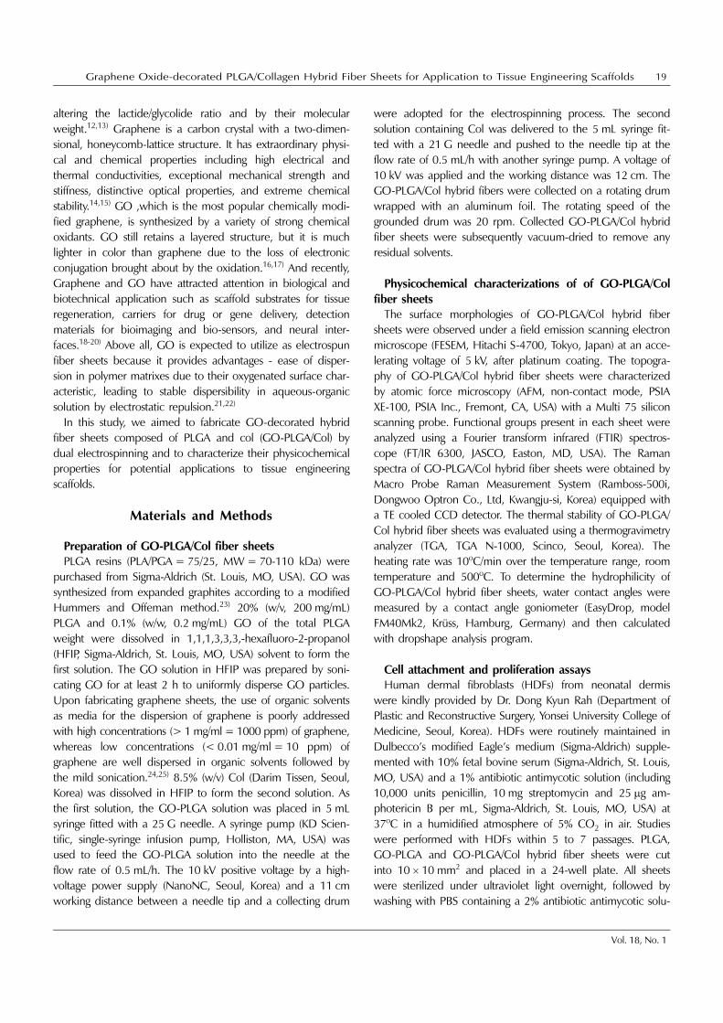
The surface morphologic imaging by FESEM of pure PLGA,
GO-PLGA and GO-PLGA/Col hybrid fiber sheets are shown in
Figure 1(A). GO-PLGA and GO-PLGA/Col hybrid fiber sheets
were randomly oriented and have a three-dimensional inter-
connected pore structure which is similar to that of pure
PLGA fiber sheets. From FESEM images combined with image
analysis software, the diameter of GO-PLGA and GO-PLGA/
Col hybrid fibers was found to range between 250 nm and
890 nm (average fiber diameter: 480 ± 210 nm, 680 ± 185
nm respectively). There were no significant difference in the
morphologies according to existence of GO and Col.
Topographic imaging by AFM of pure PLGA, GO-PLGA and
GO-PLGA/Col hybrid fiber sheets are shown in Figure 1(B).
Figure 1. Surface morphological and topographical images of PLGA, GO-PLGA and GO-PLGA/Col hybrid fiber sheets: (A) FESEMimages and (B) 3D rendered images by AFM.
Graphene Oxide-decorated PLGA/Collagen Hybrid Fiber Sheets for Application to Tissue Engineering Scaffolds 21
Vol. 18, No. 1
For each sample, a scan size of 30 × 30 µm was measured to
obtain a sufficient amount of ridges to characterize the
surfaces and to maintain enough resolution to achieve a
precise test. The structures found with the AFM measurement
correlate well with the FESEM images. The average roughness
of pure PLGA, GO-PLGA, GO-PLGA/Col hybrid fiber sheets
are 0.847 µm, 0.211 µm and 0.309 µm respectively. It was
recognized that the diameter of fibers have increasing ten-
dency, the roughness of fibers is also increased.
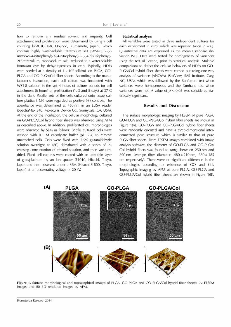
The Figure 2(A) shows the FT-IR spectra of Col, pure PLGA
fibers and GO-PLGA/Col hybrid fiber sheets. The spectra of
pure PLGA fibers showed that the C=O stretch and the C-O
stretch observed at around 1760 and 1080 cm−1, respectively.
Both bands exhibit carbonyl stretching demonstrating the pre-
sence of the ester group. The spectra of Col displayed mainly
bands at 1650, 1560, and 1230 cm−1, characteristic of the
amide I, II and III bands, respectively. The amide I absorption
is generated predominatingly by protein amide C=O stret-
ching vibrations and amide II is consisted of amide N-H ben-
ding vibrations and C-N stretching vibrations. The amide III
band is complex, consisting of components form C-N stret-
ching and N-H in plane bending from amide linkage.26) Addi-
tionally, bands at around 3550 and 2950 cm−1 were observed,
which represent the stretching of −OH and −CH3, respecti-
vely. By comparing the spectra of GO/PLGA-Col hybride fiber
sheets, it has been found that there are no additional peaks
excluding the specific bands of Col and PLGA. Raman spectra
of GO-PLGA and GO-PLGA/Col hybrid fiber sheets were
dominated by the noticeable band near 1350 cm−1 and
1600 cm−1, which was due to the D band and G band of GO
[Figure 2(B)]. The D band is activated once when the defects
are entered into sp2 hybridized carbon networks, and the
intensity of D band is proportional to the defects con-
centration.27,28) The G band is arising from the E2g vibrational
mode of sp2-carbon network. It is known fact that the
existence of strong D band at 1350□cm−1 must accompany
the existence of another defects-assisted Raman band, the D’
band, located at 1620□cm−1. The intensity of D’ band is also
proportional to the concentration of defects like as the case of
D band. And when the amount of defects was fairly in-
creased, the broadening effect of Raman bands was occurred.
Thus the broad G band of GO is usually considered as
consisting of both G and D’ bands despite the fact that it
looks like a single band due to the merge effect after
broadening.29) The specific bands of PLGA were also notified
in Raman spectrum. The noticeable band near 1768 cm−1 was
regarded as the ester group of PLGA. Also, the deformation
bands of CH2 and CH3 were observed at 1450 cm−1 and
1424 cm−1, respectively, which correspond to the anti-sym-
metric vibration of CH2 from the lactic unit of PLGA and from
the glycolic unit of PLGA.30) These results indicate that GO
was well dispersed in the surface of GO-PLGA and GO-PLGA/
Col hybrid fiber sheets. The Figure 2(C) shows the results of
TGA analysis. The main decomposition temperature was
measured as 395, 393 and 372oC for pure PLGA, for GO-
PLGA and for GO-PLGA/Col hybrid fiber sheets, respectively.
Each point of decomposition temperature at around 400oC is
signified the typical thermal degradation profile of PLGA.31)
The total mass loss of the GO-PLGA/Col was 89.97% which
was lower than 93.74% and 94.54% of pure PLGA and GO-
PLGA. This is due to the fact that the amount of PLGA was
decreased by adding the Col.32) Additionally, since the
Figure 2. Physicochemical and thermal properties of PLGA,GO-PLGA and GO-PLGA/Col fiber sheets: (A) FT-IR spectra, (B)Raman spectra and (C) TGA profiles.
22 Eun Ji Lee et al.
Biomaterials Research 2014
graphene oxide is a thermally unstable materials, there was
slight mass loss at around 100oC in case of GO-PLGA and
GO-PLGA/Col hybrid fiber sheets. The mass loss in the early
stage means the pyrolysis of labile oxygen-containing groups of
graphene oxide.33) From TGA results, we recognized that GO,
PLGA and Col were well constructed in each sheet and GO-
PLGA/Col hybrid fiber sheet was thermally stable.
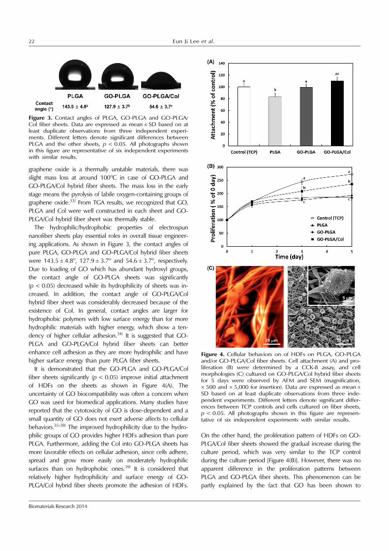
The hydrophilic/hydrophobic properties of electrospun
nanofiber sheets play essential roles in overall tissue engineer-
ing applications. As shown in Figure 3, the contact angles of
pure PLGA, GO-PLGA and GO-PLGA/Col hybrid fiber sheets
were 143.5 ± 4.8o, 127.9 ± 3.7o and 54.6 ± 3.7o, respectively.
Due to loading of GO which has abundant hydroxyl groups,
the contact angle of GO-PLGA sheets was significantly
(p < 0.05) decreased while its hydrophilicity of sheets was in-
creased. In addition, the contact angle of GO-PLGA/Col
hybrid fiber sheet was considerably decreased because of the
existence of Col. In general, contact angles are larger for
hydrophobic polymers with low surface energy than for more
hydrophilic materials with higher energy, which show a ten-
dency of higher cellular adhesion.34) It is suggested that GO-
PLGA and GO-PLGA/Col hybrid fiber sheets can better
enhance cell adhesion as they are more hydrophilic and have
higher surface energy than pure PLGA fiber sheets.
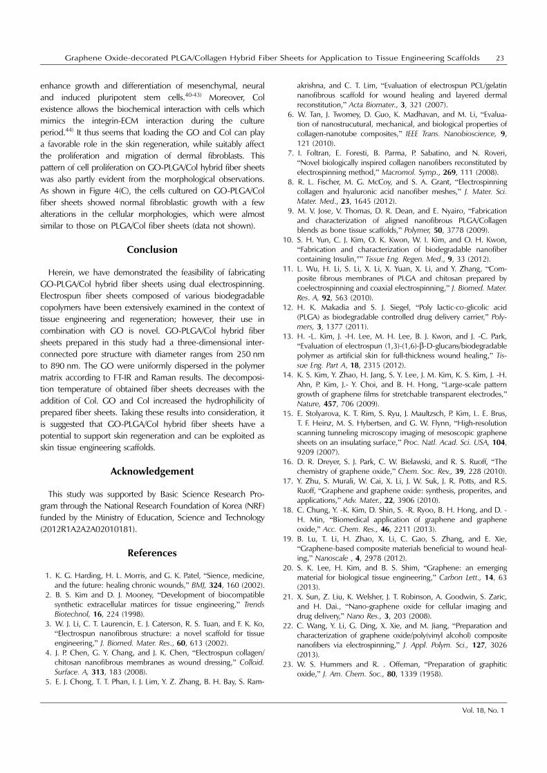
It is demonstrated that the GO-PLGA and GO-PLGA/Col
fiber sheets significantly (p < 0.05) improve initial attachment
of HDFs on the sheets as shown in Figure 4(A). The
uncertainty of GO biocompatibility was often a concern when
GO was used for biomedical applications. Many studies have
reported that the cytotoxicity of GO is dose-dependent and a
small quantity of GO does not exert adverse affects to cellular
behaviors.35-38) The improved hydrophilicity due to the hydro-
philic groups of GO provides higher HDFs adhesion than pure
PLGA. Furthermore, adding the Col into GO-PLGA sheets has
more favorable effects on cellular adhesion, since cells adhere,
spread and grow more easily on moderately hydrophilic
surfaces than on hydrophobic ones.39) It is considered that
relatively higher hydrophilicity and surface energy of GO-
PLGA/Col hybrid fiber sheets promote the adhesion of HDFs.
On the other hand, the proliferation pattern of HDFs on GO-
PLGA/Col fiber sheets showed the gradual increase during the
culture period, which was very similar to the TCP control
during the culture period [Figure 4(B)]. However, there was no
apparent difference in the proliferation patterns between
PLGA and GO-PLGA fiber sheets. This phenomenon can be
partly explained by the fact that GO has been shown to
Figure 3. Contact angles of PLGA, GO-PLGA and GO-PLGA/Col fiber sheets. Data are expressed as mean ± SD based on atleast duplicate observations from three independent experi-ments. Different letters denote significant differences betweenPLGA and the other sheets, p < 0.05. All photographs shownin this figure are representative of six independent experimentswith similar results.
Figure 4. Cellular behaviors on of HDFs on PLGA, GO-PLGAand/or GO-PLGA/Col fiber sheets. Cell attachment (A) and pro-liferation (B) were determined by a CCK-8 assay, and cellmorphologies (C) cultured on GO-PLGA/Col hybrid fiber sheetsfor 5 days were observed by AFM and SEM (magnification,× 500 and × 5,000 for insertion). Data are expressed as mean ±
SD based on at least duplicate observations from three inde-pendent experiments. Different letters denote significant differ-ences between TCP controls and cells cultured on fiber sheets,p < 0.05. All photographs shown in this figure are represen-tative of six independent experiments with similar results.
Graphene Oxide-decorated PLGA/Collagen Hybrid Fiber Sheets for Application to Tissue Engineering Scaffolds 23
Vol. 18, No. 1
enhance growth and differentiation of mesenchymal, neural
and induced pluripotent stem cells.40-43) Moreover, Col
existence allows the biochemical interaction with cells which
mimics the integrin-ECM interaction during the culture
period.44) It thus seems that loading the GO and Col can play
a favorable role in the skin regeneration, while suitably affect
the proliferation and migration of dermal fibroblasts. This
pattern of cell proliferation on GO-PLGA/Col hybrid fiber sheets
was also partly evident from the morphological observations.
As shown in Figure 4(C), the cells cultured on GO-PLGA/Col
fiber sheets showed normal fibroblastic growth with a few
alterations in the cellular morphologies, which were almost
similar to those on PLGA/Col fiber sheets (data not shown).
Conclusion
Herein, we have demonstrated the feasibility of fabricating
GO-PLGA/Col hybrid fiber sheets using dual electrospinning.
Electrospun fiber sheets composed of various biodegradable
copolymers have been extensively examined in the context of
tissue engineering and regeneration; however, their use in
combination with GO is novel. GO-PLGA/Col hybrid fiber
sheets prepared in this study had a three-dimensional inter-
connected pore structure with diameter ranges from 250 nm
to 890 nm. The GO were uniformly dispersed in the polymer
matrix according to FT-IR and Raman results. The decomposi-
tion temperature of obtained fiber sheets decreases with the
addition of Col. GO and Col increased the hydrophilicity of
prepared fiber sheets. Taking these results into consideration, it
is suggested that GO-PLGA/Col hybrid fiber sheets have a
potential to support skin regeneration and can be exploited as
skin tissue engineering scaffolds.
Acknowledgement
This study was supported by Basic Science Research Pro-
gram through the National Research Foundation of Korea (NRF)
funded by the Ministry of Education, Science and Technology
(2012R1A2A2A02010181).
References
1. K. G. Harding, H. L. Morris, and G. K. Patel, “Sience, medicine,and the future: healing chronic wounds,” BMJ, 324, 160 (2002).
2. B. S. Kim and D. J. Mooney, “Development of biocompatiblesynthetic extracellular matirces for tissue engineering,” TrendsBiotechnol, 16, 224 (1998).
3. W. J. Li, C. T. Laurencin, E. J. Caterson, R. S. Tuan, and F. K. Ko,“Electrospun nanofibrous structure: a novel scaffold for tissueengineering,” J. Biomed. Mater. Res., 60, 613 (2002).
4. J. P. Chen, G. Y. Chang, and J. K. Chen, “Electrospun collagen/chitosan nanofibrous membranes as wound dressing,” Colloid.Surface. A, 313, 183 (2008).
5. E. J. Chong, T. T. Phan, I. J. Lim, Y. Z. Zhang, B. H. Bay, S. Ram-
akrishna, and C. T. Lim, “Evaluation of electrospun PCL/gelatinnanofibrous scaffold for wound healing and layered dermalreconstitution,” Acta Biomater., 3, 321 (2007).
6. W. Tan, J. Twomey, D. Guo, K. Madhavan, and M. Li, “Evalua-tion of nanostrucutural, mechanical, and biological properties ofcollagen-nanotube composites,” IEEE Trans. Nanobioscience, 9,121 (2010).
7. I. Foltran, E. Foresti, B. Parma, P. Sabatino, and N. Roveri,“Novel biologically inspired collagen nanofibers reconstituted byelectrospinning method,” Macromol. Symp., 269, 111 (2008).
8. R. L. Fischer, M. G. McCoy, and S. A. Grant, “Electrospinningcollagen and hyaluronic acid nanofiber meshes,” J. Mater. Sci.Mater. Med., 23, 1645 (2012).
9. M. V. Jose, V. Thomas, D. R. Dean, and E. Nyairo, “Fabricationand characterization of aligned nanofibrous PLGA/Collagenblends as bone tissue scaffolds,” Polymer, 50, 3778 (2009).
10. S. H. Yun, C. J. Kim, O. K. Kwon, W. I. Kim, and O. H. Kwon,“Fabrication and characterization of biodegradable nanofibercontaining Insulin,”” Tissue Eng. Regen. Med., 9, 33 (2012).
11. L. Wu, H. Li, S. Li, X. Li, X. Yuan, X. Li, and Y. Zhang, “Com-posite fibrous membranes of PLGA and chitosan prepared bycoelectrospinning and coaxial electrospinning,” J. Biomed. Mater.Res. A, 92, 563 (2010).
12. H. K. Makadia and S. J. Siegel, “Poly lactic-co-glicolic acid(PLGA) as biodegradable controlled drug delivery carrier,” Poly-mers, 3, 1377 (2011).
13. H. -L. Kim, J. -H. Lee, M. H. Lee, B. J. Kwon, and J. -C. Park,“Evaluation of electrospun (1,3)-(1,6)-β-D-glucans/biodegradablepolymer as artificial skin for full-thickness wound healing,” Tis-sue Eng. Part A, 18, 2315 (2012).
14. K. S. Kim, Y. Zhao, H. Jang, S. Y. Lee, J. M. Kim, K. S. Kim, J. -H.Ahn, P. Kim, J.- Y. Choi, and B. H. Hong, “Large-scale patterngrowth of graphene films for stretchable transparent electrodes,”Nature, 457, 706 (2009).
15. E. Stolyarova, K. T. Rim, S. Ryu, J. Maultzsch, P. Kim, L. E. Brus,T. F. Heinz, M. S. Hybertsen, and G. W. Flynn, “High-resolutionscanning tunneling microscopy imaging of mesoscopic graphenesheets on an insulating surface,” Proc. Natl. Acad. Sci. USA, 104,9209 (2007).
16. D. R. Dreyer, S. J. Park, C. W. Bielawski, and R. S. Ruoff, “Thechemistry of graphene oxide,” Chem. Soc. Rev., 39, 228 (2010).
17. Y. Zhu, S. Murali, W. Cai, X. Li, J. W. Suk, J. R. Potts, and R.S.Ruoff, “Graphene and graphene oxide: synthesis, properites, andapplications,” Adv. Mater., 22, 3906 (2010).
18. C. Chung, Y. -K. Kim, D. Shin, S. -R. Ryoo, B. H. Hong, and D. -H. Min, “Biomedical application of graphene and grapheneoxide,” Acc. Chem. Res., 46, 2211 (2013).
19. B. Lu, T. Li, H. Zhao, X. Li, C. Gao, S. Zhang, and E. Xie,“Graphene-based composite materials beneficial to wound heal-ing,” Nanoscale , 4, 2978 (2012).
20. S. K. Lee, H. Kim, and B. S. Shim, “Graphene: an emergingmaterial for biological tissue engineering,” Carbon Lett., 14, 63(2013).
21. X. Sun, Z. Liu, K. Welsher, J. T. Robinson, A. Goodwin, S. Zaric,and H. Dai., “Nano-graphene oxide for cellular imaging anddrug delivery,” Nano Res., 3, 203 (2008).
22. C. Wang, Y. Li, G. Ding, X. Xie, and M. Jiang, “Preparation andcharacterization of graphene oxide/poly(vinyl alcohol) compositenanofibers via electrospinning,” J. Appl. Polym. Sci., 127, 3026(2013).
23. W. S. Hummers and R. . Offeman, “Preparation of graphiticoxide,” J. Am. Chem. Soc., 80, 1339 (1958).
24 Eun Ji Lee et al.
Biomaterials Research 2014
24. P. Blake, P. D. Brimicombe, R. R. Nair, T. J. Booth, D. Jiang, F.Schedin, L. A. Ponomarenko, S. V. Morozov, H. F. Gleeson, E. W.Hill, A. K. Geim, and K. S. Nvoselov, “Graphene-based liquidcrystal device,” Nano Lett., 8, 1704 (2008).
25. Y. Hernandez, V. Nicolosi, M. Lotya, F. M. Blighe, Z. Sun, S. De,I. T. McGovern, B. Holland, M. Byrne, Y. K. Gun’Ko, J. J. Boland,P. Niraj, G. Duesberg, S. Krishnamurthy, R. Goodhue, J. Hutchi-son, V. Scardaci, A. C. Ferrari, and J. N. Coleman, “High-yieldproduction of graphene by liquid-phase exfoliation of graphite,”Nat. Nanotechnol, 3, 563 (2008).
26. A. Sionkowska, M. Wisniewski, J. Skopinska, C. J. Kennedy, andT. J. Wess, “Molecular interactions in collagen and chitosanblends,” Biomaterials, 25, 795 (2004).
27. K. N. Kudin, B. Ozbas, H. C. Schniepp, R. K. Prud’homme, I. A.Aksay, and R. Car., “Raman spectra of graphite oxide and func-tionalized graphene sheets,” Nano Lett., 8, 36 (2008).
28. L. G. Cancado, A. Jorio, E. H. M. Ferreira, F. Stavale, C. A.Achete, R. B. Capaz, M. V. O. Moutinho, A. Lombardo, T. S. Kul-mala, and A. C. Ferrari, “Quantifying defects in graphene viaRaman spectroscopy at different excitation energies,” Nano Lett.,11, 3190 (2011).
29. C. Thomsen and S. Reich, “Graphite oxide under high pressure:a Raman spectroscopic study,” Phys. Rev. Lett., 85, 5214 (2000).
30. E. Vey, C. Rodger, J. Booth, M. Claybourn, A. F. Miller, and A.Saiani., “Degradation kinetics of poly(lactic-co-glycolic) acid blockcopolymer cast films in phosphate buffer solution as revealed byinfrared and Raman spectroscopies,” Polym. Degrad. Stabil., 96,1882 (2011).
31. A. D. Li, Z. Z. Sun, M. Zhou, X. X. Xu, J. Y. Ma, W. Zheng, H. M.Zhou, L. Li, and Y. F. Zheng, “Electrospun chitosan-graft-PLGAnanofibers with significantly enhanced hydrophilicity andimproved mechanical property,” Colloids Surf. B Biointerfaces,102, 674 (2013).
32. Q. Wei, J. Lu, H. Ai, and B. Jiang, “Novel method for the fabri-cation of multiscale structure collagen/hydroxyapatite-micro-sphere composties based on CaCO
3 micoparticle templates,”
Mater. Lett., 80, 91 (2012).33. M. Fang, K. Wang, H. Lu, Y. Yang, and S. Nutt., “Covalent poly-
mer functionalization of graphene nanosheets and mechanicalproperties of composites,” J. Mater. Chem., 19, 7098 (2009).
34. N. J. Hallab, K. J. Bundy, K. O’Connor, R. L. Moses, and J. J.
Jacobs, “Evaluation of metallic and polymeric biomaterial sur-face energy and surface roughness characteristics for directedcell adhesion,” Tissue Eng., 7, 55 (2011).
35. K. Wang, J. Ruan, H. Song, J. Zhang, Y. Wo, S. Guo, and D.Cui., “Biocompatibility of graphene oxide,” Nanoscale Res. Lett.,6, 1 (2011).
36. P. P. Zuo, H. F. Feng, Z. Z. Xu, L. F. Zhang, Y. L. Zhang, W. Xia,and W. Q. Zhang, “Fabrication of biocompatible and mechani-cally reinforced graphene oxide-chitosan nanocomposite films,”Chem. Cent. J., 7, 39 (2013).
37. K. H. Liao, Y. S. Lin, C. W. Macosko, and C. L Haynes, “Cytotox-icity of graphene oxide and graphene in human erythrocytes andskin fibroblasts,” ACS Appl. Mater. Interfaces, 3, 2607 (2011).
38. A. Magrez, S. Kasas, V. Salicio, N. Pasquier, J. W. Seo, M. Celio,S. Catsicas, B. Schwaller, and L. Forro, “Cellualar toxicity of car-bon-based nanomaterials,” Nano Lett., 6, 1121 (2006).
39. Y. Arima and H. Iwata, “Effect of wettability and surface func-tional groups on protein adsorption and cell adhesion using well-defined mixed self-assembled monolayers,” Biomaterials, 28,3074 (2007).
40. T. R. Nayak, H. Andersen, V. S. Makam, C. Khaw, S. Bae, X. Xu, P.-L. R. Ee, J. -H. Ahn, B. H. Hong, G. Pastorin, and B. Ozyilmaz,“Graphene for controlled and accelerated osteogenic differentia-tion of human mesenchymal stem cells,” ACS Nano, 5, 4670(2011).
41. W. C. Lee, C. H. Y. X. Lim, H. Shi, L. A. L. Tang, Y. Wang, C. T.Lim, and K. P. Loh, “Origin of enhanced stem cell growth anddifferentiation on graphene and graphene oxide,” ACS Nano, 5,7334 (2011).
42. S. Y. Park, J. S. Park, S. H. Sim, M. G. Sung, K. S. Kim, B. H.Hong, and S. H. Hong, “Enhanced differentiation of humanneural stem cells into neurons on grapheme,” Adv. Mater., 23,H263 (2011).
43. G. Y. Chen, D. W.P . Pang, S. M. Hwang, H. Y. Tuan, and Y. C.Hu, “A graphene-based platform for induced pluripotent stemcells culture and differentiation,” Biomaterials, 33, 418 (2012).
44. E. Schnell, K. Kinkhammer, S. Balzer, G. Brook, D. Klee, P. Dalton,and J. Mey, “Guidance of glial cell migration and axonal growth onelectrospun nanofibers of poly-ε-caprolactone and a collagen/poly-ε -caprolactone blend,” Biomaterials, 28, 3012 (2007).