graphic recordings of mandibular movements
DESCRIPTION
best guide for graphic recordings of mandibular movementsTRANSCRIPT
7/21/2019 Graphic Recordings of Mandibular Movements
http://slidepdf.com/reader/full/graphic-recordings-of-mandibular-movements 1/12
Graphic recordings of mandibular movements:
Research criteria
Joseph A. Clayton, D.D.S., M.S.,* W. E. Kotowicz, D.D.S., M.S.,** and
George E. Myers, F.D.S., D.D.S., M.S.***
The University of Michigan, School
of
Dentistry, Ann Arbor, Mich.
I nvestigations which have used graphic tracing devices to study mandibular move-
ments have yielded conflicting results. l-12 However, some of the inconsistencies in the
previously reported results may have been due to mechanical errors in the use of
the recording apparatus.
The purpose of this study was to determine whether or not graphic tracings of
mandibular movements could be affected by: 1) changes in the occlusal vertical
d:Lmension, 2) changes in the central bearing guidance surface, and 3) tooth
guidance. A series of pantographic surveys were made from three patients and
compared in order to analyze the ef fect that each variable had on the graphic trac-
ings.
VERTICAL DIMENSION CHANGES AND GRAPHIC TRACINGS
Method
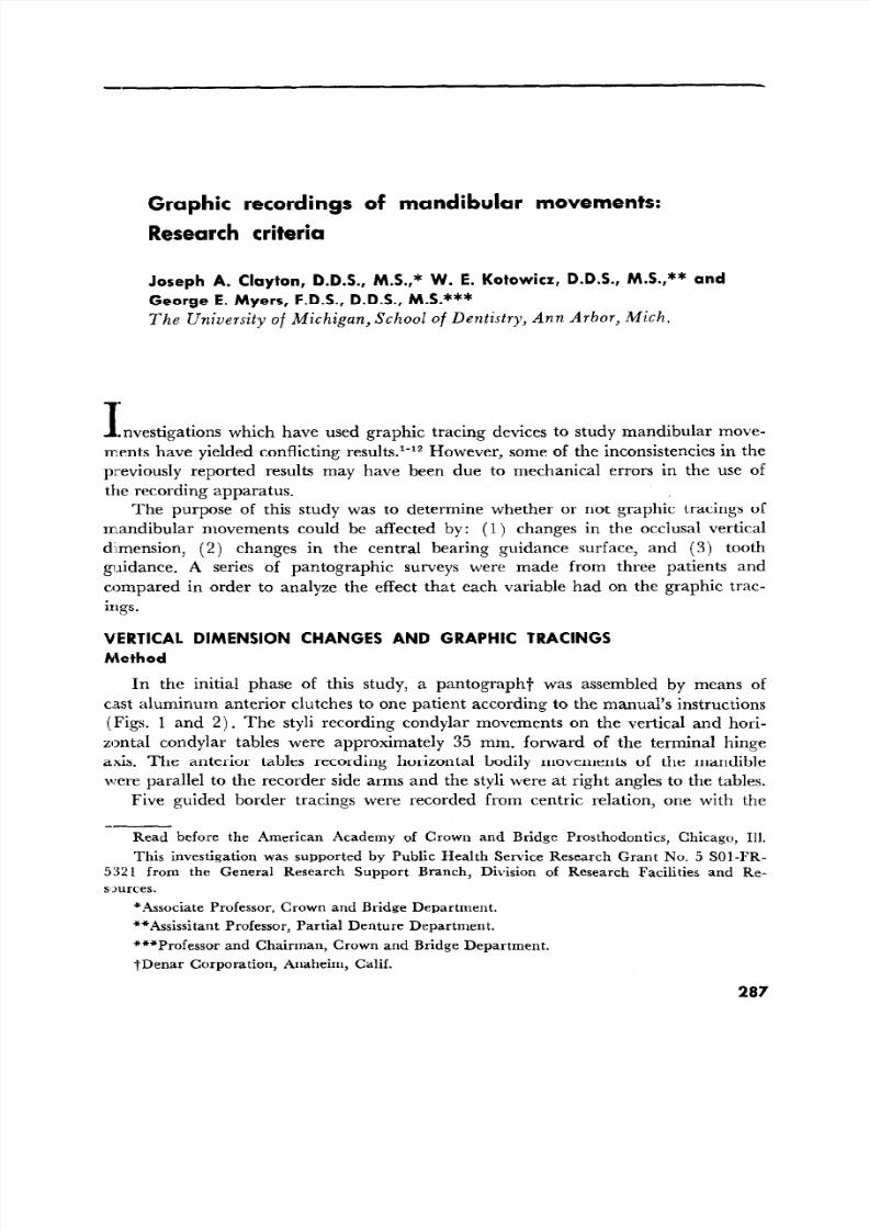
In the initial phase of this study, a pantograph? was assembled by means of
c.ast aluminum anterior clutches to one patient according to the manual’s instructions
Figs. 1 and 2). The styl i recording condylar movements on the vertical and hori-
zontal condylar tables were approximately 35 mm. forward of the terminal hinge
axis. The anterior tables recording horizontal bodily movements of the mandible
were parallel to the recorder side arms and the styl i were at right angles to the tables.
Five guided border tracings were recorded from centric relation, one with the
Read before the American Academy of Crown and Bridge Prostho dontics, Chicago, Ill.
Th is investigation was supported by Pub lic Health Service Research Grant No. 5 SOl-FR-
5321 from the General Research Support Branch, Division of Research Fac ilities and Re-
smrces.
*Ass ociate Professor, Crown and Bridge Department.
**As sissit ant Professor, Partial Denture Department.
***Professor and Chairman, Crown and Bridge Department.
tDenar Corporation, Anaheim, Calif.
287
7/21/2019 Graphic Recordings of Mandibular Movements
http://slidepdf.com/reader/full/graphic-recordings-of-mandibular-movements 2/12
Fig.
280 Clayton, Kotowicz, and Myers
.l. Prosth. Dent.
Mach, 1971
F ig. 2
Fig. 1. Cast aluminum clutch es attached to the anterior teeth with acylic resin,
Fig. ‘2. A pantograph assem bled and attached to the anterior clutche s. Copper tubing was
used for scriber and table arm extensions.
IL J\ ///
v v L
hd- >
a
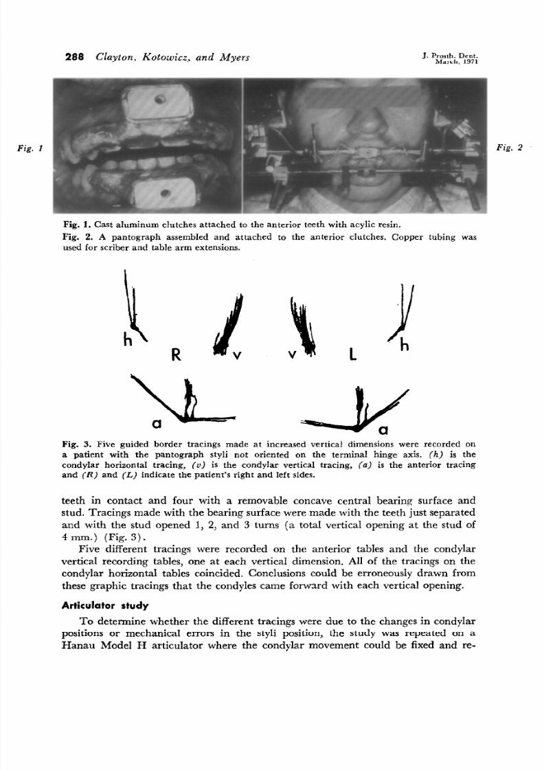
Fig. 3. Five guided border tracings made at increased vertical dimen sions were recorded on
a patient with the pantograph styli not oriented on the terminal hinge axis. (h) is the
condylar horizontal tracing, (v) is the condylar vertical tracing, (a) is the anterior tracing
and
R)
and
L)
indicate the patient’s right and left sides .
teeth in contact and four with a removable concave central bearing surface and
stud. Tracings made with the bearing surface were made with the teeth just separated
and with the stud opened 1, 2, and 3 turns a total vertical opening at the stud of
4 mm.) Fig. 3).
Five different tracings were recorded on the anterior tables and the condylar
vertical recording tables, one at each vertical dimension. All of the tracings on the
condylar horizontal tables coincided, Conclusions could be erroneously drawn from
these graphic tracings that the condyles came forward with each vertical opening.
Articulator study
To determine whether the different tracings were due to the changes n condyIar
positions or mechanical errors in the styli position, the study was repeated on a
Hanau Model H articulator where the condylar movement could be fixed and re-
7/21/2019 Graphic Recordings of Mandibular Movements
http://slidepdf.com/reader/full/graphic-recordings-of-mandibular-movements 3/12
Graphic recordings of
mandibular movements
209
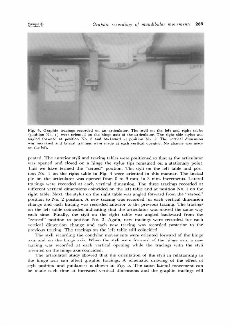
Fig. 4.
Graphic tracings recorded on an articulator, The styli on the left and right tables
(position No. I) were oriented on the hinge axis of the articulator. The right side stylus was
angled forward at position No. 2 and backward at position No. 3. The vertical dimens ion
was increased and lateral tracings were made at each vertical opening. No change was made
on the left.
peated. The anterior sty li and tracing tables were positioned so that as the articulator
was opened and closed on a hinge the stylus tips remained on a stationary point.
This we have termed the “zeroed” position. The styli on the left table and posi-
tion No. 1 on the right table in Fig. 4 were oriented in this manner. The incisal
pin on the articulator was opened from 0 to 9 mm. in 3 mm. increments. Lateral
tracings were recorded at each vertical dimension. The three tracings recorded at
different vertical dimensions coincided on the lef t table and at position No. 1 on the
Cght table. Next, the stylus on the right table was angled forward from the “zeroed”
lrosition to No. 2 position. A new tracing was recorded for each vertical dimension
change and each tracing was recorded anterior to the previous tracing. The tracings
on the lef t table coincided indicating that the articulator was moved the same way
each time. Finally, the sty li on the right table was angled backward from the
“zeroed” position to position No. 3. Again, new tracings were recorded for each
vertical dimension change and each new tracing was recorded posterior to the
previous tracing. The tracings on the lef t table still coincided.
The sty li recording the condylar movements were oriented forward of the hinge
axis and on the hinge axis. When the sty li were forward of the hinge axis, a new
-u-acing was recorded at each vertical opening while the tracings with the styli
CDrieuted on the hinge axis coincide d.
The articulator study showed that the orientation of the sty li in relationship to
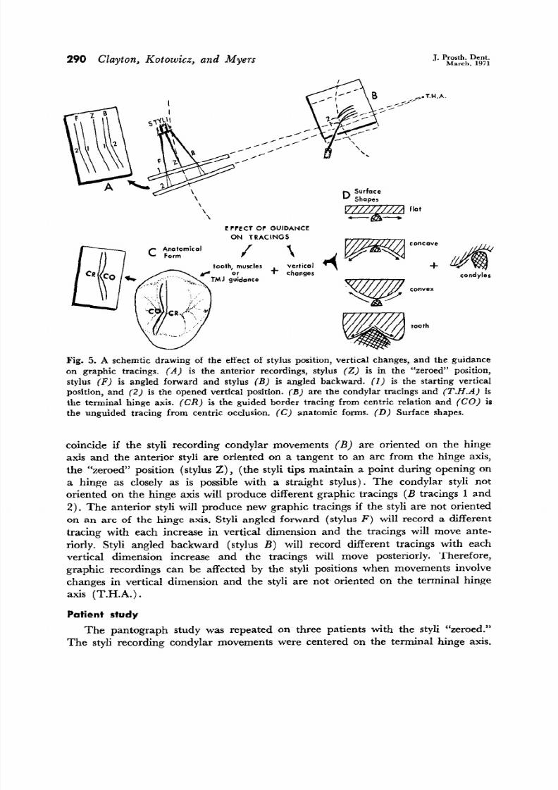
the hinge axis can af fect graphic tracings. A schematic drawing of the ef fect of
:jtyli position and guidances is shown in Fig. 5. The same lateral movement can
be made each time at increased vertical dimensions and the graphic tracings will
7/21/2019 Graphic Recordings of Mandibular Movements
http://slidepdf.com/reader/full/graphic-recordings-of-mandibular-movements 4/12
290 Clayton, Kotowicz, and Myers
.I. Prosth. Dent.
March, 1971
EFFEC T OF GUIDANCE
ON TRACINGS
vertical
changes
tooth
condyler
Fig. 5. A schemtic drawing of the ef fect of stylus position, vertical changes, and the guidance
on graphic tracings. A) is the anterior recordings, stylus 2) is in the “zeroed” position,
stylus F) is angled forward and stylus B) is angled backward. I) is the starting vertical
position, and 2) is the opened vertical position. B) are the condylar tracings and T.H.A) is
the terminal hinge axis. CR) is the guided border tracing from centric relation and CO) is
the unguided tracing from centric occlusion. C) anatomic forms. D) Surface shapes.
coincide if the styli recording condylar movements B) are oriented on the hinge
axis and the anterior styli are oriented on a tangent to an arc from the hinge axis,
the “zeroed” position stylus 2) , the styli tips maintain a point during opening on
a hinge as closely as is possible with a straight stylus). The condylar styli not
oriented on the hinge axis will produce different graphic tracings B tracings 1 and
2). The anterior styli will produce new graphic tracings if the styli are not oriented
on an arc of the hinge axis. Styli angled forward stylus F) will record a different
tracing with each increase in vertical dimension and the tracings will move ante-
riorly. Styli angled backward stylus B) will record different tracings with each
vertical dimension increase and the tracings will move posteriorly. Therefore,
graphic recordings can be affected by the styli positions when movements involve
changes n vertical dimension and the styli are not oriented on the terminal hinge
axis T.H.A.) .
Patient study
The pantograph study was repeated on three patients with the styli “zeroed.”
The styli recording condylar movements were centered on the terminal hinge axis.
7/21/2019 Graphic Recordings of Mandibular Movements
http://slidepdf.com/reader/full/graphic-recordings-of-mandibular-movements 5/12
Volume 2.i
Number 3
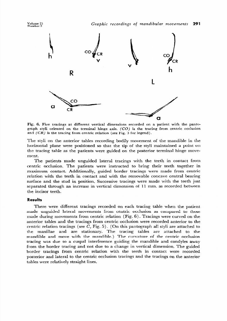
Graphic recordings of mandibular movements 291
G
h
R
\ cjR
V
h
l.
Fig:. 6. Five tracings at different vertical dimen sions recorded on a patient with the panta-
graph styli oriented on the terminal hinge axis. (CO) is the tracing from ce ntric occlu sion
and CR) is the tracing from centric relation (see Fig. 3 for legend).
The styli on the anterior tables recording bodily movement of the mandible in the
horizontal plane were positioned so that the tip of the styli maintained a point on
the tracing table as the patients were guided on the posterior terminal hinge move-
ment.
The patients made unguided lateral tracings with the teeth in contact from
centric occlusion. The patients were instructed to bring their teeth together in
m.a,ximum contact. Additionally, guided border tracings were made from centric
relation with the teeth in contact and with the removable concave central bearing
surface and the stud in position. Successive racings were made with the teeth just
separated through an increase in vertical dimension of 11 mm. as recorded between
the incisor teeth.
There were different tracings recorded on each tracing table when the patient
made unguided lateral movements from centric occlusion as compared to those
made during movements from centric relation Fig. 6). Tracings were curved on the
anterior tables and the tracings from centric occlusion were recorded anterior to the
centric relation tracings see C, Fig. 5). On this pantograph all styli are attached to
the maxillae and are stationary. The tracing tables are attached to the
mandible and move with the mandible.) The curvature of the centric occlusion
tracing was due to a cuspal interference guiding the mandible and condyles away
from the border tracing and not due to a change in vertical dimension. The guided
border tracings from centric relation with the teeth in contact were recorded
posterior and lateral to the centric occlusion tracings and the tracings on the anterior
tables were relatively straight lines.
7/21/2019 Graphic Recordings of Mandibular Movements
http://slidepdf.com/reader/full/graphic-recordings-of-mandibular-movements 6/12
292 Clayton, Kotowicz, and Myers
J. Prosth. Dent.
March, 1971
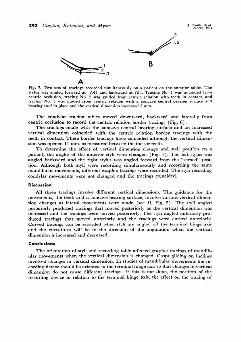
A
Fig. 7. Two sets of tracings recorded simultaneously on a patient on the anterior tables. The
stylus was angled forward at A) and backward at B). Tracing No. 1 was unguided from
centric occlusion, tracing No. 2 was guided from centric relation with teeth in contact, and
tracing No. 3 was guided from centric relation with a concave central bearing surface and
bearing stud in place and the vertical dimension increased 2 mm,
The condylar tracing tables moved downward, backward and laterally from
centric occlusion to record the centric relation border tracings Fig. 6).
The tracings made with the concave central bearing surface and an increased
vertical dimension coincided with the centric relation border tracings with the
teeth in contact. These border tracings have coincided although the vertical dimen-
sion was opened 11 mm. as measuredbetween the incisor teeth.
To determine the effect of vertical dimension change and styli position on a
patient, the angles of the anterior styli were changed Fig. 7). The left stylus was
angled backward and the right stylus was angled forward from the “zeroed” posi-
tion. Although both styli were recording simultaneously and recording the same
mandibular movements, different graphic tracings were recorded. The styli recording
condylar movements were not changed and the tracings coincided.
Discussion
All three tracings involve different vertical dimensions. The guidance for the
movements, the teeth and a concave bearing surface, involve various vertical dimen-
sion changes as lateral movements were made see D, Fig. 5). The styli angled
posteriorly produced tracings that moved posteriorly as the vertical dimension was
increased and the tracings were curved posteriorly. The styli angled anteriorly pro-
duced tracings that moved anteriorly and the tracings were curved anteriorly.
Curved tracings can be recorded when sty li are angled off the terminal hinge axis
and the curvatures will be in the direction of the angulation when the vertical
dimension s increasedand decreased.
Conclusions
The orientation of styli and recording table affected graphic tracings of mandib-
ular movements when the vertical dimension is changed. Cusps gliding on inclines
involved changes in vertical dimension. In studies of mandibular movements the re-
cording device should be oriented to the terminal hinge axis so that’ changes n vertical
dimension do not cause different tracings. If this is not done, the position of the
recording device in relation to the terminal hinge axis, the effect on the tracing o.f
7/21/2019 Graphic Recordings of Mandibular Movements
http://slidepdf.com/reader/full/graphic-recordings-of-mandibular-movements 7/12
G
ru LIL t c I tugs of tt~andibular movetnents 293
/ ‘. .e ,o .d’
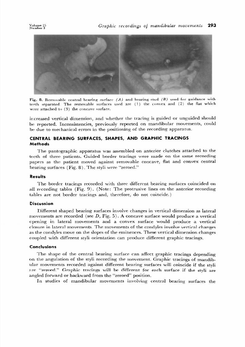
Fig. 8. Removable central bearing surfaw (A) and bearing stud Bj used for guidance 4th
te,:th separa ted. Th e removable surfa ces usrd arr (I) thr wnwx and (2)
the flat which
w’zre attached to (3) the concave surface.
ircreased vertical dimension, and whether the tracing is guided or unguided should
be reported. ‘Inconsistencies, previously reported on mandibular movements, could
be due to mechanical errors in the positioning of the recording apparatus.
CENTRAL BEARING SURFACES, SHAPES, AND GRAPHIC TRACINGS
Methods
The pantographic apparatus was assembled on anterior clutches attached to the
teeth of three patients. Guided border tracings were madt= on the same recording
papers as the patient moved against removable concave, fla t and convex central
bearing surfaces Fig. 8‘) _ The styli were “zeroed.”
Results
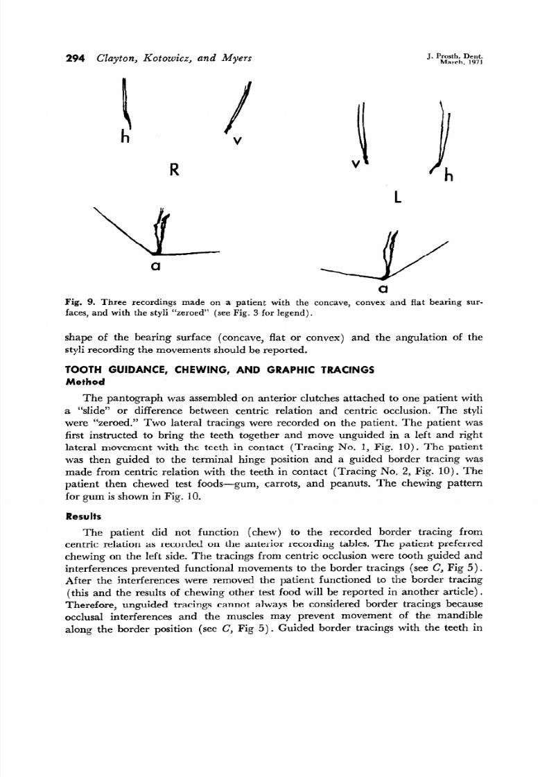
The border tracings recorded with three different bearing surfaces coincided on
all recording tables Fig. 9). Note: The protrusive lines on the anterior recording
tables are not border tracings and, therefore, do not c.oincide.1
Discussion
Different shaped bearing surfaces involve changes in vertical dimension as lateral
movements are recorded see
D,
Fig. 5). A concave surface would produce a vertical
c’pening in lateral movements and a convex surface would produce a vertical
closure in lateral movements. The movements of the condyles involve vertical changes
as the condyles move on the slopes of the eminences. These vertical dimension changes
coupled with different sty li orientation can produce different graphic tracings.
Conclusions
The shape of the central bearing surface can af fect graphic
tracings pending
on the anguIation of the sty li recording the movement. Graphic tracings of mandib-
ular movements recorded against different bearing surfaces will coincide if the sty li
a.re “zeroed.” Graphic tracings will be different for each surface if the sty li are
angled forward or backward from the “zeroed” position.
In studies of mandibular movements involving central bearing surfaces the
7/21/2019 Graphic Recordings of Mandibular Movements
http://slidepdf.com/reader/full/graphic-recordings-of-mandibular-movements 8/12
294 Clayton, Kotowicz, and Myers
J. Prosth. Dent.
March, 1971
L
a
Fig. 9. Three recordings made on a patient with the concave, convex and fla t bearing sur-
faces, and with the sty li “zeroed” see Fig. 3 for
legend).
shape of the bearing surface concave, fla t or convex) and the angulation of the
sty li recording the movements should be reported.
TOOTH GUIDANCE, CHEWING, AND GRAPHIC TRACINGS
Method
The pantograph was assembled on anterior clutches attached to one patient with
a “slide” or difference between centric relation and centric occlusion. The sty li
were “zeroed.” Two lateral tracings were recorded on the patient. The patient was
first instructed to bring the teeth together and move unguided in a lef t and right
lateral movement with the teeth in contact Tracing No. 1, Fig. 10). The patient
was then guided to the terminal hinge position and a guided border tracing was
made from centric relation with the teeth in contact Tracing No. 2, Fig. 10). The
patient then chewed test foods-gum, carrots, and peanuts. The chewing pattern
for gum is shown in Fig. 10.
Results
The patient did not function chew) to the recorded border tracing from
centric relation as recorded on the anterior recording tables. The patient preferred
chewing on the left side. The tracings from centric occlusion were tooth guided and
interferences prevented functional movements to the border tracings see C, Fig 5).
After the interferences were removed the patient functioned to the border tracing
this and the results of chewing other test food will be reported in another article).
Therefore, unguided tracings cannot always be considered border tracings because
occlusal interferences and the muscles may prevent movement of the mandible
along the border position see C, Fig 5). Guided border tracings with the teeth in
7/21/2019 Graphic Recordings of Mandibular Movements
http://slidepdf.com/reader/full/graphic-recordings-of-mandibular-movements 9/12
Graphic
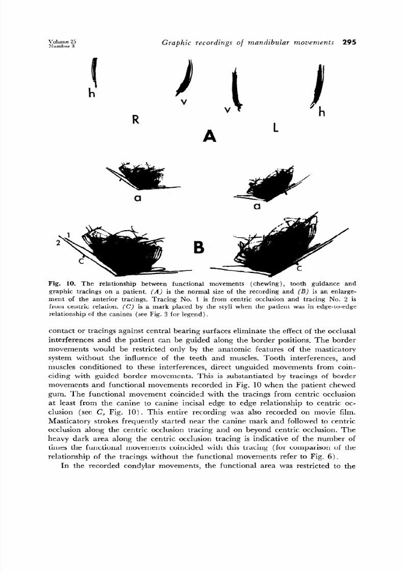
recordings of mandibular movements 295
R
a
a
Fig. 10. The relationship between func tional movements (chewing), tooth guidance and
graphic tracings on a patient. (A) is the normal size of the recording and (B) i s an enlarge-
ment of the anterior tracings. Tracing No. 1 is from cen tric occlu sion and tracing No. 2 is
from centric relation. (C) is a mark placed by the styli when the patient was in edge-to-edge
relationship of the canines (see Fig. 3 for legend).
contact or tracings against central bearing surfaces eliminate the ef fect of the occlusal
interferences and the patient can be guided along the border positions. The border
movements would be restricted only by the anatomic features of the masticatory
system without the influence of the teeth and muscles.
Tooth interferences, and
muscles conditioned to these interferences, direct unguided movements from coin-
ciding with guided border movements.
Th is is substantiated by tracings of border
movements and functional movements recorded in Fig. 10 when the patient chewed
gum. The functional movement coincided with the tracings from centric occlusion
at least from the canine to canine incisal edge to edge relationship to centric oc-
clusion see C, Fig. 10). This entire recording was also recorded on movie film.
Masticatory strokes frequently started near the canine mark and followed to centric
occlusion along the centric occlusion tracing and on beyond centric occlusion. The
heavy dark area along the centric occlusion tracing is indicative of the number of
times the functional movements coincided with this tracing for comparison of the
relationship of the tracings without the functional movements refer to Fig. 6).
In the recorded condylar movements, the functional area was restricted to the
7/21/2019 Graphic Recordings of Mandibular Movements
http://slidepdf.com/reader/full/graphic-recordings-of-mandibular-movements 10/12
296 Clayton, Kotowicz, and Myers
J. Prosth. Dent.
March, 1971
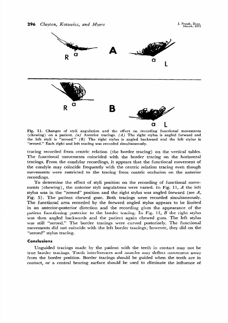
B
Fig. 11. Changes of styli angulation and the effect on recording functional movements
(chewing) on a patient. (a) Anterior tracings. A) The right stylus is angled forward and
the le ft s tyli is “zeroed.”
B)
The right stylus is angled backward and the left stylus is
“zeroed.” Each right and left tracing was recorded simultaneou sly.
tracing recorded from centric relation the border tracing) on the vertical tables.
The functional movements coincided with the border tracing on the horizontal
tracings. From the condylar recordings, it appears that the functional movement of
the condyle may coincide frequently with the centric relation tracing even though
movements were restricted to the tracing from centric occlusion on the anterior
recordings.
To determine the effect of styli position on the recording of functional move-
ments chewing), the anterior styli angulations were varied. In Fig. 11, A the left
stylus was in the “zeroed” position and the right stylus was angled forward seeA,
Fig. 5). The patient chewed gum. Both tracings were recorded simultaneously.
The functional area recorded by the forward angled stylus appears to be limited
in an anterior-posterior direction and the recording gives the appearance of the
patient functioning posterior to the border tracing. In Fig. 11, B the right stylus
was then angled backwards and the patient again chewed gum. The left stylus
was still “zeroed.” The border tracings were curved posteriorly. The functional
movements did not coincide with the left border tracings; however, they did on the
“zeroed” stylus tracing.
Conclusions
Unguided tracings made by the patient with the teeth in contact may not be
true border tracings. Tooth interferences and musclesmay deflect movement away
from the border position. Border tracings should be guided when the teeth are in
contact, or a central bearing surface should be used to eliminate the influence of
7/21/2019 Graphic Recordings of Mandibular Movements
http://slidepdf.com/reader/full/graphic-recordings-of-mandibular-movements 11/12
7/21/2019 Graphic Recordings of Mandibular Movements
http://slidepdf.com/reader/full/graphic-recordings-of-mandibular-movements 12/12
298 Clayton,
Kotowicz, and Myers
J. Prosth. Dent.
March, 1971
The recording apparatus should be tested to determine whether the graphic
tracings represent recorded mandibular movements or whether they are erroneous
tracings produced by mechanical errors.
References
1. Cohen, R.: The relationship of anterior guidance to condylar guidance in mandibular
movement, J.
PROSTH . DE NT. 6: 758-767, 1956.
2.
Kurth, L. E.: Mandibular movements in mastication,
J. Amer. Dent. Ass . 29: 1769-1790,
1942.
3. Kotowicz, W. E.: Analysis o f pantographic tracings, Ann Arbor, University of Michigan,
School
of Dentistry, 1968 (63 p. typed thesis).
4. La Pera, F.: Understanding graphic records of mandibular movements, J. PROSTH .
DENT . 18: 417-424, 1967.
5. McCollum, B. B.: Fundamentals involved in prescribing restorative dental remedies, Dent.
Items Interest 61: 852-863, 1939.
6. McCollum, B. B., and Stuart, C. E.: A research report, South Pasadena, Calif., 1955,
Scientific Press.
7. Payne, S. H.: A study of posterior occlu sion in duplicate dentures, J. PROSTH . DENT . 1:
322-326, 1951.
8. Pos selt, U.: Studies in the mobility of the human mandible, Acta Odont. Stand. 10:
l-160 (suppl. lo), 1952.
9. Shanahan, T. E. J., and Leff, A.: Mandibular and articulator movements. Part VII.
Concepts of lateral movements and condyle paths, J. PROSTH . DENT. 14: 279-289, 1964.
10. Shanahan, T. E. J., and Leff, A.: Mandibular and articulator movements. Part V.
Vertical and sagitta l axes myths, J. PROSTH. DENT . 13: 866-872, 1963.
11. Shanahan, T. E. J., and Leff, A.: Mandibular and articulator movements. Part II. Illusion
of mandibular tracings, J. PROSTH . DE NT. 12: 82-85, 1962.
12. Schweitzer, J. M.: Masticatory function in man, J. PROSTH . DENT. 11: 625-647, 1961.
THE UNIVER SITY OF MICHIGAN
SCHOOL OF DEN TISTR Y
ANN ARBOR , MICH. 48104