greaves, j., lemonidis, k., gorleku, o. a., cruchaga, c ...eprints.gla.ac.uk/73567/1/73567.pdf ·...
TRANSCRIPT
Greaves, J., Lemonidis, K., Gorleku, O. A., Cruchaga, C., Grefen, C., and Chamberlain, L. H. (2012) Palmitoylation-induced aggregation of cysteine-string protein mutants that cause neuronal ceroid lipofuscinosis. Journal of Biological Chemistry, 287 (44). pp. 37330-37339. ISSN 0021-9258
Copyright © 2012 The American Society for Biochemistry and Molecular Biology, Inc http://eprints.gla.ac.uk/73567 Deposited on: 13 February 2013
Enlighten – Research publications by members of the University of Glasgow http://eprints.gla.ac.uk

Palmitoylation-induced Aggregation of Cysteine-stringProtein Mutants That Cause Neuronal Ceroid Lipofuscinosis*
Received for publication, June 14, 2012, and in revised form, August 9, 2012 Published, JBC Papers in Press, August 19, 2012, DOI 10.1074/jbc.M112.389098
Jennifer Greaves‡, Kimon Lemonidis‡, Oforiwa A. Gorleku‡, Carlos Cruchaga§, Christopher Grefen¶,and Luke H. Chamberlain‡1
From the ‡Strathclyde Institute of Pharmacy and Biomedical Sciences, University of Strathclyde, 161 Cathedral Street, GlasgowG4 0RE, Scotland, United Kingdom, the §Department of Psychiatry and Hope Center Program on Protein Aggregation andNeurodegeneration, Washington University, Saint Louis, Missouri 63130, and the ¶Laboratory of Plant Physiology and Biophysics,Institute of Molecular, Cell, and Systems Biology, University of Glasgow, Glasgow G12 8QQ, Scotland, United Kingdom
Background: Specific mutations in the chaperone protein CSP� cause adult-onset neuronal ceroid lipofuscinosis.Results: These mutants form SDS-resistant aggregates in a palmitoylation-dependent manner in cell lines and brain samplesfrom mutation carriers.Conclusion: Palmitoylation induces disease-causing CSP� mutants to form SDS-resistant aggregates.Significance: Formation of SDS-resistant CSP� aggregates may underlie development of adult-onset neuronal ceroidlipofuscinosis.
Recently, mutations in the DNAJC5 gene encoding cysteine-string protein � (CSP�) were identified to cause the neurode-generative disorder adult-onset neuronal ceroid lipofuscinosis.The disease-causing mutations (L115R or �L116) occur withinthe cysteine-string domain, a region of the protein that is post-translationally modified by extensive palmitoylation. Here wedemonstrate that L115R and�L116mutant proteins aremistar-geted in neuroendocrine cells and form SDS-resistant aggre-gates, concordant with the properties of other mutant proteinslinked to neurodegenerative disorders. The mutant aggregatesare membrane-associated and incorporate palmitate. Indeed,co-expression of palmitoyltransferase enzymes promoted theaggregation of the CSP� mutants, and chemical depalmitoyla-tion solubilized the aggregates, demonstrating that aggregationis induced andmaintained by palmitoylation. In agreementwiththese observations, SDS-resistant CSP� aggregates were pres-ent in brain samples from patients carrying the L115Rmutationand were depleted by chemical depalmitoylation. In summary,this study identifies a novel interplay between geneticmutationsand palmitoylation in driving aggregation of CSP� mutant pro-teins.We propose that this palmitoylation-induced aggregationof mutant CSP� proteins may underlie the development ofadult-onset neuronal ceroid lipofuscinosis in affected families.
Neuronal ceroid lipofuscinoses (NCLs)2 are a group of neu-rodegenerative disorders, with a hallmark accumulation ofautofluorescent lipid- and protein-rich ceroid (lipofuscin) in
neurons and other cell types (1). NCLs are classified as earlyinfantile, late infantile, juvenile, and adult, depending upon theage of symptomatic onset. The genes affected in different NCLshave been characterized and encode predominantly lysosomalproteins (1); for example,mutations in theCLN1 gene encodingthe lysosomal enzyme protein palmitoyl thioesterase 1 cancause infantile NCL (2); protein palmitoyl thioesterase 1 func-tions in the removal of fatty acid groups from palmitoylatedproteins during their lysosomal degradation (3).Symptoms of adult-onset NCL (ANCL) usually precipitate
before the age of 40 and lead to a significant decrease in lifeexpectancy. Unlike other NCLs, which tend to be autosomalrecessive, ANCL can be either autosomal recessive or auto-somal dominant (4). Three studies published in 2011–2012identified mutations in the DNAJC5 gene encoding cysteine-string protein � (CSP�) as the cause of autosomal dominantANCL in several unrelated families (5–7).CSP� is a ubiquitously expressed DnaJ chaperone protein
that regulates proteins involved in secretory vesicle dynamics(8–10). Knock-out of CSP� in mice leads to fulminant neuro-degeneration (11), likely by destabilizing key synaptic proteinssuch as SNAP25 (12, 13) and dynamin (14, 15). The mutationsin CSP� that cause ANCL occur within the highly conservedcysteine-string region, a heavily palmitoylated domain involvedin membrane binding and intracellular targeting. The specificmutations identified were a substitution of leucine 115 by argi-nine (L115R) or a deletion of leucine 116 (�L116) (5–7).The cysteine-string domain of CSP� plays a dual role in pro-
moting stable membrane attachment (16, 17). First, the overallhydrophobicity of this region may allow transient membraneinteraction of the nonpalmitoylated protein, allowing it to con-nect with membrane-bound Asp-His-His-Cys (DHHC) palmi-toyltransferases. Subsequent palmitoylation of the cysteine-string domain by specific DHHC proteins promotes stablemembrane attachment, facilitating trafficking to secretory ves-icles and the plasmamembrane. In addition, the cysteine-string
* This work was supported by Medical Research Council Senior FellowshipAward Grant G0601597 and Wellcome Trust Grant 094184 (to L. H. C.). Thiswork was also supported by National Institutes of Health Grant P50AG05681.Author’s Choice—Final version full access.
1 To whom correspondence should be addressed. E-mail: [email protected].
2 The abbreviations used are: NCL, neuronal ceroid lipofuscinosis; ANCL,adult-onset NCL; CSP�, cysteine-string protein �; EGFP, enhanced GFP;SUS, split-ubiquitin system.
THE JOURNAL OF BIOLOGICAL CHEMISTRY VOL. 287, NO. 44, pp. 37330 –37339, October 26, 2012Author’s Choice © 2012 by The American Society for Biochemistry and Molecular Biology, Inc. Published in the U.S.A.
37330 JOURNAL OF BIOLOGICAL CHEMISTRY VOLUME 287 • NUMBER 44 • OCTOBER 26, 2012
at Glasgow
University Library, on F
ebruary 13, 2013w
ww
.jbc.orgD
ownloaded from

domain may also be involved in homodimerization and multi-merization of CSP� (18).
The role of the cysteine-string domain inmembrane binding,palmitoylation, and multimerization of CSP� suggests that�L116 and L115R mutations may perturb any one of theseparameters. However, to date, analysis of the effects of themutations have mainly used in silico modeling. A Kyte-Doolittle algorithm revealed a decrease in hydrophobicity ofthe cysteine-string domain for the L115Rmutant and a smallernonsignificant decrease for the�L116mutant (6, 7). Additionalin silico analysis revealed that both disease-causing mutationsreduce the propensity of the cysteine-string domain to movefrom water to a phosphocholine bilayer interface, reducing themembrane affinity of CSP� (7). CSS-Palm software, which isused to identify putative palmitoylation sites, suggested a min-imal effect of the mutations on palmitoylation per se, possiblywith the modification of one cysteine compromised as a conse-quence of the L115R mutation (7). In silico analysis did notreveal a consistent effect of the mutations on protein aggrega-tion; however, this analysis highlighted the high intrinsic tend-ency to form antiparallel �-sheets species of CSP� and themutants (7). In addition to these in silico analyses, some exper-imental data were presented by Nosková et al. (6), which sug-gested that the mutants were mislocalized and exhibited a verymodest decrease in palmitoylation.Although potentially powerful, caution must be exercised
when interpreting results of these in silico analyses for the fol-lowing reasons: (i) the structure of the cysteine-string domain isnot known, significantly weakening the reliability of nonexperi-mental measurements; (ii) aggregation propensity is not simplyrelated to the amino acid sequence of CSP� but may also bedependent on the palmitoylation status of the protein and rel-ative cytosol-membrane distribution; (iii) palmitoylation ofCSP� is tightly linked with the intrinsic membrane affinity ofthe cysteine-string domain; (iv) palmitoylation prediction pro-grams do not consider the properties of the individual DHHCproteins that modify CSP�. In short, the in silico analyses per-formed to date may not adequately (if at all) define how theL115R and �L116 mutations affect the cellular properties ofCSP� and cause autosomal dominant ANCL.
EXPERIMENTAL PROCEDURES
Mammalian Plasmids and Mutagenesis—The human CSP�coding sequence lacking the initiating methionine and flankedby HindIII and BamHI restriction sites was synthesized byGeneArt (Invitrogen). Human CSP� contains an intrinsic Hin-dIII restriction site, and this was removed by introducing asilent mutation (AAG3AAA). This CSP� sequence wasinserted in-frame into the pEGFP-C2 vector. To generatemyc-tagged constructs, bovine CSP� was excised from amyc-pcDNA3.1 construct (9) using BamHI and EcoRI enzymesand replaced with the human CSP� coding sequences. HA-tagged DHHC constructs were a kind gift from Masaki Fukata(19). The sequences of all plasmid constructs were verified byDNA sequencing (DNA Sequencing Service, Dundee, UK).Antibodies—Rabbit polyclonal antibody recognizing CSP�
was purchased from Stressgen (Victoria, Canada). HSC70 anti-bodywas fromNewEnglandBiolabs (Herts, UK). GFP antibody
(JL8) was fromClontech. RabbitMyc andmouse actin antibod-ies were from Abcam. Rat HA antibody (used for immunoblot-ting) was purchased from Roche Applied Science, and mouseHA antibody (used for immunofluorescence) was from Cova-nce (Paris, France).Cell Culture and Transfection—PC12 cells were cultured in
RPMI 1640 medium with 10% horse serum and 5% fetal bovineserum. HEK293T cells were cultured in DMEM with 10% fetalbovine serum. All cells were grown in a humidified atmosphereat 37 °C and 5% (HEK293T) or 7.5% (PC12) CO2.Lipofectamine 2000 (Invitrogen) was used for all transfec-
tions at 2 �l/�g DNA. For confocal microscopy, PC12 cellswere plated on poly-D-lysine glass coverslips and transfectedwith 0.5 �g of EGFP-CSP� plasmids and analyzed �40 h later.For biochemical analysis, PC12 cells growing on poly-D-lysine-coated 24-well plates were transfected with 1 �g of plasmidDNA and analyzed �40 h after transfection. HEK293T cellswere transfected with 0.8 �g of EGFP-CSP and 1.6 �g of theindicated HA-tagged DHHC constructs and analyzed �20 hlater.Cell Fixation, Labeling, and Confocal Microscopy—Trans-
fected cells were fixed in 4% formaldehyde. For antibody stain-ing, the cells were then permeabilized in 0.25%TritonX-100 (inPBS with 0.3% BSA) and incubated successively with primaryantibody (1:50) and Alexa Fluor 546-conjugated secondaryantibody (1:400; Invitrogen). The cells were then washed inPBS, air-dried, and mounted on glass slides in Mowiol. A LeicaSP5 laser scanning confocal microscope was used to view cel-lular fluorescence. Image stacks of PC12 cells were acquired atNyquist sampling rates and deconvolved using Huygens soft-ware (Scientific Volume Imaging).SDS-PAGE—Samples were diluted in SDS-dissociation
buffer (final concentration 2% SDS, 25 mM DTT, 10% glycerol,0.01% bromphenol blue, 50 mM Tris, pH 6.8) heated to 100 °Cfor 5 min and loaded onto 12% polyacrylamide gels.Cell Fractionation—Transfected PC12 or HEK293T cells on
24-well plates were resuspended in a buffer containing 5 mM
Hepes, and 1 mM EDTA, pH 7.4, supplemented with a proteaseinhibitor mixture (Sigma) and with or without 1% TritonX-100. The cells were frozen at �80 °C, thawed, and centri-fuged at 16,000 � g for 30 min at 4 °C. Recovered supernatantand pellet fractions were made up to an equal volume in SDS-dissociation buffer and resolved by SDS-PAGE.Preparation of Human Brain Lysates—Lysates from human
postmortem brain samples were prepared by homogenization(Dounce homogenizer) in ice-cold buffer composed of 20 mM
Hepes, 250 mM sucrose, 1 mM MgCl2, 2 mM EDTA, 1% TritonX-100, and protease inhibitor mixture (Sigma), pH 7.4. Insolu-ble material was removed by centrifugation 4000 � g for 10min. Cortical brain tissue from histologically characterizednormal brain tissues authorized for ethically approved scien-tific research (Lothian Research Ethical Committee; reference2003/8/37) were gratefully provided by Robert Walker at theMedical Research Council Sudden Death Brain and TissueBank, University of Edinburgh (20). Cortical tissue fromANCLpatients carrying the L115R mutation was obtained fromWashington University School of Medicine in St. Louis ADRC.All tissue used was from anonymized patients, and ethical
Aggregation of CSP� Mutants
OCTOBER 26, 2012 • VOLUME 287 • NUMBER 44 JOURNAL OF BIOLOGICAL CHEMISTRY 37331
at Glasgow
University Library, on F
ebruary 13, 2013w
ww
.jbc.orgD
ownloaded from

approval for the work was granted by the University of Strath-clyde (reference UEC1112/46).Chemical Depalmitoylation of CSP�—Human brain lysates
(100 �g of protein) were incubated overnight at room temper-ature in 0.5 M hydroxylamine, pH 7, or 0.5 M Tris, pH 7, supple-mentedwith a protease inhibitormixture (Sigma). EGFP-CSP�exhibited proteolysis following extended incubation times inhydroxylamine, and therefore cells expressing EGFP-taggedconstructs were treated with 0.5 M Tris-HA for 2 h. Followingtreatment, the cell samples were diluted in SDS-dissociationbuffer and resolved by SDS-PAGE.[3H]Palmitic Acid Labeling Experiments—Transfected
HEK293T cells were washed in DMEM supplementedwith 10mg/ml defatted BSA and then incubated in the same mediumcontaining 0.5mCi/ml [3H]palmitic acid (PerkinElmer Life Sci-ences) for 3 h. Cells were then lysed in SDS-dissociation buffer,resolved by SDS-PAGE, and transferred to nitrocellulose.Duplicate nitrocellulose membranes with either processed forimmunoblotting or used for detection of 3H signal with the aidof a Kodak Biomax Transcreen LE intensifier (PerkinElmer LifeSciences).Split Ubiquitin System—Gateway-compatible mouse DHHC17/
DHHC3 and human CSP� cDNAs were produced by PCR andinserted using Gateway technology into pDONR207 and theninto pMetYC-Dest (bait plasmid) and pNX32-Dest (prey plas-mid) respectively. After transformation of THY.AP4 andTHY.AP5 yeast strains with bait and prey plasmids, respec-tively, the two strains were mated. Growth of diploid cells wasmonitored after dropping of 5 �l of each yeast suspension (atA600 1 or 0.1) on synthetic defined medium plates (1.7 g/literyeast nitrogen base without ammonium sulfate, 5 g/literammonium sulfate, 20 g/liter glucose, 1.5 g/liter CSM-Ade,-His,-Trp,-Leu,-Ura,-Met drop-outmix, and 20 g/liter agar) andsubsequent growth for 4 days at 30 °C (for interactions withDHHC17) or 7 days (for interactions with DHHC3). As a load-ing control and to verify matings, the same amounts weredropped onto plates with synthetic defined medium supple-mented with adenine and histidine, and growth was monitoredafter 3 days at room temperature. Yeast transformations, mat-ing, and sample preparation for Western blotting were per-formed as described in Ref. 21.Quantification and Statistical Analyses—Densitometric
quantification of immunoblots was performed using ImageJsoftware (National Institutes ofHealth). Statistical analyses wasby a one-way ANOVA using the Analyze-It plugin forMicrosoft Excel; a p value of �0.05 was taken to represent sta-tistical significance. For quantification of aggregation, the den-sity of aggregates was expressed as a ratio to monomeric formsof the protein (corresponding to the sum of the nonpalmitoy-lated and palmitoylated bands).
RESULTS
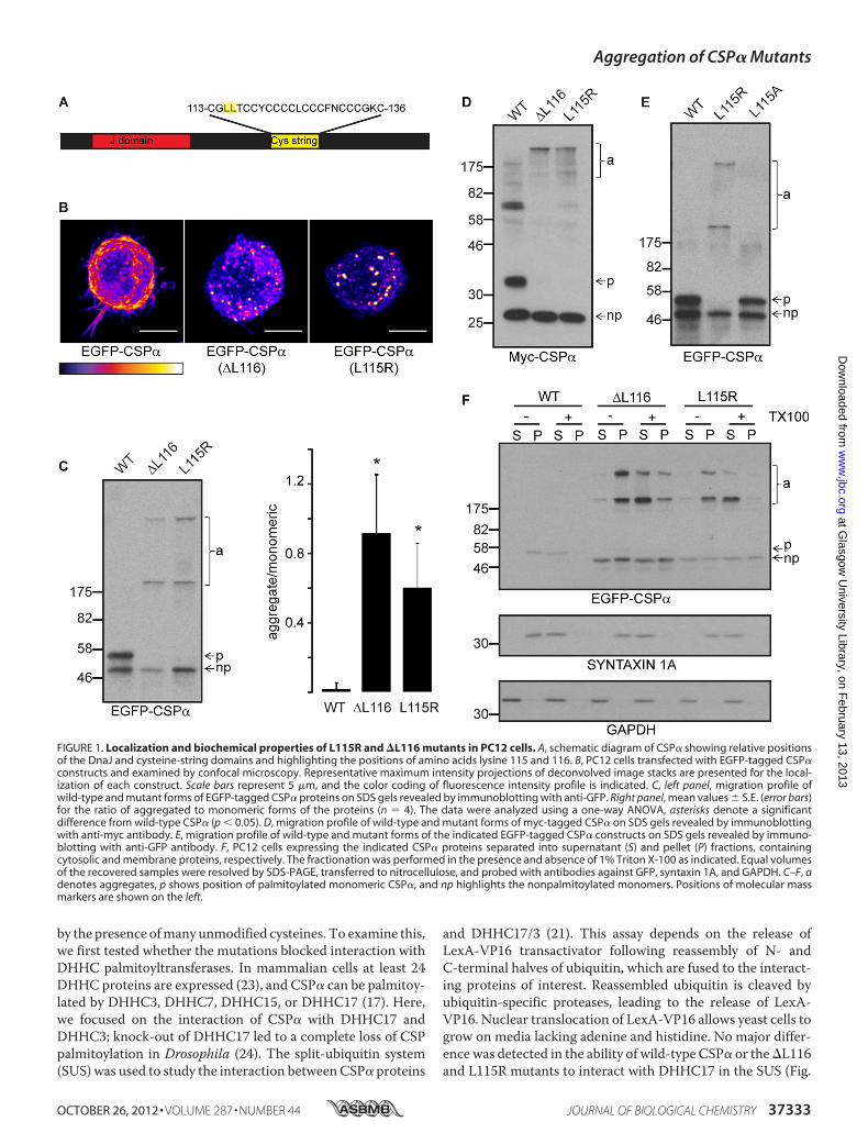
Mutant CSP� Proteins AreMistargeted and Form SDS-resist-ant Aggregates—A schematic diagram of the domain structureof CSP� is shown in Fig. 1A, highlighting the position of theresidues (leucine 115 and leucine 116) that are mutated inANCL. As a first step toward the identification of acquired bio-chemical properties of CSP� proteins containing the �L116
and L115R mutations, EGFP-tagged forms of these proteinswere expressed in neuroendocrine PC12 cells and analyzed byconfocal microscopy and SDS-PAGE/immunoblotting. Wild-type EGFP-CSP� associates with the plasma membrane andvesicles in PC12 cells (16) (Fig. 1B, left). In contrast, both the�L116 and L115R mutants displayed a more dispersed andpunctate localization and a reduced plasmamembrane staining(Fig. 1B,middle and right).
As intracellular targeting of CSP� is dependent upon multi-ple palmitoylation of the cysteine-string domain (16), we nextexamined whether this modification was perturbed for themutant proteins. The palmitoylation status of CSP� can bereadily assessed by its migration profile on SDS gels, as the fullypalmitoylated form of the protein migrates approximately 7kDa heavier than the nonpalmitoylated protein (16, 17, 22). Themigration of palmitoylated (p) and nonpalmitoylated (np)bands of wild-type EGFP-CSP� expressed in PC12 cells isshown in Fig. 1C (note that we confirm that the upper band ispalmitoylated in subsequent figures). Interestingly, for the�L116 and L115R mutants there was a clear absence of palmi-toylated monomeric forms of the proteins (Fig. 1C), and themutations induced the formation of SDS-resistant aggregates(Fig. 1C, quantified in the right panel). These aggregatesmigrated predominantly as two distinct higher molecular massbands, with the upper band remaining in the stacking gel (Fig.1C). To confirm that aggregation was not dependent on theEGFP tag, myc-tagged forms of the CSP� proteins were alsoexamined, confirming the near absence of palmitoylatedmono-meric forms of the mutant proteins and formation of highmolecular mass aggregates (Fig. 1D).In contrast to the L115R mutant, a CSP�(L115A) mutant
exhibited the same migration profile on SDS gels as wild-typeCSP� (Fig. 1E), demonstrating that the defects in the L115R and�L116mutants likely arise due to a loss of overall hydrophobic-ity rather than a specific requirement for an intact dileucinemotif.Finally, we examined whether the mutant aggregates were
cytosolic or membrane-associated. For this, transfected PC12cells were disrupted by freeze/thawing and fractionated intocytosol and membrane fractions by centrifugation. Immu-noblotting for glyceraldehyde-3-phosphate dehydrogenase(GAPDH) and syntaxin 1A confirmed the successful separationof cytosolic and membrane proteins in supernatant and pelletfractions, respectively (Fig. 1F). The �L116 and L115R aggre-gates were enriched in the membrane fraction (Fig. 1F). Toconfirm that the aggregates are trulymembrane-associated andnot pelleting due to their large size, the fractionation protocolwas also performed in the presence of Triton X-100 to solubi-lize bulk cellular membranes. Under these conditions, themajority of themutant aggregates did not pellet, supporting theidea that they are predominantly membrane-associated inPC12 cells.CSP� Mutants Display Efficient Interaction with DHHC
Palmitoyltransferases—The cysteine-string domain of CSP�,where the disease-causingmutations occur, has 14 cysteine res-idues in a span of 24 amino acids. One possibility to explain theobserved aggregation of CSP� mutants is that the mutationsprevent palmitoylation of the protein, and aggregation is caused
Aggregation of CSP� Mutants
37332 JOURNAL OF BIOLOGICAL CHEMISTRY VOLUME 287 • NUMBER 44 • OCTOBER 26, 2012
at Glasgow
University Library, on F
ebruary 13, 2013w
ww
.jbc.orgD
ownloaded from
by the presence ofmany unmodified cysteines. To examine this,we first tested whether the mutations blocked interaction withDHHC palmitoyltransferases. In mammalian cells at least 24DHHC proteins are expressed (23), and CSP� can be palmitoy-lated by DHHC3, DHHC7, DHHC15, or DHHC17 (17). Here,we focused on the interaction of CSP� with DHHC17 andDHHC3; knock-out of DHHC17 led to a complete loss of CSPpalmitoylation in Drosophila (24). The split-ubiquitin system(SUS) was used to study the interaction betweenCSP� proteins
and DHHC17/3 (21). This assay depends on the release ofLexA-VP16 transactivator following reassembly of N- andC-terminal halves of ubiquitin, which are fused to the interact-ing proteins of interest. Reassembled ubiquitin is cleaved byubiquitin-specific proteases, leading to the release of LexA-VP16. Nuclear translocation of LexA-VP16 allows yeast cells togrow on media lacking adenine and histidine. No major differ-ence was detected in the ability of wild-type CSP� or the�L116and L115R mutants to interact with DHHC17 in the SUS (Fig.
FIGURE 1. Localization and biochemical properties of L115R and �L116 mutants in PC12 cells. A, schematic diagram of CSP� showing relative positionsof the DnaJ and cysteine-string domains and highlighting the positions of amino acids lysine 115 and 116. B, PC12 cells transfected with EGFP-tagged CSP�constructs and examined by confocal microscopy. Representative maximum intensity projections of deconvolved image stacks are presented for the local-ization of each construct. Scale bars represent 5 �m, and the color coding of fluorescence intensity profile is indicated. C, left panel, migration profile ofwild-type and mutant forms of EGFP-tagged CSP� proteins on SDS gels revealed by immunoblotting with anti-GFP. Right panel, mean values � S.E. (error bars)for the ratio of aggregated to monomeric forms of the proteins (n � 4). The data were analyzed using a one-way ANOVA, asterisks denote a significantdifference from wild-type CSP� (p � 0.05). D, migration profile of wild-type and mutant forms of myc-tagged CSP� on SDS gels revealed by immunoblottingwith anti-myc antibody. E, migration profile of wild-type and mutant forms of the indicated EGFP-tagged CSP� constructs on SDS gels revealed by immuno-blotting with anti-GFP antibody. F, PC12 cells expressing the indicated CSP� proteins separated into supernatant (S) and pellet (P) fractions, containingcytosolic and membrane proteins, respectively. The fractionation was performed in the presence and absence of 1% Triton X-100 as indicated. Equal volumesof the recovered samples were resolved by SDS-PAGE, transferred to nitrocellulose, and probed with antibodies against GFP, syntaxin 1A, and GAPDH. C–F, adenotes aggregates, p shows position of palmitoylated monomeric CSP�, and np highlights the nonpalmitoylated monomers. Positions of molecular massmarkers are shown on the left.
Aggregation of CSP� Mutants
OCTOBER 26, 2012 • VOLUME 287 • NUMBER 44 JOURNAL OF BIOLOGICAL CHEMISTRY 37333
at Glasgow
University Library, on F
ebruary 13, 2013w
ww
.jbc.orgD
ownloaded from
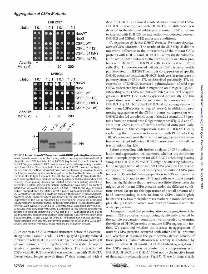
2). In contrast, a CSP� mutant truncated before the cysteine-string domain (amino acids 1–112) displayed a greatly reducedinteractionwithDHHC17under stringent conditions (with 200�M methionine), confirming the ability of the system to reportreliably on protein-protein interactions. The interaction ofCSP�withDHHC3 in the SUSwas weaker thanwithDHHC17.Nevertheless, longer growth times (7 days compared with 4
days for DHHC17) allowed a robust measurement of CSP�-DHHC3 interaction. As with DHHC17, no difference wasdetected in the ability of wild-type and mutant CSP� proteinsto interact with DHHC3; no interaction was detected betweenDHHC3 and CSP�(1–112) under any conditions.Co-expression of Active DHHC Proteins Promotes Aggrega-
tion of CSP� Mutants—The results of the SUS (Fig. 2) did notuncover a difference in the interactions of the mutant CSP�proteins with DHHC3 and DHHC17. To investigate palmitoy-lation of the CSP� mutants further, we co-expressed these pro-teins with DHHC3 in HEK293T cells. In contrast with PC12cells (Fig. 1), overexpressed wild-type CSP� is only weaklypalmitoylated in HEK293T cells, but co-expression of specificDHHC proteins (including DHHC3) leads to a large increase inpalmitoylation of CSP� (17). As described previously (17), co-expression of DHHC3 increased palmitoylation of wild-typeCSP�, as detected by a shift in migration on SDS gels (Fig. 3A).Interestingly, the CSP� mutants exhibited a low level of aggre-gation in HEK293T cells when expressed individually, and thisaggregation was markedly increased by co-expression ofDHHC3 (Fig. 3A). Note that DHHC3 did not co-aggregate withthe mutant CSP� proteins (Fig. 3A, lower). In addition to pro-moting aggregation of the CSP� mutants, co-expression withDHHC3 also led to redistribution of the�L116 and L115R pro-teins from the cytosol onto Golgi membranes (Fig. 3, B and C).Note that CSP� is not efficiently trafficked onto post-Golgimembranes in this co-expression assay in HEK293T cells,explaining the difference in localization with PC12 cells (Fig.1B). We also confirmed that the mutant aggregates were mem-brane-associated following DHHC3 co-expression by cellularfractionation (Fig. 3D).Before proceeding with further analyses of CSP� palmitoy-
lation and aggregation, we examined whether the conditionsused in sample preparation for SDS-PAGE (including heatingsamples to 100 °C in 25 mM DTT) might be affecting palmitoy-lation or aggregation of the mutant CSP� proteins. For this, wecompared the migration of wild-type and mutant CSP� pro-teins on SDS gels following preparation in SDS sample buffercontaining 1, 5, and 25 mM DTT and with or without sampleboiling. Fig. 3E shows that there was very little difference in themigration of mutant CSP� proteins under the different condi-tions tested except for the appearance of a small amount of aband corresponding in size to dimeric CSP� (band slightlybelow the 175-kDa molecular mass marker) in nonboiled sam-ples, the presence of which was more pronounced with thewild-type protein.Having confirmed that palmitoylation and aggregation of the
mutant CSP� proteins was not being significantly affected bythe sample preparation conditions, we proceeded to examinethe effects of DHHCproteins onmutant CSP� aggregation fur-ther. We examined whether the increase in aggregation ofmutant CSP� proteins occurred with other DHHC proteinsand whether it required the palmitoyltransferase activity ofthese proteins (palmitoyltransferase activity is abolished bymutation of theDHHCmotif to DHHS). Indeed, aggregation ofthe CSP� mutants was promoted by co-expression withDHHC3, DHHC7, and DHHC17 but not by the inactive formsof these palmitoyltransferases (Fig. 3F). These findings clearly
FIGURE 2. Interaction of CSP� mutants with DHHC palmitoyltransferases.Yeast diploids were created by mating cells expressing a C-terminal half ofubiquitin and PLV (protein A-LexA-VP16) tag fused to the C termini ofDHHC17 (top panel) or DHHC3 (middle panel) with cells expressing the wild-type form of the N-terminal half of ubiquitin (NubWT; positive control), amutant form of the N terminus that does not spontaneously reassemble withthe C terminus of ubiquitin (NubG; negative control), or NubG fused to the Nterminus of wild-type CSP�, or L115R, �L116, and CSP�(1–112) mutants. Dip-loids were spotted onto medium containing adenine (Ade) and histidine (His)to verify equal plating density and plated on medium lacking Ade/His todetermine protein-protein interaction; methionine was added to confirminteraction at lower expression levels. 0.1 and 1 refer to the A600 of liquidculture dropped onto the plates. Yeast diploids containing DHHC17 and allCSP� constructs displayed growth on plates without Ade/His (second panel);however, under more stringent conditions in the presence of methionine(expression of the bait is regulated by a methionine-repressible promoter)(third and fourth panels) growth of cells expressing the 1–112 mutant was lost,whereas wild-type, L115R, and �L116 constructs all supported growth. Simi-lar results were obtained with DHHC3, although in this case no growth wasdetected in diploids containing DHHC3 and the 1–112 mutant on any plateslacking Ade/His. Images for growth on plates lacking Ade/His were taken after4 days for DHHC17 and 7 days for DHHC3. The bottom panel shows an immu-noblot probed with anti-HA, confirming expression of all of the CSP� con-structs; positions of molecular mass markers are shown on the left.
Aggregation of CSP� Mutants
37334 JOURNAL OF BIOLOGICAL CHEMISTRY VOLUME 287 • NUMBER 44 • OCTOBER 26, 2012
at Glasgow
University Library, on F
ebruary 13, 2013w
ww
.jbc.orgD
ownloaded from

link palmitoylation by DHHC proteins with the induction ofaggregation of CSP� proteins carrying the L115R or �L116mutations.
CSP� Mutant Aggregates Are Disrupted by ChemicalDepalmitoylation—Only active forms ofDHHC3/7/17 are ableto promote aggregation of the CSP� �L116 and L115R
Aggregation of CSP� Mutants
OCTOBER 26, 2012 • VOLUME 287 • NUMBER 44 JOURNAL OF BIOLOGICAL CHEMISTRY 37335
at Glasgow
University Library, on F
ebruary 13, 2013w
ww
.jbc.orgD
ownloaded from

mutants, implying that palmitoylation drives aggregate forma-tion. To assess the palmitoylation status of the �L116 andL115R aggregates, HEK293T cells expressing the mutantswith or without DHHC3 co-expression were labeled with[3H]palmitic acid. Aggregated forms of the CSP� mutantsclearly incorporated radiolabel, and this was enhanced byDHHC3 co-expression (Fig. 4A, right).
To determine whether the observed palmitoylation of themutant proteins is important for the maintenance of aggrega-tion, cell lysates were treated with hydroxylamine to cleavethioester linkages between cysteine residues and palmitatechains. Hydroxylamine treatment dissolved the higher molec-ular mass forms of L115R and �L116 mutants co-expressed in
HEK293T cells with DHHC3 (Fig. 4B, left), confirming thattheir SDS-resistant aggregation requires palmitoylation. Thissame effect of hydroxylamine on mutant aggregates was alsoobserved when CSP�s were expressed in PC12 cells withoutDHHC co-expression (Fig. 4B, right).Aggregation ofMutantCSP�Proteins in PostmortemSamples
from Patients with ANCL—The results presented thus far high-light a possible role for palmitoylation-dependent aggregationof mutant CSP� in the development of ANCL. To explore fur-ther the relevance of these findings to ANCL, brain lysates wereprepared from control individuals and DNAJC5 mutation car-riers and incubated in the absence or presence of hydroxyla-mine to test whether palmitoylation-sensitive aggregates werepresent. Fig. 5 shows that high molecular mass SDS-resistantaggregates were clearly detected in samples from ANCLpatients but not in control samples. Furthermore, immunore-activity of these aggregates was greatly reduced followinghydroxylamine treatment.Co-aggregation of Mutant and Wild-type CSP� Proteins—
Because mutations in CSP� cause autosomal dominant ANCL,the �L116 and L115R mutant proteins are toxic even in thepresence of a wild-type copy of the CSP� gene. Therefore, it ispossible that the mutant proteins have the capacity to interferewith the function of wild-type CSP�.We examined this by test-ing whether mutant CSP� proteins induced co-aggregation ofwild-type CSP�. For this, HEK293T cells were transfected withmyc-tagged wild-type CSP�, DHHC3, and EGFP-tagged wild-type or mutant CSP�. Fig. 6 shows that in the presence ofEGFP-CSP� �L116 or L115Rmutants, a small fraction of myc-CSP�was recruited into SDS-resistant aggregates. Thiswas notobserved following co-expression with either EGFP or EGFP-CSP�wild-type (Fig. 6). This result clearly highlights the poten-tial of mutant CSP� proteins to interfere with the wild-typeprotein, offering a possible mechanism for the dominant effectof these disease-causing mutants in the development of ANCL.
DISCUSSION
The link between proteinmisfolding/aggregation and neuro-degeneration is well established for disorders such asAlzheimer, Parkinson, and Huntington diseases (25). Theresults presented in this study further highlight the correlationbetweenprotein aggregation andneurodegeneration,with respecttomutantCSP� andANCL. Perhaps themost intriguing observa-tion in the present study is the link between aggregation of themutant CSP� proteins and palmitoylation, highlighting theinterplay between genetic mutations and post-translationalmodification in the induction of protein aggregation. Post-
FIGURE 3. Active DHHC palmitoyltransferases promote aggregation of CSP� mutants. A, EGFP-CSP� wild-type and �L116/L115R mutants were co-transfected into HEK293T cells with HA-DHHC3 (D3) or empty vector. Lysates were resolved by SDS-PAGE, transferred to nitrocellulose, and probed withantibodies against GFP or HA. Nonpalmitoylated (np) and palmitoylated (p) CSP� are marked by arrows, and aggregated (a) CSP� is highlighted by a bracket.Positions of molecular mass standards are shown on the left. B and C, HEK293T cells were transfected with EGFP-tagged constructs (B) or EGFP-taggedconstructs together with HA-DHHC3 (C). Co-transfected cells were fixed and stained with anti-HA, followed by an anti-mouse antibody conjugated to AlexaFluor 546. Cells were viewed using a Leica SP5 confocal microscope. Scale bars represent 5 �m, and the fluorescence intensity color code is shown. D, HEK293Tcells co-expressing the indicated CSP� proteins and DHHC3 were fractionated into supernatant (S) and pellet (P) fractions, containing cytosolic and membraneproteins, respectively. The fractionation was performed in the presence and absence of 1% Triton X-100 as indicated. Equal volumes of the recovered sampleswere resolved by SDS-PAGE, transferred to nitrocellulose, and probed with antibodies against GFP, HA, and GAPDH. E, HEK293T cells co-expressing the CSP�proteins and DHHC3 were lysed in SDS-dissociation buffer containing 1, 5, or 25 mM DTT and were either heated to 100 °C (boil) or 37 °C (non-boil) for 5 minbefore running. F, cells were transfected with EGFP-CSP� and the indicated HA-DHHC proteins and analyzed as in A. The mean ratio of aggregated tomonomeric forms of each protein under the various transfection conditions was calculated and is presented together with S.E. (error bars) (n � 3). Statisticaltests were performed using a one-way ANOVA, asterisks denote a p value of �0.05 compared with proteins without DHHC co-expression.
FIGURE 4. Palmitoylation analysis of CSP� mutants. A, HEK293T cells trans-fected with the wild-type and mutant EGFP-CSP� constructs and with (�) orwithout (�) HA-DHHC3 (D3) were labeled with 0.5 mCi/ml [3H]palmitic acidfor 3 h. Cells were lysed in SDS-dissociation buffer, resolved by SDS-PAGE, andtransferred to nitrocellulose. Duplicate nitrocellulose membranes were eitherprocessed for detection of 3H signal (right) or probed by immunoblottingwith anti-GFP (left). B, HEK293T cells co-transfected with EGFP-CSP� con-structs and DHHC3 (left) or PC12 cells transfected with EGFP-CSP� constructsalone (right) were treated with hydroxylamine (�) or Tris (�) as describedunder “Experimental Procedures.” The samples were then resolved by SDS-PAGE and transferred to nitrocellulose for immunoblotting analysis usingantibodies against EGFP. Nonpalmitoylated (np) and palmitoylated (p) mono-meric CSP� are marked by arrows, and aggregated CSP� (a) is highlighted bya bracket. Positions of molecular mass standards are shown on the left.
Aggregation of CSP� Mutants
37336 JOURNAL OF BIOLOGICAL CHEMISTRY VOLUME 287 • NUMBER 44 • OCTOBER 26, 2012
at Glasgow
University Library, on F
ebruary 13, 2013w
ww
.jbc.orgD
ownloaded from

translational modifications such as phosphorylation and pro-teolytic cleavage can modulate the aggregation of cytotoxicmutant proteins in other neurodegenerative disorders (26), andindeed blocking the palmitoylation of mutant Huntingtinincreased the formation of inclusions and enhanced toxicity(27). The present study extends this link between neurodegen-eration and palmitoylation and suggests that future success incounteracting ANCL in patients carrying the disease-causingCSP� mutations might be achieved by targeting the palmitoy-lation machinery.It is intriguing that protein palmitoylation is implicated in
both infantile and adult-onset forms of NCL, albeit by differentmechanisms. A deficiency of the lysosomal thioesterase proteinpalmitoyl thioesterase 1 causes early onset NCL (2), and this
enzyme is involved in depalmitoylation of proteins during theirdegradation in lysosomes (29).We speculate that the palmitoy-lated aggregates of CSP�mutant proteinsmight be less suscep-tible to the actions of lysosomal thioesterases, leading to a grad-ual accumulation of palmitoylated CSP� peptides. Thus, thedifference in age of symptomatic onset between the infantileand adult-onset forms of ANCL may reflect a different rate ofaccumulation of nondegraded palmitoylated peptides: rapidaccumulation due to a decreased cleavage of bulk palmitoylatedproteins following protein palmitoyl thioesterase 1 deficiency(30) and a slower accumulation resulting from a decreasedsensitivity of a single protein (mutant CSP�) to lysosomalthioesterases.What could be the underlying cause of the observed aggre-
gation of L115R and�L116CSP�mutants? First, it is importantto note that wild-type CSP� has an intrinsic tendency to self-associate (18), and indeed SDS-resistant dimers of wild-typeCSP� are frequently observed in cellular samples (see Fig. 3E).This self-association is dramatically enhanced by the �L116and L115R mutations, leading to the formation of high molec-ular mass SDS-resistant aggregates that are not observed withthe wild-type protein. One possibility to explain these effects isthat the mutations promote a restructuring of the cysteine-string domain resulting in added palmitate groups being posi-tioned outside the hydrophobic core of the membrane bilayer.Palmitate chains of different mutant CSP� monomers mightthen cluster together via lipid-lipid interactions to shield theexposed palmitate chains from water. Indeed, it is possible thatthe CSP� monomers within the aggregates are not fully palmi-toylated. This is implied by the lower [3H]palmitate signal fromaggregates comparedwithmonomeric protein; for example, for
FIGURE 5. CSP� expression and aggregation in human brain. Corticallysates from control and DNAJC5 mutation carriers (three separate individualsfor each) were treated with hydroxylamine (�) or Tris (�), resolved by SDS-PAGE, and transferred to nitrocellulose for immunoblotting analysis usingantibodies against CSP�, HSC70, and actin. Nonpalmitoylated (np) and palmi-toylated (p) monomeric CSP� are marked by arrows, and aggregated CSP� (a)is highlighted by a bracket. Positions of molecular mass standards are shownon the left.
FIGURE 6. Co-aggregation of wild-type and mutant CSP�. HEK293T cellswere transfected with HA-DHHC3, myc-tagged CSP� wild-type, and EGFP,EGFP-CSP�, EGFP-CSP� (�L116), or EGFP-CSP� (L115R). Cell lysates wereresolved by SDS-PAGE and transferred to nitrocellulose for immunoblottinganalysis using antibodies against EGFP (left) and myc (right). Nonpalmitoy-lated (np) and palmitoylated (p) monomeric CSP� are marked by arrows, andaggregated CSP� (a) is highlighted by a bracket. Positions of molecular massstandards are shown on the left.
Aggregation of CSP� Mutants
OCTOBER 26, 2012 • VOLUME 287 • NUMBER 44 JOURNAL OF BIOLOGICAL CHEMISTRY 37337
at Glasgow
University Library, on F
ebruary 13, 2013w
ww
.jbc.orgD
ownloaded from

the data presented in Fig. 4A the 3H signal for the �L116 andL115R high molecular mass aggregates was 5.8- and 12.5-fold,respectively, less than that from the corresponding monomerswhen normalized to protein levels. This difference might sug-gest that the aggregates contain partially palmitoylated mutantmonomers or amixture of palmitoylated and nonpalmitoylatedmonomers. At present we remain cautious in our interpreta-tion of the different 3H signals frommonomeric and aggregatedprotein. In particular, it is not clear whether palmitate turnoveron themonomers and aggregates occurswith similar kinetics oreven whether the aggregation of mutant CSP� somehowreduces the 3H signal from associated palmitate chains. Dis-rupting the mutant CSP� aggregates into their constituentmonomers, without perturbing palmitoylation, would providea clearer indication of the palmitoylation status of individualmonomers within the aggregates; this is an area of investigationthat we are currently pursuing. Another possibility to explainthe palmitoylation-dependent aggregation is that the mutantproteins are intrinsically more prone to self-associate (in theirnonpalmitoylated state). In this case, palmitoylation-inducedmembrane binding might simply concentrate the mutant pro-teins on cellular membranes facilitating aggregate formation ina manner that does not directly involve palmitate chains.Although this model is supported by the observation that bac-terially expressed recombinant CSP� self-associates via aregion containing the cysteine-string domain (18), this does notreadily fit with our data showing that the high molecularmass mutant aggregates are disassembled by hydroxylaminetreatment.Another point of note is that complete knock-out of CSP� in
mice causes neurodegeneration (11). Thus, it is possible thatdisease occurs in humans carrying the L115R/�L116mutationsas a result of reduced levels of wild-type CSP�. However, thefinding that CSP� heterozygous knock-out mice have no overtneurodegenerative phenotype might argue against this possi-bility (11). Nevertheless, mutant forms of CSP� could exhibita toxic gain-of-function effect through interfering with thefunction or trafficking of wild-type CSP�. Indeed, it was sug-gested that there was a loss of immunofluorescence signal andsynaptic targeting of CSP� in cerebral and cerebellar cortexsamples from a L115R mutation carrier (6). We detected therecruitment of small amounts of myc-tagged wild-type CSP�protein into EGFP-tagged mutant aggregates (Fig. 6), and thusaggregates containing both mutant and wild-type CSP� in thebrains of ANCL patients are a possibility. Co-aggregation ofwild-type and mutant CSP� may perturb synaptic targeting,leading to destabilization of key synaptic proteins such as theSNARE protein SNAP25 (31). The mutant aggregates mightalso cause cellular toxicity by recruiting and sequestering otherkey cellular proteins (28).At present we are not certain why Nosková et al. (6) failed to
detect a major loss of palmitoylation of mutant EGFP-CSP�constructs in CAD5 cells. However, we note that the authorsdid not confirm the identity of immunoreactive bands thatwereproposed to represent palmitoylated and nonpalmitoylatedCSP� (for example, by using hydroxylamine treatment). Full-length immunoblots were also not presented in this study, pre-venting an assessment of protein aggregation, althoughhydrox-
ylamine treatment of brain samples from an affected individualthat lacked CSP� immunoreactivity led to the appearance of animmunoreactive band that may represent a dimeric form ofCSP� (6).In summary, the results presented further highlight the rela-
tionship between protein aggregation and neurodegeneration,while revealing a novel role for palmitoylation in driving aggre-gation of disease-causing mutants.
Acknowledgment—We thank Masaki Fukata for providing the HA-tagged DHHC constructs.
REFERENCES1. Jalanko, A., and Braulke, T. (2009) Neuronal ceroid lipofuscinoses.
Biochim. Biophys. Acta 1793, 697–7092. Vesa, J., Hellsten, E., Verkruyse, L. A., Camp, L. A., Rapola, J., Santavuori,
P., Hofmann, S. L., and Peltonen, L. (1995) Mutations in the palmitoylprotein thioesterase gene causing infantile neuronal ceroid lipofuscinosis.Nature 376, 584–587
3. Camp, L. A., and Hofmann, S. L. (1993) Purification and properties of apalmitoyl-protein thioesterase that cleaves palmitate from H-Ras. J. Biol.Chem. 268, 22566–22574
4. Shacka, J. J. (2012)Mousemodels of neuronal ceroid lipofuscinoses: usefulpre-clinical tools to delineate disease pathophysiology and validate thera-peutics. Brain Res. Bull. 88, 43–57
5. Velinov, M., Dolzhanskaya, N., Gonzalez, M., Powell, E., Konidari, I.,Hulme, W., Staropoli, J. F., Xin, W., Wen, G. Y., Barone, R., Coppel, S. H.,Sims, K., Brown, W. T., and Züchner, S. (2012) Mutations in the geneDNAJC5 cause autosomal dominant Kufs disease in a proportion of cases:study of the Parry family and 8 other families. PLoS ONE 7, e29729
6. Nosková, L., Stránecký, V., Hartmannová, H., Pristoupilová, A., Baresová,V., Ivánek, R., Hulková, H., Jahnová, H., van der Zee, J., Staropoli, J. F.,Sims, K. B., Tyynelä, J., Van Broeckhoven, C., Nijssen, P. C. , Mole, S. E.,Elleder, M., and Kmoch, S. (2011) Mutations in DNAJC5, encoding cys-teine-string protein �, cause autosomal-dominant adult-onset neuronalceroid lipofuscinosis. Am. J. Hum. Genet. 89, 241–252
7. Benitez, B. A., Alvarado, D., Cai, Y., Mayo, K., Chakraverty, S., Norton, J.,Morris, J. C., Sands, M. S., Goate, A., and Cruchaga, C. (2011) Exome-sequencing confirms DNAJC5 mutations as cause of adult neuronal ce-roid-lipofuscinosis. PLoS ONE 6, e26741
8. Chamberlain, L. H., and Burgoyne, R. D. (1998) Cysteine string proteinfunctions directly in regulated exocytosis.Mol. Biol. Cell 9, 2259–2267
9. Zhang, H., Kelley, W. L., Chamberlain, L. H., Burgoyne, R. D., Wollheim,C. B., and Lang, J. (1998) Cysteine-string proteins regulate exocytosis ofinsulin independent from transmembrane ion fluxes. FEBS Lett. 437,267–272
10. Chamberlain, L. H., and Burgoyne, R. D. (2000) Cysteine-string protein:the chaperone at the synapse. J. Neurochem. 74, 1781–1789
11. Fernández-Chacón, R., Wölfel, M., Nishimune, H., Tabares, L., Schmitz,F., Castellano-Muñoz,M., Rosenmund, C., Montesinos,M. L., Sanes, J. R.,Schneggenburger, R., and Südhof, T. C. (2004) The synaptic vesicle pro-tein CSP� prevents presynaptic degeneration. Neuron 42, 237–251
12. Sharma, M., Burré, J., Bronk, P., Zhang, Y., Xu, W., and Südhof, T. C.(2012) CSP� knockout causes neurodegeneration by impairing SNAP-25function. EMBO J. 31, 829–841
13. Jahn, R., and Scheller, R. H. (2006) SNAREs: engines formembrane fusion.Nat. Rev. Mol. Cell. Biol. 7, 631–643
14. Rozas, J. L., Gómez-Sánchez, L., Mircheski, J., Linares-Clemente, P., Ni-eto-González, J. L., Vázquez, M. E., Luján, R., and Fernández-Chacón, R.(2012) Motorneurons require cysteine string protein-� to maintain thereadily releasable vesicular pool and synaptic vesicle recycling.Neuron 74,151–165
15. Zhang, Y. Q., Henderson, M. X., Colangelo, C. M., Ginsberg, S. D., Bruce,C.,Wu, T., and Chandra, S. S. (2012) Identification of CSP� clients revealsa role in dynamin 1 regulation. Neuron 74, 136–150
Aggregation of CSP� Mutants
37338 JOURNAL OF BIOLOGICAL CHEMISTRY VOLUME 287 • NUMBER 44 • OCTOBER 26, 2012
at Glasgow
University Library, on F
ebruary 13, 2013w
ww
.jbc.orgD
ownloaded from

16. Greaves, J., and Chamberlain, L. H. (2006) Dual role of the cysteine-stringdomain in membrane binding and palmitoylation-dependent sorting ofthe molecular chaperone cysteine-string protein. Mol. Biol. Cell 17,4748–4759
17. Greaves, J., Salaun, C., Fukata, Y., Fukata, M., and Chamberlain, L. H.(2008) Palmitoylation and membrane interactions of the neuroprotectivechaperone cysteine-string protein. J. Biol. Chem. 283, 25014–25026
18. Swayne, L. A., Blattler, C., Kay, J. G., and Braun, J. E. (2003) Oligomeriza-tion characteristics of cysteine string protein. Biochem. Biophys. Res.Comm. 300, 921–926
19. Fukata, M., Fukata, Y., Adesnik, H., Nicoll, R. A., and Bredt, D. S. (2004)Identification of PSD-95 palmitoylating enzymes. Neuron 44, 987–996
20. Millar, T., Walker, R., Arango, J. C., Ironside, J. W., Harrison, D. J., Ma-cIntyre, D. J., Blackwood, D., Smith, C., and Bell, J. E. (2007) Tissue andorgan donation for research in forensic pathology: the MRC SuddenDeath Brain and Tissue Bank. J. Pathol. 213, 369–375
21. Grefen, C., Obrdlik, P., and Harter, K. (2009) The determination of pro-tein-protein interactions by the mating-based split-ubiquitin system(mbSUS).Methods Mol. Biol. 479, 217–233
22. Gorleku, O. A., Barns, A.M., Prescott, G. R., Greaves, J., and Chamberlain,L.H. (2011) Endoplasmic reticulum localization ofDHHCpalmitoyltrans-ferases mediated by lysine-based sorting signals. J. Biol. Chem. 286,39573–39584
23. Greaves, J., and Chamberlain, L. H. (2011) DHHC palmitoyl transferases:substrate interactions and (patho)physiology. Trends Biochem. Sci. 36,245–253
24. Ohyama, T., Verstreken, P., Ly, C. V., Rosenmund, T., Rajan, A., Tien,A. C., Haueter, C., Schulze, K. L., and Bellen, H. J. (2007) Huntingtin-
interacting protein 14, a palmitoyl transferase required for exocytosis andtargeting of CSP to synaptic vesicles. J. Cell Biol. 179, 1481–1496
25. Polymenidou, M., and Cleveland, D. W. (2011) The seeds of neurodegen-eration: prion-like spreading in ALS. Cell 147, 498–508
26. Humbert, S., Bryson, E. A., Cordelières, F. P., Connors, N. C., Datta, S. R.,Finkbeiner, S., Greenberg, M. E., and Saudou, F. (2002) The IGF-1/Aktpathway is neuroprotective inHuntington’s disease and involvesHunting-tin phosphorylation by Akt. Dev. Cell 2, 831–837
27. Yanai, A., Huang, K., Kang, R., Singaraja, R. R., Arstikaitis, P., Gan, L.,Orban, P. C., Mullard, A., Cowan, C. M., Raymond, L. A., Drisdel, R. C.,Green, W. N., Ravikumar, B., Rubinsztein, D. C., El-Husseini, A., andHayden, M. R. (2006) Palmitoylation of huntingtin by HIP14 is essentialfor its trafficking and function. Nat. Neurosci. 9, 824–831
28. Olzscha, H., Schermann, S. M., Woerner, A. C., Pinkert, S., Hecht, M. H.,Tartaglia, G. G., Vendruscolo,M., Hayer-Hartl,M., Hartl, F. U., andVabu-las, R. M. (2011) Amyloid-like aggregates sequester numerous metastableproteins with essential cellular functions. Cell 144, 67–78
29. Verkruyse, L. A., andHofmann, S. L. (1996) Lysosomal targeting of palmi-toyl-protein thioesterase. J. Biol. Chem. 271, 15831–15836
30. Lu, J. Y., Verkruyse, L. A., and Hofmann, S. L. (1996) Lipid thioestersderived from acylated proteins accumulate in infantile neuronal ceroidlipofuscinosis: correction of the defect in lymphoblasts by recombi-nant palmitoyl-protein thioesterase. Proc. Natl. Acad. Sci. U.S.A. 93,10046–10050
31. Sharma, M., Burré, J., and Südhof, T. C. (2011) CSP� promotes SNARE-complex assembly by chaperoning SNAP-25 during synaptic activity.Nat.Cell Biol. 13, 30–39
Aggregation of CSP� Mutants
OCTOBER 26, 2012 • VOLUME 287 • NUMBER 44 JOURNAL OF BIOLOGICAL CHEMISTRY 37339
at Glasgow
University Library, on F
ebruary 13, 2013w
ww
.jbc.orgD
ownloaded from