griscelli syndrome: characterization of a new …ctcusp.org/pdf/references2004/5.pdf · griscelli...
TRANSCRIPT
P1: JLS
pp1212-joci-487253 JOCI.cls May 18, 2004 10:38
Journal of Clinical Immunology, Vol. 24, No. 4, July 2004 (C© 2004)
Griscelli Syndrome: Characterization of a New Mutationand Rescue of T-Cytotoxic Activity by RetroviralTransfer ofRAB27AGene
JOAO C. S. BIZARIO,1,2 JEROME FELDMANN,3 FABIOLA A. CASTRO,4,5 GAEL MENASCHE,3
CRISTINA M. A. JACOB,6 L. CRISTOFANI,6 ERASMO B. CASELLA,6 JULIO C. VOLTARELLI,5
GENEVIEVE DE SAINT-BASILE,3 and ENILZA M. ESPREAFICO1,7
Accepted: March 2, 2004
Griscelli syndrome (GS) is caused by mutations in theMYO5A(GS1), RAB27A(GS2), orMLPH (GS3) genes, all of whichlead to a similar pigmentary dilution. In addition, GS1 patientsshow primary neurological impairment, whereas GS2 patientspresent immunodeficiency and periods of lymphocyte prolif-eration and activation, leading to their infiltration in many or-gans, such as the nervous system, causing secondary neurolog-ical damage. We report the diagnosis of GS2 in a 4-year-oldchild with haemophagocytic syndrome, immunodeficiency, andsecondary neurological disorders. Typical melanosome accumu-lation was found in skin melanocytes and pigment clumps wereobserved in hair shafts. Two heterozygous mutant alleles of theRAB27Agene were found, a C-T transition (C352T) that leads toQ118stop and a G-C transversion on the exon 5 splicing donorsite (G467+1C). Functional assays showed increased cellularactivation and decreased cytotoxic activity of NK and CD8+ Tcells, associated with defective lytic granules release. Myosin-Va expression and localization in the patient lymphocytes werealso analyzed. Most importantly, we show that cytotoxic activ-ity of the patient’s CD8+ T lymphocytes can be rescuedin vitro
1Departamentos de Biologia Celular, Molecular e BioagentesPatogenicos, S˜ao Paulo, Brazil.
2Departamentos de Curso de Medicina da Universidade de Ribeir˜aoPreto (UNAERP), S˜ao Paulo, Brazil.
3Departamentos de Unit´e de Recherches sur le D´evelopement Normal etPathologique du Syst`eme Immunitaire, INSERM U-429, Paris, France.
4Departamento de An´alises Cl´ınicas, Toxicologicas e Bromatol´ogicasda Faculdade de Ciˆencias Farmacˆeuticas de Ribeir˜ao Preto-USP, S˜aoPaulo, Brazil.
5Departamentos de Cl´ınica Medica da Faculdade de Medicina deRibeirao Preto-Universidade de S˜ao Paulo (USP), Brazil.
6Departamento de Pediatria da Faculdade de Medicina-USP, S˜ao Paulo,Brazil.
7To whom correspondence should be addressed at Av. Bandeirantes,3900, 14049-900 Ribeir˜ao Preto–SP–Brazil; e-mail: [email protected].
by RAB27A gene transfer mediated by a recombinant retrovi-ral vector, a first step towards a potential treatment of the acutephase of GS2 by RAB27A transduced lymphocytes.
KEY WORDS: Immunodeficiency diseases; Griscelli syndrome; celltrafficking; CTL; gene therapy.
INTRODUCTION
Griscelli syndrome (GS, MIM 214450) is a rare, fatal,autosomal recessive disorder, first described as a partialalbinism associated with immunodeficiency (1, 2). Symp-toms and evolution of this disease are quite variable andinvolve the pigmentary, immunological, and neurologi-cal systems. Pigmentary defects are always present inGS patients, characterized by hypopigmentation due tothe accumulation of melanosomes in the perinuclear re-gion of the melanocytes, thus impairing the transfer ofthese granules to the keratinocytes, resulting in a silvery-gray color of the hair and pigmentary dilution of the skin(3). Immunological features include susceptibility to re-peated infections and deficiency of T cell cytotoxic ac-tivity, which leads to the occurrence of haemophagocyticsyndrome (HS), a condition also referred to as the “accel-erated phase.” HS is often fatal and characterized by peri-ods of fever, pancytopenia, lymphocyte and macrophageactivation, and lymphocyte hyperproliferation, leading toinfiltration of spleen, liver, lymph nodes, and brain (4–6). HS usually leads to a secondary neurological involve-ment, characterized by episodes of convulsion, hemipare-sis, neuropsychomotor involution and may include othersymptoms, such as loss of cognitive memory and fatalneural degenerations (7). Pasturalet al. (8) reported thefirst gene involved in this disease,MYO5A, mapped onchromosome 15q21. This is the equivalent of thedilute
3970271-9142/04/0700-0397/0C© 2004 Plenum Publishing Corporation

P1: JLS
pp1212-joci-487253 JOCI.cls May 18, 2004 10:38
398 BIZARIO ET AL.
locus in mice (9) and encodes myosin-Va, a molecular mo-tor of the unconventional myosin superfamily, involved inthe short-range transport or positioning of vesicles, or-ganelles, mRNA and other cellular factors in many celltypes (10–12). Pasturalet al. (13) and Menasch´e et al.(4) described a second gene,RAB27A, located next to theMYO5Alocus, whose mutation accounts for most casesof GS reported. This is the equivalent ofashenlocus inmice (14) and encodes Rab27a, a small GTPase of theRas superfamily, involved in targeting-docking-fusion ofvesicles and organelles in eukaryotic cells (15, 16). Theseworks led to the subdivision of this syndrome in twogroups, GS1, caused by mutation in theMYO5Agene,and GS2, in theRAB27Agene. Both of them show thepigmentary features, whereas blood dysfunctions are onlyobserved in GS2 patients, and primary severe neurologicalinvolvement in GS1. However, GS2 patients usually de-velop neurological complications secondarily to the HS(6, 7). Bone marrow transplantation (17, 18) andchemotherapy (19, 20) have been applied in GS2 patientswith some success. The lack of immunological involve-ment in patients withMYO5Amutations has also led to thesuggestion thatMYO5Amutations would be better clas-sified within another abnormality condition called Ele-jalde syndrome (ES [MIM 256710]), a neuroectodermalmelanolysosomal disease characterized by pigmentary de-fects similar to GS and severe primary neurological dys-functions, with seizures, hypotonia, and mental retardation(21).
It is interesting to point out that a great deal of infor-mation has been made available from recent studies onthe molecular basis of the three phenotypically similarcoat color mutations in mice,dilute, ashen,and leaden,as well as human GS. Leaden encodes a rabphilin-likeprotein that was designated melanophilin and functionsas a Rab27a effector in melanocytes (22). Very recentlya third form of Griscelli syndrome (GS3) was shown toresult from melanophilin defect (23). In this case, the GSphenotype is restricted to the characteristic hypopigmenta-tion of this disease. Rab27a is targeted to the melanosomemembrane after prenylation and binds to melanophilin ina GTP-dependent manner. Melanophilin then recrutes themolecular motor myosin-Va, which allows movement ortethering of the melanosomes on the actin cytoskeleton(3, 24–28). These findings explain the common pigmen-tary features observed in GS1, GS2, and GS3 patients,and the distinct features are in part due to tissue spe-cific expression of these genes. While all three proteinsare expressed in melanocytes, only myosin-Va is highlyexpressed in neurons. In lymphocytes, Rab27a is highlyexpressed, whereas melanophilin and myosin-Va has notbeen detected in cytotoxic T lymphocytes (29), despite
that myosin-Va has been detected in leukocytes (30–32),in all mononuclear cell subtypes (33). Although not yetclarified, an alternative Rab27a effector, potentially capa-ble of recruiting a class V myosin member and/or anothermember of the myosin superfamily, is likely to functionin lymphocytes (34), explaining the lack of immunolog-ical dysfunctions in GS1 and GS3 patients or indiluteand leadenmice (4, 23, 29, 35). The immune defects ofGS2 patient have been attributed to the lack of cytotoxicactivity of CD8+ T lymphocytes due to a deficient lyticgranules release in these cells (4, 35, 36). In fact, geneti-cally determined cytotoxic defect resulting fromperforinmutation in patients with familial hemophagocytic syn-drome (FHL) leads to identical immune phenotype as theone observed in GS2 patients (37). In the present work,we report two mutant alleles of theRAB27Agene found ina Brazilian child and describe the clinical and cellular fea-tures of the disease in this patient. We also show analysesof myosin-Va expression and localization in the patient’slymphocytes. In addition, we demonstratein vitro rescueof CD8+ T lymphocyte cytotoxic activity using a retrovi-ral vector mediatedRAB27Agene transfer.
PATIENT AND CONTROLS
Case Report
A male Brazilian patient was the first child of non-consanguineous parents. At birth he presented silvery-gray hair (Fig. 1A) and postnatal jaundice treated withphototherapy. At 5 months he was referred to the Chil-dren’s Institute of the Faculty of Medicine of the Univer-sity of Sao Paulo (ICrHCFMUSP), presenting fever, hep-atosplenomegaly, and pancytopenia. Agnogenic myeloidmetaplasia was diagnosed and treated with corticosteroids.He remained with recurrent fever episodes including threepneumonias and cytopenias, such as neutropenia andthrombocytopenia and bone marrow smears haemophago-cytosis. During the first 4 years he showed normal psy-chomotor development, but at 4-years-old he presentedrepeated convulsive episodes, hemigeneralized, remain-ing in the intensive care unit during 45 days. A CT scanrevealed right parieto-occipital edema, diffuse infiltrationin the left putamen and bilateral cortex. Then, neuropsy-chomotor status deteriorated, with appearance of bilateralhemiparesis predominating on the left side. In addition,his motor coordination, equilibrium and speech were im-paired, associated with multiple convulsive episodes. Thehypothesis of Ch`ediak-Higashi syndrome (CHS) diagno-sis was ruled out because of the absence of giant intracel-lular granules in peripheral blood granulocytes. Then, the
Journal of Clinical Immunology, Vol. 24, No. 4, 2004

P1: JLS
pp1212-joci-487253 JOCI.cls May 18, 2004 10:38
GRISCELLI SYNDROME ANDCTL RESCUE 399
Fig. 1. Altered pigment distribution in the hair and skin from the patient withRAB27Amutation.(A) Patient PO. Note the silvery-gray color of the hair, eyelashes, and eyebrows. (B and C)Optical microscopy images of hair samples from the patient (B) and a healthy individual (C).Note the aggregates of pigment and empty areas within the hair shaft. (D and E) HE stainedskin sections from the patient (D) and a healthy individual (E). Note the accumulation ofbrown pigment in cells present in the basal layer of the epidermis (D, arrows). (F) Electron-microscopy image depicting a melanocyte with a large accumulation of mature melanosomesin the perinuclear region. [Note: The on-line version of this figure appears in color.]
Journal of Clinical Immunology, Vol. 24, No. 4, 2004
P1: JLS
pp1212-joci-487253 JOCI.cls May 18, 2004 10:38
400 BIZARIO ET AL.
accelerated phase of Griscelli disease was suggested as apossible diagnosis. Morphofunctional and molecular diag-noses of GS were done and are presented here. No familialHLA compatible donor was identified and bone marrowtransplantation was excluded as a treatment option. Im-munosuppressor agents, including corticosteroid associ-ated to cyclosporine, and antithymocyte immunoglobulinsustained a clinical remission until 7-years-old when a sec-ond accelerated phase developed and the patient was putin an induced coma for 20 days. Convulsive episodes andrecurrent infections were observed during the evolutionof the disease. The same treatment was applied, but thepatient continued with deterioration of his neurologicalconditions. At the beginning of a new accelerated phase,standard chemotherapy protocol was applied. The patientdid not respond to this treatment and died from multipleorgan failure at 9-years-old. Note: Blood samples and skinfragment from the patient were collected only after obtain-ing of an informed consent to the investigations from hisparents.
Controls
A group of patients was selected, during routine sam-ple collection at the ICrHCFMUSP. Children from 4- to8-years-old with no immunodeficiency, or any other dis-eases which could interfere with the tests, were submittedto blood collection after the signature of an informed con-sent by their parents. We also used samples obtained from19 consenting adult donors from the Regional Blood Cen-ter Foundation of Ribeir˜ao Preto – SP – Brazil.
MATERIAL AND METHODS
Optical Microscopy
Skin fragments were fixed with 4% glutaraldehydein 0.1 M cacodylate buffer, pH 7.4, for 24 h, at 4◦C.After washing 3 times with the same buffer, the frag-ments were processed by routine histological proceduresand stained with hematoxylin-eosin. Hair samples wereobserved without any previous treatment. Photomicro-graphs were obtained using an AXYOPHOT-ZEISS (Jena,Germany) microscope.
Electron Microscopy
Skin fragments were fixed as described above and post-fixed in 1% osmium tetroxide in cacodylate buffer, for1 h, at 4◦C, and transferred to 1% uranyl acetate in 0.1 Msodium acetate, pH 5.0, on ice, overnight. Fragmentswere then gradually dehydrated in ethanol and embed-
ded in araldite. Ultrathin sections were taken and placedon pioloform-coated copper grids, contrasted with 2%uranyl acetate for 20 min, 0.3% lead acetate for 3 min,and observed under a Philips electron microscope, modelEM-208.
Cell Isolation
Peripheral blood mononuclear cells (PBMC) were iso-lated from whole heparinized blood by Ficoll-Hypaquedensity-gradient technique (Hystopaque-1077; Sigma, St.Louis, USA).
Mutation Detection
DNA and RNA were isolated from PBMC follow-ing standard procedures. Myosin-Va cDNAs were ob-tained by reverse transcription using Superscript-II en-zyme (GIBCO-BRL, Carlsbad, USA) and PCR using theenzyme ELONGASE (GIBCO-BRL, Carlsbad, USA) orTaq DNA polymerase (PHARMACIA, Uppsala, Sweden,USA), using the following pair of primers: 5′-GCC CTGCCC TGC CCT GCT C-3′ and 5′-AGA CTC GTT GCTGCT GTG G-3′, to amplify from nucleotide 131 to 3616, acDNA fragment which encodes the head, neck, and prox-imal tail domain of myosin-Va; and 5′-CCA CTG GGCGGG TCC TT-3′ and 5′-TGG TGC CTT CCT AAC AACAGC-3′, to amplify from nucleotide 3192 to 6212, whichencodes from the proximal tail to the STOP codon. For am-plification of the Rab27a gene, we used primers designedfrom intronic sequences flanking exons 2 to 6 of RAB27A,which encodes the whole open reading frame, to amplifygenomic DNA from the patient, his brother and parents.Standard Taq DNA polymerase and the following pair ofprimers were used, respectively: 5′-TCA TAC AAC CCTAGA CAT ACA-3′ and 5′-TGT TGA CTT AAC GATTAC ATT TTT-3′; 5′-TTG TTT TCT CTT TCA CTTG-3′ and 5′-TTT TCC CTT TCC TTC AG-3′; 5′-GCTGAA GGC ATT GCT TGT-3′ and AGA TCT CCT CCAAAA CGA TT-3′; 5′-TTT TGC ATG TAT TGT TCA CTGA-3′ and 5′-TG GCT GAG GTT TTG CTT TA-3′; 5′-TGTCTT CCA GAA TCC CCT ACT-3′ and 5′-ATG CCC ATTAAT CTC TCA CTG T-3′.
Sequencing
Direct sequencing of the PCR products was performedusing the Big DyeDeoxy terminator kit RR Mix pro-tocol and an ABI 377 sequencer (Applied Biosystems,Foster City, USA). Sequences were aligned with theRAB27A (genomic contig: NT010194; mRNA sequence:NM 183236) or MYO5A (NM000259) sequences ob-tained from GENBANK.
Journal of Clinical Immunology, Vol. 24, No. 4, 2004

P1: JLS
pp1212-joci-487253 JOCI.cls May 18, 2004 10:38
GRISCELLI SYNDROME ANDCTL RESCUE 401
Genotype Analysis Using Polymorphic Markers
Primers against CHLC.GCT1E8, D15S1049, andD15S121 polymorphic regions, located within theRAB27Alocus, on chromosome 15q21, were used, as de-scribed by Menasch´eet al.(3). Radioactive PCR was per-formed to amplify the polymorphic regions. Labeled PCRproducts were subjected to electrophoresis in polyacry-lamide gel, in order to separate the fragments obtained.An X-ray film (Kodak, Rochester, USA) was exposed tothe gel and the developed bands were analyzed and enu-merated on the basis of the relative migration, from thetop to the bottom of the gel.
Proliferation Assays Upon Mitogen Stimulationand Mixed Lymphocyte Culture
For the mitogen stimulus, 100µL of 2 × 10⁶ cells/mLsuspension were incubated in the presence of 2.5µg/mLPhytohemagglutinin (PHA) or 3µg/mL Concanavalin A(Con-A) (both lectins were from Sigma, St. Louis, USA),for 72 h. Then, all cultures were pulsed with 1µCi/well3H-Thymidine (Amersham, Buckinghamshire, UK) andincubated for an additional 24 h period, in the same con-ditions as specified above. Cells were then harvested andβ-emission, in counts per minute (cpm), was measured in aβ-counter (Beckman LS8100, Fullerton, USA). All assayswere performed on triplicate. Stimulation Index was deter-mined by the ratio of cpm in the mitogen-stimulated sam-ples to cpm in nonstimulated samples. Two-way mixedlymphocyte culture was performed according to standardprotocols (38), using cells from the patient and unre-lated control individuals. Freshly isolated peripheral bloodmononuclear cells were ressuspended in RPMI-1640, sup-plemented with 20% human AB serum in a density of 1×10⁶ cell/mL. For the mixed culture, samples of a 100µLof cell suspensions (1× 10⁵ cells) from each individ-ual, the patient and an unrelated individual or from twounrelated controls, were plated in a 96-wells round bot-tom microtiter plate and incubated in a humidified atmo-sphere with 5% CO2, at 37◦C, during 5 days. To determinethe background level of3H-Thymidine incorporation, thesame number of cells (2× 10⁵) was plated separately foreach individual, and to determine the maximal prolifera-tion levels, a pool of 2× 10⁵ cells from 5 unrelated in-dividuals were plated. Within the fifth day of incubation,cultures were pulsed with 0.5µCi 3H-Thymidine and in-cubated for an additional 18 h period, following whichcells were harvested on filter paper andβ-emission (cpm)was measured. The relative response was determined ac-cording to the formula: (test cpm – autologous cpm)/(poolcpm – autologous cpm)× 100. Where “autologous cpm”corresponds to spontaneous proliferation and “pool cpm”
is the maximal proliferation from the pool of five unrelatedcells.
Immunolabeling and Flow Cytometry
For HLA-DR staining, cells from the patient and sixhealthy chidren were simultaneously labeled with a Fluo-rescein Isothiocyanate (FITC)-conjugated monomorphicantibody to HLA-DR (clone L243) and a PE-conjugatedantibody to CD3 (SK7), CD14 (MφP9), and CD19(SJ25C1) markers, respectively, each one at 10µg/mL.Three independent assays were done for the patient andcontrols.
For myosin-Va staining, mononuclear cells from thepatient and from 19 health adult blood donors were used.The detailed procedure employed here and the results ofmyosin-Va fluorescence intensity for the 19 healthy donorshave been previously reported (29). Briefly, freshly iso-lated cells (100µL, 5× 10⁶ cells/mL) were labeled withPhycoerytrin-conjugated monoclonal antibodies againstthe following human CD markers: CD3 (clone SK7), CD4(SK3), CD8 (SK1), CD16 (Leu 11C), CD19 (SJ25C1),CD56 (NCAM16.2), and CD14 (MφP9). Cells were thenwashed in Phosphate buffer saline (PBS), pH 7.2, fixedand permeabilized in the same buffer containing 2%paraformaldehyde and 0.01% Saponin, during 10 min, at37◦C. After washing (3×), cells were blocked for 1 hin PBS containing 2% BSA and 5% goat serum, fol-lowed by incubation with a polyclonal antibody againstthe tail domain of myosin-Va (39), at 10µg/mL, for 1 h,at room temperature (RT), washing and incubation with aFITC-conjugated goat anti-rabbit IgG (Molecular Probes,Eugene, USA), also at 10µg/mL, for 1 h, at RT. Anti-bodies to CD markers and HLA-DR were obtained fromBecton-Dickinson, San Jose, USA. A single experimentwas performed for the patient and controls.
As a control for nonspecific staining and to set the back-ground levels, cells were labeled, under the same con-ditions specified above, with PE and FITC-conjugatedγ2a/γ1 antibodies (clones X39/X40). Flow cytometryanalyses were carried out on a FACSORT equipment(Becton-Dickinson, San Jose, USA), using a linear ampli-fier with resolution of 1024 units and the CELLQUESTanalysis software. Fluorescence intensity was measured inarbitrary units (number of channel shifting) in histogramplot on a 0–1023 linear scale, for 10,000 cells countedin the lymphocyte gate and 15,000 in the monocyte gate,per blood sample analyzed. The median of fluorescenceintensity was calculated from 10,000 or 15,000 cells, sub-tracted from the background fluorescence, for each cellsubpopulation from the patient and from each one of thecontrol blood samples analyzed.
Journal of Clinical Immunology, Vol. 24, No. 4, 2004

P1: JLS
pp1212-joci-487253 JOCI.cls May 18, 2004 10:38
402 BIZARIO ET AL.
Immunolabeling and Confocal Microscopy
Cells were plated on Cell-Tak (Becton Dickinson,Franklin Lakes, USA)-treated glass coverslips inside 35mm diameter Petri dishes. Cells were colabeled with mon-oclonal antibody toβ-tubulin (Transduction Laboratories,Lexington, KY) and a polyclonal to myosin-Va tail (39),essentially as described above. Coverslips were mountedin 1 mg/mL p-phenylenediamine in 90% glycerol andPBS. Image aquisition and processing were done usinga Leica TCS-NT Confocal System (Leica MicrosystemsGmbH, Mannheim, Germany). Two independent assayswere performed for the patient cells and a control samplein parallel.
NK Activity
NK activity against K562 target cells was assessed bya flow cytometry assay (40), using the DIO (MolecularProbes, Eugene, USA) membrane dye to stain live K562cells and propidium iodide (PI, Sigma, St. Louis, USA)nuclear dye to stain dead cells. Unstained effector cells(monocyte-depleted PBMC) and DIO-stained target cells(K562) were added to each of six polystyrene 12× 75 mmassay tubes at effector:target ratios of 40:1, 20:1, 10:1, and5:1. As control, two of the tubes contained effector or tar-get cells only. Propidium iodide was added at 13µg/tubeand the tubes were incubated in a 5% CO2 atmosphere, at37◦C, for 2 h. Detection of target cells was done by flowcytometry using two-parameter dot plots. Damaged targetcells were identified as the doubly labeled (green and red)cells. NK cells remains unstained and intact target cellsare stained green only. Percentage of specific lysis wascalculated by determining the fraction (%) of dead targetcells subtracted from % of debris and fragments. NK ac-tivity was expressed as the number of lytic units 40% per107 cells. A single assay was performed on triplicate forthe patient and control children (n = 10).
CD8+ T Cell Cytotoxicity and Degranulation Assays
In order to obtain IL-2-dependent T cells, patient andcontrol cells were stimulated for 24 h, with 3µg/mL ofPHA (Difco; Surrey, UK) in PANSERIN medium (BiotechGMBH; Berlin, Germany) supplemented with 5% HumanAB serum and 40 IU/mL IL-2 (PeproTech; London, UK),under standard cell culture conditions. Then, cells weremaintained in culture for an additional 6 day period un-der the same conditions, except for the absence of PHA.For assaying the cytotoxic activity, we measured the ly-sis of Fas-deficient L1210-3 target cells in a standard 4 h51Cr-release assay in the presence of a monoclonal an-tibody to CD3 (10µg, OKT3, Ortoclone, Belgium), af-
ter incubation with different ratios of effector/target cells,as described by Steppet al. (37). For the degranulationassay, 6× 10³ cells were plated in wells coated withanti-CD3 (OKT3, 20µg/mL) and incubated for 4 h. TheN-a-benzyloxycarbonyl-L-lysine thiobenzyl ester (BLT)esterase (granzyme A) activity on a 50-µL aliquot of cell-free supernatant was measured (41). The effector/targetratio was calculated from the number of CD8+ T cells,as determined by flow cytometry using a FACScan equip-ment (Becton-Dickinson, San Jose, USA). These assayswere done once, in quadruplicate, using cells from thepatient and a single healthy control of matched age.
Cloning Strategies for Obtaining the RetroviralVector and Rab27a Construct
We cloned the RAB27A cDNA into the pBS-MFGB2-IRES-EGFP vector, constructed and kindly given to usby Dr. Frederic Rieux-Laucat, from the INSERM U-429, Paris, France. In order to obtain the vector, theHindIII-EcoRI fragment of the GC-B2 plasmid (con-taining the Mo-MLV DNA)(42) was subcloned into theHindIII-EcoRI sites of pBluescript SK (pBS-MFG-B2).In addition, a NcoI-BglII IRES-EGFP cassette was sub-cloned from a PCR-modified pIRES2-EGFP into theNcoI-BamHI sites of pBS-MFG-B2 (pBS-MFGB2-IRES-GFP). RAB27A sequence was amplified using strategicprimers containing a 5′ AflIII site and a 3′ BamHI site(5′-GGG GGA CAT GTC TGA TGG AGA TTA TGA-3′ and 5′-GCG GAT CCG CCC ACC TGA ACT ACTATG TCA-3′) and subcloned into the pGEM-T easy vec-tor system I (Promega, Madison, USA). The insert wasdigested from pGEM with AflIII and BamHI and lig-ated into the NcoI (compatible with AflIII) and BamHIsites in the pBS-MFGB2-IRES-GFP vector constructed.The clone was confirmed by sequencing, and preparativeamounts of purified plasmids (Rab27a construct and vec-tor alone) were obtained by Maxi-preparation (QIAGEN,Valencia, USA).
Transfection of Packaging Phoenix Cells
Transfection of Phoenix cells (43) was done us-ing FUGENETM 6 Transfection kit (Roche, Mannheim,Germany), in semiconfluent 10 cm culture dishes (NUNC,Naperville, USA). Three different conditions were as-sayed: RAB27A construct, vector alone, and no plasmid.After 24 and 48 h, the medium was collected, filtered ina 0.22µm filter and frozen at –80◦C. These retroviralsupernatants were used to infect T cells. The percent-age of Phoenix transfected cells was estimated by flowcytometry.
Journal of Clinical Immunology, Vol. 24, No. 4, 2004
P1: JLS
pp1212-joci-487253 JOCI.cls May 18, 2004 10:38
GRISCELLI SYNDROME ANDCTL RESCUE 403
Transduction of IL2-Dependent T Cellswith Retroviral Particles
Human Fibronectin (Sigma, St. Louis, USA) coated 6-well plates (COSTAR, Cambridge, England) were usedto plate 8× 10⁵ IL2-dependent T cells, cultured for 7days, as describe above. After 24 h, the medium was re-placed by the retroviral supernatants supplemented with4 mg/mL of protamine sulfate (Sigma, St. Louis, USA).Three cycles of infection, 12 h each, were done by re-placing the supernatant, and after 48 h from the last cycle,the cells were submitted to the cytotoxicity assay as de-scribed above. The percentage of transduced cells was es-timated by flow cytometry using the GFP fluorescence as areference.
Statistical Analysis
In order to statistically compare immunological param-eters (lymphocyte proliferation, NK activity and HLA-DRexpression), as well as levels of myosin-Va expression inimmune cells, between the patient and controls, we calcu-lated and indicated on the graphs, the percentiles 25 and 75from the control data set. All calculations were performedusing the Prism 2.01 software.
RESULTS
Cellular Diagnosis of Griscelli Syndrome
A photograph of the patient when he was 4-years-old(with authorization from his parents) is shown in Fig. 1A.The silvery-gray hair is noticeable over the head, face,eyelashes, and eyebrowns. Also his skin appears to behypopigmented as compared with his brother and par-ents (not shown). Direct examination of the hair underan optical microscope showed abnormal distribution ofpigments, characterized by the presence of large clumpsof pigments and extended areas free of pigment in theshaft (Fig. 1B), compared with the homogeneous distri-bution of pigment in the hair shaft from a normal con-trol individual (Fig. 1C). This pigmentation pattern of thehair is observed in all GS patients and can be used as adifferential diagnosis between other pigmentary diseases,such as CHS, which is characterized by the presence ofsmall aggregates of pigments distributed throughout thehair shaft (1). Hematoxylin-eosin stained skin sections ob-served under the optical microscope revealed the presenceof highly pigmented, dark melanocytes, in the basal layerof the epidermis (Fig. 1D). In contrast, the melanocytes ofnormal skin appear as clear cells not distinguished fromthe surrounding keratinocytes (Fig. 1E). Electron micro-
scopic observation revealed melanosomes of normal sizeand morphology, but densely accumulated in the cell bodyof the melanocytes (Fig. 1F), markedly distinct from thelarge inclusions observed in CHS (1).
The Patient Carries Two Different MutantAlleles of the RAB27A Gene
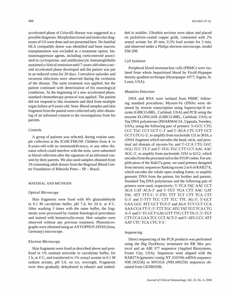
Exons 2–6 and flanking regions ofRAB27Agene ofthe patient were sequenced and analyzed, leading to theidentification of two heterozygous mutations in this gene(Fig. 2A). One allele has a C-T transition on exon 5,
Fig. 2. Mutations detected in the Rab27a gene and pedigree analysis.(A) Electrophoregrams showing the genomic sequence from two regionsof theRAB27Agene. The two mutations detected (C352T and G467+1 C) are indicated by arrows. (B and C) Pedigree from the patient’sfamily (B) and a second Brazilian family (C) and segregation analysisof the polymorphic markers linked to the GS2 locus on chromosome15q21. Asterisks indicate affected haplotypes. Note that the mothersshare common marker haplotype. [Note: The on-line version of thisfigure appears in color.]
Journal of Clinical Immunology, Vol. 24, No. 4, 2004

P1: JLS
pp1212-joci-487253 JOCI.cls May 18, 2004 10:38
404 BIZARIO ET AL.
corresponding to position 352 on the RAB27A cDNA.This nonsense mutation leads to a STOP codon, TAG,in place of a CAG codon, which encodes for the Glu-tamine 118 in the predicted Rab27a amino acid se-quence. The predicted amino acid sequence of the humanRab27a protein is 221 amino acids. The other allele con-tains a G-C transversion on the splicing donor site ofexon 5, in the position 467+ 1. This mutation willprobably interfere with mRNA splicing leading to aframeshift.
Pedigree Analysis
Sequencing and analysis of theRAB27Agene of thepatient’s parents revealed that the C352T mutation wasinherited from the father and the G467+ 1C mutationfrom the mother (data not shown). The second child ofthis family, who is healthy, carries only the C352T muta-tion, being therefore, heterozygous for this disease (datanot shown). On the basis of this data, the family wasadvised regarding the risks of conceiving other childrencarrying this disease. Polymorphic markers were used toidentify a possible relation between this family and an-other Brazilian family carrying the G467+ 1C mutation,which had previously been determined (4). The pedigreeof the family described in the present work, with the mu-tations and polymorphic markers indicated, is shown inFig. 2B, and the pedigree for the second family is shownin Fig. 2C. Note that the mothers share common markerhaplotypes within a small genetic region of theRAB27Alocus, suggesting that the mutation arose from a commonancestor.
Patient Lymphocytes are Capable of Respondingto Mitogens and Alloantigens
Our results show that the patient’s circulating lympho-cytes are capable to proliferate when stimulated by mito-gens or alloantigens (Fig. 3A and B). Patient cells incorpo-rated3H-Thymidine under stimuli by PHA (17,500 cpm)and Con-A (24,780 cpm), presenting stimulation indexof 28 and 34, respectively. Median values from 10 con-trol individuals were 12,480 cpm for PHA and 12,630for Con-A stimuli, giving stimulation index of 22 and18.8, respectively. We also verified that the patient’s cir-culating lymphocytes presented an elevated rate of spon-taneous proliferation (2500 cpm) compared to controls,which was on the order of 520 cpm (median from 10 indi-viduals). Therefore, patient lymphocytes showed a prolif-eration rate and stimulation index above control range forboth mitogens (Fig. 3A), whereas, in the mixed lympho-cyte cultures (Fig. 3B), similar rates of lymphocyte prolif-
Fig. 3. Lymphocyte proliferation in response to mitogens or alloantigensand surface expression of HLA-DR. (A) Stimulation index for PHA orCon-A induced lymphocyte proliferation in peripheral blood lympho-cytes from the patient and 10 controls. Median and the percentiles 25and 75 from the control data points are indicated by horizontal lines. (B)Proliferation relative response to alloantigens stimulation (mixed lym-phocyte cultures). Note that patient lymphocytes showed proliferationindices above control range in response to both mitogens but not to al-loantigens. (C) Flow cytometric quantification of HLA-DR staining onCD3, CD14 and CD19 cell subsets in the patient and 6 control children.The mean and standard deviation (SD) values of the median of fluo-rescence intensity for the patient’s CD3 (135, SD= 2.8), CD14 (345.5,SD= 4.2), and CD19 (757, SD= 15.5) cell subsets were obtained fromthree independent assays. Median values from the 6 control data points(CD3= 61.3, CD14= 193.3 and CD19= 332.4) and the percentiles25 and 75 are indicated by horizontal lines.
Journal of Clinical Immunology, Vol. 24, No. 4, 2004
P1: JLS
pp1212-joci-487253 JOCI.cls May 18, 2004 10:38
GRISCELLI SYNDROME ANDCTL RESCUE 405
eration were obtained for the patient (relative response=79%) or control cells (relative response= 86%).
HLA-DR Expression Suggests an ActivationStatus of the Patient Lymphocytes
Fluorescence intensity for the surface staining of HLA-DR molecules on nonstimulated T or B lymphocytes (CD3or CD19) or monocytes (CD14) is plotted in Fig. 3C,for the patient and the control children (n = 6). Thisanalysis shows that HLA-DR expression on the patientcells is above control range. HLA-DR expression on CD3cells suggests an activation status of these circulatinglymphocytes.
Differential Myosin-Va Expression and Localization:A Functional Link with Rab27a in Lymphocytesor a Result of Lymphocyte Activation?
To test the originally proposed working hypothesis ofMYO5Agene mutation we analyzed myosin-Va proteinexpression in the mononuclear blood cells of the patient.The polypeptide of expected size was detected in West-ern blots (data not shown). Also, flow cytometry showedstaining for myosin-Va in all mononuclear cell subsets(Fig. 4A). The results (in Fig. 4A) suggest an increasein myosin-Va levels in the patient cells, most prominentin lymphocytes and monocytes, in comparison with adultcontrol individuals. Given the association of myosin-Vaand Rab27a in melanocytes and the high levels for myosin-Va shown here we reasoned that it would be important tocharacterize the cellular localization of myosin-Va in thepatient’s mononuclear cells. Confocal microscopy imagesof myosin-Va staining (Fig. 4B–E) shows a typical punc-tate distribution of myosin-Va throughout the cytoplasmand also remarkably colocalized with the microtubule or-ganizing center in both mutant or control lymphocytes. Inseveral instances centrosomal staining for myosin-Va ap-peared to be more striking in the patient lymphocytes thanin controls (see Fig. 4 B–D).
Cytotoxic Response Is Impairedin the Patient’s NK and CD8+ Cells
NK and T cell cytotoxic activities are dependent onlytic granule release. The NK cytotoxic activity againstK562 target cells (Fig. 5A), assessed by flow cytometry,was shown to be about 1.5-fold lower in the patient cellscompared with the control median from 10 control chil-dren, while this is not a remarkable deficiency it is belowthe activity found in every control individual. By the51Cr-release assay, we found that the cytotoxic activity of IL2-dependent, CD8+ T cells against Fas-deficient L1210-3
target cells (Fig. 5B) was on the order of 3- to 5-fold lowerin the patient cells compared with the control. Also, thedegranulation activity of the CD8+ T cells (Fig. 5C) fromthe patient was markedly lower than the control.
Retroviral Gene Transfer Restores Cytotoxicityof RAB27A Mutant T Cells
RAB27A cDNA was cloned into the retroviral vectorpBSMFGB2IresEGFP and Phoenix packaging cells weretransfected as shown in Fig. 6A. Around 50% of the cellswere transfected and the supernatant was collected 24 and48 h after transfection and used to infect the patient’s IL2-dependent T cells. After three cycles of infection, the pro-portion of transduced cells was about 10%, as analyzed byflow cytometry (Fig. 6B). The cytotoxic activity of thesecells was then measured, and as shown in Fig. 6C, rescueof the cytoxic function of patient cells was obtained uponRAB27A transduction, which was not observed upon GFPtransduction.
DISCUSSION
Griscelli syndrome is a rare and not widely known dis-ease. Its diagnosis has been often incorrectly made dueto the remarkable similarity of symptoms with Chediak-Higashi syndrome or Familial Hemophagocytic lym-phohistiocytosis. Examination of the pigment alterationswithin the hair shaft and skin melanocytes led us to es-tablish the differential diagnosis between these diseases(Fig. 1), as previously indicated by several authors (1, 2,8). On the basis of a previous report on the involvementof MYO5Agene with GS (7), we first searched for muta-tions in this gene, which was, however, found to be intact(data not shown). More recent studies (4, 14) then ledus to direct our search for mutations towardsRAB27A.Evaluation of the clinical history of the patient describedhere was clearly indicative of GS2 subtype, i.e., causedby RAB27Amutation. We found two heterozygous mu-tant alleles ofRAB27Ain this patient, a C352T mutationinherited from the father and a G467+ 1C mutation, fromthe mother (Fig. 2). M´enasch´e et al. (4) have previouslycharacterized a homozygous G467+ 1C mutation in aBrazilian child. Since both patients’ mother are heterozy-gous for this mutation and share the same marker hap-lotypes, this finding is compatible with a mutation in-herited from a common ancestor. The patient’s brotherwithout disease expression was heterozygous for paternalRAB27Amutation and affected haplotype. These findingswere used in genetic counseling to the family. Interest-ingly, a nonsense mutation C346T (Q116stop), close to the
Journal of Clinical Immunology, Vol. 24, No. 4, 2004

P1: JLS
pp1212-joci-487253 JOCI.cls May 18, 2004 10:38
406 BIZARIO ET AL.
Fig. 4. Expression and localization analyses of myosin-Va in mononuclear cells. (A) Medianof myosin-Va fluorescence intensity, measured by flow cytometry, in mononuclear cells fromthe patient, compared to 19 healthy, adult blood donors (controls). Median values from thecontrol data points for all cell subsets and the percentiles 25 and 75 are indicated by horizontallines. (B–E) Confocal microscopy images of myosin-Va staining (green) alone (B and D) orsuperimposed images of the myosin-Va (green) andβ-tubulin (red) staining (C and E). Theimages were generated from reconstruction of multiple optical sections. Note that myosin-Va has a typical punctate distribution throughout the cytoplasm in both patient and controllymphocytes, but its accumulation in the centrosome appears to be more intense in the patientcells.
C352T (Q118stop) identified here, was previously identi-fied leading to lymphohistiocytic infiltration, low plateletlevels, and secondary neurological symptoms with onset at1-year-old (6). Regarding the effects of these mutationsin mRNA and protein expression, no characterization has
been done either for the patient describe here or for the pre-vious patients (4, 6). In order to characterize these mutantalleles and be able to make functional or clinical correla-tions it will be necessary to determine whether the mRNAand a truncated Rab27a protein are detected.
Journal of Clinical Immunology, Vol. 24, No. 4, 2004

P1: JLS
pp1212-joci-487253 JOCI.cls May 18, 2004 10:38
GRISCELLI SYNDROME ANDCTL RESCUE 407
Fig. 5. Defective cytotoxic activity in NK and CTLs from the patientwith RAB27Amutation. (A) NK cytotoxic activity against K562 targetcells, assessed by flow cytometry. The values of the lytic unit 40% per107 cells obtained from a single assay for the patient (6.5) and 10 con-trol healthy chidren are plotted, and the median (10.33) from the controldata points as well as the percentiles 25 and 75 (8.6 and 11.5) are indi-cated by horizontal lines. (B) Cytotoxic activity of CD8+T lymphocytesagainst Fas-deficient L1210-3 target cells. (C) Degranulation activity ofCD8+ T lymphocytes. The relative number of CD8+ cells in the pa-tient (79.2%) and control (71.1%) samples is indicated. BLT esteraseactivity was measured in the cell supernatants. The % of specific BLTesterase release was calculated according to the formula: (specific ac-tivity in the supernatant of the anti-CD3 activated cells – spontaneousrelease)/(activity in the total cell lysate – spontaneous release)× 100,where spontaneous release was obtained from nonactivated cells.
The observation of an elevated rate of spontaneous andmitogen-induced proliferation and the high expression ofHLA-DR molecules on lymphocyte and monocyte cellsurfaces (Fig. 3) are compatible with generalized lym-phocyte and macrophage activation leading to HS in thispatient. It is generally accepted that the “spontaneous”lymphocyte proliferation (potentially triggered by an in-fectious agent), causing HS in GS2 patients, is a conse-quence of the lack of T cell cytotoxic activity, although theprecise mechanism is not fully understood (44). Our re-sults are also consistent with a strong deficiency in CD8+T cell cytotoxicity associated withRAB27Amutation, aspreviously shown (4, 35, 36). Additionally, we show a de-ficiency in NK cytotoxic activity in this patient (Fig. 5),compatible with the interpretation that both CD8+and NKcells citotoxicity rely on Rab27a-dependent mechanismsfor lytic granule release. We have not an explanation towhy the NK deficiency shown here was subtle comparedto the deficiency found in CD8+ cells. One possibility isthat the flow cytometric analysis used for NK activity alsodetects cell damage induced by a lytic granule indepen-dent pathway, though we can not rule out the possibility ofan alternative overlapping mechanism playing a functionin NK cells.
Although myosin-Va is not required for the cytotoxicactivity (4, 35, 36), we found it pertinent to report hereour findings, since they suggest an increase in the myosin-Va levels and a more prominent centrosomal staining forthis myosin in theRAB27Amutant lymphocytes (Fig. 4).Despite that the flow cytometric analysis was not donewith a matched age control group, nor could it be re-peated due to limitation in obtaining cells from the pa-tient, this finding is consistent with previously reportedevidence of higher myosin-Va levels inRAB27Amutantfibroblasts (13). The patient lymphocytes exhibited a typ-ical punctate distribution pattern of myosin-Va, thougha more prominent centrosomal staining was often noted.In spite that future studies will be necessary to con-firm this observation, an accumulation of myosin-Va atthe centrosomal region is consistent with previously re-ported results onRAB27Amutant melanocytes (45). Thesefindings might simply be related to lymphocyte activa-tion, since an increase in myosin-Va expression in re-sponse to lymphocyte activation has been demonstrated(33). Lymphocyte activation is one of the main concernsin this pathological condition, and is in fact likely tooccur in this patient as discussed above. On the otherhand, these results could be interpreted as due to a feed-back mechanism that compensates for the lack of func-tion of Rab27a GTPase or a decreased turnover of themyosin-Va protein. SinceMYO5Amutations do not af-fect the degranulation pathway of cytotoxic cells, either
Journal of Clinical Immunology, Vol. 24, No. 4, 2004

P1: JLS
pp1212-joci-487253 JOCI.cls May 18, 2004 10:38
408 BIZARIO ET AL.
Fig. 6. Restoration of the cytotoxic activity upon transduction of the patient’sCD8+ lymphocytes with retrovirus carrying the RAB27A cDNA. (A) Quan-tification of transfected Phoenix cells by GFP detection, using flow cytometry.R4 indicates the nontransfected cell population (GFP negative). R5 indicates thepopulation of cells expressing GFP (GFP positive). (B) Quantification of T cellsinfected with the retrovirus particles produced by the PHOENIX cells. R7 indi-cates the GFP negative population of cells and R6 represents the GFP positivecells. (C) Cytotoxic activity of IL2-dependent T cells against Fas-deficient L1210-3 target cells. Note that the cytotoxic activity of the Rab27a-retrovirus infectedcells is comparable to healthy control cells, whereas the patient’s cells infectedwith GFP-retrovirus or noninfected cells showed low levels of activity. The per-centage of CD8+ cells, as measured by FACS, was: Control= 73.3%; Patient=74.5%; Patient-Vector= 75.20%; Patient-Rab27a= 76.6%. Cells were culturedfor 10 days.
Journal of Clinical Immunology, Vol. 24, No. 4, 2004
P1: JLS
pp1212-joci-487253 JOCI.cls May 18, 2004 10:38
GRISCELLI SYNDROME ANDCTL RESCUE 409
myosin-Va plays a complementary role in this function orits connection with Rab27a suggested here involves ad-ditional lymphocyte Rab27a-dependent function(s). Al-though these possibilities are intriguing, elucidation ofmyosin-Va function in lymphocytes is beyond the scopeof this work.
One of the most important aspects of the present workwas to demonstrate the rescuing of T lymphocyte cyto-toxic activity, using retroviral vector mediated gene trans-fer, which was successful in restoringin vitro the cyto-toxic function of RAB27Amutant CD8+ T cells, evenwith only about 10% of the cells being expressing theretroviral GFP-IRES-Rab27a. This is not surprising, sincethe efficacy of T lymphocyte cytotoxic activity is remark-able, and cytotoxic cells have the ability to kill severaltarget cells in few minutes. Thus even a few cytotoxiccells may be highly efficient. Hence, this functional res-cue confirms that the mutations mapped in theRAB27Agene are those responsible for the lack of cytotoxic ac-tivity in CD8+ T lymphocytes of this patient and alsostrengthens that Rab27a GTPase plays indeed an essen-tial role in the lytic granule release. The correction shownwas done with transiently transduced T cells not allowingdetection of the protein level or further functional stud-ies. Although the expression of GFP is not an absoluteproof of Rab27a expression, a unique RNA containingboth genes (GFP and Rab27a) is produced and then pro-cessed in IRES system. Additionally, since a correction ofthe cytotoxicity is observed in GFP-IRES-RAB27a andnot with GFP-IRES alone, it is difficult to conceive thatRab27a was not properly expressed. Therefore, the datashown here represent the first step in evaluation of thefeasibility of acute phase treatment of GS2 patients, usinggenetically modified cytotoxic T lymphocytes. Until nowthe first choice of treatment for GS2 has been bone mar-row transplantation when possible (17, 18). Alternatively,chemotherapy has been used in order to maintain the pa-tient in long-term remission (19, 20). Undoubtedly, effortstowards finding an efficient treatment, available and appli-cable for all patients are desirable. In future experimentsaiming at genetic correction of these cells, stable trans-fection of the cells will be required. The ashen mice, thenatural Rab27a murine mutant, may be a useful model toevaluatein vivo the possibilities ofRAB27Agene correc-tion approaches.
ACKNOWLEDGMENTS
We especially thank: Dr. Fr´ederic Rieux-Laucat, whokindly gave us the retroviral vector used in this work.Silmara Reis Banzi for excellent technical assistance.
Maria D. Seabra Ferreira, Maria Tereza P. Maglia, Jos´eAugusto Maulin, Izilda R. Violante, Vani M. AlvesCorreia, and M´arcia Graeff from the optical and electronmicroscopy laboratories of the Departamento de BiologiaCelular e Molecular e Bioagentes Patogˆenicos-FMRP-USP; Patricia Vianna Bonini Palma and Fabiana Rossetode Morais, from FUNDHERP, for the flow-cytometry ex-pertise; Nathalie Lambert and St´ephanie Certain, fromHospital Necker Enfants Malades and INSERM-U429.Grant Support: This work was supported by FAPESP,CNPq, FUNDHERP, FAEPA, and UNAERP, in Brazil,and INSERM in France.
REFERENCES
1. Griscelli C, Durandy A, Guy-Grand D, Daguillard F, Herzog C,Prunieras M: A syndrome associating partial albinism and immun-odeficiency. Am J Med 65:691–702, 1978
2. Klein C, Philippe N, Le Deist F, Fraitag S, Prost C, Durandy A,Fischer A, Griscelli C: Partial albinism with immunodeficiency(Griscelli syndrome). J Pediatr 125:886–895, 1994
3. Bahadoran P, Aberdam E, Mantoux F, Busca R, Bille K, Yalman N,Saint-Basile G, Casaroli-Marano R, Ortonne JP, Ballotti R: Rab27a:A key to melanosome transport in human melanocytes. J Cell Biol152:843–850, 2001
4. Menasch´e G, Pastural E, Feldmann J, Certain S, Ersoy F, DupuisS, Wulffraat N, Bianchi D, Fischer A, Le Deist F, de Saint-BasileG: Mutations in RAB27A cause Griscelli syndrome associated withhaemophagocytic syndrome. Nat Genet 25:173–176, 2000
5. de Saint Basile G: Implication du traffic intracellulaire danstroiss maladies h´ereditaires du syst’eme h´ematopo¨ıetique. Med Sci16:745–750, 2000
6. Sanal O, Ersoy F, Tezcan I, Metin A, Yel L, Menasche G, Gurgey A,Berkel I, de Saint-Basile G: Griscelli disease: Genotype–phenotypecorrelation in an array of clinical heterogeneity. J Clin Immunol22:237–243, 2002
7. Haraldsson A, Weemaes CM, Bakkeren JA, Happle R: Griscellidisease with cerebral involvement. Eur J Pediatr 150:419–422, 1991
8. Pastural E, Barrat FJ, Dufourcq-Lagelouse R, Certain S, Sanal O,Jabado N, Seger R, Griscelli C, Fischer A, de Saint-Basile G:Griscelli disease maps to chromosome 15q21 and is associated withmutations in the myosin-Va gene. Nat Genet 16:289–292, 1997
9. Mercer JA, Seperack PK, Strobel MC, Copeland NG, Jenkins NA:Novel myosin heavy chain encoded by murine dilute coat colourlocus. Nature 349:709–713, 1991
10. Titus MA: Motor proteins: Myosin-V the multi-purpose transportmotor. Curr Biol 7:R301–R304, 1997
11. Reck-Peterson SL, Tyska MJ, Novick PJ, Mooseker MS: The yeastclass V myosins, Myo2p and Myo4p, are nonprocessive actin-basedmotors. J Cell Biol 153:1121–1126, 2001
12. Langford GM: Myosin-V, a versatile motor for short-range vesicletransport. Traffic 3:859–865, 2002
13. Pastural E, Ersoy F, Yalman N, Wulffraat N, Grillo E, Ozkinay F,Tezcan I, Gedikoglu G, Philippe N, Fischer A, de Saint-Basile G:Two genes are responsible for Griscelli syndrome at the same 15q21locus. Genomics 63:299–306, 2000
14. Wilson SM, Yip R, Swing DA, O’Sullivan TN, Zhang Y, Novak EK,Swank RT, Russell LB, Copeland NG, Jenkins NA: A mutation in
Journal of Clinical Immunology, Vol. 24, No. 4, 2004

P1: JLS
pp1212-joci-487253 JOCI.cls May 18, 2004 10:38
410 BIZARIO ET AL.
Rab27a causes the vesicle transport defects observed in ashen mice.Proc Natl Acad Sci USA 97:7933–7938, 2000
15. Deacon SW, Gelfand VI: Of yeast, mice, and men. Rab proteins andorganelle transport. J Cell Biol 152:F21–F24, 2001
16. Seabra MC, Mules EH, Hume AN: Rab GTPases, intracellular trafficand disease. Trends Mol Med 8:23–30, 2002
17. Schneider LC, Berman RS, Shea CR, Perez-Atayde AR, WeinsteinH, Geha RS: Bone marrow transplantation (BMT) for the syndromeof pigmentary dilution and lymphohistiocytosis (Griscelli’s syn-drome). J Clin Immunol 10:146–153, 1990
18. Tezcan I, Sanal O, Ersoy F, Uckan D, Kilic S, Metin A, Cetin M,Akin R, Oner C, Tuncer A: Successful bone marrow transplantationin a case of Griscelli disease which presented in accelerated phasewith neurological involvement. Bone Marrow Transplant 24:931–933, 1999
19. Gurgey A, Sayli T, Gunay M, Ersoy F, Kucukali T, Kale G, Caglar M:High-dose methylprednisolone and VP-16 in treatment of Griscellisyndrome with central nervous system involvement. Am J Hematol47:331–332, 1994
20. Imashuku S, Hibi S, Ohara T, Iwai A, Sako M, Kato M, ArakawaH, Sotomatsu M, Kataoka S, Asami K, Hasegawa D, Kosaka Y,Sano K, Igarashi N, Maruhashi K, Ichimi R, Kawasaki H, MaedaN, Tanizawa A, Arai K, Abe T, Hisakawa H, Miyashita H, HenterJI: Effective control of Epstein-Barr virus-related hemophagocyticlymphohistiocytosis with immunochemotherapy. Blood 93:1869–1874, 1999
21. Anikster Y, Huizing M, Anderson PD, Fitzpatrick DL, Klar A, Gross-Kieselstein E, Berkun Y, Shazberg G, Gahl WA, Hurvitz H: Evidencethat Griscelli syndrome with neurological involvement is caused bymutations in RAB27A, not MYO5A. Am J Hum Genet 71:407–414,2002
22. Matesic LE, Yip R, Reuss AE, Swing DA, O’Sullivan TN, FletcherCF, Copeland NG, Jenkins NA: Mutations in Mlph, encoding a mem-ber of the Rab effector family, cause the melanosome transport de-fects observed in leaden mice. Proc Natl Acad Sci USA 98:10238–10243, 2001
23. Menasch´e G, Ho CH, Sanal O, Feldmann J, Tezcan I, Ersoy F,Houdusee A, Fisher A, de Saint-Basile G: Griscelli syndromerestricted to hypopigmentation results from a melanophilin defect(GS3) or a MYO5A F-exon deletion (GS1). J Clin Invest 112:450–456, 2003
24. Wu XS, Rao K, Zhang H, Wang F, Sellers JR, Matesic LE, CopelandNG, Jenkins NA, Hammer JA, III: Identification of an organellereceptor for myosin-Va. Nat Cell Biol 4:271–278, 2002
25. Nagashima K, Torii S, Yi Z, Igarashi M, Okamoto K, Takeuchi T,Izumi T: Melanophilin directly links Rab27a and myosin Va throughits distinct coiled-coil regions. FEBS Lett 517:233–238, 2002
26. Fukuda M, Kuroda TS, Mikoshiba K: Slac2-a/melanophilin, themissing link between Rab27 and myosin Va: Implications of a tri-partite protein complex for melanosome transport. J Biol Chem277:12432–12436, 2002
27. Provance DW, Jr, James TL, Mercer JA: Melanophilin, the prod-uct of the Leaden locus, is required for targeting of myosin-Va tomelanosomes. Traffic 3:124–132, 2002
28. Nascimento AA, Roland JT, Gelfand VI: Pigment cells: A model forthe study of organelle transport. Annu Rev Cell Dev Biol 19:469–491, 2003
29. Hume AN, Collinson LM, Hopkins CR, Strom M, Barral DC, BossiG, Griffiths GM, Seabra MC: The leaden gene product is required
with Rab27a to recruit myosin Va to melanosomes in melanocytes.Traffic 3:193–202, 2002
30. Espindola FS, Espreafico EM, Coelho MV, Martins AR, Costa FR,Mooseker MS, Larson RE: Biochemical and immunological char-acterization of p190-calmodulin complex from vertebrate brain: Anovel calmodulin-binding myosin. J Cell Biol 118:359–368, 1992
31. Reis D, Souza M, Mineo J, Espindola F: Myosin V and iNOS ex-pression is enhanced in J774 murine macrophages treated with IFN-gamma. Braz J Med Biol Res 34:221–226, 2001
32. Rodriguez OC, Cheney RE: Human myosin-Vc is a novel class Vmyosin expressed in epithelial cells. J Cell Sci 115:991–1004, 2002
33. Bizario JC, Castro FA, Sousa JF, Fernandes RN, Damiao AD,Oliveira MK, Palma PV, Larson RE, Voltarelli JC, Espreafico EM:Myosin-V colocalizes with MHC class II in blood mononuclearcells and is up-regulated by T-lymphocyte activation. J Leukoc Biol71:195–204, 2002
34. Strom M, Hume AN, Tarafder AK, Barkagianni E, Seabra MC:A family of Rab27-binding proteins. Melanophilin links Rab27aand myosin Va function in melanosome transport. J Biol Chem277:25423–25430, 2002
35. Haddad EK, Wu X, Hammer JA, III, Henkart PA: Defective granuleexocytosis in Rab27a-deficient lymphocytes from Ashen mice. J CellBiol 152:835–842, 2001
36. Stinchcombe JC, Barral DC, Mules EH, Booth S, Hume AN,Machesky LM, Seabra MC, Griffiths GM: Rab27a is required forregulated secretion in cytotoxic T lymphocytes. J Cell Biol 152:825–834, 2001
37. Stepp SE, Dufourcq-Lagelouse R, Le Deist F, Bhawan S, CertainS, Mathew PA, Henter JI, Bennett M, Fischer A, de Saint-Basile G,Kumar V: Perforin gene defects in familial hemophagocytic lym-phohistiocytosis. Science 286:1957–1959, 1999
38. Bach FH, Bach ML: Mixed lymphocyte cultures in transplantationimmunology. Transplant Proc 3:942–948, 1971
39. Espreafico EM, Cheney RE, Matteoli M, Nascimento AAC, deCamili PV, Larson RE, Mooseker MS: Primary structure and cellularlocalization of chicken brain myosin-V (p190), an unconventionalmyosin with calmodulin light chains. J Cell Biol 119:1541–1558,1992
40. Chang L, Gusewitch GA, Chritton DB, Folz JC, Lebeck LK,Nehlsen-Cannarella SL: Rapid flow cytometric assay for the assess-ment of natural killer cell activity. J Immunol Methods 166:45–54,1993
41. Kim S, Yokoyama WM: NK cell granule exocytosis and cy-tokine production inhibited by Ly-49A engagement. Cell Immunol183:106–112, 1998
42. Hacein-Bey H, Cavazzana-Calvo M, Le Deist F, Dautry-Varsat A,Hivroz C, Riviere I, Danos O, Heard JM, Sugamura K, Fisher A,de Saint-Basile G: Gamma-c gene transfer into SCID X1 patients’B-cell lines restores normal high-affinity interleukin-2 receptor ex-pression and function. Blood 87:3108–3116, 1996
43. Yang S, Delgado R, King SR, Woffendin C, Barker CS, Yang ZY, XuL, Nolan GP, Nabel GJ: Generation of retroviral vector for clinicalstudies using transient transfection. Hum Gene Ther 10:123–132,1999
44. de Saint-Basile G, Fisher A: The role of cytotoxicity in lymphocytehomeostasis. Curr Opin Immunol 13:549–354, 2001
45. Hume AN, Collinson LM, Rapak A, Gomes AQ, Hopkins CR,Seabra MC: Rab27a regulates the peripheral distribution ofmelanosomes in melanocytes. J Cell Biol 152:795–808, 2001
Journal of Clinical Immunology, Vol. 24, No. 4, 2004