growth hormone control of hepatic lipid...
TRANSCRIPT

Zhongbo Liu,1 Jose Cordoba-Chacon,2,3 Rhonda D. Kineman,2,3 Bruce N. Cronstein,4
Radhika Muzumdar,5 Zhenwei Gong,5 Haim Werner,6 and Shoshana Yakar1
Growth Hormone Control of HepaticLipid MetabolismDiabetes 2016;65:3598–3609 | DOI: 10.2337/db16-0649
In humans, low levels of growth hormone (GH) and itsmediator, IGF-1, associate with hepatic lipid accumula-tion. In mice, congenital liver-specific ablation of the GHreceptor (GHR) results in reductions in circulating IGF-1and hepatic steatosis, associated with systemic insulinresistance. Due to the intricate relationship between GHand IGF-1, the relative contribution of each hormone tothe development of hepatic steatosis is unclear. Ourgoal was to dissect the mechanisms by which hepaticGH resistance leads to steatosis and overall insulinresistance, independent of IGF-1. We have generated acombined mouse model with liver-specific ablation ofGHR in which we restored liver IGF-1 expression via thehepatic IGF-1 transgene. We found that liver GHR abla-tion leads to increases in lipid uptake, de novo lipogen-esis, hyperinsulinemia, and hyperglycemia accompaniedwith severe insulin resistance and increased body adi-posity and serum lipids. Restoration of IGF-1 improvedoverall insulin sensitivity and lipid profile in serum andreduced body adiposity, but was insufficient to protectagainst steatosis-induced hepatic inflammation or oxida-tive stress. We conclude that the impaired metabolism instates of GH resistance results from direct actions of GHon lipid uptake and de novo lipogenesis, whereas itsactions on extrahepatic tissues are mediated by IGF-1.
According to Browning et al. (1), nonalcoholic fatty liverdisease (NAFLD) affects almost one-third of the adult pop-ulation in North America. Low levels of growth hormone(GH) in the general population associate with NAFLD (2).
Multi–single nucleotide polymorphism analyses of genome-wide association study have revealed 18 single nucleotidepolymorphisms in the GH pathway that relate to the de-velopment and progression of NAFLD (3). Also, patientswith GH receptor (GHR) loss of function (Laron syndrome)exhibit NAFLD (4). Likewise, cessation of GH treatment inGH-deficient (GHD) children after achieving adult heightleads to development of NAFLD and dyslipidemia in 29% ofpatients surveyed 10 years after therapy (2). Importantly,reductions in circulating GH (2,5) or its mediator, IGF-1(6–8), associate with NAFLD even after adjusting to BMI(9). Obese patients manifest GH resistance and can showdeclines in GH (10,11) that may be as low as those observedin GHD subjects (12,13). GH therapy reduces fat in youngmen with abdominal obesity (14), patients with primaryGHD (5,15,16), and hepatosteatosis in patients with HIVlipodystrophy (17). In contrast, humans treated with theGHR antagonist (pegvisomant and somatostatin analogs)to control for endogenous GH levels (18) show increasedhepatic triglyceride (TG) content.
In rodents, diet-induced fatty liver associates withreduced circulating GH (19,20) and reduced signal trans-ducer and activator of transcription-5 (STAT5) phosphory-lation in response to GH stimulation (21). Liver-specificdeletion of GHR in mice results in a marked decrease inserum IGF-1 levels, significant increases in fat mass andserum lipids, and severe hepatic steatosis associated withsystemic insulin resistance (22). A similar metabolic pheno-type is observed in mice with liver-specific disruption of theGHR mediators Janus kinase-2 (JAK2) (23) or STAT5 (24).
1Department of Basic Science & Craniofacial Biology, David B. Kriser DentalCenter, NYU College of Dentistry, New York, NY2Research and Development, Jesse Brown VA Medical Center, Chicago, IL3Department of Medicine, Section of Endocrinology, Diabetes and Metabolism,University of Illinois at Chicago, Chicago, IL4Department of Medicine, NYU School of Medicine, New York, NY5Division of Pediatric Endocrinology, Diabetes and Metabolism Consultation,Children’s Hospital of Pittsburgh of UPMC, University of Pittsburgh School ofMedicine, Pittsburgh, PA6Department of Human Molecular Genetics and Biochemistry, The Sackler Schoolof Medicine, Tel Aviv University, Ramat Aviv, Israel
Corresponding author: Shoshana Yakar, [email protected].
Received 23 May 2016 and accepted 20 September 2016.
This article contains Supplementary Data online at http://diabetes.diabetesjournals.org/lookup/suppl/doi:10.2337/db16-0649/-/DC1.
© 2016 by the American Diabetes Association. Readers may use this article aslong as the work is properly cited, the use is educational and not for profit, and thework is not altered. More information is available at http://www.diabetesjournals.org/content/license.
3598 Diabetes Volume 65, December 2016
METABOLISM

GH treatment in mice reduced diet-induced hepatosteatosis(21,25), and overexpression of a GHR antagonist increasedhepatic steatosis (26).
Clearly, clinical and experimental data indicate thatreductions in circulating GH levels or hepatic responseto GH are a typical component of NAFLD. On the basis ofthe above evidence, a clinical trial is currently underway totest the effectiveness of GH treatment in reducing hepaticlipid content in patients with NAFLD (NCT02217345).
Addressing the molecular mechanisms by which GHregulates hepatic lipid metabolism is challenging; modelswith ablation of GHR action in liver show significantlyreduced serum IGF-1 levels accompanied with high GHlevels that affect carbohydrate and lipid metabolism inextrahepatic tissues. Thus, the direct roles of hepatic GH inlipid and carbohydrate metabolism are not known. Wehypothesized that hepatic GHR regulates lipid metabolismindependent of IGF-1 and that reductions in hepatic GHRsignaling are not merely a consequence of NAFLD but acontributor to hepatic lipid accumulation in NAFLD. Toaddress our hypothesis, we generated a mouse model withhepatic ablation of the GHR (Li-GHRKO) and restoredIGF-1 gene expression via hepatic IGF-1 transgene (HIT).The Li-GHRKO-HIT mice show normal levels of serum IGF-1and exhibit hepatic GH resistance.
RESEARCH DESIGN AND METHODS
AnimalsThe generations of HIT mice (27) and floxed ghr mice (28)were previously described. Gene inactivation of the ghr inliver was achieved by the cre/lox-P system, as described byus previously (29). All mice were in the C57BL/6J geneticbackground. Weaned mice were allocated randomly intocages separated according to their sex. Mice were housedtwo to five animals per cage in a facility with 12-h light/dark cycles and free access to food/water. The differentanalyses were performed in male mice at the indicated ages.
All animal procedures were approved by the Institu-tional Animal Care and Use Committee of the NYU Schoolof Medicine (assurance number A3435–01; U.S. Depart-ment of Agriculture license no. 465).
Serum HormonesSerum/plasma were collected via orbital bleeding imme-diately after euthanasia with CO2 between 8 and 10 A.M.
Hormones were measured by ELISA: GH (EZRMGH-45K;Millipore), IGF-1 (22-IG1MS-E01; ALPCO), leptin (EZRL-83K; Millipore), insulin (NC9440604; Mercodia), IGF-1binding protein-3 (IGFBP-3; EMIGFBP3; Thermo FisherScientific), cytokines (K152A0H-1; Meso Scale Discovery),serum IGFBP-1 (CL0383; Cell Applications, Inc.), andIGFBP-2 (IRKTAH5375; Innovative Research, Inc.). Serumacid labile subunit (ALS) levels were determined by West-ern immunoblotting (AF1436; R&D Systems).
Serum and Tissue LipidsFree fatty acids (FFAs) were measured using calorimet-ric assay (11383175001; Roche). Liver TG content was
assessed following chloroform-methanol extraction andquantified using the TG reagent ( T7531; Pointe Scientific).Serum samples were sent to ANTECH Diagnostics (NewHyde Park, NY) for cholesterol measurements.
Tissue FA CompositionTissue FA composition was determined by gas chroma-tography/mass spectrometry (GC/MS) after lipid extrac-tion, as described previously (30).
Liver Glycogen ContentLiver glycogen content was measured using colorimetricassay (glycogen kit NC0294986; Cayman Chemical).
Liver EnzymesSerum samples were sent to ANTECH Diagnostics formeasurements of aspartate transaminase (AST), alaninetransaminase (ALT), and alkaline phosphatase.
Blood Glucose Measurements, Insulin, Glucose, andPyruvate Tolerance TestsMice were injected with 0.5 U/kg insulin, 2 mg/g glucose,or 2 g/kg sodium pyruvate (S8636; Sigma-Aldrich) fortolerance tests. Blood glucose levels were measured using aglucometer (Elite; Bayer, Mishawaka, IN). Insulin tolerancewas measured in fed mice, glucose tolerance in 8-h–fastedmice, and pyruvate tolerance in 15-h–fasted mice.
Lipid PeroxidationThiobarbituric acid–reactive substances were measuredusing a commercial kit (10009055; Cayman Chemical).
Protein OxidationCarbonylated proteins were detected using an OxiSelectProtein Carbonyl Spectrophotometric assay (STA-315; CellBiolabs, Inc.).
Gene ExpressionRNA was extracted using TRIzol (Invitrogen, Carlsbad,CA) or RNeasy Plus (74134; Qiagen), reverse-transcribed(18080-051; Life Technologies), and subjected to real-time PCR using SYBR master mix (4385612; Life Tech-nologies/Applied Biosystems). Transcript levels werecorrected to 18S. The primer sequences are presented inSupplementary Table 2.
Western Immunoblot AssayProteins were extracted using CHAPS buffer (1.25%CHAPS; 28300; Thermo Fisher Scientific) with proteaseinhibitor cocktail (04693132001; Roche), separated on4–20% SDS-PAGE (NP0335; Life Technologies), and trans-ferred to nitrocellulose membranes (170-4158; Bio-Rad).Antibodies used were anti-F4/80 (MCA497R; AbD Serotec),anti–insulin receptor (IR; sc-711; Santa Cruz Biotechnology),actin (4970; Cell Signaling Technology), and secondary anti-bodies (7074; Cell Signaling Technology).
HistologyTissues were fixed in 10% zinc formalin, then processed forparaffin sectioning (5-mm sections), and stained with he-matoxylin and eosin. Anti-F4/80 (MCA497R; AbD Serotec)was used to assess macrophage infiltration in the liver.
diabetes.diabetesjournals.org Liu and Associates 3599

Statistical AnalysisData are presented as means6 SEM. Differences betweengroups were tested using one-way ANOVA and post hocTukey test, with significance accepted at P , 0.05.
RESULTS
Restoration of Hepatic IGF-1 in the Li-GHRKO MiceTo restore IGF-1 in the liver-specific GHRKO mice, wecrossed the Li-GHRKO mice with the HIT mice (27) (Fig.1A), in which the rat IGF-1 transgene expressed specifi-cally in liver under the transthyretin promoter.
Ghr gene ablation in liver resulted in ;95% reductionsin serum IGF-1 in Li-GHRKO mice (Fig. 1B), whereas ex-pression of the Igf-1 transgene in the HIT mice increasedserum IGF-1 levels by approximately twofold. Crossing ofthe Li-GHRKO and HIT mice normalized serum IGF-1 lev-els (in Li-GHRKO-HIT). Serum GH levels increased fivefoldin the Li-GHRKO, whereas restoration of liver IGF-1 (Li-GHRKO-HIT) normalized GH levels (Fig. 1C).
The ALS is a surrogate marker of GHR action in liver.Accordingly, ALS protein levels in serum were undetect-able in both Li-GHRKO and Li-GHRKO-HIT mice (Fig. 1D).Serum IGFBP-3 levels reduced significantly in Li-GHRKOand Li-GHRKO-HIT mice (Fig. 1E), likely due to bluntedexpression of ALS (that stabilizes the IGF-1/IGFBP3 com-plex in serum) and increased susceptibility of unboundIGFBP-3 to degradation.
IGF-1 Improves Systemic Glucose/Lipid Homeostasisand Body Composition in Li-GHRKO-HIT MiceLi-GHRKO mice show increased blood glucose, increasedserum insulin levels, and severely impaired insulin tolerancetests (Fig. 2). Restoration of hepatic Igf-1 (Li-GHRKO-HITmice) normalized the fed and fasting blood glucose (Fig.2A), insulin (Fig. 2B), and insulin tolerance tests (Fig.2D). In accordance with a recent study (31) showing thatelevated IGFBP-1 improves whole-body insulin sensitivityand glucose tolerance, we found that in the Li-GHRKO-HITmice serum IGFBP-1 increased fivefold (Fig. 2E), suggestingthat it may play a role in the improvement of overall insulinsensitivity. A previous study using the Li-GHRKO mice (32)showed that IGFBP-1 levels increased by 2.5-fold despiteelevated levels of its inhibitor insulin. This may be in partdue to hepatic insulin resistance, progressive hepaticsteatosis (33), or, alternatively, a compensatory responseto significant reductions in IGFBP-3. Our results differfrom those previously reported (22), in which adminis-tration of recombinant human (rh)IGF-1 via osmoticpump into the Li-GHRKO mice did not correct the met-abolic phenotype. We have previously shown that in theabsence of ALS, the half-life of rhIGF-1 is very short, andit is rapidly cleared from circulation (34). Thus, infusingrhIGF-1 via osmotic pumps for a relatively short time(4 weeks) to Li-GHRKO mice with already severe hepaticsteatosis is inefficient.
Figure 1—Restoration of hepatic IGF-1 gene expression in Li-GHRKOmice. A: HIT were crossed with Li-GHRKOmice to yield the followinggroups: control mice harbored the floxed ghr gene, HIT mice harbored the rat IGF-1 transgene and floxed ghr gene, Li-GHRKO harboredthe floxed ghr gene and albumin (Alb) promoter–derived Cre transgene, and the Li-GHRKO-HIT mice harbored the floxed ghr gene, albuminpromoter–derived Cre transgene, and HIT. B: Serum IGF-1 levels in male mice at 16 weeks of age. C: Serum GH levels in male mice at8–16 weeks of age. D: Serum ALS levels of 16-week-old male mice. E: Serum IGFBP-3 in male mice at 16 weeks of age. Data presentedas mean 6 SEM. N indicates sample size. Significance accepted at P < 0.05: control vs. Li-GHRKO (a), control vs. Li-GHRKO-HIT (b),control vs. HIT (c), Li-GHRKO vs. Li-GHRKO-HIT (d), Li-GHRKO vs. HIT (e), and Li-GHRKO-HIT vs. HIT (f).
3600 Liver GH Resistance Diabetes Volume 65, December 2016
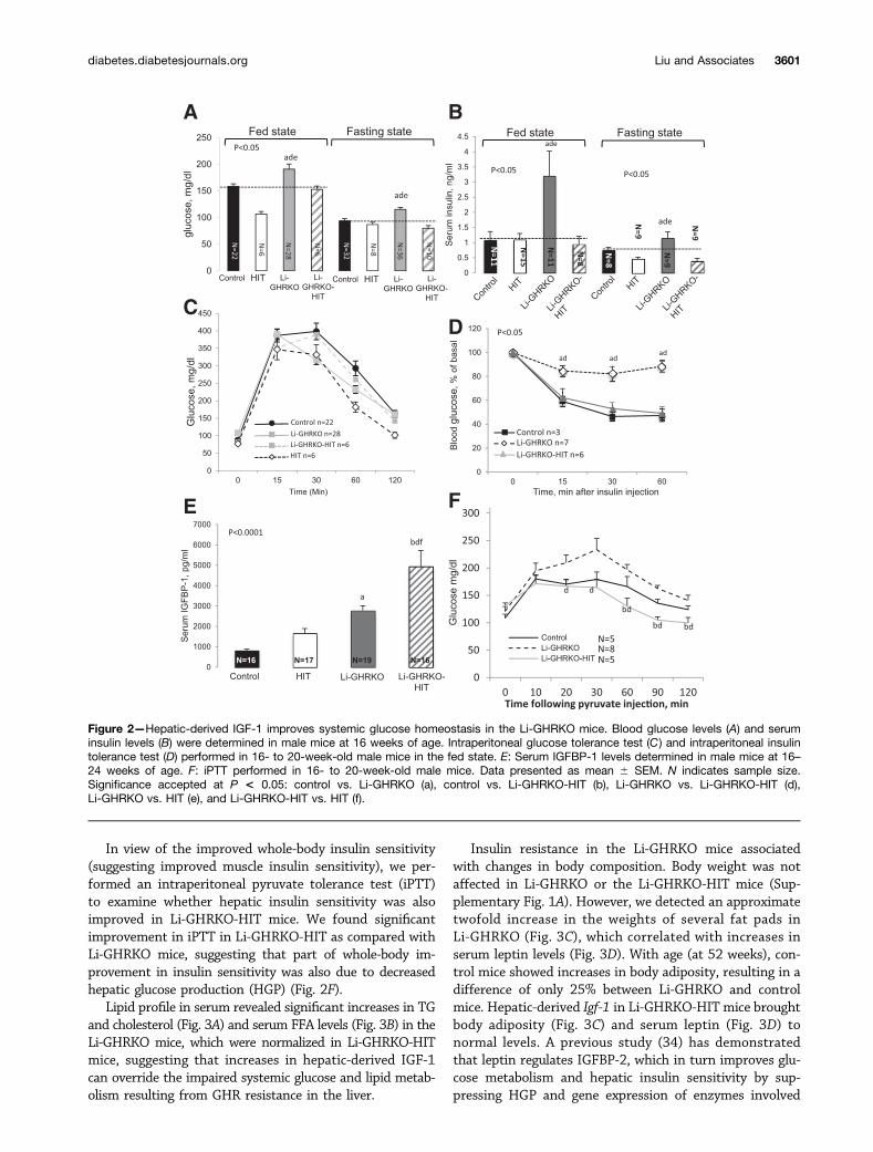
In view of the improved whole-body insulin sensitivity(suggesting improved muscle insulin sensitivity), we per-formed an intraperitoneal pyruvate tolerance test (iPTT)to examine whether hepatic insulin sensitivity was alsoimproved in Li-GHRKO-HIT mice. We found significantimprovement in iPTT in Li-GHRKO-HIT as compared withLi-GHRKO mice, suggesting that part of whole-body im-provement in insulin sensitivity was also due to decreasedhepatic glucose production (HGP) (Fig. 2F).
Lipid profile in serum revealed significant increases in TGand cholesterol (Fig. 3A) and serum FFA levels (Fig. 3B) in theLi-GHRKO mice, which were normalized in Li-GHRKO-HITmice, suggesting that increases in hepatic-derived IGF-1can override the impaired systemic glucose and lipid metab-olism resulting from GHR resistance in the liver.
Insulin resistance in the Li-GHRKO mice associatedwith changes in body composition. Body weight was notaffected in Li-GHRKO or the Li-GHRKO-HIT mice (Sup-plementary Fig. 1A). However, we detected an approximatetwofold increase in the weights of several fat pads inLi-GHRKO (Fig. 3C), which correlated with increases inserum leptin levels (Fig. 3D). With age (at 52 weeks), con-trol mice showed increases in body adiposity, resulting in adifference of only 25% between Li-GHRKO and controlmice. Hepatic-derived Igf-1 in Li-GHRKO-HIT mice broughtbody adiposity (Fig. 3C) and serum leptin (Fig. 3D) tonormal levels. A previous study (34) has demonstratedthat leptin regulates IGFBP-2, which in turn improves glu-cose metabolism and hepatic insulin sensitivity by sup-pressing HGP and gene expression of enzymes involved
Figure 2—Hepatic-derived IGF-1 improves systemic glucose homeostasis in the Li-GHRKO mice. Blood glucose levels (A) and seruminsulin levels (B) were determined in male mice at 16 weeks of age. Intraperitoneal glucose tolerance test (C ) and intraperitoneal insulintolerance test (D) performed in 16- to 20-week-old male mice in the fed state. E: Serum IGFBP-1 levels determined in male mice at 16–24 weeks of age. F: iPTT performed in 16- to 20-week-old male mice. Data presented as mean 6 SEM. N indicates sample size.Significance accepted at P < 0.05: control vs. Li-GHRKO (a), control vs. Li-GHRKO-HIT (b), Li-GHRKO vs. Li-GHRKO-HIT (d),Li-GHRKO vs. HIT (e), and Li-GHRKO-HIT vs. HIT (f).
diabetes.diabetesjournals.org Liu and Associates 3601

in gluconeogenesis and FA synthesis. We found that serumIGFBP-2 levels increased .10-fold in the Li-GHRKO-HITmice (Fig. 3E), which may explain in part the improvementin overall insulin sensitivity in those mice.
IGF-1 Is Insufficient to Restore Lipid Metabolism in theLivers of GH-Resistant MiceRelative liver weight in the Li-GHRKO mice increased at allages and was reduced in the Li-GHRKO-HIT mice (Fig. 4A).Li-GHRKO mice exhibited steatotic livers by histology(Fig. 4B) and showed an approximately sixfold increase inliver TG content (Fig. 4C) and FA content detected by GC/MS(Fig. 4D and Supplementary Table 1). Hepatic steatosis per-sisted even after restoration of liver Igf-1 (Li-GHRKO-HITmice), although to a lesser degree (Fig. 4B).
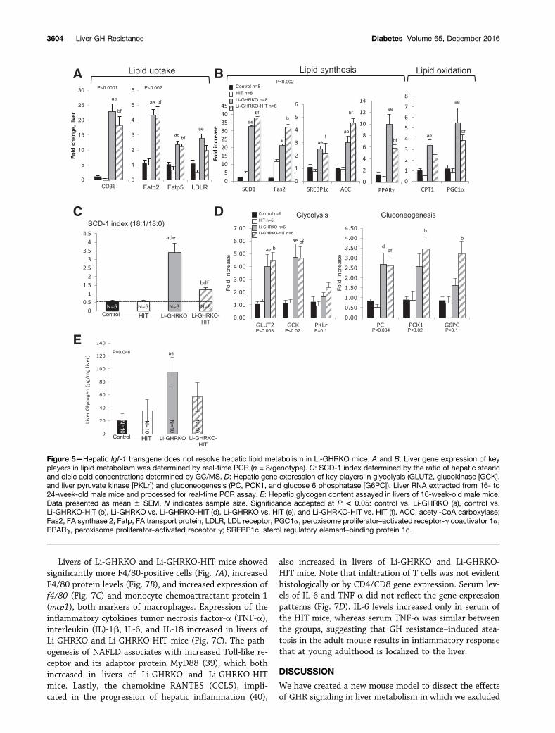
In accordance with previous findings with mutated GHRand liver-specific STAT5 knockout (35) mice showing anegative regulation of the FA translocase CD36 by STAT5,we found significantly increased CD36 expression in bothLi-GHRKO and the Li-GHRKO-HIT mice (Fig. 5A). Likewise,the FA transport proteins 2 and 5 and the LDL receptorthat regulates cholesterol metabolism increased in the
Li-GHRKO and Li-GHRKO-HIT mice (Fig. 5A), suggestingincreased hepatic lipid uptake in the absence of GHR. Fur-ther, we found that liver content of linoleic and docosahex-aenoic acids, which are not synthesized de novo and mustbe taken up by hepatocytes, are increased in Li-GHRKOmice (Supplementary Table 1). Although the levels of he-patic linoleic acid were reduced in Li-GHRKO-HIT whencompared with Li-GHRKO mice, they were not normalized.In an attempt to decrease GH-mediated lipolysis in fat, wedeleted the GHR in fat of the Li-GHRKO-HIT mice usingthe adiponectin-driven cre-transgenic mice. We show thatdespite normalization of serum GH, IGF-1, and insulin, aswell as inactivation of the GHR in fat, hepatic lipid contentin the Li/Fat-GHRKO-HIT mice was not normalized (Sup-plementary Fig. 1B). Overall, the data suggest that GHR inliver controls lipid uptake independent of IGF-1 or insulin.
Gene expression of stearoyl-CoA desaturase-1 (SCD-1),the rate-limiting enzyme that converts the palmitate(16:0) and stearate (18:0) to palmitoleate (16:1n7) andoleate (18:1n9), respectively, increased significantly inboth Li-GHRKO and the Li-GHRKO-HIT mice, suggesting
Figure 3—Hepatic-derived IGF-1 improves systemic lipid homeostasis and body composition in the Li-GHRKO mice. A: Serum TG andcholesterol in males at 16 weeks of age. B: Serum FFAs in the fed state in males at 16 weeks of age. C: Relative gonadal fat pad wet-weightto body weight at the indicated ages. D: Serum leptin levels in males at 16 weeks of age. E: Serum IGFBP-2 levels in males at 16–24 weeksof age. Data presented as mean 6 SEM. N indicates sample size. Significance accepted at P < 0.05: control vs. Li-GHRKO (a), control vs.Li-GHRKO-HIT (b), Li-GHRKO vs. Li-GHRKO-HIT (d), Li-GHRKO vs. HIT (e), and Li-GHRKO-HIT vs. HIT (f).
3602 Liver GH Resistance Diabetes Volume 65, December 2016

an increase in de novo lipogenesis (DNL) (Fig. 5B). In accor-dance with previous publications (36,37), we show that theSCD-1 index (the ratio of 18:1/18:0 by GC/MS) increasedthreefold in the Li-GHRKO mice (Fig. 5C and Supplemen-tary Table 1), suggesting an increased DNL. Li-GHRKO-HITshowed a significant decrease in SCD-1 index as comparedwith the Li-GHRKO mice, but this was still significantlyhigher than controls, suggesting that hepatic IGF-1 im-proved but did not resolve DNL in the absence of GHR.Increased DNL correlated with increased gene expressionof the sterol regulatory element-binding protein 1, acetyl-CoA carboxylase, and FA synthase (Fig. 5B) in Li-GHRKOand the Li-GHRKO-HIT mice. Finally, carnitine palmitoyl-transferase 1 (CPT-1) and peroxisome proliferator–activatedreceptor-g coactivator 1a increased in Li-GHRKO and theLi-GHRKO-HIT mice, suggesting increased FA oxidation.
Compromised hepatic lipid metabolism in Li-GHRKOand the Li-GHRKO-HIT mice was associated with im-paired glucose metabolism (Fig. 5D). Gene expression of theGLUT2 and glucokinase increased in Li-GHRKO mice, in-dicating enhanced glucose flux. Pyruvate carboxylase (PC),PCK1/PEPCK, and glucose 6 phosphatase, which are criticalenzymes for gluconeogenesis, were also elevated in liversof Li-GHRKO. Despite significant improvement in iPTT inLi-GHRKO-HIT mice, expression of these genes was stillelevated. Expression of genes involved in glycogen synthesiswas highly variable (Supplementary Fig. 1C). However, wefound significant increases in hepatic glycogen content inLi-GHRKO mice (Fig. 5E), which is in agreement with stud-ies in humans showing that in states of hyperinsulinemia
with hyperglycemia (Li-GHRKOmice), hepatic glycogen syn-thesis is maximized (38). Improvement in insulin sensitivityassociated with decreased glycogen content in the livers ofthe Li-GHRKO-HIT mice (Fig. 5E).
IGF-1 Modulates Oxidative Stress, but Is Insufficient toResolve Liver Inflammation in GH-Resistant MiceLiver expression of glutathione peroxidase, superoxidedismutase (SOD), catalase 2, and the nuclear factorerythroid 2–related factor-2 increased significantly inLi-GHRKO and Li-GHRKO-HIT mice (Fig. 6A). Protein car-bonylation (oxidatively damaged proteins) in the liverdid not differ between the groups (Fig. 6B). However,Li-GHRKO mice showed significantly increased levels ofoxidized lipids in liver (Fig. 6C) and serum (Fig. 6D) thatwere normalized in the Li-GHRKO-HIT mice. Because adulthepatocytes express very low levels of the IGF-1 receptor,we postulated that IGF-1 elicited its effects on lipid perox-idation via its actions on hepatic stellate cells or via the IRon hepatocytes. We found that IR levels increased signi-ficantly in livers of Li-GHRKO-HIT as compared withLi-GHRKO mice (Supplementary Fig. 1D), suggesting thathepatic-derived IGF-1 may act locally via the IR to reduceoxidative stress in liver. Hepatic steatosis in the Li-GHRKOmice associated with liver injury as indicated by elevatedlevels of AST and ALT (Fig. 6E) and elevated lipid perox-idation (Fig. 6C) that were normalized with hepatic-IGF-1.Note that muscle protein carbonylation (SupplementaryFig. 1E) and the levels of oxidized lipids (SupplementaryFig. 1F) reduced in Li-GHRKO-HIT mice and may contrib-ute to overall improved insulin sensitivity.
Figure 4—Hepatic Igf-1 transgene does not resolve hepatic steatosis in the Li-GHRKO mice. A: Relative liver wet-weight to body weightwas followed in several age groups as indicated. B: Hematoxylin/eosin staining of liver sections from 16-week-old control, Li-GHRKO, HIT,and Li-GHRKO-HIT mice. Scale bars 5 100 mm. C: Hepatic TG content. D: Hepatic FA content in 16-week-old male mice measured byGC/MS. Data presented as mean6 SEM. N indicates sample size. Significance accepted at P< 0.05: control vs. Li-GHRKO (a), control vs.Li-GHRKO-HIT (b), Li-GHRKO vs. Li-GHRKO-HIT (d), Li-GHRKO vs. HIT (e), and Li-GHRKO-HIT vs. HIT (f).
diabetes.diabetesjournals.org Liu and Associates 3603
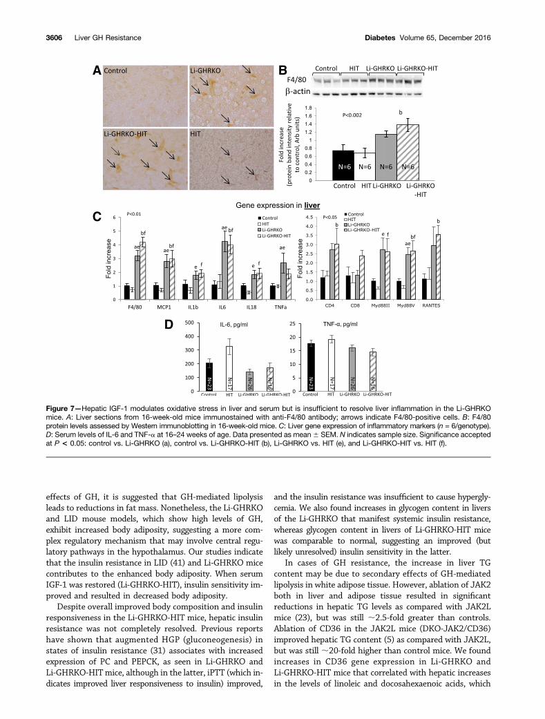
Livers of Li-GHRKO and Li-GHRKO-HIT mice showedsignificantly more F4/80-positive cells (Fig. 7A), increasedF4/80 protein levels (Fig. 7B), and increased expression off4/80 (Fig. 7C) and monocyte chemoattractant protein-1(mcp1), both markers of macrophages. Expression of theinflammatory cytokines tumor necrosis factor-a (TNF-a),interleukin (IL)-1b, IL-6, and IL-18 increased in livers ofLi-GHRKO and Li-GHRKO-HIT mice (Fig. 7C). The path-ogenesis of NAFLD associates with increased Toll-like re-ceptor and its adaptor protein MyD88 (39), which bothincreased in livers of Li-GHRKO and Li-GHRKO-HITmice. Lastly, the chemokine RANTES (CCL5), impli-cated in the progression of hepatic inflammation (40),
also increased in livers of Li-GHRKO and Li-GHRKO-HIT mice. Note that infiltration of T cells was not evidenthistologically or by CD4/CD8 gene expression. Serum lev-els of IL-6 and TNF-a did not reflect the gene expressionpatterns (Fig. 7D). IL-6 levels increased only in serum ofthe HIT mice, whereas serum TNF-a was similar betweenthe groups, suggesting that GH resistance–induced stea-tosis in the adult mouse results in inflammatory responsethat at young adulthood is localized to the liver.
DISCUSSION
We have created a new mouse model to dissect the effectsof GHR signaling in liver metabolism in which we excluded
Figure 5—Hepatic Igf-1 transgene does not resolve hepatic lipid metabolism in Li-GHRKO mice. A and B: Liver gene expression of keyplayers in lipid metabolism was determined by real-time PCR (n = 8/genotype). C: SCD-1 index determined by the ratio of hepatic stearicand oleic acid concentrations determined by GC/MS. D: Hepatic gene expression of key players in glycolysis (GLUT2, glucokinase [GCK],and liver pyruvate kinase [PKLr]) and gluconeogenesis (PC, PCK1, and glucose 6 phosphatase [G6PC]). Liver RNA extracted from 16- to24-week-old male mice and processed for real-time PCR assay. E: Hepatic glycogen content assayed in livers of 16-week-old male mice.Data presented as mean 6 SEM. N indicates sample size. Significance accepted at P < 0.05: control vs. Li-GHRKO (a), control vs.Li-GHRKO-HIT (b), Li-GHRKO vs. Li-GHRKO-HIT (d), Li-GHRKO vs. HIT (e), and Li-GHRKO-HIT vs. HIT (f). ACC, acetyl-CoA carboxylase;Fas2, FA synthase 2; Fatp, FA transport protein; LDLR, LDL receptor; PGC1a, peroxisome proliferator–activated receptor-g coactivator 1a;PPARg, peroxisome proliferator–activated receptor g; SREBP1c, sterol regulatory element–binding protein 1c.
3604 Liver GH Resistance Diabetes Volume 65, December 2016

the possible contribution of concomitant reductions inIGF-1 to the overall phenotype. Our study clearly indicatesthat restoration of IGF-1 in the context of hepatic GHresistance normalized whole-body insulin sensitivity andserum lipid profile in the adult mouse (16–24 weeks), aphenotype that persisted to 52 weeks of age. However,IGF-1 did not resolve hepatic steatosis, as enhanced hepaticlipid uptake and DNL were still evident. Importantly, IGF-1was insufficient for protecting against steatosis-inducedhepatic inflammation.
Li-GHRKO and liver-specific IGF-1–deficient (LID) mice,with high levels of GH show insulin resistance (41). Hepaticproduction of IGF-1 (Li-GHRKO-HIT) rescued overall in-sulin sensitivity likely via normalization of serum GH levelsand improved muscle insulin responsiveness. Improvementin overall insulin sensitivity in the Li-GHRKO-HIT miceassociated also with normalized iPTT, suggesting partialimprovement in hepatic insulin sensitivity. IGFBP-1, a
surrogate marker of insulin resistance, was elevated in theLi-GHRKO-HIT mice and may also contribute to the im-provement in whole-body insulin sensitivity. Li-GHRKO-HIT mice also showed improved lipid profile in serum andnormalized body adiposity that correlated with normal-ized serum leptin levels and .10-fold increase in IGFBP-2levels that were previously shown to enhance insulin sen-sitivity (34). At this point, improvement in whole-bodyinsulin sensitivity together with normalized leptin levelsin the Li-GHRKO-HIT mice most likely involves central(neuroendocrine) regulation, which controls body composi-tion via leptin-dependent and -independent mechanisms.
The contribution of GH to body adiposity is docu-mented in both humans and mice. Patients with Laronsyndrome (42), GH-deficient subjects (43), and GHRKOmice exhibit increases in body adiposity (44), whereas pa-tients with acromegaly (45) and the bovine GH-transgenicmice are lean (46). Together with the documented lipolytic
Figure 6—Hepatic IGF-1 modulates oxidative stress in liver and serum. A: Liver gene expression of enzymes involved in oxidative stressresponse in males at 16 weeks measured by real-time PCR. B: Protein carbonylation was determined using a spectrophotometric assay ofliver protein extracts from male mice at 24 weeks of age. Lipid peroxidation measured using thiobarbituric acid–reactive substances assayin liver (C ) and serum (D) from male mice at 24 weeks of age. E: Serum levels of AST, ALT, and alkaline phosphatase (Alk Phos) in males at16 weeks of age. Data presented as mean 6 SEM. N indicates sample size. Significance accepted at P < 0.05: control vs. Li-GHRKO (a),control vs. Li-GHRKO-HIT (b), Li-GHRKO vs. Li-GHRKO-HIT (d), Li-GHRKO vs. HIT (e), and Li-GHRKO-HIT vs. HIT (f). CAT2, catalase 2;GPX, glutathione peroxidase; MDA, malondialdehyde; Nrf2, nuclear factor erythroid 2–related factor-2.
diabetes.diabetesjournals.org Liu and Associates 3605
effects of GH, it is suggested that GH-mediated lipolysisleads to reductions in fat mass. Nonetheless, the Li-GHRKOand LID mouse models, which show high levels of GH,exhibit increased body adiposity, suggesting a more com-plex regulatory mechanism that may involve central regu-latory pathways in the hypothalamus. Our studies indicatethat the insulin resistance in LID (41) and Li-GHRKO micecontributes to the enhanced body adiposity. When serumIGF-1 was restored (Li-GHRKO-HIT), insulin sensitivity im-proved and resulted in decreased body adiposity.
Despite overall improved body composition and insulinresponsiveness in the Li-GHRKO-HIT mice, hepatic insulinresistance was not completely resolved. Previous reportshave shown that augmented HGP (gluconeogenesis) instates of insulin resistance (31) associates with increasedexpression of PC and PEPCK, as seen in Li-GHRKO andLi-GHRKO-HITmice, although in the latter, iPTT (which in-dicates improved liver responsiveness to insulin) improved,
and the insulin resistance was insufficient to cause hypergly-cemia. We also found increases in glycogen content in liversof the Li-GHRKO that manifest systemic insulin resistance,whereas glycogen content in livers of Li-GHRKO-HIT micewas comparable to normal, suggesting an improved (butlikely unresolved) insulin sensitivity in the latter.
In cases of GH resistance, the increase in liver TGcontent may be due to secondary effects of GH-mediatedlipolysis in white adipose tissue. However, ablation of JAK2both in liver and adipose tissue resulted in significantreductions in hepatic TG levels as compared with JAK2Lmice (23), but was still ;2.5-fold greater than controls.Ablation of CD36 in the JAK2L mice (DKO-JAK2/CD36)improved hepatic TG content (5) as compared with JAK2L,but was still ;20-fold higher than control mice. We foundincreases in CD36 gene expression in Li-GHRKO andLi-GHRKO-HIT mice that correlated with hepatic increasesin the levels of linoleic and docosahexaenoic acids, which
Figure 7—Hepatic IGF-1 modulates oxidative stress in liver and serum but is insufficient to resolve liver inflammation in the Li-GHRKOmice. A: Liver sections from 16-week-old mice immunostained with anti-F4/80 antibody; arrows indicate F4/80-positive cells. B: F4/80protein levels assessed by Western immunoblotting in 16-week-old mice. C: Liver gene expression of inflammatory markers (n = 6/genotype).D: Serum levels of IL-6 and TNF-a at 16–24 weeks of age. Data presented as mean6 SEM. N indicates sample size. Significance acceptedat P < 0.05: control vs. Li-GHRKO (a), control vs. Li-GHRKO-HIT (b), Li-GHRKO vs. HIT (e), and Li-GHRKO-HIT vs. HIT (f).
3606 Liver GH Resistance Diabetes Volume 65, December 2016
are not synthesized de novo, suggesting increased lipiduptake. Hepatic IGF-1 significantly reduced the levels ofthese acids (Li-GHRKO-HIT) but not to normal levels. Fur-ther, ablation of GHR in both liver and adipose tissue(liver/fat-GHRKO-HIT mice) did not resolved hepatic stea-tosis, despite normal levels of blood glucose and insulin,again suggesting that an increase in white adipose tissuelipolysis is not the only cause for hepatic lipid accumulationin states of GH resistance.
On the basis of the theory of Li et al. (47), fatty livercan become resistant to insulin-mediated suppression ofHGP, but remains sensitive to insulin-mediated stimula-tion of lipogenesis. In the Li-GHRKO-HIT mice, despitenormal insulin levels and iPTT, hepatic steatosisremained, suggesting that lipid synthesis in livers of thesemice is independent of insulin signaling and most likelyrelates to ablation of the GHR. Our data are also supportedby Vatner et al. (48), who showed that the rate of hepaticTG production depends on the rate of FA uptake, is in-dependent of hepatic insulin signaling, and, with clinicaldata showing that low levels of circulating IGF-1 may havea role in the development of NAFLD, is independent ofinsulin resistance. Accordingly, we note that the high levelsof insulin found in the LID mice did not lead to hepaticsteatosis (15,41), suggesting that increased hepatic lipidsynthesis in the Li-GHRKO mice relates directly to the
ablation of GHR action in liver. Consequently, we detectedincreases in SCD-1 gene expression and SCD-1 index in theLi-GHRKO and Li-GHRKO-HIT mice, suggesting that he-patic GHR regulates liver DNL independent of IGF-1.Notably, in the Li-GHRKO-HIT mice, SCD-1 index wasreduced but not normalized, suggesting that GHR signal-ing directly suppresses hepatic DNL. These data are inagreement with previous study showing a twofold in-crease in DNL in GH-resistant mice (49). Overall, it ispostulated that both lipid uptake and DNL occur instates of hepatic GH resistance independent of IGF-1.
Excess lipid storage in the liver leads to enhancedb-oxidation of FFAs, which is regulated by CPT-1. Li-GHRKOand Li-GHRKO-HIT mice show increases in CPT-1 geneexpression, suggesting increases in b-oxidation. Increasedb-oxidation results in overproduction of reactive oxygenspecies (ROS), eventually leading to mitochondrial dys-function. Oxidation of lipids by extramitochondrial oxi-dation systems, such as microsomal and peroxisomaloxidation, also leads to increased ROS production (40).Accumulation of ROS and nitrogen species is a hallmarkof NAFLD (26). We found increases in lipids peroxida-tion in the Li-GHRKO mice in serum and liver, whichwas restored to normal by hepatic IGF-1 (Li-GHRKO-HIT mice). Despite improvement in lipid peroxidation,an increase in oxidative stress may still be occurring in the
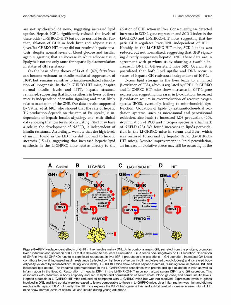
Figure 8—IGF-1–independent effects of GHR in liver involve mainly DNL. A: In control animals, GH, secreted from the pituitary, promotesliver production and secretion of IGF-1 that is delivered to tissues via circulation. IGF-1 feeds back negatively on GH secretion. B: Ablationof GHR in liver (Li-GHRKO) results in significant reductions in liver IGF-1 production and elevations in GH secretion. Increased GH levelscontribute to overall increased insulin resistance (reflected by high levels of serum insulin and elevated blood glucose) and increased bodyadiposity (evident by increased circulating leptin levels). Li-GHRKO mice show severe hepatic steatosis, resulting from increased DNL andincreased lipid uptake. The impaired lipid metabolism in the Li-GHRKO mice associates with protein and lipid oxidation in liver, as well asinflammation in the liver. C: Restoration of hepatic IGF-1 in the Li-GHRKO-HIT mice normalizes serum IGF-1 and GH secretion. Thisassociates with reduction in body adiposity and serum leptin and normalization of serum lipids, blood glucose, and serum insulin levels.Hepatic steatosis in Li-GHRKO-HIT mice reduced as compared with Li-GHRKO mice but was not resolved. Expression levels of genesinvolved in DNL and lipid uptake were increased to levels comparable to those in Li-GHRKO mice. Liver inflammation was high and did notresolve with hepatic IGF-1. D: Lastly, the HIT mice express the IGF-1 transgene in liver and exhibit twofold increase in serum IGF-1. HITmice show normal levels of serum GH and insulin during young adulthood.
diabetes.diabetesjournals.org Liu and Associates 3607
Li-GHRKO-HIT liver because we found significant increasesin SOD and catalase in both Li-GHRKO and Li-GHRKO-HITmice.
NAFLD associates with low-grade inflammation, whichoften proceeds to chronic inflammation (7). Kupffer cellsare liver-resident macrophages that show high levels ofthe cell-surface marker F4/80. Activated Kupffer cells pro-duce proinflammatory cytokines such as IL-1b and TNF-aand secrete chemokines to recruit blood-derived monocy-tes/macrophages into the liver and play key roles in theinitiation and progression of liver inflammation (50). In-deed, we found increases in F4/80 and MCP-1 in bothLi-GHRKO and Li-GHRKO-HIT mice, which associatedwith increased expression of MyD88, RANTES (CCL5),IL-6, IL-1b, and TNF-a. Serum levels of IL-1b and TNF-aat 16–24 weeks did not differ from controls, but duringadvanced adulthood (1 and 2 years), they increased in bothLi-GHRKO and Li-GHRKO-HIT mice (data not shown).
In summary, we have shown that hepatic GH re-sistance associates with overall impaired lipid metabolism,insulin resistance, and increased body adiposity. Hepaticlipid accumulation in states of chronic GH resistanceresults from increased lipid uptake and increased DNL,indicating that GH directly regulates these processes inthe liver. The impaired lipid metabolism, along withsevere hepatic insulin resistance, leads to overall increasedoxidative stress and initiates inflammation (Fig. 8). Resto-ration of hepatic IGF-1 (Li-GHRKO-HIT) resolved over-all insulin resistance and partially alleviated hepaticoxidative stress, but did not resolve hepatic steatosis orinflammation.
Acknowledgments. The authors thank Dr. Papasani V. Subbaiah, Sectionof Endocrinology, Diabetes and Metabolism, University of Illinois at Chicago,for providing technical expertise in setting up the GC/MS for analysis of FAcomposition.Funding. This work was supported by National Institutes of Health grantDK-100246 (to S.Y.) and Bi-national Science Foundation grant 2013282 (to S.Y.and H.W.). R.D.K. was supported by Department of Veterans Affairs Merit AwardBX001114. B.N.C. was supported by National Institutes of Health grantsAR-056672, AR-068593, and UL1-TR-001445.Duality of Interest. No potential conflicts of interest relevant to this articlewere reported.Author Contributions. Z.L. conducted the experiments. J.C.-C. andR.D.K. measured FA composition by GC/MS. B.N.C. conducted liver histology andserum lipid profiles. R.M. and Z.G. conducted liver protein carbonylation assay.H.W. conducted liver histology and serum lipid profiles and helped in experimen-tal design and discussion. S.Y. designed the experiments and wrote the paper.S.Y. is the guarantor of this work and, as such, had full access to all the data inthe study and takes responsibility for the integrity of the data and the accuracyof the data analysis.
References1. Browning JD, Szczepaniak LS, Dobbins R, et al. Prevalence of hepatic
steatosis in an urban population in the United States: impact of ethnicity. Hep-
atology 2004;40:1387–13952. Xu L, Xu C, Yu C, et al. Association between serum growth hormone levels and
nonalcoholic fatty liver disease: a cross-sectional study. PLoS One 2012;7:e44136
3. Chen QR, Braun R, Hu Y, et al. Multi-SNP analysis of GWAS data identifiespathways associated with nonalcoholic fatty liver disease. PLoS One 2013;8:e659824. Laron Z, Ginsberg S, Webb M. Nonalcoholic fatty liver in patients with Laronsyndrome and GH gene deletion - preliminary report. Growth Horm IGF Res 2008;18:434–4385. Hazlehurst JM, Tomlinson JW. Non-alcoholic fatty liver disease in commonendocrine disorders. Eur J Endocrinol 2013;169:R27–R376. Arturi F, Succurro E, Procopio C, et al. Nonalcoholic fatty liver disease isassociated with low circulating levels of insulin-like growth factor-I. J Clin En-docrinol Metab 2011;96:E1640–E16447. Fusco A, Miele L, D’Uonnolo A, et al. Nonalcoholic fatty liver disease isassociated with increased GHBP and reduced GH/IGF-I levels. Clin Endocrinol(Oxf) 2012;77:531–5368. Sumida Y, Yonei Y, Tanaka S, et al. Lower levels of insulin-like growthfactor-1 standard deviation score are associated with histological severity of non-alcoholic fatty liver disease. Hepatol Res 2015;45:771–7819. Völzke H, Nauck M, Rettig R, et al. Association between hepatic steatosisand serum IGF1 and IGFBP-3 levels in a population-based sample. Eur J En-docrinol 2009;161:705–71310. Huang L, Steyn FJ, Tan HY, et al. The decline in pulsatile GH secretionthroughout early adulthood in mice is exacerbated by dietary-induced weightgain. Endocrinology 2012;153:4380–438811. Iranmanesh A, Lizarralde G, Veldhuis JD. Age and relative adiposity arespecific negative determinants of the frequency and amplitude of growth hor-mone (GH) secretory bursts and the half-life of endogenous GH in healthy men.J Clin Endocrinol Metab 1991;73:1081–108812. Dichtel LE, Yuen KC, Bredella MA, et al. Overweight/Obese adults withpituitary disorders require lower peak growth hormone cutoff values on glucagonstimulation testing to avoid overdiagnosis of growth hormone deficiency. J ClinEndocrinol Metab 2014;99:4712–471913. Shalet SM, Toogood A, Rahim A, Brennan BM. The diagnosis of growthhormone deficiency in children and adults. Endocr Rev 1998;19:203–22314. Bredella MA, Gerweck AV, Lin E, et al. Effects of GH on body compositionand cardiovascular risk markers in young men with abdominal obesity. J ClinEndocrinol Metab 2013;98:3864–387215. Nishizawa H, Iguchi G, Murawaki A, et al. Nonalcoholic fatty liver disease inadult hypopituitary patients with GH deficiency and the impact of GH replacementtherapy. Eur J Endocrinol 2012;167:67–7416. Takahashi Y, Iida K, Takahashi K, et al. Growth hormone reverses non-alcoholic steatohepatitis in a patient with adult growth hormone deficiency.Gastroenterology 2007;132:938–94317. Schwarz JM, Mulligan K, Lee J, et al. Effects of recombinant human growthhormone on hepatic lipid and carbohydrate metabolism in HIV-infected patientswith fat accumulation. J Clin Endocrinol Metab 2002;87:94218. Madsen M, Krusenstjerna-Hafstrøm T, Møller L, et al. Fat content in liverand skeletal muscle changes in a reciprocal manner in patients with acromegalyduring combination therapy with a somatostatin analog and a GH receptor an-tagonist: a randomized clinical trial. J Clin Endocrinol Metab 2012;97:1227–123519. Steyn FJ, Xie TY, Huang L, et al. Increased adiposity and insulin correlateswith the progressive suppression of pulsatile GH secretion during weight gain.J Endocrinol 2013;218:233–24420. Luque RM, Kineman RD. Impact of obesity on the growth hormone axis:evidence for a direct inhibitory effect of hyperinsulinemia on pituitary function.Endocrinology 2006;147:2754–276321. Qin Y, Tian YP. Preventive effects of chronic exogenous growth hormonelevels on diet-induced hepatic steatosis in rats. Lipids Health Dis 2010;9:7822. Fan Y, Menon RK, Cohen P, et al. Liver-specific deletion of the growthhormone receptor reveals essential role of growth hormone signaling in hepaticlipid metabolism. J Biol Chem 2009;284:19937–1994423. Nordstrom SM, Tran JL, Sos BC, Wagner KU, Weiss EJ. Disruption of JAK2in adipocytes impairs lipolysis and improves fatty liver in mice with elevated GH.Mol Endocrinol 2013;27:1333–1342
3608 Liver GH Resistance Diabetes Volume 65, December 2016

24. Cui Y, Hosui A, Sun R, et al. Loss of signal transducer and activator oftranscription 5 leads to hepatosteatosis and impaired liver regeneration. Hep-atology 2007;46:504–51325. List EO, Palmer AJ, Berryman DE, Bower B, Kelder B, Kopchick JJ. Growthhormone improves body composition, fasting blood glucose, glucose toleranceand liver triacylglycerol in a mouse model of diet-induced obesity and type 2diabetes. Diabetologia 2009;52:1647–165526. Yang T, Householder LA, Lubbers ER, et al. Growth hormone receptor an-tagonist transgenic mice are protected from hyperinsulinemia and glucose in-tolerance despite obesity when placed on a HF diet. Endocrinology 2015;156:555–56427. Wu Y, Sun H, Basta-Pljakic J, et al. Serum IGF-1 is insufficient to restoreskeletal size in the total absence of the growth hormone receptor. J Bone MinerRes 2013;28:1575–158628. Wu Y, Wang C, Sun H, LeRoith D, Yakar S. High-efficient FLPo deleter micein C57BL/6J background. PLoS One 2009;4:e805429. Yakar S, Liu JL, Stannard B, et al. Normal growth and development in theabsence of hepatic insulin-like growth factor I. Proc Natl Acad Sci U S A 1999;96:7324–732930. Kineman RD, Majumdar N, Subbaiah PV, Cordoba-Chacon J. Hepatic PPARgIs Not Essential for the Rapid Development of Steatosis After Loss of Hepatic GHSignaling, in Adult Male Mice. Endocrinology 2016;157:1728–173531. Rajwani A, Ezzat V, Smith J, et al. Increasing circulating IGFBP1 levelsimproves insulin sensitivity, promotes nitric oxide production, lowers bloodpressure, and protects against atherosclerosis. Diabetes 2012;61:915–92432. List EO, Berryman DE, Funk K, et al. Liver-specific GH receptor gene-disrupted (LiGHRKO) mice have decreased endocrine IGF-I, increased local IGF-I,and altered body size, body composition, and adipokine profiles. Endocrinology2014;155:1793–180533. Li HH, Doiron K, Patterson AD, Gonzalez FJ, Fornace AJ Jr. Identification ofserum insulin-like growth factor binding protein 1 as diagnostic biomarker forearly-stage alcohol-induced liver disease. J Transl Med 2013;11:26634. Hedbacker K, Birsoy K, Wysocki RW, et al. Antidiabetic effects of IGFBP2, aleptin-regulated gene. Cell Metab 2010;11:11–2235. Barclay JL, Nelson CN, Ishikawa M, et al. GH-dependent STAT5 signalingplays an important role in hepatic lipid metabolism. Endocrinology 2011;152:181–19236. Lee JJ, Lambert JE, Hovhannisyan Y, et al. Palmitoleic acid is elevated in fattyliver disease and reflects hepatic lipogenesis. Am J Clin Nutr 2015;101:34–43
37. Silbernagel G, Kovarova M, Cegan A, et al. High hepatic SCD1 activity isassociated with low liver fat content in healthy subjects under a lipogenic diet.J Clin Endocrinol Metab 2012;97:E2288–E229238. Petersen KF, Laurent D, Rothman DL, Cline GW, Shulman GI. Mechanism bywhich glucose and insulin inhibit net hepatic glycogenolysis in humans. J ClinInvest 1998;101:1203–120939. Miura K, Ohnishi H. Role of gut microbiota and Toll-like receptors in non-alcoholic fatty liver disease. World J Gastroenterol 2014;20:7381–739140. Bonekamp NA, Völkl A, Fahimi HD, Schrader M. Reactive oxygen speciesand peroxisomes: struggling for balance. Biofactors 2009;35:346–35541. Yakar S, Setser J, Zhao H, et al. Inhibition of growth hormone action im-proves insulin sensitivity in liver IGF-1-deficient mice. J Clin Invest 2004;113:96–10542. Laron Z. Lessons from 50 Years of Study of Laron Syndrome. Endocr Pract2015;21:1395–140243. Maison P, Griffin S, Nicoue-Beglah M, Haddad N, Balkau B, Chanson P;Metaanalysis of Blinded, Randomized, Placebo-Controlled Trials. Impact ofgrowth hormone (GH) treatment on cardiovascular risk factors in GH-deficientadults: a Metaanalysis of Blinded, Randomized, Placebo-Controlled Trials. J ClinEndocrinol Metab 2004;89:2192–219944. Masternak MM, Bartke A, Wang F, et al. Metabolic effects of intra-abdominal fat in GHRKO mice. Aging Cell 2012;11:73–8145. Freda PU, Shen W, Heymsfield SB, et al. Lower visceral and subcutaneousbut higher intermuscular adipose tissue depots in patients with growth hormoneand insulin-like growth factor I excess due to acromegaly. J Clin EndocrinolMetab 2008;93:2334–234346. Benencia F, Harshman S, Duran-Ortiz S, et al. Male bovine GH transgenicmice have decreased adiposity with an adipose depot-specific increase in im-mune cell populations. Endocrinology 2015;156:1794–180347. Li S, Brown MS, Goldstein JL. Bifurcation of insulin signaling pathway in ratliver: mTORC1 required for stimulation of lipogenesis, but not inhibition of glu-coneogenesis. Proc Natl Acad Sci U S A 2010;107:3441–344648. Vatner DF, Majumdar SK, Kumashiro N, et al. Insulin-independent regulationof hepatic triglyceride synthesis by fatty acids. Proc Natl Acad Sci U S A 2015;112:1143–114849. Cordoba-Chacon J, Majumdar N, List EO, et al. Growth Hormone InhibitsHepatic De Novo Lipogenesis in Adult Mice. Diabetes 2015;64:3093–310350. Lanthier N. Targeting Kupffer cells in non-alcoholic fatty liver disease/non-alcoholic steatohepatitis: Why and how? World J Hepatol 2015;7:2184–2188
diabetes.diabetesjournals.org Liu and Associates 3609