growth hormone-dependent insulin-like growth factor (igf) binding protein from human plasma differs...
TRANSCRIPT

Vol. 139, No. 3, 1986
September 30, 1986
BIOCHEMICAL AND BIOPHYSICAL RESEARCH COMMUNICATIONS
Pages ]256-]26]
GROWTH HORMONE-DEPENDENT INSULIN-LIKE GROWTH FACTOR (IGF) BINDING PROTEIN FROM HUMAN PLASMA DIFFERS FROM OTHER
HUMAN IGF BINDING PROTEINS
Robert C. Baxter*, Janet L. Martin*, Margaret I. Tyler** and Merlin E.H. Howden**
*Department of Endocrinology, Royal Prince Alfred Hospital, Camperdown, NSW 2050, Australia, and **School of Chemistry, Macquarie University, North Ryde, NSW 2113, Australia
Received July 14, 1986
SUMMARY : A growth hormone-dependent binding protein for insulin-like growth factors (IGF-I and IGF-II) has been isolated from human plasma. Analyzed on SDS gels, the preparation contained a major protein band of 53 kDa, and a minor band of 47 kDa. After transfer to nitrocellulose, both species bound iodinated IGF-I, and could be detected using an antibody raised against the purified preparation. In contrast, an IGF binding protein purified from human amniotic fluid bound IGF-I but was not detectable immunologically. The amino acid composition of the plasma binding protein preparation was different from that reported for amniotic fluid and HEP G2 hepatoma proteins, and the unique amino-terminal sequence, Gly-Ala-Ser-Ser-Ala-Gly-Leu-Gly-Pro-Val-, was different from that of the amniotic fluid and hepatoma proteins. This study indicates that the growth hormone-dependent IGF binding protein of human plasma is structurally and immunologically distinct from other IGF binding proteins. ® 1986 Academic P .. . . . inc.
A variety of human cell types and body fluids have been shown to produce or contain proteins
which bind peptides of the insulin-like growth factor (IGF) family (1-9). Recently several of these
binding proteins (BP) have been purified. The first human BP to be isolated and characterized was
from amniotic fluid (3,4). Using conventional biochemical techniques, a protein of apparent
molecular mass 32 kDa on SDS PAGE was obtained. Subsequently, an antibody raisedagainst this
protein was used to isolate an IGF BP by immunoafflnity chromatography from culture medium
conditioned by the human hepatoma cell line HEP G2 (10). The hepatoma protein was of similar
molecular mass to that from amniotic fluid, and by the criteria of amino acid analysis and
amino-terminal sequence determination, could not be distinguished from the amniotic fluid BP.
At least two IGF BP species have been identified in the human circulation, a protein of molecular
mass about 35 kDa, which cross-reacts with antibodies to the amniotic fluid BP (11,12), and
another, non-cross-reactive protein of about 150 kDa, which is growth hormone-dependent (13) and
carries most of the endogenous IGFs (13,14). Affinity labeling experiments also indicate several BP
species of intermediate molecular masses (15), although their contribution to the total binding
Abbreviations : IGF, insulin-like growth factor; BP, binding protein; SDS PAGE; sodium dodecyl sulfate polyacrylamide gel electrophoresis.
0006-291X/86 $1.50 Copyright © 1986 by Academic" Press, Inc. All rights of reproduction in an)' form reserved. 1256

Vol. 139, No. 3, 1986 BIOCHEMICAL AND BIOPHYSICAL RESEARCH COMMUNICATIONS
capacity of serum is not yet known. The 150 kDa protein is irreversibly broken down upon
exposure to dilute acid, to an active, stable form of 50-60 kDa (16,17), which also shows growth
hormone-dependence (18). We recently reported the purification of this acid-stable protein (19,20),
and demonstrated that antibodies raised against it recognize the 150 kDa, growth hormone-
dependent complex almost exclusively in whole human plasma (19). The pure BP preparation
appeared as a major and a minor glycoprotein band, of apparent molecular masses 53 kDa and 47
kDa respectively, on SDS PAGE at pH 7.0 (20).
Since the 30-35 kDa BP species appear not to be glycosylated (15), the possibility existed that the
difference between these proteins and the proteins we had isolated was simply one of glycosylation.
We now report binding, immunological and structural studies on the ~50 kDa, acid-stable IGF BP,
which indicate that its two protein components are structurally related to each other, but different
from the proteins isolated from amniotic fluid and HEP G2 cell medium.
MATERIALS AND METHODS
Acid-stable Plasma IGF Binding.Protein. Acid-stable IGF-binding protein was pttrified from Cohn fraction IV of human plasma by affinity chromatography on an agarose-IGF-II column, followed by reverse-phase high performance chromatography, as previously described (19,20).
Amniotic Fluid IGF Binding Protein. An affinity column was prepared by coupling 1.2 mg recombinant human IGF-I ( generously provided by Dr. B. D. Burleigh, International Minerals and Chemical Corp., Terre Haute, IN) to 3 g moist wt. of activated agarose (Affigel-10, Bio-Rad, Richmond, CA). Pooled human amniotic fluid was diluted with an equal volume of 0.05 M Na phosphate buffer, pH 6.5, and pumped onto the affinity column at 4.5 ml/h. The column was washed with 100 ml of 0.25 M NaC1, and binding protein was eluted with 0.5 M acetic acid, pH 3.0, at 1 ml/min. This process was carried out on two 125 ml pools of amniotic fluid. The active eluate fractions were combined and further purified by high performance chromatography on a Hi-Pore RP-318 column (Bio-Rad), eluted with a 15-60% acetonitrile gradient in 0.1% trifluoroacetic acid. The major peak of binding activity emerged at 43% acetonitrile. This preparation appeared as a single band by Coomassie staining after SDS PAGE.
SDS PAGE and ElectroblottinN Electrophoresis was performed on 7-15% linear gradient slab gels, 17 x 20 cm, as described by Laemmli (21). Electrophoresis at 120 V was continued for 7-8 h at 22°C. Gels were then stained for protein in 0.025% Coomassie blue in 25% isopropanol/10% acetic acid, or equilibrated for at least 1 h in blotting buffer (25 mM Tris-C1, 192 mM glycine, 20% methanol, pH 8.3). Western blotting onto nitrocellulose (22), using Bio-Rad Trans-Blot apparatus, was for 16 h at 30 V and 22°C. Nitrocellulose sheets were then blocked in 10 mM Tris-C1, 0.1 M NaC1, pH 7.4 (buffer A), containing 3% bovine albumin, for 1 h at 37°C.
To detect proteins with IGF binding activity, the sheets were incubated 3-4 h at 22°C in 50 ml of 0.05 M sodium phosphate buffer, pH 6.5 (buffer B), containing 0.5% bovine albumin and 2 x 10⁶ cpm of iodinated IGF-I or IGF-II, prepared as described previously (20). After 3 washes in buffer B, one in buffer plus 0.05 % Nonidet P-40, and another in buffer alone, the sheets were dried in air and exposed to Kodak X-Omat AR film in a cassette fitted with a Du Pont Cronex intensifying screen, for 6 h at -70°C.
To detect proteins immunologically, nitrocellulose sheets, after blocking with 3% albumin, were incubated 2 h at 22°C with anti-BP antiserum R1-4 (19) at 1:1000 final dilution in buffer A containing 3% albumin. Sheets Were then washed in buffer A according to the schedule described
1257

Vol. 139, No. 3, 1986 BIOCHEMICAL AND BIOPHYSICAL RESEARCH COMMUNICATIONS
for the binding studies, and incubated 1 h at 22°C in affinity-purified goat anti-rabbit IgG conjugated to horseradish peroxidase (Bio-Rad), 1:1000 final dilution in buffer A containing 3% bovine albumin. For color development, the sheets were again washed as described above, and placed in a fresh solution of 3,3'-diaminobenzidine tetrahydrochloride (Sigma), 30 mg in 50 ml of buffer B containing 50~tl of 30% hydrogen peroxide.
Amino Acid Composition. Samples were hydrolyzed in evacuated tubes for 24 h at 110°C with 6 M HC1, 1% phenol. Hydrolysates were dried, then derivatized with phenylisothiocyanate to produce phenylthiocarbamyl amino acids, which were analyzed by the Waters PICO-TAG method using reverse-phase high performance liquid chromatography on a Waters C 18 Novapak column.
N-terminal Sequence Analyses. An unreduced sample (1 nmol) and a reduced carboxymethylated sample (5 nmol) of plasma BP were sequenced by automated Edman degradation on an Applied Biosystems 470 A protein sequencer (23). The phenylthiohydantoins, after automatic conversion from the phenylthiazolinones, were identified by reverse-phase liquid chromatography on a Waters C18 Novapak column. Amniotic fluid BP ( approx. 1 nmol, unreduced) was sequenced similarly.
RESULTS
Figure 1 illustrates SDS PAGE of purified plasma IGF BP at pH 8.8. As previously seen on
electrophoresis at pH 7.0 (20), a major protein band of molecular mass 53 kDa was observed on
Coomassie staining, with a weaker band present at 47 kDa (lane A). Amniotic fluid BP showed a
single major band of apparent molecular mass 28 kDa (lane B). After electrophoresis and transfer
onto nitrocellulose, both of the bands in the plasma BP preparation, as well as the amniotic fluid BP
preparation, showed IGF-I binding activity (lanes C.D ). A similar result was seen when IGF-II
tracer was used (data not shown). However, while both components of the plasma BP reacted with
9 4 - ~
6 7 - ~
43-~.
3 0 - ~
20-~"
A B C D E F
Figure 1. SDS PAGE of IGF binding proteins from human plasma and human amniotic fluid. Detection was by Coomassie blue staining (lane A, plasma BP, 20 I.tg; lane B, amniotic fluid BP, 10 p.g), by radioiodinated IGF-I binding followed by autoradiography (lane C, plasma BP, 0.5 ~tg; lane D. amniotic fluid BP, 0.5 ~tg), or by incubation with anti-plasma BP antiserum and peroxidase-labeled second antibody (lane E, plasma BP, 1 ~tg ; lane F, amniotic fluid BP, 5 ~tg). Arrows indicate the positions of marker proteins (molecular masses in kDa).
1258

Vol. 139, No. 3, 1986 BIOCHEMICAL AND BIOPHYSICAL RESEARCH COMMUNICATIONS
Table 1. Amino acid composition of plasma IGF binding protein Molar Ratios
Amino Acid 1st Sample 2nd Sample Amniotic Fluid BP
Asx 9.2 9.2 7.7 Glx 13.7 13.0 13.5 Ser 11.5 11.0 9.8 Gly 12.5 12.7 8.6 His 2.3 2.1 2.6 Arg 8.8 8.9 4.3 Thr 4.3 3.8 4.5 Ala 9.0 8.7 10.6 Pro 8.8 8.7 7.2 Tyr 4.4 4.0 2.6 Val 5.5 5.5 4.1 Met 1.0 1.2 0.7 Cys 4.0 3.6 6.3 Ile 2.5 2.9 2.5 Leu 9.4 9.5 7.3 Phe 2.3 2.5 2.3 Lys 7.7 9.4 4.2 Trp n.d. n.d. (0.4)
Determined after hydrolysis with 6 M HC1, 1% phenol for 24 h at 110°C. Trp was not determined (n.d.) and Cys values are probably low. Values for the amniotic fluid BP (4) are shown for comparison.
an antiserum raised against the same purified BP preparation (lan~ E), no reactivity was seen
towards the amniotic fluid protein (lane F).
Table 1 compares the amino acid composition of two different preparations of plasma IGF BP
with that of the amniotic fluid BP described by Povoa e t al . (4). While there are some similarities
between the plasma and arnniotic fluid proteins, a notable difference is the higher proportion of
lysine and arginine in the plasma protein.
Table 2 shows the 10 amino acid residues at the amino-terminus of plasma IGF BP. Although this
preparation appeared to contain two proteins on analytical electrophoresis, only a single,
Table 2. Amino-terminal sequence of plasma insulin-like growth factor binding protein
Plasma BP
Amniotic Fluid BP
1 2 3 4 5 6 7 8 9 10
Gly - Ala - Ser- Ser- Ala - Gly - Leu- Gly- Pro- Val- 0.7 1.5 0.17 0.2 0.4 0.4 0.1 0.4 0.2 0.1
Ala - Pro - Trp - Gin - X - Ala - Pro - X - Ser- Ala-
The plasma BP sequence was determined by gas-phase Edman degradation of 5 nmol of reduced and carboxymethylated protein, and confLrmed using 1 nmol of unreduced sample. Values show nmol of phenylthiohydantoin recovered. The sequence for the amniotic fluid BP was determined similarly on approximately 1 nmol of protein, unreduced. Residues 5 and 8, both Cys according to data of Povoa et al. (4), were not determined.
1259
Vol. 139, No. 3, 1986 BIOCHEMICAL AND BIOPHYSICAL RESEARCH COMMUNICATIONS
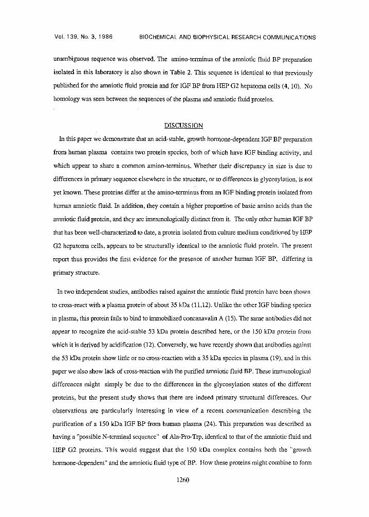
unambiguous sequence was observed. The amino-terminus of the amniotic fluid BP preparation
isolated in this laboratory is also shown in Table 2. This sequence is identical to that previously
published for the amniotic fluid protein and for IGF BP from HEP G2 hepatoma cells (4, 10). No
homology was seen between the sequences of the plasma and amniotic fluid proteins.
DISCUSSION
In this paper we demonstrate that an acid-stable, growth hormone-dependent IGF BP preparation
from human plasma contains two protein species, both of which have IGF binding activity, and
which appear to share a common amino-terminus. Whether their discrepancy in size is due to
differences in primary sequence elsewhere in the structure, or to differences in glycosylation, is not
yet known. These proteins differ at the amino-terminus from an IGF binding protein isolated from
human amniofic fluid. In addition, they contain a higher proportion of basic amino acids than the
amniotic fluid protein, and they are immunologically distinct from it. The only other human IGF BP
that has been well-characterized to date, a protein isolated from culture medium conditioned by HEP
G2 hepatoma cells, appears to be structurally identical to the amniotic fluid protein. The present
report thus provides the first evidence for the presence of another human IGF BP, differing in
primary structure.
In two independent studies, antibodies raised against the amniotic fluid protein have been shown
to cross-react with a plasma protein of about 35 kDa (11,12). Unlike the other IGF binding species
in plasma, this protein fails to bind to immobilized concanavalin A (15). The same antibodies did not
appear to recognize the acid-stable 53 kDa protein described here, or the 150 kDa protein from
which it is derived by acidification (12). Conversely, we have recently shown that antibodies against
the 53 kDa protein show little or no cross-reaction with a 35 kDa species in plasma (19), and in this
paper we also show lack of cross-reaction with the purified amniotic fluid BP. These immunological
differences might simply be due to the differences in the glyeosylation states of the different
proteins, but the present study shows that there are indeed primary structural differences. Our
observations are particularly interesting in view of a recent communication describing the
purification of a 150 kDa IGF BP from human plasma (24). This preparation was described as
having a "possible N-terminal sequence" of Ala-Pro-Trp, identical to that of the amniotic fluid and
HEP G2 proteins. This would suggest that the 150 kDa complex contains both the "growth
hormone-dependent" and the amniotic fluid type of BP. How these proteins might combine to form
1260
Vol. 139, No. 3, 1986 BIOCHEMICAL AND BIOPHYSICAL RESEARCH COMMUNICATIONS
the 150 kDa species, and the functional significance of their combination, remain important areas for
investigation.
ACKNOWLEDGEMENT
This study was supported by the National Health and Medical Research Council, Australia.
REFERENCES
1. Zapf, J., Waldvogel, M., and Froesch, E.R. (1975) Arch. Biochem. Biophys. 168, 638-645. 2. Hintz, R.L., and Liu, F. (1977) J. Clin. Endocrinol. Metab. 45, 988-995. 3. Drop, S.L.S., Kortleve, D.J., and Guyda, H.J. (1984) J. Clin. Endocrinol. Metab. 59,
899-907. 4. Povoa, G., Enberg, G., Jornvall, H., and Hall, K. (1984) Eur. J. Biochem. 144, 199-204. 5. Binoux, M., Hardouin, S., Lassarre, C., and Hossenlopp, P. (1982) J. Clin. Endocrinol.
Metab. 55, 600-602. 6. Baxter, R.C., Maitland, J.E., Raison, R.L., Reddel, R.R., and Sutherland, R.L. (1983) In :
Insulin-like Growth Factors - Somatomedins, pp. 615-618 (ed. E.M. Spencer), de Gruyter, Berlin.
7. Baxter, R.C., Zaltsman, Z., and Turtle, J.R. (1984) J. Clin. Endocrinol. Metab. 58, 955-959.
8. Baxter, R.C., Martin, J.L., and Handelsman, D. J. (1984) Acta Endocrinol. 106; 420-427. 9. Adams, S.O., Kapadia, M., Mills, B., and Daughaday, W.H. (1984) Endocrinology 115,
520-526. 10. Povoa, G., Isaksson, M., Jornvall, H., and Hall, K. (1985) Biochem. Biophys. Res.
Commun. 128, 1071-1078. 11. Drop, S.L.S., Kortleve,D.J., Guyda, H.J., and Posner, B.I. (1984) J. Clin. Endocrinol.
Metab. 59, 908-915. 12. Povoa, G., Roovete, A., and Hall, K. (1984) Acta Endocrinol. 107, 563-570. 13. Hintz, R.L., Liu, F., Rosenfeld, R.G., and Kemp, S.F. (1981) J. Clin. Endocrinol. Metab.
53, 100-104. 14. Daughaday, W.H., Ward, A.P., Goldberg, A.C., Trivedi, B., and Kapadia, M. (1982) J. Clin.
Endocrinol. Metab. 55, 916-921. 15. Wilkins, J.R., and D'Ercole, A.J. (1985) J. Clin. Invest. 75, 1350-1358. 16. Furlanetto, R.W. (1980) J. Clin. Endocrinol. Metab. 51, 12-19. 17. Morris, D.H., and Schalch, D.S. (1982) Endocrinology 111,801-805 18. Binoux, M., Seurin, D., Lassarre, C., and Gourmelen, M. (1984) J. Clin. Endocrinol. Metab.
59, 453-462. 19. Martin, J.L., and Baxter, R.C. (1985) J. Clin. Endocrinol. Metab. 61,799-801. 20. Martin, J.L., and Baxter, R.C. (1986) J. Biol. Chem., in press. 21. Laemmli, U.K. (1970) Nature 227, 680-685. 22. Towbin, H., Staehelin, T., and Gordon, J. (1979) Proc. Natl. Acad. Sci. USA 76, 4350-4354. 23. Hewick, R.M., Hunkapiller, M.W., Hood, L.E., and Dreyer, W.J. (1981) J. Biol. Chem.
256, 7990-7997. 24. Enberg, G. (1986) Biochem. Biophys. Res. Commun. 135, 178-182.
1261