growth hormone-releasing hormone receptor … · growth hormone-releasing hormone receptor ......
TRANSCRIPT
Growth hormone-releasing hormone receptorantagonists inhibit human gastric cancer throughdownregulation of PAK1–STAT3/NF-κB signalingJinfeng Gana,1, Xiurong Kea,1, Jiali Jianga,1, Hongmei Donga,1, Zhimeng Yaoa, Yusheng Lina, Wan Lina, Xiao Wub,Shumei Yanc, Yixuan Zhuangb, Wai Kit Chud, Renzhi Caie,f,g,h,i, Xianyang Zhange,f,g,h,i, Herman S. Cheunge,f,j,Norman L. Blockk, Chi Pui Pangd,l, Andrew V. Schallye,f,g,h,i,k,2, and Hao Zhanga,b,m,2
aCancer Research Center, Shantou University Medical College, Shantou 515041, China; bTumor Tissue Bank, Affiliated Cancer Hospital of Shantou UniversityMedical College, Shantou 515041, China; cDepartment of Pathology, Sun Yat-sen University Cancer Center, Guangzhou 510060, China; dDepartment ofOphthalmology & Visual Sciences, The Chinese University of Hong Kong, Hong Kong, China; eEndocrine, Polypeptide, and Cancer Institute, Veterans AffairsMedical Center, Miami, FL 33125; fSouth Florida Veterans Affairs Foundation for Research and Education, Miami, FL 33125; gDivision of Hematology andOncology, Department of Medicine, Miller School of Medicine, University of Miami, Miami, FL 33136; hDivision of Endocrinology, Department of Medicine,Miller School of Medicine, University of Miami, Miami, FL 33136; iSylvester Comprehensive Cancer Center, Miller School of Medicine, University of Miami,Miami, FL 33136; jDepartment of Biomedical Engineering, University of Miami, Coral Gables, FL 33146; kDepartment of Pathology, Miller School ofMedicine, University of Miami, Miami, FL 33136; lJoint Shantou International Eye Center, Shantou University and The Chinese University of Hong Kong,Shantou 515041, China; and mDepartment of Biotherapy, Affiliated Cancer Hospital of Shantou University Medical College, Shantou 515041, China
Contributed by Andrew V. Schally, November 16, 2016 (sent for review August 23, 2016; reviewed by Mong-Hong Lee, Oliver Sartor, Ratna K. Vadlamudi,and Qiang Yu)
Gastric cancer (GC) ranks as the fourth most frequent in incidenceand second in mortality among all cancers worldwide. The develop-ment of effective treatment approaches is an urgent requirement.Growth hormone-releasing hormone (GHRH) and GHRH receptor(GHRH-R) have been found to be present in a variety of tumoraltissues and cell lines. Therefore the inhibition of GHRH-Rwas proposedas a promising approach for the treatment of these cancers. However,little is known about GHRH-R and the relevant therapy in humanGC. By survival analyses of multiple cohorts of GC patients, weidentified that increased GHRH-R in tumor specimens correlates withpoor survival and is an independent predictor of patient prognosis.We next showed that MIA-602, a highly potent GHRH-R antagonist,effectively inhibited GC growth in cultured cells. Further, thisinhibitory effect was verified in multiple models of human GC celllines xenografted into nude mice. Mechanistically, GHRH-R antago-nists target GHRH-R and down-regulate the p21-activated kinase 1(PAK1)-mediated signal transducer and activator of transcription 3(STAT3)/nuclear factor-κB (NF-κB) inflammatory pathway. Overall,our studies establish GHRH-R as a potential molecular target in humanGC and suggest treatment with GHRH-R antagonist as a promisingtherapeutic intervention for this cancer.
GHRH receptor | GHRH-R antagonist | PAK1 | stomach cancer |prognostic predictor
Gastric cancer (GC) ranks as the fourth most common cancerin incidence and the second most frequent in mortality
among all cancers worldwide (1). Surgery remains the only cu-rative therapy for GC, and it must be accomplished in a timelymanner. The 5-y relative survival rate of GC is less than 25% (2).Despite recent advances, the molecular mechanisms underlyinggastric tumorigenesis remain largely unknown. Several moleculartargets including human epidermal growth factor receptor 2 (HER2),epidermal growth factor receptor (EGFR), c-MET, VEGR2, HGF,and mTOR have been proposed in human GC (2–4). However, theresponse rates related to these therapeutic approaches vary con-siderably (4). Thus, the development of novel molecular targets andderived therapeutic strategies is an urgent need.Increasing evidence suggests that GC is a type of inflammation-
associated cancer caused by the complex interaction between hostand environmental factors (5). Infection with the pathogenHelicobacter pylori, which triggers chronic gastritis, remains thestrongest single risk factor for human GC (6). Numerous cellularand molecular pathways, which converge at the level of the signaltransducer and activator of transcription 3 (STAT3) and nuclear
factor-κB (NF-κB) (7–10), are involved in this inflammation-drivengastric tumorigenesis and progression.Growth hormone-releasing hormone (GHRH) is a neuropeptide
produced in the hypothalamus. In the anterior pituitary, GHRHregulates the synthesis and secretion of growth hormone (GH)(11, 12) upon binding to GHRH receptor (GHRH-R) and sub-sequently exerts mitogenic activity for pituitary cells (11, 12).GHRH and GHRH-R had been demonstrated to be expressedpredominantly in the anterior pituitary gland but also foundmodestly in other somatic cells. However, accumulating evidenceshows that both GHRH and GHRH-R are significantly presentin various cancers including breast, prostate, ovarian, pancreatic,colon, gastric, and lung cancers, lymphoma, and glioblastoma
Significance
Gastric cancer (GC) is a leading and lethal malignancy in the world.Unfortunately, therapeutics targeting GC lag behind those of manyother cancers, and the effective treatment options are limited. Herewe report that increased expression of growth hormone-releasinghormone receptor (GHRH-R) in tumor specimens is significantlyassociated with tumorigenic progression and poor clinical outcomeof GC patients. MIA-602, a highly potent GHRH-R antagonist, ef-fectively inhibits GC growth in vitro and in vivo. The anti-neoplasticeffects were mediated by blockage of the p21-activated kinase 1(PAK1)–signal transducer and activator of transcription 3 (STAT3)/nuclear factor-κB (NF-κB) axis. Thus, GHRH-R presents a molecularmarker and therapeutic target of GC, and MIA-602 demonstratesexcellent therapeutic potential against human GC.
Author contributions: C.P.P., A.V.S., and H.Z. designed research; J.G., X.K., J.J., H.D., Z.Y.,Y.L., W.L., X.W., S.Y., and Y.Z. performed research; R.C. and A.V.S. contributed newreagents/analytic tools; W.K.C., X.Z., H.S.C., C.P.P., A.V.S., and H.Z. analyzed data; andH.S.C., N.L.B., C.P.P., A.V.S., and H.Z. wrote the paper.
Reviewers: M.-H.L., University of Texas MD Anderson Cancer Center; O.S., Tulane CancerCenter; R.K.V., University of Texas Health Science Center at San Antonio; and Q.Y., GenomeInstitute of Singapore.
Conflict of interest statement: A.V.S. and R.C. are listed as co-inventors on patents onGHRH analogs which were assigned to the University of Miami and VeteransAffairs Department.
Freely available online through the PNAS open access option.1J.G., X.K., J.J., and H.D. contributed equally to this work.2To whom correspondence may be addressed. Email: [email protected] or [email protected].
This article contains supporting information online at www.pnas.org/lookup/suppl/doi:10.1073/pnas.1618582114/-/DCSupplemental.
www.pnas.org/cgi/doi/10.1073/pnas.1618582114 PNAS | December 20, 2016 | vol. 113 | no. 51 | 14745–14750
CELL
BIOLO
GY
(11–19). The GHRH/GHRH-R pathway is considered a growthfactor-signaling pathway in these cancers and may modulate theactivities of multiple intracellular pathways (11–13). Thus, targetingthe GHRH/GHRH-R pathway has been proposed for the treat-ment of cancer (11, 12). Over the past three decades, various classesof GHRH-R antagonists have been developed that have shownstrong growth-inhibitory effects in cancer both in vitro and in vivo(11–14, 20). MIA-602 (14) represents the latest in a series ofGHRH-R antagonists and has been chosen for clinical develop-ment. We previously reported that GHRH-R is present in gastricmucosa (13), and GHRH-R mRNA is detectable in two GC celllines (17). However, little is known about the effects of a GHRH-Rantagonist, particularly on GC in vivo, and the mechanisms bywhich GHRH-R antagonist achieves anti-neoplastic effects in GC.Moreover, although GHRH-R expression has been discovered inmultiple cancers, the clinical relevance of GHRH-R in tumorigenicprogression and in clinical outcomes is largely elusive.
Among the multiple oncoproteins, a serine/threonine protein ki-nase designated “p21-activated kinase 1” (PAK1), which is stimulatedby active Rac1 and Cdc42-GTPases, works as a node of cancer-signaling networks (21, 22). PAK1 overexpression has been ob-served frequently in a variety of cancers. Aberrant PAK1 plays acritical role in tumor cell proliferation and invasiveness, therebymodulating oncogenesis and tumorigenic progression (21, 22). Be-cause PAK1 has been recognized as a potential pharmacologicalmolecular target, the clinical pipeline of molecular therapy targetingPAK1 has been growing (21–23). Our previous studies and those ofother investigators demonstrated that PAK1 is up-regulated incancers of the gastrointestinal tract, including GC (23–28). Intrigu-ingly, PAK1 also activates the inflammatory signaling induced byH. pylori infection in epithelial cells, further linking PAK1 to humanGC pathogenesis (29). Indeed, PAK1 not only induces the oncogenicsignaling but also promotes inflammatory pathways, such as STAT3and NF-κB (29–32), which are associated with inflammation-associated malignancy (7–10), suggesting that PAK1 has a functionin inflammation-related tumor progression.In this study, we show that aberrant GHRH-R is clinically
important in GC progression and patient prognosis. TargetingGHRH-R by using MIA-602 induces the inhibition of GC growthboth in vitro and in vivo. The inhibitory effects of GHRH-R an-tagonist are mediated by targeting the inflammatory signalingpathway of PAK1–STAT3/NF-κB.
ResultsOverexpression of GHRH-R Is Clinically Important for TumorigenicProgression and Is Associated with Overall Survival in Human GC.To evaluate the clinical significance of GHRH-R in human GC,we investigated GHRH-R expression in GC specimens from a106-patient cohort. By analysis of specimens immunohistochemicallystained for GHRH-R in primary GC specimens, along with paired
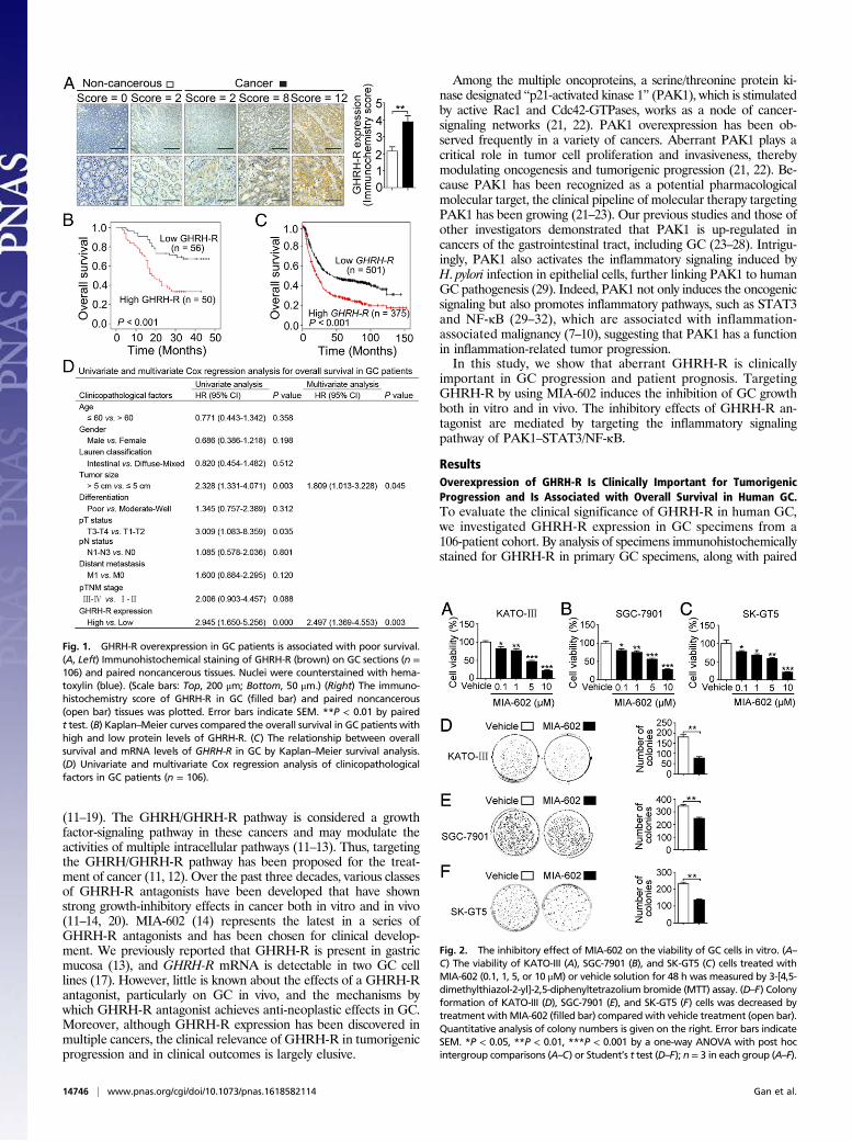
Fig. 1. GHRH-R overexpression in GC patients is associated with poor survival.(A, Left) Immunohistochemical staining of GHRH-R (brown) on GC sections (n =106) and paired noncancerous tissues. Nuclei were counterstained with hema-toxylin (blue). (Scale bars: Top, 200 μm; Bottom, 50 μm.) (Right) The immuno-histochemistry score of GHRH-R in GC (filled bar) and paired noncancerous(open bar) tissues was plotted. Error bars indicate SEM. **P < 0.01 by pairedt test. (B) Kaplan–Meier curves compared the overall survival in GC patients withhigh and low protein levels of GHRH-R. (C) The relationship between overallsurvival and mRNA levels of GHRH-R in GC by Kaplan–Meier survival analysis.(D) Univariate and multivariate Cox regression analysis of clinicopathologicalfactors in GC patients (n = 106).
Fig. 2. The inhibitory effect of MIA-602 on the viability of GC cells in vitro. (A–C) The viability of KATO-III (A), SGC-7901 (B), and SK-GT5 (C) cells treated withMIA-602 (0.1, 1, 5, or 10 μM) or vehicle solution for 48 h was measured by 3-[4,5-dimethylthiazol-2-yl]-2,5-diphenyltetrazolium bromide (MTT) assay. (D–F) Colonyformation of KATO-III (D), SGC-7901 (E), and SK-GT5 (F) cells was decreased bytreatment withMIA-602 (filled bar) compared with vehicle treatment (open bar).Quantitative analysis of colony numbers is given on the right. Error bars indicateSEM. *P < 0.05, **P < 0.01, ***P < 0.001 by a one-way ANOVA with post hocintergroup comparisons (A–C) or Student’s t test (D–F); n = 3 in each group (A–F).
14746 | www.pnas.org/cgi/doi/10.1073/pnas.1618582114 Gan et al.
adjacent normal tissues, we discovered that the GC tissues exhibitedrobust expression of GHRH-R compared with normal tissues (P <0.01) (Fig. 1A). To validate a reasonable cutoff score for GHRH-Roverexpression, a receiver operating characteristic (ROC) curveanalysis was performed (Fig. S1). Accordingly, the cutoff score forGHRH-R overexpression was 2.5 (immunochemistry score), whichwas closest to the point with both maximum sensitivity and speci-ficity. The cases with scores ≤2.5 were defined as having low ex-pression of GHRH-R, and those with scores >2.5 were defined ashaving overexpression of GHRH-R. Overexpression of GHRH-Rwas detected in 50 of 106 human primary GCs (47.17%). Wefurther examined the association of GHRH-R expression withclinicopathological features of malignant behavior. Correlationanalysis demonstrated that the overexpression of GHRH-R waspositively correlated with tumor size (P = 0.031) and patholog-ical tumor (pT) status (P = 0.001) (Table S1). Thus, GHRH-R ishighly enriched in human GC tissues and correlates closely withcancer progression.Further, Kaplan–Meier analysis demonstrated that GC pa-
tients with increasing expression of GHRH-R showed a pooroverall survival (P < 0.001, log-rank test) (Fig. 1B). We furtherconfirmed our findings in a larger cohort containing 876 GCpatients from a published dataset (kmplot.com/analysis/index.php?p=service&cancer=gastric) (33), which was multi-institutional andrepresented various races and countries, including Germany,Australia, Brazil, Korea, Singapore, and China. In line with aboveobservations, this dataset showed that higher GHRH-R expressionis associated with a poorer overall survival of GC patients (P <0.001, log-rank test) (Fig. 1C). Multivariate Cox regression analysisof the 106-patient cohort determined that GHRH-R expression isan independent predictor of prognosis for GC patients [hazardratio (HR) = 2.497, 95% confidence interval (CI) = 1.369–4.553,P = 0.003] (Fig. 1D). The survival analyses from these multiplecohorts confirmed that GHRH-R overexpression is positively as-sociated with poor overall survival of GC patients and is a potentialpredictor for GC patients.Because increased transcription and gene copy number gain or
amplification are alternative mechanisms of oncoprotein over-
expression, we examined the status of mRNA expression andgene copy number of GHRH-R in human GC. In a publisheddataset (GSE13861) including 65 GC specimens and 19 adjacentnormal tissues (34), GHRH-R mRNA was found to be signifi-cantly elevated in GC specimens vs. normal controls (P < 0.001)(Fig. S2A). Supporting the above findings, 21 of 24 GC cell lineshad higher expression of GHRH-R mRNA than did normalgastric tissues (n = 10, MERAV database, merav.wi.mit.edu/)(Fig. S2B) (35). Next, the amplification of the GHRH-R gene inhuman GC was observed by analyzing DNA copy number usingthe Oncomine database (Fig. S2C) (36). Thus, in addition toprotein overexpression, GHRH-R mRNA is also overexpressedin human GC tissues, along with gene amplification. Togetherthese findings show that increased GHRH-R is clinically im-portant in tumorigenic progression in human GC and correlatesclosely with patient outcome.
The GHRH-R Antagonist MIA-602 Inhibits the Growth of Human GCCells in Vitro. The clinical importance of GHRH-R involvementin tumor progression and patient outcomes underscores thegreat therapeutic promise of targeting GHRH-R in human GC.We therefore determined the inhibitory efficacy of MIA-602,one of the latest GHRH-R antagonists, on human GC cells. Wetreated three human GC cell lines, KATO-III, SGC-7901, andSK-GT5, with MIA-602 at 0.1-, 1-, 5-, and 10-μM concentrationsfor 48 h (15, 16, 37). MIA-602 treatment resulted in reduced cellviability in all cell lines in a dose-dependent manner, as comparedwith vehicle controls (P < 0.05 for all) (Fig. 2 A–C). Because astriking decrease, by ∼25% (IC25), was seen in cell viability at 1 μMfollowing MIA-602 treatment (Fig. 2 A–C), we chose the dosage of1 μM for subsequent studies. After exposure to 1 μM MIA-602 for9 d, all treated cell lines formed fewer colonies than the same celllines treated with vehicle (control) (P < 0.01 for all) (Fig. 2 D–F).Cellular proliferation represented by phospho-Histone H3 (pH3)immunofluorescence was decreased in all MIA-602–treated cellscompared with controls (P < 0.01 for all) (Fig. S3). Therefore, theGHRH-R antagonist MIA-602 inhibits GC cell growth.
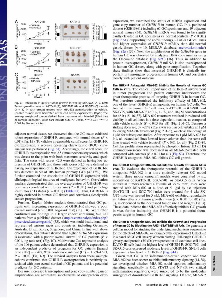
The GHRH-R Antagonist MIA-602 Inhibits the Growth of Human GC inVivo. To evaluate the therapeutic potential of the GHRH-Rantagonist MIA-602 in a more clinically relevant GC modelsystem, three mouse xenograft models were generated by s.c.inoculation of KATO-III, SGC-7901, or SK-GT5 cells. Whenindividual tumors reached a volume of 75.89 mm3, mice weretreated with MIA-602 at a dose of 5 μg/d by s.c. injection(KATO-III- and SGC-7901-mice were treated for 4 wk; SK-GT5-mice was treated for 5 wk). MIA-602 exhibited remarkableinhibitory effects on tumor growth in vivo (P < 0.001 for all) (Fig.3), as evidenced by the decreased tumor size and weight (Fig. 3).These data indicate that MIA-602 effectively inhibits GC growthin vivo, further indicating that GHRH-R is a potential thera-peutic target in human GC.
The GHRH-R Antagonist MIA-602 Inhibits the Growth and Progressionof Human GC by Blocking the PAK1–STAT3/NF-κB Axis. To generate acellular model for studying the underlying mechanisms responsiblefor the effects of MIA-602, we examined the expression of GHRH-Rin a panel of GC cell lines by Western blotting. Full-length GHRH-Rglycosylated protein (55 kDa) was present in all examined cell lines.KATO-III cells had the highest level of GHRH-R; SGC-7901 andSK-GT5 cells expressed moderate levels of GHRH-R; SNU-1 cellsexpressed a low level of GHRH-R (Fig. S4A).Given that GC is an inflammation-driven cancer, and that
MIA-602 has been shown to inhibit inflammatory signaling (14, 38),we investigated whether MIA-602 could suppress inflammatorysignaling, specifically in GC. STAT3 and NF-κB, two criticalinflammation regulators, were suspected to be the molecularsurrogates of downstream GHRH-R signaling. Of note, MIA-602
Fig. 3. Inhibition of gastric tumor growth in vivo by MIA-602. (A–C, Left)Tumor growth curves of KATO-III (A), SGC-7901 (B), and SK-GT5 (C) models(n = 12 in each group) treated with MIA-602 administration or vehicle.(Center) Tumors were harvested at the end of the experiments. (Right) Theaverage weights of tumors derived from treatment with MIA-602 (filled bar)or control (open bar). Error bars indicate SEM. *P < 0.05, **P < 0.01, ***P <0.001 by Student’s t test.
Gan et al. PNAS | December 20, 2016 | vol. 113 | no. 51 | 14747
CELL
BIOLO
GY
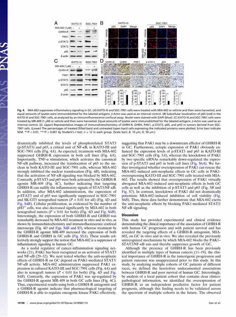
dramatically inhibited the levels of phosphorylated STAT3(p-STAT3) and p65, a critical unit of NF-κB, in KATO-III andSGC-7901 cells (Fig. 4A). As expected, treatment with MIA-602suppressed GHRH-R expression in both cell lines (Fig. 4A).Importantly, TNF-α stimulation, which activates the canonicalNF-κB pathway, increased the translocation of p65 to the nu-cleus in both KATO-III and SGC-7901 cells, whereas MIA-602strongly inhibited the nuclear translocation (Fig. 4B), indicatingthat the activation of NF-κB signaling was blocked by MIA-602.Contrarily, p-STAT3 and p65 were both activated by the GHRH-Ragonist MR-409 (Fig. 4C), further indicating that targetingGHRH-R can nullify the inflammatory signals of STAT3/NF-κB.In addition, after MIA-602 administration, the expression ofp-STAT3 and of p65 was significantly suppressed in SGC-7901and SK-GT5 xenografted tumors (P < 0.01 for all) (Fig. 4D andFig. S4B). Cellular proliferation, as evidenced by the number ofpH3+ cells, was also decreased significantly by MIA-602 in bothxenografted tumors (P < 0.01 for both) (Fig. 4D and Fig. S4B).Interestingly, the expression of both GHRH-R and GHRH wasremarkably decreased by MIA-602 treatment in vitro and in vivo, asshown by immunohistochemistry and immunofluorescence confocalmicroscopy (Fig. 4D and Figs. S4B and S5), whereas treatment bythe GHRH-R agonist MR-409 increased the expression of bothGHRH-R and GHRH in GC cells (Fig. S5A). These results col-lectively strongly support the notion that MIA-602 is a suppressor ofinflammatory signaling in human GC.As a nodal regulator of cancer-inflammation signaling net-
works (21), PAK1 has been recognized as an activator of STAT3and NF-κB (29–32). We next tested whether the anti-neoplasticeffects of GHRH-R on GC depend on PAK1-mediated STAT3/NF-κB activity. MIA-602 administration suppressed PAK1 ex-pression in cultured KATO-III and SGC-7901 cells (Fig. 4A) andalso in xenograft tumors (P < 0.01 for both) (Fig. 4D and Fig.S4B). Contrarily, the expression of PAK1 was up-regulated bythe GHRH-R agonist MR-409 in both GC cells lines (Fig. 4C).Thus, experimental results using both a GHRH-R antagonist anda GHRH-R agonist indicate that pharmacological targeting ofGHRH-R is able to regulate oncogenic kinase PAK1 effectively,
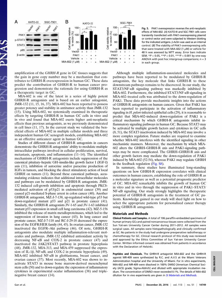
suggesting that PAK1 may be a downstream effector of GHRH-Rin GC. Furthermore, ectopic expression of PAK1 obviously en-hanced the expression levels of p-STAT3 and p65 in KATO-IIIand SGC-7901 cells (Fig. 5A), whereas the knockdown of PAK1by two specific siRNAs remarkably down-regulated the expres-sion of p-STAT3 and p65 in both cell lines (Fig. S6A). We fur-ther investigated whether overexpression of PAK1 can rescue theMIA-602–induced anti-neoplastic effects in GC cells in PAK1-overexpressing KATO-III and SGC-7901 cells treated with MIA-602. The results showed that overexpression of PAK1 virtuallyabrogated MIA-602–induced anti-neoplastic effects in both GCcells as well as the inhibition of p-STAT3 and p65 (Fig. 5B andFig. S7). In contrast, knockdown of PAK1 did not dramaticallypotentiate MIA-602–induced inhibition of cell viability (Fig.S6B). Thus, these data further demonstrate that MIA-602 exertsthe anti-neoplastic effects by blocking PAK1-mediated STAT3/NF-κB signaling.
DiscussionThis study has provided experimental and clinical evidencedemonstrating the clinical importance of the association of GHRH-Rwith human GC progression and with patient survival and hasrevealed the targeting effects of a GHRH-R antagonist, MIA-602, on GC in vitro and in vivo. We also revealed the previouslyunrecognized mechanisms by which MIA-602 blocks the PAK1–STAT3/NF-κB axis and thereby suppresses growth of GC.Although the presence of GHRH-R has been previously
identified in multiple types of human cancers (11–19), the clin-ical importance of GHRH-R in the tumorigenic progression andpatient outcome was unappreciated prior to this study. In thisstudy, by analyzing multiple cohorts of GC patients of differentraces, we defined the heretofore undocumented associationsbetween GHRH-R and poor survival of human GC. Interestingly,by analysis of a local patient cohort that contains clear clinico-pathological information, we identified the overexpression ofGHRH-R as an independent predictive factor for patientprognosis, although this finding needs to be validated acrossthe spectrum of multiple cohorts in the future. The observed
Fig. 4. MIA-602 suppresses inflammatory signaling in GC. (A) KATO-III and SGC-7901 cells were treated with MIA-602 or vehicle and then were harvested, andequal amounts of lysates were immunoblotted for the labeled antigens. β-Actin was used as an internal control. (B) Subcellular localization of p65 (red) in theKATO-III and SGC-7901 cells, as analyzed by an immunofluorescence confocal assay. Nuclei were stained with DAPI (blue). (C) KATO-III and SGC-7901 cells weretreated by MR-409 (1 μM) or vehicle and then were harvested. Equal amounts of lysates were immunoblotted for the labeled antigens. β-Actin was used as aninternal control. (D, Upper) Representative images of immunohistochemistry of GHRH-R, GHRH, PAK1, p-STAT3, p65, and pH3 in tumors derived from SGC-7901 cells. (Lower) The percentages of treated (filled bars) and untreated (open bars) cells expressing the indicated proteins were plotted. Error bars indicateSEM. **P < 0.01, ***P < 0.001 by Student’s t test; n = 12 in each group. (Scale bars: B, 10 μm; D, 50 μm.)
14748 | www.pnas.org/cgi/doi/10.1073/pnas.1618582114 Gan et al.
amplification of the GHRH-R gene in GC tissues suggests thatthe gain in gene copy number may be a mechanism that con-tributes to GHRH-R overexpression in human GC. These datapredict the contribution of GHRH-R to human cancer pro-gression and demonstrate the rationale for using GHRH-R asa therapeutic target in GC.MIA-602 is one of the latest in a series of highly potent
GHRH-R antagonists and is based on an earlier antagonist,JMR-132 (11, 15, 16, 37). MIA-602 has been reported to possessgreater potency and stability in antitumor activity than JMR-132(15). Using MIA-602, we systemically examined its therapeuticeffects by targeting GHRH-R in human GC cells in vitro andin vivo and found that MIA-602 exerts higher anti-neoplasticeffects than previous antagonists, as we previously demonstratedin cell lines (11, 17). In the current study we validated the ben-eficial effects of MIA-602 in multiple cellular models and threeindependent human GC xenograft models, establishing MIA-602as an effective anticancer agent in human GC.Studies of different classes of GHRH-R antagonists in cancers
demonstrate the GHRH-R antagonists’ ability to modulate multipleintracellular pathways involved in cellular proliferation, survival,metastasis, apoptosis, and inflammation (11–14). The identifiedmechanisms of GHRH-R antagonists include suppression of thecanonical pituitary–hepatic GH–insulin-like growth factor I (IGF-I)axis (11), inhibition of autocrine and/or paracrine production ofIGFs in tumors, and blocking the direct stimulatory action of localGHRH on tumors (11). Beyond these canonical pathways, accu-mulating evidence indicates that additional intracellular moleculesand pathways can be modulated by GHRH-R antagonists. JMR-132 induced cell-growth inhibition and apoptosis through PKCδ-mediated activation of p53/p21 in endometrial cancer (39) andcaused p21-mediated S-phase arrest in colon cancer (40). AnotherGHRH-R antagonist, MZ-J-7-138, up-regulated wild-type p53 butdown-regulated mutant p53 and p21 in prostate cancer (41).Similarly, the GHRH-R antagonists JV-1-65 and JV-1-63 inhibitedmutant p53 expression in small cell lung carcinoma (42). MZ-5-156inhibited the release of matrix metalloproteinases, which led to thesuppression of invasion in lung cancer (43). In lung cancer andprostate cancer, MZ-J-7-118 and RC-J-29-18 decreased the expres-sion of the EGFR/HER family (44, 45). In ovarian cancer, JMR-132inactivated the EGFR–Akt pathway (46). Of note, GHRH-Rantagonists also modulate multiple inflammation-relevant mol-ecules and pathways. JMR-132 exhibited antioxidant activity bydecreasing NF-κB and COX-2 in prostate cancer (47) andinactivated the JAK2/STAT3 pathway in prostate hyperplasia(48). JMR-132, MIA-313, and MIA-459 suppressed the expres-sion of IL-1β, NF-κB, and COX-2 in prostatic hyperplasia (49).MIA-602 inhibited NF-κB in glioblastoma, breast cancer, andovarian cancer (37). Most recently, MIA-602 was shown to in-activate STAT3 in mouse bone marrow-derived mesenchymalstem cells (50) and to down-regulate the expression of inflammatorycytokines in experimental ocular inflammation (38) and triple-negative breast cancer (14).
Although multiple inflammation-associated molecules andpathways have been reported to be modulated by GHRH-Rantagonists, the key molecule that links GHRH-R to thesedownstream pathways remains to be discovered. In our study, theSTAT3/NF-κB signaling pathway was markedly inhibited byMIA-602. Furthermore, the inhibited STAT3/NF-κB signaling inMIA-602–treated cells was attributed to the down-regulation ofPAK1. These data provide mechanistic insights into the actionsof GHRH-R antagonists on human cancers. Given that PAK1 hasbeen reported to participate in the activation of inflammatorysignaling inH. pylori-infected epithelial cells (29), it is reasonable topredict that MIA-602–induced down-regulation of PAK1 is acritical mechanism by which GHRH-R antagonists inhibit in-flammatory signaling pathways in human GC. Because STAT3 maybe activated by multiple growth factors and cytokines in GC cells(8, 51), the STAT3 inactivation induced by MIA-602 may involve amore complex regulation. Future studies are required to examinewhether MIA-602 interferes with the identified signaling in othermechanistic manners. Moreover, the mechanism by which MIA-602 alters the GHRH–GHRH-R axis and PAK1-signaling path-ways may be more complicated. The GHRH–GHRH-R axis andother growth factors may mediate the down-regulation of PAK1induced by MIA-602 (52-54), whereas PAK1 may regulate GHRHin the feedback regulation (Fig. S8).In summary, these studies have illuminated long-standing
questions on how GHRH-R expression correlates with clinicaloutcomes in human cancers, establishing the role of GHRH-R asa molecular signature as well as a potential prognostic predictorof GC. MIA-602 remarkably inhibits the growth of human GCin vitro and in vivo through the suppression of PAK1–STAT3/NF-κB signaling. Our study strongly highlights the therapeuticpotential of GHRH-R antagonists in the treatment of GC pa-tients. Knowledge gained in our study will shed light on how toselect the appropriate patients for personalized cancer therapyusing GHRH-R antagonists.
Materials and MethodsClinical Patients and Samples. A total of 106 paraffin-embedded specimens ofhuman primary GCs and paired noncancerous tissues were collected from theSun Yat-sen University Cancer Center during 2011–2012. All samples weresurgical cases. All samples were histopathologically and clinically confirmedas GC. No patients in the study had undergone preoperative radiotherapy orchemotherapy for GC. Clinical research protocol of this study was reviewedand approved by the Ethics Committee of Sun Yat-sen University CancerCenter. Written informed consent was obtained from patients in accordancewith the Declaration of Helsinki.
Peptides and Chemicals. The GHRH-R antagonist MIA-602 and the GHRH-Ragonist MR-409 were synthesized by R.C. and A.V.S at the Miami VeteransAdministration hospital and the University of Miami. For in vitro experiments,MIA-602 and MR-409 were dissolved in 100% DMSO [American Chemical Soci-ety (ACS) grade; Sigma-Aldrich] and diluted with appropriate incubation me-dium. The concentration of DMSO never exceeded 0.1%. The details of MIA-602dilution for in vivo experiments are given in SI Materials and Methods.
Fig. 5. PAK1 overexpression reverses the anti-neoplasticeffects of MIA-602. (A) KATO-III and SGC-7901 cells weretransiently transfected with PAK1-overexpressing plasmidor control vector and were subjected to Western blottingfor the indicated antigens. β-Actin was used as an internalcontrol. (B) The viability of PAK1-overexpressing cellsthat were treated with MIA-602 (1 μM) or vehicle for48 h was assessed by MTT assay. Error bars indicateSEM. *P < 0.05, **P < 0.01, ***P < 0.001 by one-wayANOVA with post hoc intergroup comparisons; n = 3in each group.
Gan et al. PNAS | December 20, 2016 | vol. 113 | no. 51 | 14749
CELL
BIOLO
GY
Tumor Xenografts. A total of 2 × 10⁶ KATO-III, SGC-7901, or SK-GT5 cellswere resuspended in 150 μL PBS and were injected s.c. into the flanks of 5- to6-wk-old nude mice (Vital River Laboratory Animal Technology Co. Ltd.).Details of experiments with the tumor xenografts are given in SI Materialsand Methods. These animal experiments were reviewed and approved bythe Ethics Committee of Shantou University Medical College.
Materials and methods are described further in SI Materials and Methods.
ACKNOWLEDGMENTS. We thank the Li Ka Shing Foundation for generoussupport. This work was supported by a block grant from the UniversityGrants Committee Hong Kong (to C.P.P.) and in part by National Natural
Science Foundation of China Grants 81071736, 30973508, and 81572876 andthe Clinical Research Enhancement Initiative of Shantou University MedicalCollege Grants 201412 and 201421 (all to H.Z.), by funding for the Collabo-rative and Creative Center, Molecular Diagnosis and Personalized Medicine,Shantou University, Guangdong Province (H.Z.), and by funding from theDepartment of Education, Guangdong Government under the Top-tier Uni-versity Development Scheme for Research and Control of Infectious Diseases.The work at the Veterans Affairs Medical Center, Miami, was supportedby the Medical Research Service of the Department of Veterans Affairs(A.V.S.), the South Florida Veterans Affairs Foundation for Research andEducation (A.V.S.), and the Sylvester Comprehensive Cancer Center, MillerSchool of Medicine of the University of Miami (A.V.S.).
1. Torre LA, et al. (2015) Global cancer statistics, 2012. CA Cancer J Clin 65(2):87–108.2. Van Cutsem E, Sagaert X, Topal B, Haustermans K, Prenen H (2016) Gastric cancer.
Lancet S0140-6736(16)30354-3, 10.1016/S0140-6736(16)30354-3.3. Wadhwa R, et al. (2013) Gastric cancer-molecular and clinical dimensions. Nat Rev Clin
Oncol 10(11):643–655.4. Lordick F, Janjigian YY (2016) Clinical impact of tumour biology in the management
of gastroesophageal cancer. Nat Rev Clin Oncol 13(6):348–360.5. Elinav E, et al. (2013) Inflammation-induced cancer: Crosstalk between tumours, im-
mune cells and microorganisms. Nat Rev Cancer 13(11):759–771.6. Polk DB, Peek RM, Jr (2010) Helicobacter pylori: Gastric cancer and beyond. Nat Rev
Cancer 10(6):403–414.7. Bollrath J, Greten FR (2009) IKK/NF-kappaB and STAT3 pathways: Central signalling hubs in
inflammation-mediated tumour promotion and metastasis. EMBO Rep 10(12):1314–1319.8. Yu H, Lee H, Herrmann A, Buettner R, Jove R (2014) Revisiting STAT3 signalling in
cancer: New and unexpected biological functions. Nat Rev Cancer 14(11):736–746.9. Lee ST, et al. (2011) Context-specific regulation of NF-κB target gene expression by
EZH2 in breast cancers. Mol Cell 43(5):798–810.10. Feng Y, et al. (2014) Metformin promotes autophagy and apoptosis in esophageal
squamous cell carcinoma by downregulating Stat3 signaling. Cell Death Dis 5:e1088.11. Schally AV, Varga JL, Engel JB (2008) Antagonists of growth-hormone-releasing hormone:
An emerging new therapy for cancer. Nat Clin Pract Endocrinol Metab 4(1):33–43.12. Schally AV, Varga JL (1999) Antagonistic analogs of growth hormone-releasing hor-
mone: New potential antitumor agents. Trends Endocrinol Metab 10(10):383–391.13. Barabutis N, Schally AV (2010) Growth hormone-releasing hormone: Extrapituitary
effects in physiology and pathology. Cell Cycle 9(20):4110–4116.14. Perez R, et al. (2012) Antagonists of growth hormone-releasing hormone suppress in vivo
tumor growth and gene expression in triple negative breast cancers.Oncotarget 3(9):988–997.15. Pozsgai E, et al. (2010) The effect of GHRH antagonists on human glioblastomas and
their mechanism of action. Int J Cancer 127(10):2313–2322.16. Klukovits A, et al. (2012) Novel antagonists of growth hormone-releasing hormone inhibit
growth and vascularization of human experimental ovarian cancers. Cancer 118(3):670–680.17. Busto R, et al. (2002) The expression of growth hormone-releasing hormone (GHRH)
and splice variants of its receptor in human gastroenteropancreatic carcinomas. ProcNatl Acad Sci USA 99(18):11866–11871.
18. Fahrenholtz CD, et al. (2014) Preclinical efficacy of growth hormone-releasing hor-mone antagonists for androgen-dependent and castration-resistant human prostatecancer. Proc Natl Acad Sci USA 111(3):1084–1089.
19. Havt A, et al. (2005) The expression of the pituitary growth hormone-releasing hor-mone receptor and its splice variants in normal and neoplastic human tissues. ProcNatl Acad Sci USA 102(48):17424–17429.
20. Zarandi M, et al. (1994) Synthesis and biological activities of highly potent antagonistsof growth hormone-releasing hormone. Proc Natl Acad Sci USA 91(25):12298–12302.
21. Dammann K, Khare V, Gasche C (2014) Tracing PAKs from GI inflammation to cancer.Gut 63(7):1173–1184.
22. Vadlamudi RK, et al. (2005) An essential role of Pak1 phosphorylation of SHARP inNotch signaling. Oncogene 24(28):4591–4596.
23. Li Z, et al. (2015) Personalizing risk stratification by addition of PAK1 expression toTNM staging: Improving the accuracy of clinical decision for gastroesophageal junc-tion adenocarcinoma. Int J Cancer 136(7):1636–1645.
24. Ching YP, et al. (2007) P21-activated protein kinase is overexpressed in hepatocellularcarcinoma and enhances cancer metastasis involving c-Jun NH2-terminal kinase acti-vation and paxillin phosphorylation. Cancer Res 67(8):3601–3608.
25. Carter JH, et al. (2004) Pak-1 expression increases with progression of colorectalcarcinomas to metastasis. Clin Cancer Res 10(10):3448–3456.
26. Wang G, et al. (2015) PAK1-mediated MORC2 phosphorylation promotes gastric tu-morigenesis. Oncotarget 6(12):9877–9886.
27. Li Z, et al. (2013) Prognostic importance and therapeutic implications of PAK1, a drugableprotein kinase, in gastroesophageal junction adenocarcinoma. PLoS One 8(11):e80665.
28. Gan J, et al. (2015) Dysregulation of PAK1 is associated with DNA damage and is ofprognostic importance in primary esophageal small cell carcinoma. Int J Mol Sci 16(6):12035–12050.
29. Foryst-Ludwig A, Naumann M (2000) p21-activated kinase 1 activates the nuclear factorkappa B (NF-kappa B)-inducing kinase-Ikappa B kinases NF-kappa B pathway and proin-flammatory cytokines in Helicobacter pylori infection. J Biol Chem 275(50):39779–39785.
30. Shrestha Y, et al. (2012) PAK1 is a breast cancer oncogene that coordinately activatesMAPK and MET signaling. Oncogene 31(29):3397–3408.
31. Zhou W, et al. (2014) PAK1 mediates pancreatic cancer cell migration and resistanceto MET inhibition. J Pathol 234(4):502–513.
32. Dammann K, et al. (2015) PAK1 modulates a PPARγ/NF-κB cascade in intestinal in-flammation. Biochim Biophys Acta 1853(10 Pt A):2349–2360.
33. Szász AM, et al. (2016) Cross-validation of survival associated biomarkers in gastric cancerusing transcriptomic data of 1,065 patients. Oncotarget, 10.18632/oncotarget.10337.
34. Cho JY, et al. (2011) Gene expression signature-based prognostic risk score in gastriccancer. Clin Cancer Res 17(7):1850–1857.
35. Shaul YD, et al. (2016) MERAV: A tool for comparing gene expression across humantissues and cell types. Nucleic Acids Res 44(D1):D560–D566.
36. Rhodes DR, et al. (2004) ONCOMINE: A cancer microarray database and integrateddata-mining platform. Neoplasia 6(1):1–6.
37. Bellyei S, et al. (2010) GHRH antagonists reduce the invasive and metastatic potentialof human cancer cell lines in vitro. Cancer Lett 293(1):31–40.
38. Qin YJ, et al. (2014) Antagonist of GH-releasing hormone receptors alleviates ex-perimental ocular inflammation. Proc Natl Acad Sci USA 111(51):18303–18308.
39. Wu HM, et al. (2010) Growth hormone-releasing hormone antagonist induces apo-ptosis of human endometrial cancer cells through PKCδ-mediated activation of p53/p21. Cancer Lett 298(1):16–25.
40. Hohla F, et al. (2009) GHRH antagonist causes DNA damage leading to p21 mediatedcell cycle arrest and apoptosis in human colon cancer cells. Cell Cycle 8(19):3149–3156.
41. Stangelberger A, et al. (2012) Inhibitory effects of antagonists of growth hormone re-leasing hormone on experimental prostate cancers are associated with upregulation ofwild-type p53 and decrease in p21 and mutant p53 proteins. Prostate 72(5):555–565.
42. Kanashiro CA, et al. (2003) Inhibition of mutant p53 expression and growth of DMS-153 small cell lung carcinoma by antagonists of growth hormone-releasing hormoneand bombesin. Proc Natl Acad Sci USA 100(26):15836–15841.
43. Siejka A, Barabutis N, Schally AV (2012) GHRH antagonist inhibits focal adhesion ki-nase (FAK) and decreases expression of vascular endothelial growth factor (VEGF) inhuman lung cancer cells in vitro. Peptides 37(1):63–68.
44. Stangelberger A, et al. (2005) Antagonists of growth hormone releasing hormone(GHRH) and of bombesin/gastrin releasing peptide (BN/GRP) suppress the expressionof VEGF, bFGF, and receptors of the EGF/HER family in PC-3 and DU-145 human an-drogen-independent prostate cancers. Prostate 64(3):303–315.
45. Kanashiro CA, Schally AV, Zarandi M, Hammann BD, Varga JL (2007) Alterations ofEGFR/HER, angiogenesis and apoptosis pathways after therapy with antagonists ofgrowth hormone releasing hormone and bombesin in non-small cell lung cancer. Int JOncol 30(4):1019–1028.
46. Guo J, Schally AV, Zarandi M, Varga J, Leung PC (2010) Antiproliferative effect ofgrowth hormone-releasing hormone (GHRH) antagonist on ovarian cancer cellsthrough the EGFR-Akt pathway. Reprod Biol Endocrinol 8:54.
47. Barabutis N, Schally AV (2008) Antioxidant activity of growth hormone-releasinghormone antagonists in LNCaP human prostate cancer line. Proc Natl Acad Sci USA105(51):20470–20475.
48. Siejka A, Schally AV, Block NL, Barabutis N (2010) Antagonists of growth hormone-releasing hormone inhibit the proliferation of human benign prostatic hyperplasiacells. Prostate 70(10):1087–1093.
49. Rick FG, et al. (2011) Antagonists of growth hormone-releasing hormone (GHRH)reduce prostate size in experimental benign prostatic hyperplasia. Proc Natl Acad SciUSA 108(9):3755–3760.
50. Ma Q, et al. (2016) Profound actions of an agonist of growth hormone-releasinghormone on angiogenic therapy by mesenchymal stem cells. Arterioscler Thromb VascBiol 36(4):663–672.
51. Giraud AS, Menheniott TR, Judd LM (2012) Targeting STAT3 in gastric cancer. ExpertOpin Ther Targets 16(9):889–901.
52. Rider L, Diakonova M (2011) Adapter protein SH2B1beta binds filamin A to regulateprolactin-dependent cytoskeletal reorganization and cell motility. Mol Endocrinol25(7):1231–1243.
53. Qiao M, Shapiro P, Kumar R, Passaniti A (2004) Insulin-like growth factor-1 regulates en-dogenous RUNX2 activity in endothelial cells through a phosphatidylinositol 3-kinase/ERK-dependent and Akt-independent signaling pathway. J Biol Chem 279(41):42709–42718.
54. Yang Y, et al. (2011) Activation of Rac1-PI3K/Akt is required for epidermal growthfactor-induced PAK1 activation and cell migration in MDA-MB-231 breast cancer cells.J Biomed Res 25(4):237–245.
55. Dong H, et al. (2016) PTPRO represses ERBB2-driven breast oncogenesis by dephosphoryla-tion and endosomal internalization of ERBB2. Oncogene, 10.1038/onc.2016.213.
56. Zhang H, et al. (2013) Aberrant chimeric RNA GOLM1-MAK10 encoding a secretedfusion protein as a molecular signature for human esophageal squamous cell carci-noma. Oncotarget 4(11):2135–2143.
57. Tang Q, et al. (2013) Resveratrol-induced apoptosis is enhanced by inhibition ofautophagy in esophageal squamous cell carcinoma. Cancer Lett 336(2):325–337.
58. Barrett T, et al. (2007) NCBI GEO: Mining tens of millions of expression profiles–database and tools update. Nucleic Acids Res 35(Database issue):D760–D765.
14750 | www.pnas.org/cgi/doi/10.1073/pnas.1618582114 Gan et al.