growth! the plant cell—tissue systems, tissues, and cellsncrane/bio1c/botpdfs/plantstructure...•...
TRANSCRIPT
1
Growth! The plant cell—tissue systems, tissues, and cells
1. Review of the plant body 2. The three tissue systems 3. Tissues that make up the tissue systems 4. Cell types that make up the tissues 5. Components of a cell
Plant ‘systems’ • The plant body has a hierarchy of organs,
tissues, and cells, like multicellular animals – Have organs composed of different tissues, which are
in turn composed of cells • The basic morphology of vascular plants
– Reflects their evolutionary history as terrestrial organisms that draw nutrients from two very different environments: below-ground and above-ground
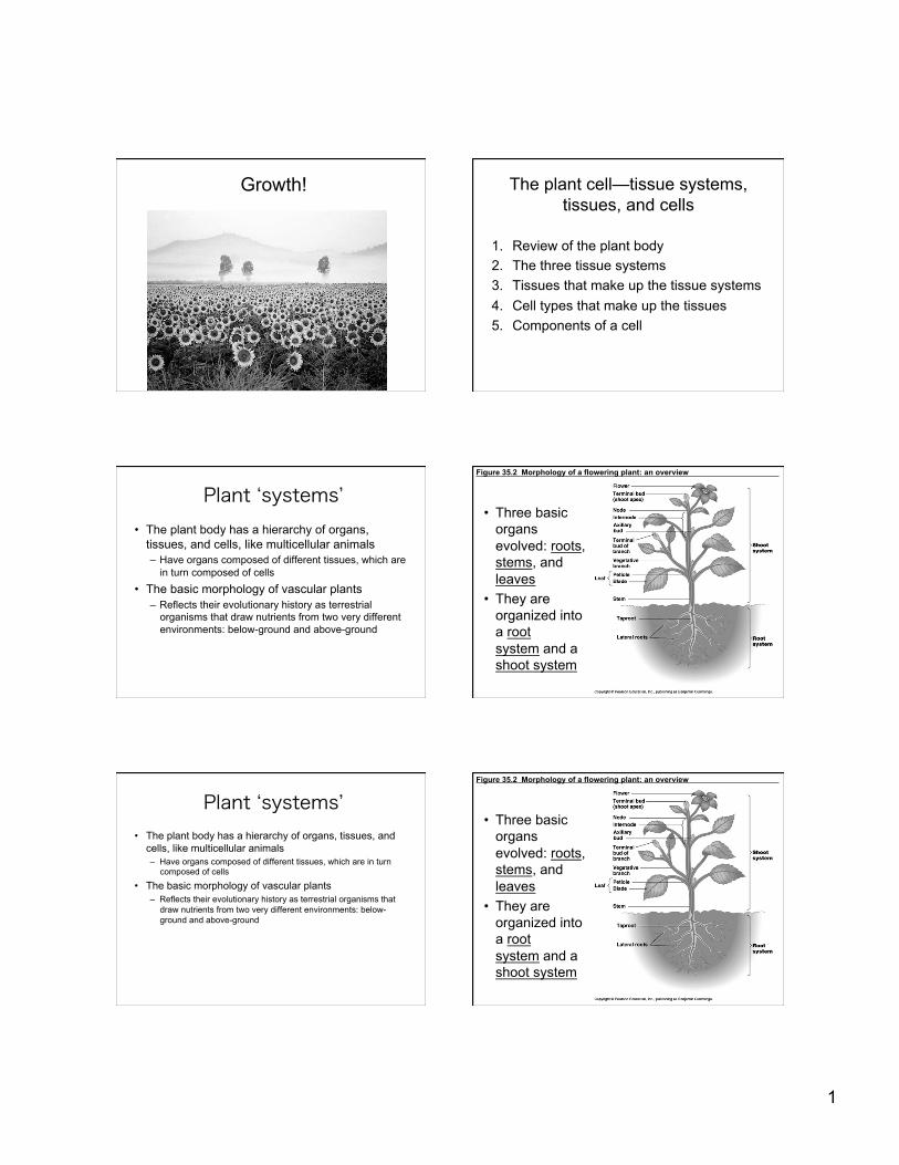
Figure 35.2 Morphology of a flowering plant: an overview
• Three basic organs evolved: roots, stems, and leaves
• They are organized into a root system and a shoot system
Plant ‘systems’ • The plant body has a hierarchy of organs, tissues, and
cells, like multicellular animals – Have organs composed of different tissues, which are in turn
composed of cells
• The basic morphology of vascular plants – Reflects their evolutionary history as terrestrial organisms that
draw nutrients from two very different environments: below-ground and above-ground
Figure 35.2 Morphology of a flowering plant: an overview
• Three basic organs evolved: roots, stems, and leaves
• They are organized into a root system and a shoot system

2
Roots • A root
– Is an organ that anchors the vascular plant – Absorbs minerals and water – Often stores organic nutrients
• In most plants – The absorption of water and
minerals occurs near the root tips, where vast numbers of tiny root hairs increase the surface area of the root
Roots cont.
• Gymnosperms and eudicots: taproots with lateral roots
• Seedless vascular and monocots: fibrous root system: spread out
• Many plants have modified roots: adventitious roots arise above ground from stems and even leaves
Many plants have modified roots
(a) Prop roots (b) Storage roots (c) “Strangling” aerial roots
(d) Buttress roots (e) Pneumatophores
Stems (shoot system)
• Nodes: point of leaf attachment • Internodes: segments between nodes • Axillary buds can form new shoots or
branches • Terminal buds can lead to apical
dominance (grow up!)
Figure 35.4 Modified shoots: Stolons, strawberry (top left); rhizomes, iris (top right); tubers, potato (bottom left); bulb, onion (bottom right)
3
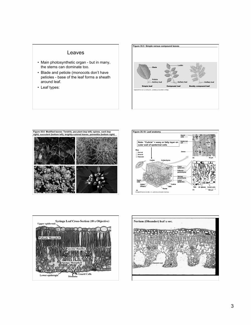
Leaves
• Main photosynthetic organ - but in many, the stems can dominate too.
• Blade and petiole (monocots don’t have petioles - base of the leaf forms a sheath around leaf.
• Leaf types:
Figure 35.5 Simple versus compound leaves
Figure 35.6 Modified leaves: Tendrils, pea plant (top left); spines, cacti (top right); succulent (bottom left); brightly-colored leaves, poinsettia (bottom right) Figure 35.19 Leaf anatomy
Note: “Cuticle” = waxy or fatty layer on outer wall of epidermal cells
4
Leaf types The plant cell—tissue systems, tissues, and cells
1. Review of the plant body 2. The three tissue systems 3. Tissues that make up the tissue systems 4. Cell types that make up the tissues 5. Components of a cell
Figure 35.7 The three tissue systems The plant cell—tissue systems,
tissues, and cells
1. Review of the plant body 2. The three tissue systems 3. Tissues that make up the tissue systems 4. Cell types that make up the tissues 5. Components of a cell
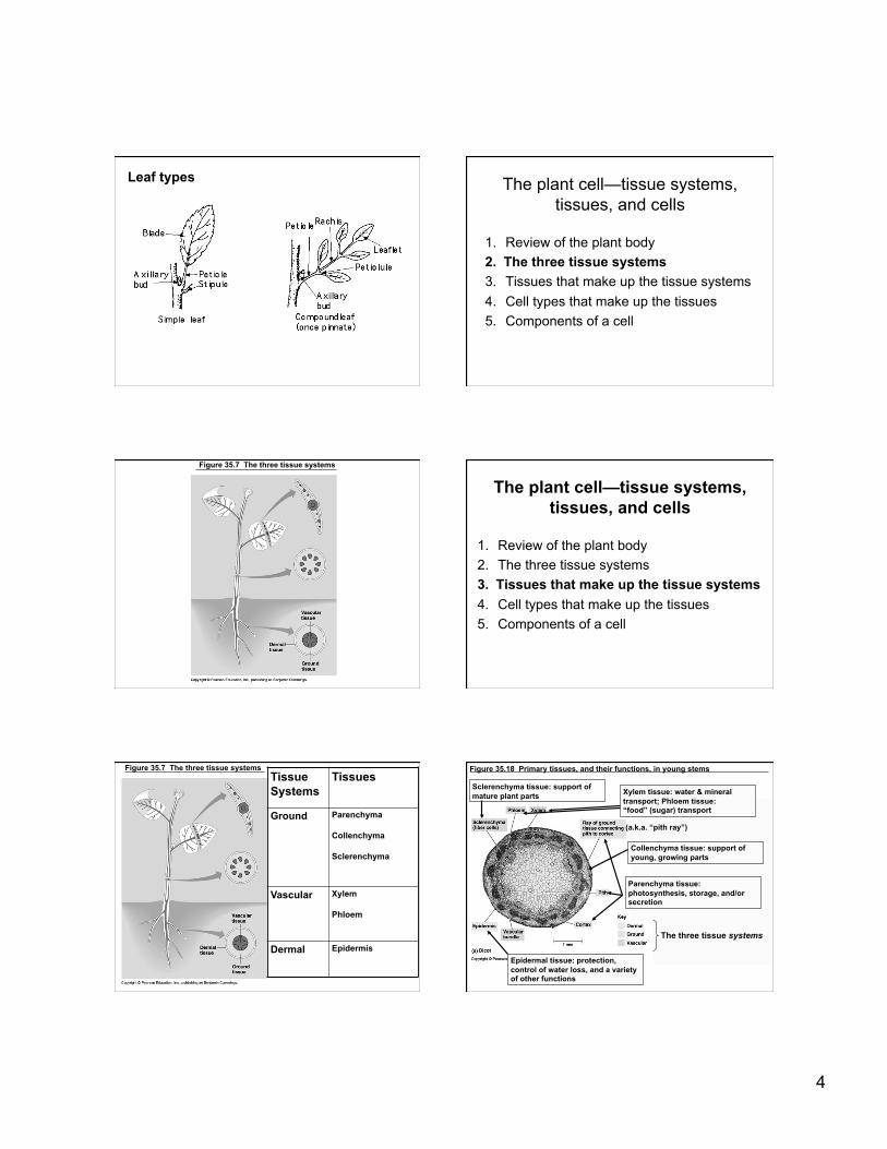
Figure 35.7 The three tissue systems Tissue Systems
Tissues
Ground Parenchyma
Collenchyma
Sclerenchyma
Vascular Xylem
Phloem
Dermal Epidermis
Figure 35.18 Primary tissues, and their functions, in young stems
The three tissue systems
Epidermal tissue: protection, control of water loss, and a variety of other functions
Parenchyma tissue: photosynthesis, storage, and/or secretion
Collenchyma tissue: support of young, growing parts
Xylem tissue: water & mineral transport; Phloem tissue: “food” (sugar) transport
Sclerenchyma tissue: support of mature plant parts
(a.k.a. “pith ray”)

5
Figure 35.16 Organization of primary tissues in young stems. Note difference from root: vascular tissue is arranged in bundles, with ground tissue in center. Also note difference in arrangement of bundles between dicot and monocot.
Figure 35.15 Organization of tissue systems and tissues in young roots
“Epidermis:” Dermal system, epidermal tissue
“Cortex:” Ground system, parenchyma tissue
“Stele:” Vascular system, xylem & phloem tissues
Figure 35.13 Organization of primary tissues in young roots. Note the difference between the monocot and the dicot in the arrangement of the xylem & phloem in the stele. The plant cell—tissue systems,
tissues, and cells
1. Review of the plant body 2. The three tissue systems 3. Tissues that make up the tissue systems 4. Cell types that make up the tissues 5. Components of a cell
Tissue Systems Tissues (& cell types)
Ground Parenchyma (parenchyma cells, transfer cells)
Collenchyma (collenchyma cells)
Sclerenchyma (fibers & sclereids)
Vascular Xylem (tracheids or vessel members, also some parenchyma cells, fibers, & sclereids )
Phloem (sieve cells or sieve-tube members, also specialized parenchyma cells called companion or albuminous cells, some fibers & sclereids)
Dermal Epidermis (ground cells, guard cells, trichomes, and others, also some fibers & sclereids)
Three tissue systems of plants
• Dermal tissue - outer protective covering – Epidermis/periderm analogous to skin – Cuticle - waxy coating to preserve water
• Vascular tissue - transport system – Xylem: carries water and nutrients from roots to
leaves. Support and food storage too. – Phloem: transport organic nutrients (sugar),
amino acids, lipids, hormones etc. • Ground tissue - “everything else”.
– Pith (internal to vascular), Cortex – Function in storage, photosynthesis, & support
6
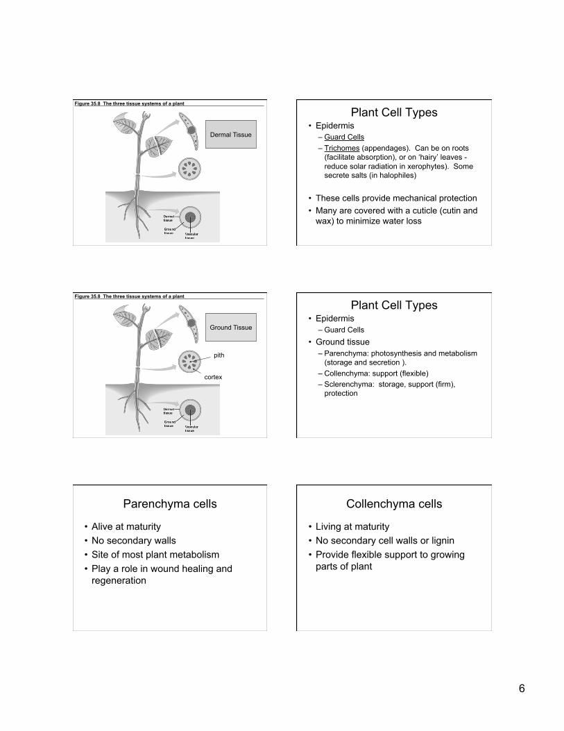
Figure 35.8 The three tissue systems of a plant
Dermal Tissue
Plant Cell Types • Epidermis
– Guard Cells – Trichomes (appendages). Can be on roots
(facilitate absorption), or on ‘hairy’ leaves - reduce solar radiation in xerophytes). Some secrete salts (in halophiles)
• These cells provide mechanical protection • Many are covered with a cuticle (cutin and
wax) to minimize water loss
Figure 35.8 The three tissue systems of a plant
pith
cortex
Ground Tissue
Plant Cell Types • Epidermis
– Guard Cells • Ground tissue
– Parenchyma: photosynthesis and metabolism (storage and secretion ).
– Collenchyma: support (flexible) – Sclerenchyma: storage, support (firm),
protection
Parenchyma cells
• Alive at maturity • No secondary walls • Site of most plant metabolism • Play a role in wound healing and
regeneration
Collenchyma cells
• Living at maturity • No secondary cell walls or lignin • Provide flexible support to growing
parts of plant

7
Sclerenchyma cells • Thick secondary walls, usually with lignin • Usually dead at maturity • Usually specialized for support and strengthening of
parts that have ceased elongating. – Sclereids impart hardness to seed coats, shells of
nuts (give pears their grit) – Fibers are usually long, slender, tapered (hemp and
flax fibers)
Figure 35.8 The three tissue systems of a plant
Vascular Tissue
Plant Cell Types • Epidermis
– Guard Cells • Ground tissue
– Parenchyma: photosynthesis and metabolism – Collenchyma: support – Scelerenchyma: support, storage, protection
• Vascular tissues – Xylem: water and nutrients from roots. Also
support and food storage • Tracheids, vessel elements
– Phloem: sugars from leaves • Sieve-tube members, companion cells
Xylem cells • Dead at maturity • Tracheids found in all vascular plants
– Long and thin with tapered ends – Lignin for structural support – Less specialized than vessel elements (‘safer’ though)
• Vessel elements found mainly in angiosperms (flowering plants) – Generally wider, shorter, and less tapered than
tracheids – Has perforations for more efficient water flow - but
perforations are open systems and can be less safe.
Figure 35.8 Water-conducting cells of xylem tissue Phloem
• Primary and secondary phloem. Primary phloem is often destroyed during elongation of the organ.
• Principal conducting cells are the sieve elements (‘with pores’)
8
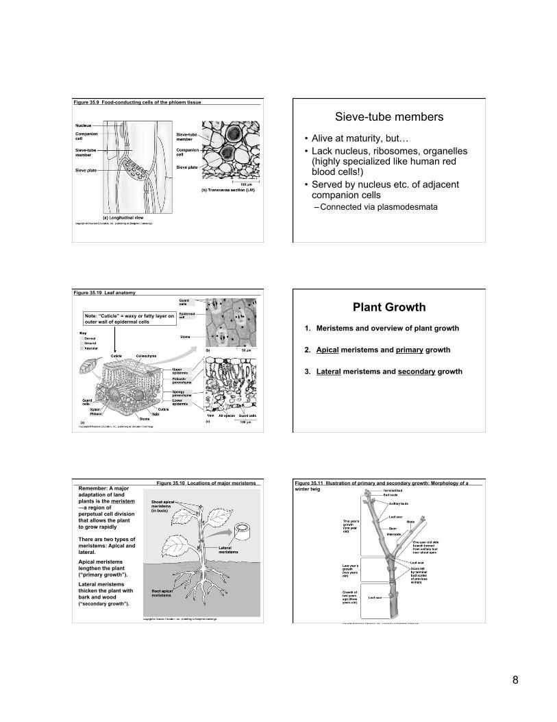
Figure 35.9 Food-conducting cells of the phloem tissue Sieve-tube members
• Alive at maturity, but… • Lack nucleus, ribosomes, organelles
(highly specialized like human red blood cells!)
• Served by nucleus etc. of adjacent companion cells – Connected via plasmodesmata
Figure 35.19 Leaf anatomy
Note: “Cuticle” = waxy or fatty layer on outer wall of epidermal cells
Plant Growth 1. Meristems and overview of plant growth
2. Apical meristems and primary growth
3. Lateral meristems and secondary growth
Figure 35.10 Locations of major meristems Remember: A major adaptation of land plants is the meristem—a region of perpetual cell division that allows the plant to grow rapidly
There are two types of meristems: Apical and lateral.
Apical meristems lengthen the plant (“primary growth”).
Lateral meristems thicken the plant with bark and wood (“secondary growth”).
Figure 35.11 Illustration of primary and secondary growth: Morphology of a winter twig
9
Plant Growth
1. Meristems and overview of plant growth
2. Apical meristems and primary growth
3. Lateral meristems and secondary growth
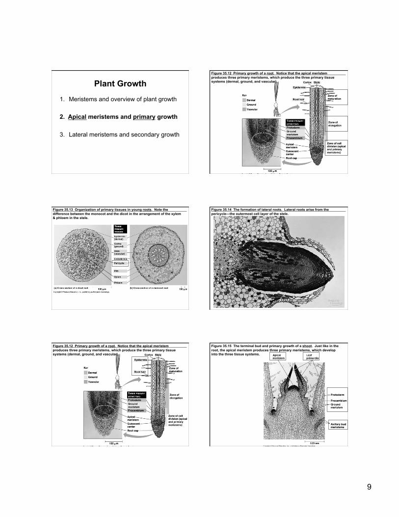
Figure 35.12 Primary growth of a root. Notice that the apical meristem produces three primary meristems, which produce the three primary tissue systems (dermal, ground, and vascular).
Figure 35.13 Organization of primary tissues in young roots. Note the difference between the monocot and the dicot in the arrangement of the xylem & phloem in the stele.
Figure 35.14 The formation of lateral roots. Lateral roots arise from the pericycle—the outermost cell layer of the stele.
Figure 35.12 Primary growth of a root. Notice that the apical meristem produces three primary meristems, which produce the three primary tissue systems (dermal, ground, and vascular).
Figure 35.15 The terminal bud and primary growth of a shoot. Just like in the root, the apical meristem produces three primary meristems, which develop into the three tissue systems.
10
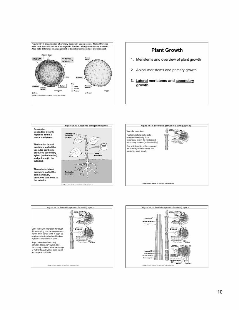
Figure 35.16 Organization of primary tissues in young stems. Note difference from root: vascular tissue is arranged in bundles, with ground tissue in center. Also note difference in arrangement of bundles between dicot and monocot. Plant Growth
1. Meristems and overview of plant growth
2. Apical meristems and primary growth
3. Lateral meristems and secondary growth
Figure 35.10 Locations of major meristems Remember: Secondary growth happens at the 2 lateral meristems
The interior lateral meristem, called the vascular cambium, produces secondary xylem (to the interior) and phloem (to the exterior).
The exterior lateral meristem, called the cork cambium, produces cork cells to the exterior.
Figure 35.18 Secondary growth of a stem (Layer 1) Vascular cambium:
Fusiform initials make cells elongated vertically, form secondary xylem (to inside) and secondary phloem (to the outside)
Ray initials make cells elongated horizontally-transfer water and nutrients, store starch
Figure 35.18 Secondary growth of a stem (Layer 2)
Cork cambium: meristem for tough thick covering - replaces epidermis. Forms from cortex to fill in gaps as epidermis is stretched and broken by lateral expansion of stem
Rays maintain connectivity between secondary xylem and secondary phloem, allow exchange of nutrients and water, store starch and organic nutrients
Figure 35.18 Secondary growth of a stem (Layer 3)
11
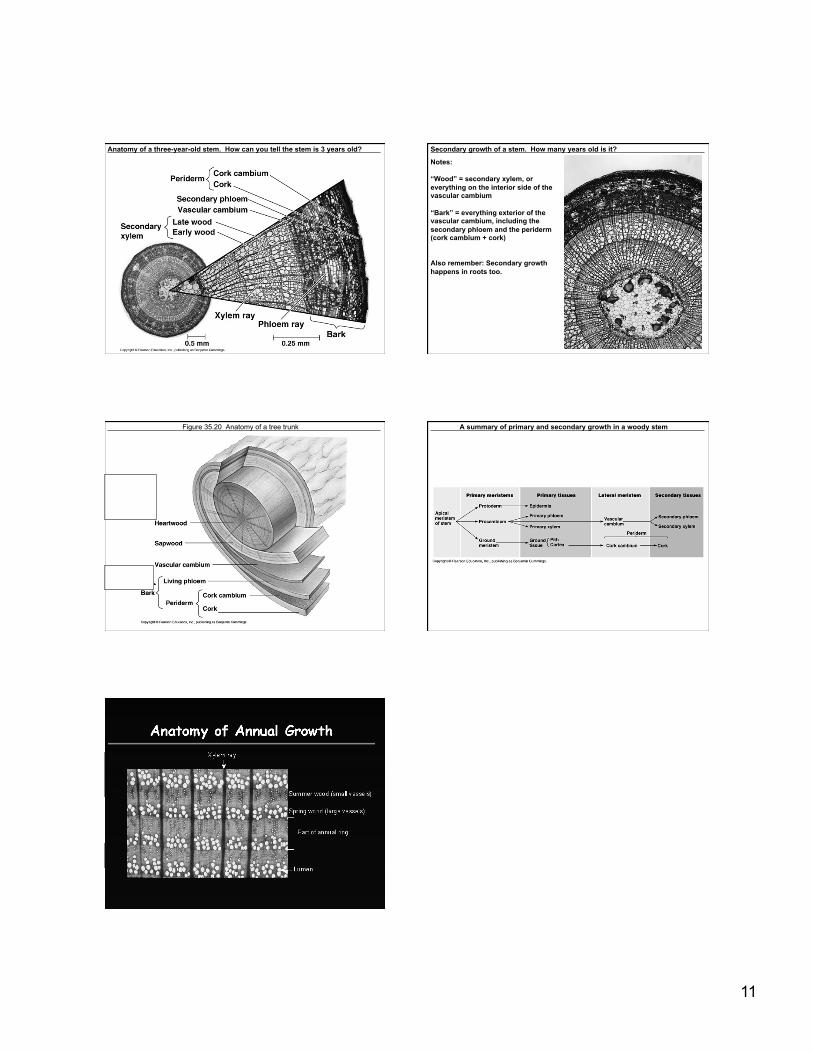
Anatomy of a three-year-old stem. How can you tell the stem is 3 years old? Secondary growth of a stem. How many years old is it? Notes:
“Wood” = secondary xylem, or everything on the interior side of the vascular cambium
“Bark” = everything exterior of the vascular cambium, including the secondary phloem and the periderm (cork cambium + cork)
Also remember: Secondary growth happens in roots too.
Figure 35.20 Anatomy of a tree trunk
Oldest xyelm nonfuctional
Oldest phloem
sloughed off
A summary of primary and secondary growth in a woody stem
Oldest xyelm nonfuctional
Oldest phloem
sloughed off