gtp-binding proteins in intracellular transport
TRANSCRIPT

REVIEWS
The mechanism by which membrane and secretory proteins are transported from their sites of syn- thesis on the endoplasmic reticulum (ER) to their final destinations has intrigued cell biologists for many years. These classes of proteins are trans- located across the membrane bilayer of the ER and interorganelle transfer is then accomplished by transport vesicles that shuttle proteins from the ER to the Golgi complex, between Golgi cisternae, and from the Golgi complex to the plasma membrane or prelysosomes. For every transfer step, a vesicle carrying the appropriate cargo molecules must bud from a donor compartment, identify its target and fuse with the target membrane. Resident proteins of the donor compartment must be excluded from newly forming transport vesicles and retention mechanisms exist for this purpose. In addition, retrieval mechanisms capture resident proteins that escape the retention apparatus, thereby in- creasing the efficiency with which proteins are local- ized in a given compartment. A combination of gen- etic and biochemical approaches is now beginning to provide a glimpse of the molecular transactions that underlie vesicular transport between mem- brane-bound compartments.
iuzanne R. PfeffeJ
Small GTP-binding proteins in membrane traffic The potential role of GTP-binding proteins (G
proteins) in regulating vesicular transport was first realized when Novick and colleagues determined the primary structure of the yeast SEC4 gene product I. Temperature-sensitive sec4 mutations lead to the accumulation of invertase-containing secretory vesicles at the restrictive temperature. The sequence of the SEC4 gene product (SEC4) indicated that it is a Ras-like or 'small' G protein, and subsequent analyses revealed that it Is present on the surface of secretory vesicles 2 and can bind and hydrolyse GTP :~. The yeast protein YPT1 is another Ras-ilke G protein, which Is 48% Identical to SEC44. Mutations in YPT1 inhibit vesicular transport between the ER and the Goigi complex, and lead to proliferation of the ER s,6.
The accumulation of transport vesicles in yeast strains harbouring see4 mutations strongly suggests that SEC4 functions in the targeting and/or fusion of secretory vesicles with the plasma membrane. Functional analyses of YPT1 have demonstrated an analogous role for this protein in transport between the ER and Golgi complex (see below). Together, these observations underscore the po- tential importance of G proteins in regulating vesicular transport events.
A search for the mammalian counterparts of SEC4 and YPT1 has led to the identification of a large number of Ras-related G proteins that may include more than 20 different gene products (see Refs 7-10 for reviews). The SEC4- and YPTl-related proteins have been grouped into a family termed rab proteins ('Ras-like proteins from rat brain'). This terminology has been adopted even for pro- teins from species other than rat. The rab proteins are 21-25 kDa in mass and are -30% identical to the oncogene product Ras. The amino acid residues
GTP-binding proteins in
intracellular transport
>
One of the most exciting recent discoveries in the area of
intracellular protein transport is the finding that many organelles
involved in exocytic and endocytic membrane traffic have one or
more Ras-like GTP-binding proteins on their cytoplasmic face that
are specific for each membranous compartment. These proteins
are attractive candidates for regulators of transport vesicle
formation and the accurate delivery of transport vesicles to their
correct targets.
that comprise the GTP-binding domain are the most highly conserved.
In addition to rab proteins, another class of small G proteins also functions in intracellular transport. This family is termed ARF for ADP ri- bosylation factor. ARF was first discovered as a co- factor for cholera toxin-catalysed ADP ribosylation of the alpha subunit of 'large' trimeric stimulatory G proteins. Mammalian ARF is located in the Golgi complex and yeast strains lacking ARF1 display a defect in invertase secretion 11. The family of ARF- related proteins is also likely to have many members.
Intracellular location of rab and ARF proteins The rab and ARF proteins have distinct sub-
cellular locations. Indeed, i t is now clear that most organelles involved in exocytic and endocytic pro- cesses have at least one distinct Ras-like O protein on their surfaces (Table 1). The rab and ARF genes encode cytosolic proteins, and post-translational modifications of both families are required for their functional activity and association wi th intra- cellular organelles. Certain ARF proteins acquire N-terminal myristate. By contrast, rab proteins are modified by a geranylgeranyl (Cz0) prenyl moiety on one or both of the C-terminal cysteine residues in the sequence CC, CXC, CCXX or C C X X X 12,13. Mutation of the C-terminal cysteine residues of YPT1, SEC4 and tab5 interferes with the mem- brane association and functions of these pro- teins 14-16. In addition, YPT1 and SEC4 both require the action of the putative prenyltransferase BET2 to support intracellular transport 17.
Most tab proteins have two C-terminal cysteine residues, and the rab3A protein (also termed
Suzanne Pfeffer is at the Department of Biochemistry, Stanford University School of Medicine, Stanford, CA 94305, USA.
TRENDS IN CELL BIOLOGY VOL 2 FEBRUARY 1992 © 1992 Elsevier Science Publishers Ltd (UK) 0962-8924/92/$05.00 41
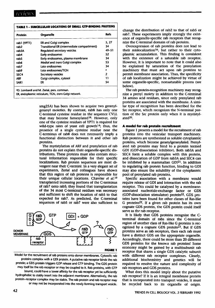
TABLE 1 - SUBCELLULAR LOCATIONS OF SMALL GTP-BINDING PROTEINS
Protein Organelle Refs
tab1 (YPT1) ER and Golgi complex 5, 27 rab2 Transitional ER (intermediate compartment) 34 rab3A Regulated secretory vesicles 49-51 rab4 Early endosomes 52 rabS Early endosomes, plasma membrane 34 rab6 Medial and trans Golgi complex 53 rab7 Late endosomes 34 rab9 Late endosomes/TGN a SEC4 Secretory vesicles 2 ARF Golgi complex, cytosol 11 SAR1 ER 54
aD. Lombardi and M. Zerial, pers. commun. ER, endoplasmic reticulum, TGN, trans Golgi network.
smg25A) has been shown to acquire two geranyl- geranyl moieties. By contrast, rab8 has only one C-terminal cysteine residue in the sequence CVLL that may become farnesylated 18. However, only one of the cysteine residues of YPT1 is required for wild-type rates of yeast cell growth14; thus, the presence of a single cysteine residue near the C-terminus of rab8 does not necessarily imply a functional distinction between it and other rab proteins.
The myristylation of ARF and prenylation of rab proteins do not explain their organelle-specific dis- tributions. These proteins must also contain struc- tural Information responsible for their specific Iocalizatlons. Rab protein sequences are most di- vergent near their C-termini. In a very elegant set of experiments, Zerlal and colleagues have shown that this region of rab proteins is responsible for their unique cellular locations. Chavrier et al. 19 transplanted increasing portions of the C.terminus of rab7 onto rabS; they found that transplantation of the 34 most C-terminal residues was necessary and sufficient to shift the location of rab5 to that expected for rab7. As predicted, the C-terminal sequences of rab$ or rab7 were also sufficient to
GTP [ - ~ + GDP
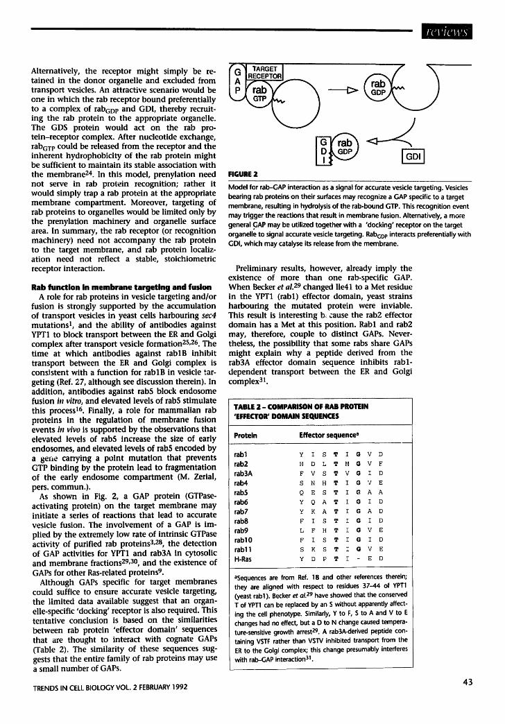
KICt I FIGURE 1
Model for the recruitment of rab proteins onto donor membranes. Cytosolic rab proteins complex with a GDI protein. A putative rab receptor binds the rab
protein; a GDS protein triggers GDP release and GTP binding. The GDS protein may itself be the tab receptor or may be coupled to it. Rab proteins, with GTP
bound, could have a lower affinity for the tab receptor yet be sufficiently hydrophobic to stably insert into the adjacent membrane. Alternatively, the tab protein-receptor complex may be stable. The rab protein and rab receptor may
or may not be incorporated into the newly forming transport vesicle.
change the distribution of rab2 to that of rabS or rab7. These experiments imply strongly the exist- ence of organelle-specific rab receptors that recog- nize the C-terminal domain of rab proteins.
Overexpression of rab proteins does not lead to their mislocalization 16, but rather to their cyto- plasmic accumulation. This finding is consistent with the existence of a saturable tab receptor. However, it is important to note that it could also be explained by saturation of the prenylation machinery that must act upon rab proteins to permit membrane association. Thus, the specificity of rab localization might he achieved by virtue of some organelle-specific, nonsaturable process (see below).
The rab protein-recognition machinery may recog- nize a prenyl moiety in addition to the C-terminal 34 amino acid residues, since only prenylated tab proteins are associated with the membrane. A simi- lar type of recognition has been described for the Src receptor, which recognizes the N-terminal por- tion of the Src protein only when it is myristyl- ated 2o.
A model for rab protein recruitment Figure 1 presents a model for the recruitment of rab
proteins into the vesicular transport machinery. Rab proteins are synthesized as soluble cytoplasmic proteins, which become geranylgeranylated. Prenyl- ated rab proteins may bind to a protein termed GDI (GDP-dissociation inhibitor). Both rab3A and SEC4 form a soluble complex with this protein, and dissociation of GDP from rab3A and SEC4 can be inhibited by a mammalian GD121. In addition to regulating rab protein function, binding to GDI may also ensure the solubil i ty of the cytoplasmic pool of prenylated rab proteins.
Specific association wi th a membrane would require release of GDI and Interaction with the rab receptor. This could be catalysed by a membrane- associated nucleotide-exchange factor or GDS (GDP-dlssoclatlon stimulator) protein22. GDS pro- teins have been found for other classes of Ras-like G proteins 22. If a given rab protein has its own cognate GDS protein, the GDS protein could itself serve as the rab receptor.
It is likely that GDS proteins recognize the C- terminal domain of tabs since the C-terminal region of another class of Ras-like G proteins is rec- ognized by a cognate GDS protein 23. But if GDS proteins serve as rab receptors, then each tab must have a distinct GDS on the appropriate organelle. Accordingly, there could be more than 20 different GDS proteins for the known rab proteins! Some economy might be gained by a multisubunit rab receptor that shares a single GDS catalytic subunit with different rab receptor complexes. Clearly, additional biochemistry and genetics will be required to resolve the nature and complexity of rab receptors and GDS proteins.
What does this model imply about the putative rab receptor? If it is an integral membrane protein that is incorporated into transport vesicles, it must be recycled back to its organelle of origin.
42 TRENDS IN CELL BIOLOGY VOL. 2 FEBRUARY 1992
Alternatively, the receptor might simply be re- tained in the donor organelle and excluded from transport vesicles. An attractive scenario would be one in which the rab receptor bound preferentially to a complex of rabGD P and GDI, thereby recruit- ing the tab protein to the appropriate organe!!e. The GDS protein would act on the rab pro- tein-receptor complex. After nucleotide exchange, rabGT P could be released from the receptor and the inherent hydrophobicity of the tab protein might be sufficient to maintain its stable association with the membrane 24. In this model, prenylation need not serve in rab protein recognition; rather it would simply trap a tab protein at the appropriate membrane compartment. Moreover, targeting of rab proteins to organelles would be limited only by the prenylation machinery and organelle surface area. In summary, the rab receptor (or recognition machinery) need not accompany the tab protein to the target membrane, and rab protein localiz- ation need not reflect a stable, stoichiometric receptor interaction.
Rab function in membrane targeting and fusion A role for rab proteins in vesicle targeting and/or
fusion is strongly supported by the accumulation of transport vesicles in yeast cells harbouring sec4 mutations 1, and the ability of antibodies against YPT1 to block transport between the ER and Golgi complex after transport vesicle formation2S, 26. The time at which antibodies against rablB inhibit transport between the ER and Golgi complex is consistent with a function for rablB in vesicle tar- geting (Ref. 27, although see discussion therein). In addition, antibodies against rab5 block endosome fusion in vitro, and elevated levels of rab5 stimulate this process 16. Finally, a role for mammalian tab proteins in the regulation of membrane fusion events i , vivo is supported by the observations that elevated levels of rab5 increase the size of early endosomes, and elevated levels of tab5 encoded by a 8ene carrying a point mutation that prevents GTP binding by the protein lead to fragmentation of the early endosome compartment (M. Zerial, pers. commun.).
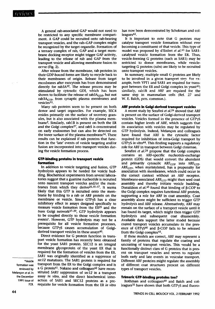
As shown in Fig. 2, a GAP protein (GTPase- activating protein) on the target membrane may initiate a series of reactions that lead to accurate vesicle fusion. The involvement of a GAP is im- plied by the extremely low rate of intrinsic GTPase activity of purified tab proteins 3,2s, the detection of GAP activities for YPT1 and rab3A in cytosolic and membrane fractions 29,3°, and the existence of GAPs for other Ras-related proteins%
Although GAPs specific for target membranes could suffice to ensure accurate vesicle targeting, the limited data available suggest that an organ- elle-specific 'docking' receptor is also required. This tentative conclusion is based on the similarities between tab protein 'effector domain' sequences that are thought to interact with cognate GAPs (Table 2). The similarity of these sequences sug- gests that the entire family of rab proteins may use a small number of GAPs.
~-G~ TARGET I ~ ( ~ ~
FIGURE 2
Model for rab-GAP interaction as a signal for accurate vesicle targeting. Vesicles bearing tab proteins on their surfaces may recognize a GAP specific to a target membrane, resulting in hydrolysis of the rab-bound GTP. This recognition event may trigger the reactions that result in membrane fusion. Alternatively, a more general GAP may be utilized together with a 'docking' receptor on the target organelle to signal accurate vesicle targeting. RabGD P interacts preferentially with GDI, which may catalyse its release from the membrane.
Preliminary results, however, already imply the existence of more than one rab-specific GAP. When Becket et al. 29 changed lie41 to a Met residue in the YPT1 (rabl) effector domain, yeast strains harbouring the mutated protein were inviable. This result is interesting b~ cause the rab2 effector domain has a Met at this position. Rabl and rab2 may, therefore, couple to distinct GAPs. Never- theless, the possibility that some tabs share GAPs might explain why a peptide derived from the rab3A effector domain sequence inhibits rabl- dependent transport between the ER and Golgi complex 31.
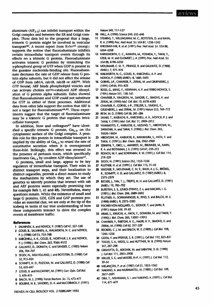
TABLE 2 - COMPARISON OF RAB PROTEIN 'EFFECTOR' DOMAIN SEQUENCES
Protein Effector sequence a
rabl Y I s T ~ G v D
rab2 H D L T M G V E
rab3A F V S T V G I D
tab4 s N H T I G V E
rab5 Q E S T I C~ A A
tab6 Y Q A T I G I D
rab7 Y K A T I G A D
rab8 F I S T I G I D
tab9 L F H T I G V E
rab10 F I S T I G I D
rab11 s K S T I G V E
H-Ras Y D P T I E D
aSequences are from Ref. 18 and other references therein; they are aligne d with respect to residues 37-44 of YPT1 (yeast rabl ). Becker et aL 29 have showed that the conserved T of YPT1 can be replaced by an S without apparently affect- ing the cell phenotype. Similarly, Y to F, S to A and V to E changes had no effect, but a D to N change caused tempera- ture-sensitive growth arrest 29. A rab3A-derived peptide con- taining VSTF rather than VSI"V inhibited transport from the ER to the Golgi complex; this change presumably interferes with rab-GAP interaction 31.
TRENDS IN CELL BIOLOGY VOL. 2 FEBRUARY 1992 43
*Vesicle formation was
reviewed by Melan~:on et oL in
the December 1991 issue of
TCB.
A general rab-associated GAP would not need to be restricted to any specific membrane compart- ment. A GAP could be recruited onto rab-bearing transport vesicles, and the rab-GAP complex might be recognized by the target organelle. Formation of a ternary complex of rab, GAP and a target mem- brane docking receptor might trigger GAP activity, leading to the release of rab and GAP from the transport vesicle and allowing membrane fusion to occur (Fig. 2).
After release from the membrane, rab proteins in their GDP-bound forms are likely to recycle back to their membranes of origin. Release from target membranes after exocytosis has been demonstrated directly for rab3A 32. The release process may be stimulated by cytosolic GDI, which has been shown to facilitate the release of rab3AcoI,, but not rab3AGT P, from synaptic plasma membranes and vesicles 33.
Many rab proteins seem to be present on both donor and target organelles. For example, SEC4 resides primarily on the surface of secretory gran- ules, but is also associated with the plasma mem- brane 2. Similarly, rablB is present on both the ER and the Golgi complex 27, and rab5 is concentrated on early endosomes but can also be detected on the inner surface of the plasma membrane a4. These results can be explained if rab proteins that func- tion in the 'late' events of vesicle targeting and/or fusion are incorporated into transport vesicles dur- ing the vesicle formation process.
GTP.bindlng proteins in transport vesicle formation
In addition to vesicle targeting and fusion, GTP hydrolysis appears to be needed for vesicle bud- ding. Biochemical experiments from several labora. tories suggest that a guanine nucleotlde is recruited onto nascent transport vesicles and/or the mem- branes from which they derive2S, :~s-:~7. It seems likely that this GTP is recruited onto the mem- brane by binding to a tab or ARF protein on the membrane or vesicle. Since GTPyS has a clear inhibitory effect in assays designed specifically to measure vesicle formation from the ERZS and the trans Golgi network 37-39, GTP hydrolysis appears to be coupled directly to those vesicle formation events*. However, GTP hydrolysis may not be a prerequisite for all vesicle formation processes, because GTPTS causes accumulation of Golgi- derived transport vesicles in these assays40.
Direct evidence for G protein function in trans- port vesicle formation has recently been obtained for the yeast SAR1 protein. SEC12 is an integral membrane glycoprotein of the yeast ER that is required for the formation of vesicles from the ER. SAR1 was originally identified as a suppressor of secl2 mutations. The SAR1 protein is required for transport from the ER to the Golgi complex and is a G protein 41. Nakano and colleagues 42 have recon- stituted SAR1 suppression of secl2 in a transport assay in vitro, and the direct biochemical inter- action of SAR1 and SEC12 proteins as a pre- requisite for vesicle formation from the ER #1 vitro
has now been demonstrated by Schekman and col- leagues 43.
It is important to note that G proteins may catalyse transport vesicle formation without ever becoming a constituent of that vesicle. This type of model was proposed by d'Enfert et ai. 43 for SAR1- catalysed vesicle formation from the ER. Thus, vesicle-forming G proteins (such as SAR1) may be restricted to donor membranes, while vesicle- targeting G proteins (rabs) are likely to be recruited onto transport vesicles.
In summary, multiple small G proteins are likely to be involved in a given transport step For ex- ample, both YPT1 and SAR1 are required for trans- port between the ER and Golgi complex in yeast43; similarly, rablB and ARF are required for the same step in mammalian cells (Ref. 27 and W. E. Balch, pers. commun.).
ARF protein in Golgi-derived transport vesicles A recent study by Serafini et al. 24 showed that ARF
is present on the surface of Golgi-derived transport vesicles. Vesicles formed in the presence of GTP7S contain higher levels of ARF, which suggests that its association with vesicles may be regulated by GTP hydrolysis. Indeed, Melan(;on and colleagues have found that ARF is the cytosolic factor required for inhibition of intra-Golgi transport by GTPyS in vitro 44. This finding supports a regulatory role for ARF in transport between Golgi cisternae.
Serafini et al. 24 propose that the Golgi complex contains an ARF-specific nucleotide.exchange protein (GDS) that would convert the abundant and primarily cytosolic ARFGD I, into ARFc;Tp. ARFGT P, when myristylated, has a propensity for association with membranes, which could occur In the correct context without an ARF receptor. Membrane-associated ARFc;.rl, might then seed coat assembly and thereby initiate vesicle budding. Donaldson et al. 4s found that binding of It-COP to the Golgl complex requires functional ARF protein, supporting a role for ARF in coat assembly. Coat assembly alone might be sufficient to trigger GTP hydrolysis and ARF release. Alternatively, ARF may remain on the surface of a transport vesicle until it has found its target, which might then trigger GTP hydrolysis and subsequent coat disassembly. Available data support the latter model because coated transport vesicles accumulate in the pres- ence of GTPTS 40 and ~-COP fails to be released from the Golgi complex 4s.
If these models are correct, ARF may represent a family of proteins that regulate the coating and uncoating of transport vesicles. This would be a functionally distinct class of G proteins that is pres- ent on transport vesicles and serves to regulate both early and late events in vesicular transport. Different ARF proteins might regulate the assembly of different coat structures present on different types of transport vesicles.
Trimeric GTP-binding proteins too? Rothman and colleagues 4o and Balch and col-
leagues 3s have shown that both GTP7S and fluoro-
44 TRENDS IN CELL BIOLOGY VOL. 2 FEBRUARY 1992
aluminate (AIF3_ s) can inhibit transport within the Golgi complex and between the ER and Golgi com- plex. These data led to the proposal that a large, trimeric G protein might be involved in vesicular transport 4o. A recent report from Kahn 46 strong;y supports the notion that fluoroaluminate inhibits certain intracellular transport events through its effects on a trimeric G protein. Fluoroaluminate activates trimeric G proteins by mimicking the ¥-phosphoryl group of GTP when GDP is present in the guanine nucleotide-binding site. Fluoroalumi- nate decreases the rate of GDP release from G pro- tein alpha subunits, but it did not affect the release of GDP from rablA, rablB, rab3B or ARF 46. With GTP bound, ARF binds phospholipid vesicles and can activate cholera to~n-catal~sed ADP ribosyl- ation of G protein alpha subunits. Kahn showed that fluoroaluminate and GDP could not substitute for GTP in either of these processes. Additional data from other labs support the notion that ARF is not a target for fluoroaluminate 44,4s. These exper- iments suggest that the target of fluoroaluminate may be a trimeric G protein that regulates intra- cellular transport.
In addition, Stow and colleagues 47 have ident- ified a specific trimeric G protein, Gc~i_ 3, on the cytoplasmic surface of the Golgi complex. A poss- ible role for this protein in intracellular transport is implied by the ability of Gcxi_ 3 to retard the rate of constitutive secretion when it is overexpressed three-fold. Strikingly, this effect was reversed in the presence of pertussis toxin, which specifically inactivates Gai_ 3 by covalent ADP ribosylation 47.
G proteins, small and large, appear to be key regulators of intracellular transport. Their roles in distinct transport steps, and their localization to distinct organelles, provide a direct means to study the mechanisms by which they act. The use of genetics to identify proteins that interact with rab and ARF proteins seems especially promising (see for example Refs 1, 41 and 48). Nevertheless, many questions remain. While the existence of small and large G proteins, GDI, GDS and GAP proteins pro- vides an essential clue, we are only at the tip of the iceberg in terms of our true understanding of how these components interact to drive the complex events of membrane traffic.
References 1 SALMINEN, A. and NOVICK, P. (1987) Ce1147, 527-538 2 GOUD, B., SALMINEN, A., WALWORTH, N. C. and NOVlCK,
P. J. (1988) Cell 53, 753-768 3 KABCENELL, A. K., GOUD, B., NORTHUP, I. K. and NOVICK,
P. I. (1990)I. Biol. Chem. 265, 9366-9372 4 GALLWITZ, D., DONATH, C. and SANDER, C. (1983) Nature
306, 704-707 5 SEGEV, N., MULHOLLAND, I. and BOTSTEIN, D. (1988) Cell
52, 915-924 6 SCHMII-r, H. D., PUZICHA, M. and GALLWITZ, D. (1988)Cell
53, 635-647 7 GOUD, B. and McCAFFREY, M. (1991) Curr. Opin. Cell Biol.
3, 626-633 8 BALCH, W. E. (1990) Trends Biochem. 5ci. 15, 473-477 9 BOURNE, H. R., SANDERS, D. A. and McCORMICK, F. (199~)
TRENDS IN CELL BIOLOGY VOL. 2 FEBRUARY 1992
Nature 349, 117-127 10 HALL, A. (1990) Science 249, 635-640 11 STEARNS, T., WILLINGHAM, M. C., BOTSTEIN, D. and KAHN,
R. A. (1990) Proc. NatlAcad. Sci. USA 87, 1238-1242 12 KHOSRAVI-FAR, R. et al. (i991) Proc. Natl Acad. Sci. USA 88,
6264-6268 13 FARNSWORTH, C. C., KAWATA, M., YOSHIDA, Y., TAKAI, Y.,
GELB, M. H. and Gt.OMSET, J. A. (1991) Proc. NatlAcad. Sci. USA 88, 6196-6200
14 MOLENAAR, C. M. T., PRANGE, R. and GALLWITZ, D. (1988) EMBO I. 7, 971-976
15 WALWORTH, N. C., GOUD, B., KABCENELL, A. P. and NOVICK, P. (1989) EMBO I. 8, 1685-1693
16 GORVEL, J-P., CHAVRIER, P., ZERIAL, M. and GRUENBERG, I. (1991) Cell 64, 915-925
17 ROSSI, G., IIANG, Y., NEWMAN, A. P. and FERRO-NOVICK, S. (!99i) Nature 351,158-161
18 CHAVRIER, P., VINGRON, M., SANDER, C., SIMONS, K. and ZERIAL, M. (1990) Mol. Cell. BioL 10, 6578-6585
19 CHAVRIER, P., GORVEL, I-P., STELZER, E., SIMONS, K., GRUENBERG, J. and ZERIAL, M. (1991) Nature 353, 769-772
20 RESH, M. D. (1989) Cell58, 281-286 21 SASAKI, T., KAIBUCHI, K., KABCENELL, A. K., NOVICK, P. J. and
TAKAI, Y. (1991) Mol. Cell. Biol. 11, 2909-2912 22 YAMAMOTO, T., KAIBUCHI, K., MIZUNO, T., HIROYOSHI, M.,
SHIRATAKI, H. and TAKAI, Y. (1990) J. Biol. Chem. 265, 16626-16634
23 HIROYOSHI, M., KAIBUCHI, K., KAWAMURA, S., HATA, Y. and TAKAI, Y. (1991) I. BioL Chem. 266, 2962-2969
24 SERAFINI, T., ORCI, L., AMHERST, M., BRUNNER, M., KAHN, R. A. and ROTHMAN, J. E. (1991) Cell67, 239-253
25 REXACH, M. F. and SCHEKMAN, R. W. (1991) I. Cell Biol. 114, 219-229
26 SEGEV, N. (1991) Science 252, 1553-1556 27 PLUTNER, H. etal. (1991)/. CellBiol. 115, 31-43 28 WAGNER, P., MOLENAAR, C. M. T., RAUH, A. I. G., BROKEL,
R., SCHMITF, H. O. and GALLWITZ, D. (1987) EMBO I. 6, 2373-2379
29 BECKER, J., TAN, T. I., TREPTE, H. H. and GALLWITZ, D. (1991) EMBO I. 10, 785-792
30 BURSTEIN, E. S., LINKO-STENTZ, Z. L. and MACARA, I. G. (1991) I. Biol. Chem. 266, 2689-2692
31 PLUTNER, H., SCHWANINGER, R., PIND, S. and BALCH, W. E. (1990) EMBO J. 9, 2375-2383
32 FISCHER.VaN-MaLLARD, G., SODHOF, T. and JAHN, R. (1991) Nature 349, 79-81
33 AP, AKI, S., KIKUCHI, A., HATA, Y., ISOMURA, M. and TAKAI, Y. (1990) 1. Biol. Chem. 265, 13007-13015
34 CHAVRIER, P., PARTON, R. G., HAURI, H. P., SIMONS, K. and ZERIAL, M. (1990) Cell 62, 317-329
35 BECKERS, C. I. M. and BALCH, W. E. (1989) I. Cell Biol. 108, 1245-1256
36 GODA, Y. and PFEFFER, S. R. (1991) J. Cell Biol. 112, 823-831 37 TOOZE, S. A., WEISS, U. and HUI-FNER, W. B. (1990) Nature
347, 207-208 38 GRAVOTTA, D., ADESNIK, M. and SABATINI, D. D. (1990)
i. Cell Biol. 111, 2893-2908 39 MILLER, S. G. and MOORE, H-P. H. (1991) I. Cell Biol. 112,
39-54 40 MELAN~ON, P. et al. (1987) Cell51, 1053-1062 41 NAKANO, A. and MURAMATSU, M. (1989)/. CellBiol. 109,
2677-2691 42 OKA, T., NISHIKAWA, S. I. and NAKANO, A. (1991) I. Cell Biol.
114, 671-679
45
Acknowledgements
I am grateful to Marino Zerial,
Paul Melan~on, ]im Rothman and
members of the Pfeffer lab for all their thoughtful
suggestions.
43 D'ENFERT, C., WUESTEHUBE, L. I., LILA, T. and SCHEKMAN, R. (1991) I. Cell Biol. 114, 663-670
44 TAYLOR, T. C. and MELAN(~ON, P. (1991) I. Cell Biol. 115, 245a 45 DONALDSON, J. G., KAHN, R. A. and KLAUSNER, R. D. (1991)
I. Cell Biol. 115, 245a 46 KAHN, R. A. (1991) I. Biol. Chem. 266, 15595-15597 47 STOW, J. L., DE ALMEIDA, J. B., NARULA, N., HOLTZMAN,
E. J., ERCOLANI, L. and AUSIELLO, D. A. (1991)I. Cell Biol. 114, 1113-1124
48 DASCHER, C., OSSIG, R., GALLWITZ, D. and SCHMITI', H. D. (1991) Mol. Cell. Biol. 11,872-885
49 FISCHER-VON-MOLLARD, G. et al. (1990) Proc. Natl Acad. Sci.
USA 87, 1988-1992 50 DARCHEN, R., ZAHROUHI, A., HAMMEL, F., MONTEILS, M.P.,
TAVlTIAN, A. and SCHERMAN, D. (1990) Proc. Hall Acad. Sci. USA 87, 5692-5695
51 MIZOGUCHI, A. et al. (1990) 1. Biol. Chem. 265, 11872-11879 52 VAN DER SLUIJS, P., HULL, M., ZAHROUHI, A., TAVITIAN, A.,
GOUD, B. and MELLMAN, I. (1991) Proc. NatlAcad. Sci. USA 88, 6313-6317
53 GOUD, B., ZAHROUHI, A., TAVITIAN, A. and SARASTE, J. (1990) Nature 345, 553-556
54 NISHIKAWA, S. I. and NAKANO, A. Biochim. Biophys. Acta (in press)
The plasma membrane calcium
pump: a multiregulated
transporter
Activation of many cells, especially nonexcitable ceils, results in a
Ca z+ transient that is influenced in part by the kinetics of active
extrusion of Ca 2+ across the plasma membrane. The molecular
clonin8 of the plasma membrane CaZ+.pump has helped to clarify
the relationship between its stnicture and function. The
Ca2+-pump is controlled by multiple regulators, including
cahnodulin, phospholipids and various kinases. Longer term
control is achieved through regulation ofl its gene expression, and
the presence ofl a number of CaZ+.pump isoforms that differ in
their regulatory domains provides potential functional diversity. In
this review, we focus on the mechanisms that regulate the
fii:~ction of the Ca2+.pump, and their physiological significance.
Cell activation by hormones, neurotransmitters and membrane depolarization is mediated by intracellular messengers such as Ca 2+. A low intra- cellular Ca 2+ concentration (20-100 nM in most cells) is maintained by the low Ca 2+ permeability of the plasma membrane (PM) and by active extru- sion of Ca 2+ against a large electrochemical gradient.
When cells are activated, a change in intracellular Ca 2+ occurs in the form of a Ca 2+ transient or a series of oscillating spikes 1. The source of Ca 2+ for the rapid rise phase of the transient varies in dif- ferent cell types; Ca 2+ enters the cell through volt- age-sensitive or receptor-operated channels in excitable cells, but is released from inositol 1,4,5- trisphosphate- or Ca2+-sensitive intracellular stores in muscle and nonexcitable cells. A number of mechanisms may contribute to the declining phase of the Ca 2+ transient; these include Ca 2+ buffering, sequestration, and extrusion across the PM. For example, in skeletal and cardiac muscle Ca 2+ is rapidly sequestered by a high-affinity Ca 2+- pump in the sarcoplasmic reticulum (SR) mere- brane 2.
The significance of extrusion of Ca 2+ across the PM in the reduction of lntracellular Ca 2+ levels varies between ceil types. This mechanism appears to predominate in cells that mainly mobilize extra- cellular Ca 2÷ for activation, for example following chollnergic stimulation of acid secretion in rat parietal cells 3. Ca 2÷ extrusion may be mediated by a Na÷-Ca 2÷ exchanger dependent on the Na ÷ gradient generated by the ATP-dependent Na+-pump, and/or a Ca2+-stimulated Mg2+-dependent ATPase (Ca2+-ATPase or 'Ca2+-pump'). For example, human erythrocytes lack the Na+-Ca 2+ exchange mech- anism, and a low intracellular Ca 2+ concentration is maintained solely by the Ca2+-pump4; in con- trast, Ca 2+ extrusion from neuronal cells is almost entirely dependent on the Na+-Ca z+ exchange mech- anismS. The Na+-Ca2+ exchanger generally has a lower affinity for Ca2+ (Ko.s(Ca) - 1-2 pM in the acti- vated state 6) than the CaZ+-pump (K0.s(Ca) -- 0.2-0.6 ~M), but a higher capacity (see for example Ref. 7).
In addition to its role in the declining phase of the Ca 2+ transient, the PM CaZ+-pump may influ- ence cellular function in other ways. In the plateau phase that follows the rapid decline of intraceilular Ca 2+, when there is a balance between sustained Ca 2+ influx and Ca 2+ extrusion, the Ca2+-pump could modulate the subPM Ca 2+ concentration and in turn the activity of membrane-associated Ca 2+- dependent enzymesS. Furthermore, oscillations in Ca 2+ concentration are modulated by Ca 2+ influx, and in the two-pool oscillatory model of Berridge ]
46 © 1992 Elsevier Science Publishers Ltd (UK) 0962-8924/92/$05.00 TRENDS IN CELL BIOLOGY VOL. 2 FEBRUARY 1992