guía de ayuda para la parálisis cerebral.pdf
TRANSCRIPT
8/9/2019 Guía de ayuda para la parálisis cerebral.pdf
http://slidepdf.com/reader/full/guia-de-ayuda-para-la-paralisis-cerebralpdf 1/143
Contents ..................................................................1Preface.....................................................................2Global-HELP ..........................................................2Contributors ...........................................................3Foreword .................................................................4
Introduction
General concepts .....................................................5Classification.........................................................10
Associated problems .............................................12Physical examination & making the diagnosis .....15Gait ........................................................................26Prognosis & goals of management .......................32
Management
Principles...............................................................36Rehabilitation & physiotherapy ............................38Bracing ..................................................................47Mobility aids & assistive devices .........................52Orthopaedic surgery ..............................................58Anesthesia & chronic pain management...............67
Spasticity
Pathophysiology ....................................................71
Essentials of treatment ..........................................74Oral medications ...................................................76Botulinum toxin & neuromuscular blocks ............77Intrathecal baclofen ..............................................86Selective dorsal rhizotomy ....................................88
Types of CP
Hemiplegia ............................................................89Diplegia .................................................................97Quadriplegia ........................................................103Dyskinesia ...........................................................110Special situations The neglected child .......................................112 The adult ......................................................115
Management with limited resources .............118Appendix
For families ........................................................119Resources ............................................................129
AuthorsNadire BERKER Selim YALÇIN
Consultants
Leon ROOT
Lynn STAHELI
Contributors
Antigone PAPAVASSILIOU
Dhiren GANJWALA
Garen KOLOYAN
Zeynep ETI
The HELP Guide to
The HELP Guide to Cerebral Palsy
1
8/9/2019 Guía de ayuda para la parálisis cerebral.pdf
http://slidepdf.com/reader/full/guia-de-ayuda-para-la-paralisis-cerebralpdf 2/143
2
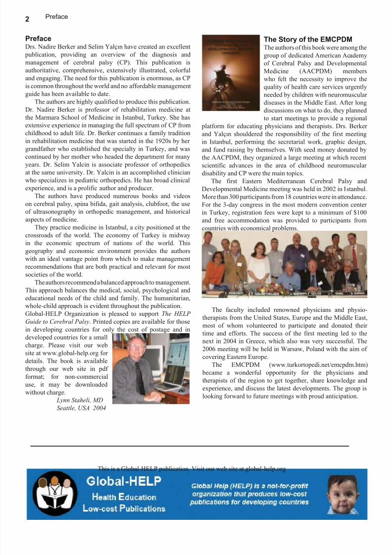
The Story of the EMCPDMThe authors of this book were among thegroup of dedicated American Academyof Cerebral Palsy and DevelopmentalMedicine (AACPDM) memberswho felt the necessity to improve thequality of health care services urgently
needed by children with neuromusculardiseases in the Middle East. After longdiscussions on what to do, they plannedto start meetings to provide a regional
platform for educating physicians and therapists. Drs. Berkerand Yalçın shouldered the responsibility of the first meetingin Istanbul, performing the secretarial work, graphic design,and fund raising by themselves. With seed money donated bythe AACPDM, they organized a large meeting at which recentscientific advances in the area of childhood neuromusculardisability and CP were the main topics.
The first Eastern Mediterranean Cerebral Palsy andDevelopmental Medicine meeting was held in 2002 in Istanbul.
More than 300 participants from 18 countries were in attendance.For the 3-day congress in the most modern convention centerin Turkey, registration fees were kept to a minimum of $100and free accommodation was provided to participants fromcountries with economical problems.
PrefaceDrs. Nadire Berker and Selim Yalçın have created an excellent
publication, providing an overview of the diagnosis andmanagement of cerebral palsy (CP). This publication isauthoritative, comprehensive, extensively illustrated, colorfuland engaging. The need for this publication is enormous, as CPis common throughout the world and no affordable management
guide has been available to date.The authors are highly qualified to produce this publication.Dr. Nadire Berker is professor of rehabilitation medicine atthe Marmara School of Medicine in Istanbul, Turkey. She hasextensive experience in managing the full spectrum of CP fromchildhood to adult life. Dr. Berker continues a family traditionin rehabilitation medicine that was started in the 1920s by hergrandfather who established the specialty in Turkey, and wascontinued by her mother who headed the department for manyyears. Dr. Selim Yalcin is associate professor of orthopedicsat the same university. Dr. Yalcin is an accomplished clinicianwho specializes in pediatric orthopedics. He has broad clinicalexperience, and is a prolific author and producer.
The authors have produced numerous books and videoson cerebral palsy, spina bifida, gait analysis, clubfoot, the useof ultrasonography in orthopedic management, and historicalaspects of medicine.
They practice medicine in Istanbul, a city positioned at thecrossroads of the world. The economy of Turkey is midwayin the economic spectrum of nations of the world. Thisgeography and economic environment provides the authorswith an ideal vantage point from which to make managementrecommendations that are both practical and relevant for mostsocieties of the world.
The authors recommend a balanced approach to management.
This approach balances the medical, social, psychological andeducational needs of the child and family. The humanitarian,whole-child approach is evident throughout the publication.Global-HELP Organization is pleased to support The HELP
Guide to Cerebral Palsy. Printed copies are available for thosein developing countries for only the cost of postage and indeveloped countries for a smallcharge. Please visit our website at www.global-help.org fordetails. The book is availablethrough our web site in pdfformat; for non-commercialuse, it may be downloadedwithout charge.
Lynn Staheli, MD
Seattle, USA 2004
Preface
This is a Global-HELP publication. Visit our web site at global-help.org
The faculty included renowned physicians and physio-therapists from the United States, Europe and the Middle East,most of whom volunteered to participate and donated theirtime and efforts. The success of the first meeting led to thenext in 2004 in Greece, which also was very successful. The2006 meeting will be held in Warsaw, Poland with the aim ofcovering Eastern Europe.
The EMCPDM (www.turkortopedi.net/emcpdm.htm) became a wonderful opportunity for the physicians andtherapists of the region to get together, share knowledge and
experience, and discuss the latest developments. The group islooking forward to future meetings with proud anticipation.
EASTERN MEDITERRANEAN
CEREBRAL PALSY &
DEVELOPMENTAL MEDICINE
CONGRESS
8/9/2019 Guía de ayuda para la parálisis cerebral.pdf
http://slidepdf.com/reader/full/guia-de-ayuda-para-la-paralisis-cerebralpdf 3/143
3
Authors
Nadire Berker, MDDr. Berker has treated disabled children for nearly10 years. She has pioneered the application ofmodern methods in pediatric rehabilitation, lecturedextensively, and co-authored many books inTurkish on various subjects of childhood [email protected]
Selim Yalçın, MDDr. Yalçın is a prominent Turkish pediatricorthopaedic surgeon with a chief interest indevelopmental disorders and the pediatric spine. Heloves to teach and has authored many educational books, organized meetings, created short moviesand web sites. [email protected]
Leon Root, MDDr. Root is one of the leading names in cerebral palsyorthopaedic surgery worldwide. A former presidentof the AACPDM, he has dedicated his career tocerebral palsy, given many lectures and authoredmore than 100 articles on the subject.
Lynn Staheli, MDDr. Staheli is one of the most prominent names in pediatric orthopaedics. He is the founder and chiefeditor of the Journal of Pediatric Orthopaedics, authorof six books and a wizard of desktop publishing. Hefounded the Global-HELP organization to createaffordable medical textbooks worldwide.
Contributors
Lana Staheli, Ph DLana Staheli, PhD is a certified counselor,marriage therapist, and consultant on interpersonalrelationships and life strategies. She authoredseveral relationship books. Lana is co-founder andvice-president of Global-HELP.
Zeynep Eti, MDDr. Eti is head of the department of Algology inMarmara University School of Medicine. She hasdedicated part of her work to pediatric anesthesiologyand algology.
Dhiren Ganjwala, MDDr. Ganjwala is a pediatric orthopaedic surgeonform India. Teaching is his passion and heconducts workshops and deliver lectures at variousinstitutions. He has published and edited many books on variety of topics for doctors, residents and patient education.
Garen Koloyan, MDDr. Koloyan is a pediatric orthopaedic surgeon fromYerevan, Armenia. He has been one of the creatorsof the EMCPDM and has done pioneer work fordisabled children of Armenia and Georgia.
Antigone Papavassiliou, MDDr Papavassiliou is the director of Pediatric Neurology at the Pendeli Children’s Hospital inAthens, Greece. She has been treating children andadolescents with CP for many years and has devoteda lot of time in teaching physicians and therapists.She has co-authored two medical textbooks on CPand many others for patient education in Greek.
Idil ÇilingiroğluMs. Çilingiroğlu is an architect who devoted hertime and talent to draw all the illustrations in thesection for families.
Dear Reader,
Years of treating children with cerebral palsy (CP) has shownus that a worldwide need exists for a concise, illustrated bookto guide health professionals regarding this difficult problem.This book is an attempt to fulfill that need. The Guide isintended for use by physicians, residents, medical studentsand allied health professionals who treat children with CP. Wefocus on the latest concepts in the treatment of musculoskeletal
problems and describe the associated impairments, providingsuggestions for further reading. The chapters on total bodyinvolvement, diplegia, hemiplegia and dyskinesia include themost common treatments applied for these patients. Note that
problems described in one section may occur in other typesof CP. We present the most frequently used and acceptedtreatment methods with scientifically proved efficacy andinclude references at the end of each chapter.
The illustrations and photographs of patients are fromours and Dr. Leon Root’s archives unless stated otherwise.We would like to thank our patients and their families forsharing their problems with us and also for allowing us to use
their pictures. We are indebted to Ms. Dory Kelly for helpingus with text editing.
Treatment of the child with CP is difficult, often frustratingand sometimes depressing. This is even more pronouncedin countries with limited resources for the disabled. We tryto provide information on how to proceed in places whereresources are limited. An interdisciplinary managementapproach is the only means to integrate children with CP withthe society and lessen the impact of the problem. We hope thatreaders will benefit from our work and use this guidebookin the treatment of unfortunate millions of patients with CPworldwide.
Nadire Berker and Selim Yalçın Istanbul, Turkey - 2005
Contributors
Graphic design: Selim Yalçın and Nadire Berker Page design: Selim Yalçın and Tevfik PekcanlıPrepress: Rotamat Press Co. Ltd.Printed at: Mart Printing Co. Ltd.
Istanbul, Turkey, March 2005
ISBN: 975-6257-12-1
No: 8 in Pediatric Orthopedics & Rehabilitation Series
prepared by Drs. Berker & Yalçın
Published jointly by Avrupa Medical Bookshop Co. Ltd. &
Global-HELP Organization
© Selim Yalçın & Nadire Berker
Every effort has been made to confirm the accuracy of the presented infor-mation. The authors and publisher are not responsible for errors of omission
or for any consequences from the application of the information in this bookand make no warranty, expressed or implied, with respect to the currency,completeness, or accuracy of the contents of this publication. Application ofthis information in a particular situation remains the professional responsi- bility of the practitioner. Care has been taken to reach all copyright holders.
8/9/2019 Guía de ayuda para la parálisis cerebral.pdf
http://slidepdf.com/reader/full/guia-de-ayuda-para-la-paralisis-cerebralpdf 4/143
4
Dedication
We would like to thank
ForewordCerebral palsy (CP) is the most common chronic disability ofchildhood today. It is ubiqitious and it occurs all around theworld. In developed nations, the incidence is about 1 to2 per1000 births. In spite of improved obstetrical and perinatal care,CP remains with us. As a result of injury to the brain, thesechildren have motor defects which will affect them for their
entire lifetime. Treatment often starts when they are infants,and continues throughout their life, even into adulthood. The problems involved are complex; not only do these childrenhave problems of mobility, but they can also have seizuredisorders, gastrointestinal system problems, learning and
perceptual difficulties, visual problems, hearing problems, andgrowth deficiency. In spite of all these numerous difficulties,cerebral palsied children can be helped.What the authors attempt in this book is to divide informationfor physicians, therapists or other paramedical personnelwho are interested and will be treating and taking care ofthese children for their lifetime. They present the basicunderstanding of what CP is and the fact that it takes a team
to treat them. The child and his parents become the focusof treatment because you cannot treat the child withoutinvolving the parents as well. The team has to consist of the
physician who will be the captain of the team. That physiciancan be a pediatrician, orthopaedic surgeon, physiatrist or evena neurologist, but they must take overall control of the childand make sure that all parameters of care are attended to. Theteam has to also include the occupational therapist, the speechtherapist, the physical therapist, teachers and social workers.
Dr. Wallace Salter of Toronto, Canada is fond of sayingthat after you operate on the child with CP, he still has CP.This may seem discouraging because we cannot cure the CPwith our present knowledge, but we can make life better forthese children, and that is important. Even small degrees ofimprovement makes a great difference. Getting a child to
walk, be it in crutches, in braces or with a walker, is much better than having him in a wheelchair. Having a child be ableto live in a wheelchair, as is true for children with total bodyinvolvement, is much better than having him be on a stretcheror in a bed for the rest of his life. These are important thingsto consider.The authors have carefully defined the types of CP, the
prognosis of CP, therapies that are at present available, thesurgical indications and most important of all, the pre and
postoperative care that these children must have. They writein a very clear and concise way which provides a readyreference for the interested reader in treating these children.In my own experience, I have found that working with the
cerebral palsied children and their families has been the mostrewarding aspect of my medical career. The children andfamilies are deeply grateful to you for whatever you can offerthem and particularly, they respond to the fact that you care.And it’s with your caring and your ability to help that makesa difference.
Foreword
Mustafa Berker Ender Berker
Our mothers and fathers, for their guidance and inspiration.Our children, for their compassion towards
those less fortunate than themselves.
Asaf Yalçın Sabahat Yalçın Deniz Yalçın Deniz Özaras Güneş Yalçın
Leon Root, MDOrthopaedic SurgeonHospital for Special Surgery
New York, February 2005
8/9/2019 Guía de ayuda para la parálisis cerebral.pdf
http://slidepdf.com/reader/full/guia-de-ayuda-para-la-paralisis-cerebralpdf 5/143
5General Concepts
General Concepts
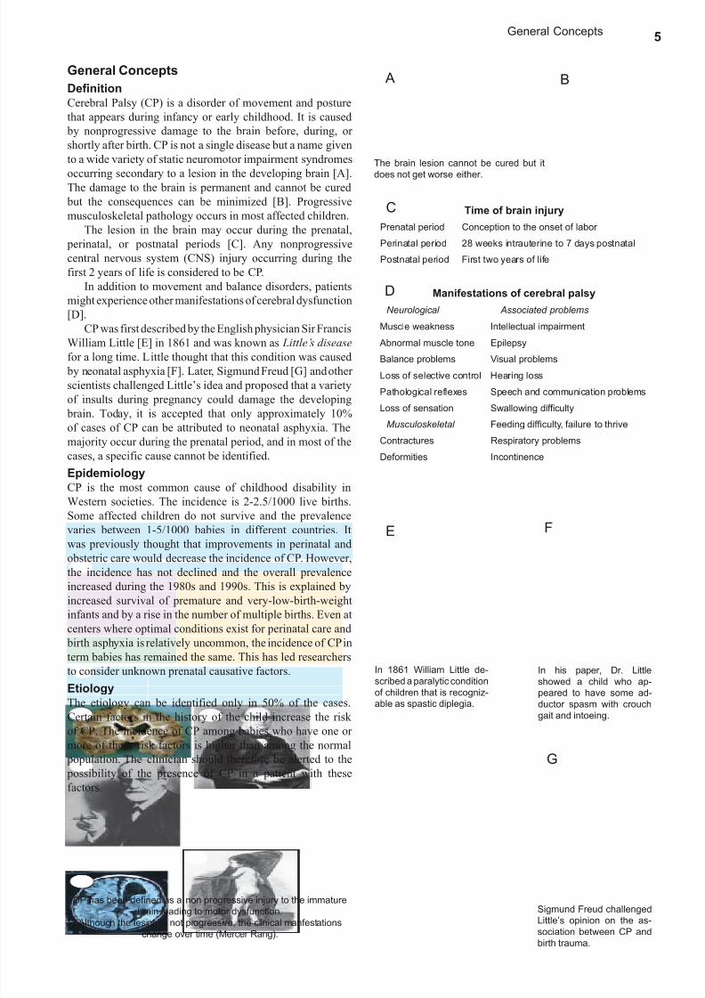
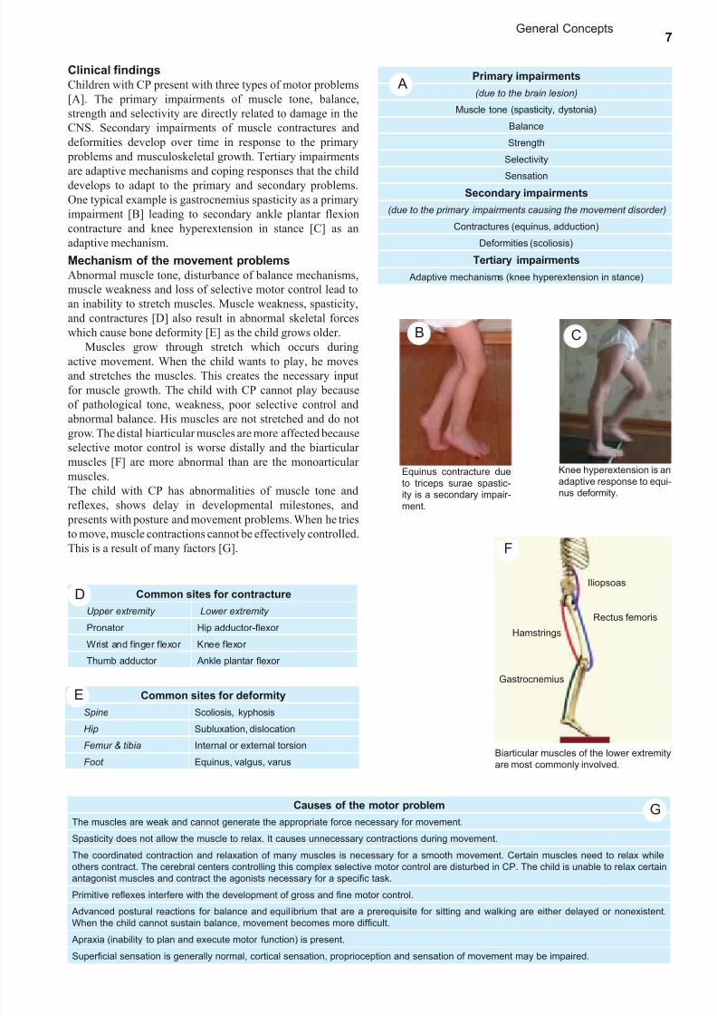
Definition
Cerebral Palsy (CP) is a disorder of movement and posturethat appears during infancy or early childhood. It is caused
by nonprogressive damage to the brain before, during, orshortly after birth. CP is not a single disease but a name givento a wide variety of static neuromotor impairment syndromes
occurring secondary to a lesion in the developing brain [A].The damage to the brain is permanent and cannot be cured
but the consequences can be minimized [B]. Progressivemusculoskeletal pathology occurs in most affected children.
The lesion in the brain may occur during the prenatal, perinatal, or postnatal periods [C]. Any nonprogressivecentral nervous system (CNS) injury occurring during thefirst 2 years of life is considered to be CP.
In addition to movement and balance disorders, patientsmight experience other manifestations of cerebral dysfunction[D].
CP was first described by the English physician Sir FrancisWilliam Little [E] in 1861 and was known as Little’s disease
for a long time. Little thought that this condition was caused by neonatal asphyxia [F]. Later, Sigmund Freud [G] and otherscientists challenged Little’s idea and proposed that a varietyof insults during pregnancy could damage the developing
brain. Today, it is accepted that only approximately 10%of cases of CP can be attributed to neonatal asphyxia. Themajority occur during the prenatal period, and in most of thecases, a specific cause cannot be identified.
Epidemiology
CP is the most common cause of childhood disability inWestern societies. The incidence is 2-2.5/1000 live births.Some affected children do not survive and the prevalencevaries between 1-5/1000 babies in different countries. Itwas previously thought that improvements in perinatal andobstetric care would decrease the incidence of CP. However,the incidence has not declined and the overall prevalenceincreased during the 1980s and 1990s. This is explained byincreased survival of premature and very-low-birth-weightinfants and by a rise in the number of multiple births. Even atcenters where optimal conditions exist for perinatal care and
birth asphyxia is relatively uncommon, the incidence of CP interm babies has remained the same. This has led researchersto consider unknown prenatal causative factors.
Etiology
The etiology can be identified only in 50% of the cases.Certain factors in the history of the child increase the riskof CP. The incidence of CP among babies who have one ormore of these risk factors is higher than among the normal
population. The clinician should therefore be alerted to the possibility of the presence of CP in a patient with thesefactors.
In 1861 William Little de-
scribed a paralytic condition
of children that is recogniz-able as spastic diplegia.
In his paper, Dr. Little
showed a child who ap-peared to have some ad-
ductor spasm with crouch
gait and intoeing.
Sigmund Freud challengedLittle’s opinion on the as-
sociation between CP and
birth trauma.
CP has been defined as a non progressive injury to the immature
brain leading to motor dysfunction.
Although the lesion is not progressive, the clinical manfestations
change over time (Mercer Rang).
The brain lesion cannot be cured but it
does not get worse either.
Time of brain injury
Prenatal period Conception to the onset of labor
Perinatal period 28 weeks intrauterine to 7 days postnatal
Postnatal period First two years of life
Manifestations of cerebral palsy
Neurological Associated problems
Muscle weakness Intellectual impairment
Abnormal muscle tone Epilepsy
Balance problems Visual problems
Loss of selective control Hearing loss
Pathological reflexes Speech and communication problems
Loss of sensation Swallowing difficulty
Musculoskeletal Feeding difficulty, failure to thrive
Contractures Respiratory problems
Deformities Incontinence
E
E
D
D
C
C
B A
G
G
F
F
8/9/2019 Guía de ayuda para la parálisis cerebral.pdf
http://slidepdf.com/reader/full/guia-de-ayuda-para-la-paralisis-cerebralpdf 6/143
6
F
F
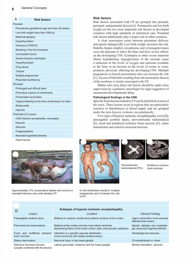
Risk factors
Prenatal
Prematurity (gestational age less than 36 weeks)
Low birth weight (less than 2500 g)
Maternal epilepsy
Hyperthyroidism
Infections (TORCH)Bleeding in the third trimester
Incompetent cervix
Severe toxemia, eclampsia
Hyperthyroidism
Drug abuse
Trauma
Multiple pregnancies
Placental insufficiency
Perinatal
Prolonged and difficult labor
Premature rupture of membranes
Presentation anomalies
Vaginal bleeding at the time of admission for labor
Bradycardia
Hypoxia
Postnatal (0-2 years)
CNS infection (encephalitis, meningitis)
Hypoxia
Seizures
Coagulopathies
Neonatal hyperbilirubinemia
Head trauma
General Concepts
Risk factors
Risk factors associated with CP are grouped into prenatal, perinatal, and postnatal factors [A]. Prematurity and low birthweight are the two most important risk factors in developedcountries with high standards of obstetrical care. Postnatalrisk factors additionally play a major role in other countries.
A clear association exists between premature deliveryand spastic diplegia [B]. Low birth weight increases the risk.Rubella, herpes simplex, toxoplasma, and cytomegalovirusescross the placenta to infect the fetus and have severe effectson the developing CNS. Eclampsia or other severe maternalillness hypothermia, hypoglycemia of the neonate causea reduction in the levels of oxygen and nutrients availableto the fetus or an increase in the levels of toxins or waste
products, adversely affecting the developing CNS. Multiple pregnancies or breech presentation also can increase the risk[C]. Excess of bilirubin resulting from the haemolytic diseaseof the newborn is clearly associated with CP.
Babies who carry these risk factors should be under closesupervision by a pediatric neurologist for signs suggestive of
neuromotor developmental delay.Pathological findings in the CNS
Specific brain lesions related to CP can be identified in most ofthe cases. These lesions occur in regions that are particularlysensitive to disturbances in blood supply and are groupedunder the term hypoxic ischemic encephalopathy.
Five types of hypoxic ischemic encephalopathy exist [D]; parasagittal cerebral injury, periventricular leukomalacia[E], focal and multifocal ischemic brain necrosis [F], statusmarmoratus and selective neuronal necrosis.
Approximately 11% of premature babies who survive in
neonatal intensive care units develop CP.
In vitro fertilization results in multiple
pregnancies and increases the risk
of CP.
Periventricularleukomalacia (PVL)
Multifocal ischemicbrain necrosis
E
E
Subtypes of hypoxic ischemic encephalopathy
Lesion Location Clinical Finding
Parasagittal cerebral injury Bilateral in superior medial and posterior portions of the cortex Upper extremities more severely
affected than lowers
Periventricular leukomalacia Bilateral white matter necrosis near lateral ventricles
descending fibers of the motor cortex, optic and acoustic radiations
Spastic diplegia and quadriple-
gia visual and cognitive deficits
Focal and multifocal ischemic
brain necrosis
Infarction in a specific vascular distribution
(most commonly left middle cerebral artery)
Hemiplegia and seizures
Status marmoratus Neuronal injury in the basal ganglia Choreoathetosis or mixed
Selective neuronal necrosis
(usually combined with the above)
Lateral geniculate, thalamus and the basal ganglia Mental retardation, seizures
D
D
C
C
B
A
8/9/2019 Guía de ayuda para la parálisis cerebral.pdf
http://slidepdf.com/reader/full/guia-de-ayuda-para-la-paralisis-cerebralpdf 7/143
8/9/2019 Guía de ayuda para la parálisis cerebral.pdf
http://slidepdf.com/reader/full/guia-de-ayuda-para-la-paralisis-cerebralpdf 8/143
8
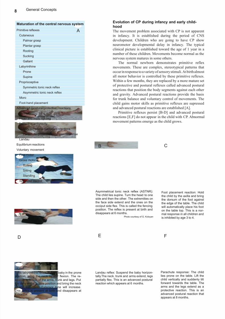
Asymmetrical tonic neck reflex (ASTNR):The child lies supine. Turn the head to one
side and then the other. The extremities on
the face side extend and the ones on the
occiput side flex. This is called the fencingposition. The reflex is present at birth and
disappears at 6 months.
General Concepts
Evolution of CP during infancy and early child-
hood
The movement problem associated with CP is not apparentin infancy. It is established during the period of CNSdevelopment. Children who are going to have CP showneuromotor developmental delay in infancy. The typicalclinical picture is established toward the age of 1 year in anumber of these children. Movements become normal as thenervous system matures in some others.
The normal newborn demonstrates primitive reflexmovements. These are complex, stereotypical patterns thatoccur in response to a variety of sensory stimuli. At birth almostall motor behavior is controlled by these primitive reflexes.Within a few months, they are replaced by a more mature setof protective and postural reflexes called advanced posturalreactions that position the body segments against each otherand gravity. Advanced postural reactions provide the basisfor trunk balance and voluntary control of movements. Thechild gains motor skills as primitive reflexes are supressedand advanced postural reactions are established [A].
Primitive reflexes persist [B-D] and advanced posturalreactions [E,F] do not appear in the child with CP. Abnormalmovement patterns emerge as the child grows.
Maturation of the central nervous system
Primitive reflexes
Cutaneous
Palmar grasp
Plantar grasp
Rooting
Sucking
Gallant
Labyrinthine
Prone
Supine
Proprioceptive
Symmetric tonic neck reflex
Asymmetric tonic neck reflex
Moro
Foot-hand placement
Advanced (postural/protective) reactions
Head righting
Head and body righting
Protective-antigravity
Forward-lateral-backward reactions
Parachute-protective extension response
Landau
Equilibrium reactions
Voluntary movement
Fine motor
Gross motor
Rolling
Sitting
Standing
Sphincter control
Foot placement reaction: Hold
the child by the axilla and bringthe dorsum of the foot against
the edge of the table. The child
will automatically place his foot
on the table top. This is a nor-mal response in all children and
is inhibited by age 3 to 4.
Tonic labyrinthine reflex: Put the baby in the proneposition, bring the neck to 45o flexion. The re-
sponse is flexion of the arms, trunk and legs. Putthe baby in the supine position and bring the neck
to 45o extension. Extensor tone will increase.This reflex is present at birth and disappears at
4 months.
Landau reflex: Suspend the baby horizon-tally.The neck, trunk and arms extend, legs
partially flex. This is an advanced posturalreaction which appears at 6 months.
Parachute response: The child
lies prone on the table. Lift the
child vertically and suddenly tiltforward towards the table. The
arms and the legs extend as a
protective reaction. This is an
advanced postural reaction thatappears at 8 months.
E
ED
D
C
CB
A
F
F
Photo courtesy of G. Koloyan

8/9/2019 Guía de ayuda para la parálisis cerebral.pdf
http://slidepdf.com/reader/full/guia-de-ayuda-para-la-paralisis-cerebralpdf 9/143
9
Absent traction response indicating
poor head control in a 10 month old
baby.
General Concepts
The child’s ability to achieve head control, sit, crawl, stand,and walk is always delayed. Late achievement of a milestonesuch as sitting indicates the presence of a motor deficit andthe degree of delay correlates with the severity of the problem[A,B].
Babies with CP usually have a period of hypotonicityduring the early months of life. Between the ages of 6 to 18
months, muscle tone gradually increases in those who aregoing to develop spasticity. Fluctuations in tone from hypo-to hypertonicity is a characteristic of developing dyskineticCP. Athetosis becomes obvious after 18 to 24 months. Ataxiamay not be apparent until even later.
Early signs suggestive of CP in the infant are abnormal behavior, oromotor problems and poor mobility [C]. Theinfant is irritable, too docile, or difficult to handle. He doesnot suck well, sleeps poorly, vomits frequently and has
poor eye contact. Deviant oromotor patterns include tongueretraction and thrust, tonic bite and grimacing. Early motorsigns are poor head control [D] with normal or increased tonein the limbs [E], and persistent or asymmetric fisting. Motor
development is both delayed and abnormal [F]. Instead ofcrawling, the child moves by creeping or hopping like a
bunny. Hand preference during the first two years of life is asign of hemiplegic CP.
The clinical picture of CP is established in early childhoodas the movement problem becomes prominent [G,H].
References2004 Baxter P. ‘Birth asphyxia and cerebral palsy’ Brain&Development 26 S6-
72004 Cans C, McManus V, Crowley M, et al. Surveillance of Cerebral Palsy
in Europe Collaborative Group ‘Cerebral palsy of post-neonatal origin:
characteristics and risk factors’ Paediatr Perinat Epidemiol 18(3):214-202004 Shapiro BK. ‘Cerebral palsy: A reconceptualization of the spectrum’ J
Pediatr 145(2 Suppl):S3-72002 Han TR, Bang MS, Lim JY, et al. ‘Risk factors of cerebral palsy in preterminfants’ Am J Phys Med Rehabil 81(4):297-3032002 Russman BS. ‘Cerebral Palsy: Definition, Manifestations And Etiology’Turk J Phys Med Rehabil 48 (2): 4-6
2002 Stromberg B, Dahlquist G, Ericson A, et al. ‘Neurological sequelae
in children born after in-vitro fertilisation: a population-based study’ Lancet9;359(9305):461-5
1999 Molnar GE, Sobus KM. ‘Growth and Development’ In Pediatric
Rehabilitation 3rd Edition pp: 13-28 Molnar GE, Alexander MA Hanley BelfusPhiladelphia
1998 Dormans JP,Copley LA. ‘Musculoskeletal impairments’ In Caring forChildren with Cerebral Palsy A Team Approach pp:125-141 Dormans JP,
Pellegrino L, Paul H Brookes Co Baltimore
1998 Pellegrino L, Dormans JP. ‘Definitions, etiology and epiemiology ofcerebral palsy’ In Caring for Children with Cerebral Palsy A Team Approach
pp:3-30 Dormans JP, Pellegrino L, Paul H Brookes Co Baltimore
1994 Campbell SK. ‘The child’s development of functional movement’ In
Campbell SK Physical Therapy for Children pp:3-38 WB Saunders Co.Philadelphia
1992 Blasco PA. ‘Pathology of cerebral palsy’ In The Diplegic Child: Evaluationand Management pp:3-20 Sussman MD AAOS, Rosemont
1990 Scherzer AL, Tscharnuter I. ‘Early Diagnosis and Treatment in Cerebral
Palsy: A Primer on Infant Developmental Problems’ 2nd Edition PediatricHabilitation Series Vol 6 Marcel Dekker Inc New York
Signs suggestive of CP in an infant
Abnormal behavior
Excessive docility or irritability
Poor eye contact
Poor sleep
Oromotor problems
Frequent vomiting
Poor sucking
Tongue retraction
Persistent bite
Grimacing
Poor mobility
Poor head control
Hand preference before 2 years of age
Abnormal tone
Children with increased femoral ante-
version and adductor spasticity sit in
the W-position to maintain balance.
Pathological asymmetrical pos-
ture in a 6 year old child
Increased tone in the limbs
and truncal hypotonia is com-
mon in spastic quadriplegia.
CP is likely
if there is no
Head control 3 months
Sitting 6 months
Rolling over 6 months
Walking 18 months
Absent Landau reflex at 11months is a sign of develop-
mental delay.
Major deficits in patients with CP
Loss of selective motor control and dependence on primitive reflex
patterns for movement
Abnormal muscle tone that is strongly influenced by body posture,
position & movement
Imbalance between agonist and antagonist muscles that, with time
and growth, leads to fixed muscle contracture and bony deformity
Impaired body balance mechanisms
Sensory loss
Vision
Hearing
Superficial & deep sensation
Associated problems
Seizures
Mental retardation
Behavior problems
Nutrition
Constipation
F
F
E
E
C
C
B A
H
H
G
G
D
D
8/9/2019 Guía de ayuda para la parálisis cerebral.pdf
http://slidepdf.com/reader/full/guia-de-ayuda-para-la-paralisis-cerebralpdf 10/143
10
Anatomical classification
Location Description
Hemiplegia Upper and lower extremity on one side of body
Diplegia Four extremities, legs more affected than the arms
Quadriplegia Four extremities plus the trunk, neck and face
Triplegia Both lower extremities and one upper extremity
Monoplegia One extremity (rare)
Double hemiplegia Four extremities, arms more affected than the legs
Clinical classification
Tonus Lesion site
Spastic Cortex
Dyskinetic Basal ganglia - extrapyramidal system
Hypotonic / Ataxic Cerebellum
Mixed Diffuse
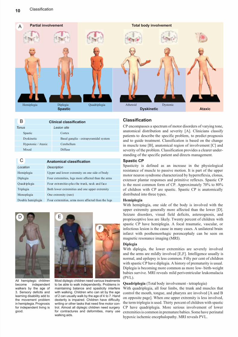
Classification
CP encompasses a spectrum of motor disorders of varying tone,anatomical distribution and severity [A]. Clinicians classify
patients to describe the specific problem, to predict prognosisand to guide treatment. Classification is based on the changein muscle tone [B], anatomical region of involvement [C] andseverity of the problem. Classification provides a clearer under-standing of the specific patient and directs management.
Spastic CP
Spasticity is defined as an increase in the physiologicalresistance of muscle to passive motion. It is part of the upper
motor neuron syndrome characterized by hyperreflexia, clonus,extensor plantar responses and primitive reflexes. Spastic CPis the most common form of CP. Approximately 70% to 80%of children with CP are spastic. Spastic CP is anatomicallydistributed into three types.
Hemiplegia
With hemiplegia, one side of the body is involved with theupper extremity generally more affected than the lower [D].Seizure disorders, visual field deficits, astereognosis, and
proprioceptive loss are likely. Twenty percent of children withspastic CP have hemiplegia. A focal traumatic, vascular, orinfectious lesion is the cause in many cases. A unilateral braininfarct with posthemorrhagic porencephaly can be seen onmagnetic resonance imaging (MRI).
Diplegia
With diplegia, the lower extremities are severely involvedand the arms are mildly involved [E,F]. Intelligence usually isnormal, and epilepsy is less common. Fifty per cent of childrenwith spastic CP have diplegia. A history of prematurity is usual.Diplegia is becoming more common as more low- birth-weight
babies survive. MRI reveals mild periventricular leukomalacia(PVL).
Quadriplegia (Total body involvement - tetraplegia)With quadriplegia, all four limbs, the trunk and muscles thatcontrol the mouth, tongue, and pharynx are involved [A and Bon opposite page]. When one upper extremity is less involved,the term triplegia is used. Thirty percent of children with spasticCP have quadriplegia. More serious involvement of lowerextremities is common in premature babies. Some have perinatalhypoxic ischemic encephalopathy. MRI reveals PVL.
Classification
All hemiplegic children
become independent
walkers by the age of
3. Sensory deficits and
learning disability add tothe movement problem
in hemiplegia. Prognosis
for independent living isgood.
Most diplegic children need various treatments
to be able to walk independently. Problems in
maintaining balance and spasticity interfere
with walking. Children who can sit by the age
of 2 can usually walk by the age of 4 to 7. Handdexterity is impaired. Children have difficulty
writing or other tasks that need fine motor con-
trol. Almost all diplegic children need surgeryfor contractures and deformities, many use
walking aids.
F
F
E
ED
B
A
C
C
Spastic Ataxic Hemiplegia Diplegia Quadriplegia
Partial involvement Total body involvement
Dyskinetic
Athetoid Dystonic
A
8/9/2019 Guía de ayuda para la parálisis cerebral.pdf
http://slidepdf.com/reader/full/guia-de-ayuda-para-la-paralisis-cerebralpdf 11/143
11
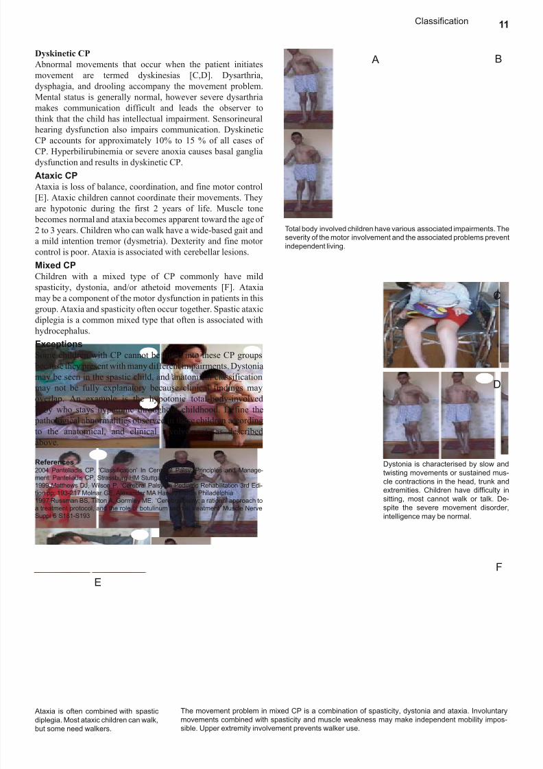
Dyskinetic CP
Abnormal movements that occur when the patient initiatesmovement are termed dyskinesias [C,D]. Dysarthria,dysphagia, and drooling accompany the movement problem.Mental status is generally normal, however severe dysarthriamakes communication difficult and leads the observer tothink that the child has intellectual impairment. Sensorineuralhearing dysfunction also impairs communication. DyskineticCP accounts for approximately 10% to 15 % of all cases ofCP. Hyperbilirubinemia or severe anoxia causes basal gangliadysfunction and results in dyskinetic CP.
Ataxic CP
Ataxia is loss of balance, coordination, and fine motor control[E]. Ataxic children cannot coordinate their movements. Theyare hypotonic during the first 2 years of life. Muscle tone
becomes normal and ataxia becomes apparent toward the age of2 to 3 years. Children who can walk have a wide-based gait anda mild intention tremor (dysmetria). Dexterity and fine motorcontrol is poor. Ataxia is associated with cerebellar lesions.
Mixed CPChildren with a mixed type of CP commonly have mildspasticity, dystonia, and/or athetoid movements [F]. Ataxiamay be a component of the motor dysfunction in patients in thisgroup. Ataxia and spasticity often occur together. Spastic ataxicdiplegia is a common mixed type that often is associated withhydrocephalus.
Exceptions
Some children with CP cannot be fitted into these CP groups because they present with many different impairments. Dystoniamay be seen in the spastic child, and anatomical classificationmay not be fully explanatory because clinical findings mayoverlap. An example is the hypotonic total-body-involved
baby who stays hypotonic throughout childhood. Define the pathological abnormalities observed in these children accordingto the anatomical, and clinical involvement, as describedabove.
References2004 Panteliadis CP. ‘Classification’ In Cerebral Palsy: Principles and Manage-ment. Panteliadis CP, Strassburg HM Stuttgart Thieme
1999 Matthews DJ, Wilson P. ‘Cerebral Palsy’ In Pediatric Rehabilitation 3rd Edi-tion pp: 193-217 Molnar GE, Alexander MA Hanley Belfus Philadelphia
1997 Russman BS, Tilton A, Gormley ME. ‘Cerebral palsy: a rational approach to
a treatment protocol, and the role of botulinum toxin in treatment’ Muscle NerveSuppl 6 S181-S193
Classification
Total body involved children have various associated impairments. Theseverity of the motor involvement and the associated problems prevent
independent living.
Dystonia is characterised by slow and
twisting movements or sustained mus-
cle contractions in the head, trunk andextremities. Children have difficulty in
sitting, most cannot walk or talk. De-
spite the severe movement disorder,
intelligence may be normal.
The movement problem in mixed CP is a combination of spasticity, dystonia and ataxia. Involuntary
movements combined with spasticity and muscle weakness may make independent mobility impos-sible. Upper extremity involvement prevents walker use.
Ataxia is often combined with spastic
diplegia. Most ataxic children can walk,
but some need walkers.
F
F
E
E
D
D
B A
C
C
8/9/2019 Guía de ayuda para la parálisis cerebral.pdf
http://slidepdf.com/reader/full/guia-de-ayuda-para-la-paralisis-cerebralpdf 12/143
8/9/2019 Guía de ayuda para la parálisis cerebral.pdf
http://slidepdf.com/reader/full/guia-de-ayuda-para-la-paralisis-cerebralpdf 13/143
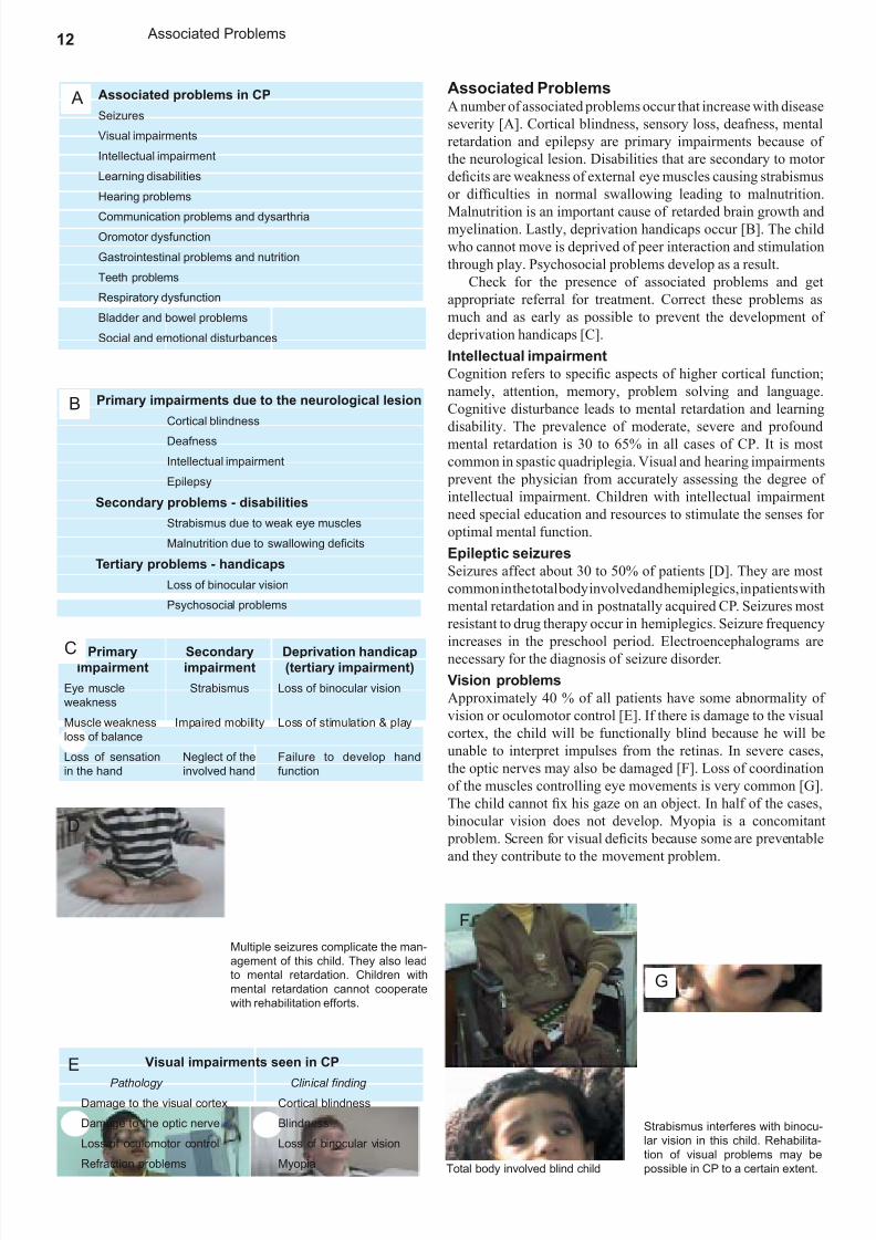
13 Associated Problems
Reasons for failure to thrive
Inadequate food intake
Recurrent vomiting
Aspiration
High basal metabolic rate
Hearing
Sensorineural hearing loss is seen in 10 % of children [A]. Children
born prematurely are at high risk for hearing loss. It is generally
not diagnosed early because of other handicaps. Test all babies for
hearing loss because appropriate hearing devices prevent many
future problems resulting from loss of hearing ability.
Communication problems and dysarthria
Dysarthria refers to speech problems. The child has difficulty producing sound and articulating words. Dysarthria occurs in
40% of patients. The causes are respiratory difficulties due to
respiratory muscle involvement, phonation difficulties due to
laryngeal involvement, and articulation difficulty due to oromotor
dysfunction. Spasticity or athetosis of the muscles of the tongue,
mouth and larynx cause dysarthria. It is important that every child
is provided with an alternative means of communication as early as
possible to avoid further disability [B,C].
Oromotor dysfunction
Sucking, swallowing, and chewing mechanisms are impaired
[D]. Drooling [E], dysarthria and inability to eat result in failure
to thrive, delayed growth and nutrition, poor hygiene [F,G] andimpaired socialization.
Gastrointestinal problems and nutrition
There is a general deficiency of growth and development. Children
with dyskinesia and spastic quadriplegia fail to thrive [H,I]. This is
related to inadequate intake of food [K], recurrent vomiting with
aspiration secondary to gastroesophageal reflux and pseudobulbar
palsy. Difficulties in swallowing (dysphagia), hyperactive gag
reflex, spasticity or loss of fine motor control impair feeding.
Gastroesophageal reflux and impaired swallowing cause aspiration
pneumonia. Many children with CP have high basal metabolic rates.
Increase in basal metabolic rate coupled with feeding difficulties
cause malnutrition. Malnutrition may be severe enough to affect brain growth and myelination in the first 3 years of life. There is
immune system suppression and increased risk of infection.
Drooling and strabismus coexist in this child.
Drooling is caused by oromotor dysfunction
and is a difficult problem to treat. Consideroral medications and botulinum toxin in
management.
Mouth hygiene is poor and dental
caries is common. Obtain regulardental care.
Teeth problems
Dentin Primary or hyperbilirubinemia
Malocclusion Spasticity
Tooth decay Feeding, swallowing problems
Gingival hyperplasia Antiepileptic drug use
Oromotor dysfunction
Drooling
Dysarthria
Inability to chew
Inability to swallow
Causes of inadequate food intake
Difficulty chewing and swallowing
Hyperactive gag reflex
Spasticity of oropharyngeal muscles
Loss of selective control of oropharyngeal muscles
Gastroesophageal reflux
This severely involved diplegic child with
hearing impairment has been using hearing
aids ever since he was a baby.
Communication aids range from advancedcomputer systems to simple picture boards.
Children with adequate mental functionlearn to use these to interact with their en-
vironment.
F
F
E
E
D
D
C
C
B
A
H
G
G
Spastic quadriplegic child with
malnutrition
K
K
I
I
8/9/2019 Guía de ayuda para la parálisis cerebral.pdf
http://slidepdf.com/reader/full/guia-de-ayuda-para-la-paralisis-cerebralpdf 14/143
14 Associated Problems
Respiratory problems
Aspiration in small quantities leads to pneumonia in children
who have difficulty swallowing. Premature babies have
bronchopulmonary dysplasia. This leads to frequent upper
respiratory tract infections. Respiratory muscle spasticity
contributes to the pulmonary problems.
Bladder and bowel dysfunction
Loss of coordination of bowel and bladder sphincters results inconstipation and/or incontinence. Enuresis, frequency, urgency,
urinary tract infections and incontinence are common problems
[A]. The causes are poor cognition, decreased mobility, poor
communication and neurogenic dysfunction [B]. Urodynamic
assessment has demonstrated bladder hyperreflexia, detrusor
sphincter dyssynergia, hypertonic bladders with incomplete
leakage and periodic relaxation of the distal sphincter during
filling.
Constipation is a common but overlooked phenomenon. It
causes distress in the child, increases spasticity and results in
poor appetite. It is a result of many factors, including poor diet
and decreased mobility. Establishing a routine for bowel training
and encouraging upright posture help reduce constipation.
Psychosocial problems
A diagnosis of CP is extremely stressful for the family and the
child when he grows up. This causes various reactions ranging
from denial to anger, guilt and depression. Coping with the
emotional burden of disability is easier if the family has strong
relationships, financial security, and supportive members of
the community. The child and the family need to find ways to
connect to each other. A healthy relationship between the mother
and the child forms the basis of future happiness.
Prevention or appropriate treatment of associated problems
improves the quality of life of the child and the family [C].
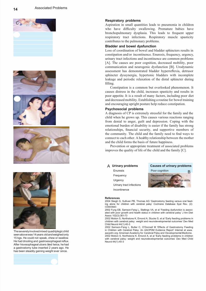
The severely involved mixed quadriplegic childseen above was 14 years old and weighed only
15 kgs. He could not speak, chew or swallow.
He had drooling and gastroesophageal reflux.
After his esophageal ulcers bled twice, he hada gastrostomy tube inserted 2 years ago. He
has been steadily gaining weight ever since.
Urinary problems
Enuresis
Frequency
Urgency
Urinary tract infections
Incontinence
Causes of urinary problems
Poor cognition
Decreased mobility
Decreased communication skills
Neurogenic dysfunction
References2004 Sleigh G, Sullivan PB, Thomas AG ‘Gastrostomy feeding versus oral feed-
ing alone for children with cerebral palsy’ Cochrane Database Syst Rev. (2):CD003943
2002 Fung EB, Samson-Fang L, Stallings VA, et al ‘Feeding dysfunction is associ-ated with poor growth and health status in children with cerebral palsy’ J Am Diet
Assoc 102(3):361-73
2002 Motion S, Northstone K, Emond A, Stucke S, et al ‘Early feeding problems in
children with cerebral palsy: weight and neurodevelopmental outcomes’ Dev Med
Child Neurol 44(1):40-3
2002 Samson-Fang L, Butler C, O’Donnell M ‘Effects of Gastrostomy Feeding
in Children with Cerebral Palsy: An AACPDM Evidence Report’ Internet at www.
aacpdm.org: American Academy for Cerebral Palsy and Developmental Medicine.
2002 Motion S, Northstone K, Emond A, et al ‘Early feeding problems in children
with cerebral palsy: weight and neurodevelopmental outcomes’ Dev Med Child
Neurol 44(1):40-3
C
C
B
A
8/9/2019 Guía de ayuda para la parálisis cerebral.pdf
http://slidepdf.com/reader/full/guia-de-ayuda-para-la-paralisis-cerebralpdf 15/143
15Physical Examination
Physical Examination and Making the DiagnosisPhysical examination of a child with movement problem has two
basic purposes [A]. First, physical examination accompanying a
detailed history enables an accurate diagnosis. Second, it allows
the treating physicians to define the impairments and disabilities,
determine the functional prognosis and set treatment goals in
children with CP. These then help devise a treatment plan for
each child.It is difficult to identify the cause of CP. When faced with
a motor disorder in the child, the physician must be careful to
rule out conditions that are results of genetic defects, such as
hereditary spastic paraplegia, that are similar to CP. A detailed
history and physical examination help the clinician exclude
these rare syndromes and prevent expensive and extensive
work-up.
Physical examination of the child with CP is not easy. It is
a three-way relationship between the child, the physician and
the family [B,C]. Adjustment problems can cause fear, distrust,
confusion, and anxiety in the family and in the child. This
disturbs their capability to understand the problem and cooperate
with the treatment team. The physician must be willing to deal
with anxious, confused, frustrated and unhappy families and
frightened children. The examination cannot succeed unless the
physician gains the parents’ confidence and trust. Parents will
trust a physician who takes a genuine interest in their child.
History
History is a key component in evaluating the child [D]. It
provides valuable information for diagnosis. In children with a
definite diagnosis, the timing of achievement of developmental
milestones and the presence of associated impairments help to
decide a functional prognosis. The physician gains insight into
the parents’ expectations and disappointments from previous
treatment procedures. Knowledge of previous botulinum toxininjections, physiotherapy, surgical procedures, outcomes,
complications, and psychological burden are key issues when
making a treatment plan [E].
History taking provides the time and room to build a sense of
understanding between the family and the physician. The goal is
to make the child and the family comfortable so that the clinical
examination will be accurate.
Goals of physical examination in a child with
movement disorder
Establish an accurate diagnosis
Differentiate CP from progressive causes of childhood neuromotor
disability
Classify the type and severity of involvement
Define the musculoskeletal impairment (spasticity, balance, weak-
ness, contractures and deformities) and decide on ways of treatment
Evaluate associated impairments and get appropriate treatment
Determine functional prognosis
Set treatment goals
Devise a treatment plan
Evaluate the outcome of previous treatment procedures
Assess the changes that occur with treatment as well as with growth
& development
A detailed history provides knowledge about
Risk factors
Timing of achievement of developmental milestones
The presence of associated impairments
Progression of child’s capabilities
Insight into the family’s resources
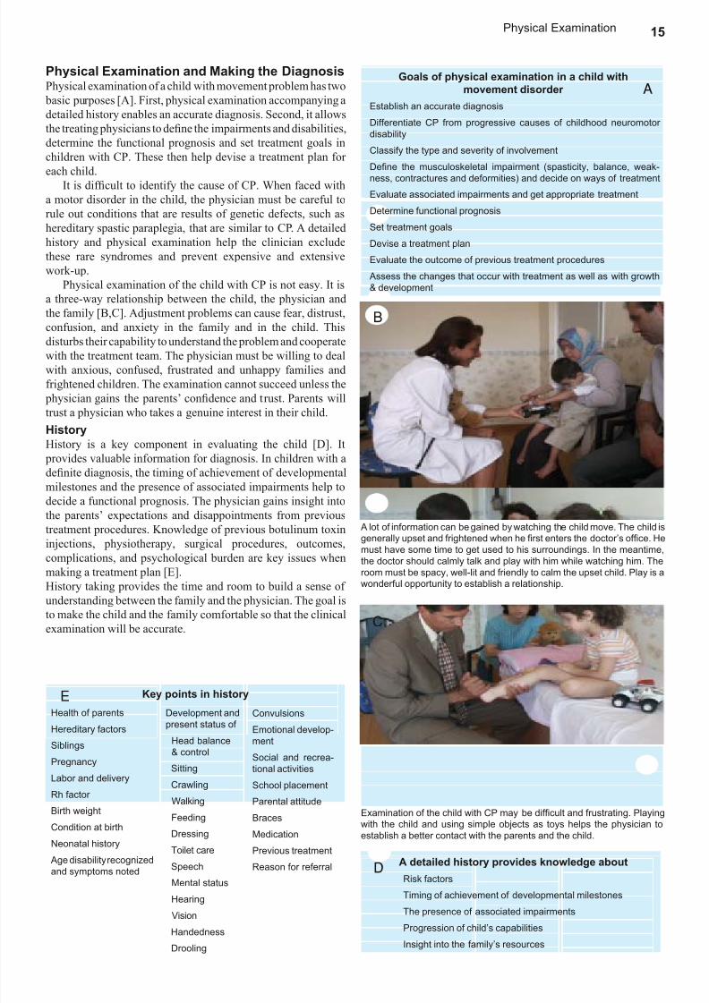
A lot of information can be gained by watching the child move. The child isgenerally upset and frightened when he first enters the doctor’s office. He
must have some time to get used to his surroundings. In the meantime,
the doctor should calmly talk and play with him while watching him. The
room must be spacy, well-lit and friendly to calm the upset child. Play is awonderful opportunity to establish a relationship.
Key points in history
DD
B
A
Examination of the child with CP may be difficult and frustrating. Playingwith the child and using simple objects as toys helps the physician to
establish a better contact with the parents and the child.
Health of parents
Hereditary factors
Siblings
Pregnancy
Labor and delivery
Rh factor
Birth weight
Condition at birth
Neonatal history
Age disability recognized
and symptoms noted
Development and
present status of
Head balance
& control
Sitting
Crawling
Walking
Feeding
Dressing
Toilet care
Speech
Mental status
Hearing
Vision
Handedness
Drooling
Convulsions
Emotional develop-
ment
Social and recrea-
tional activities
School placement
Parental attitude
Braces
Medication
Previous treatment
Reason for referral
EE
CC
8/9/2019 Guía de ayuda para la parálisis cerebral.pdf
http://slidepdf.com/reader/full/guia-de-ayuda-para-la-paralisis-cerebralpdf 16/143
16 Physical Examination
Let the child sit in the mother’s lap. Provide the older child
with a chair of his or her own. Remember to smile at and talk
to the child. Generally the parents provide the information.
Older children should answer for themselves [A,B]. The
parents can help fill in the details later. Including the child
in the conversation builds trust. If the parents trust the
physician, they will be more open-hearted when expressing
their expectations and disappointments.The child and the parents will remember less than 20%
of the information provided. Provide them with a written
summary of the results and the treatment plan for future
reference.
Clinical Examination
Observing the child’s movements is the initial and a crucial
part of the examination. Observe before you touch [C]. If the
child is young, apprehensive or tearful, let him or her stay on
mother’s lap while you watch and talk to the mother. As the
child adapts to the environment, slowly place him or her on
the examination table or on the floor and watch him or her
move around. If the child cries a lot and does not cooperate,continue while he or she is in the mother’s lap [D].
Tools required for the examination are very simple: toys,
small wooden blocks, round beads or pebbles, triangular,
circular and square shaped objects, a few coins, objects with
different textures and a tape measure.
Perform a neurological, musculoskeletal and functional
examination, although not necessarily in that order [E]. Every
physician develops his or her own style and sequence of
examination over the years [F,G].
Examination outline
Neurological examination
Skull, head circumference
Spine
Mental status
Cranial nerves
Vision - hearing - speech
Motor system
Muscle tone
Muscle power
Muscle bulk
Degree of voluntary control
Reflexes
Involuntary movements
Sensory examination
Sphincters
Developmental milestones
Musculoskeletal examination
Range of motion
Deformities, contractures
Posture
Functional examination
Sitting
Balance
Gait
Hand function
Examine the young and
frightened child in his moth-er’s lap. Evaluate tonus ab-
normalities when the child
is comfortable.
Start the examination by giv-ing the child something to
play with. A ball or a balloon
will help to test upper extrem-
ity function.
FF
EE
DD
CC
BA
GG
Try to be friends with the older child and the adolescent. Talk to them alone
and if necessary perform the examination when members of the oppositesex are not present. Do not treat older children like babies.
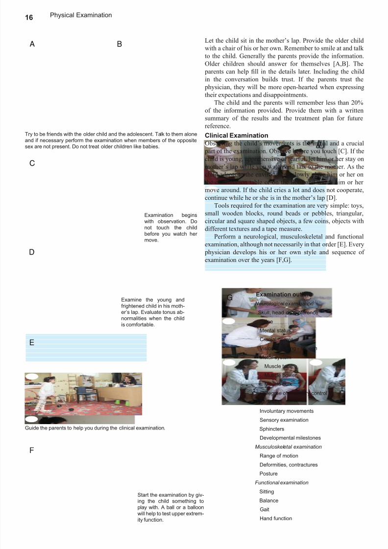
Guide the parents to help you during the clinical examination.
Examination begins
with observation. Do
not touch the child
before you watch hermove.
8/9/2019 Guía de ayuda para la parálisis cerebral.pdf
http://slidepdf.com/reader/full/guia-de-ayuda-para-la-paralisis-cerebralpdf 17/143
17Physical Examination
Neurological examination
Neurological evaluation of the infant and the child requires
adequate knowledge of neurological developmental stages [A].
Mental status
Observe the child’s orientation and interest in the surroundings.
Watch for eye contact, following objects, alertness, and ability to
obey simple commands.
Vision and hearing
The diagnosis of visual and hearing loss in infants can be easy. Call
the child when he is not looking. Clap your hands or deliberately
drop an object to make a noise behind the child and watch the
response. If the child does not seem to hear, look in the child’s
ears for wax or signs of infection. Considering the high incidence
of visual and oculomotor problems in cases of CP, all children
with a definite diagnosis of neurodevelopmental delay and/or
CP should undergo a detailed ophthalmological and audiological
examinations during early infancy. The examinations should be
repeated at yearly intervals until school age.
Muscle strength and selective motor control
Many children with CP cannot voluntarily contract or relax theirmuscles in isolation and therefore are unable to move their joints
separately. For example, when the child attempts to extend his
elbow, he involuntarily moves his whole arm. Lack of selective
motor control makes it impossible to determine muscle strength
using simple manual muscle testing [B]. Observe muscle strength
by watching the child perform certain tasks, such as throwing or
hitting a ball.
Reflexes
Evaluate the persistence of primitive reflexes and the absence
of advanced postural reactions [C,D]. The presence of primitive
reflexes beyond 6 months of age is a sign of poor prognosis [E].
Muscle tone and involuntary movements
The child must be calm for assessment of muscle tone. Place the
head in neutral position because turning or flexion can trigger
tonic neck reflexes and interfere with muscle tone. Spasticity is
the resistance felt while moving the joint through a passive range
of motion. Use the modified Ashworth or Tardieu scales to grade
spasticity. Also record tremor, chorea, athetosis, dystonia and
ataxia [F].
This 8 month old baby has diffi-
culty maintaining head control on
traction response indicating devel-opmental delay.
Differences between spasticity & dystonia
Spasticity Dystonia
Examination You feel You see
Tendon reflexes Increased Generally normal
Clonus Present Absent
Pathological reflexes Present Rare
Signs of poor prognosis
Present Absent
ASTNR Parachute response
STNR Neck righting reactions
Moro
Extensor thrust
Stepping reflex
Normal developmental stages of the child
Age (months) Milestones
1 Lifts head
3 Good head control, follows, laughs, smiles
5 Reaches and grasps objects
6 Propped sitting
8 Independent sitting, equilibrium reflexes
9 Gets to sitting position, presents parachute reflex
10 Pulls to stand, cruises
12-14 Walks, first words
18 Removes clothes, uses spoon
24 Uses two word phrases, throws overhand
30 Knows full name, puts on clothing
36 Jumps, pedals tricycle, learns rhymes
48 Hops, plays with others
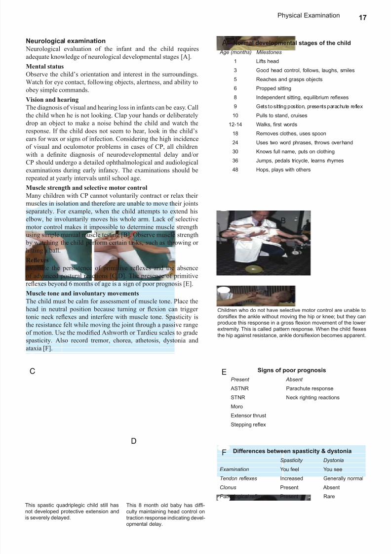
Children who do not have selective motor control are unable to
dorsiflex the ankle without moving the hip or knee; but they canproduce this response in a gross flexion movement of the lower
extremity. This is called pattern response. When the child flexes
the hip against resistance, ankle dorsiflexion becomes apparent.
This spastic quadriplegic child still has
not developed protective extension andis severely delayed.
FF
EE
DD
CC
B
A
8/9/2019 Guía de ayuda para la parálisis cerebral.pdf
http://slidepdf.com/reader/full/guia-de-ayuda-para-la-paralisis-cerebralpdf 18/143
18 Physical Examination
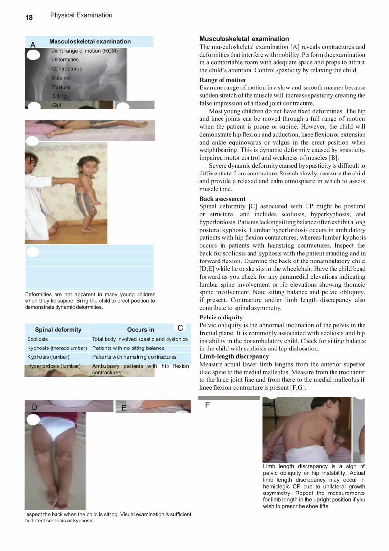
Musculoskeletal examination
The musculoskeletal examination [A] reveals contractures and
deformities that interfere with mobility. Perform the examination
in a comfortable room with adequate space and props to attract
the child’s attention. Control spasticity by relaxing the child.
Range of motion
Examine range of motion in a slow and smooth manner because
sudden stretch of the muscle will increase spasticity, creating thefalse impression of a fixed joint contracture.
Most young children do not have fixed deformities. The hip
and knee joints can be moved through a full range of motion
when the patient is prone or supine. However, the child will
demonstrate hip flexion and adduction, knee flexion or extension
and ankle equinovarus or valgus in the erect position when
weightbearing. This is dynamic deformity caused by spasticity,
impaired motor control and weakness of muscles [B].
Severe dynamic deformity caused by spasticity is difficult to
differentiate from contracture. Stretch slowly, reassure the child
and provide a relaxed and calm atmosphere in which to assess
muscle tone.
Back assessment
Spinal deformity [C] associated with CP might be postural
or structural and includes scoliosis, hyperkyphosis, and
hyperlordosis. Patients lacking sitting balance often exhibit a long
postural kyphosis. Lumbar hyperlordosis occurs in ambulatory
patients with hip flexion contractures, whereas lumbar kyphosis
occurs in patients with hamstring contractures. Inspect the
back for scoliosis and kyphosis with the patient standing and in
forward flexion. Examine the back of the nonambulatory child
[D,E] while he or she sits in the wheelchair. Have the child bend
forward as you check for any paramedial elevations indicating
lumbar spine involvement or rib elevations showing thoracic
spine involvement. Note sitting balance and pelvic obliquity,
if present. Contracture and/or limb length discrepancy also
contribute to spinal asymmetry.
Pelvic obliquity
Pelvic obliquity is the abnormal inclination of the pelvis in the
frontal plane. It is commonly associated with scoliosis and hip
instability in the nonambulatory child. Check for sitting balance
in the child with scoliosis and hip dislocation.
Limb-length discrepancy
Measure actual lower limb lengths from the anterior superior
iliac spine to the medial malleolus. Measure from the trochanter
to the knee joint line and from there to the medial malleolus if
knee flexion contracture is present [F,G].
Inspect the back when the child is sitting. Visual examination is sufficientto detect scoliosis or kyphosis.
Spinal deformity Occurs in
Scoliosis Total body involved spastic and dystonics
Kyphosis (thoracolumbar) Patients with no sitting balance
Kyphosis (lumbar) Patients with hamstring contractures
Hyperlordosis (lumbar) Ambulatory patients with hip flexion
contractures
Musculoskeletal examination
Joint range of motion (ROM)
Deformities
Contractures
Balance
Posture
Sitting
Gait
Deformities are not apparent in many young childrenwhen they lie supine. Bring the child to erect position to
demonstrate dynamic deformities.
EED
CC
A
Limb length discrepancy is a sign of
pelvic obliquity or hip instability. Actual
limb length discrepancy may occur in
hemiplegic CP due to unilateral growth
asymmetry. Repeat the measurements
for limb length in the upright position if you
wish to prescribe shoe lifts.
F G
B
8/9/2019 Guía de ayuda para la parálisis cerebral.pdf
http://slidepdf.com/reader/full/guia-de-ayuda-para-la-paralisis-cerebralpdf 19/143
19Physical Examination
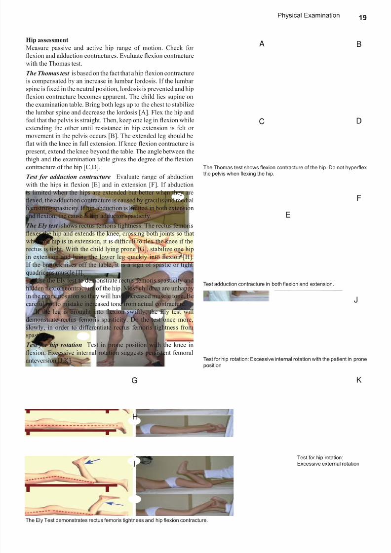
Hip assessment
Measure passive and active hip range of motion. Check for
flexion and adduction contractures. Evaluate flexion contracture
with the Thomas test.
The Thomas test is based on the fact that a hip flexion contracture
is compensated by an increase in lumbar lordosis. If the lumbar
spine is fixed in the neutral position, lordosis is prevented and hip
flexion contracture becomes apparent. The child lies supine onthe examination table. Bring both legs up to the chest to stabilize
the lumbar spine and decrease the lordosis [A]. Flex the hip and
feel that the pelvis is straight. Then, keep one leg in flexion while
extending the other until resistance in hip extension is felt or
movement in the pelvis occurs [B]. The extended leg should be
flat with the knee in full extension. If knee flexion contracture is
present, extend the knee beyond the table. The angle between the
thigh and the examination table gives the degree of the flexion
contracture of the hip [C,D].
Test for adduction contracture Evaluate range of abduction
with the hips in flexion [E] and in extension [F]. If abduction
is limited when the hips are extended but better when they areflexed, the adduction contracture is caused by gracilis and medial
hamstring spasticity. If hip abduction is limited in both extension
and flexion, the cause is hip adductor spasticity.
The Ely test shows rectus femoris tightness. The rectus femoris
flexes the hip and extends the knee, crossing both joints so that
when the hip is in extension, it is difficult to flex the knee if the
rectus is tight. With the child lying prone [G], stabilize one hip
in extension and bring the lower leg quickly into flexion [H].
If the buttock rises off the table, it is a sign of spastic or tight
quadriceps muscle [I].
Use the Ely test to demonstrate rectus femoris spasticity and
hidden flexion contracture of the hip. Most children are unhappyin the prone position so they will have increased muscle tone. Be
careful not to mistake increased tone from actual contracture.
If the leg is brought into flexion swiftly, the Ely test will
demonstrate rectus femoris spasticity. Do the test once more,
slowly, in order to differentiate rectus femoris tightness from
spasticity.
Test for hip rotation Test in prone position with the knee in
flexion. Excessive internal rotation suggests persistent femoral
anteversion [J,K].
Test adduction contracture in both flexion and extension.
The Thomas test shows flexion contracture of the hip. Do not hyperflexthe pelvis when flexing the hip.
Test for hip rotation: Excessive internal rotation with the patient in prone
position
Test for hip rotation:Excessive external rotation
The Ely Test demonstrates rectus femoris tightness and hip flexion contracture.
FF
EE
DDC
BA
KK
JJ
II
HH
GG
8/9/2019 Guía de ayuda para la parálisis cerebral.pdf
http://slidepdf.com/reader/full/guia-de-ayuda-para-la-paralisis-cerebralpdf 20/143
20 Physical Examination
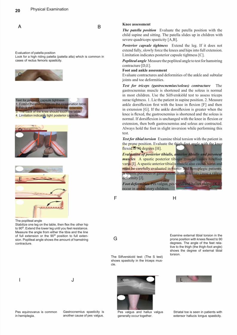
Knee assessment
The patella position Evaluate the patella position with the
child supine and sitting. The patella slides up in children with
severe quadriceps spasticity [A,B].
Posterior capsule tightness Extend the leg. If it does not
extend fully, slowly force the knees and hips into full extension.
Limitation indicates posterior capsule tightness [C].
Popliteal angle Measure the popliteal angle to test for hamstring
contracture [D,E].
Foot and ankle assessment
Evaluate contractures and deformities of the ankle and subtalar
joints and toe deformities.
Test for triceps (gastrocnemius/soleus) contracture The
gastrocnemius muscle is shortened and the soleus is normal
in most children. Use the Silfverskiöld test to assess triceps
surae tightness. 1. Lie the patient in supine position. 2. Measure
ankle dorsiflexion first with the knee in flexion [F] and then
in extension [G]. If the ankle dorsiflexion is greater when the
knee is flexed, the gastrocnemius is shortened and the soleus is
normal. If dorsiflexion is unchanged with the knee in flexion or
extension, then both gastrocnemius and soleus are contracted.
Always hold the foot in slight inversion while performing this
test.
Test for tibial torsion Examine tibial torsion with the patient in
the prone position. Evaluate the thigh-foot angle with the knee
flexed to 90 degrees [H].
Evaluation of posterior tibialis, anterior tibialis and peroneal
muscles A spastic posterior tibialis muscle causes hindfoot
varus [I]. A spastic anterior tibialis muscle also causes varus and
must be carefully evaluated in mono- and hemiplegic patients.
A spastic peroneus or gastrocnemius muscle may cause a valgus
deformity [J].
Foot deformities Pes valgus, pes varus and hallux valgus [K,L]
occur in ambulatory children.
The Silfverskiold test: (The S test)
shows spasticity in the triceps mus-
cle.
Examine external tibial torsion in theprone position with knees flexed to 90degrees. The angle of the feet rela-
tive to the thigh (the thigh-foot angle)
shows the degree of external tibialtorsion.
Pes valgus and hallux valgus
generally occur together.
Gastrocnemius spasticity isanother cause of pes valgus.
Striatal toe is seen in patients with
extensor hallucis longus spasticity.
Evaluation of patella position
Look for a high riding patella (patella alta) which is common in
cases of rectus femoris spasticity.
Test for posterior capsule tightness
1. Extend the child’s legs on the examination table.2. Force the knees and the hips in full extension.
3. The back of the knee should touch the table.
4. Limitation indicates tight posterior capsule.
The popliteal angle
Stabilize one leg on the table, then flex the other hip
to 90o. Extend the lower leg until you feel resistance.
Measure the angle from either the tibia and the line
of full extension or the 90o position to full exten-sion. Popliteal angle shows the amount of hamstring
contracture.
Pes equinovarus is common
in hemiplegia.
FF
EE
DD
CC
BA
HH
GG
II JJ KK LL
8/9/2019 Guía de ayuda para la parálisis cerebral.pdf
http://slidepdf.com/reader/full/guia-de-ayuda-para-la-paralisis-cerebralpdf 21/143
21Physical Examination
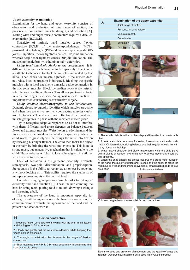
Flexion contracture
1. Measure flexion contracture of the wrist with the wrist in full flexionand the fingers in full extension.
2. Slowly and gently pull the wrist into extension while keeping thefinger joints in extension.
3. The angle of wrist with the forearm is the angle of flexioncontracture.
4. Then evaluate the PIP & DIP joints separately to determine thespastic muscle group.
1. The small child sits in the mother’s lap and the older in a comfortable
chair.
2. A desk or a table is necessary for testing fine motor control and coordi-nation. Children without sitting balance use their regular wheelchair with
a tray placed on their lap.
3. Watch active shoulder and elbow movements while the child playswith a plastic or wooden cylindrical toy to detect movement disorders
and spasticity.
4. When the child grasps the object, observe the gross motor function
of the hand, the quality of grasp and release and the ability to cross themidline. For wrist and finger fine movements, small plastic beads or toys
are better.
Volkmann angle demonstrates wrist flexion contracture.
Upper extremity examination
Examination for the hand and upper extremity consists of
observation and evaluation of joint range of motion, the
presence of contracture, muscle strength, and sensation [A].
Testing wrist and finger muscle contracture requires a detailed
examination [B,C,D,E].
Spasticity of intrinsic hand muscles causes flexion
contracture [F,G,H] of the metacarpophalangeal (MCP), proximal interphalangeal (PIP) and distal interphalangeal (DIP)
joints. Superficial flexor tightness causes PIP joint limitation
whereas deep flexor tightness causes DIP joint limitation. The
most common deformity is thumb in palm deformity.
Using local anesthetic blocks to test contractures It is
difficult to assess each hand muscle separately. Inject local
anesthetic to the nerve to block the muscles innervated by that
nerve. Then check for muscle tightness. If the muscle does
not relax, fixed contracture is indicated. Blocking the spastic
muscles with a local anesthetic unmasks active contraction in
the antagonist muscles. Block the median nerve at the wrist to
relax the wrist and finger flexors. This allows you to see activity
in wrist and finger extensors. Antagonist muscle function is
important when considering reconstructive surgery.
Using dynamic electromyography to test contractures
Dynamic electromyography identifies which muscles are active
and when they are active. Actively contracting muscles can be
used for transfers. Transfers are more effective if the transferred
muscle group fires in phase with the recipient muscle group.
Try to recognize adaptive responses so as not to interfere
with them. Efficient hand grasp depends on balance between
flexor and extensor muscles. Wrist flexors are dominant and the
finger extensors are weak in the hand with spasticity. When the
child wants to grasp objects, he brings the wrist into flexion
by releasing his finger flexors. The child then locks the objectin the palm by bringing the wrist into extension. This is not a
strong grasp, but an adaptive mechanism that is valuable to the
child. Flexor releases will lead to loss of hand grasp in children
with this adaptive response.
Lack of sensation is a significant disability. Evaluate
stereognosis, two-point discrimination, and proprioception.
Stereognosis is the ability to recognize an object by touching
it without looking at it. This ability requires the synthesis of
multiple sensory inputs at the cortical level.
Consider using age-appropriate simple tasks to test upper
extremity and hand function [I]. These include combing the
hair, brushing teeth, putting food to mouth, drawing a triangleand throwing a ball.
The appearance of the hand is important especially for
older girls with hemiplegia since the hand is a social tool for
communication. Evaluate the appearance of the hand and the
patient’s satisfaction with it.
Examination of the upper extremity
Joint range of motion
Presence of contracture
Muscle strength
Coordination
Sensation
Function
FF
EE
DD
CC
A
Note the speed and precision of movement and the quality of grasp and
release. Observe how much the child uses his involved extremity.
II
GG
B
HH
E: Courtesy of M. Carlsson
8/9/2019 Guía de ayuda para la parálisis cerebral.pdf
http://slidepdf.com/reader/full/guia-de-ayuda-para-la-paralisis-cerebralpdf 22/143
22 Physical Examination
Perform the Romberg test by asking the child to closehis eyes when standing. The child loses his balance if he
relies on his eyesight for balance.
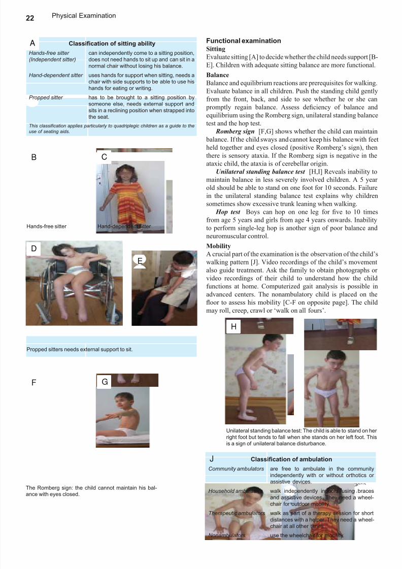
Propped sitters needs external support to sit.
Hand-dependent sitterHands-free sitter
Functional examination
Sitting
Evaluate sitting [A] to decide whether the child needs support [B-
E]. Children with adequate sitting balance are more functional.
Balance
Balance and equilibrium reactions are prerequisites for walking.
Evaluate balance in all children. Push the standing child gently
from the front, back, and side to see whether he or she can promptly regain balance. Assess deficiency of balance and
equilibrium using the Romberg sign, unilateral standing balance
test and the hop test.
Romberg sign [F,G] shows whether the child can maintain
balance. If the child sways and cannot keep his balance with feet
held together and eyes closed (positive Romberg’s sign), then
there is sensory ataxia. If the Romberg sign is negative in the
ataxic child, the ataxia is of cerebellar origin.
Unilateral standing balance test [H,I] Reveals inability to
maintain balance in less severely involved children. A 5 year
old should be able to stand on one foot for 10 seconds. Failure
in the unilateral standing balance test explains why children
sometimes show excessive trunk leaning when walking.
Hop test Boys can hop on one leg for five to 10 times
from age 5 years and girls from age 4 years onwards. Inability
to perform single-leg hop is another sign of poor balance and
neuromuscular control.
Mobility
A crucial part of the examination is the observation of the child’s
walking pattern [J]. Video recordings of the child’s movement
also guide treatment. Ask the family to obtain photographs or
video recordings of their child to understand how the child
functions at home. Computerized gait analysis is possible in
advanced centers. The nonambulatory child is placed on the
floor to assess his mobility [C-F on opposite page]. The child
may roll, creep, crawl or ‘walk on all fours’.
Classification of sitting ability
Hands-free sitter
(Independent sitter)
can independently come to a sitting position,
does not need hands to sit up and can sit in a
normal chair without losing his balance.
Hand-dependent sitter uses hands for support when sitting, needs a
chair with side supports to be able to use his
hands for eating or writing.
Propped sitter has to be brought to a sitting position bysomeone else, needs external support and
sits in a reclining position when strapped into
the seat.
This classification applies particularly to quadriplegic children as a guide to the
use of seating aids.
Classification of ambulation
Community ambulators are free to ambulate in the community
independently with or without orthotics or
assistive devices.
Household ambulators walk independently indoors using braces
and assistive devices. They need a wheel-chair for outdoor mobility.
Therapeutic ambulators walk as part of a therapy session for short
distances with a helper. They need a wheel-
chair at all other times.
Nonambulators use the wheelchair for mobility.
Unilateral standing balance test: The child is able to stand on her
right foot but tends to fall when she stands on her left foot. Thisis a sign of unilateral balance disturbance.
FF
EE
DD
CCB
A
HH
GG
II
JJ
The Romberg sign: the child cannot maintain his bal-ance with eyes closed.
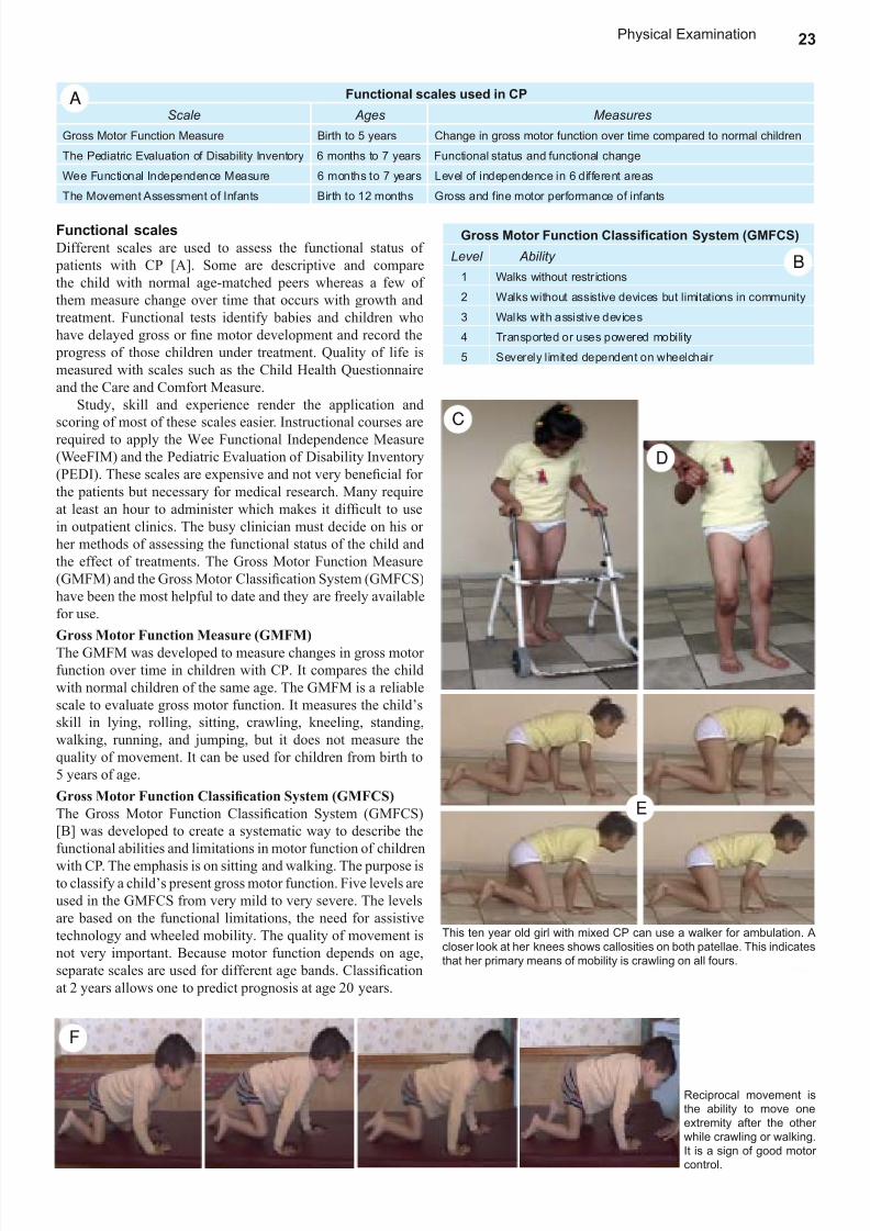
8/9/2019 Guía de ayuda para la parálisis cerebral.pdf
http://slidepdf.com/reader/full/guia-de-ayuda-para-la-paralisis-cerebralpdf 23/143
23Physical Examination
Functional scales
Different scales are used to assess the functional status of
patients with CP [A]. Some are descriptive and compare
the child with normal age-matched peers whereas a few of
them measure change over time that occurs with growth and
treatment. Functional tests identify babies and children who
have delayed gross or fine motor development and record the
progress of those children under treatment. Quality of life is
measured with scales such as the Child Health Questionnaire
and the Care and Comfort Measure.
Study, skill and experience render the application and
scoring of most of these scales easier. Instructional courses are
required to apply the Wee Functional Independence Measure
(WeeFIM) and the Pediatric Evaluation of Disability Inventory
(PEDI). These scales are expensive and not very beneficial for
the patients but necessary for medical research. Many require
at least an hour to administer which makes it difficult to use
in outpatient clinics. The busy clinician must decide on his or
her methods of assessing the functional status of the child and
the effect of treatments. The Gross Motor Function Measure
(GMFM) and the Gross Motor Classification System (GMFCS)
have been the most helpful to date and they are freely available
for use.
Gross Motor Function Measure (GMFM)
The GMFM was developed to measure changes in gross motor
function over time in children with CP. It compares the child
with normal children of the same age. The GMFM is a reliable
scale to evaluate gross motor function. It measures the child’s
skill in lying, rolling, sitting, crawling, kneeling, standing,
walking, running, and jumping, but it does not measure the
quality of movement. It can be used for children from birth to
5 years of age.
Gross Motor Function Classification System (GMFCS)
The Gross Motor Function Classification System (GMFCS)
[B] was developed to create a systematic way to describe the
functional abilities and limitations in motor function of children
with CP. The emphasis is on sitting and walking. The purpose isto classify a child’s present gross motor function. Five levels are
used in the GMFCS from very mild to very severe. The levels
are based on the functional limitations, the need for assistive
technology and wheeled mobility. The quality of movement is
not very important. Because motor function depends on age,
separate scales are used for different age bands. Classification
at 2 years allows one to predict prognosis at age 20 years.
Functional scales used in CP
Scale Ages Measures
Gross Motor Function Measure Birth to 5 years Change in gross motor function over time compared to normal children
The Pediatric Evaluation of Disability Inventory 6 months to 7 years Functional status and functional change
Wee Functional Independence Measure 6 months to 7 years Level of independence in 6 different areas
The Movement Assessment of Infants Birth to 12 months Gross and fine motor performance of infants
Gross Motor Function Classification System (GMFCS)
Level Ability
1 Walks without restr ictions
2 Walks without assistive devices but limitations in community
3 Walks with assistive devices
4 Transported or uses powered mobility
5 Severely limited dependent on wheelchair
This ten year old girl with mixed CP can use a walker for ambulation. A
closer look at her knees shows callosities on both patellae. This indicates
that her primary means of mobility is crawling on all fours.
Reciprocal movement isthe ability to move one
extremity after the other
while crawling or walking.
It is a sign of good motorcontrol.
EE
DD
CC
B
FF
A
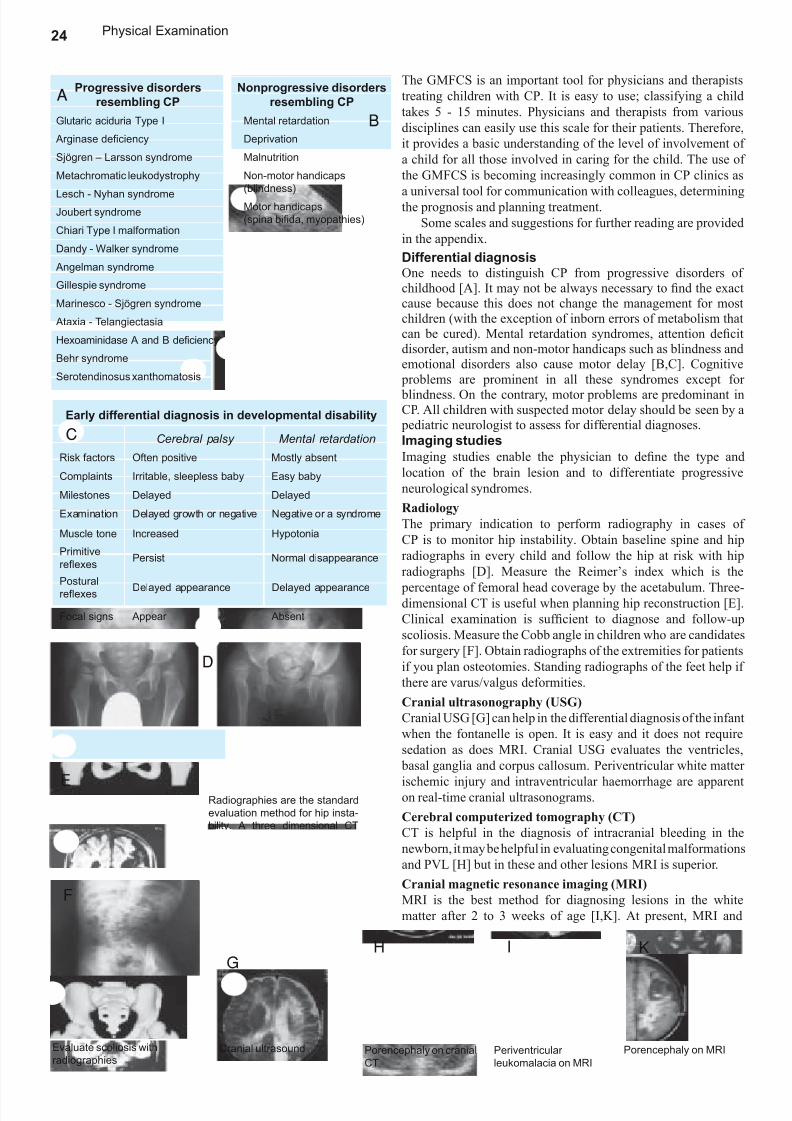
8/9/2019 Guía de ayuda para la parálisis cerebral.pdf
http://slidepdf.com/reader/full/guia-de-ayuda-para-la-paralisis-cerebralpdf 24/143
24 Physical Examination
The GMFCS is an important tool for physicians and therapists
treating children with CP. It is easy to use; classifying a child
takes 5 - 15 minutes. Physicians and therapists from various
disciplines can easily use this scale for their patients. Therefore,
it provides a basic understanding of the level of involvement of
a child for all those involved in caring for the child. The use of
the GMFCS is becoming increasingly common in CP clinics as
a universal tool for communication with colleagues, determiningthe prognosis and planning treatment.
Some scales and suggestions for further reading are provided
in the appendix.
Differential diagnosisOne needs to distinguish CP from progressive disorders ofchildhood [A]. It may not be always necessary to find the exactcause because this does not change the management for mostchildren (with the exception of inborn errors of metabolism thatcan be cured). Mental retardation syndromes, attention deficitdisorder, autism and non-motor handicaps such as blindness andemotional disorders also cause motor delay [B,C]. Cognitive
problems are prominent in all these syndromes except for
blindness. On the contrary, motor problems are predominant inCP. All children with suspected motor delay should be seen by a pediatric neurologist to assess for differential diagnoses.
Imaging studies
Imaging studies enable the physician to define the type and
location of the brain lesion and to differentiate progressive
neurological syndromes.
Radiology
The primary indication to perform radiography in cases of
CP is to monitor hip instability. Obtain baseline spine and hip
radiographs in every child and follow the hip at risk with hip
radiographs [D]. Measure the Reimer’s index which is the
percentage of femoral head coverage by the acetabulum. Three-dimensional CT is useful when planning hip reconstruction [E].
Clinical examination is sufficient to diagnose and follow-up
scoliosis. Measure the Cobb angle in children who are candidates
for surgery [F]. Obtain radiographs of the extremities for patients
if you plan osteotomies. Standing radiographs of the feet help if
there are varus/valgus deformities.
Cranial ultrasonography (USG)
Cranial USG [G] can help in the differential diagnosis of the infant
when the fontanelle is open. It is easy and it does not require
sedation as does MRI. Cranial USG evaluates the ventricles,
basal ganglia and corpus callosum. Periventricular white matter
ischemic injury and intraventricular haemorrhage are apparenton real-time cranial ultrasonograms.
Cerebral computerized tomography (CT)
CT is helpful in the diagnosis of intracranial bleeding in the
newborn, it may be helpful in evaluating congenital malformations
and PVL [H] but in these and other lesions MRI is superior.
Cranial magnetic resonance imaging (MRI)
MRI is the best method for diagnosing lesions in the white
matter after 2 to 3 weeks of age [I,K]. At present, MRI and
Porencephaly on cranial
CT
Periventricular
leukomalacia on MRI
Cranial ultrasoundEvaluate scoliosis with
radiographies
Nonprogressive disorders
resembling CP
Mental retardation
Deprivation
Malnutrition
Non-motor handicaps
(blindness)
Motor handicaps
(spina bifida, myopathies)
Progressive disorders
resembling CP
Glutaric aciduria Type I
Arginase deficiency
Sjögren – Larsson syndrome
Metachromatic leukodystrophy
Lesch - Nyhan syndromeJoubert syndrome
Chiari Type I malformation
Dandy - Walker syndrome
Angelman syndrome
Gillespie syndrome
Marinesco - Sjögren syndrome
Ataxia - Telangiectasia
Hexoaminidase A and B deficiency
Behr syndrome
Serotendinosus xanthomatosis
Early differential diagnosis in developmental disability
Cerebral palsy Mental retardation
Risk factors Often positive Mostly absent
Complaints Irritable, sleepless baby Easy baby
Milestones Delayed Delayed
Examination Delayed growth or negative Negative or a syndrome
Muscle tone Increased Hypotonia
Primitive
reflexesPersist Normal disappearance
Postural
reflexes Delayed appearance Delayed appearance
Focal signs Appear Absent
Porencephaly on MRI
FF
EE
DD
CC
B
A
HH
GG
II KK
Radiographies are the standard
evaluation method for hip insta-
bility. A three dimensional CT
scan provides valuable data inthe preoperative evaluation for
hip reconstruction.
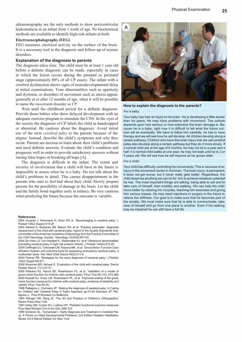
8/9/2019 Guía de ayuda para la parálisis cerebral.pdf
http://slidepdf.com/reader/full/guia-de-ayuda-para-la-paralisis-cerebralpdf 25/143
25Physical Examination
ultrasonography are the only methods to show periventricular
leukomalacia in an infant from 1 week of age. No biochemical
methods are available to identify high-risk infants at birth.
Electroencephalography (EEG)
EEG measures electrical activity on the surface of the brain.
It is a necessary tool in the diagnosis and follow-up of seizure
disorders.
Explanation of the diagnosis to parentsThe diagnosis takes time. The child must be at least 1 year old
before a definite diagnosis can be made, especially in cases
in which the lesion occurs during the prenatal or perinatal
stage (approximately 80% of all CP cases). The infant with a
cerebral dysfunction shows signs of neurodevelopmental delay
at initial examinations. Tone abnormalities such as spasticity
and dystonia, or disorders of movement such as ataxia appear
generally at or after 12 months of age, when it will be possible
to name the movement disorder as CP.
Wait until the childhood period for a definite diagnosis.
Provide those babies who show delayed development with an
adequate exercise program to stimulate the CNS. In the eyes ofthe society the diagnosis of CP labels the child as handicapped
or abnormal. Be cautious about the diagnosis. Avoid initial
use of the term cerebral palsy to the parents because of the
stigma. Instead, describe the child’s symptoms and why they
occur. Parents are anxious to learn about their child’s problems
and need definite answers. Evaluate the child’s condition and
prognosis well in order to provide satisfactory answers without
raising false hopes or breaking all hope [A].
The diagnosis is difficult in the infant. The extent and
severity of involvement that a child will have in the future is
impossible to assess when he is a baby. Do not talk about the
child’s problems in detail. This causes disappointment in the
parents who start to feel bad about their child. Slowly prepare
parents for the possibility of damage to the brain. Let the child
and the family bond together early in infancy. Be very cautious
when predicting the future because the outcome is variable.
How to explain the diagnosis to the parents?
For a baby:
Your baby has had an injury to his brain. He is developing a little slower
than his peers. He may have problems with movement. The outlookdepends upon how serious or how extensive the brain damage is. Be-
cause he is a baby, right now it is difficult to tell what the future out-look will be eventually. We have to follow him carefully, he has to have
therapy and we will see how he will develop. All children develop along a
certain pathway. Children who have this brain injury that we call cerebral
palsy also develop along a certain pathway but they do it more slowly. Ifa normal child sits at the age of 6 months, he may not sit to a year and a
half; if a normal child walks at one year, he may not walk until he is 3 or
4 years old. We will see how he will improve as he grows older.
For a child:
Your child has difficulty controlling his movements. This is because of an
injury in the movement center in the brain. The brain injury is permanent.
It does not get worse, but it never really gets better. Regardless, the
child deserves anything we can do for him to achieve whatever potentialhe has. The most important things are talking, being able to eat and to
take care of himself, then mobility and walking. We can help the child
move better by relaxing his muscles, teaching him exercises and givinghim various braces. He may need injections or surgery in the future to
relieve the stiffness. Our goal is to make sure that he becomes part of
the society. We must make sure that he is able to communicate, take
care of himself and go from one place to another. Even if his walkingmay be impaired he can still have a full life.
A
References2004 Accardo J, Kammann H, Hoon AH Jr. ‘Neuroimaging in cerebral palsy’ J
Pediatr 145(2 Suppl):S19-272004 Ashwal S, Russman BS, Blasco PA, et al. ‘Practice parameter: diagnostic
assessment of the child with cerebral palsy: report of the Quality Standards Sub-committee of the American Academy of Neurology and the Practice Committee of
the Child Neurology Society.’ Neurology 23;62(6):851-63
2004 De Vries LS, Van Haastert IL, Rademaker KJ, et al ‘Ultrasound abnormalitiespreceding cerebral palsy in high-risk preterm infants’ J Pediatr 144(6):815-20
2004 Oeffinger DJ, Tylkowski CM, Rayens MK, et al. ‘Gross Motor Function Clas-
sification System and outcome tools for assessing ambulatory cerebral palsy: amulticenter study’ Dev Med Child Neurol 46(5):311-9
2004 Palmer FB. ‘Strategies for the early diagnosis of cerebral palsy’ J Pediatr
145(2 Suppl):S8-S112004 Russman BS, Ashwal S. ‘Evaluation of the child with cerebral palsy’ Semin
Pediatr Neurol 11(1):47-57
2000 Palisano RJ, Hanna SE, Rosenbaum PL, et al. ‘Validation of a model ofgross motor function for children with cerebral palsy’ Phys Ther 80 (10), 974-985
2000 Russell DJ, Avery LM, Rosenbaum PL, et al. ‘Improved scaling of the grossmotor function measure for children with cerebral palsy: evidence of reliability and
validity’ Phys Ther 80 (9)
1998 Pellegrino L, Dormans JP. ‘Making the diagnosis of cerebral palsy’ In Caringfor Children with Cerebral Palsy A Team Approach pp:31-54 Dormans JP, Pel-
legrino L, Paul H Brookes Co Baltimore
1993 Wenger DR, Rang M. ‘The Art and Practice of Children’s Orthopaedics’Raven Press New York
1991 Haley SM, Coster WJ, Ludlow HH. ‘Pediatric functional outcome measures’
Phys Med Rehabil Clin N Am 2(4): 689-7231990 Scherzer AL, Tscharnuter I. ‘Early Diagnosis and Treatment in Cerebral Pal-
sy: A Primer on Infant Developmental Problems’ 2nd Edition Pediatric Habilitation
Series Vol 6 Marcel Dekker Inc New York
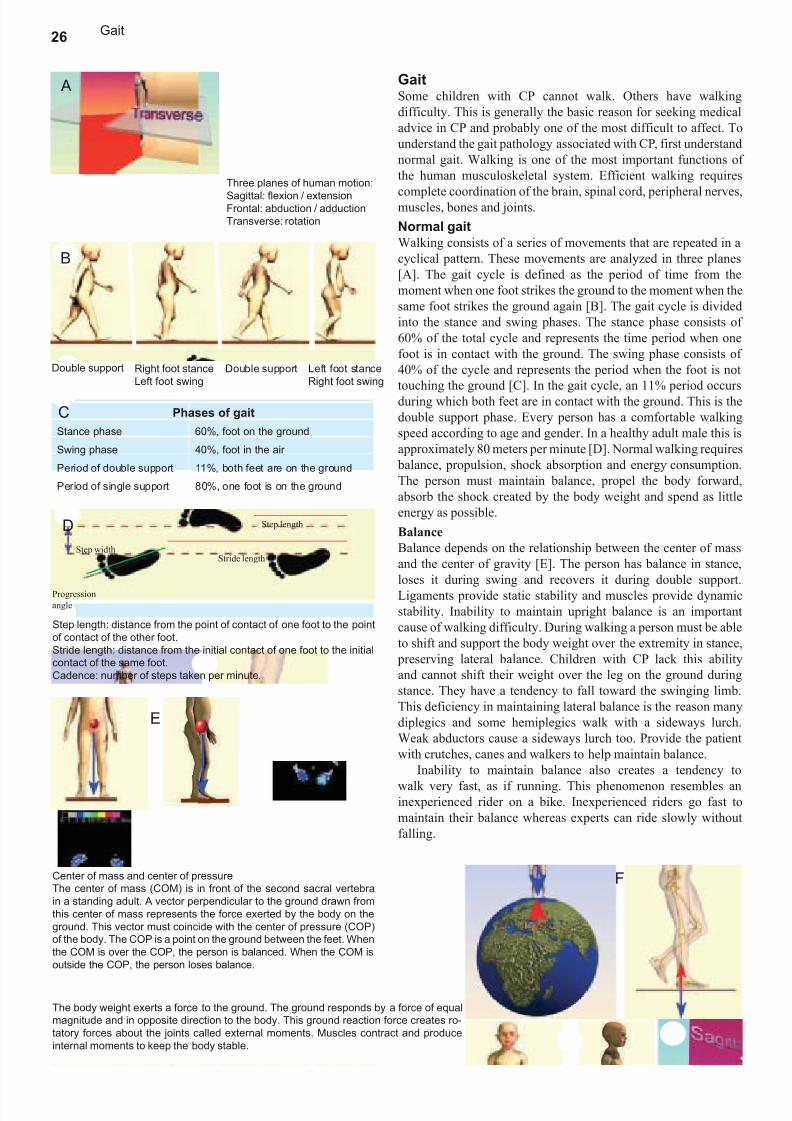
8/9/2019 Guía de ayuda para la parálisis cerebral.pdf
http://slidepdf.com/reader/full/guia-de-ayuda-para-la-paralisis-cerebralpdf 26/143
26Gait
GaitSome children with CP cannot walk. Others have walkingdifficulty. This is generally the basic reason for seeking medicaladvice in CP and probably one of the most difficult to affect. Tounderstand the gait pathology associated with CP, first understandnormal gait. Walking is one of the most important functions ofthe human musculoskeletal system. Efficient walking requires
complete coordination of the brain, spinal cord, peripheral nerves,muscles, bones and joints.
Normal gait
Walking consists of a series of movements that are repeated in acyclical pattern. These movements are analyzed in three planes[A]. The gait cycle is defined as the period of time from themoment when one foot strikes the ground to the moment when thesame foot strikes the ground again [B]. The gait cycle is dividedinto the stance and swing phases. The stance phase consists of60% of the total cycle and represents the time period when onefoot is in contact with the ground. The swing phase consists of40% of the cycle and represents the period when the foot is nottouching the ground [C]. In the gait cycle, an 11% period occursduring which both feet are in contact with the ground. This is thedouble support phase. Every person has a comfortable walkingspeed according to age and gender. In a healthy adult male this isapproximately 80 meters per minute [D]. Normal walking requires
balance, propulsion, shock absorption and energy consumption.The person must maintain balance, propel the body forward,absorb the shock created by the body weight and spend as littleenergy as possible.
Balance
Balance depends on the relationship between the center of massand the center of gravity [E]. The person has balance in stance,loses it during swing and recovers it during double support.Ligaments provide static stability and muscles provide dynamicstability. Inability to maintain upright balance is an importantcause of walking difficulty. During walking a person must be ableto shift and support the body weight over the extremity in stance,
preserving lateral balance. Children with CP lack this abilityand cannot shift their weight over the leg on the ground duringstance. They have a tendency to fall toward the swinging limb.This deficiency in maintaining lateral balance is the reason manydiplegics and some hemiplegics walk with a sideways lurch.Weak abductors cause a sideways lurch too. Provide the patientwith crutches, canes and walkers to help maintain balance.
Inability to maintain balance also creates a tendency to
walk very fast, as if running. This phenomenon resembles aninexperienced rider on a bike. Inexperienced riders go fast tomaintain their balance whereas experts can ride slowly withoutfalling.
Step length: distance from the point of contact of one foot to the point
of contact of the other foot.
Stride length: distance from the initial contact of one foot to the initialcontact of the same foot.
Cadence: number of steps taken per minute.
Step length
Stride lengthStep width
Progressionangle
Three planes of human motion:
Sagittal: flexion / extensionFrontal: abduction / adduction
Transverse: rotation
Center of mass and center of pressureThe center of mass (COM) is in front of the second sacral vertebra
in a standing adult. A vector perpendicular to the ground drawn from
this center of mass represents the force exerted by the body on the
ground. This vector must coincide with the center of pressure (COP)of the body. The COP is a point on the ground between the feet. When
the COM is over the COP, the person is balanced. When the COM is
outside the COP, the person loses balance.
The body weight exerts a force to the ground. The ground responds by a force of equalmagnitude and in opposite direction to the body. This ground reaction force creates ro-
tatory forces about the joints called external moments. Muscles contract and produce
internal moments to keep the body stable.
Phases of gait
Stance phase 60%, foot on the ground
Swing phase 40%, foot in the air
Period of double support 11%, both feet are on the ground
Period of single support 80%, one foot is on the ground
Double support Right foot stance
Left foot swing
Double support Left foot stance
Right foot swing
E
E
D
D
C
C
B
A
F
F
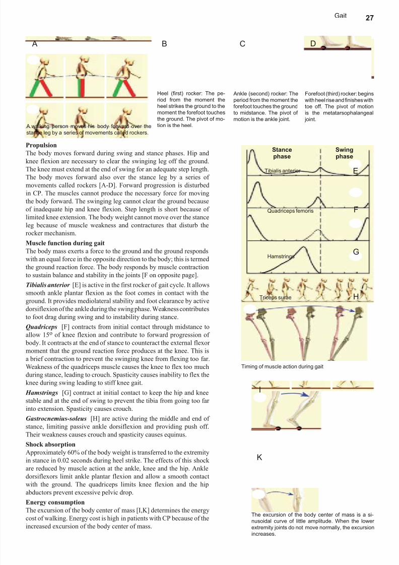
8/9/2019 Guía de ayuda para la parálisis cerebral.pdf
http://slidepdf.com/reader/full/guia-de-ayuda-para-la-paralisis-cerebralpdf 27/143
27Gait
A walking person moves his body forward over the
stance leg by a series of movements called rockers.
Heel (first) rocker: The pe-
riod from the moment theheel strikes the ground to the
moment the forefoot touches
the ground. The pivot of mo-
tion is the heel.
Ankle (second) rocker: The
period from the moment theforefoot touches the ground
to midstance. The pivot of
motion is the ankle joint.
Forefoot (third) rocker: begins
with heel rise and finishes withtoe off. The pivot of motion
is the metatarsophalangeal
joint.
Propulsion
The body moves forward during swing and stance phases. Hip andknee flexion are necessary to clear the swinging leg off the ground.The knee must extend at the end of swing for an adequate step length.The body moves forward also over the stance leg by a series ofmovements called rockers [A-D]. Forward progression is disturbedin CP. The muscles cannot produce the necessary force for movingthe body forward. The swinging leg cannot clear the ground becauseof inadequate hip and knee flexion. Step length is short because oflimited knee extension. The body weight cannot move over the stanceleg because of muscle weakness and contractures that disturb therocker mechanism.
Muscle function during gait
The body mass exerts a force to the ground and the ground respondswith an equal force in the opposite direction to the body; this is termedthe ground reaction force. The body responds by muscle contractionto sustain balance and stability in the joints [F on opposite page].
Tibialis anterior [E] is active in the first rocker of gait cycle. It allowssmooth ankle plantar flexion as the foot comes in contact with theground. It provides mediolateral stability and foot clearance by activedorsiflexion of the ankle during the swing phase. Weakness contributesto foot drag during swing and to instability during stance.
Quadriceps [F] contracts from initial contact through midstance toallow 15o of knee flexion and contribute to forward progression of
body. It contracts at the end of stance to counteract the external flexormoment that the ground reaction force produces at the knee. This isa brief contraction to prevent the swinging knee from flexing too far.Weakness of the quadriceps muscle causes the knee to flex too muchduring stance, leading to crouch. Spasticity causes inability to flex theknee during swing leading to stiff knee gait.
Hamstrings [G] contract at initial contact to keep the hip and kneestable and at the end of swing to prevent the tibia from going too farinto extension. Spasticity causes crouch.
Gastrocnemius-soleus [H] are active during the middle and end ofstance, limiting passive ankle dorsiflexion and providing push off.Their weakness causes crouch and spasticity causes equinus.
Shock absorption
Approximately 60% of the body weight is transferred to the extremityin stance in 0.02 seconds during heel strike. The effects of this shockare reduced by muscle action at the ankle, knee and the hip. Ankledorsiflexors limit ankle plantar flexion and allow a smooth contactwith the ground. The quadriceps limits knee flexion and the hip
abductors prevent excessive pelvic drop.Energy consumption
The excursion of the body center of mass [I,K] determines the energycost of walking. Energy cost is high in patients with CP because of theincreased excursion of the body center of mass.
The excursion of the body center of mass is a si-nusoidal curve of little amplitude. When the lower
extremity joints do not move normally, the excursion
increases.
Tibialis anterior
Quadriceps femoris
Hamstrings
Triceps surae
Stancephase
K
K
I
I
F
F
E
E
B A
H
H
G
G
D
DC
Swingphase
Timing of muscle action during gait
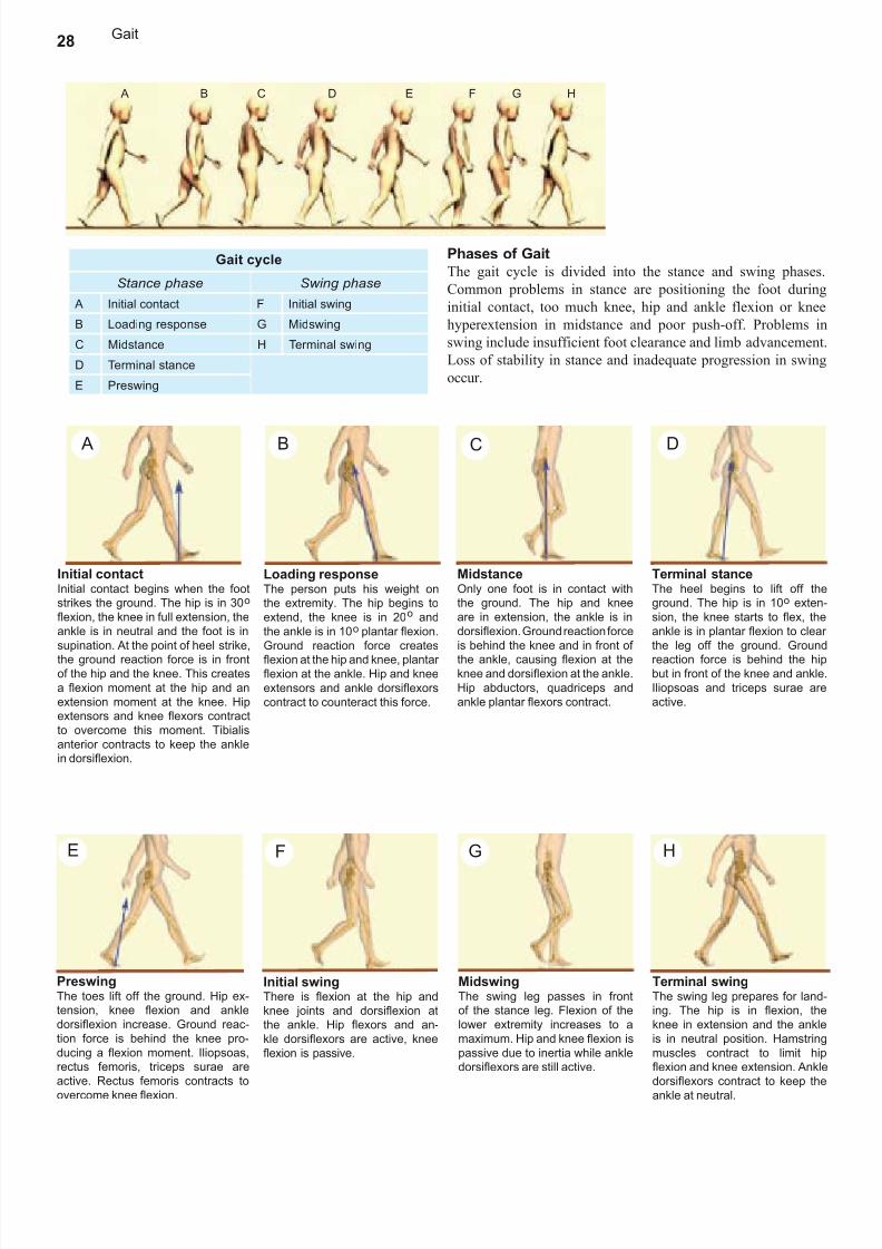
8/9/2019 Guía de ayuda para la parálisis cerebral.pdf
http://slidepdf.com/reader/full/guia-de-ayuda-para-la-paralisis-cerebralpdf 28/143
Phases of Gait
The gait cycle is divided into the stance and swing phases.Common problems in stance are positioning the foot duringinitial contact, too much knee, hip and ankle flexion or kneehyperextension in midstance and poor push-off. Problems inswing include insufficient foot clearance and limb advancement.Loss of stability in stance and inadequate progression in swingoccur.
Gait cycle
Stance phase Swing phase
A Initial contact F Initial swing
B Loading response G Midswing
C Midstance H Terminal swing
D Terminal stance
E Preswing
Initial contactInitial contact begins when the footstrikes the ground. The hip is in 30o
flexion, the knee in full extension, the
ankle is in neutral and the foot is in
supination. At the point of heel strike,
the ground reaction force is in frontof the hip and the knee. This creates
a flexion moment at the hip and an
extension moment at the knee. Hipextensors and knee flexors contract
to overcome this moment. Tibialis
anterior contracts to keep the anklein dorsiflexion.
MidstanceOnly one foot is in contact withthe ground. The hip and knee
are in extension, the ankle is in
dorsiflexion. Ground reaction force
is behind the knee and in front of
the ankle, causing flexion at theknee and dorsiflexion at the ankle.
Hip abductors, quadriceps and
ankle plantar flexors contract.
Loading responseThe person puts his weight onthe extremity. The hip begins to
extend, the knee is in 20o and
the ankle is in 10o plantar flexion.
Ground reaction force creates
flexion at the hip and knee, plantarflexion at the ankle. Hip and knee
extensors and ankle dorsiflexors
contract to counteract this force.
Terminal stanceThe heel begins to lift off theground. The hip is in 10o exten-
sion, the knee starts to flex, the
ankle is in plantar flexion to clear
the leg off the ground. Ground
reaction force is behind the hipbut in front of the knee and ankle.
Iliopsoas and triceps surae are
active.
PreswingThe toes lift off the ground. Hip ex-tension, knee flexion and ankle
dorsiflexion increase. Ground reac-
tion force is behind the knee pro-
ducing a flexion moment. Iliopsoas,rectus femoris, triceps surae are
active. Rectus femoris contracts to
overcome knee flexion.
Initial swingThere is flexion at the hip and
knee joints and dorsiflexion atthe ankle. Hip flexors and an-
kle dorsiflexors are active, knee
flexion is passive.
MidswingThe swing leg passes in frontof the stance leg. Flexion of the
lower extremity increases to a
maximum. Hip and knee flexion is
passive due to inertia while ankledorsiflexors are still active.
Terminal swingThe swing leg prepares for land-ing. The hip is in flexion, the
knee in extension and the ankle
is in neutral position. Hamstring
muscles contract to limit hipflexion and knee extension. Ankle
dorsiflexors contract to keep the
ankle at neutral.
A B C D E F G H
F
FE
D
DCB A
H
HG
28Gait
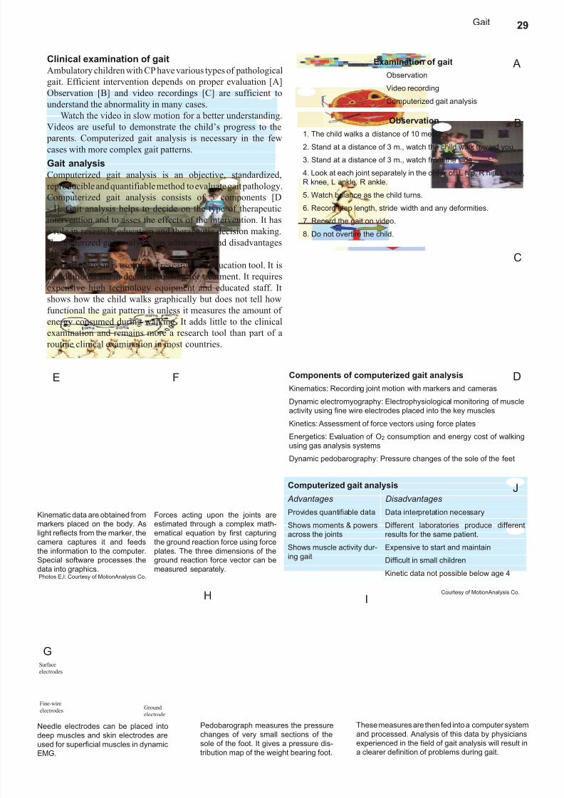
8/9/2019 Guía de ayuda para la parálisis cerebral.pdf
http://slidepdf.com/reader/full/guia-de-ayuda-para-la-paralisis-cerebralpdf 29/143
Courtesy of MotionAnalysis Co.
Kinematic data are obtained frommarkers placed on the body. As
light reflects from the marker, the
camera captures it and feeds
the information to the computer.
Special software processes thedata into graphics.
Needle electrodes can be placed into
deep muscles and skin electrodes are
used for superficial muscles in dynamicEMG.
Pedobarograph measures the pressure
changes of very small sections of the
sole of the foot. It gives a pressure dis-
tribution map of the weight bearing foot.
Forces acting upon the joints areestimated through a complex math-
ematical equation by first capturing
the ground reaction force using force
plates. The three dimensions of the
ground reaction force vector can bemeasured separately.
These measures are then fed into a computer system
and processed. Analysis of this data by physicians
experienced in the field of gait analysis will result ina clearer definition of problems during gait.
Clinical examination of gait
Ambulatory children with CP have various types of pathologicalgait. Efficient intervention depends on proper evaluation [A]Observation [B] and video recordings [C] are sufficient tounderstand the abnormality in many cases.
Watch the video in slow motion for a better understanding.Videos are useful to demonstrate the child’s progress to the
parents. Computerized gait analysis is necessary in the fewcases with more complex gait patterns.
Gait analysis
Computerized gait analysis is an objective, standardized,reproducible and quantifiable method to evaluate gait pathology.Computerized gait analysis consists of 5 components [D- I]. Gait analysis helps to decide on the type of therapeuticintervention and to asses the effects of the intervention. It hasa role in research, education and therapeutic decision making.Computerized gait analysis has advantages and disadvantages[J].
Gait analysis is useful as a research and education tool. It is
an additional aid in decision making for treatment. It requiresexpensive high technology equipment and educated staff. Itshows how the child walks graphically but does not tell howfunctional the gait pattern is unless it measures the amount ofenergy consumed during walking. It adds little to the clinicalexamination and remains more a research tool than part of aroutine clinical examination in most countries.
Observation
1. The child walks a distance of 10 meters.
2. Stand at a distance of 3 m., watch the child walk toward you.
3. Stand at a distance of 3 m., watch from the side.
4. Look at each joint separately in the order of: L hip, R hip, L knee,
R knee, L ankle, R ankle.
5. Watch balance as the child turns.
6. Record step length, stride width and any deformities.
7. Record the gait on video.
8. Do not overtire the child.
Computerized gait analysis
Advantages Disadvantages
Provides quantifiable data Data interpretation necessary
Shows moments & powers
across the joints
Different laboratories produce different
results for the same patient.
Shows muscle activity dur-
ing gait
Expensive to start and maintain
Difficult in small children
Kinetic data not possible below age 4
Examination of gait
Observation
Video recording
Computerized gait analysis
Components of computerized gait analysis
Kinematics: Recording joint motion with markers and cameras
Dynamic electromyography: Electrophysiological monitoring of muscle
activity using fine wire electrodes placed into the key muscles
Kinetics: Assessment of force vectors using force plates
Energetics: Evaluation of O2 consumption and energy cost of walking
using gas analysis systems
Dynamic pedobarography: Pressure changes of the sole of the feet
Surface
electrodes
Ground
electrode
Fine-wire
electrodes
F
FE D
D
C
C
B
A
H
H
G
G
Photos E,I: Courtesy of MotionAnalysis Co.
I
I
J
J
29Gait
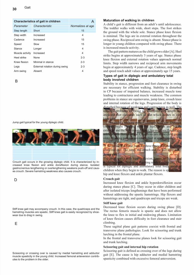
8/9/2019 Guía de ayuda para la parálisis cerebral.pdf
http://slidepdf.com/reader/full/guia-de-ayuda-para-la-paralisis-cerebralpdf 30/143
30Gait
Characteristics of gait in children
Parameter Characteristic Normalizes at age
Step length Short 15
Step width Increased 4
Cadence Increased 15
Speed Slow 15
Stance Longer 4
Muscle activity Increased 4
Heel strike None 2-3
Knee flexion Minimal in stance 2-3
Legs External rotation during swing 2-3
Arm swing Absent 4
Maturation of walking in children
A child’s gait is different from an adult’s until adolescence.The toddler walks with wide, short steps. The foot strikesthe ground with the whole sole. Stance phase knee flexionis minimal. The legs are in external rotation throughout theswing phase. Reciprocal arm swing is absent. Stance phase islonger in young children compared with swing phase. There
is increased muscle activity.The gait pattern matures as the child grows older [A]. Heelstrike begins at approximately 3 years of age. Stance phaseknee flexion and external rotation values approach normallimits. Step width narrows and reciprocal arm movements
begin at approximately 4 years of age. Cadence, step lengthand speed reach adult values at approximately age 15 years.
Types of gait in diplegic and ambulatory total
body involved children
Stability in stance, progression and foot clearance in swingare necessary for efficient walking. Stability is disturbedin CP because of impaired balance, increased muscle tone
leading to contractures and muscle weakness. The common problems in stance are equinovarus, jump knee, crouch kneeand internal rotation of the legs. Progression of the body isdisturbed because of weakness and contractures as well. Thecommon problems of swing are shortened step length andimpaired foot clearance such as that which occurs in stiffknee gait.
The child’s walking pattern changes with age. Diplegicchildren begin standing with the hips, knees and anklesextended and the legs crossed. Later, hip and knee flexion andankle plantar flexion occur. Crouch occurs as the child growsolder. Walking patterns are established at approximately 5 to7 years of age.
In the sagittal plane, look for three types of pathologicallyabnormal gait: The jump, the crouch and the stiff knee gait.
Jump gait
The child walks with hips in flexion, knees in flexion andankles in plantar flexion as if getting ready to jump [B]. Thisis typical for diplegic and ambulatory total body involvedchildren when they begin to walk. The reason is spasticity ofhip and knee flexors and ankle plantar flexors.
Crouch gait
Increased knee flexion and ankle hyperdorsiflexion occurduring stance phase [C]. They occur in older children andafter isolated triceps lengthenings that have been performedwithout addressing the spastic hamstrings. Hip flexors andhamstrings are tight, and quadriceps and triceps are weak.
Stiff knee gait
Decreased knee flexion occurs during swing phase [D].The rectus femoris muscle is spastic and does not allowthe knee to flex in initial and midswing phases. Limitationof knee flexion causes difficulty in foot clearance and stairclimbing.These sagittal plane gait patterns coexist with frontal andtransverse plane pathologies. Look for scissoring and trunklurching in the frontal plane.In the frontal and transverse planes look for scissoring gait
and trunk lurching.Scissoring gait and internal hip rotation
Scissoring gait is defined as crossing over of the legs duringgait [E]. The cause is hip adductor and medial hamstringspasticity combined with excessive femoral anteversion.
Jump gait typical for the young diplegic child.
Crouch gait occurs in the growing diplegic child. It is characterized by in-
creased knee flexion and ankle dorsiflexion during stance. Isolated
gastrocnemius lengthening or overlengthening weakens push-off and caus-es crouch. Severe hamstring weakness also causes crouch.
Stiff knee gait may accompany crouch. In this case, the quadriceps and thehamstring muscles are spastic. Stiff knee gait is easily recognized by shoe-
wear due to drag in swing.
D
D
Scissoring or crossing over is caused by medial hamstring and adductor
muscle spasticity in the young child. Increased femoral anteversion contrib-
utes to the problem in the older.
E
E
C
C
B
A
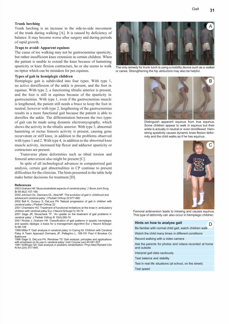
8/9/2019 Guía de ayuda para la parálisis cerebral.pdf
http://slidepdf.com/reader/full/guia-de-ayuda-para-la-paralisis-cerebralpdf 31/143
31Gait
Hints on how to analyse gait
Be familiar with normal child gait, watch children walk
Watch the child many times in different conditions
Record walking with a video camera
Ask the parents for photos and videos recorded at home
and outside
Interpret gait data cautiously
Test balance and stability
Test in real life situations (at school, on the street)
Test speed
Trunk lurching
Trunk lurching is an increase in the side-to-side movementof the trunk during walking [A]. It is caused by deficiency of
balance. It may become worse after surgery and during periodsof rapid growth.
Traps to avoid: Apparent equinus
The cause of toe walking may not be gastrocnemius spasticity,
but rather insufficient knee extension in certain children. Whenthe patient is unable to extend the knee because of hamstringspasticity or knee flexion contracture, he or she seems to walkon tiptoe which can be mistaken for pes equinus.
Types of gait in hemiplegic children
Hemiplegic gait is subdivided into four types. With type 1,no active dorsiflexion of the ankle is present, and the foot inequinus. With type 2, a functioning tibialis anterior is present,and the foot is still in equinus because of the spasticity ingastrocnemius. With type 1, even if the gastrocnemius muscleis lengthened, the patient still needs a brace to keep the foot inneutral; however with type 2, lengthening of the gastrocnemius
results in a more functional gait because the patient is able todorsiflex the ankle. The differentiation between the two typesof gait can be made using dynamic electromyography, whichshows the activity in the tibialis anterior. With type 3, abnormalhamstring or rectus femoris activity is present, causing genurecurvatum or stiff knee, in addition to the problems observedwith types 1 and 2. With type 4, in addition to the abnormal kneemuscle activity, increased hip flexor and adductor spasticity orcontracture are present.
Transverse plane deformities such as tibial torsion andfemoral anteversion also might be present [C].
In spite of all technological advances in computerized gaitanalysis, certain gait abnormalities in CP continue to presentdifficulties for the clinician. The hints presented in the table helpmake better decisions for treatment [D].
References2003 Graham HK ‘Musculoskeletal aspects of cerebral palsy’ J Bone Joint Surg
Br 85-B(2) 157-1662002 Johnson Dc, Damiano DL, Abel MF ‘The evolution of gait in childhood and
adolescent cerebral palsy’ J Pediatr Orthop 22:677-682
2002 Bell K, Ounpuu S, DeLuca PA ‘Natural progression of gait in children withcerebral palsy J Pediatr Orthop 22
2001 Chambers HG ‘Treatment of functional limitations at the knee in ambulatory
children with cerebral palsy Eur J Neurol 8(Suppl 5) 59-742001 Gage JR, Novacheck TF. ‘An update on the treatment of gait problems in
cerebral palsy’ J Pediatr Orthop B 10(4):265-74
2001 Rodda J, Graham HK ‘Classification of gait patterns in spastic hemiplegiaand spastic diplegia: a basis for a management algorithm Eur J Neurol 8(Suppl
5) 98-1081998 Miller F ‘Gait analysis in cerebral palsy’ In Caring for Children with CerebralPalsy A Team Approach Dormans JP, Pellegrino L, 169-191 Paul H Brookes Co
Baltimore1996 Gage G, DeLuca PA, Renshaw TS ‘Gait analysis: principles and applicationswith emphasis on its use in cerebral palsy’ Instr Course Lect 45:491-5071991 Hoffinger SA ‘Gait analysis in pediatric rehabilitation’ Phys Med Rehabil ClinN Am 2(4): 817-845
The only remedy for trunk lurch is using a mobility device such as a walkeror canes. Strengthening the hip abductors may also be helpful.
Femoral anteversion leads to intoeing and causes equinus.This type of deformity can also occur in hemiplegic children.
D
D
C
C
B
A
Distinguish apparent equinus from true equinus.Some children appear to walk in equinus but their
ankle is actually in neutral or even dorsiflexed. Ham-
string spasticity causes dynamic knee flexion defor-
mity and the child walks as if he has equinus.
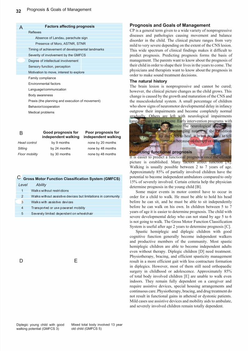
8/9/2019 Guía de ayuda para la parálisis cerebral.pdf
http://slidepdf.com/reader/full/guia-de-ayuda-para-la-paralisis-cerebralpdf 32/143
8/9/2019 Guía de ayuda para la parálisis cerebral.pdf
http://slidepdf.com/reader/full/guia-de-ayuda-para-la-paralisis-cerebralpdf 33/143
33Prognosis & Goals of Management
Treatment modalities
Support growth and nutrition
Ophthalmologic therapy
Rehabilitation
Glasses
Surgery
Dental hygiene
Gastrointestinal problems
Medications for reflux
Gastrostomy
Anti-reflux surgery
Therapy for motor function
Physical therapy
Occupational therapy
Adaptive seating
Bracing
Wheeled mobility
Orthopaedic surgery
Oromotor therapy
Chewing
Swallowing
Speech
Seizure prevention
Spasticity and dyskinesia
Medical treatment
Botulinum toxin
ITB, SDR
Treatment team
Physicians
Pediatric neurologist
Pediatric orthopaedic surgeon
Pediatric physiatrist
Allied health professionals
Physiotherapists
Occupational therapists
Orthotists
Psychologists
Speech therapists
Consulting members
Pediatric neurosurgeon
Ophthalmologist
Ear-nose-throat specialist
Audiologist
Dentist
Pediatric gastroenterologist
Nutrition specialist
Teamwork is essential when treating children with CP. Even the best
teams have controversies. Work close together and have educational
activities which everyone participates in.
Growing up with CP
‘The child with CP grows up to be an adult with CP’
The disabled individual grows up to become a disabled adult
trying to establish a sense of belonging to the society and to lead
an independent life. Dealing with the lifelong disability that CP
causes is mentally, spiritually, and financially exhausting for
the family and the growing individual. Children undergo an
especially difficult period during adolescence when the effectsof spasticity worsen, rapid growth and increase in body weight
complicate the physical disabilities, and social development
is at a critical stage. Mental retardation, severity of disability,
prolonged therapy programs preventing socialization,
overprotection of the adolescent and denial of disability by the
family negatively affect the prognosis for independent living in
the adult [A]. There are many barriers for the child to overcome
to integrate with the society as a happy and productive human
being. Many societies still regard CP as an economic and social
burden. Social, cultural and economic factors play major roles
in the community integration of the adolescent with CP.
Life expectancyMortality in CP is extremely variable. Life expectancy is
normal in most diplegic and hemiplegic children who receive
adequate medical care and have strong family support. The
survival rate of severely affected quadriplegics is low, and most
die of malnutrition, infections or respiratory problems before
reaching adolescence.
Treatment team
Many impairments associated with the primary neurological
lesion accompany the motor disorder seen in CP. A large
team of experts who work together for treatment is necessary
because the impairments vary both in severity and in character.
The team consists of physicians, surgeons and allied health
professionals such as physiotherapists, occupational therapists
and child development professionals [B] who apply various
treatment procedures [C]. The team must work together in
harmony for success. The members must be aware of all the
needs and limitations of the child and know what each other is
doing for the child. The team should work also in cooperation
with non-governmental organizations to support the integration
of disabled children with society including sport, recreation
and fun. Close interaction of the team members is vital [D].
Physicians and health professionals who work with patients
with CP need to be aware of the implications of the diagnosis,
the hardships these children undergo, and the value of each
step gained towards function, no matter how small. A thoroughknowledge of all the aspects of CP including the timing of
diagnosis, the probable natural history, the prognosis and
outcome is absolutely necessary in order to be able to help
these children become part of the normal world.
The ultimate goal in the management is to minimize
disability while promoting independence and full participation
in society. Direct all efforts to gain independence in activities
of daily living, ability to go to school, earn a living and a
successful integration with the community.
Factors adversely affecting independent
living in the adult
Mental retardation
Severity of disability
Prolonged therapy preventing socialization
Overprotection of the adolescent
Denial of disability
DD
CCB
A
Photo courtesy of D. Gaebler-Spira
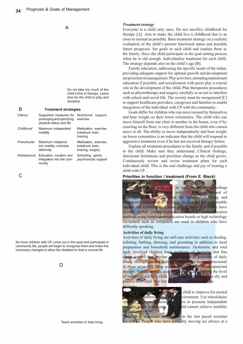
8/9/2019 Guía de ayuda para la parálisis cerebral.pdf
http://slidepdf.com/reader/full/guia-de-ayuda-para-la-paralisis-cerebralpdf 34/143
34 Prognosis & Goals of Management
Treatment strategies
Infancy Supportive measures for
prolonging and optimizingphysical status and life
Nutritional support,
exercise
Childhood Maximum independent
mobility
Medication, exercise,
botulinum toxin,
bracing
Preschooler Maximum independ-
ent mobility, minimize
deformity
Medication, exercise,
botulinum toxin,
bracing, surgery
Adolescence Education, vocation and
integration into the com-
munity
Schooling, sports,
psychosocial support
Treatment strategy
Everyone is a child only once. Do not sacrifice childhood for
therapy [A]. Aim to make the child live a childhood that is as
close to normal as possible. Base treatment strategy on a realistic
evaluation of the child’s present functional status and possible
future prognosis. Set goals in each child and explain them to
the family. Have the child participate in the goal-setting process
when he is old enough. Individualize treatment for each child.The strategy depends also on the child’s age [B].
Family education, addressing the specific needs of the infant,
providing adequate support for optimal growth and development
are priorities in management. Play activities, attending mainstream
education if possible, and socialization with peers play a crucial
role in the development of the child. Plan therapeutic procedures
such as physiotherapy and surgery carefully so as not to interfere
with school and social life. The society must be reorganized [C]
to support healthcare providers, caregivers and families to enable
integration of the individual with CP with the community.
Goals differ for children who can move around by themselves
and bear weight on their lower extremities. The child who can
move himself from one chair to another in the house, even if by
crawling on the floor, is very different from the child who cannot
move at all. The ability to move independently and bear weight
on lower extremities is an indicator that the child will respond to
aggressive treatment even if he has not received therapy before.
Explain all treatment procedures to the family and if possible
to the child. Make sure they understand. Clinical findings,
functional limitations and priorities change as the child grows.
Continuously review and revise treatment plans for each
individual child. This is the real challenge and joy of treating a
child with CP.
Priorities in function / treatment (From E. Bleck)
Communication
Communication is necessary to express thoughts, feelings
and needs. Every individual with CP needs a method of
communication. It is essential for independent living and
community integration. If the child can produce comprehensible
sounds and syllables by 2 years of age, he will probably have
normal verbal communication. Alternative communication
methods using simple communication boards or high technology
equipment such as computers are used in children who have
difficulty speaking.
Activities of daily living
Activities of daily living are self-care activities such as feeding,
toileting, bathing, dressing, and grooming in addition to meal preparation and household maintenance. Dyskinetic and total
body involved children have problems of dexterity and fine
motor control that prevent independence in activities of daily
living. Hemiplegic and diplegic children can become functional
in these areas [D]. They sometimes need help of occupational
therapy. Family attitude is a critical factor determining the level
of independence of a child. Overprotection results in a shy and
passive individual who has not gained self-care abilities.
Mobility
Mobilization is crucial for the young child to improve his mental
capacity and understanding of the environment. Use wheelchairs
or other mechanical assistive devices to promote independentmobility in the community if the child cannot achieve mobility
by walking.
Mobility is important to function in the fast paced societies
we live in. People who have difficulty moving are always at a
Do not take too much of the
child’s time in therapy. Leave
time for the child to play and
socialize.
As more children with CP come out in the open and participate incommunity life, people will begin to recognize them and make the
necessary changes to allow the disabled to lead a normal life.
DD
CC
B
A
Teach activities of daily living.
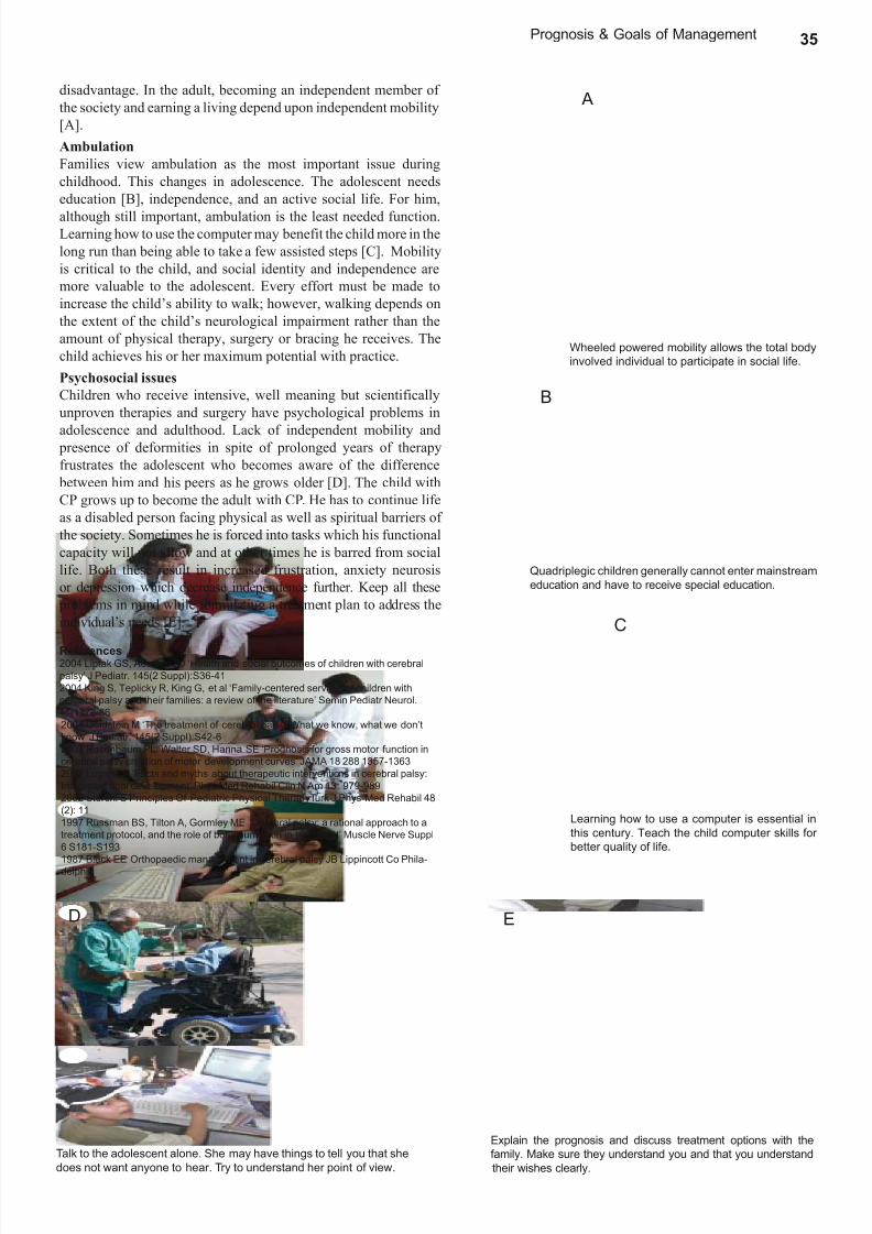
8/9/2019 Guía de ayuda para la parálisis cerebral.pdf
http://slidepdf.com/reader/full/guia-de-ayuda-para-la-paralisis-cerebralpdf 35/143
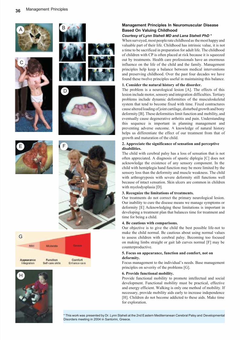
8/9/2019 Guía de ayuda para la parálisis cerebral.pdf
http://slidepdf.com/reader/full/guia-de-ayuda-para-la-paralisis-cerebralpdf 36/143
36 Management Principles
Management Principles In Neuromuscular Disease
Based On Valuing Childhood
Courtesy of Lynn Staheli MD and Lana Staheli PhD *
When surveyed, most people rate childhood as the most happy and
valuable part of their life. Childhood has intrinsic value, it is not
a time to be sacrificed in preparation for adult life. The childhood
of children with CP is often placed at risk because it is squeezed
out by treatments. Health care professionals have an enormousinfluence on the life of the child and the family. Management
principles help keep a balance between medical interventions
and preserving childhood. Over the past four decades we have
found these twelve principles useful in maintaining this balance.
1. Consider the natural history of the disorder.
The problem is a neurological lesion [A]. The effects of this
lesion include motor, sensory and integration difficulties. Tertiary
problems include dynamic deformities of the musculoskeletal
system that tend to become fixed with time. Fixed contractures
cause altered loading of joint cartilage, disturbed growth and bony
deformity [B]. These deformities limit function and mobility, and
eventually cause degenerative arthritis and pain. Understandingthis sequence is important in planning management and
preventing adverse outcome. A knowledge of natural history
helps us differentiate the effect of our treatment from that of
growth and maturation of the child.
2. Appreciate the significance of sensation and perceptive
disabilities.
The child with cerebral palsy has a loss of sensation that is not
often appreciated. A diagnosis of spastic diplegia [C] does not
acknowledge the existence of any sensory component. In the
child with hemiplegia hand function may be more limited by the
sensory loss than the deformity and muscle weakness. The child
with arthrogryposis with severe deformity still functions well because of intact sensation. Skin ulcers are common in children
with myelodysplasia [D].
3. Recognize the limitations of treatments.
Our treatments do not correct the primary neurological lesion.
Our inability to cure the disease means we manage symptoms or
deformity [E] Acknowledging these limitations is important in
developing a treatment plan that balances time for treatment and
time for being a child.
4. Be cautious with comparisons.
Our objective is to give the child the best possible life-not to
make the child normal. Be cautious about using normal values
to assess children with cerebral palsy. Becoming too focusedon making limbs straight or gait lab curves normal [F] may be
counterproductive.
5. Focus on appearance, function and comfort, not on
deformity.
Focus management to the individual’s needs. Base management
principles on severity of the problems [G].
6. Provide functional mobility.
Provide functional mobility to promote intellectual and social
development. Functional mobility must be practical, effective
and energy efficient. Walking is only one method of mobility. If
necessary, provide mobility aids early to increase independence
[H]. Children do not become addicted to these aids. Make timefor exploration.
* This work was presented by Dr. Lynn Staheli at the 2nd Eastern Mediterranean Cerebral Palsy and Developmental
Disorders meeting in 2004 in Santorini, Greece.
FFE
DD
CC
B A
HH
G
G
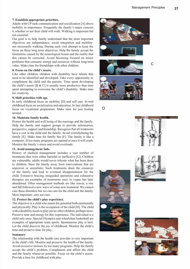
8/9/2019 Guía de ayuda para la parálisis cerebral.pdf
http://slidepdf.com/reader/full/guia-de-ayuda-para-la-paralisis-cerebralpdf 37/143
37Management Principles
7. Establish appropriate priorities.
Adults with CP rank communication and socialization [A] above
mobility in importance. Frequently the family’s major concern
is whether or not their child will walk. Walking is important but
not essential.
Our goal is to help family understand that the most important
objectives are independence, social integration and mobility-
not necessarily walking. During each visit attempt to keep thefocus on these long term objectives. Help the family accept the
limitations caused by the neurological lesion and the reality that
this cannot be corrected. Avoid becoming focused on minor
problems that consume energy and resources without long-term
value. Make time for friendships with other children.
8. Focus on the child’s assets.
Like other children, children with disability have talents that
need to be identified and developed. Take every opportunity to
compliment the child and the parents. Time spent developing
the child’s assets [B & C] is usually more productive than time
spent attempting to overcome the child’s disability. Make time
for creativity.9. Shift priorities with age.
In early childhood focus on mobility [D] and self care. In mid
childhood focus on socialization and education. In late childhood
focus on vocational preparation. Make time for just fooling
around.
10. Maintain family health.
Protect the health and well being of the marriage and the family.
Help the family and support groups to provide information,
perspective, support and friendship. Recognize that all treatments
have a cost to the child and the family. Avoid overwhelming the
family [E]. Make time for family fun [F]. The family is like a
computer. If too many programs are opened at once it will crash.Monitor the family’s stress and avoid overloads.
11. Avoid management fads.
History of medical management includes a vast number of
treatments that were either harmful or ineffective [G]. Children
are vulnerable, adults would never tolerate what has been done
to children. Steer the family away from interventions that are
unproven or unrealistic. Such treatments drain the resources
of the family and lead to eventual disappointment for the
child. Extensive bracing, misguided operations and exhaustive
therapies are examples of treatments once in vogue but later
abandoned. Often management methods are like waves, a rise
and fall followed a new wave of some new treatment. We cannotcure these disorders but we can care for the child and the family.
Most important- care not cure.
12. Protect the child’s play experience.
The objective is a child who meets his potential both emotionally
and physically. Play is the occupation of the child [H]. The child
with a disability needs to play just as other children, perhaps more.
Preserve time and energy for this experience. The individual is a
child only once. Special Olympics and wheelchair basketball are
examples of appropriate team sports. Spontaneous play is best.
Let the child discover the joy of childhood. Monitor the child’s
time and preserve time for play.
SummaryThe relationship with the health care provider is very important
in the child’s life. Monitor and preserve the health of the family.
Avoid excessive stresses by too many programs. Help the family
accept the child’s problem. Compliment and affirm the child
and the family whenever possible. Focus on the child’s assets.
Provide a time for childhood with play.
FF
EE
DD
CCB
A
HH
GG
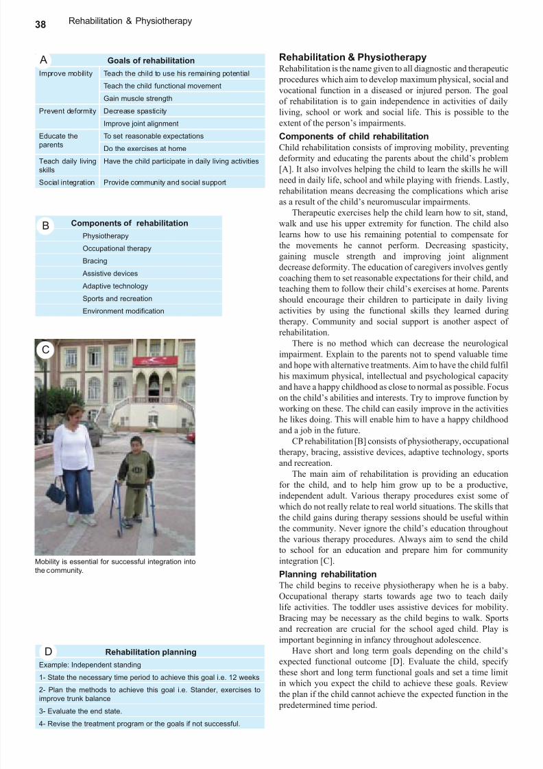
8/9/2019 Guía de ayuda para la parálisis cerebral.pdf
http://slidepdf.com/reader/full/guia-de-ayuda-para-la-paralisis-cerebralpdf 38/143
38 Rehabilitation & Physiotherapy
Rehabilitation & PhysiotherapyRehabilitation is the name given to all diagnostic and therapeutic
procedures which aim to develop maximum physical, social and
vocational function in a diseased or injured person. The goal
of rehabilitation is to gain independence in activities of daily
living, school or work and social life. This is possible to the
extent of the person’s impairments.
Components of child rehabilitationChild rehabilitation consists of improving mobility, preventing
deformity and educating the parents about the child’s problem
[A]. It also involves helping the child to learn the skills he will
need in daily life, school and while playing with friends. Lastly,
rehabilitation means decreasing the complications which arise
as a result of the child’s neuromuscular impairments.
Therapeutic exercises help the child learn how to sit, stand,
walk and use his upper extremity for function. The child also
learns how to use his remaining potential to compensate for
the movements he cannot perform. Decreasing spasticity,
gaining muscle strength and improving joint alignment
decrease deformity. The education of caregivers involves gentlycoaching them to set reasonable expectations for their child, and
teaching them to follow their child’s exercises at home. Parents
should encourage their children to participate in daily living
activities by using the functional skills they learned during
therapy. Community and social support is another aspect of
rehabilitation.
There is no method which can decrease the neurological
impairment. Explain to the parents not to spend valuable time
and hope with alternative treatments. Aim to have the child fulfil
his maximum physical, intellectual and psychological capacity
and have a happy childhood as close to normal as possible. Focus
on the child’s abilities and interests. Try to improve function by
working on these. The child can easily improve in the activities
he likes doing. This will enable him to have a happy childhood
and a job in the future.
CP rehabilitation [B] consists of physiotherapy, occupational
therapy, bracing, assistive devices, adaptive technology, sports
and recreation.
The main aim of rehabilitation is providing an education
for the child, and to help him grow up to be a productive,
independent adult. Various therapy procedures exist some of
which do not really relate to real world situations. The skills that
the child gains during therapy sessions should be useful within
the community. Never ignore the child’s education throughout
the various therapy procedures. Always aim to send the childto school for an education and prepare him for community
integration [C].
Planning rehabilitation
The child begins to receive physiotherapy when he is a baby.
Occupational therapy starts towards age two to teach daily
life activities. The toddler uses assistive devices for mobility.
Bracing may be necessary as the child begins to walk. Sports
and recreation are crucial for the school aged child. Play is
important beginning in infancy throughout adolescence.
Have short and long term goals depending on the child’s
expected functional outcome [D]. Evaluate the child, specify
these short and long term functional goals and set a time limitin which you expect the child to achieve these goals. Review
the plan if the child cannot achieve the expected function in the
predetermined time period.
Mobility is essential for successful integration into
the community.
Goals of rehabilitation
Improve mobility Teach the child to use his remaining potential
Teach the child functional movement
Gain muscle strength
Prevent deformity Decrease spasticity
Improve joint alignment
Educate theparents
To set reasonable expectations
Do the exercises at home
Teach daily living
skills
Have the child participate in daily living activities
Social integration Provide community and social support
Components of rehabilitation
Physiotherapy
Occupational therapy
Bracing
Assistive devices
Adaptive technology
Sports and recreation
Environment modification
Rehabilitation planning
Example: Independent standing
1- State the necessary time period to achieve this goal i.e. 12 weeks
2- Plan the methods to achieve this goal i.e. Stander, exercises to
improve trunk balance
3- Evaluate the end state.
4- Revise the treatment program or the goals if not successful.
DD
CC
B
A
8/9/2019 Guía de ayuda para la parálisis cerebral.pdf
http://slidepdf.com/reader/full/guia-de-ayuda-para-la-paralisis-cerebralpdf 39/143
39Rehabilitation & Physiotherapy
Medical problems of the child
which prevent rehabilitation
Respiratory problems
Epileptic seizures
Dysphagia
Depression
Gastroesophageal reflux
Sleep disorders
Visual and hearing deficits
Mental retardation
Cortical sensory deficits
Communication deficits
Allow room and time for play and educa-
tion.
Factors influencing rehabilitation outcome
Consider the following factors in the rehabilitation plan [A].
Treatment team
The pediatrician provides diagnosis and preventive health care.
The orthopaedic surgeon tries to minimise static and dynamic
contractures to improve mobility. The pediatric physiatrist
evaluates the child’s overall medical, surgical and therapy
options and helps the child and the family to set functional,achievable goals. Together, the rehabilitation team works to
assist the person with CP to achieve his place in the society. A
productive interaction between the physicians and the therapists
is essential for the maximum benefit of the child. All those
involved with the child must have a basic understanding on the
diagnosis, family expectations, degree of motor dysfunction,
functional goals and the therapy program.
Medical problems of the child
The rehabilitation physician and the team must be prepared
to anticipate certain acute and chronic problems [B] during
the rehabilitation of the child with CP. The disabled child is
more prone than his able-bodied peers to respiratory problems,convulsions, dysphagia, depression, gastroesophageal reflux
and sleep disorders. Total body and some severely involved
diplegics have visual and hearing deficits, mental retardation,
cortical sensory deficits and communication deficits that prevent
the child from reaching his maximum potential.
The child’s character
The motivation to move, temperament, behavior/cooperation
and the willingness to take risks are important determinants of
rehabilitation outcome. These personality characteristics of the
child are independent of impairment or disability.
The family
Some families provide their children with ample experiencesand opportunities that enrich their environment and increase
their ability to achieve new skills. Family resources, quality
of home environment, family support [C] and parent/caregiver
expectations guide the plans of long-term care for the disabled
child.
Physiotherapy
Physiotherapy helps improve mobility. It is the basic treatment
in all children with CP [D]. It consists of exercises, bracing and
activities towards reaching specific functional goals. It aims to
bring the child to an erect position, give the child independent
mobility and prevent deformity. Organize physiotherapy to fit
into the family’s lifestyle [E].Parents have to learn exercises from the
therapists and treatment team.
Factors influencing rehabilitation outcome
Treatment team Productive interaction
Basic understanding
Medical problems Respiratory
Convulsions
Dysphagia
Depression
Gastroesophageal reflux
Sleep disorders
Visual and hearing deficits
Mental retardation
Sensation
Communication
Child’s character Motivation to move
Temperament
Behaviour/cooperation
Willingness to take risks
The family Resources
Quality of home environment
Support
Expectations
Physiotherapy tries to improve
Postural control
Muscle strength
Range of motion
Decreasing spasticity and contracture
Increasing muscle elasticity and joint laxity
Joint alignment
Motor control
Muscular/cardiovascular endurance and mobility skillsIncreasing coordination / agility
Balance
Transitions
Use of assistive devices
EE
DD
CC
A
B
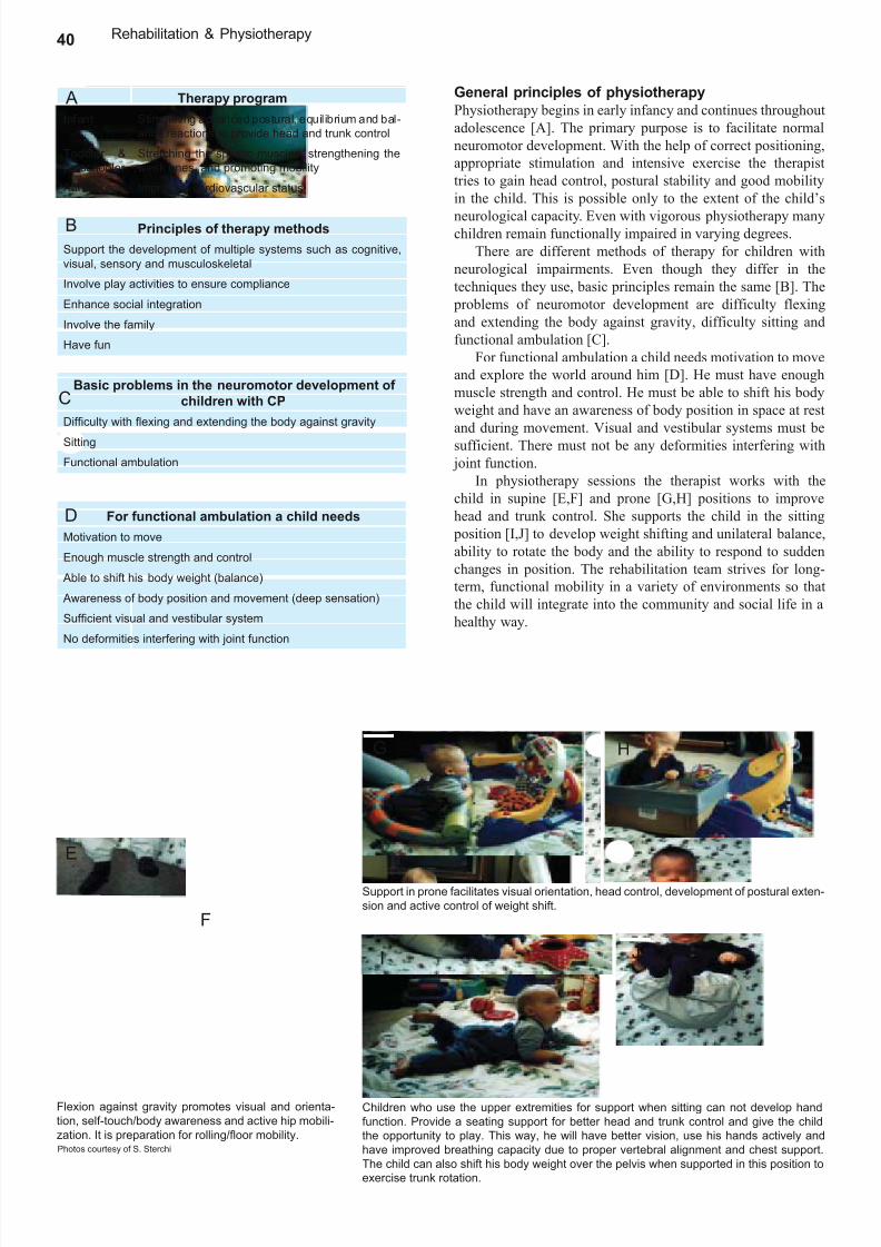
8/9/2019 Guía de ayuda para la parálisis cerebral.pdf
http://slidepdf.com/reader/full/guia-de-ayuda-para-la-paralisis-cerebralpdf 40/143
40 Rehabilitation & Physiotherapy
General principles of physiotherapy
Physiotherapy begins in early infancy and continues throughout
adolescence [A]. The primary purpose is to facilitate normal
neuromotor development. With the help of correct positioning,
appropriate stimulation and intensive exercise the therapist
tries to gain head control, postural stability and good mobility
in the child. This is possible only to the extent of the child’s
neurological capacity. Even with vigorous physiotherapy manychildren remain functionally impaired in varying degrees.
There are different methods of therapy for children with
neurological impairments. Even though they differ in the
techniques they use, basic principles remain the same [B]. The
problems of neuromotor development are difficulty flexing
and extending the body against gravity, difficulty sitting and
functional ambulation [C].
For functional ambulation a child needs motivation to move
and explore the world around him [D]. He must have enough
muscle strength and control. He must be able to shift his body
weight and have an awareness of body position in space at rest
and during movement. Visual and vestibular systems must be
sufficient. There must not be any deformities interfering with
joint function.
In physiotherapy sessions the therapist works with the
child in supine [E,F] and prone [G,H] positions to improve
head and trunk control. She supports the child in the sitting
position [I,J] to develop weight shifting and unilateral balance,
ability to rotate the body and the ability to respond to sudden
changes in position. The rehabilitation team strives for long-
term, functional mobility in a variety of environments so that
the child will integrate into the community and social life in a
healthy way.
Principles of therapy methods
Support the development of multiple systems such as cognitive,
visual, sensory and musculoskeletal
Involve play activities to ensure compliance
Enhance social integration
Involve the family
Have fun
Basic problems in the neuromotor development of
children with CP
Difficulty with flexing and extending the body against gravity
Sitting
Functional ambulation
For functional ambulation a child needs
Motivation to move
Enough muscle strength and control
Able to shift his body weight (balance)
Awareness of body position and movement (deep sensation)
Sufficient visual and vestibular system
No deformities interfering with joint function
Children who use the upper extremities for support when sitting can not develop hand
function. Provide a seating support for better head and trunk control and give the child
the opportunity to play. This way, he will have better vision, use his hands actively and
have improved breathing capacity due to proper vertebral alignment and chest support.
The child can also shift his body weight over the pelvis when supported in this position to
exercise trunk rotation.
Therapy program
Infant Stimulating advanced postural, equilibrium and bal-
ance reactions to provide head and trunk control
Toddler &
preschooler
Stretching the spastic muscles, strengthening the
weak ones, and promoting mobility
Adolescent Improving cardiovascular status
Flexion against gravity promotes visual and orienta-
tion, self-touch/body awareness and active hip mobili-
zation. It is preparation for rolling/floor mobility.
FF
EE
DD
CC
B
A
II
GG
Support in prone facilitates visual orientation, head control, development of postural exten-
sion and active control of weight shift.
Photos courtesy of S. Sterchi
HH
JJ
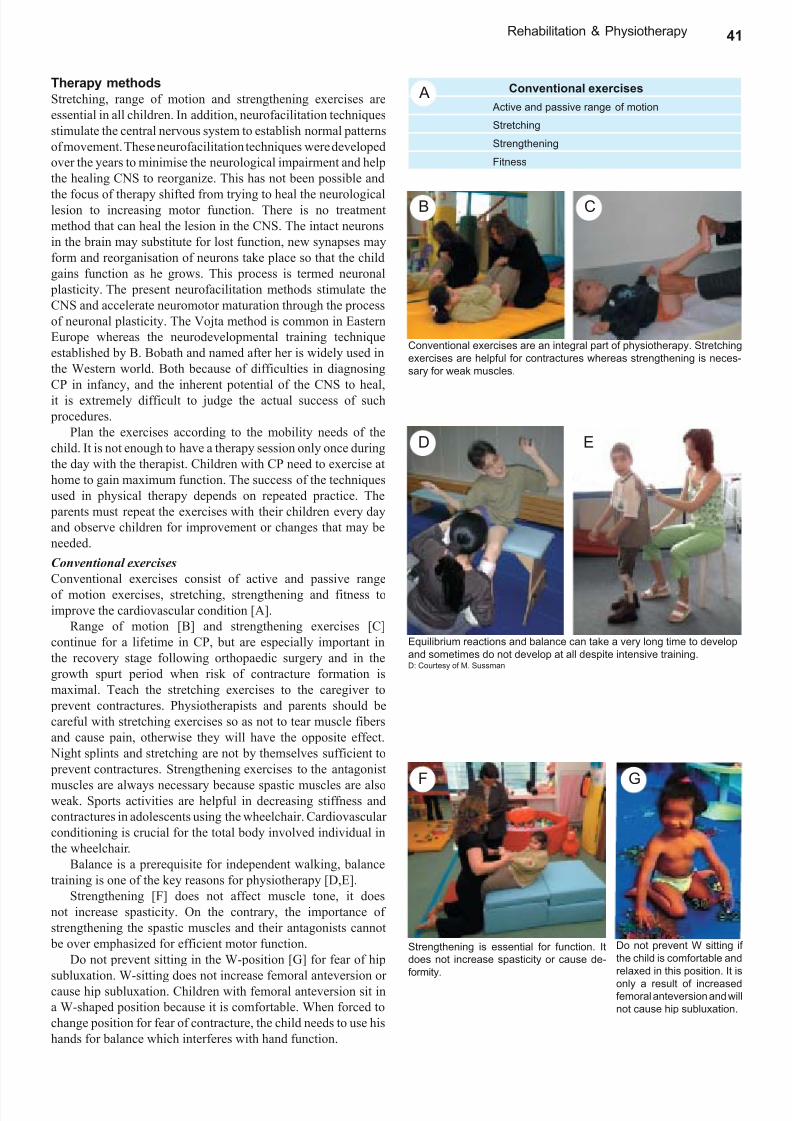
8/9/2019 Guía de ayuda para la parálisis cerebral.pdf
http://slidepdf.com/reader/full/guia-de-ayuda-para-la-paralisis-cerebralpdf 41/143
41Rehabilitation & Physiotherapy
Conventional exercises are an integral part of physiotherapy. Stretching
exercises are helpful for contractures whereas strengthening is neces-
sary for weak muscles.
Therapy methods
Stretching, range of motion and strengthening exercises are
essential in all children. In addition, neurofacilitation techniques
stimulate the central nervous system to establish normal patterns
of movement. These neurofacilitation techniques were developed
over the years to minimise the neurological impairment and help
the healing CNS to reorganize. This has not been possible and
the focus of therapy shifted from trying to heal the neurologicallesion to increasing motor function. There is no treatment
method that can heal the lesion in the CNS. The intact neurons
in the brain may substitute for lost function, new synapses may
form and reorganisation of neurons take place so that the child
gains function as he grows. This process is termed neuronal
plasticity. The present neurofacilitation methods stimulate the
CNS and accelerate neuromotor maturation through the process
of neuronal plasticity. The Vojta method is common in Eastern
Europe whereas the neurodevelopmental training technique
established by B. Bobath and named after her is widely used in
the Western world. Both because of difficulties in diagnosing
CP in infancy, and the inherent potential of the CNS to heal,
it is extremely difficult to judge the actual success of such
procedures.
Plan the exercises according to the mobility needs of the
child. It is not enough to have a therapy session only once during
the day with the therapist. Children with CP need to exercise at
home to gain maximum function. The success of the techniques
used in physical therapy depends on repeated practice. The
parents must repeat the exercises with their children every day
and observe children for improvement or changes that may be
needed.
Conventional exercises
Conventional exercises consist of active and passive range
of motion exercises, stretching, strengthening and fitness to
improve the cardiovascular condition [A].
Range of motion [B] and strengthening exercises [C]
continue for a lifetime in CP, but are especially important in
the recovery stage following orthopaedic surgery and in the
growth spurt period when risk of contracture formation is
maximal. Teach the stretching exercises to the caregiver to
prevent contractures. Physiotherapists and parents should be
careful with stretching exercises so as not to tear muscle fibers
and cause pain, otherwise they will have the opposite effect.
Night splints and stretching are not by themselves sufficient to
prevent contractures. Strengthening exercises to the antagonist
muscles are always necessary because spastic muscles are alsoweak. Sports activities are helpful in decreasing stiffness and
contractures in adolescents using the wheelchair. Cardiovascular
conditioning is crucial for the total body involved individual in
the wheelchair.
Balance is a prerequisite for independent walking, balance
training is one of the key reasons for physiotherapy [D,E].
Strengthening [F] does not affect muscle tone, it does
not increase spasticity. On the contrary, the importance of
strengthening the spastic muscles and their antagonists cannot
be over emphasized for efficient motor function.
Do not prevent sitting in the W-position [G] for fear of hip
subluxation. W-sitting does not increase femoral anteversion orcause hip subluxation. Children with femoral anteversion sit in
a W-shaped position because it is comfortable. When forced to
change position for fear of contracture, the child needs to use his
hands for balance which interferes with hand function.
Equilibrium reactions and balance can take a very long time to develop
and sometimes do not develop at all despite intensive training.
Conventional exercises
Active and passive range of motion
Stretching
Strengthening
Fitness
Do not prevent W sitting if
the child is comfortable and
relaxed in this position. It is
only a result of increasedfemoral anteversion and will
not cause hip subluxation.
Strengthening is essential for function. It
does not increase spasticity or cause de-
formity.
F
F
EED
CCB
A
G
G
D: Courtesy of M. Sussman
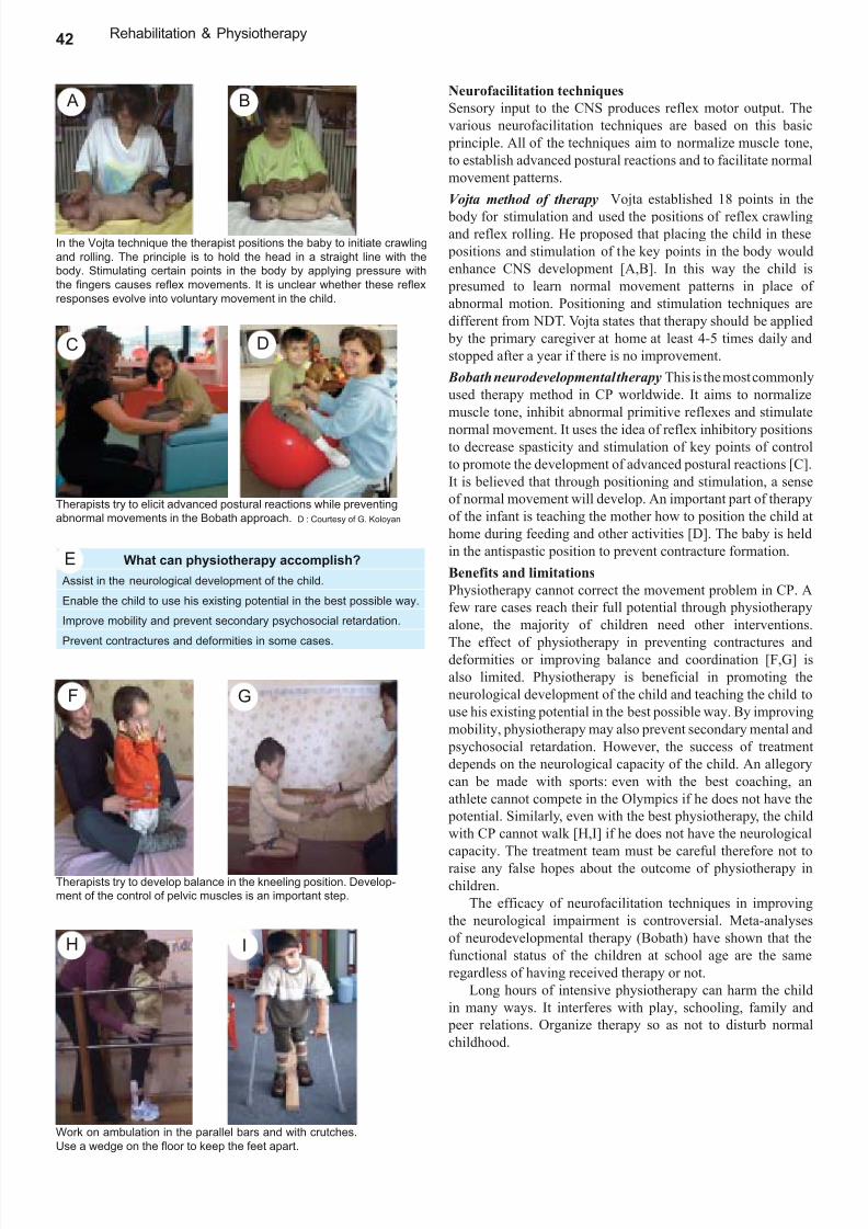
8/9/2019 Guía de ayuda para la parálisis cerebral.pdf
http://slidepdf.com/reader/full/guia-de-ayuda-para-la-paralisis-cerebralpdf 42/143
42 Rehabilitation & Physiotherapy
Neurofacilitation techniques
Sensory input to the CNS produces reflex motor output. The
various neurofacilitation techniques are based on this basic
principle. All of the techniques aim to normalize muscle tone,
to establish advanced postural reactions and to facilitate normal
movement patterns.
Vojta method of therapy Vojta established 18 points in the
body for stimulation and used the positions of reflex crawlingand reflex rolling. He proposed that placing the child in these
positions and stimulation of the key points in the body would
enhance CNS development [A,B]. In this way the child is
presumed to learn normal movement patterns in place of
abnormal motion. Positioning and stimulation techniques are
different from NDT. Vojta states that therapy should be applied
by the primary caregiver at home at least 4-5 times daily and
stopped after a year if there is no improvement.
Bobath neurodevelopmental therapy This is the most commonly
used therapy method in CP worldwide. It aims to normalize
muscle tone, inhibit abnormal primitive reflexes and stimulate
normal movement. It uses the idea of reflex inhibitory positionsto decrease spasticity and stimulation of key points of control
to promote the development of advanced postural reactions [C].
It is believed that through positioning and stimulation, a sense
of normal movement will develop. An important part of therapy
of the infant is teaching the mother how to position the child at
home during feeding and other activities [D]. The baby is held
in the antispastic position to prevent contracture formation.
Benefits and limitations
Physiotherapy cannot correct the movement problem in CP. A
few rare cases reach their full potential through physiotherapy
alone, the majority of children need other interventions.
The effect of physiotherapy in preventing contractures anddeformities or improving balance and coordination [F,G] is
also limited. Physiotherapy is beneficial in promoting the
neurological development of the child and teaching the child to
use his existing potential in the best possible way. By improving
mobility, physiotherapy may also prevent secondary mental and
psychosocial retardation. However, the success of treatment
depends on the neurological capacity of the child. An allegory
can be made with sports: even with the best coaching, an
athlete cannot compete in the Olympics if he does not have the
potential. Similarly, even with the best physiotherapy, the child
with CP cannot walk [H,I] if he does not have the neurological
capacity. The treatment team must be careful therefore not to
raise any false hopes about the outcome of physiotherapy in
children.
The efficacy of neurofacilitation techniques in improving
the neurological impairment is controversial. Meta-analyses
of neurodevelopmental therapy (Bobath) have shown that the
functional status of the children at school age are the same
regardless of having received therapy or not.
Long hours of intensive physiotherapy can harm the child
in many ways. It interferes with play, schooling, family and
peer relations. Organize therapy so as not to disturb normal
childhood.
In the Vojta technique the therapist positions the baby to initiate crawling
and rolling. The principle is to hold the head in a straight line with the
body. Stimulating certain points in the body by applying pressure with
the fingers causes reflex movements. It is unclear whether these reflex
responses evolve into voluntary movement in the child.
Therapists try to elicit advanced postural reactions while preventing
abnormal movements in the Bobath approach.
What can physiotherapy accomplish?
Assist in the neurological development of the child.
Enable the child to use his existing potential in the best possible way.
Improve mobility and prevent secondary psychosocial retardation.
Prevent contractures and deformities in some cases.
Therapists try to develop balance in the kneeling position. Develop-
ment of the control of pelvic muscles is an important step.
FF
EE
DDC
B A
GG
Work on ambulation in the parallel bars and with crutches.
Use a wedge on the floor to keep the feet apart.
HH II
D : Courtesy of G. Koloyan
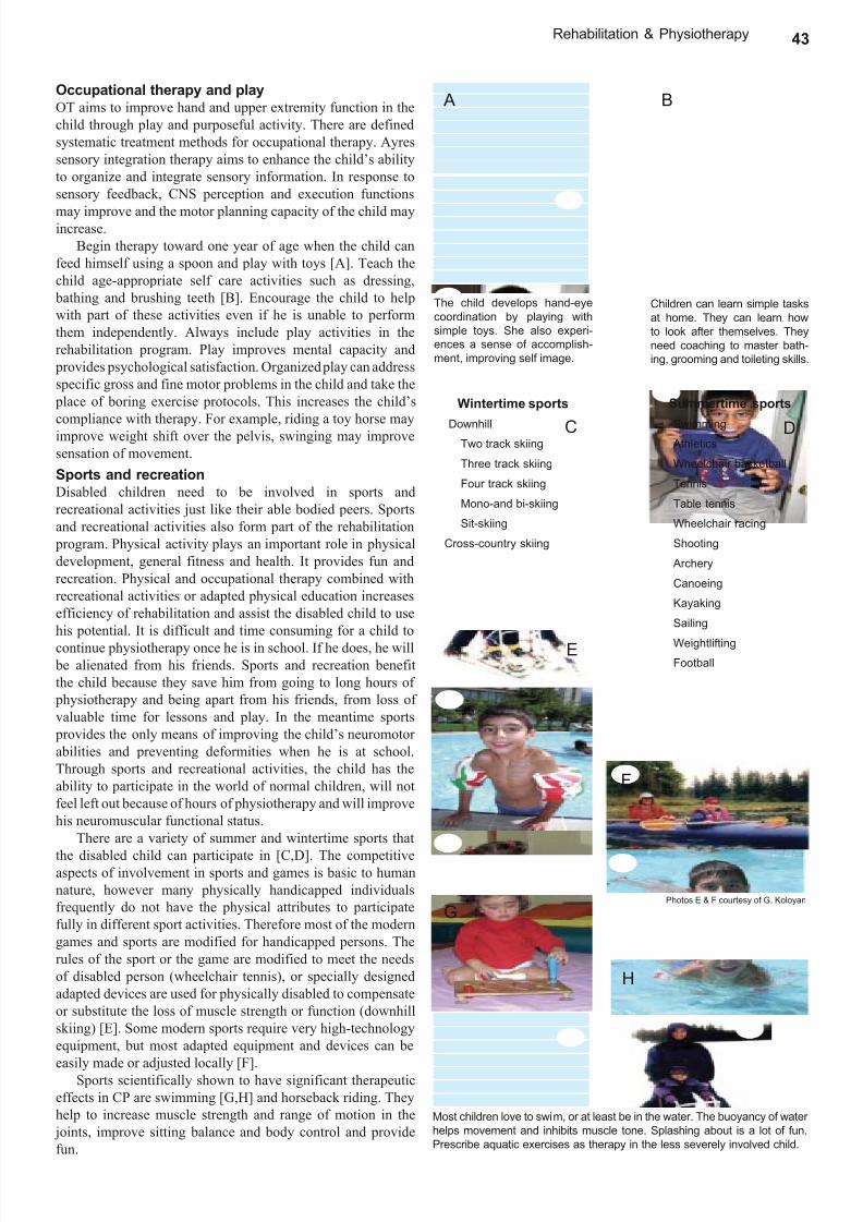
8/9/2019 Guía de ayuda para la parálisis cerebral.pdf
http://slidepdf.com/reader/full/guia-de-ayuda-para-la-paralisis-cerebralpdf 43/143
43Rehabilitation & Physiotherapy
HH
Occupational therapy and play
OT aims to improve hand and upper extremity function in the
child through play and purposeful activity. There are defined
systematic treatment methods for occupational therapy. Ayres
sensory integration therapy aims to enhance the child’s ability
to organize and integrate sensory information. In response to
sensory feedback, CNS perception and execution functions
may improve and the motor planning capacity of the child mayincrease.
Begin therapy toward one year of age when the child can
feed himself using a spoon and play with toys [A]. Teach the
child age-appropriate self care activities such as dressing,
bathing and brushing teeth [B]. Encourage the child to help
with part of these activities even if he is unable to perform
them independently. Always include play activities in the
rehabilitation program. Play improves mental capacity and
provides psychological satisfaction. Organized play can address
specific gross and fine motor problems in the child and take the
place of boring exercise protocols. This increases the child’s
compliance with therapy. For example, riding a toy horse may
improve weight shift over the pelvis, swinging may improve
sensation of movement.
Sports and recreation
Disabled children need to be involved in sports and
recreational activities just like their able bodied peers. Sports
and recreational activities also form part of the rehabilitation
program. Physical activity plays an important role in physical
development, general fitness and health. It provides fun and
recreation. Physical and occupational therapy combined with
recreational activities or adapted physical education increases
efficiency of rehabilitation and assist the disabled child to use
his potential. It is difficult and time consuming for a child to
continue physiotherapy once he is in school. If he does, he will
be alienated from his friends. Sports and recreation benefit
the child because they save him from going to long hours of
physiotherapy and being apart from his friends, from loss of
valuable time for lessons and play. In the meantime sports
provides the only means of improving the child’s neuromotor
abilities and preventing deformities when he is at school.
Through sports and recreational activities, the child has the
ability to participate in the world of normal children, will not
feel left out because of hours of physiotherapy and will improve
his neuromuscular functional status.
There are a variety of summer and wintertime sports that
the disabled child can participate in [C,D]. The competitiveaspects of involvement in sports and games is basic to human
nature, however many physically handicapped individuals
frequently do not have the physical attributes to participate
fully in different sport activities. Therefore most of the modern
games and sports are modified for handicapped persons. The
rules of the sport or the game are modified to meet the needs
of disabled person (wheelchair tennis), or specially designed
adapted devices are used for physically disabled to compensate
or substitute the loss of muscle strength or function (downhill
skiing) [E]. Some modern sports require very high-technology
equipment, but most adapted equipment and devices can be
easily made or adjusted locally [F].
Sports scientifically shown to have significant therapeutic
effects in CP are swimming [G,H] and horseback riding. They
help to increase muscle strength and range of motion in the
joints, improve sitting balance and body control and provide
fun.
Children can learn simple tasks
at home. They can learn how
to look after themselves. They
need coaching to master bath-
ing, grooming and toileting skills.
Summertime sports
Swimming Athletics
Wheelchair basketball
Tennis
Table tennis
Wheelchair racing
Shooting
Archery
Canoeing
Kayaking
Sailing
Weightlifting
Football
Wintertime sports
DownhillTwo track skiing
Three track skiing
Four track skiing
Mono-and bi-skiing
Sit-skiing
Cross-country skiing
DDC
B A
GG
The child develops hand-eye
coordination by playing with
simple toys. She also experi-
ences a sense of accomplish-
ment, improving self image.
Most children love to swim, or at least be in the water. The buoyancy of water
helps movement and inhibits muscle tone. Splashing about is a lot of fun.
Prescribe aquatic exercises as therapy in the less severely involved child.
EE
FF
Photos E & F courtesy of G. Koloyan
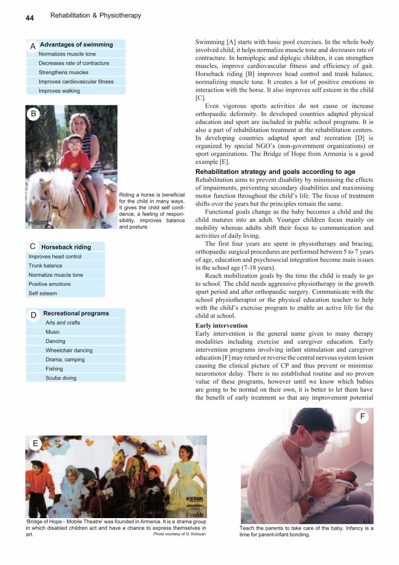
8/9/2019 Guía de ayuda para la parálisis cerebral.pdf
http://slidepdf.com/reader/full/guia-de-ayuda-para-la-paralisis-cerebralpdf 44/143
44 Rehabilitation & Physiotherapy
Horseback riding
Improves head control
Trunk balance
Normalize muscle tone
Positive emotions
Self esteem
Advantages of swimming
Normalizes muscle tone
Decreases rate of contracture
Strengthens muscles
Improves cardiovascular fitness
Improves walking
B
Swimming [A] starts with basic pool exercises. In the whole body
involved child, it helps normalize muscle tone and decreases rate of
contracture. In hemiplegic and diplegic children, it can strengthen
muscles, improve cardiovascular fitness and efficiency of gait.
Horseback riding [B] improves head control and trunk balance,
normalizing muscle tone. It creates a lot of positive emotions in
interaction with the horse. It also improves self esteem in the child
[C].Even vigorous sports activities do not cause or increase
orthopaedic deformity. In developed countries adapted physical
education and sport are included in public school programs. It is
also a part of rehabilitation treatment at the rehabilitation centers.
In developing countries adapted sport and recreation [D] is
organized by special NGO’s (non-government organizations) or
sport organizations. The Bridge of Hope from Armenia is a good
example [E].
Rehabilitation strategy and goals according to age
Rehabilitation aims to prevent disability by minimising the effects
of impairments, preventing secondary disabilities and maximising
motor function throughout the child’s life. The focus of treatmentshifts over the years but the principles remain the same.
Functional goals change as the baby becomes a child and the
child matures into an adult. Younger children focus mainly on
mobility whereas adults shift their focus to communication and
activities of daily living.
The first four years are spent in physiotherapy and bracing,
orthopaedic surgical procedures are performed between 5 to 7 years
of age, education and psychosocial integration become main issues
in the school age (7-18 years).
Reach mobilization goals by the time the child is ready to go
to school. The child needs aggressive physiotherapy in the growth
spurt period and after orthopaedic surgery. Communicate with the
school physiotherapist or the physical education teacher to help
with the child’s exercise program to enable an active life for the
child at school.
Early intervention
Early intervention is the general name given to many therapy
modalities including exercise and caregiver education. Early
intervention programs involving infant stimulation and caregiver
education [F] may retard or reverse the central nervous system lesion
causing the clinical picture of CP and thus prevent or minimise
neuromotor delay. There is no established routine and no proven
value of these programs, however until we know which babies
are going to be normal on their own, it is better to let them have
the benefit of early treatment so that any improvement potential
Recreational programs
Arts and crafts
Music
Dancing
Wheelchair dancing
Drama, camping
Fishing
Scuba diving
DD
CC
A
FF
Riding a horse is beneficial
for the child in many ways.
It gives the child self confi-
dence, a feeling of respon-
sibility, improves balance
and posture.
Teach the parents to take care of the baby. Infancy is a
time for parent-infant bonding.
EE
‘Bridge of Hope - Mobile Theatre’ was founded in Armenia. It is a drama group
in which disabled children act and have a chance to express themselves in
art. Photo courtesy of G. Koloyan
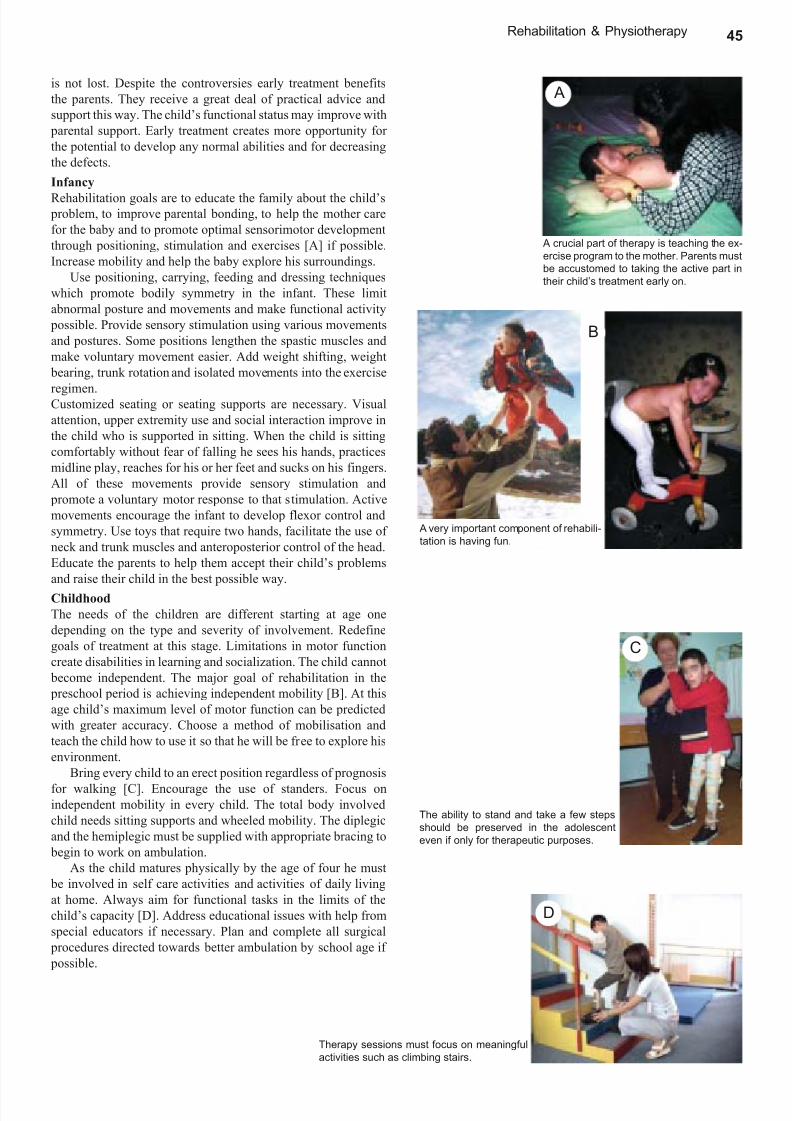
8/9/2019 Guía de ayuda para la parálisis cerebral.pdf
http://slidepdf.com/reader/full/guia-de-ayuda-para-la-paralisis-cerebralpdf 45/143
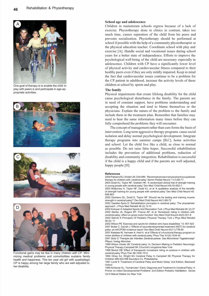
8/9/2019 Guía de ayuda para la parálisis cerebral.pdf
http://slidepdf.com/reader/full/guia-de-ayuda-para-la-paralisis-cerebralpdf 46/143
46 Rehabilitation & Physiotherapy
School age and adolescence
Children in mainstream schools regress because of a lack of
exercise. Physiotherapy done in clinics in contrast, takes too
much time, causes separation of the child from his peers and
prevents socialization. Physiotherapy should be performed at
school if possible with the help of a community physiotherapist or
the physical education teacher. Coordinate school with play and
exercise [A]. Handle social and vocational issues during schoolyears for a better state of independence. Efforts to improve the
psychological well being of the child are necessary especially in
adolescence. Children with CP have a significantly lower level
of physical activity and cardiovascular fitness compared to their
healthy peers even if they are only mildly impaired. Keep in mind
the fact that cardiovascular issues continue to be a problem for
the CP patient in adulthood, increase the activity levels of these
children at school by sports and play.
The family
Physical impairments that create lifelong disability for the child
cause psychological disturbance in the family. The parents are
in need of constant support, have problems understanding andaccepting the situation and tend to blame themselves or the
physicians. Explain the nature of the problem to the family and
include them in the treatment plan. Remember that families may
need to hear the same information many times before they can
fully comprehend the problems they will encounter.
The concept of management rather than cure forms the basis of
intervention. Long term aggressive therapy programs cause social
isolation and delay normal psychological development. Integrate
therapy programs into summer camps [B,C], home activities
and school. Let the child live like a child, as close to normal
as possible. Do not raise false hopes. Successful rehabilitation
includes the prevention of additional problems, reduction of
disability and community integration. Rehabilitation is successful
if the child is a happy child and if the parents are well adjusted,
happy people [D].
References
2004 Palisano RJ, Snider LM, Orlin MN. ‘Recent advances in physical and occupational
therapy for children with cerebral palsy’ Semin Pediatr Neurol 11(1):66-77.
2003 Dodd KJ, Taylor NF, Graham HK. ‘A randomized clinical trial of strength training
in young people with cerebral palsy’ Dev Med Child Neurol 45(10):652-7.
2003 McBurney H, Taylor NF, Dodd KJ, et al ‘A qualitative analysis of the benefits
of strength training for young people with cerebral palsy’ Dev Med Child Neurol 45:
658-663
2002 Damiano DL, Dodd K, Taylor NF ‘Should we be testing and training muscle
strength in cerebral palsy?’ Dev Med Child Neurol 44(1):68-72
2002 Gaebler-Spira D: Rehabilitation principles in cerebral palsy: The physiatrists’
approach J Phys Med Rehabil 48 (2): 9-102002 Koloyan G Adapted Sports And Recreation Turk J Phys Med Rehabil 48 (2):37
2002 Sterba JA, Rogers BT, France AP, et al ‘Horseback riding in children with
cerebral palsy: effect on gross motor function’ Dev Med Child Neurol 44(5):301-8
2002 Sterchi S Principles Of Pediatric Physical Therapy Turk J Phys Med Rehabil
48 (2): 11
2002 Wilson PE ‘Exercise and sports for children who have disabilities’ 13: 907-923
2001 Butler C, Darrah J ‘Effects of neurodevelopmental treatment (NDT) for cerebral
palsy: an AACPDM evidence report’ Dev Med Child Neurol 43(11):778-90
2001 Ketelaar M, Vermeer A, Hart H, et al ‘Effects of a functional therapy program on
motor abilities of children with cerebral palsy’ Phys Ther 81(9):1534-45
2001 Stotz S ‘Therapie der infantilen Zerebral-parese. Das Münchener Tageskonzept’
Pflaum Verlag München
1999 Wilson Howle JM ‘Cerebral palsy’ In: Decision Making in Pediatric Neurologic
Physical Therapy Campbell SK Churchill Livingstone New York
1998 Bertoti DB ‘Effect of therapeutic horseback riding on posture in children with
cerebral palsy’ Phys Ther 68:1505-1512
1994 Olney SJ, Wright MJ Cerebral Palsy In Campbell SK Physical Therapy for
Children 489-524 WB Saunders Co. Philadelphia1991 Levitt S ‘Treatment of Cerebral Palsy and Motor Delay’ 2nd Edition, Blackwell
Oxford
1990 Scherzer AL, Tscharnuter I ‘Early Diagnosis and Treatment in Cerebral Palsy: A
Primer on Infant Developmental Problems’ 2nd Edition Pediatric Habilitation Series
Vol 6 Marcel Dekker Inc New York
DD
CC
B
Functional gains may be few in many children with CP. Mini-
mizing medical problems and comorbidities sustains family
health and happiness. This ten year old girl with quadriplegicCP is happy among her large family who are well adjusted to
her disability.
A
One goal of therapy is to enable the child to
play with peers is and participate in age-ap-
propriate activities.
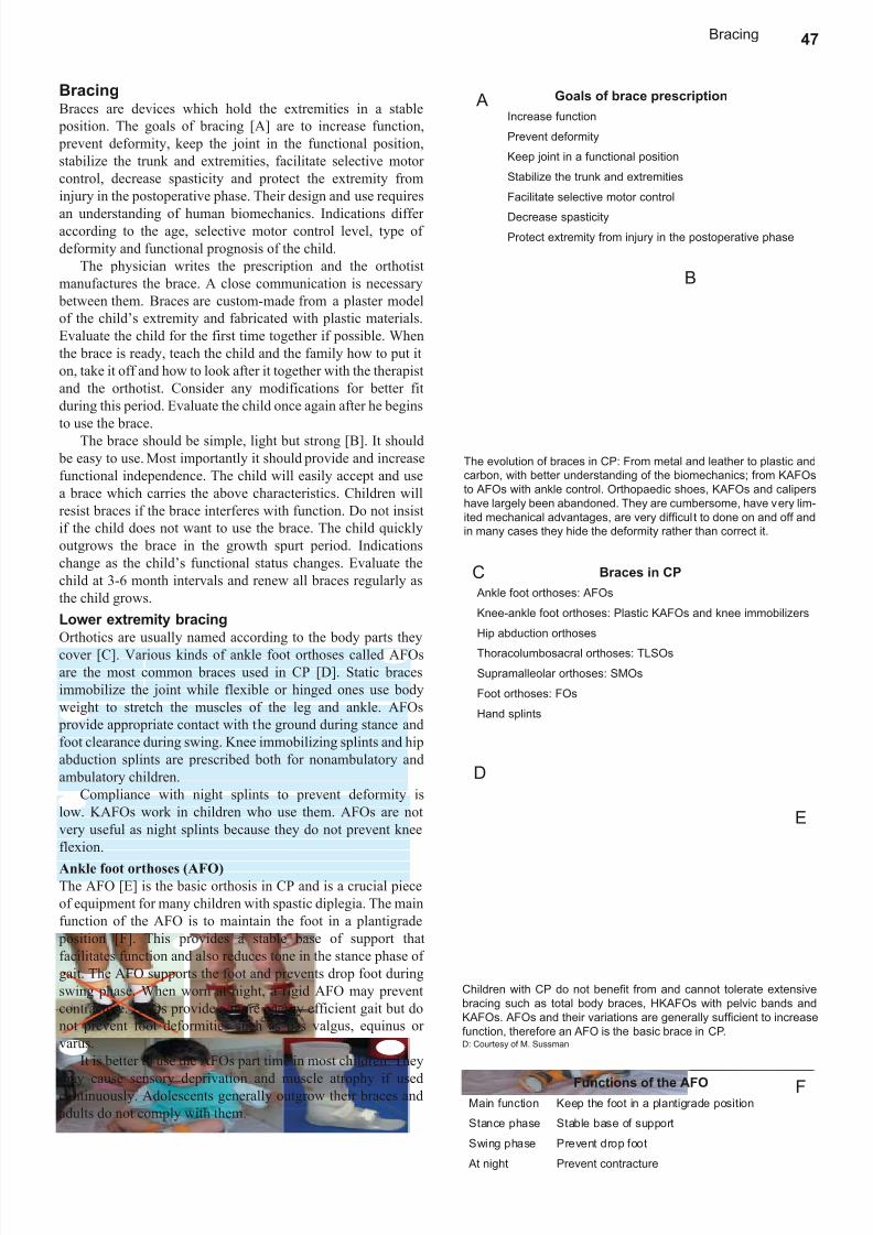
8/9/2019 Guía de ayuda para la parálisis cerebral.pdf
http://slidepdf.com/reader/full/guia-de-ayuda-para-la-paralisis-cerebralpdf 47/143
47Bracing
Goals of brace prescription
Increase function
Prevent deformity
Keep joint in a functional position
Stabilize the trunk and extremities
Facilitate selective motor control
Decrease spasticity
Protect extremity from injury in the postoperative phase
BracingBraces are devices which hold the extremities in a stable
position. The goals of bracing [A] are to increase function,
prevent deformity, keep the joint in the functional position,
stabilize the trunk and extremities, facilitate selective motor
control, decrease spasticity and protect the extremity from
injury in the postoperative phase. Their design and use requires
an understanding of human biomechanics. Indications differaccording to the age, selective motor control level, type of
deformity and functional prognosis of the child.
The physician writes the prescription and the orthotist
manufactures the brace. A close communication is necessary
between them. Braces are custom-made from a plaster model
of the child’s extremity and fabricated with plastic materials.
Evaluate the child for the first time together if possible. When
the brace is ready, teach the child and the family how to put it
on, take it off and how to look after it together with the therapist
and the orthotist. Consider any modifications for better fit
during this period. Evaluate the child once again after he begins
to use the brace.
The brace should be simple, light but strong [B]. It should
be easy to use. Most importantly it should provide and increase
functional independence. The child will easily accept and use
a brace which carries the above characteristics. Children will
resist braces if the brace interferes with function. Do not insist
if the child does not want to use the brace. The child quickly
outgrows the brace in the growth spurt period. Indications
change as the child’s functional status changes. Evaluate the
child at 3-6 month intervals and renew all braces regularly as
the child grows.
Lower extremity bracing
Orthotics are usually named according to the body parts they
cover [C]. Various kinds of ankle foot orthoses called AFOs
are the most common braces used in CP [D]. Static braces
immobilize the joint while flexible or hinged ones use body
weight to stretch the muscles of the leg and ankle. AFOs
provide appropriate contact with the ground during stance and
foot clearance during swing. Knee immobilizing splints and hip
abduction splints are prescribed both for nonambulatory and
ambulatory children.
Compliance with night splints to prevent deformity is
low. KAFOs work in children who use them. AFOs are not
very useful as night splints because they do not prevent knee
flexion.
Ankle foot orthoses (AFO)The AFO [E] is the basic orthosis in CP and is a crucial piece
of equipment for many children with spastic diplegia. The main
function of the AFO is to maintain the foot in a plantigrade
position [F]. This provides a stable base of support that
facilitates function and also reduces tone in the stance phase of
gait. The AFO supports the foot and prevents drop foot during
swing phase. When worn at night, a rigid AFO may prevent
contracture. AFOs provide a more energy efficient gait but do
not prevent foot deformities such as pes valgus, equinus or
varus.
It is better to use the AFOs part time in most children. They
may cause sensory deprivation and muscle atrophy if usedcontinuously. Adolescents generally outgrow their braces and
adults do not comply with them.
Children with CP do not benefit from and cannot tolerate extensive
bracing such as total body braces, HKAFOs with pelvic bands and
KAFOs. AFOs and their variations are generally sufficient to increase
function, therefore an AFO is the basic brace in CP.
Braces in CP
Ankle foot orthoses: AFOs
Knee-ankle foot orthoses: Plastic KAFOs and knee immobilizers
Hip abduction orthoses
Thoracolumbosacral orthoses: TLSOs
Supramalleolar orthoses: SMOs
Foot orthoses: FOs
Hand splints
Functions of the AFOMain function Keep the foot in a plantigrade position
Stance phase Stable base of support
Swing phase Prevent drop foot
At night Prevent contracture
The evolution of braces in CP: From metal and leather to plastic and
carbon, with better understanding of the biomechanics; from KAFOs
to AFOs with ankle control. Orthopaedic shoes, KAFOs and calipers
have largely been abandoned. They are cumbersome, have very lim-
ited mechanical advantages, are very difficult to done on and off and
in many cases they hide the deformity rather than correct it.
FF
EE
DD
CC
B
A
D: Courtesy of M. Sussman
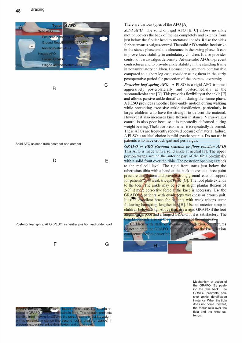
8/9/2019 Guía de ayuda para la parálisis cerebral.pdf
http://slidepdf.com/reader/full/guia-de-ayuda-para-la-paralisis-cerebralpdf 48/143
48 Bracing
Types of AFO
Solid AFO
Posterior leafspring AFO (PLSO)
Ground Reaction AFO (GRAFO)
Antirecurvatum AFO
Hinged AFO
Hinged GRAFO Hinged antirecurvatum AFO
There are various types of the AFO [A].
Solid AFO The solid or rigid AFO [B, C] allows no ankle
motion, covers the back of the leg completely and extends from
just below the fibular head to metatarsal heads. Raise the sides
for better varus-valgus control. The solid AFO enables heel strike
in the stance phase and toe clearance in the swing phase. It can
improve knee stability in ambulatory children. It also provides
control of varus/valgus deformity. Advise solid AFOs to preventcontractures and to provide ankle stability in the standing frame
in nonambulatory children. Because they are more comfortable
compared to a short leg cast, consider using them in the early
postoperative period for protection of the operated extremity.
Posterior leaf spring AFO A PLSO is a rigid AFO trimmed
aggressively posterolaterally and posteromedially at the
supramalleolar area [D]. This provides flexibility at the ankle [E]
and allows passive ankle dorsiflexion during the stance phase.
A PLSO provides smoother knee-ankle motion during walking
while preventing excessive ankle dorsiflexion, particularly in
larger children who have the strength to deform the material.
However it also increases knee flexion in stance. Varus-valgus
control is also poor because it is repeatedly deformed during
weight bearing. The brace breaks when it is repeatedly deformed.
These AFOs are frequently renewed because of material failure.
A PLSO is an ideal choice in mild spastic equinus. Do not use in
patients who have crouch gait and pes valgus.
GRAFO or FRO (Ground reaction or floor reaction AFO)
This AFO is made with a solid ankle at neutral [F]. The upper
portion wraps around the anterior part of the tibia proximally
with a solid front over the tibia. The posterior opening extends
to the malleoli level. The rigid front starts just below the
tuberositas tibia with a band at the back to create a three point
pressure distribution and provide strong ground reaction support
for patients with weak triceps surae [G]. The foot plate extendsto the toes. The ankle may be set in slight plantar flexion of
2-3o if more corrective force at the knee is necessary. Use the
GRAFO in patients with quadriceps weakness or crouch gait.
It is an excellent brace for patients with weak triceps surae
following hamstring lengthening [H]. Use an anterior strap in
children below 15 kg. Above that, use a rigid GRAFO if the foot
alignment is poor and a hinged GRAFO if it is satisfactory. The
benefit depends also on the work quality of the orthotist.
Children with static or dynamic knee flexion contractures
do not tolerate the GRAFO. Surgically release the knee flexion
contracture before prescribing the GRAFO.
Mechanism of action of
the GRAFO: By push-
ing the tibia back, the
GRAFO prevents pas-
sive ankle dorsiflexion
in stance. When the tibiadoes not come forward,
the femur rolls over the
tibia and the knee ex-
tends.
GRAFO as seen from posterior, lateral and anterior. The character-
istic of a GRAFO is the tibia restraint in front. This restraint prevents
the tibia from moving forward as the person starts to put his weight
on his extremity in stance (the second rocker phase of stance) It
prevents excessive ankle dorsiflexion and crouch gait.
Solid AFO as seen from posterior and anterior
Posterior leaf spring AFO (PLSO) in neutral position and under load
EED
CC B
A
HHF GG
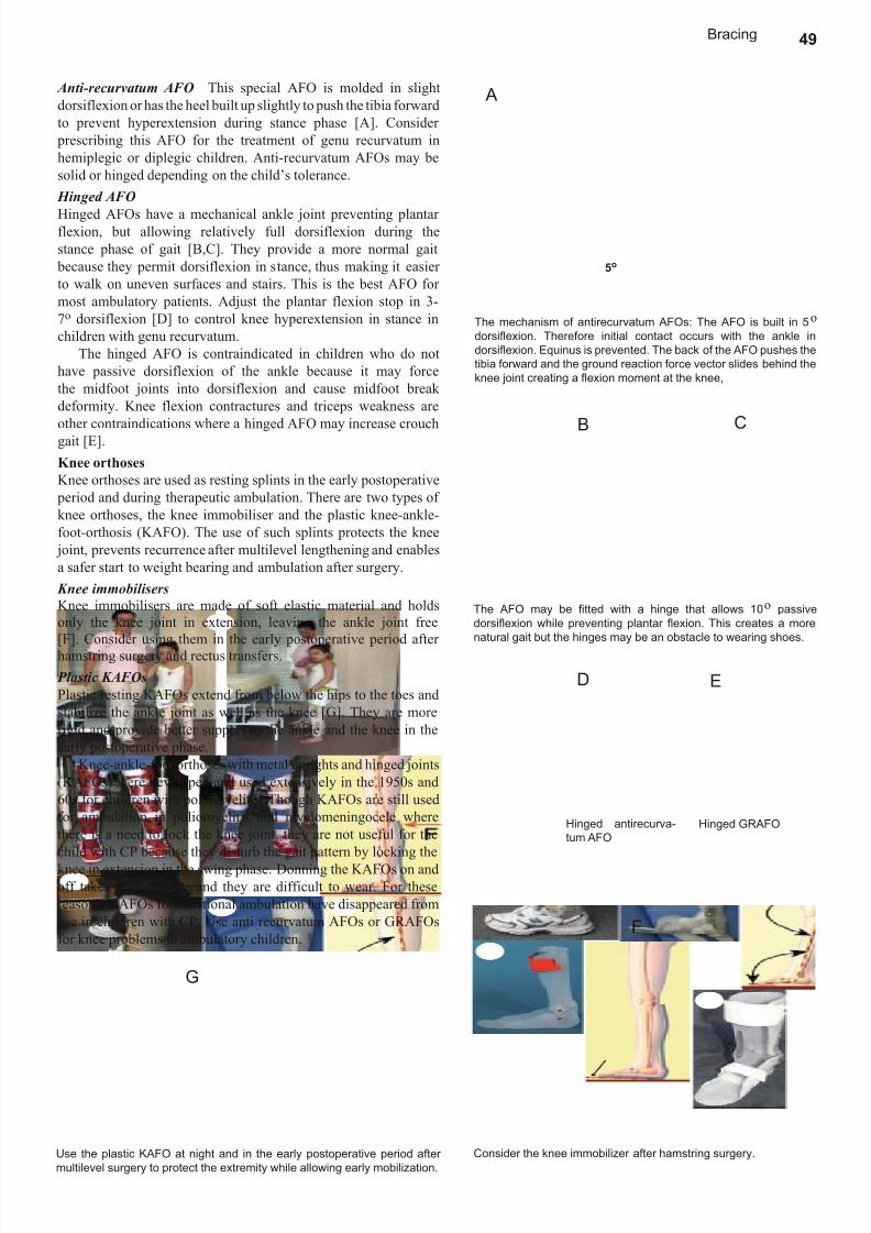
8/9/2019 Guía de ayuda para la parálisis cerebral.pdf
http://slidepdf.com/reader/full/guia-de-ayuda-para-la-paralisis-cerebralpdf 49/143
49Bracing
Anti-recurvatum AFO This special AFO is molded in slight
dorsiflexion or has the heel built up slightly to push the tibia forward
to prevent hyperextension during stance phase [A]. Consider
prescribing this AFO for the treatment of genu recurvatum in
hemiplegic or diplegic children. Anti-recurvatum AFOs may be
solid or hinged depending on the child’s tolerance.
Hinged AFO
Hinged AFOs have a mechanical ankle joint preventing plantarflexion, but allowing relatively full dorsiflexion during the
stance phase of gait [B,C]. They provide a more normal gait
because they permit dorsiflexion in stance, thus making it easier
to walk on uneven surfaces and stairs. This is the best AFO for
most ambulatory patients. Adjust the plantar flexion stop in 3-
7o dorsiflexion [D] to control knee hyperextension in stance in
children with genu recurvatum.
The hinged AFO is contraindicated in children who do not
have passive dorsiflexion of the ankle because it may force
the midfoot joints into dorsiflexion and cause midfoot break
deformity. Knee flexion contractures and triceps weakness are
other contraindications where a hinged AFO may increase crouch
gait [E].
Knee orthoses
Knee orthoses are used as resting splints in the early postoperative
period and during therapeutic ambulation. There are two types of
knee orthoses, the knee immobiliser and the plastic knee-ankle-
foot-orthosis (KAFO). The use of such splints protects the knee
joint, prevents recurrence after multilevel lengthening and enables
a safer start to weight bearing and ambulation after surgery.
Knee immobilisers
Knee immobilisers are made of soft elastic material and holds
only the knee joint in extension, leaving the ankle joint free
[F]. Consider using them in the early postoperative period after
hamstring surgery and rectus transfers.
Plastic KAFOs
Plastic resting KAFOs extend from below the hips to the toes and
stabilize the ankle joint as well as the knee [G]. They are more
rigid and provide better support to the ankle and the knee in the
early postoperative phase.
Knee-ankle-foot orthoses with metal uprights and hinged joints
(KAFOs) were developed and used extensively in the 1950s and
60s for children with poliomyelitis. Though KAFOs are still used
for ambulation in poliomyelitis and myelomeningocele where
there is a need to lock the knee joint, they are not useful for the
child with CP because they disturb the gait pattern by locking the
knee in extension in the swing phase. Donning the KAFOs on and
off takes a lot of time and they are difficult to wear. For these
reasons, KAFOs for functional ambulation have disappeared from
use in children with CP. Use anti recurvatum AFOs or GRAFOs
for knee problems in ambulatory children.
Consider the knee immobilizer after hamstring surgery.
The AFO may be fitted with a hinge that allows 10o passive
dorsiflexion while preventing plantar flexion. This creates a more
natural gait but the hinges may be an obstacle to wearing shoes.
The mechanism of antirecurvatum AFOs: The AFO is built in 5o
dorsiflexion. Therefore initial contact occurs with the ankle in
dorsiflexion. Equinus is prevented. The back of the AFO pushes the
tibia forward and the ground reaction force vector slides behind the
knee joint creating a flexion moment at the knee,
FF
C
CB
A
GG
Use the plastic KAFO at night and in the early postoperative period after
multilevel surgery to protect the extremity while allowing early mobilization.
Hinged GRAFO
EED
Hinged antirecurva-
tum AFO
5o
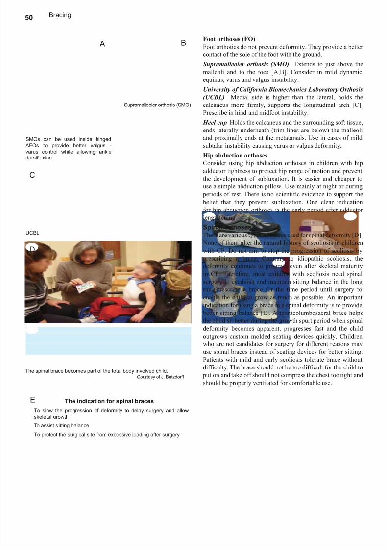
8/9/2019 Guía de ayuda para la parálisis cerebral.pdf
http://slidepdf.com/reader/full/guia-de-ayuda-para-la-paralisis-cerebralpdf 50/143
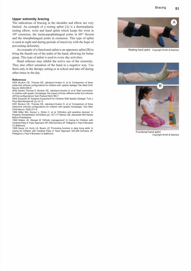
8/9/2019 Guía de ayuda para la parálisis cerebral.pdf
http://slidepdf.com/reader/full/guia-de-ayuda-para-la-paralisis-cerebralpdf 51/143
51Bracing
Upper extremity bracing
The indications of bracing in the shoulder and elbow are very
limited. An example of a resting splint [A] is a thermoplastic
resting elbow, wrist and hand splint which keeps the wrist in
10o extension, the metacarpophalangeal joints in 60o flexion
and the interphalangeal joints in extension. This type of splint
is used at night and during periods of inactivity with the hope of
preventing deformity.An example of a functional splint is an opponens splint [B] to
bring the thumb out of the palm of the hand, allowing for better
grasp. This type of splint is used in every day activities.
Hand orthoses may inhibit the active use of the extremity.
They also effect sensation of the hand in a negative way. Use
them only in the therapy setting or at school and take off during
other times in the day.
References2004 Buckon CE, Thomas SS, Jakobson-Huston S, et al ‘Comparison of three
ankle-foot orthosis configurations for children with spastic diplegia’ Dev Med Child
Neurol. 46(9):590-8
2002 Sienko Thomas S, Buckon CE, Jakobson-Huston S, et al ‘Stair locomotion
in children with spastic hemiplegia: the impact of three different ankle foot orthosis
(AFOs) configurations’ Gait Posture16(2):180-7.
2002 Sussman M ‘Adaptive Equipment For Children With Spastic Diplegia’ Turk J
Phys Med Rehabil 48 (2):12-13
2001 Buckon CE, Thomas SS, Jakobson-Huston S, et al ‘Comparison of three
ankle-foot orthosis configurations for children with spastic hemiplegia.’ Dev Med
Child Neurol. 43(6):371-8.
1999 Miller MA, Koczur L, Strine C, et al ‘Orthotics and assistive devices’ In
Pediatric Rehabilitation 3rd Edition pp: 157-177 Molnar GE, Alexander MA Hanley
Belfus Philadelphia
1998 Walker JS, Stanger M ‘Orthotic management’ In Caring for Children with
Cerebral Palsy A Team Approach 391-426 Dormans JP, Pellegrino L Paul H Brookes
Co Baltimore
1998 Geyer LA, Kurtz LA, Byarm LE ‘Promoting function in daily living skills’ In
Caring for Children with Cerebral Palsy A Team Approach 323-346 Dormans JP,
Pellegrino L Paul H Brookes Co BaltimoreFunctional hand splint
Copyright Smith & Nephew
Resting hand splint Copyright Smith & Nephew
B
A
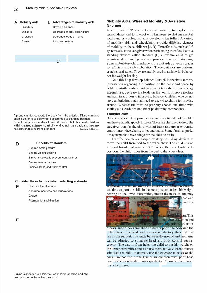
8/9/2019 Guía de ayuda para la parálisis cerebral.pdf
http://slidepdf.com/reader/full/guia-de-ayuda-para-la-paralisis-cerebralpdf 52/143
52 Mobility Aids & Assistive Devices
Mobility Aids, Wheeled Mobility & Assistive
DevicesA child with CP needs to move around, to explore his
surroundings and to interact with his peers so that his mental,
social and psychological skills develop to the fullest. A variety
of mobility aids and wheelchairs provide differing degrees
of mobility to these children [A,B]. Transfer aids such as lift
systems assist the caregiver when performing transfers. Passivestanding devices called standers [C] allow the child to get
accustomed to standing erect and provide therapeutic standing.
Some ambulatory children have to use gait aids as well as braces
for efficient and safe ambulation. These gait aids are walkers,
crutches and canes. They are mainly used to assist with balance,
not for weight bearing.
Gait aids help develop balance. The child receives sensory
information regarding the position of the body and space by
holding onto the walker, crutch or cane. Gait aids decrease energy
expenditure, decrease the loads on the joints, improve posture
and pain in addition to improving balance. Children who do not
have ambulation potential need to use wheelchairs for moving
around. Wheelchairs must be properly chosen and fitted with
seating aids, cushions and other positioning components.
Transfer aids
Different types of lifts provide safe and easy transfer of the older
and heavy handicapped children. These are designed to help the
caregiver transfer the child without trunk and upper extremity
control into wheelchairs, toilet and baths. Some families prefer
lift systems that have slings for the child to sit in.
Transfer boards are simple rotatory or sliding devices to
move the child from bed to the wheelchair. The child sits on
a round board that rotates 360o. When the board rotates to
position, the child slides from the bed to the wheelchair.
Grab bars and rails may be placed around and over the bed
to improve bed mobility.
Standers
Standing in an erect posture contributes to the child with CP
in many ways [D]. It establishes the sense of verticality, helps
develop better eye contact, improves communication and balance
reactions. The pulmonary, cardiovascular, gastrointestinal and
urological system functions all improve by standing. Passive
standers support the child in the erect posture and enable weight
bearing on the lower extremities, stretch the muscles and may
prevent contractures, decrease muscle tone, improve head and
trunk control. There are supine standers, prone standers and the
parapodium [E].
Prone frames
Prone frames support the body and the chest from the front. This
position stretches the hip flexors, providing knee extension and
ankle dorsiflexion. Lateral body supports, hip guides, abductor
blocks, knee blocks and shoe holders support the body and the
extremities. If the head control is not satisfactory, the child may
use a chin support. The angle between the ground and the frame
can be adjusted to stimulate head and body control against
gravity. The tray in front helps the child to put his weight on
the upper extremities and also use them actively. Prone frames
stimulate the child to actively use the extensor muscles of the
back. Do not use prone frames in children with poor headcontrol and increased extensor spasticity. Choose supine frames
in such children.
Mobility aids
Standers
Walkers
Crutches
Canes
Advantages of mobility aids
Develop balance
Decrease energy expenditure
Decrease loads on joints
Improve posture
Benefits of standers
Support erect posture
Enable weight bearingStretch muscles to prevent contractures
Decrease muscle tone
Improve head and trunk control
Consider these factors when selecting a stander
Head and trunk control
Abnormal postures and muscle tone
Growth
Potential for mobilisation
A prone stander supports the body from the anterior. Tilting standers
enable the child to slowly get accustomed to standing position.
Do not use prone standers if the child cannot hold his head. Children
with increased extensor spasticity tend to arch their back and they are
not comfortable in prone standers. Courtesy G. Koloyan
Supine standers are easier to use in large children and chil-
dren who do not have head support.
A
EE
FF
DD
B
CC
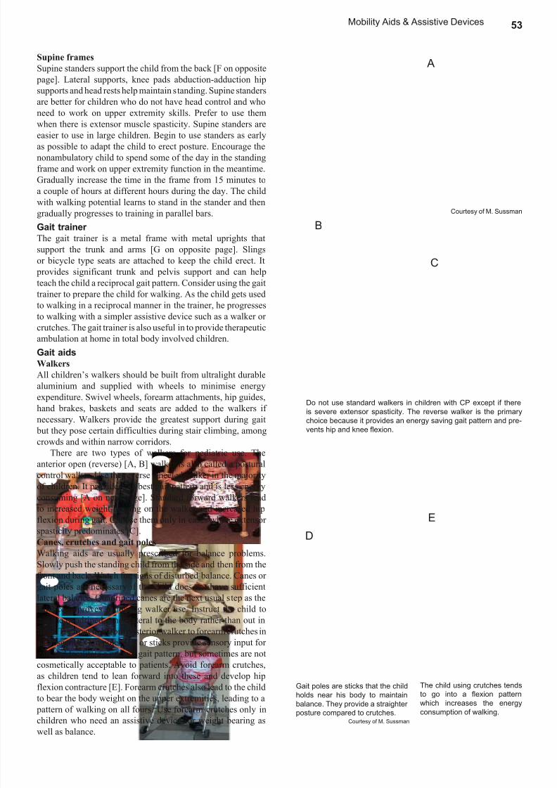
8/9/2019 Guía de ayuda para la parálisis cerebral.pdf
http://slidepdf.com/reader/full/guia-de-ayuda-para-la-paralisis-cerebralpdf 53/143
53Mobility Aids & Assistive Devices
Gait poles are sticks that the child
holds near his body to maintain
balance. They provide a straighter
posture compared to crutches.
Do not use standard walkers in children with CP except if there
is severe extensor spasticity. The reverse walker is the primarychoice because it provides an energy saving gait pattern and pre-
vents hip and knee flexion.
Supine frames
Supine standers support the child from the back [F on opposite
page]. Lateral supports, knee pads abduction-adduction hip
supports and head rests help maintain standing. Supine standers
are better for children who do not have head control and who
need to work on upper extremity skills. Prefer to use them
when there is extensor muscle spasticity. Supine standers are
easier to use in large children. Begin to use standers as earlyas possible to adapt the child to erect posture. Encourage the
nonambulatory child to spend some of the day in the standing
frame and work on upper extremity function in the meantime.
Gradually increase the time in the frame from 15 minutes to
a couple of hours at different hours during the day. The child
with walking potential learns to stand in the stander and then
gradually progresses to training in parallel bars.
Gait trainer
The gait trainer is a metal frame with metal uprights that
support the trunk and arms [G on opposite page]. Slings
or bicycle type seats are attached to keep the child erect. It
provides significant trunk and pelvis support and can helpteach the child a reciprocal gait pattern. Consider using the gait
trainer to prepare the child for walking. As the child gets used
to walking in a reciprocal manner in the trainer, he progresses
to walking with a simpler assistive device such as a walker or
crutches. The gait trainer is also useful in to provide therapeutic
ambulation at home in total body involved children.
Gait aids
Walkers
All children’s walkers should be built from ultralight durable
aluminium and supplied with wheels to minimise energy
expenditure. Swivel wheels, forearm attachments, hip guides,
hand brakes, baskets and seats are added to the walkers if
necessary. Walkers provide the greatest support during gait
but they pose certain difficulties during stair climbing, among
crowds and within narrow corridors.
There are two types of walkers for pediatric use. The
anterior open (reverse) [A, B] walker is also called a postural
control walker. Use the reverse wheeled walker in the majority
of children. It provides the best gait pattern and is less energy
consuming [A on next page]. Standard forward walkers lead
to increased weight bearing on the walker and increased hip
flexion during gait. Choose them only in cases where extensor
spasticity predominates [C].
Canes, crutches and gait poles
Walking aids are usually prescribed for balance problems.Slowly push the standing child from the side and then from the
front and back. Watch for signs of disturbed balance. Canes or
gait poles are necessary if the child does not have sufficient
lateral balance. Quadriped canes are the next usual step as the
balance improves following walker use. Instruct the child to
use the quadriped canes lateral to the body rather than out in
front. Try to switch from posterior walker to forearm crutches in
adolescents. Gait poles [D] or sticks provide sensory input for
gait and facilitate a normal gait pattern, but sometimes are not
cosmetically acceptable to patients. Avoid forearm crutches,
as children tend to lean forward into these and develop hip
flexion contracture [E]. Forearm crutches also lead to the childto bear the body weight on the upper extremities, leading to a
pattern of walking on all fours. Use forearm crutches only in
children who need an assistive device for weight bearing as
well as balance.
Courtesy of M. Sussman
Courtesy of M. Sussman
The child using crutches tends
to go into a flexion pattern
which increases the energy
consumption of walking.
A
EE
CC
DD
B
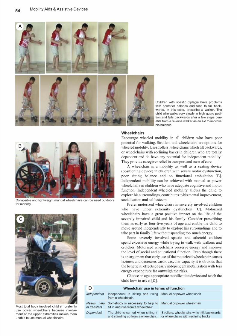
8/9/2019 Guía de ayuda para la parálisis cerebral.pdf
http://slidepdf.com/reader/full/guia-de-ayuda-para-la-paralisis-cerebralpdf 54/143
54 Mobility Aids & Assistive Devices
Collapsible and lightweight manual wheelchairs can be used outdoors
for mobility.
Wheelchairs
Encourage wheeled mobility in all children who have poor
potential for walking. Strollers and wheelchairs are options for
wheeled mobility. Use strollers, wheelchairs which tilt backwards,
or wheelchairs with reclining backs in children who are totally
dependent and do have any potential for independent mobility.
They provide caregiver relief in transport and ease of care.
A wheelchair is a mobility as well as a seating device
(positioning device) in children with severe motor dysfunction,
poor sitting balance and no functional ambulation [B].Independent mobility can be achieved with manual or power
wheelchairs in children who have adequate cognitive and motor
function. Independent wheeled mobility allows the child to
explore his surroundings, contributes to his mental improvement,
socialization and self esteem.
Prefer motorized wheelchairs in severely involved children
who have upper extremity dysfunction [C]. Motorized
wheelchairs have a great positive impact on the life of the
severely impaired child and his family. Consider prescribing
them as early as four-five years of age and enable the child to
move around independently to explore his surroundings and to
take part in family life without spending too much energy.
Some severely involved spastic and athetoid children
spend excessive energy while trying to walk with walkers and
crutches. Motorized wheelchairs preserve energy and improve
the level of social and educational function. Even though there
is an argument that early use of the motorized wheelchair causes
laziness and decreases cardiovascular capacity it is obvious that
the beneficial effects of early independent mobilization with less
energy expenditure far outweigh the risks.
Choose an age-appropriate mobilization device and teach the
child how to use it [D].
Wheelchair use in terms of function
Independent Independent in sitting and risingfrom a wheelchair.
Manual or power wheelchair
Needs help
in transfers
Somebody is necessary to help to
sit in and rise from a wheelchair.
Manual or power wheelchair
Dependent The child is carried when sitting in
and standing up from a wheelchair.
Strollers, wheelchairs which tilt backwards,
or wheelchairs with reclining backs
Children with spastic diplegia have problems
with posterior balance and tend to fall back-
wards. In this case, prescribe a walker. The
child who walks very slowly in high guard posi-
tion and falls backwards after a few steps ben-efits from a reverse walker as an aid to improve
his balance.
Most total body involved children prefer to
use power wheelchairs because involve-
ment of the upper extremities makes them
unable to use manual wheelchairs.
A
CC
DD
B
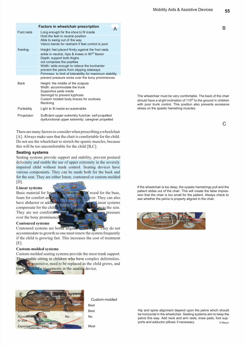
8/9/2019 Guía de ayuda para la parálisis cerebral.pdf
http://slidepdf.com/reader/full/guia-de-ayuda-para-la-paralisis-cerebralpdf 55/143
55Mobility Aids & Assistive Devices
Hip and spine alignment depend upon the pelvis which should
be horizontal in the wheelchair. Seating systems aim to keep the
pelvis this way. Add neck and arm rests, knee pads, foot sup-
ports and adductor pillows if necessary.
If the wheelchair is too deep, the spastic hamstrings pull and the
patient slides out of the chair. This will create the false impres-
sion that the chair is too small for the patient. Always check to
see whether the pelvis is properly aligned in the chair.
Factors in wheelchair prescription
Foot rests Long enough for the shoe to fit inside
Hold the feet in neutral position
Able to swing out of the way
Velcro bands for restraint if feet control is poor
Seating Height: feet placed firmly against the foot rests
ankle in neutral, hips & knees in 90o flexion
Depth: support both thighs
not compress the poplitea
Width: wide enough to relieve the trochanter
prevent the pelvis from slipping sideways
Firmness: to limit of tolerability for maximum stability
prevent pressure sores over the bony prominences
Back Height: the middle of the scapula
Width: accommodate the trunk
Supportive peds inside
Semirigid to prevent kyphosis
Custom molded body braces for scoliosis
Reclining
Portability Light to fit inside an automobile
Propulsion Sufficient upper extremity function: self propelled
dysfunctional upper extremity: caregiver propelled
The wheelchair must be very comfortable. The back of the chair
should have a slight inclination of 110o to the ground in children
with poor trunk control. This position also prevents excessive
stress on the spastic hamstring muscles.
There are many factors to consider when prescribing a wheelchair
[A]. Always make sure that the chair is comfortable for the child.
Do not use the wheelchair to stretch the spastic muscles, because
this will be too uncomfortable for the child [B,C].
Seating systems
Seating systems provide support and stability, prevent postural
deformity and enable the use of upper extremity in the severely
impaired child without trunk control. Seating devices have
various components. They can be made both for the back and
for the seat. They are either linear, contoured or custom-molded
[D].Linear systems
Basic material for linear systems consists of wood for the base,
foam for comfort and pressure relief and a cover. They can also
have abductor or adductor supports at the sides. Linear systems
compensate for the child’s growth and accommodate to the size.
They are not comfortable and insufficient to relieve pressure
over the bony prominences.
Contoured systems
Contoured systems are better aligned to the body. They do not
accommodate to growth so one must renew the system frequently
if the child is growing fast. This increases the cost of treatment
[E].
Custom-molded systems
Custom-molded seating systems provide the most trunk support.
They enable sitting in children who have complex deformities.
They are expensive, need to be replaced as the child grows, and
limit the child’s movements in the seating device.
Seating systems
Linear Contoured Custom-molded
Trunk support Good Better Best
Comfort Good Better Best
Accommodate
to size
Yes No No
Expense Less More Most
A
EE
CC
DD
B
© Meyra
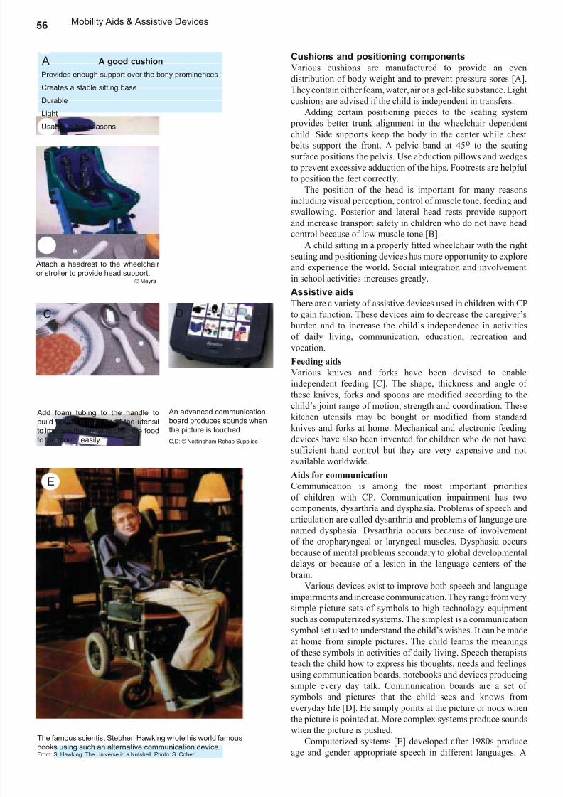
8/9/2019 Guía de ayuda para la parálisis cerebral.pdf
http://slidepdf.com/reader/full/guia-de-ayuda-para-la-paralisis-cerebralpdf 56/143
56 Mobility Aids & Assistive Devices
A good cushion
Provides enough support over the bony prominences
Creates a stable sitting base
Durable
Light
Usable in hot seasons
Add foam tubing to the handle to
build up the grip or bend the utensilto improve the angle to bring the food
to the mouth easily.
The famous scientist Stephen Hawking wrote his world famous
books using such an alternative communication device.From: S. Hawking: The Universe in a Nutshell. Photo: S. Cohen
An advanced communication
board produces sounds whenthe picture is touched.
Attach a headrest to the wheelchair
or stroller to provide head support.
Cushions and positioning componentsVarious cushions are manufactured to provide an even
distribution of body weight and to prevent pressure sores [A].
They contain either foam, water, air or a gel-like substance. Light
cushions are advised if the child is independent in transfers.
Adding certain positioning pieces to the seating system
provides better trunk alignment in the wheelchair dependent
child. Side supports keep the body in the center while chest
belts support the front. A pelvic band at 45o to the seating
surface positions the pelvis. Use abduction pillows and wedges
to prevent excessive adduction of the hips. Footrests are helpful
to position the feet correctly.
The position of the head is important for many reasons
including visual perception, control of muscle tone, feeding and
swallowing. Posterior and lateral head rests provide support
and increase transport safety in children who do not have head
control because of low muscle tone [B].
A child sitting in a properly fitted wheelchair with the right
seating and positioning devices has more opportunity to explore
and experience the world. Social integration and involvement
in school activities increases greatly.Assistive aidsThere are a variety of assistive devices used in children with CP
to gain function. These devices aim to decrease the caregiver’s
burden and to increase the child’s independence in activities
of daily living, communication, education, recreation and
vocation.
Feeding aids
Various knives and forks have been devised to enable
independent feeding [C]. The shape, thickness and angle of
these knives, forks and spoons are modified according to the
child’s joint range of motion, strength and coordination. These
kitchen utensils may be bought or modified from standardknives and forks at home. Mechanical and electronic feeding
devices have also been invented for children who do not have
sufficient hand control but they are very expensive and not
available worldwide.
Aids for communication
Communication is among the most important priorities
of children with CP. Communication impairment has two
components, dysarthria and dysphasia. Problems of speech and
articulation are called dysarthria and problems of language are
named dysphasia. Dysarthria occurs because of involvement
of the oropharyngeal or laryngeal muscles. Dysphasia occurs
because of mental problems secondary to global developmental
delays or because of a lesion in the language centers of the
brain.
Various devices exist to improve both speech and language
impairments and increase communication. They range from very
simple picture sets of symbols to high technology equipment
such as computerized systems. The simplest is a communication
symbol set used to understand the child’s wishes. It can be made
at home from simple pictures. The child learns the meanings
of these symbols in activities of daily living. Speech therapists
teach the child how to express his thoughts, needs and feelings
using communication boards, notebooks and devices producing
simple every day talk. Communication boards are a set of
symbols and pictures that the child sees and knows fromeveryday life [D]. He simply points at the picture or nods when
the picture is pointed at. More complex systems produce sounds
when the picture is pushed.
Computerized systems [E] developed after 1980s produce
age and gender appropriate speech in different languages. A
EE
DD
A
CC
B
© Meyra
C,D: © Nottingham Rehab Supplies
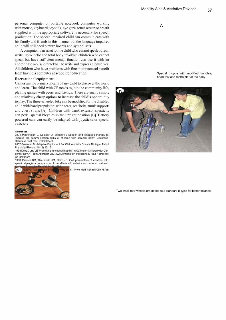
8/9/2019 Guía de ayuda para la parálisis cerebral.pdf
http://slidepdf.com/reader/full/guia-de-ayuda-para-la-paralisis-cerebralpdf 57/143
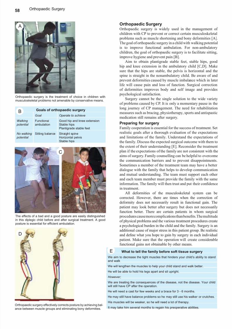
8/9/2019 Guía de ayuda para la parálisis cerebral.pdf
http://slidepdf.com/reader/full/guia-de-ayuda-para-la-paralisis-cerebralpdf 58/143
58 Orthopaedic Surgery
Orthopaedic SurgeryOrthopaedic surgery is widely used in the management of
children with CP to prevent or correct certain musculoskeletal
problems such as muscle shortening and bony deformities [A].
The goal of orthopaedic surgery in a child with walking potential
is to improve functional ambulation. For non-ambulatory
children, the goal of orthopaedic surgery is to facilitate sitting,
improve hygiene and prevent pain [B].Aim to obtain plantigrade stable feet, stable hips, good
hip and knee extension in the ambulatory child [C,D]. Make
sure that the hips are stable, the pelvis is horizontal and the
spine is straight in the nonambulatory child. Be aware of and
prevent deformities caused by muscle imbalance which in later
life will cause pain and loss of function. Surgical correction
of deformities improves body and self image and provides
psychological satisfaction.
Surgery cannot be the single solution to the wide variety
of problems caused by CP. It is only a momentary pause in the
long journey of CP management. The need for rehabilitation
measures such as bracing, physiotherapy, sports and antispastic
medication still remains after surgery.
Preparing for surgery
Family cooperation is essential for the success of treatment. Set
realistic goals after a thorough evaluation of the expectations
and limitations of the family. Understand the expectations of
the family. Discuss the expected surgical outcome with them to
the extent of their understanding [E]. Reconsider the treatment
plan if the expectations of the family are not consistent with the
aims of surgery. Family counselling can be helpful to overcome
the communication barriers and to prevent disappointments.
Sometimes a member of the treatment team may have a better
dialogue with the family that helps to develop communication
and mutual understanding. The team must support each other
and each team member must provide the family with the same
information. The family will then trust and put their confidence
in treatment.
All deformities of the musculoskeletal system can be
corrected. However, there are times when the correction of
deformity does not necessarily result in functional gain. The
patient may look better after surgery but does not necessarily
function better. There are certain patients in whom surgical
procedures cause more complications than benefits. The multitude
of physical problems and the various treatment procedures create
a psychological burden in the child and the family. Surgery is an
additional cause of major stress in this patient group. Be realisticand define what you hope to gain by surgery in each individual
patient. Make sure that the operation will create considerable
functional gains not obtainable by other means.
Orthopaedic surgery is the treatment of choice in children with
musculoskeletal problems not amenable by conservative means.
The effects of a bad and a good posture are easily distinguished
in this diplegic child before and after surgical treatment. A good
posture is essential for efficient ambulation.
Goals of orthopaedic surgery
Goal Operate to achieve
Walking
potential
Functional
ambulation
Good hip and knee extension
Stable hips
Plantigrade stable feet
No walking
potential
Sitting balance Straight spine
Horizontal pelvis
Stable hips
A
B
CC
DD
Orthopaedic surgery effectively corrects posture by achieving bal-
ance between muscle groups and eliminating bony deformities.
What to tell the family before soft tissue surgery
We aim to decrease the tight muscles that hinders your child’s ability to stand
and walk
We will lengthen the muscles to help your child stand and walk better.
He will be able to hold his legs apart and sit upright.
However;
We are treating the consequences of the disease, not the disease. Your child
will still have CP after the operation.
He will need a cast for few weeks and a brace for 3 - 6 months.
He may still have balance problems so he may still use his walker or crutches.
His muscles will be weaker, so he will need a lot of therapy.
It may take him several months to regain his preoperative abilities.
EE
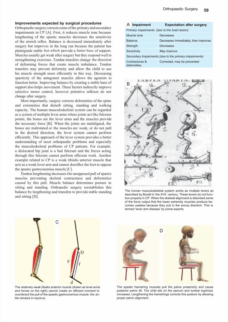
8/9/2019 Guía de ayuda para la parálisis cerebral.pdf
http://slidepdf.com/reader/full/guia-de-ayuda-para-la-paralisis-cerebralpdf 59/143
59Orthopaedic Surgery
Impairment Expectation after surgery
Primary impairments (due to the brain lesion)
Muscle tone Decreases
Balance Decreases immediately, then improves
Strength Decreases
Selectivity May improve
Secondary Impairments (due to the primary impairments)
Contractures &
deformities
Corrected, may be prevented
The human musculoskeletal system works as multiple levers as
described by Borelli in the XVII. century. These levers do not func-
tion properly in CP. When the skeletal alignment is disturbed some
of the force output that the lower extremity muscles produce be-
comes useless because they pull in the wrong direction. This is
termed ‘lever arm disease’ by some experts.
Improvements expected by surgical procedures
Orthopaedic surgery corrects some of the primary and secondary
impairments in CP [A]. First, it reduces muscle tone because
lengthening of the spastic muscles decreases the sensitivity
of the stretch reflex. Balance is decreased immediately after
surgery but improves in the long run because the patient has
plantigrade stable feet which provide a better base of support.
Muscles usually get weak after surgery but they respond well tostrengthening exercises. Tendon transfers change the direction
of deforming forces that create muscle imbalance. Tendon
transfers may prevent deformity and allow the child to use
his muscle strength more efficiently in this way. Decreasing
spasticity of the antagonist muscles allows the agonists to
function better. Improving balance by creating a stable base of
support also helps movement. These factors indirectly improve
selective motor control, however primitive reflexes do not
change after surgery.
Most importantly, surgery corrects deformities of the spine
and extremities that disturb sitting, standing and walking
capacity. The human musculoskeletal system can be regarded
as a system of multiple lever arms where joints act like fulcrum
points, the bones are the lever arms and the muscles provide
the necessary force [B]. When the joints are malaligned, the
bones are malrotated or the muscles are weak, or do not pull
in the desired direction, the lever system cannot perform
efficiently. This approach of the lever system provides a better
understanding of most orthopaedic problems and especially
the musculoskeletal problems of CP patients. For example,
a dislocated hip joint is a bad fulcrum and the forces acting
through this fulcrum cannot perform efficient work. Another
example related to CP is a weak tibialis anterior muscle that
acts as a weak lever arm and cannot dorsiflex the foot to oppose
the spastic gastrocnemius muscle [C].Tendon lengthening decreases the unopposed pull of spastic
muscles preventing skeletal contractures and deformities
caused by this pull. Muscle balance determines posture in
sitting and standing. Orthopedic surgery reestablishes this
balance by lengthening and transfers to provide stable standing
and sitting [D].
A
B
The spastic hamstring muscles pull the pelvis posteriorly and cause
posterior pelvic tilt. The child sits on the sacrum and lumbar kyphosis
increases. Lengthening the hamstrings corrects this posture by allowing
proper pelvic alignment.
DD
CC
f F
d
D
The relatively weak tibialis anterior muscle (drawn as level arms
and forces on the right) cannot create an efficient moment to
counteract the pull of the spastic gastrocnemius muscle, the an-
kle remains in equinus.
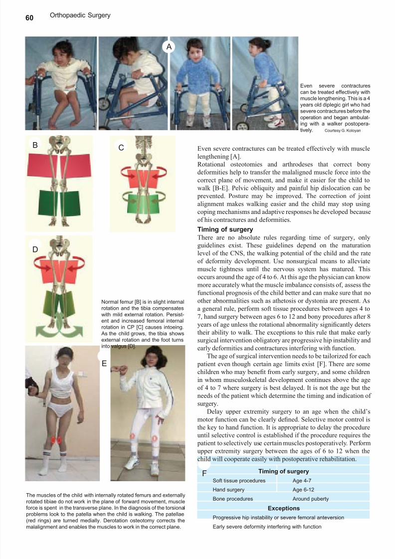
8/9/2019 Guía de ayuda para la parálisis cerebral.pdf
http://slidepdf.com/reader/full/guia-de-ayuda-para-la-paralisis-cerebralpdf 60/143
60 Orthopaedic Surgery
Timing of surgery
Soft tissue procedures Age 4-7
Hand surgery Age 6-12
Bone procedures Around puberty
Exceptions
Progressive hip instability or severe femoral anteversion
Early severe deformity interfering with function
Even severe contractures can be treated effectively with muscle
lengthening [A].
Rotational osteotomies and arthrodeses that correct bony
deformities help to transfer the malaligned muscle force into the
correct plane of movement, and make it easier for the child to
walk [B-E]. Pelvic obliquity and painful hip dislocation can be
prevented. Posture may be improved. The correction of jointalignment makes walking easier and the child may stop using
coping mechanisms and adaptive responses he developed because
of his contractures and deformities.
Timing of surgery
There are no absolute rules regarding time of surgery, only
guidelines exist. These guidelines depend on the maturation
level of the CNS, the walking potential of the child and the rate
of deformity development. Use nonsurgical means to alleviate
muscle tightness until the nervous system has matured. This
occurs around the age of 4 to 6. At this age the physician can know
more accurately what the muscle imbalance consists of, assess the
functional prognosis of the child better and can make sure that noother abnormalities such as athetosis or dystonia are present. As
a general rule, perform soft tissue procedures between ages 4 to
7, hand surgery between ages 6 to 12 and bony procedures after 8
years of age unless the rotational abnormality significantly deters
their ability to walk. The exceptions to this rule that make early
surgical intervention obligatory are progressive hip instability and
early deformities and contractures interfering with function.
The age of surgical intervention needs to be tailorized for each
patient even though certain age limits exist [F]. There are some
children who may benefit from early surgery, and some children
in whom musculoskeletal development continues above the age
of 4 to 7 where surgery is best delayed. It is not the age but the
needs of the patient which determine the timing and indication of
surgery.
Delay upper extremity surgery to an age when the child’s
motor function can be clearly defined. Selective motor control is
the key to hand function. It is appropriate to delay the procedure
until selective control is established if the procedure requires the
patient to selectively use certain muscles postoperatively. Perform
upper extremity surgery between the ages of 6 to 12 when the
child will cooperate easily with postoperative rehabilitation.
FF
Normal femur [B] is in slight internal
rotation and the tibia compensates
with mild external rotation. Persist-
ent and increased femoral internal
rotation in CP [C] causes intoeing.
As the child grows, the tibia shows
external rotation and the foot turns
into valgus [D].
The muscles of the child with internally rotated femurs and externally
rotated tibiae do not work in the plane of forward movement, muscle
force is spent in the transverse plane. In the diagnosis of the torsional
problems look to the patella when the child is walking. The patellae
(red rings) are turned medially. Derotation osteotomy corrects the
malalignment and enables the muscles to work in the correct plane.
BCC
DD
A
EE
Even severe contractures
can be treated effectively with
muscle lengthening. This is a 4years old diplegic girl who had
severe contractures before the
operation and began ambulat-
ing with a walker postopera-
tively. Courtesy G. Koloyan
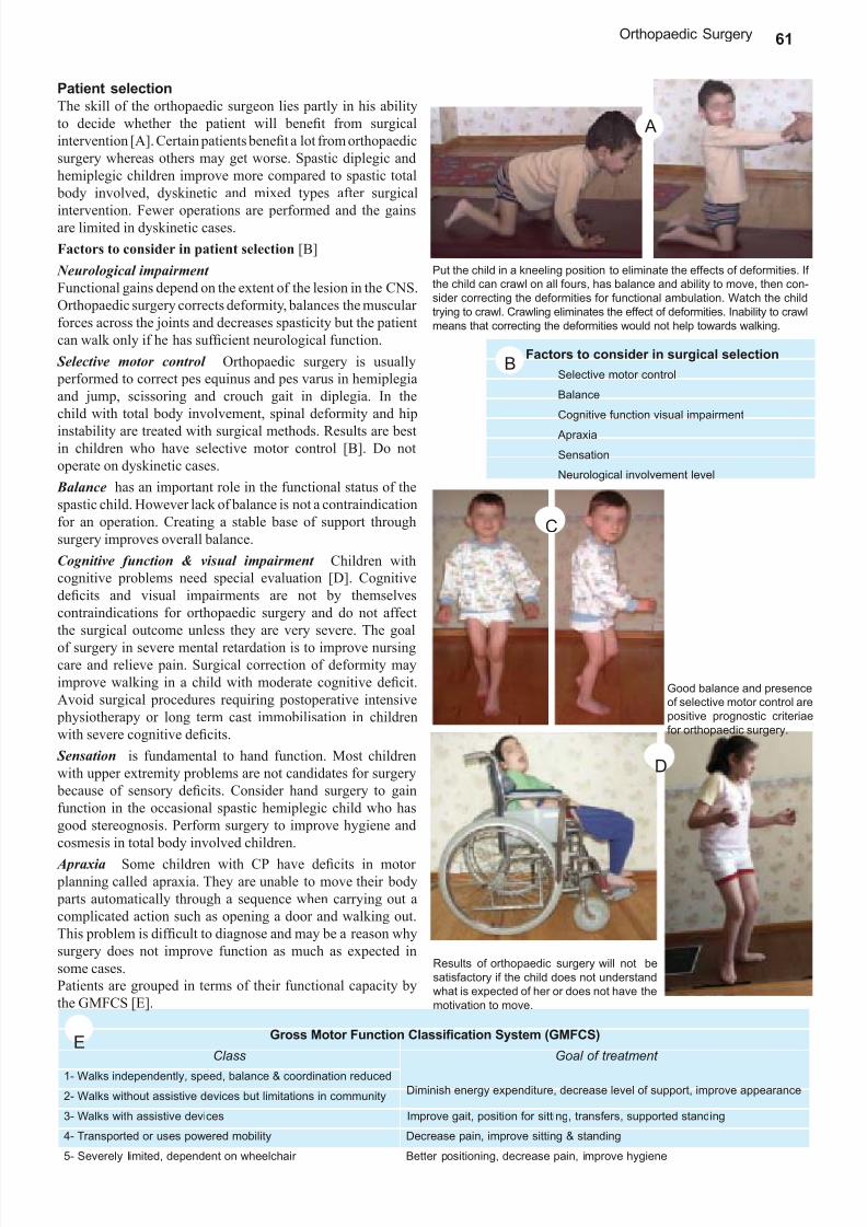
8/9/2019 Guía de ayuda para la parálisis cerebral.pdf
http://slidepdf.com/reader/full/guia-de-ayuda-para-la-paralisis-cerebralpdf 61/143
61Orthopaedic Surgery
Put the child in a kneeling position to eliminate the effects of deformities. If
the child can crawl on all fours, has balance and ability to move, then con-
sider correcting the deformities for functional ambulation. Watch the child
trying to crawl. Crawling eliminates the effect of deformities. Inability to crawl
means that correcting the deformities would not help towards walking.
A
Patient selection
The skill of the orthopaedic surgeon lies partly in his ability
to decide whether the patient will benefit from surgical
intervention [A]. Certain patients benefit a lot from orthopaedic
surgery whereas others may get worse. Spastic diplegic and
hemiplegic children improve more compared to spastic total
body involved, dyskinetic and mixed types after surgical
intervention. Fewer operations are performed and the gainsare limited in dyskinetic cases.
Factors to consider in patient selection [B]
Neurological impairment
Functional gains depend on the extent of the lesion in the CNS.
Orthopaedic surgery corrects deformity, balances the muscular
forces across the joints and decreases spasticity but the patient
can walk only if he has sufficient neurological function.
Selective motor control Orthopaedic surgery is usually
performed to correct pes equinus and pes varus in hemiplegia
and jump, scissoring and crouch gait in diplegia. In the
child with total body involvement, spinal deformity and hip
instability are treated with surgical methods. Results are bestin children who have selective motor control [B]. Do not
operate on dyskinetic cases.
Balance has an important role in the functional status of the
spastic child. However lack of balance is not a contraindication
for an operation. Creating a stable base of support through
surgery improves overall balance.
Cognitive function & visual impairment Children with
cognitive problems need special evaluation [D]. Cognitive
deficits and visual impairments are not by themselves
contraindications for orthopaedic surgery and do not affect
the surgical outcome unless they are very severe. The goal
of surgery in severe mental retardation is to improve nursingcare and relieve pain. Surgical correction of deformity may
improve walking in a child with moderate cognitive deficit.
Avoid surgical procedures requiring postoperative intensive
physiotherapy or long term cast immobilisation in children
with severe cognitive deficits.
Sensation is fundamental to hand function. Most children
with upper extremity problems are not candidates for surgery
because of sensory deficits. Consider hand surgery to gain
function in the occasional spastic hemiplegic child who has
good stereognosis. Perform surgery to improve hygiene and
cosmesis in total body involved children.
Apraxia Some children with CP have deficits in motor planning called apraxia. They are unable to move their body
parts automatically through a sequence when carrying out a
complicated action such as opening a door and walking out.
This problem is difficult to diagnose and may be a reason why
surgery does not improve function as much as expected in
some cases.
Patients are grouped in terms of their functional capacity by
the GMFCS [E].
Factors to consider in surgical selection
Selective motor control
Balance
Cognitive function visual impairment
Apraxia
Sensation
Neurological involvement level
Gross Motor Function Classification System (GMFCS)
Class Goal of treatment
1- Walks independently, speed, balance & coordination reducedDiminish energy expenditure, decrease level of support, improve appearance
2- Walks without assistive devices but limitations in community
3- Walks with assistive devices Improve gait, position for sitting, transfers, supported standing
4- Transported or uses powered mobility Decrease pain, improve sitting & standing
5- Severely limited, dependent on wheelchair Better positioning, decrease pain, improve hygiene
DD
B
Good balance and presence
of selective motor control are
positive prognostic criteriae
for orthopaedic surgery.
Results of orthopaedic surgery will not be
satisfactory if the child does not understand
what is expected of her or does not have the
motivation to move.
CC
EE
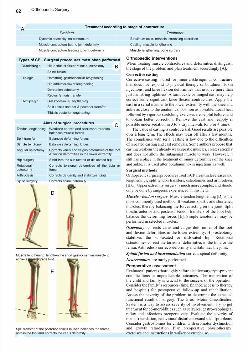
8/9/2019 Guía de ayuda para la parálisis cerebral.pdf
http://slidepdf.com/reader/full/guia-de-ayuda-para-la-paralisis-cerebralpdf 62/143
62 Orthopaedic Surgery
Orthopaedic interventions
When treating muscle contractures and deformities distinguish
the stage of the problem and plan treatment accordingly [A].
Corrective casting
Corrective casting is used for minor ankle equinus contracture
that does not respond to physical therapy or botulinum toxin
injections; and knee flexion deformities that involve more than
just hamstring tightness. A turnbuckle or hinged cast may help
correct some significant knee flexion contractures. Apply the
cast in a serial manner to the lower extremity with the knee and
ankle as close to the anatomical position as possible. Local heat
followed by vigorous stretching exercises are helpful beforehand
to obtain better correction. Remove the cast and reapply if
possible under sedation in 3 to 7 day intervals for 3 or 4 times.The value of casting is controversial. Good results are possible
over a long term. The effects may wear off after a few months.
The compliance with serial casting is low due to the difficulties
of repeated casting and cast removals. Some authors propose that
casting weakens the already weak spastic muscles, creates atrophy
and does not allow the antagonist muscle to work. However, it
still has a place in the treatment of minor deformities of the knee
and ankle. It is used after botulinum toxin injections as well.
Surgical methods
Orthopaedic surgical procedures used in CP are muscle releases and
lengthenings, split tendon transfers, osteotomies and arthrodeses
[B,C]. Upper extremity surgery is much more complex and shouldonly be done by surgeons experienced in this field.
Muscle - tendon surgery Muscle-tendon lengthening [D] is the
most commonly used method. It weakens spastic and shortened
muscles, thereby balancing the forces acting on the joint. Split
tibialis anterior and posterior tendon transfers of the foot help
balance the deforming forces [E]. Simple tenotomies may be
performed in selected muscles.
Osteotomy corrects varus and valgus deformities of the foot
and flexion deformities in the lower extremity. Hip osteotomy
stabilizes the subluxated or dislocated hip. Rotational
osteotomies correct the torsional deformities in the tibia or the
femur. Arthrodesis corrects deformity and stabilizes the joint.
Spinal fusion and instrumentation corrects spinal deformity.
Neurectomies are rarely performed.
Preoperative assessment
Evaluate all patients thoroughly before elective surgery to preventcomplications or unpredictable outcomes. The motivation ofthe child and family is crucial to the success of the operation.Consider the family’s resources (time, finance, access to therapyand hospital) for postoperative follow-up and rehabilitation.Assess the severity of the problem to determine the expected
functional result of surgery. The Gross Motor ClassificationSystem is a way to assess severity of involvement. Try to get
treatment for co-morbidities such as seizures, gastro-esophagealreflux and infections preoperatively. Evaluate the severity ofmental retardation, behavioural disturbances and social problems.Consider gastrostomies for children with oromotor dysfunctionand growth retardation. Plan preoperative physiotherapy,
exercises and instructions in walker or crutch use.
Types of CP Surgical procedures most often performedQuadriplegic Hip adductor flexor release, osteotomy
Spine fusion
Diplegic Hamstring-gastrocnemius lengthening
Hip adductor-flexor lengthening
Derotation osteotomy
Rectus femoris transfer
Hemiplegic Gastrocnemius lengthening
Split tibialis anterior & posterior transfer
Tibialis posterior lengthening
Aims of surgical proceduresTendon lengthening Weakens spastic and shortened muscles,
balances muscle forces
Split transfer Balances deforming forces
Simple tenotomy Balances deforming forces
Angular osteotomy Corrects varus and valgus deformities of the foot
& flexion deformities in the lower extremity
Hip surgery Stabil izes the subluxated or dislocated hip
Rotational
osteotomy
Corrects torsional deformities of the tibia or
femur
Arthrodesis Corrects deformity and stabilizes joints
Spine surgery Corrects spinal deformity
B
CC
EE
Muscle lengthening: lengthen the short gastrocnemius muscle to
achieve a plantigrade foot.
Split transfer of the posterior tibialis muscle balances the forces
across the foot and corrects the varus deformity.
Treatment according to stage of contracture
Problem Treatment
Dynamic spasticity, no contracture Botulinum toxin, orthosis, stretching exercises
Muscle contracture but no joint deformity Casting, muscle lengthening
Muscle contracture leading to joint deformity Muscle lengthening, bone surgery
A
DD
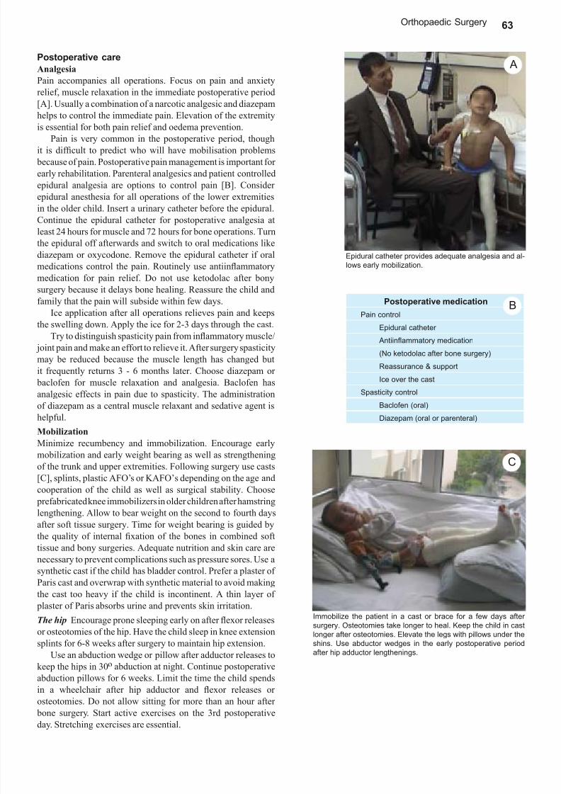
8/9/2019 Guía de ayuda para la parálisis cerebral.pdf
http://slidepdf.com/reader/full/guia-de-ayuda-para-la-paralisis-cerebralpdf 63/143
63Orthopaedic Surgery
A
Epidural catheter provides adequate analgesia and al-
lows early mobilization.
Postoperative care
Analgesia
Pain accompanies all operations. Focus on pain and anxiety
relief, muscle relaxation in the immediate postoperative period
[A]. Usually a combination of a narcotic analgesic and diazepam
helps to control the immediate pain. Elevation of the extremity
is essential for both pain relief and oedema prevention.
Pain is very common in the postoperative period, thoughit is difficult to predict who will have mobilisation problems
because of pain. Postoperative pain management is important for
early rehabilitation. Parenteral analgesics and patient controlled
epidural analgesia are options to control pain [B]. Consider
epidural anesthesia for all operations of the lower extremities
in the older child. Insert a urinary catheter before the epidural.
Continue the epidural catheter for postoperative analgesia at
least 24 hours for muscle and 72 hours for bone operations. Turn
the epidural off afterwards and switch to oral medications like
diazepam or oxycodone. Remove the epidural catheter if oral
medications control the pain. Routinely use antiinflammatory
medication for pain relief. Do not use ketodolac after bonysurgery because it delays bone healing. Reassure the child and
family that the pain will subside within few days.
Ice application after all operations relieves pain and keeps
the swelling down. Apply the ice for 2-3 days through the cast.
Try to distinguish spasticity pain from inflammatory muscle/
joint pain and make an effort to relieve it. After surgery spasticity
may be reduced because the muscle length has changed but
it frequently returns 3 - 6 months later. Choose diazepam or
baclofen for muscle relaxation and analgesia. Baclofen has
analgesic effects in pain due to spasticity. The administration
of diazepam as a central muscle relaxant and sedative agent is
helpful.Mobilization
Minimize recumbency and immobilization. Encourage early
mobilization and early weight bearing as well as strengthening
of the trunk and upper extremities. Following surgery use casts
[C], splints, plastic AFO’s or KAFO’s depending on the age and
cooperation of the child as well as surgical stability. Choose
prefabricated knee immobilizers in older children after hamstring
lengthening. Allow to bear weight on the second to fourth days
after soft tissue surgery. Time for weight bearing is guided by
the quality of internal fixation of the bones in combined soft
tissue and bony surgeries. Adequate nutrition and skin care are
necessary to prevent complications such as pressure sores. Use asynthetic cast if the child has bladder control. Prefer a plaster of
Paris cast and overwrap with synthetic material to avoid making
the cast too heavy if the child is incontinent. A thin layer of
plaster of Paris absorbs urine and prevents skin irritation.
The hip Encourage prone sleeping early on after flexor releases
or osteotomies of the hip. Have the child sleep in knee extension
splints for 6-8 weeks after surgery to maintain hip extension.
Use an abduction wedge or pillow after adductor releases to
keep the hips in 30o abduction at night. Continue postoperative
abduction pillows for 6 weeks. Limit the time the child spends
in a wheelchair after hip adductor and flexor releases or
osteotomies. Do not allow sitting for more than an hour after
bone surgery. Start active exercises on the 3rd postoperative
day. Stretching exercises are essential.
Immobilize the patient in a cast or brace for a few days after
surgery. Osteotomies take longer to heal. Keep the child in cast
longer after osteotomies. Elevate the legs with pillows under the
shins. Use abductor wedges in the early postoperative period
after hip adductor lengthenings.
Postoperative medication
Pain control
Epidural catheter
Antiinflammatory medication
(No ketodolac after bone surgery)
Reassurance & support
Ice over the cast
Spasticity control
Baclofen (oral)
Diazepam (oral or parenteral)
B
CC
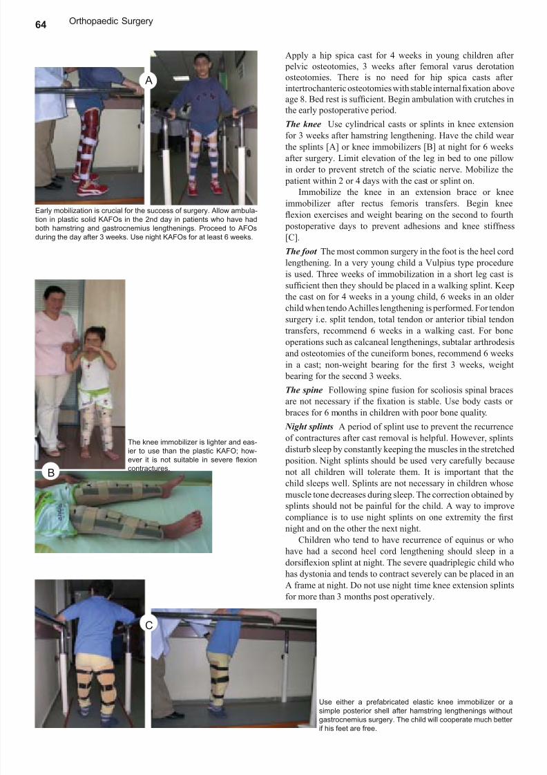
8/9/2019 Guía de ayuda para la parálisis cerebral.pdf
http://slidepdf.com/reader/full/guia-de-ayuda-para-la-paralisis-cerebralpdf 64/143
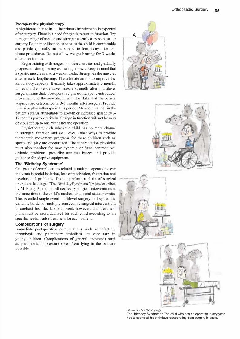
8/9/2019 Guía de ayuda para la parálisis cerebral.pdf
http://slidepdf.com/reader/full/guia-de-ayuda-para-la-paralisis-cerebralpdf 65/143
65Orthopaedic Surgery
The ‘Birthday Syndrome’: The child who has an operation every year
has to spend all his birthdays recuperating from surgery in casts.
A
Illustration by Idil Çilingiroğlu
Postoperative physiotherapy
A significant change in all the primary impairments is expected
after surgery. There is a need for gentle return to function. Try
to regain range of motion and strength as early as possible after
surgery. Begin mobilisation as soon as the child is comfortable
and painless, usually on the second to fourth day after soft
tissue procedures. Do not allow weight bearing for 3 weeks
after osteotomies.Begin training with range of motion exercises and gradually
progress to strengthening as healing allows. Keep in mind that
a spastic muscle is also a weak muscle. Strengthen the muscles
after muscle lengthening. The ultimate aim is to improve the
ambulatory capacity. It usually takes approximately 3 months
to regain the preoperative muscle strength after multilevel
surgery. Immediate postoperative physiotherapy re-introduces
movement and the new alignment. The skills that the patient
acquires are established in 3-6 months after surgery. Provide
intensive physiotherapy in this period. Monitor changes in the
patient’s status attributable to growth or increased spasticity 6-
12 months postoperatively. Change in function will not be veryobvious for up to one year after the operation.
Physiotherapy ends when the child has no more change
in strength, function and skill level. Other ways to provide
therapeutic movement programs for these children such as
sports and play are encouraged. The rehabilitation physician
must also monitor for new dynamic or fixed contractures,
orthotic problems, prescribe accurate braces and provide
guidance for adaptive equipment.
The ‘Birthday Syndrome’
One group of complications related to multiple operations over
the years is social isolation, loss of motivation, frustration and
psychosocial problems. Do not perform a chain of surgicaloperations leading to ‘The Birthday Syndrome’ [A] as described
by M. Rang. Plan to do all necessary surgical interventions at
the same time if the child’s medical and social status permits.
This is called single event multilevel surgery and spares the
child the burden of multiple consecutive surgical interventions
throughout his life. Do not forget, however, that treatment
plans must be individualized for each child according to his
specific needs. Tailor treatment for each patient.
Complications of surgery
Immediate postoperative complications such as infection,
thrombosis and pulmonary embolism are very rare in
young children. Complications of general anesthesia suchas pneumonia or pressure sores from lying in the bed are
possible.
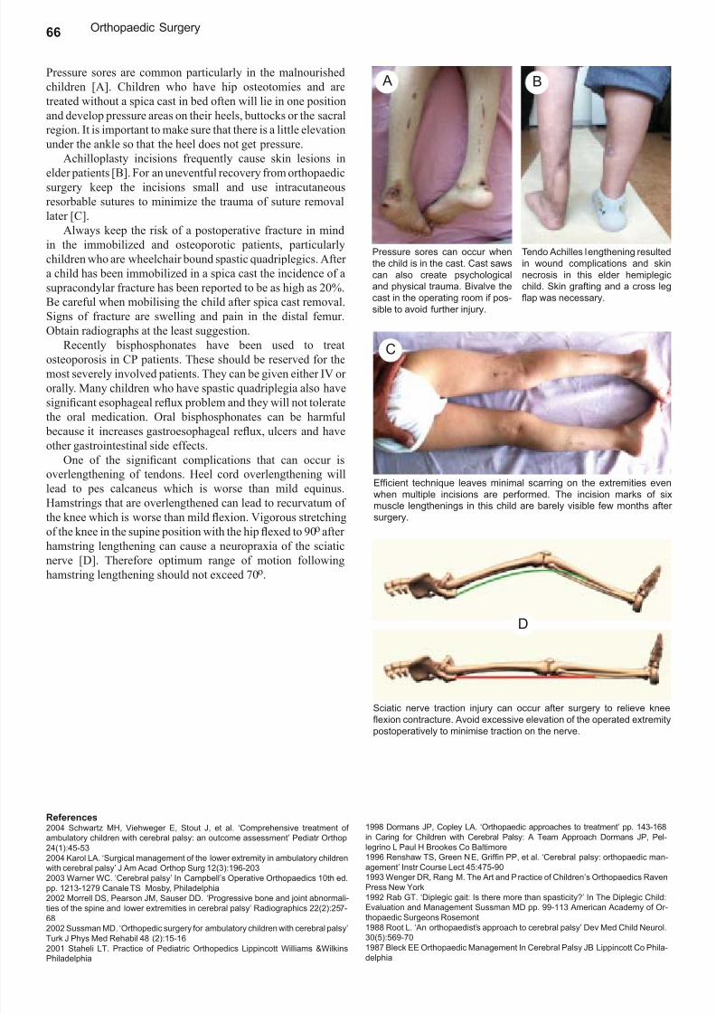
8/9/2019 Guía de ayuda para la parálisis cerebral.pdf
http://slidepdf.com/reader/full/guia-de-ayuda-para-la-paralisis-cerebralpdf 66/143
66 Orthopaedic Surgery
Pressure sores can occur when
the child is in the cast. Cast saws
can also create psychological
and physical trauma. Bivalve the
cast in the operating room if pos-
sible to avoid further injury.
Tendo Achilles lengthening resulted
in wound complications and skin
necrosis in this elder hemiplegic
child. Skin grafting and a cross leg
flap was necessary.
A B
Efficient technique leaves minimal scarring on the extremities even
when multiple incisions are performed. The incision marks of sixmuscle lengthenings in this child are barely visible few months after
surgery.
C
C
Sciatic nerve traction injury can occur after surgery to relieve kneeflexion contracture. Avoid excessive elevation of the operated extremity
postoperatively to minimise traction on the nerve.
DD
References2004 Schwartz MH, Viehweger E, Stout J, et al. ‘Comprehensive treatment of
ambulatory children with cerebral palsy: an outcome assessment’ Pediatr Orthop
24(1):45-53
2004 Karol LA. ‘Surgical management of the lower extremity in ambulatory children
with cerebral palsy’ J Am Acad Orthop Surg 12(3):196-203
2003 Warner WC. ‘Cerebral palsy’ In Campbell’s Operative Orthopaedics 10th ed.
pp. 1213-1279 Canale TS Mosby, Philadelphia2002 Morrell DS, Pearson JM, Sauser DD. ‘Progressive bone and joint abnormali-
ties of the spine and lower extremities in cerebral palsy’ Radiographics 22(2):257-
68
2002 Sussman MD. ‘Orthopedic surgery for ambulatory children with cerebral palsy’
Turk J Phys Med Rehabil 48 (2):15-16
2001 Staheli LT. Practice of Pediatric Orthopedics Lippincott Williams &Wilkins
Philadelphia
1998 Dormans JP, Copley LA. ‘Orthopaedic approaches to treatment’ pp. 143-168
in Caring for Children with Cerebral Palsy: A Team Approach Dormans JP, Pel-
legrino L Paul H Brookes Co Baltimore
1996 Renshaw TS, Green NE, Griffin PP, et al. ‘Cerebral palsy: orthopaedic man-
agement’ Instr Course Lect 45:475-90
1993 Wenger DR, Rang M. The Art and Practice of Children’s Orthopaedics Raven
Press New York1992 Rab GT. ‘Diplegic gait: Is there more than spasticity?’ In The Diplegic Child:
Evaluation and Management Sussman MD pp. 99-113 American Academy of Or-
thopaedic Surgeons Rosemont
1988 Root L. ‘An orthopaedist’s approach to cerebral palsy’ Dev Med Child Neurol.
30(5):569-70
1987 Bleck EE Orthopaedic Management In Cerebral Palsy JB Lippincott Co Phila-
delphia
Pressure sores are common particularly in the malnourished
children [A]. Children who have hip osteotomies and are
treated without a spica cast in bed often will lie in one position
and develop pressure areas on their heels, buttocks or the sacral
region. It is important to make sure that there is a little elevation
under the ankle so that the heel does not get pressure.
Achilloplasty incisions frequently cause skin lesions in
elder patients [B]. For an uneventful recovery from orthopaedicsurgery keep the incisions small and use intracutaneous
resorbable sutures to minimize the trauma of suture removal
later [C].
Always keep the risk of a postoperative fracture in mind
in the immobilized and osteoporotic patients, particularly
children who are wheelchair bound spastic quadriplegics. After
a child has been immobilized in a spica cast the incidence of a
supracondylar fracture has been reported to be as high as 20%.
Be careful when mobilising the child after spica cast removal.
Signs of fracture are swelling and pain in the distal femur.
Obtain radiographs at the least suggestion.
Recently bisphosphonates have been used to treat
osteoporosis in CP patients. These should be reserved for the
most severely involved patients. They can be given either IV or
orally. Many children who have spastic quadriplegia also have
significant esophageal reflux problem and they will not tolerate
the oral medication. Oral bisphosphonates can be harmful
because it increases gastroesophageal reflux, ulcers and have
other gastrointestinal side effects.
One of the significant complications that can occur is
overlengthening of tendons. Heel cord overlengthening will
lead to pes calcaneus which is worse than mild equinus.
Hamstrings that are overlengthened can lead to recurvatum of
the knee which is worse than mild flexion. Vigorous stretching
of the knee in the supine position with the hip flexed to 90o afterhamstring lengthening can cause a neuropraxia of the sciatic
nerve [D]. Therefore optimum range of motion following
hamstring lengthening should not exceed 70o.
8/9/2019 Guía de ayuda para la parálisis cerebral.pdf
http://slidepdf.com/reader/full/guia-de-ayuda-para-la-paralisis-cerebralpdf 67/143
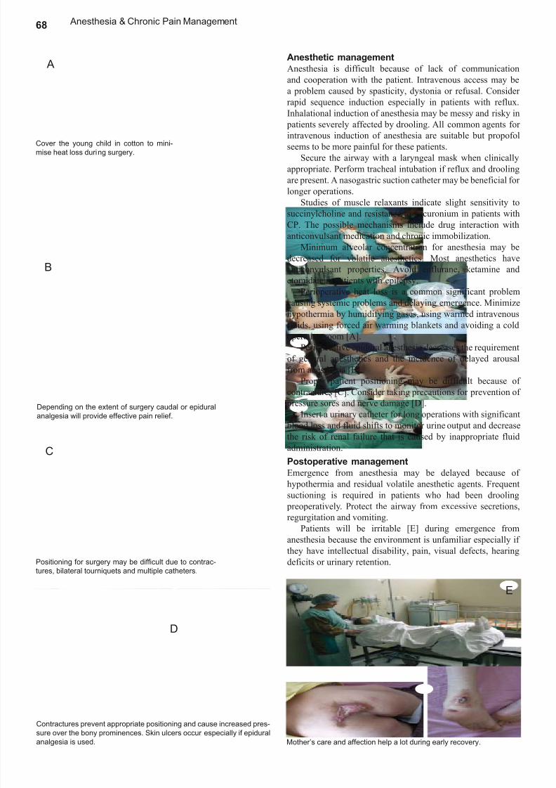
8/9/2019 Guía de ayuda para la parálisis cerebral.pdf
http://slidepdf.com/reader/full/guia-de-ayuda-para-la-paralisis-cerebralpdf 68/143
68 Anesthesia & Chronic Pain Management
Cover the young child in cotton to mini-
mise heat loss during surgery.
Anesthetic management
Anesthesia is difficult because of lack of communication
and cooperation with the patient. Intravenous access may be
a problem caused by spasticity, dystonia or refusal. Consider
rapid sequence induction especially in patients with reflux.
Inhalational induction of anesthesia may be messy and risky in
patients severely affected by drooling. All common agents for
intravenous induction of anesthesia are suitable but propofolseems to be more painful for these patients.
Secure the airway with a laryngeal mask when clinically
appropriate. Perform tracheal intubation if reflux and drooling
are present. A nasogastric suction catheter may be beneficial for
longer operations.
Studies of muscle relaxants indicate slight sensitivity to
succinylcholine and resistance to vecuronium in patients with
CP. The possible mechanisms include drug interaction with
anticonvulsant medication and chronic immobilization.
Minimum alveolar concentration for anesthesia may be
decreased for volatile anesthetics. Most anesthetics have
anticonvulsant properties. Avoid enflurane, ketamine and
etomidate in patients with epilepsy.
Perioperative heat loss is a common significant problem
causing systemic problems and delaying emergence. Minimize
hypothermia by humidifying gases, using warmed intravenous
fluids, using forced air warming blankets and avoiding a cold
operating room [A].
Perioperative epidural anesthesia decreases the requirement
of general anesthetics and the incidence of delayed arousal
from anesthesia [B].
Proper patient positioning may be difficult because of
contractures [C]. Consider taking precautions for prevention of
pressure sores and nerve damage [D].
Insert a urinary catheter for long operations with significant blood loss and fluid shifts to monitor urine output and decrease
the risk of renal failure that is caused by inappropriate fluid
administration.
Postoperative management
Emergence from anesthesia may be delayed because of
hypothermia and residual volatile anesthetic agents. Frequent
suctioning is required in patients who had been drooling
preoperatively. Protect the airway from excessive secretions,
regurgitation and vomiting.
Patients will be irritable [E] during emergence from
anesthesia because the environment is unfamiliar especially if
they have intellectual disability, pain, visual defects, hearingdeficits or urinary retention.Positioning for surgery may be difficult due to contrac-
tures, bilateral tourniquets and multiple catheters.
CC
B
A
Depending on the extent of surgery caudal or epidural
analgesia will provide effective pain relief.
Contractures prevent appropriate positioning and cause increased pres-
sure over the bony prominences. Skin ulcers occur especially if epidural
analgesia is used. Mother’s care and affection help a lot during early recovery.
DD
EE
8/9/2019 Guía de ayuda para la parálisis cerebral.pdf
http://slidepdf.com/reader/full/guia-de-ayuda-para-la-paralisis-cerebralpdf 69/143
69 Anesthesia & Chronic Pain Management
Differential diagnosis of chronic pain and restlessness in
the quadriplegic patient
Etiology Symptom Treatment
Hip sub-
luxation
Pain during transfers
Night pain
Less with position
change
Analgesics (paracetamol),
NSAID, intraarticular steroids
Surgery
Reflux Anemia Antacids, ranitidine
Weight loss Proton pump inhibitors
Anorexia Erect posture, proper sitting
position
Low back
pain
Pain after sitting for a
longtime
Physiotherapy
Night pain Tricyclic antidepressants
Paracetamol, NSAID,
Facet joint injections
Wheelchair modifications
Menses
pain
During menses Oral contraceptives
Continue intrathecal baclofen during the perioperative period
to prevent abrupt withdrawal symptoms such as convulsions,
hallucinations and dyskinesias. Maintain anticonvulsant
medication for children with epilepsy. During the early
postoperative period diazepam can be administered rectally,
sodium valproate may be given IV in countries where this
preparation is available.
Decreased mobility, inefficient fluid intake and loss of peristaltism increase the tendency to develop constipation. Oral,
intravenous or epidural opioids contribute to this problem.
Pain and spasticity
Focus on relieving pain, anxiety and muscle spasm. Postoperative
pain management is crucial for the success of surgery. Epidural
analgesia diminishes both postoperative pain and muscle
spasm. It is accepted as the gold standard for pain management
in patients with CP. Epidural administration of opioid and local
anesthetic combination (bupivacaine+fentanyl) is the most
effective and safest method for postoperative analgesia because
it causes less sedation. Add clonidine to achieve better control
of muscle spasm.Epidural analgesia has certain advantages and dysadvantages
[A,B]. A common side effect of epidural analgesia is urinary
retention. Insert a Foley catheter preoperatively to prevent
postoperative urination problems. Leave the epidural catheter
in place for 2 to 3 days after soft tissue procedures and 4 to 5
days in cases of bone surgery. Turn off the infusion and start
oral medications such as diazepam or oxycodone after 5 days.
Remove the epidural catheter if oral medications control the
pain. For the young child who has minor soft tissue surgery or
simple hip plate removal, it is not necessary to use an epidural
catheter. A postoperative caudal block will allow such patients
to emerge from anesthesia without pain.
Nonsteroidal antiinflammatory analgesics or paracetamol isthe second choice after epidural analgesia. Rectal paracetamol
(40 mg/kg loading dose, then 20 mg/kg/ four times daily)
will relieve postoperative pain in most cases. If not sufficient,
consider parenteral opioids (morphine, tramodol and
meperidine).
Postoperative muscle spasms are a common and serious
problem. Epidural analgesia is advantageous in children
undergoing lower extremity surgery because it decreases reflex
muscle spasms triggered by pain. For patients in whom epidural
analgesia is not appropriate, benzodiazepin (diazepam 0.1- 0.2
mg/kg or midazolam 0.05 - 0.07 mg/kg ) administered orally,
rectally, or intravenously can be used to alleviate spasm butoften results in prolonged sedation.
An alternative drug for the management of postoperative
spasticity is oral baclofen. Baclofen has analgesic effects on
pain caused by spasticity.
Chronic pain management
Chronic pain results from hip instability, gastro-esophageal
reflux, menstrual discomfort in adolescent girls, back pain
caused by scoliosis, facet joint arthritis, capsulitis and spasticity.
It is difficult to determine the cause of and treat chronic
pain in the child who has a communication disorder [C]. A
multidisciplinary approach is necessary with the team members
ready for consultation, evaluation and treatment on the sameday if possible. Depression during the adolescent period can
increase chronic pain. Patients with CP are undertreated for
pain because of various reasons [D].
Dysadvantages of epidural analgesia
Urinary retention
Side effects of opioids
(Itching, nausea, vomiting)
Increased risk of pressure ulcers or compartment syndrome
Complications
Spinal hematoma
Nerve root damage
InfectionDural puncture
Advantages of epidural analgesia
The most effective analgesia
Decreased muscle spasm
Early mobilization
Decreased pulmonary complications
Decreased risk of deep venous thrombosis
(not a major issue in the young child)
Causes of undertreatment for pain
Too many medical problems that may cause pain
Frequent painful procedures
Behavioural problems that mask expression of pain
Difficult to interpret pain indicators because of physical problems
Comfort of the patients valued less by society
CC
B
A
DD
8/9/2019 Guía de ayuda para la parálisis cerebral.pdf
http://slidepdf.com/reader/full/guia-de-ayuda-para-la-paralisis-cerebralpdf 70/143
70 Anesthesia & Chronic Pain Management
References2002 Hadden KL, von Baeyer CL. ‘Pain in children with cerebral palsy: common
triggers and expressive behaviors’ Pain 99: 281-288.
2002 Wongprasartsuk P, Stevens J. ‘Cerebral palsy and anaesthesia’ Paed
Anaesth 12:296-303.
2000 Nolan J, Chalkiadis GA, Low J, et al ‘Anaesthesia and pain management in
cerebral palsy’ Anaesthesia 55: 32-41.
1999 Ershov VL, Ostreikov IF. ‘Complications of anesthesia and their prevention
in children with spastic cerebral palsy during ambulatory surgery’ Anesteziol
Reanimatol 4: 33-35.
1999 Malviya S, Pandit UA, Merkel S, et al ‘A comparison of continuous epiduralinfusion and intermittent intravenous bolus doses of morphine in children undergoing
selective dorsal rhizotomy’ Reg Anesth Pain Med 24: 438-443.
1998 Brenn BR, Brislin RP, Rose JB. ‘Epidural analgesia in children with cerebral
palsy’ Can J Anaesth 45: 1156-1161.
1998 McGrath PJ, Rosmus C, Camfield C, et al ‘Behaviors care givers use to
determine pain in non-verbal, cognitively impaired children’ Dev Med Child Neurol
40: 340-343.
1997 Brett EM, Scrutton D. ‘Cerebral palsy, perinatal injury to the spinal cord and
brachial plexus birth injury’ In: Brett ED, ed. Paediatric Neurology Textbook. 291-
331 Churchill Livingstone, New York
1996 DeLuca PA ‘The musculoskeletal management of children with cerebral palsy’
Ped Clin North Am 5:1135-1151
1996 Landwehr LP, Boguniewicz M. ‘Current perspective on latex allergy’ J Paediatr
128: 305-312.
1995 Antognini JF, Gronert GA. ‘Succinylcholine sensitivity in cerebral palsy’
Anesth Analg 80: 1248-1253.
1994 Geiduschek JM, Haberkern CM, McLaughlin JF, et al ‘Pain management for
children following selective dorsal rhizotomy’ Can J Anaesth 41: 492-496.1991 Moorthy SS, Krishna G, Dierdorf S. ‘Resistance to vecuronium in patients
with cerebral palsy’ Anesth Analg 73: 275-277.
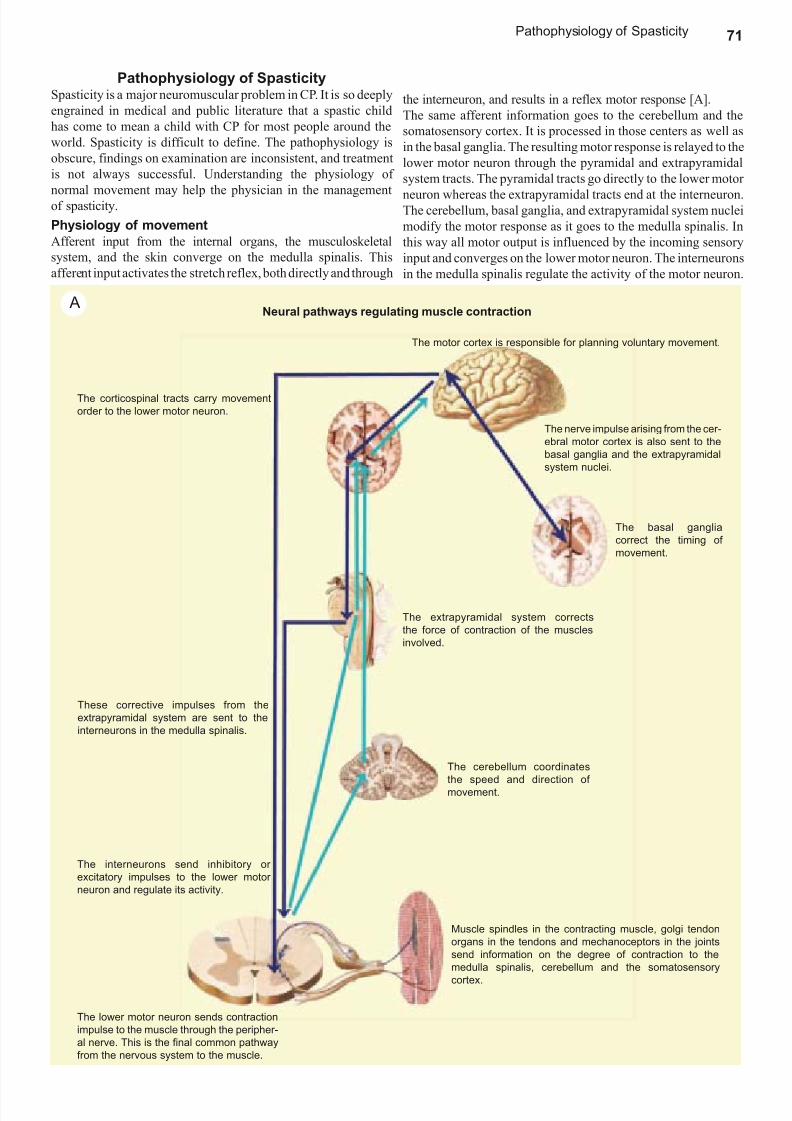
8/9/2019 Guía de ayuda para la parálisis cerebral.pdf
http://slidepdf.com/reader/full/guia-de-ayuda-para-la-paralisis-cerebralpdf 71/143
71Pathophysiology of Spasticity
The motor cortex is responsible for planning voluntary movement.
The corticospinal tracts carry movement
order to the lower motor neuron.
The lower motor neuron sends contraction
impulse to the muscle through the peripher-
al nerve. This is the final common pathway
from the nervous system to the muscle.
The nerve impulse arising from the cer-
ebral motor cortex is also sent to the
basal ganglia and the extrapyramidal
system nuclei.
Muscle spindles in the contracting muscle, golgi tendon
organs in the tendons and mechanoceptors in the joints
send information on the degree of contraction to the
medulla spinalis, cerebellum and the somatosensory
cortex.
The cerebellum coordinates
the speed and direction of
movement.
The basal ganglia
correct the timing of
movement.
The extrapyramidal system corrects
the force of contraction of the muscles
involved.
These corrective impulses from the
extrapyramidal system are sent to the
interneurons in the medulla spinalis.
The interneurons send inhibitory or
excitatory impulses to the lower motor
neuron and regulate its activity.
Pathophysiology of SpasticitySpasticity is a major neuromuscular problem in CP. It is so deeplyengrained in medical and public literature that a spastic childhas come to mean a child with CP for most people around theworld. Spasticity is difficult to define. The pathophysiology isobscure, findings on examination are inconsistent, and treatmentis not always successful. Understanding the physiology of
normal movement may help the physician in the managementof spasticity.
Physiology of movement
Afferent input from the internal organs, the musculoskeletalsystem, and the skin converge on the medulla spinalis. Thisafferent input activates the stretch reflex, both directly and through
Neural pathways regulating muscle contraction A
the interneuron, and results in a reflex motor response [A].The same afferent information goes to the cerebellum and thesomatosensory cortex. It is processed in those centers as well asin the basal ganglia. The resulting motor response is relayed to thelower motor neuron through the pyramidal and extrapyramidalsystem tracts. The pyramidal tracts go directly to the lower motor
neuron whereas the extrapyramidal tracts end at the interneuron.The cerebellum, basal ganglia, and extrapyramidal system nucleimodify the motor response as it goes to the medulla spinalis. Inthis way all motor output is influenced by the incoming sensoryinput and converges on the lower motor neuron. The interneuronsin the medulla spinalis regulate the activity of the motor neuron.
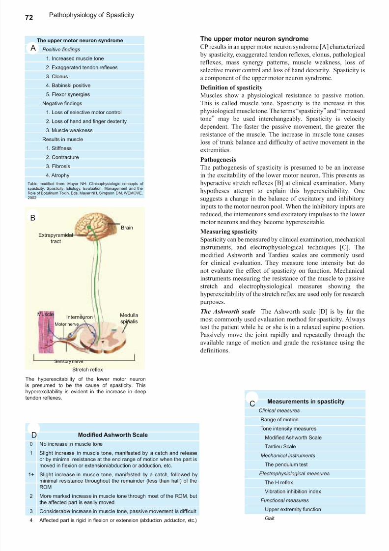
8/9/2019 Guía de ayuda para la parálisis cerebral.pdf
http://slidepdf.com/reader/full/guia-de-ayuda-para-la-paralisis-cerebralpdf 72/143
72 Pathophysiology of Spasticity
Measurements in spasticity
Clinical measures
Range of motion
Tone intensity measures
Modified Ashworth Scale
Tardieu Scale
Mechanical instruments
The pendulum test
Electrophysiological measures
The H reflex
Vibration inhibition index
Functional measures
Upper extremity function
Gait
The upper motor neuron syndrome
CP results in an upper motor neuron syndrome [A] characterized by spasticity, exaggerated tendon reflexes, clonus, pathologicalreflexes, mass synergy patterns, muscle weakness, loss ofselective motor control and loss of hand dexterity. Spasticity isa component of the upper motor neuron syndrome.
Definition of spasticity
Muscles show a physiological resistance to passive motion.This is called muscle tone. Spasticity is the increase in this physiological muscle tone. The terms “spasticity” and “increasedtone” may be used interchangeably. Spasticity is velocitydependent. The faster the passive movement, the greater theresistance of the muscle. The increase in muscle tone causesloss of trunk balance and difficulty of active movement in theextremities.
Pathogenesis
The pathogenesis of spasticity is presumed to be an increasein the excitability of the lower motor neuron. This presents ashyperactive stretch reflexes [B] at clinical examination. Many
hypotheses attempt to explain this hyperexcitability. Onesuggests a change in the balance of excitatory and inhibitoryinputs to the motor neuron pool. When the inhibitory inputs arereduced, the interneurons send excitatory impulses to the lowermotor neurons and they become hyperexcitable.
Measuring spasticity
Spasticity can be measured by clinical examination, mechanicalinstruments, and electrophysiological techniques [C]. Themodified Ashworth and Tardieu scales are commonly usedfor clinical evaluation. They measure tone intensity but donot evaluate the effect of spasticity on function. Mechanicalinstruments measuring the resistance of the muscle to passivestretch and electrophysiological measures showing thehyperexcitability of the stretch reflex are used only for research purposes.
The Ashworth scale The Ashworth scale [D] is by far themost commonly used evaluation method for spasticity. Alwaystest the patient while he or she is in a relaxed supine position.Passively move the joint rapidly and repeatedly through theavailable range of motion and grade the resistance using thedefinitions.
The upper motor neuron syndrome
Positive findings
1. Increased muscle tone
2. Exaggerated tendon reflexes
3. Clonus
4. Babinski positive
5. Flexor synergiesNegative findings
1. Loss of selective motor control
2. Loss of hand and finger dexterity
3. Muscle weakness
Results in muscle
1. Stiffness
2. Contracture
3. Fibrosis
4. Atrophy
Table modified from: Mayer NH: Clinicophysiologic concepts of
spasticity, Spasticity: Etiology, Evaluation, Management and the
Role of Botulinum Toxin. Eds. Mayer NH, Simpson DM, WEMOVE,2002
The hyperexcitability of the lower motor neuron
is presumed to be the cause of spasticity. Thishyperexcitability is evident in the increase in deep
tendon reflexes.
Brain
Extrapyramidal
tract
Muscle
Stretch reflex
Medulla
spinalisInterneuron
Sensory nerve
Motor nerve
CC
B
A
Modified Ashworth Scale
0 No increase in muscle tone
1 Slight increase in muscle tone, manifested by a catch and release
or by minimal resistance at the end range of motion when the part is
moved in flexion or extension/abduction or adduction, etc.
1+ Slight increase in muscle tone, manifested by a catch, followed by
minimal resistance throughout the remainder (less than half) of theROM
2 More marked increase in muscle tone through most of the ROM, but
the affected part is easily moved
3 Considerable increase in muscle tone, passive movement is difficult
4 Affected part is rigid in flexion or extension (abduction ,adduction, etc.)
DD
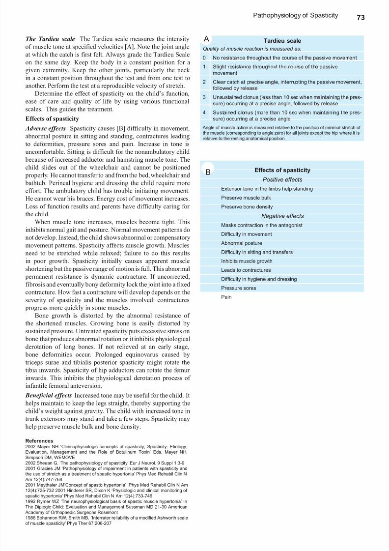
8/9/2019 Guía de ayuda para la parálisis cerebral.pdf
http://slidepdf.com/reader/full/guia-de-ayuda-para-la-paralisis-cerebralpdf 73/143
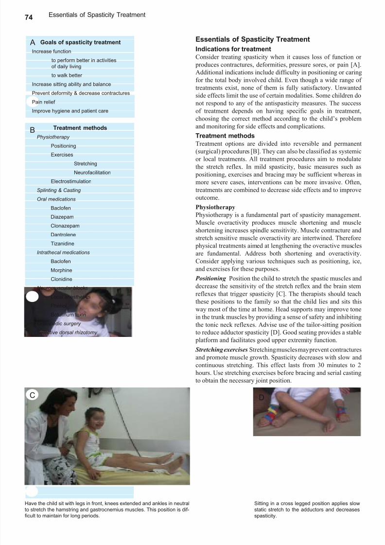
8/9/2019 Guía de ayuda para la parálisis cerebral.pdf
http://slidepdf.com/reader/full/guia-de-ayuda-para-la-paralisis-cerebralpdf 74/143
74 Essentials of Spasticity Treatment
Essentials of Spasticity Treatment
Indications for treatment
Consider treating spasticity when it causes loss of function or produces contractures, deformities, pressure sores, or pain [A].Additional indications include difficulty in positioning or caringfor the total body involved child. Even though a wide range oftreatments exist, none of them is fully satisfactory. Unwanted
side effects limit the use of certain modalities. Some children donot respond to any of the antispasticity measures. The successof treatment depends on having specific goals in treatment,choosing the correct method according to the child’s problemand monitoring for side effects and complications.
Treatment methods
Treatment options are divided into reversible and permanent(surgical) procedures [B]. They can also be classified as systemicor local treatments. All treatment procedures aim to modulatethe stretch reflex. In mild spasticity, basic measures such as positioning, exercises and bracing may be sufficient whereas inmore severe cases, interventions can be more invasive. Often,
treatments are combined to decrease side effects and to improveoutcome.
Physiotherapy
Physiotherapy is a fundamental part of spasticity management.Muscle overactivity produces muscle shortening and muscleshortening increases spindle sensitivity. Muscle contracture andstretch sensitive muscle overactivity are intertwined. Therefore physical treatments aimed at lengthening the overactive musclesare fundamental. Address both shortening and overactivity.Consider applying various techniques such as positioning, ice,and exercises for these purposes.
Positioning Position the child to stretch the spastic muscles and
decrease the sensitivity of the stretch reflex and the brain stemreflexes that trigger spasticity [C]. The therapists should teachthese positions to the family so that the child lies and sits thisway most of the time at home. Head supports may improve tonein the trunk muscles by providing a sense of safety and inhibitingthe tonic neck reflexes. Advise use of the tailor-sitting positionto reduce adductor spasticity [D]. Good seating provides a stable platform and facilitates good upper extremity function.
Stretching exercises Stretching muscles may prevent contracturesand promote muscle growth. Spasticity decreases with slow andcontinuous stretching. This effect lasts from 30 minutes to 2hours. Use stretching exercises before bracing and serial casting
to obtain the necessary joint position.
Goals of spasticity treatment
Increase function
to perform better in activities
of daily living
to walk better
Increase sitting ability and balance
Prevent deformity & decrease contracturesPain relief
Improve hygiene and patient care
Treatment methods
Physiotherapy
Positioning
Exercises
Stretching
Neurofacilitation
Electrostimulation
Splinting & Casting
Oral medications
Baclofen
Diazepam
Clonazepam
Dantrolene
Tizanidine
Intrathecal medications
Baclofen
Morphine
Clonidine
Neuromuscular blocks
Local anesthetics
Phenol
Botulinum toxin
Orthopedic surgery
Selective dorsal rhizotomy
Sitting in a cross legged position applies slow
static stretch to the adductors and decreases
spasticity.
Have the child sit with legs in front, knees extended and ankles in neutral
to stretch the hamstring and gastrocnemius muscles. This position is dif-
ficult to maintain for long periods.
DDC
B
A
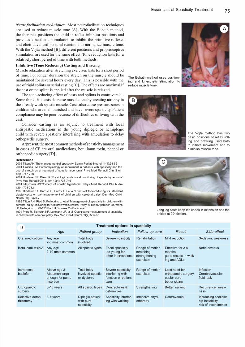
8/9/2019 Guía de ayuda para la parálisis cerebral.pdf
http://slidepdf.com/reader/full/guia-de-ayuda-para-la-paralisis-cerebralpdf 75/143
75Essentials of Spasticity Treatment
Neurofacilitation techniques Most neurofacilitation techniquesare used to reduce muscle tone [A]. With the Bobath method,the therapist positions the child in reflex inhibitor positions and provides kinesthetic stimulation to inhibit the primitive reflexesand elicit advanced postural reactions to normalize muscle tone.With the Vojta method [B], different positions and proprioceptivestimulation are used for the same effect. Tone reduction lasts for a
relatively short period of time with both methods.Inhibitive (Tone Reducing) Casting and Bracing
Muscle relaxation after stretching exercises lasts for a short periodof time. For longer duration the stretch on the muscle should bemaintained for several hours every day. This is possible with theuse of rigid splints or serial casting [C]. The effects are maximal ifthe cast or the splint is applied after the muscle is relaxed.
The tone-reducing effect of casts and splints is controversial.Some think that casts decrease muscle tone by creating atrophy inthe already weak spastic muscle. Casts also cause pressure sores inchildren who are malnourished and have severe spasticity. Patientcompliance may be poor because of difficulties of living with thecast.
Consider casting as an adjunct to treatment with localantispastic medications in the young diplegic or hemiplegicchild with severe spasticity interfering with ambulation to delayorthopaedic surgery.
At present, the most common methods of spasticity managementin cases of CP are oral medications, botulinum toxin, phenol ororthopaedic surgery [D].
References2004 Tilton AH ‘The management of spasticity’ Semin Pediatr Neurol 11(1):58-65
2001 Gracies JM ‘Pathophysiology of impairment in patients with spasticity and the
use of stretch as a treatment of spastic hypertonia’ Phys Med Rehabil Clin N Am
12(4):747-768
2001 Hinderer SR, Dixon K ‘Physiologic and clinical monitoring of spastic hypertonia’
Phys Med Rehabil Clin N Am 12(4):733-746
2001 Meythaler JM’Concept of spastic hypertonia’ Phys Med Rehabil Clin N Am
12(4):725-732
1988 Hinderer KA, Harris SR, Purdy AH, et al ‘Effects of ‘tone-reducing’ vs. standard
plaster-casts on gait improvement of children with cerebral palsy’ Dev Med Child
Neurol 30(3):370-7
1998 Tilton AH, Ried S, Pellegrino L, et al ‘Management of spasticity in children with
cerebral palsy’ In Caring for Children with Cerebral Palsy: A Team Approach Dormans
JP, Pellegrino L, 99-123 Paul H Brookes Co Baltimore
1991 Price R, Bjornson KF, Lehmann JF, et al ‘Quantitative measurement of spasticity
in children with cerebral palsy’ Dev Med Child Neurol 33(7):585-95
Treatment options in spasticity
Age Patient group Indication Follow-up care Result Side-effect
Oral medications Any age
2-5 most common
Total body
involved
Severe spasticity Rehabilitation Mild reduction Sedation, weakness
Botulinum toxin A Any age
2-10 most common
All spastic types Focal spasticity
too young for
other interventions
Range of motion,
stretching,
strengthening
exercises
Effective for 3-6
months
good results in walk-
ing and ADLs
None obvious
Intrathecal
baclofen
Above age 3
Abdomen large
enough for pump
insertion
Total body
involved spastic
or dystonic
Severe spasticity
interfering with
function or patient
care
Range of motion
exercises
Less need for
orthopaedic surgery
easier care
better sitting
Infection
Cerebrovascular
fluid leak
Orthopaedic
surgery
5-15 years All spastic types Contractures &
deformities
Strengthening Better walking Recurrence, weak-
ness
Selective dorsal
rhizotomy
3-7 years Diplegic patient
with pure
spasticity
Spasticity interfer-
ing with walking
Intensive physi-
otherapy
Controversial Increasing scoliosis,
hip instability
risk of incontinence
CC
Long leg casts keep the knees in extension and the
ankles at 90o flexion.
The Vojta method has two
basic positions of reflex roll-
ing and crawling used both
to initiate movement and to
diminish muscle tone.
The Bobath method uses position-
ing and kinesthetic stimulation to
reduce muscle tone.
DD
B
A
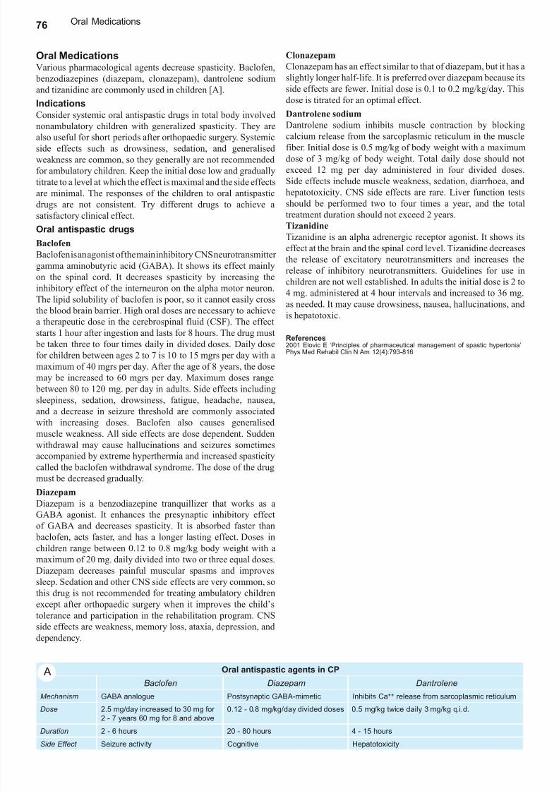
8/9/2019 Guía de ayuda para la parálisis cerebral.pdf
http://slidepdf.com/reader/full/guia-de-ayuda-para-la-paralisis-cerebralpdf 76/143
76 Oral Medications
Oral MedicationsVarious pharmacological agents decrease spasticity. Baclofen, benzodiazepines (diazepam, clonazepam), dantrolene sodiumand tizanidine are commonly used in children [A].
Indications
Consider systemic oral antispastic drugs in total body involvednonambulatory children with generalized spasticity. They are
also useful for short periods after orthopaedic surgery. Systemicside effects such as drowsiness, sedation, and generalisedweakness are common, so they generally are not recommendedfor ambulatory children. Keep the initial dose low and graduallytitrate to a level at which the effect is maximal and the side effectsare minimal. The responses of the children to oral antispasticdrugs are not consistent. Try different drugs to achieve asatisfactory clinical effect.
Oral antispastic drugs
Baclofen
Baclofen is an agonist of the main inhibitory CNS neurotransmittergamma aminobutyric acid (GABA). It shows its effect mainly
on the spinal cord. It decreases spasticity by increasing theinhibitory effect of the interneuron on the alpha motor neuron.The lipid solubility of baclofen is poor, so it cannot easily crossthe blood brain barrier. High oral doses are necessary to achievea therapeutic dose in the cerebrospinal fluid (CSF). The effectstarts 1 hour after ingestion and lasts for 8 hours. The drug must be taken three to four times daily in divided doses. Daily dosefor children between ages 2 to 7 is 10 to 15 mgrs per day with amaximum of 40 mgrs per day. After the age of 8 years, the dosemay be increased to 60 mgrs per day. Maximum doses range between 80 to 120 mg. per day in adults. Side effects includingsleepiness, sedation, drowsiness, fatigue, headache, nausea,
and a decrease in seizure threshold are commonly associatedwith increasing doses. Baclofen also causes generalisedmuscle weakness. All side effects are dose dependent. Suddenwithdrawal may cause hallucinations and seizures sometimesaccompanied by extreme hyperthermia and increased spasticitycalled the baclofen withdrawal syndrome. The dose of the drugmust be decreased gradually.
Diazepam
Diazepam is a benzodiazepine tranquillizer that works as aGABA agonist. It enhances the presynaptic inhibitory effectof GABA and decreases spasticity. It is absorbed faster than baclofen, acts faster, and has a longer lasting effect. Doses inchildren range between 0.12 to 0.8 mg/kg body weight with amaximum of 20 mg. daily divided into two or three equal doses.Diazepam decreases painful muscular spasms and improvessleep. Sedation and other CNS side effects are very common, sothis drug is not recommended for treating ambulatory childrenexcept after orthopaedic surgery when it improves the child’stolerance and participation in the rehabilitation program. CNSside effects are weakness, memory loss, ataxia, depression, anddependency.
Clonazepam
Clonazepam has an effect similar to that of diazepam, but it has aslightly longer half-life. It is preferred over diazepam because itsside effects are fewer. Initial dose is 0.1 to 0.2 mg/kg/day. Thisdose is titrated for an optimal effect.
Dantrolene sodium
Dantrolene sodium inhibits muscle contraction by blocking
calcium release from the sarcoplasmic reticulum in the musclefiber. Initial dose is 0.5 mg/kg of body weight with a maximumdose of 3 mg/kg of body weight. Total daily dose should notexceed 12 mg per day administered in four divided doses.Side effects include muscle weakness, sedation, diarrhoea, andhepatotoxicity. CNS side effects are rare. Liver function testsshould be performed two to four times a year, and the totaltreatment duration should not exceed 2 years.Tizanidine
Tizanidine is an alpha adrenergic receptor agonist. It shows itseffect at the brain and the spinal cord level. Tizanidine decreasesthe release of excitatory neurotransmitters and increases therelease of inhibitory neurotransmitters. Guidelines for use inchildren are not well established. In adults the initial dose is 2 to4 mg. administered at 4 hour intervals and increased to 36 mg.as needed. It may cause drowsiness, nausea, hallucinations, andis hepatotoxic.
References2001 Elovic E ‘Principles of pharmaceutical management of spastic hypertonia’Phys Med Rehabil Clin N Am 12(4):793-816
Oral antispastic agents in CP
Baclofen Diazepam Dantrolene
Mechanism GABA analogue Postsynaptic GABA-mimetic Inhibits Ca++ release from sarcoplasmic reticulum
Dose 2.5 mg/day increased to 30 mg for
2 - 7 years 60 mg for 8 and above
0.12 - 0.8 mg/kg/day divided doses 0.5 mg/kg twice daily 3 mg/kg q.i.d.
Duration 2 - 6 hours 20 - 80 hours 4 - 15 hours
Side Effect Seizure activity Cognitive Hepatotoxicity
A
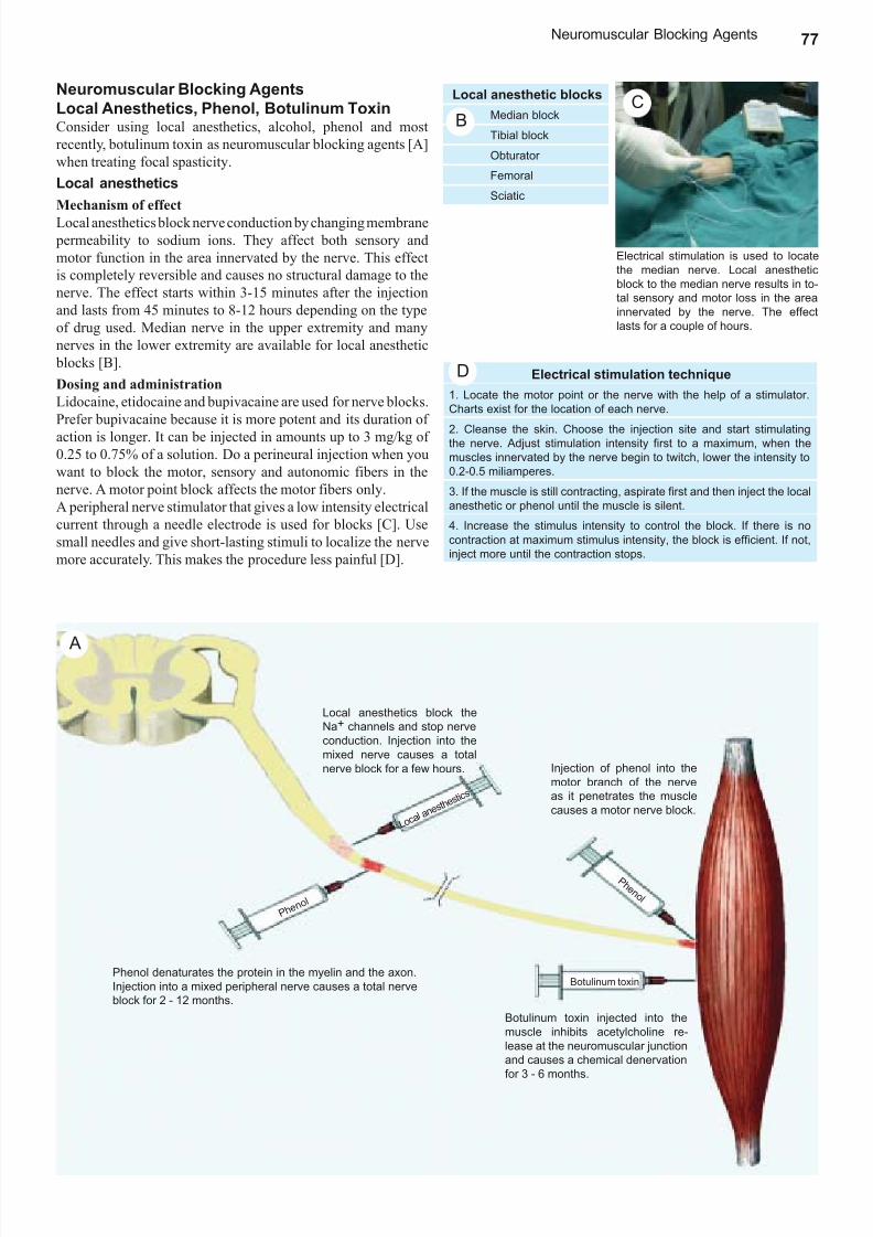
8/9/2019 Guía de ayuda para la parálisis cerebral.pdf
http://slidepdf.com/reader/full/guia-de-ayuda-para-la-paralisis-cerebralpdf 77/143
77Neuromuscular Blocking Agents
Neuromuscular Blocking Agents
Local Anesthetics, Phenol, Botulinum ToxinConsider using local anesthetics, alcohol, phenol and mostrecently, botulinum toxin as neuromuscular blocking agents [A]when treating focal spasticity.
Local anesthetics
Mechanism of effect
Local anesthetics block nerve conduction by changing membrane permeability to sodium ions. They affect both sensory andmotor function in the area innervated by the nerve. This effectis completely reversible and causes no structural damage to thenerve. The effect starts within 3-15 minutes after the injectionand lasts from 45 minutes to 8-12 hours depending on the typeof drug used. Median nerve in the upper extremity and manynerves in the lower extremity are available for local anesthetic blocks [B].
Dosing and administration
Lidocaine, etidocaine and bupivacaine are used for nerve blocks.Prefer bupivacaine because it is more potent and its duration of
action is longer. It can be injected in amounts up to 3 mg/kg of0.25 to 0.75% of a solution. Do a perineural injection when youwant to block the motor, sensory and autonomic fibers in thenerve. A motor point block affects the motor fibers only.A peripheral nerve stimulator that gives a low intensity electricalcurrent through a needle electrode is used for blocks [C]. Usesmall needles and give short-lasting stimuli to localize the nervemore accurately. This makes the procedure less painful [D].
Local anesthetic blocks
Median block
Tibial block
Obturator
Femoral
Sciatic
Electrical stimulation technique
1. Locate the motor point or the nerve with the help of a stimulator.
Charts exist for the location of each nerve.
2. Cleanse the skin. Choose the injection site and start stimulatingthe nerve. Adjust stimulation intensity first to a maximum, when the
muscles innervated by the nerve begin to twitch, lower the intensity to
0.2-0.5 miliamperes.
3. If the muscle is still contracting, aspirate first and then inject the local
anesthetic or phenol until the muscle is silent.
4. Increase the stimulus intensity to control the block. If there is no
contraction at maximum stimulus intensity, the block is efficient. If not,
inject more until the contraction stops.
DD
CC B
Electrical stimulation is used to locate
the median nerve. Local anesthetic
block to the median nerve results in to-
tal sensory and motor loss in the area
innervated by the nerve. The effect
lasts for a couple of hours.
L o c a l
a n e s t
h e s t i c
s
P h e n
o l
Botulinum toxin
P h e n o l
Phenol denaturates the protein in the myelin and the axon.
Injection into a mixed peripheral nerve causes a total nerve
block for 2 - 12 months.
A
Botulinum toxin injected into the
muscle inhibits acetylcholine re-
lease at the neuromuscular junction
and causes a chemical denervation
for 3 - 6 months.
Local anesthetics block the
Na+ channels and stop nerve
conduction. Injection into the
mixed nerve causes a total
nerve block for a few hours. Injection of phenol into the
motor branch of the nerve
as it penetrates the muscle
causes a motor nerve block.
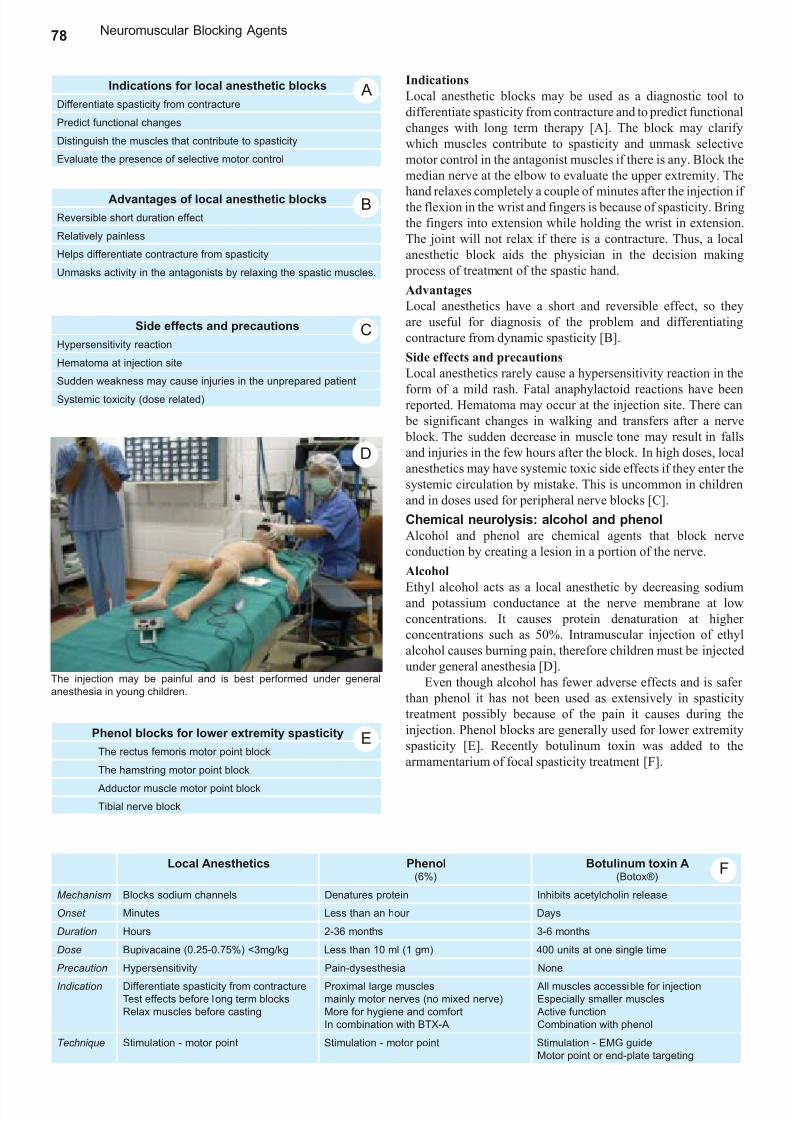
8/9/2019 Guía de ayuda para la parálisis cerebral.pdf
http://slidepdf.com/reader/full/guia-de-ayuda-para-la-paralisis-cerebralpdf 78/143
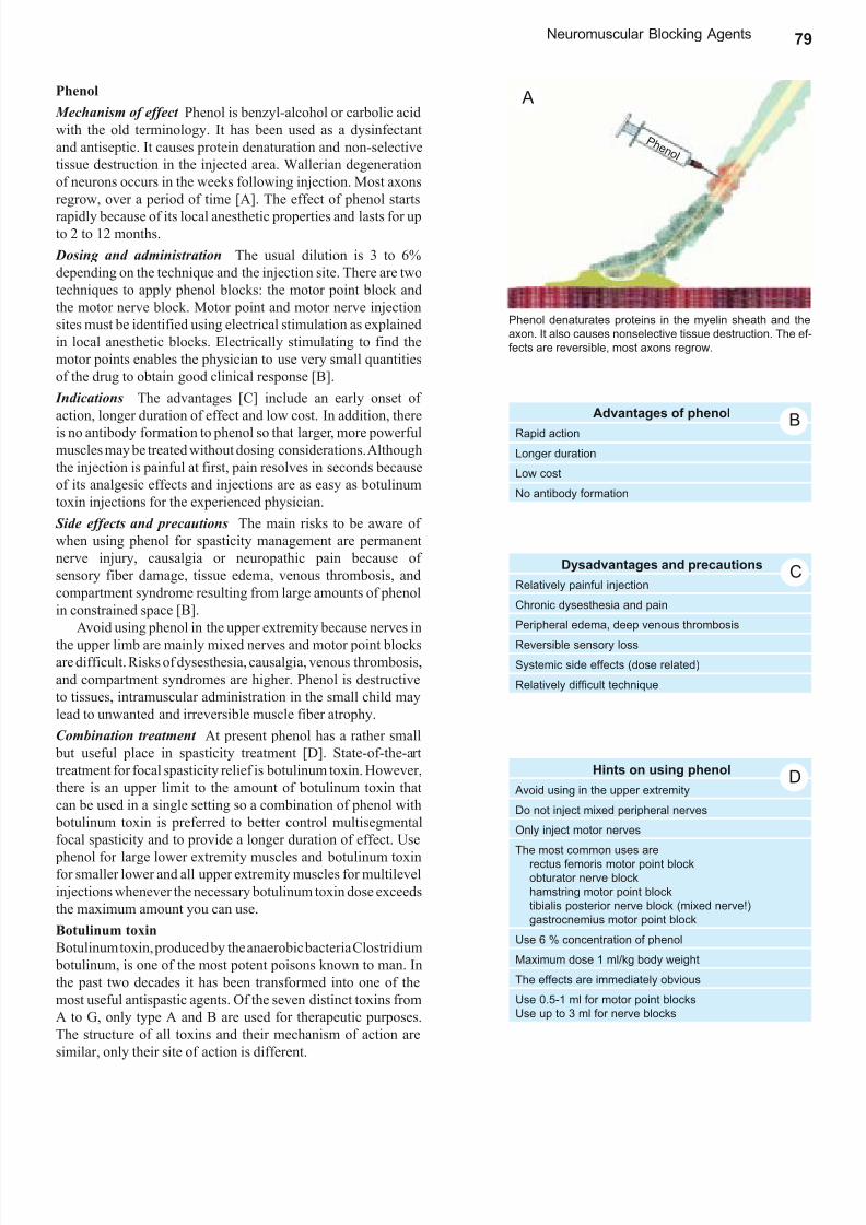
8/9/2019 Guía de ayuda para la parálisis cerebral.pdf
http://slidepdf.com/reader/full/guia-de-ayuda-para-la-paralisis-cerebralpdf 79/143
79Neuromuscular Blocking Agents
Hints on using phenol
Avoid using in the upper extremity
Do not inject mixed peripheral nerves
Only inject motor nerves
The most common uses are
rectus femoris motor point block
obturator nerve block
hamstring motor point block
tibialis posterior nerve block (mixed nerve!)
gastrocnemius motor point block
Use 6 % concentration of phenol
Maximum dose 1 ml/kg body weight
The effects are immediately obvious
Use 0.5-1 ml for motor point blocks
Use up to 3 ml for nerve blocks
Phenol
Mechanism of effect Phenol is benzyl-alcohol or carbolic acidwith the old terminology. It has been used as a dysinfectantand antiseptic. It causes protein denaturation and non-selectivetissue destruction in the injected area. Wallerian degenerationof neurons occurs in the weeks following injection. Most axonsregrow, over a period of time [A]. The effect of phenol starts
rapidly because of its local anesthetic properties and lasts for upto 2 to 12 months.
Dosing and administration The usual dilution is 3 to 6%depending on the technique and the injection site. There are twotechniques to apply phenol blocks: the motor point block andthe motor nerve block. Motor point and motor nerve injectionsites must be identified using electrical stimulation as explainedin local anesthetic blocks. Electrically stimulating to find themotor points enables the physician to use very small quantitiesof the drug to obtain good clinical response [B].
Indications The advantages [C] include an early onset ofaction, longer duration of effect and low cost. In addition, there
is no antibody formation to phenol so that larger, more powerfulmuscles may be treated without dosing considerations. Althoughthe injection is painful at first, pain resolves in seconds becauseof its analgesic effects and injections are as easy as botulinumtoxin injections for the experienced physician.
Side effects and precautions The main risks to be aware ofwhen using phenol for spasticity management are permanentnerve injury, causalgia or neuropathic pain because ofsensory fiber damage, tissue edema, venous thrombosis, andcompartment syndrome resulting from large amounts of phenolin constrained space [B].
Avoid using phenol in the upper extremity because nerves in
the upper limb are mainly mixed nerves and motor point blocksare difficult. Risks of dysesthesia, causalgia, venous thrombosis,and compartment syndromes are higher. Phenol is destructiveto tissues, intramuscular administration in the small child maylead to unwanted and irreversible muscle fiber atrophy.
Combination treatment At present phenol has a rather small but useful place in spasticity treatment [D]. State-of-the-arttreatment for focal spasticity relief is botulinum toxin. However,there is an upper limit to the amount of botulinum toxin thatcan be used in a single setting so a combination of phenol with botulinum toxin is preferred to better control multisegmentalfocal spasticity and to provide a longer duration of effect. Use
phenol for large lower extremity muscles and botulinum toxinfor smaller lower and all upper extremity muscles for multilevelinjections whenever the necessary botulinum toxin dose exceedsthe maximum amount you can use.
Botulinum toxin
Botulinum toxin, produced by the anaerobic bacteria Clostridium botulinum, is one of the most potent poisons known to man. Inthe past two decades it has been transformed into one of themost useful antispastic agents. Of the seven distinct toxins fromA to G, only type A and B are used for therapeutic purposes.The structure of all toxins and their mechanism of action aresimilar, only their site of action is different.
DD
Dysadvantages and precautions
Relatively painful injection
Chronic dysesthesia and pain
Peripheral edema, deep venous thrombosis
Reversible sensory loss
Systemic side effects (dose related)
Relatively difficult technique
Advantages of phenol
Rapid action
Longer duration
Low cost
No antibody formation
A
B
CC
Phenol denaturates proteins in the myelin sheath and the
axon. It also causes nonselective tissue destruction. The ef-
fects are reversible, most axons regrow.
A
P h e n o l
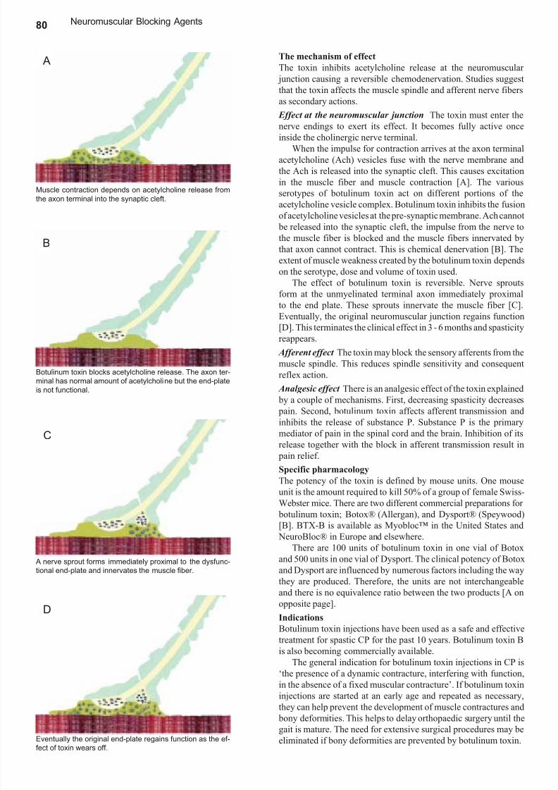
8/9/2019 Guía de ayuda para la parálisis cerebral.pdf
http://slidepdf.com/reader/full/guia-de-ayuda-para-la-paralisis-cerebralpdf 80/143
80 Neuromuscular Blocking Agents
DD
The mechanism of effect
The toxin inhibits acetylcholine release at the neuromuscular junction causing a reversible chemodenervation. Studies suggestthat the toxin affects the muscle spindle and afferent nerve fibersas secondary actions.
Effect at the neuromuscular junction The toxin must enter thenerve endings to exert its effect. It becomes fully active once
inside the cholinergic nerve terminal.When the impulse for contraction arrives at the axon terminal
acetylcholine (Ach) vesicles fuse with the nerve membrane andthe Ach is released into the synaptic cleft. This causes excitationin the muscle fiber and muscle contraction [A]. The variousserotypes of botulinum toxin act on different portions of theacetylcholine vesicle complex. Botulinum toxin inhibits the fusionof acetylcholine vesicles at the pre-synaptic membrane. Ach cannot be released into the synaptic cleft, the impulse from the nerve tothe muscle fiber is blocked and the muscle fibers innervated bythat axon cannot contract. This is chemical denervation [B]. Theextent of muscle weakness created by the botulinum toxin dependson the serotype, dose and volume of toxin used.
The effect of botulinum toxin is reversible. Nerve sproutsform at the unmyelinated terminal axon immediately proximalto the end plate. These sprouts innervate the muscle fiber [C].Eventually, the original neuromuscular junction regains function[D]. This terminates the clinical effect in 3 - 6 months and spasticityreappears.
Afferent effect The toxin may block the sensory afferents from themuscle spindle. This reduces spindle sensitivity and consequentreflex action.
Analgesic effect There is an analgesic effect of the toxin explained by a couple of mechanisms. First, decreasing spasticity decreases
pain. Second, botulinum toxin affects afferent transmission andinhibits the release of substance P. Substance P is the primarymediator of pain in the spinal cord and the brain. Inhibition of itsrelease together with the block in afferent transmission result in pain relief.
Specific pharmacology
The potency of the toxin is defined by mouse units. One mouseunit is the amount required to kill 50% of a group of female Swiss-Webster mice. There are two different commercial preparations for botulinum toxin; Botox® (Allergan), and Dysport® (Speywood)[B]. BTX-B is available as Myobloc™ in the United States and NeuroBloc® in Europe and elsewhere.
There are 100 units of botulinum toxin in one vial of Botoxand 500 units in one vial of Dysport. The clinical potency of Botoxand Dysport are influenced by numerous factors including the waythey are produced. Therefore, the units are not interchangeableand there is no equivalence ratio between the two products [A onopposite page].
Indications
Botulinum toxin injections have been used as a safe and effectivetreatment for spastic CP for the past 10 years. Botulinum toxin Bis also becoming commercially available.
The general indication for botulinum toxin injections in CP is‘the presence of a dynamic contracture, interfering with function,in the absence of a fixed muscular contracture’. If botulinum toxin
injections are started at an early age and repeated as necessary,they can help prevent the development of muscle contractures and bony deformities. This helps to delay orthopaedic surgery until thegait is mature. The need for extensive surgical procedures may beeliminated if bony deformities are prevented by botulinum toxin.
CC
B
A
Muscle contraction depends on acetylcholine release from
the axon terminal into the synaptic cleft.
Botulinum toxin blocks acetylcholine release. The axon ter-
minal has normal amount of acetylcholine but the end-plate
is not functional.
A nerve sprout forms immediately proximal to the dysfunc-
tional end-plate and innervates the muscle fiber.
Eventually the original end-plate regains function as the ef-
fect of toxin wears off.
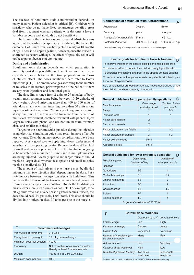
8/9/2019 Guía de ayuda para la parálisis cerebral.pdf
http://slidepdf.com/reader/full/guia-de-ayuda-para-la-paralisis-cerebralpdf 81/143
81Neuromuscular Blocking Agents
Comparison of botulinum toxin A preparations
Preparation Dysport Botox
Company Ipsen Allergan
1 ng toksin-hemagglutinin 20 m.u. > 5 m.u.
Contents of one vial 500 m.u. (12,5 ng) 100 m.u.(40 ng)
The relative potency of these preparations has not been established yet.
Recommended dosages
Per muscle of lower limb 3-6 U/kg
Per kg total body weight: 12 U/kg proven dosage
Maximum dose per session 400 U
Frequency: Not more than once every 3 months
Usually at least 6 month intervals
Dilution 100 U in 1 or 2 ml 0.9% NaCl
Maximum dose per site 50 U
General guidelines for upper extremity spasticity
Muscles injectedDose range
(units/kg of bw)
Number of sites
per muscle
Biceps 2 2-3
Pronator teres 1 1
Flexor carpi radialis 2 1
Flexor carpi ulnaris 2 1
Flexor digitorum superficialis 2 1-2
Flexor digitorum profundus 2 1-2
Flexor pollicis longus 0.5-1 1
Adductor pollicis 0.5-1 1
General guidelines for lower extremity spasticity
Muscles injectedDose range
(units/kg of bw)
Number of
sites per muscle
Iliopsoas 2 2
Quadriceps 3-6 4
Medial hamstrings 3-6 3-4
Lateral hamstrings 2-3 2
Adductors 3-6 2
Gastrocnemius 3-6 1-2
Soleus 2-3 1
Tibialis posterior 1-3 1
In general maximum of 50 U/site
Specific goals for botulinum toxin A treatment
To improve walking in the spastic diplegic and hemiplegic child
To minimise adductor tone in the child with early hip subluxation
To decrease the spasms and pain in the spastic-athetoid patients
To reduce tone in the psoas muscle in patients with back pain
because of hyperlordosis
As a simulation for orthopedic surgery, to have a general idea of how
the child will be when spasticity is reduced.
B
A
DD
The success of botulinum toxin administration depends onmany factors. Patient selection is critical [B]. Children withspasticity who do not have fixed contractures benefit a greatdeal from treatment whereas patients with dyskinesia have avariable response and athetoids do not benefit at all.
The timing of the injections is controversial. Most cliniciansagree that the earlier the spasticity is reduced, the better the
outcome. Botulinum toxin can be injected as early as 18 monthsof age. There is no upper age limit, however, once the muscle isshortened as occurs with age, the effect of spasticity relief willnot be apparent because of contracture.
Dosing and administration
Botulinum toxin dosing depends on which preparation isused. Dysport dosing is different than Botox and there is noequivalence ratio between the two preparations in termsof clinical effect. The doses mentioned here refer to Botoxinjections [C,D]. The amount changes according to the numberof muscles to be treated, prior response of the patient if thereare any prior injections and functional goals.
The dose limits range from 2 units to 29 units/kg of bodyweight, most common range being between 10-20 units/kg of body weight. Avoid injecting more than 400 to 600 units oftotal dose at any one time, injecting more than 50 units at oneinjection site and exceeding 20 units per kilogram per muscleat any one time. If there is a need for more toxin because ofmultilevel involvement, combine treatment with phenol. Injectlarger muscles with phenol and use botulinum toxin for moredistal and smaller muscles [E].
Targeting the neuromuscular junction during the injectionusing electrical stimulation guide may result in more effect forless volume. Even though no serious complications have beenreported, it is a good idea to apply high doses under general
anesthesia in the operating theatre. Reduce the dose if the childis small and has atrophic muscles, if the treatment is goingto be repeated for a number of times and if multiple musclesare being injected. Severely spastic and larger muscles shouldreceive a larger dose whereas less spastic and small musclesreceive a smaller dose [F].
The amount of toxin given to one muscle must be dividedinto more than two injection sites, depending on the dose. Put asafe distance between two injection sites with high doses. Thisincreases the diffusion of the toxin in the muscle and prevents itfrom entering the systemic circulation. Divide the total dose permuscle over more sites as much as possible. For example, for a
20 kg child who has a very spastic gastrocnemius muscle, thedose should be 6 U/kg/muscle, 120 U total. This dose should bedivided into 4 injection sites, 30 units per site in the muscle.
CC
Botox® dose modifiers
Decrease dose if Increase dose if
Patient weight Low High
Duration of therapy Chronic Acute
Muscle bulk Very small Very large
Number of muscles inject-
ed simultaneously
Many Few
Ashworth score Low Very high
Concern about weakness High Low
Results of previous therapy Too much
weakness
Inadequate
response
Table reproduced with permission from WE MOVE New York www.mdvu.org.
EE
FF
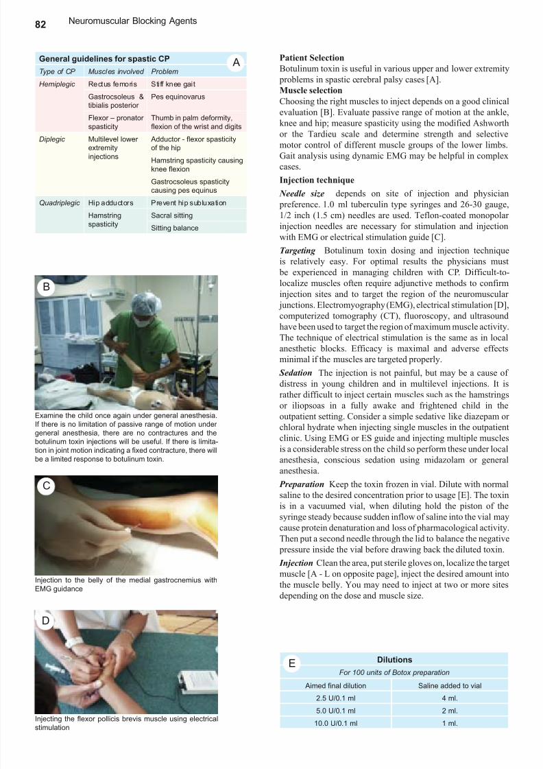
8/9/2019 Guía de ayuda para la parálisis cerebral.pdf
http://slidepdf.com/reader/full/guia-de-ayuda-para-la-paralisis-cerebralpdf 82/143
82 Neuromuscular Blocking Agents
Dilutions
For 100 units of Botox preparation
Aimed final dilution Saline added to vial
2.5 U/0.1 ml 4 ml.
5.0 U/0.1 ml 2 ml.
10.0 U/0.1 ml 1 ml.
General guidelines for spastic CP
Type of CP Muscles involved Problem
Hemiplegic Rectus femoris Stiff knee gait
Gastrocsoleus &
tibialis posterior
Pes equinovarus
Flexor – pronator
spasticity
Thumb in palm deformity,
flexion of the wrist and digits
Diplegic Multilevel lower
extremity
injections
Adductor - flexor spasticity
of the hip
Hamstring spasticity causing
knee flexion
Gastrocsoleus spasticity
causing pes equinus
Quadriplegic Hip adductors Prevent hip subluxation
Hamstring
spasticity
Sacral sitting
Sitting balance
DD
CC
B
Patient Selection
Botulinum toxin is useful in various upper and lower extremity problems in spastic cerebral palsy cases [A].Muscle selection
Choosing the right muscles to inject depends on a good clinicalevaluation [B]. Evaluate passive range of motion at the ankle,knee and hip; measure spasticity using the modified Ashworth
or the Tardieu scale and determine strength and selectivemotor control of different muscle groups of the lower limbs.Gait analysis using dynamic EMG may be helpful in complexcases.
Injection technique
Needle size depends on site of injection and physician preference. 1.0 ml tuberculin type syringes and 26-30 gauge,1/2 inch (1.5 cm) needles are used. Teflon-coated monopolarinjection needles are necessary for stimulation and injectionwith EMG or electrical stimulation guide [C].
Targeting Botulinum toxin dosing and injection techniqueis relatively easy. For optimal results the physicians must
be experienced in managing children with CP. Difficult-to-localize muscles often require adjunctive methods to confirminjection sites and to target the region of the neuromuscular junctions. Electromyography (EMG), electrical stimulation [D],computerized tomography (CT), fluoroscopy, and ultrasoundhave been used to target the region of maximum muscle activity.The technique of electrical stimulation is the same as in localanesthetic blocks. Efficacy is maximal and adverse effectsminimal if the muscles are targeted properly.
Sedation The injection is not painful, but may be a cause ofdistress in young children and in multilevel injections. It israther difficult to inject certain muscles such as the hamstrings
or iliopsoas in a fully awake and frightened child in theoutpatient setting. Consider a simple sedative like diazepam orchloral hydrate when injecting single muscles in the outpatientclinic. Using EMG or ES guide and injecting multiple musclesis a considerable stress on the child so perform these under localanesthesia, conscious sedation using midazolam or generalanesthesia.
Preparation Keep the toxin frozen in vial. Dilute with normalsaline to the desired concentration prior to usage [E]. The toxinis in a vacuumed vial, when diluting hold the piston of thesyringe steady because sudden inflow of saline into the vial maycause protein denaturation and loss of pharmacological activity.
Then put a second needle through the lid to balance the negative pressure inside the vial before drawing back the diluted toxin.
Injection Clean the area, put sterile gloves on, localize the targetmuscle [A - L on opposite page], inject the desired amount intothe muscle belly. You may need to inject at two or more sitesdepending on the dose and muscle size.
A
Examine the child once again under general anesthesia.
If there is no limitation of passive range of motion under
general anesthesia, there are no contractures and the
botulinum toxin injections will be useful. If there is limita-
tion in joint motion indicating a fixed contracture, there will
be a limited response to botulinum toxin.
Injection to the belly of the medial gastrocnemius with
EMG guidance
Injecting the flexor pollicis brevis muscle using electrical
stimulation
EE
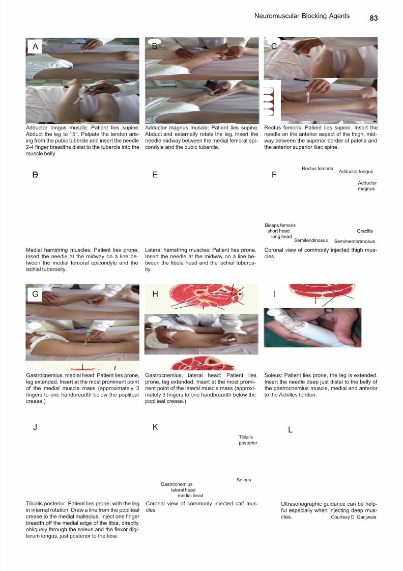
8/9/2019 Guía de ayuda para la parálisis cerebral.pdf
http://slidepdf.com/reader/full/guia-de-ayuda-para-la-paralisis-cerebralpdf 83/143
83Neuromuscular Blocking Agents
EE
HH
Rectus femoris: Patient lies supine. Insert the
needle on the anterior aspect of the thigh, mid-
way between the superior border of patella and
the anterior superior iliac spine.
Adductor magnus muscle: Patient lies supine.
Abduct and externally rotate the leg. Insert the
needle midway between the medial femoral epi-
condyle and the pubic tubercle.
Adductor longus muscle: Patient lies supine.
Abduct the leg to 15o. Palpate the tendon aris-
ing from the pubic tubercle and insert the needle
2-4 finger breadths distal to the tubercle into the
muscle belly.
Medial hamstring muscles: Patient lies prone,
Insert the needle at the midway on a line be-
tween the medial femoral epicondyle and the
ischial tuberosity.
Tibialis posterior: Patient lies prone, with the leg
in internal rotation. Draw a line from the popliteal
crease to the medial malleolus. Inject one finger
breadth off the medial edge of the tibia, directly
obliquely through the soleus and the flexor digi-
torum longus, just posterior to the tibia.
Soleus: Patient lies prone, the leg is extended.
Insert the needle deep just distal to the belly of
the gastrocnemius muscle, medial and anterior
to the Achilles tendon.
Gastrocnemius, medial head: Patient lies prone,
leg extended. Insert at the most prominent point
of the medial muscle mass (approximately 3
fingers to one handbreadth below the popliteal
crease.)
Gastrocnemius, lateral head: Patient lies
prone, leg extended. Insert at the most promi-
nent point of the lateral muscle mass (approxi-
mately 3 fingers to one handbreadth below the
popliteal crease.)
Lateral hamstring muscles: Patient lies prone.
Insert the needle at the midway on a line be-
tween the fibula head and the ischial tuberos-
ity.
Biceps femoris
short head
long headSemitendinosus Semimembranosus
Rectus femoris Adductor longus
Adductor
magnus
Gracilis
Gastrocnemius
lateral head
medial head
Soleus
Tibialis
posterior
Coronal view of commonly injected thigh mus-
cles
Coronal view of commonly injected calf mus-
cles
FFE
CCB A
JJ KK
GG II
DD
Ultrasonographic guidance can be help-
ful especially when injecting deep mus-
cles. Courtesy D. Ganjwala
LL
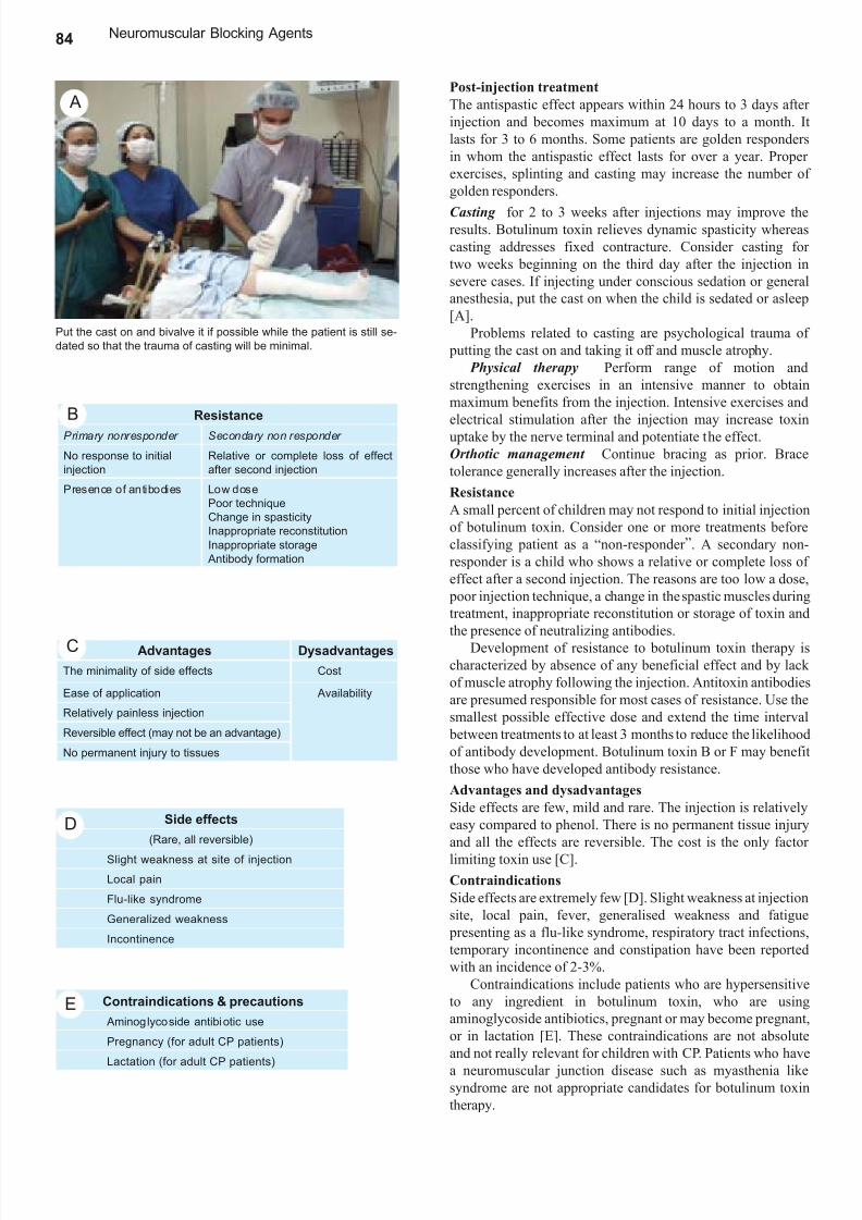
8/9/2019 Guía de ayuda para la parálisis cerebral.pdf
http://slidepdf.com/reader/full/guia-de-ayuda-para-la-paralisis-cerebralpdf 84/143
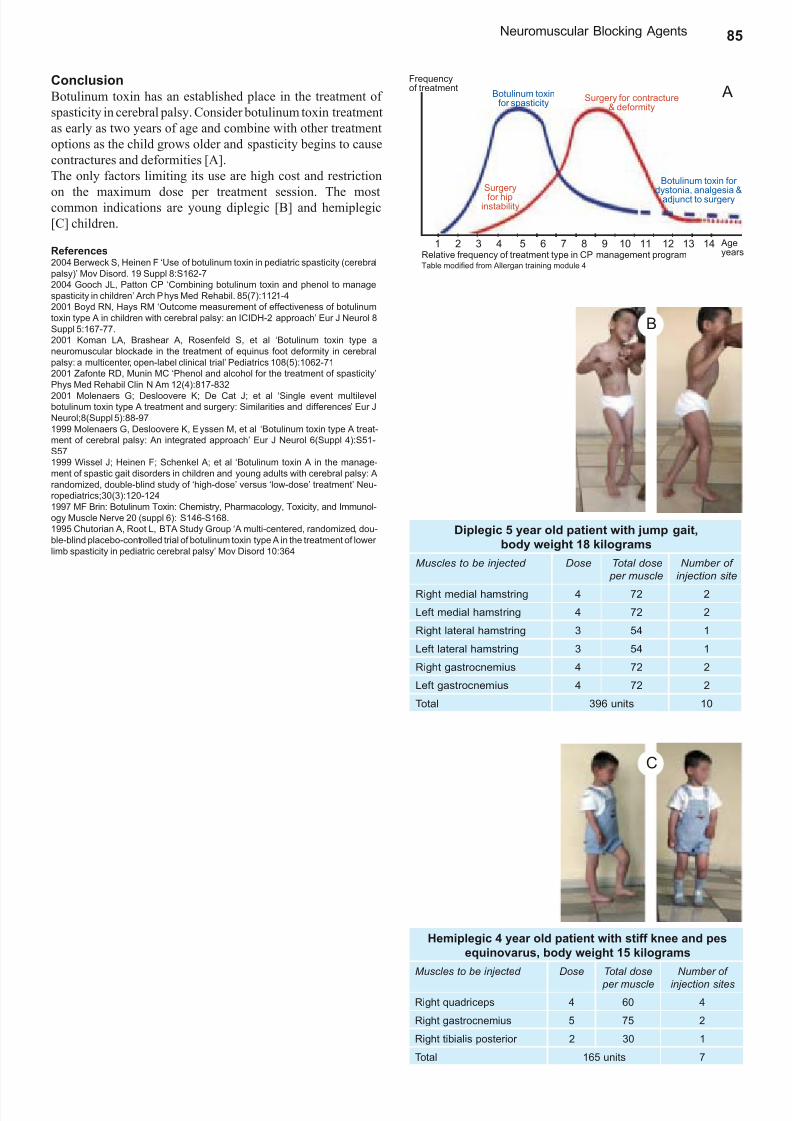
8/9/2019 Guía de ayuda para la parálisis cerebral.pdf
http://slidepdf.com/reader/full/guia-de-ayuda-para-la-paralisis-cerebralpdf 85/143
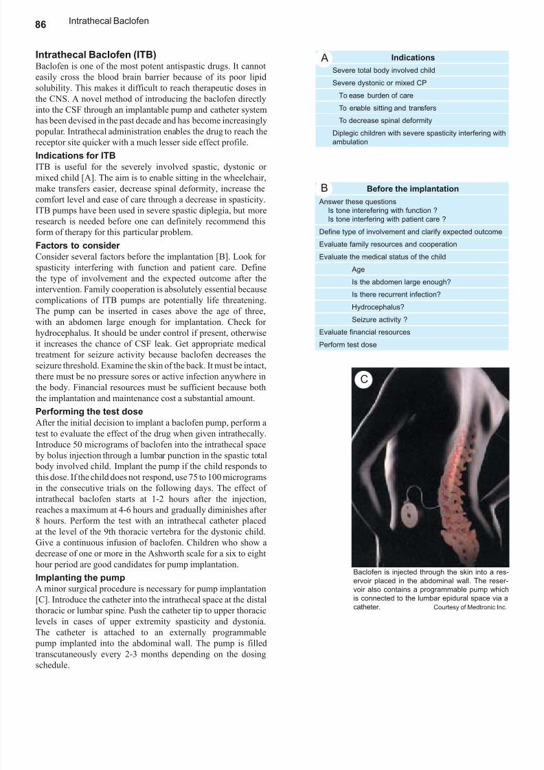
8/9/2019 Guía de ayuda para la parálisis cerebral.pdf
http://slidepdf.com/reader/full/guia-de-ayuda-para-la-paralisis-cerebralpdf 86/143
86 Intrathecal Baclofen
Intrathecal Baclofen (ITB)Baclofen is one of the most potent antispastic drugs. It cannoteasily cross the blood brain barrier because of its poor lipidsolubility. This makes it difficult to reach therapeutic doses inthe CNS. A novel method of introducing the baclofen directlyinto the CSF through an implantable pump and catheter systemhas been devised in the past decade and has become increasingly
popular. Intrathecal administration enables the drug to reach thereceptor site quicker with a much lesser side effect profile.
Indications for ITB
ITB is useful for the severely involved spastic, dystonic ormixed child [A]. The aim is to enable sitting in the wheelchair,make transfers easier, decrease spinal deformity, increase thecomfort level and ease of care through a decrease in spasticity.ITB pumps have been used in severe spastic diplegia, but moreresearch is needed before one can definitely recommend thisform of therapy for this particular problem.
Factors to consider
Consider several factors before the implantation [B]. Look for
spasticity interfering with function and patient care. Definethe type of involvement and the expected outcome after theintervention. Family cooperation is absolutely essential becausecomplications of ITB pumps are potentially life threatening.The pump can be inserted in cases above the age of three,with an abdomen large enough for implantation. Check forhydrocephalus. It should be under control if present, otherwiseit increases the chance of CSF leak. Get appropriate medicaltreatment for seizure activity because baclofen decreases theseizure threshold. Examine the skin of the back. It must be intact,there must be no pressure sores or active infection anywhere inthe body. Financial resources must be sufficient because boththe implantation and maintenance cost a substantial amount.
Performing the test dose
After the initial decision to implant a baclofen pump, perform atest to evaluate the effect of the drug when given intrathecally.Introduce 50 micrograms of baclofen into the intrathecal space by bolus injection through a lumbar punction in the spastic total body involved child. Implant the pump if the child responds tothis dose. If the child does not respond, use 75 to 100 microgramsin the consecutive trials on the following days. The effect ofintrathecal baclofen starts at 1-2 hours after the injection,reaches a maximum at 4-6 hours and gradually diminishes after8 hours. Perform the test with an intrathecal catheter placedat the level of the 9th thoracic vertebra for the dystonic child.
Give a continuous infusion of baclofen. Children who show adecrease of one or more in the Ashworth scale for a six to eighthour period are good candidates for pump implantation.
Implanting the pump
A minor surgical procedure is necessary for pump implantation[C]. Introduce the catheter into the intrathecal space at the distalthoracic or lumbar spine. Push the catheter tip to upper thoraciclevels in cases of upper extremity spasticity and dystonia.The catheter is attached to an externally programmable pump implanted into the abdominal wall. The pump is filledtranscutaneously every 2-3 months depending on the dosingschedule.
Indications
Severe total body involved child
Severe dystonic or mixed CP
To ease burden of care
To enable sitting and transfers
To decrease spinal deformity
Diplegic children with severe spasticity interfering withambulation
Before the implantation
Answer these questions
Is tone interefering with function ?
Is tone interfering with patient care ?
Define type of involvement and clarify expected outcome
Evaluate family resources and cooperation
Evaluate the medical status of the child
Age
Is the abdomen large enough?
Is there recurrent infection?
Hydrocephalus?
Seizure activity ?
Evaluate financial resources
Perform test dose
Baclofen is injected through the skin into a res-
ervoir placed in the abdominal wall. The reser-
voir also contains a programmable pump which
is connected to the lumbar epidural space via a
catheter. Courtesy of Medtronic Inc.
CC
B
A
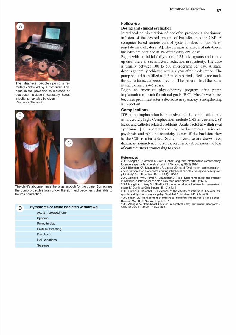
8/9/2019 Guía de ayuda para la parálisis cerebral.pdf
http://slidepdf.com/reader/full/guia-de-ayuda-para-la-paralisis-cerebralpdf 87/143
87Intrathecal Baclofen
Follow-up
Dosing and clinical evaluation
Intrathecal administration of baclofen provides a continuousinfusion of the desired amount of baclofen into the CSF. Acomputer based remote control system makes it possible toregulate the daily dose [A]. The antispastic effects of intrathecal baclofen are obtained at 1% of the daily oral dose.
Begin with an initial daily dose of 25 micrograms and titrateup until there is a satisfactory reduction in spasticity. The doseis usually between 100 to 500 micrograms per day. A staticdose is generally achieved within a year after implantation. The pump should be refilled at 1-3 month periods. Refills are madethrough a transcutaneous injection. The battery life of the pumpis approximately 4-5 years.Begin an intensive physiotherapy program after pumpimplantation to reach functional goals [B,C]. Muscle weakness becomes prominent after a decrease in spasticity. Strengtheningis important.
Complications
ITB pump implantation is expensive and the complication rateis moderately high. Complications include CNS infections, CSFleaks, and catheter related problems. Acute baclofen withdrawalsyndrome [D] characterized by hallucinations, seizures, psychosis and rebound spasticity occurs if the baclofen flowto the CSF is interrupted. Signs of overdose are drowsiness,dizziness, somnolence, seizures, respiratory depression and lossof consciousness progressing to coma.
References2003 Albright AL, Gilmartin R, Swift D, et al ‘Long-term intrathecal baclofen therapy
for severe spasticity of cerebral origin’ J Neurosurg. 98(2):291-5
2003 Bjornson KF, McLaughlin JF, Loeser JD, et al ‘Oral motor, communication,
and nutritional status of children during intrathecal baclofen therapy: a descriptive
pilot study’ Arch Phys Med Rehabil 84(4):500-6
2002 Campbell WM, Ferrel A, McLaughlin JF, et al ‘Long-term safety and efficacy
of continuous intrathecal baclofen’ Dev Med Child Neurol 44(10):660-5
2001 Albright AL, Barry MJ, Shafton DH, et al ‘Intrathecal baclofen for generalized
dystonia’ Dev Med Child Neurol 43(10):652-7
2000 Butler C, Campbell S ‘Evidence of the effects of intrathecal baclofen for
spastic and dystonic cerebral palsy’ Dev Med Child Neurol 42: 634–645
1999 Krach LE ‘Management of intrathecal baclofen withdrawal: a case series’
Develop Med Child Neurol. Suppl 80:111996 Albright AL ‘Intrathecal baclofen in cerebral palsy movement disorders’ JChild Neurol. 11 (Suppl 1): S29-S35
The intrathecal baclofen pump is re-
motely controlled by a computer. This
enables the physician to increase or
decrease the dose if necessary. Bolus
injections may also be given.
Courtesy of Medtronic
The child’s abdomen must be large enough for the pump. Sometimes
the pump protrudes from under the skin and becomes vulnerable to
trauma or infection.
Symptoms of acute baclofen withdrawal
Acute increased tone
Spasms
Paresthesias
Profuse sweating
Dysphoria
Hallucinations
Seizures
DD
CC
B
A
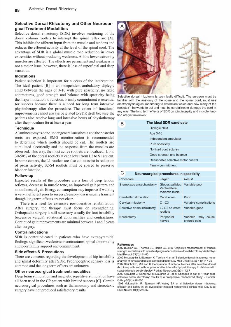
8/9/2019 Guía de ayuda para la parálisis cerebral.pdf
http://slidepdf.com/reader/full/guia-de-ayuda-para-la-paralisis-cerebralpdf 88/143
88 Selective Dorsal Rhizotomy
Selective Dorsal Rhizotomy and Other Neurosur-
gical Treatment ModalitiesSelective dorsal rhizotomy (SDR) involves sectioning of thedorsal column rootlets to interrupt the spinal reflex arc [A].This inhibits the afferent input from the muscle and tendons andreduces the efferent activity at the level of the spinal cord. Theadvantage of SDR is a global muscle tone reduction in lower
extremities without producing weakness. All the lower extremitymuscles are affected. The effects are permanent and weakness isnot a major issue, however, there is loss of superficial and deepsensation.
Indications
Patient selection is important for success of the intervention.The ideal patient [B] is an independent ambulatory diplegicchild between the ages of 3-10 with pure spasticity, no fixedcontractures, good strength and balance with spasticity beingthe major limitation to function. Family commitment is essentialfor success because there is a need for long term intensive physiotherapy after the procedure. The extent of functional
improvements cannot always be related to SDR itself because the patients also receive long and intensive hours of physiotherapyafter the procedure for at least a year.
Technique
A laminectomy is done under general anesthesia and the posteriorroots are exposed. EMG monitorization is recommendedto determine which rootlets should be cut. The rootlets arestimulated electrically and the response from the muscles areobserved. This way, the most active rootlets are localized. Up to30-50% of the dorsal rootlets at each level from L2 to S1 are cut.In some centers, the L1 rootlets are also cut to assist in reductionof psoas activity. S2-S4 rootlets must be spared to preserve
bladder function.Follow-up
Expected results of the procedure are a loss of deep tendonreflexes, decrease in muscle tone, an improved gait pattern andsmoothness of gait. Energy consumption may improve if walkingis very inefficient prior to surgery. Sensory loss is usually transientthough long term effects are not clear.
There is a need for extensive postoperative rehabilitation.After surgery, the therapy must focus on strengthening.Orthopaedic surgery is still necessary usually for foot instability(excessive valgus), rotational abnormalities and contractures.Continued gait improvements are minimal between 1 and 2 yearsafter surgery.
Contraindications
SDR is contraindicated in patients who have extrapyramidalfindings, significant weakness or contractures, spinal abnormalityand poor family support and commitment.
Side effects & Precautions
There are concerns regarding the development of hip instabilityand spinal deformity after SDR. Proprioceptive sensory loss iscommon and the long term effects are unknown.
Other neurosurgical treatment modalities
Deep brain stimulation and magnetic repetitive stimulation haveall been tried in the CP patient with limited success [C]. Certain
neurosurgical procedures such as thalamotomy and stereotaxicsurgery have not produced satisfactory results.
The ideal SDR candidate
Diplegic child
Age 3-10
Independent ambulator
Pure spasticity
No fixed contractures
Good strength and balance
Reasonable selective motor control
Family commitment
Selective dorsal rhizotomy is technically difficult. The surgeon must be
familiar with the anatomy of the spine and the spinal cord, must use
electrophysiological monitoring to determine which and how many of the
rootlets (*) he wants to cut and must be careful not to damage the cord in
any way. The long term effects of SDR on joint integrity and muscle func-
tion are yet unknown.
Neurosurgical procedures in spasticity
Procedure Target Result
Stereotaxic encephalotomy Globus pallidus
Ventrolateral
thalamic nuclei
Variable-poor
Cerebellar stimulation Cerebellum Poor
Cervical rhizotomy C1-C3 Variable-complications
Selective dorsal rhizotomy L2-S2 selected
rootlets
Variable-good
Neurectomy Peripheral
nerves
Variable, may cause
chronic pain
References2002 Buckon CE, Thomas SS, Harris GE, et al ‘Objective measurement of muscle
strength in children with spastic diplegia after selective dorsal rhizotomy’ Arch Phys
Med Rehabil 83(4):454-60
2002 McLaughlin J, Bjornson K, Temkin N, et al ‘Selective dorsal rhizotomy: meta-
analysis of three randomized controlled trials’ Dev Med Child Neurol 44(1):17-25
2002 Steinbok P, McLeod K ‘Comparison of motor outcomes after selective dorsal
rhizotomy with and without preoperative intensified physiotherapy in children with
spastic diplegic cerebral palsy’ Pediatr Neurosurg 36(3):142-7
2000 Graubert C, Song KM, McLaughlin JF, et al ‘Changes in gait at 1 year post-
selective dorsal rhizotomy: results of a prospective randomized study’ J Pediatr
Orthop 20(4):496-500
1998 McLaughlin JF, Bjornson KF, Astley SJ, et al Selective dorsal rhizotomy:
efficacy and safety in an investigator-masked randomized clinical trial’ Dev MedChild Neurol 40(4):220-32.
CC
B
A
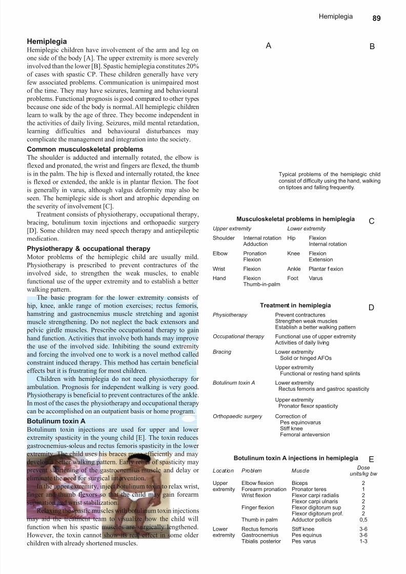
8/9/2019 Guía de ayuda para la parálisis cerebral.pdf
http://slidepdf.com/reader/full/guia-de-ayuda-para-la-paralisis-cerebralpdf 89/143
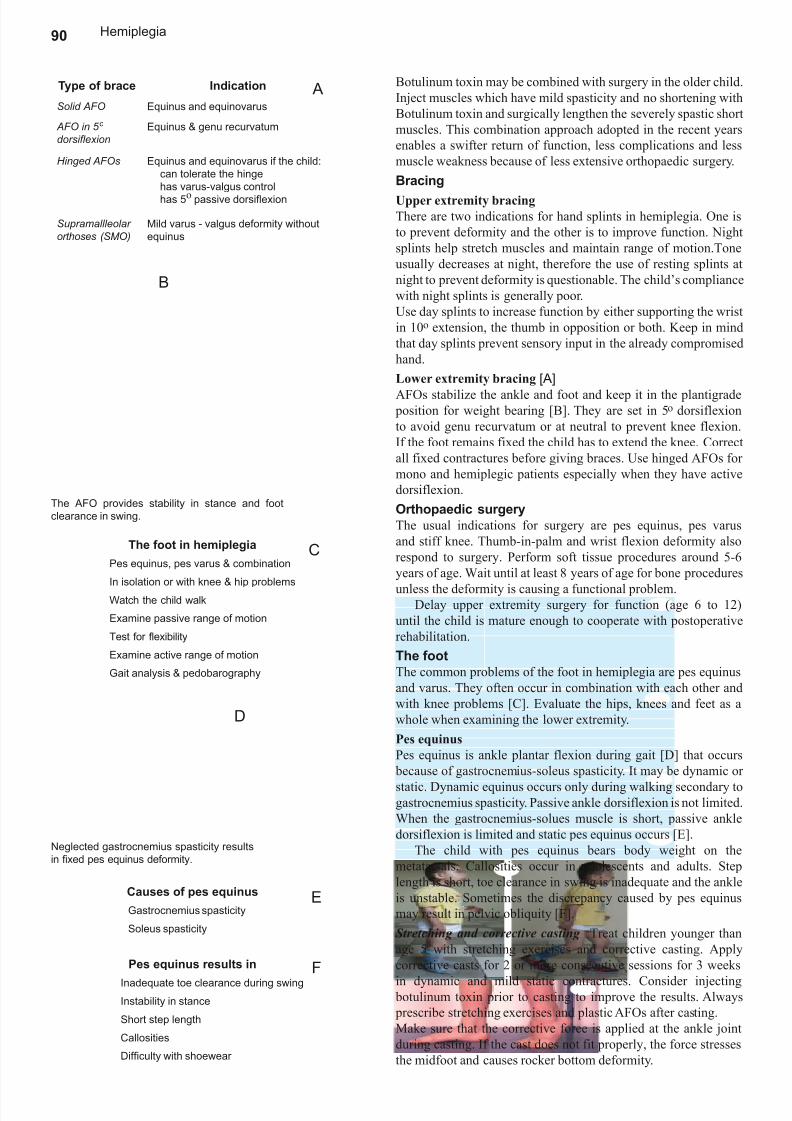
8/9/2019 Guía de ayuda para la parálisis cerebral.pdf
http://slidepdf.com/reader/full/guia-de-ayuda-para-la-paralisis-cerebralpdf 90/143
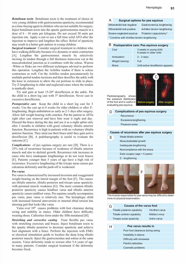
8/9/2019 Guía de ayuda para la parálisis cerebral.pdf
http://slidepdf.com/reader/full/guia-de-ayuda-para-la-paralisis-cerebralpdf 91/143
91Hemiplegia
Pes varus results in
Poor foot clearance during swing
Instability in stance
Difficulty with shoewear
Painful callosities
Cosmetic problems
Causes of the varus foot
Tibialis posterior spasticity Hindfoot varus
Tibialis anterior spasticity Midfoot varus
Triceps surae spasticity Ankle varus
Causes of recurrence after pes equinus surgery
Weak tibialis anterior
Musculoskeletal growth
Inadequate lengthening
Noncompliance with the brace
Early surgery (age < 5 years)
Z - lengthening
Postoperative care: Pes equinus surgeryCast 3 weeks in young child
6 weeks in older child
Ambulation 2 - 3 days
Weight bearing Full
Brace Until active dorsiflexion appears
Complications of pes equinus surgery
Recurrence
Excessive lengthening
Pressure sores
Surgical options for pes equinus
Silfverskiöld test negative Gastrocnemius lengthening
Silfverskiöld test positive Achilles tendon lengthening
Severe neglected equinus Posterior capsulotomy*
* Combine with Achilles tendon lengthening
G
G
H
H
E
E
D
D
B
B
A
Pedobarography shows
the pressure distributionof the foot and is useful in
evaluating pes equinus.
The muscle responsible for pes varus may be difficult to deter-
mine on physical examination.
C
C
Before surgeryBefore surgery fter surgery After surgery
F
F
Botulinum toxin Botulinum toxin is the treatment of choice invery young children with gastrocnemius spasticity, recommendedas a time-buying agent in children who are not suitable for surgery.Inject botulinum toxin into the spastic gastrocnemius muscle in adose of 6 - 10 units per kilogram. Do not exceed 50 units perinjection site. Apply a cast or use a full time solid AFO after theinjection to improve and lengthen the effect. Relief of spasticity
may result in a better gait pattern in young children. Surgical treatment Consider surgical treatment in children whohave walking difficulty because of a dynamic or static contracture[A]. Lengthen the gastrocnemius muscle by selectivelyincising its tendon through a full thickness transverse cut at themusculoskeletal junction as it combines with the soleus. Warren-White or Hoke are two different techniques advised to performthis operation. Lengthen the Achilles tendon if there is soleuscontracture as well. Cut the Achilles tendon percutaneously bymultiple partial tendon incisions and then dorsiflex the ankle withthe knee in extension to allow the cut portions to slide in place.Try Z-lengthening in older and neglected cases where the tendonis markedly short.
Try and gain at least 15-20o dorsiflexion at the ankle. Putthe child in a short leg cast with 5o dorsiflexion. Never cast inexcessive dorsiflexion.
Postoperative care Keep the child in a short leg cast for 3weeks. Use the cast up to 6 weeks for older children or after Z-lengthening. Begin ambulation as early as 2-3 days after surgery.Allow full weight bearing with crutches. Put the patient in AFOsright after cast removal and have him wear it night and day.Discard the brace during the day and use it as a night splint onlyafter 3 months in children with good voluntary tibialis anteriorfunction. Recurrence is high in patients with no voluntary tibialisanterior function. They must use their brace until they gain active
dorsiflexion [B]. A pedobarography is useful to evaluate theoutcome [C].
Complications of pes equinus surgery are rare [D]. There is a25% risk of recurrence because of weakness of tibialis anteriormuscle and also to skeletal growth. Recurrence risk increases incases who have inadequate lengthenings or do not wear braces[E]. Patients younger than 5 years of age have a high risk ofrecurrence. Excessive lengthening of the triceps surae causes pescalcaneus deformity and the push-off is weakened.
Pes varus
Pes varus is characterized by increased inversion and exaggeratedweight bearing on the lateral margin of the foot [F]. The causesare tibialis anterior, tibialis posterior and triceps surae spasticitywith peroneal muscle weakness [G]. The more common tibialis posterior spasticity causes hindfoot varus and tibialis anteriorspasticity causes midfoot varus. Pes equinus usually accompanies pes varus, pure varus is relatively rare. The hemiplegic childwith increased femoral anteversion or internal tibial torsion hasintoeing gait that looks like varus.
Varus over 10o causes problems with foot clearance duringswing and stability in stance. Older children have difficultywearing shoes. Callosities form under the fifth metatarsal [H].
Stretching and corrective casting Treat flexible pes varuswith stretching exercises and braces. Inject botulinum toxin tothe spastic tibialis posterior to decrease spasticity and achieve
foot alignment with a brace. Perform the injection with EMGor electrical stimulation guide to localize the deep lying tibialis posterior muscle. Inject the gastrocnemius and soleus at the samesession. Varus deformity tends to worsen after 5-6 years of agein many patients. Consider surgical treatment if the deformity becomes fixed.
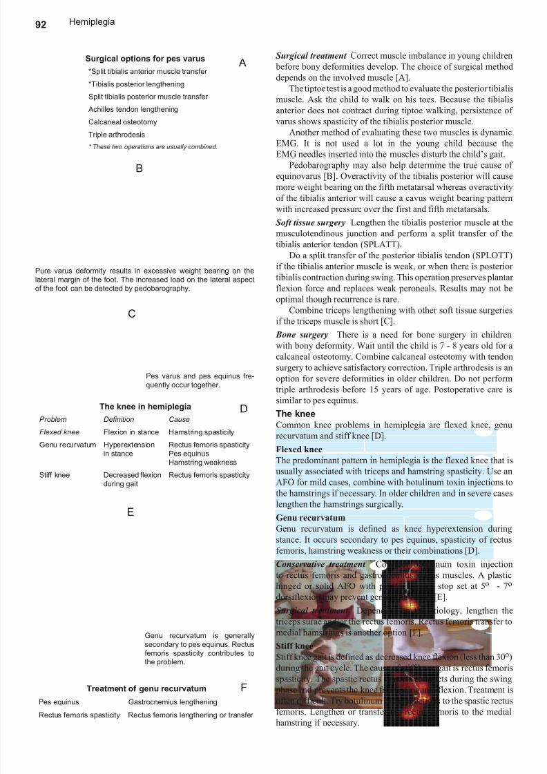
8/9/2019 Guía de ayuda para la parálisis cerebral.pdf
http://slidepdf.com/reader/full/guia-de-ayuda-para-la-paralisis-cerebralpdf 92/143
92 Hemiplegia
Treatment of genu recurvatum
Pes equinus Gastrocnemius lengthening
Rectus femoris spasticity Rectus femoris lengthening or transfer
The knee in hemiplegiaProblem Definition Cause
Flexed knee Flexion in stance Hamstring spasticity
Genu recurvatum Hyperextension
in stance
Rectus femoris spasticity
Pes equinus
Hamstring weakness
Stiff knee Decreased flexion
during gait
Rectus femoris spasticity
F
F
Surgical treatment Correct muscle imbalance in young children before bony deformities develop. The choice of surgical methoddepends on the involved muscle [A].
The tiptoe test is a good method to evaluate the posterior tibialismuscle. Ask the child to walk on his toes. Because the tibialisanterior does not contract during tiptoe walking, persistence ofvarus shows spasticity of the tibialis posterior muscle.
Another method of evaluating these two muscles is dynamicEMG. It is not used a lot in the young child because theEMG needles inserted into the muscles disturb the child’s gait.
Pedobarography may also help determine the true cause ofequinovarus [B]. Overactivity of the tibialis posterior will causemore weight bearing on the fifth metatarsal whereas overactivityof the tibialis anterior will cause a cavus weight bearing patternwith increased pressure over the first and fifth metatarsals.
Soft tissue surgery Lengthen the tibialis posterior muscle at themusculotendinous junction and perform a split transfer of thetibialis anterior tendon (SPLATT).
Do a split transfer of the posterior tibialis tendon (SPLOTT)if the tibialis anterior muscle is weak, or when there is posteriortibialis contraction during swing. This operation preserves plantarflexion force and replaces weak peroneals. Results may not beoptimal though recurrence is rare.
Combine triceps lengthening with other soft tissue surgeriesif the triceps muscle is short [C].
Bone surgery There is a need for bone surgery in childrenwith bony deformity. Wait until the child is 7 - 8 years old for acalcaneal osteotomy. Combine calcaneal osteotomy with tendonsurgery to achieve satisfactory correction. Triple arthrodesis is anoption for severe deformities in older children. Do not performtriple arthrodesis before 15 years of age. Postoperative care issimilar to pes equinus.
The knee
Common knee problems in hemiplegia are flexed knee, genurecurvatum and stiff knee [D].
Flexed knee
The predominant pattern in hemiplegia is the flexed knee that isusually associated with triceps and hamstring spasticity. Use anAFO for mild cases, combine with botulinum toxin injections tothe hamstrings if necessary. In older children and in severe caseslengthen the hamstrings surgically.
Genu recurvatum
Genu recurvatum is defined as knee hyperextension during
stance. It occurs secondary to pes equinus, spasticity of rectusfemoris, hamstring weakness or their combinations [D].
Conservative treatment Consider botulinum toxin injectionto rectus femoris and gastrocnemius-soleus muscles. A plastichinged or solid AFO with plantar flexion stop set at 5o - 7o dorsiflexion may prevent genu recurvatum [E].
Surgical treatment Depending on the etiology, lengthen thetriceps surae and/or the rectus femoris. Rectus femoris transfer tomedial hamstrings is another option [F].
Stiff knee
Stiff knee gait is defined as decreased knee flexion (less than 30o)during the gait cycle. The cause of stiff knee gait is rectus femoris
spasticity. The spastic rectus femoris contracts during the swing phase and prevents the knee from going into flexion. Treatment isoften difficult. Try botulinum toxin injections to the spastic rectusfemoris. Lengthen or transfer the rectus femoris to the medialhamstring if necessary.
Surgical options for pes varus
*Split tibialis anterior muscle transfer
*Tibialis posterior lengthening
Split tibialis posterior muscle transfer
Achilles tendon lengthening
Calcaneal osteotomy
Triple arthrodesis* These two operations are usually combined.
D
D
A
E
E
Genu recurvatum is generally
secondary to pes equinus. Rectus
femoris spasticity contributes tothe problem.
Pes varus and pes equinus fre-
quently occur together.
B
B
Pure varus deformity results in excessive weight bearing on thelateral margin of the foot. The increased load on the lateral aspect
of the foot can be detected by pedobarography.
C
C
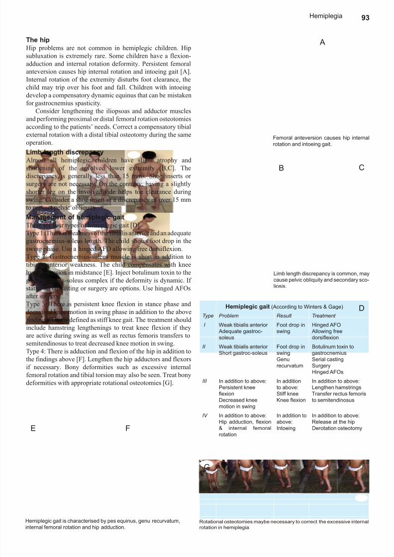
8/9/2019 Guía de ayuda para la parálisis cerebral.pdf
http://slidepdf.com/reader/full/guia-de-ayuda-para-la-paralisis-cerebralpdf 93/143
93Hemiplegia
The hip
Hip problems are not common in hemiplegic children. Hipsubluxation is extremely rare. Some children have a flexion-adduction and internal rotation deformity. Persistent femoralanteversion causes hip internal rotation and intoeing gait [A].Internal rotation of the extremity disturbs foot clearance, thechild may trip over his foot and fall. Children with intoeing
develop a compensatory dynamic equinus that can be mistakenfor gastrocnemius spasticity.Consider lengthening the iliopsoas and adductor muscles
and performing proximal or distal femoral rotation osteotomiesaccording to the patients’ needs. Correct a compensatory tibialexternal rotation with a distal tibial osteotomy during the sameoperation.
Limb length discrepancy
Almost all hemiplegic children have slight atrophy andshortening of the involved lower extremity [B,C]. Thediscrepancy is generally less than 15 mms. Shoe inserts orsurgery are not necessary. On the contrary, having a slightly
shorter leg on the involved side helps toe clearance duringswing. Consider a shoe insert in a discrepancy of over 15 mmto prevent pelvic obliquity.
Management of hemiplegic gait
There are four types of hemiplegic gait [D].Type 1: There is weakness of the tibialis anterior and an adequategastrocnemius-soleus length. The child shows foot drop in theswing phase. Use a hinged AFO allowing free dorsiflexion.Type 2: Gastrocnemius-soleus muscle is short in addition totibialis anterior weakness. The child compensates with kneehyperextension in midstance [E]. Inject botulinum toxin to thegastrocnemius-soleus complex if the deformity is dynamic. Ifstatic, serial casting or surgery are options. Use hinged AFOsafter surgery.Type 3: There is persistent knee flexion in stance phase anddecreased knee motion in swing phase in addition to the abovefindings. This is defined as stiff knee gait. The treatment shouldinclude hamstring lengthenings to treat knee flexion if theyare active during swing as well as rectus femoris transfers tosemitendinosus to treat decreased knee motion in swing.Type 4: There is adduction and flexion of the hip in addition tothe findings above [F]. Lengthen the hip adductors and flexorsif necessary. Bony deformities such as excessive internalfemoral rotation and tibial torsion may also be seen. Treat bonydeformities with appropriate rotational osteotomies [G].
F
F
Hemiplegic gait (According to Winters & Gage)
Type Problem Result Treatment
I Weak tibialis anterior
Adequate gastroc-
soleus
Foot drop in
swing
Hinged AFO
Allowing free
dorsiflexion
II Weak tibialis anterior
Short gastroc-soleus
Foot drop in
swing
Genu
recurvatum
Botulinum toxin to
gastrocnemius
Serial casting
Surgery
Hinged AFOs
III In addition to above:
Persistent knee
flexion
Decreased knee
motion in swing
In addition
to above:
Stiff knee
Knee flexion
In addition to above:
Lengthen hamstrings
Transfer rectus femoris
to semitendinosus
IV In addition to above:
Hip adduction, flexion
& internal femoral
rotation
In addition to
above:
Intoeing
In addition to above:
Release at the hip
Derotation osteotomy
D
D
G
G
C
CB
A
E
E
Femoral anteversion causes hip internal
rotation and intoeing gait.
Limb length discrepancy is common, maycause pelvic obliquity and secondary sco-
liosis.
Hemiplegic gait is characterised by pes equinus, genu recurvatum,
internal femoral rotation and hip adduction.Rotational osteotomies maybe necessary to correct the excessive internal
rotation in hemiplegia.
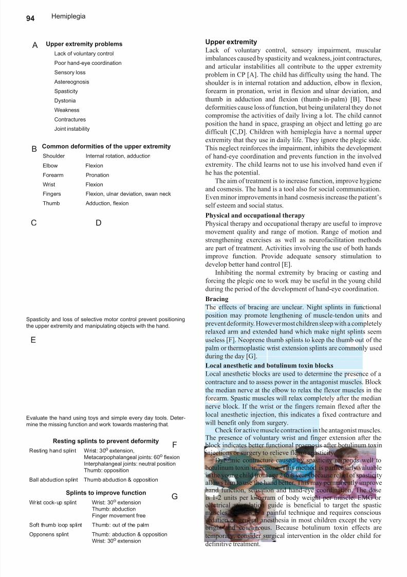
8/9/2019 Guía de ayuda para la parálisis cerebral.pdf
http://slidepdf.com/reader/full/guia-de-ayuda-para-la-paralisis-cerebralpdf 94/143
94 Hemiplegia
Resting splints to prevent deformity
Resting hand splint Wrist: 30o extension,
Metacarpophalangeal joints: 60o flexion
Interphalangeal joints: neutral position
Thumb: opposition
Ball abduction splint Thumb abduction & opposition
Splints to improve function
Wrist cock-up splint Wrist: 30o extension
Thumb: abduction
Finger movement free
Soft thumb loop splint Thumb: out of the palm
Opponens splint Thumb: abduction & opposition
Wrist: 30o extension
G
G
E
E
Upper extremity
Lack of voluntary control, sensory impairment, muscularimbalances caused by spasticity and weakness, joint contractures,and articular instabilities all contribute to the upper extremity problem in CP [A]. The child has difficulty using the hand. Theshoulder is in internal rotation and adduction, elbow in flexion,forearm in pronation, wrist in flexion and ulnar deviation, and
thumb in adduction and flexion (thumb-in-palm) [B]. Thesedeformities cause loss of function, but being unilateral they do notcompromise the activities of daily living a lot. The child cannot position the hand in space, grasping an object and letting go aredifficult [C,D]. Children with hemiplegia have a normal upperextremity that they use in daily life. They ignore the plegic side.This neglect reinforces the impairment, inhibits the developmentof hand-eye coordination and prevents function in the involvedextremity. The child learns not to use his involved hand even ifhe has the potential.
The aim of treatment is to increase function, improve hygieneand cosmesis. The hand is a tool also for social communication.Even minor improvements in hand cosmesis increase the patient’sself esteem and social status.
Physical and occupational therapy
Physical therapy and occupational therapy are useful to improvemovement quality and range of motion. Range of motion andstrengthening exercises as well as neurofacilitation methodsare part of treatment. Activities involving the use of both handsimprove function. Provide adequate sensory stimulation todevelop better hand control [E].
Inhibiting the normal extremity by bracing or casting andforcing the plegic one to work may be useful in the young childduring the period of the development of hand-eye coordination.
Bracing
The effects of bracing are unclear. Night splints in functional position may promote lengthening of muscle-tendon units and prevent deformity. However most children sleep with a completelyrelaxed arm and extended hand which make night splints seemuseless [F]. Neoprene thumb splints to keep the thumb out of the palm or thermoplastic wrist extension splints are commonly usedduring the day [G].
Local anesthetic and botulinum toxin blocks
Local anesthetic blocks are used to determine the presence of acontracture and to assess power in the antagonist muscles. Blockthe median nerve at the elbow to relax the flexor muscles in theforearm. Spastic muscles will relax completely after the median
nerve block. If the wrist or the fingers remain flexed after thelocal anesthetic injection, this indicates a fixed contracture andwill benefit only from surgery.
Check for active muscle contraction in the antagonist muscles.The presence of voluntary wrist and finger extension after the block indicates better functional prognosis after botulinum toxininjections or surgery to relieve flexor spasticity.
Dynamic contracture caused by spasticity responds well to botulinum toxin injections. This method is particularly valuablein the young child from age 2 to 6 years because relief of spasticityallows him to use the hand better. This may permanently improvehand function, sensation and hand-eye coordination. The doseis 1-2 units per kilogram of body weight per muscle. EMG or
electrical stimulation guide is beneficial to target the spasticmuscles, but this is a painful technique and requires conscioussedation or general anesthesia in most children except the very bright and courageous. Because botulinum toxin effects aretemporary, consider surgical intervention in the older child fordefinitive treatment.
Upper extremity problems
Lack of voluntary control
Poor hand-eye coordination
Sensory loss
Astereognosis
Spasticity
Dystonia
Weakness
Contractures
Joint instability
Common deformities of the upper extremity
Shoulder Internal rotation, adduction
Elbow Flexion
Forearm Pronation
Wrist Flexion
Fingers Flexion, ulnar deviation, swan neck
Thumb Adduction, flexion
D
DC
B
B
A
Evaluate the hand using toys and simple every day tools. Deter-
mine the missing function and work towards mastering that.
Spasticity and loss of selective motor control prevent positioning
the upper extremity and manipulating objects with the hand.
F
F
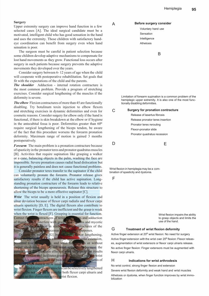
8/9/2019 Guía de ayuda para la parálisis cerebral.pdf
http://slidepdf.com/reader/full/guia-de-ayuda-para-la-paralisis-cerebralpdf 95/143
95Hemiplegia
Indications for wrist arthrodesis
No wrist control, strong finger flexion and extension
Severe wrist flexion deformity and weak hand and wrist muscles
Athetosis or dystonia, when finger function improves by wrist immo-
bilization
Treatment of wrist flexion deformity
Active finger extension at 20o wrist flexion: No need for surgery
Active finger extension with the wrist over 20o flexion: Flexor releas-
es, augmentation of wrist extensors or flexor carpi ulnaris release.
No active finger flexion: Finger extensors must be augmented with
flexor carpi ulnaris.
Surgery for pronation contracture
Release of lasertus fibrosis
Release pronator teres insertion
Pronator teres rerouting
Flexor-pronator slide
Pronator quadratus recession
Surgery
Upper extremity surgery can improve hand function in a fewselected cases [A]. The ideal surgical candidate must be amotivated, intelligent child who has good sensation in the handand uses the extremity. Those children with satisfactory hand-eye coordination can benefit from surgery even when handsensation is poor.
The surgeon must be careful in patient selection becausesome children develop adaptive mechanisms to compensate forlost hand movements as they grow. Functional loss occurs aftersurgery in such patients because surgery prevents the adaptivemovements they developed over the years.
Consider surgery between 6- 12 years of age when the childwill cooperate with postoperative rehabilitation. Set goals thatfit with the expectations of the child and the parents.The shoulder Adduction - internal rotation contracture isthe most common problem. Provide a program of stretchingexercises. Consider surgical lengthening of the muscles if thedeformity is severe.
The elbow Flexion contractures of more than 45 are functionallydisabling. Try botulinum toxin injection to elbow flexorsand stretching exercises in dynamic deformities and even forcosmetic reasons. Consider surgery for elbow only if the hand isfunctional, if there is skin breakdown at the elbow or if hygienein the antecubital fossa is poor. Deformities greater than 60o require surgical lengthening of the biceps tendon, be awareof the fact that this procedure worsens the forearm pronationdeformity. Maximum range of motion is gained 3 months postoperatively.
Forearm The main problem is a pronation contracture becauseof spasticity in the pronator teres and pronator quadratus muscles
[B]. Activities that require supination like grasping a walkeror a cane, balancing objects in the palm, washing the face areimpossible. Severe pronation causes radial head dislocation butit is generally painless and does not cause functional problems.
Consider pronator teres transfer to the supinator if the childcan voluntarily pronate the forearm. Pronator release givessatisfactory results if the child has active supination. Long-standing pronation contracture of the forearm leads to relativeshortening of the biceps aponeurosis. Release this structure toallow the biceps to be a more effective supinator [C].
Wrist The wrist usually is held in a position of flexion andulnar deviation because of flexor carpi radialis and flexor carpiulnaris spasticity [D, E]. The digital flexors also contribute towrist flexion. Finger flexors are inefficient and the grasp is weakwhen the wrist is flexed [F]. Grasping is essential for function.Correct flexion contractures of wrist and fingers and adductionof thumb if they interfere with grasp. Macerations and mycoticinfections are common in severe flexion contractures of thehand. Surgery becomes necessary for hygienic purposes.
Options for surgery [G] include wrist flexor lengthening,flexor origin slide, tendon transfer to improve wrist extension, proximal row carpectomy, and wrist fusion with or withoutcarpal shortening [H]. Avoid wrist arthrodesis because the patient loses the tenodesis effect of wrist extension that resultsin finger flexion and facilitates grasp and release. Consider wrist
arthrodesis only to relieve the pain and improve the cosmesis ofthe hand when there is no or limited hand function.
Wrist and digital flexor muscles can be selectively lengtheneddistally. Do not release or transfer both flexor carpi ulnaris andradialis as this eliminates active wrist flexion.
Before surgery consider
Voluntary hand use
Sensation
Intelligence
Athetosis
F
F
E
E
H
H
G
G
C
C
A
B
B
Limitation of forearm supination is a common problem of the
hemiplegic upper extremity. It is also one of the most func-
tionally disabling deformities.
D
D
Wrist flexion impairs the ability
to grasp objects and limits theuse of the hand.
Wrist flexion in hemiplegia may be a com-
bination of spasticity and dystonia.
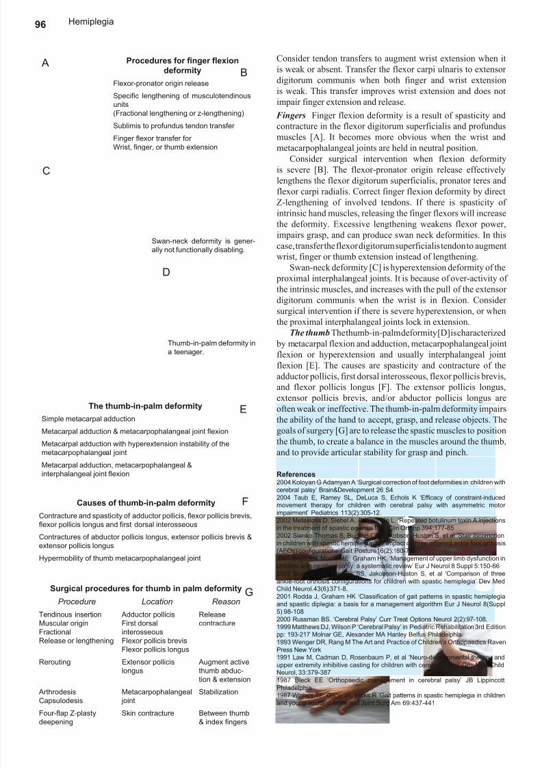
8/9/2019 Guía de ayuda para la parálisis cerebral.pdf
http://slidepdf.com/reader/full/guia-de-ayuda-para-la-paralisis-cerebralpdf 96/143
96 Hemiplegia
Surgical procedures for thumb in palm deformity
Procedure Location Reason
Tendinous insertion
Muscular origin
Fractional
Release or lengthening
Adductor pollicis
First dorsal
interosseous
Flexor pollicis brevis
Flexor pollicis longus
Release
contracture
Rerouting Extensor pollicis
longus
Augment active
thumb abduc-tion & extension
Arthrodesis
Capsulodesis
Metacarpophalangeal
joint
Stabilization
Four-flap Z-plasty
deepening
Skin contracture Between thumb
& index fingers
Causes of thumb-in-palm deformity
Contracture and spasticity of adductor pollicis, flexor pollicis brevis,
flexor pollicis longus and first dorsal interosseous
Contractures of abductor pollicis longus, extensor pollicis brevis &extensor pollicis longus
Hypermobility of thumb metacarpophalangeal joint
The thumb-in-palm deformity
Simple metacarpal adduction
Metacarpal adduction & metacarpophalangeal joint flexion
Metacarpal adduction with hyperextension instability of the
metacarpophalangeal joint
Metacarpal adduction, metacarpophalangeal &
interphalangeal joint flexion
Procedures for finger flexion
deformity
Flexor-pronator origin release
Specific lengthening of musculotendinous
units
(Fractional lengthening or z-lengthening)
Sublimis to profundus tendon transfer
Finger flexor transfer for
Wrist, finger, or thumb extension
F
F
E
E
D
D
G
G
Consider tendon transfers to augment wrist extension when itis weak or absent. Transfer the flexor carpi ulnaris to extensordigitorum communis when both finger and wrist extensionis weak. This transfer improves wrist extension and does notimpair finger extension and release.
Fingers Finger flexion deformity is a result of spasticity andcontracture in the flexor digitorum superficialis and profundus
muscles [A]. It becomes more obvious when the wrist andmetacarpophalangeal joints are held in neutral position.
Consider surgical intervention when flexion deformityis severe [B]. The flexor-pronator origin release effectivelylengthens the flexor digitorum superficialis, pronator teres andflexor carpi radialis. Correct finger flexion deformity by directZ-lengthening of involved tendons. If there is spasticity ofintrinsic hand muscles, releasing the finger flexors will increasethe deformity. Excessive lengthening weakens flexor power,impairs grasp, and can produce swan neck deformities. In thiscase, transfer the flexor digitorum superficialis tendon to augmentwrist, finger or thumb extension instead of lengthening.
Swan-neck deformity [C] is hyperextension deformity of the proximal interphalangeal joints. It is because of over-activity ofthe intrinsic muscles, and increases with the pull of the extensordigitorum communis when the wrist is in flexion. Considersurgical intervention if there is severe hyperextension, or whenthe proximal interphalangeal joints lock in extension.
The thumb The thumb-in-palm deformity [D] is characterized by metacarpal flexion and adduction, metacarpophalangeal jointflexion or hyperextension and usually interphalangeal jointflexion [E]. The causes are spasticity and contracture of theadductor pollicis, first dorsal interosseous, flexor pollicis brevis,and flexor pollicis longus [F]. The extensor pollicis longus,extensor pollicis brevis, and/or abductor pollicis longus areoften weak or ineffective. The thumb-in-palm deformity impairsthe ability of the hand to accept, grasp, and release objects. Thegoals of surgery [G] are to release the spastic muscles to positionthe thumb, to create a balance in the muscles around the thumb,and to provide articular stability for grasp and pinch.
References2004 Koloyan G Adamyan A ‘Surgical correction of foot deformities in children with
cerebral palsy’ Brain&Development 26 S4
2004 Taub E, Ramey SL, DeLuca S, Echols K ‘Efficacy of constraint-inducedmovement therapy for children with cerebral palsy with asymmetric motor
impairment’ Pediatrics 113(2):305-12.
2002 Metaxiotis D, Siebel A, Doederlein L. ‘Repeated botulinum toxin A injectionsin the treatment of spastic equinus foot’ Clin Orthop 394:177-85
2002 Sienko Thomas S, Buckon CE, Jakobson-Huston S, et al ‘Stair locomotionin children with spastic hemiplegia: the impact of three different ankle foot orthosis(AFOs) configurations’ Gait Posture16(2):180-7.
2001 Boyd RN, Morris ME, Graham HK. ‘Management of upper limb dysfunction inchildren with cerebral palsy: a systematic review’ Eur J Neurol 8 Suppl 5:150-66
2001 Buckon CE, Thomas SS, Jakobson-Huston S, et al ‘Comparison of three
ankle-foot orthosis configurations for children with spastic hemiplegia’ Dev MedChild Neurol.43(6):371-8.
2001 Rodda J, Graham HK ‘Classification of gait patterns in spastic hemiplegia
and spastic diplegia: a basis for a management algorithm Eur J Neurol 8(Suppl5) 98-108
2000 Russman BS. ‘Cerebral Palsy’ Curr Treat Options Neurol 2(2):97-108.
1999 Matthews DJ, Wilson P ‘Cerebral Palsy’ in Pediatric Rehabilitation 3rd Editionpp: 193-217 Molnar GE, Alexander MA Hanley Belfus Philadelphia
1993 Wenger DR, Rang M The Art and Practice of Children’s Orthopaedics Raven
Press New York1991 Law M, Cadman D, Rosenbaum P, et al ‘Neuro-developmental therapy and
upper extremity inhibitive casting for children with cerebral palsy’ Dev Med Child
Neurol, 33:379-3871987 Bleck EE ‘Orthopaedic management in cerebral palsy’ JB Lippincott
Philadelphia1987 Winters TF, Gage JR, Hicks R ‘Gait patterns in spastic hemiplegia in children
and young adults’ J Bone and Joint Surg Am 69:437-441
C
C
B
B
A
Thumb-in-palm deformity in
a teenager.
Swan-neck deformity is gener-
ally not functionally disabling.
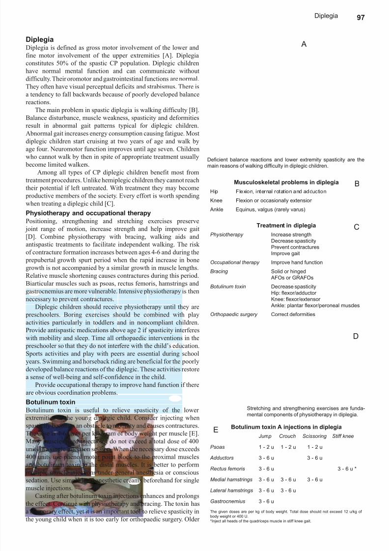
8/9/2019 Guía de ayuda para la parálisis cerebral.pdf
http://slidepdf.com/reader/full/guia-de-ayuda-para-la-paralisis-cerebralpdf 97/143
97Diplegia
Botulinum toxin A injections in diplegia
Jump Crouch Scissoring Stiff knee
Psoas 1 - 2 u 1 - 2 u 1 - 2 u
Adductors 3 - 6 u 3 - 6 u
Rectus femoris 3 - 6 u 3 - 6 u *
Medial hamstrings 3 - 6 u 3 - 6 u 3 - 6 u
Lateral hamstrings 3 - 6 u 3 - 6 u
Gastrocnemius 3 - 6 u
The given doses are per kg of body weight. Total dose should not exceed 12 u/kg of
body weight or 400 U.
*Inject all heads of the quadriceps muscle in stiff knee gait.
DiplegiaDiplegia is defined as gross motor involvement of the lower andfine motor involvement of the upper extremities [A]. Diplegiaconstitutes 50% of the spastic CP population. Diplegic childrenhave normal mental function and can communicate withoutdifficulty. Their oromotor and gastrointestinal functions are normal.They often have visual perceptual deficits and strabismus. There is
a tendency to fall backwards because of poorly developed balancereactions.The main problem in spastic diplegia is walking difficulty [B].
Balance disturbance, muscle weakness, spasticity and deformitiesresult in abnormal gait patterns typical for diplegic children.Abnormal gait increases energy consumption causing fatigue. Mostdiplegic children start cruising at two years of age and walk byage four. Neuromotor function improves until age seven. Childrenwho cannot walk by then in spite of appropriate treatment usually become limited walkers.
Among all types of CP diplegic children benefit most fromtreatment procedures. Unlike hemiplegic children they cannot reachtheir potential if left untreated. With treatment they may become
productive members of the society. Every effort is worth spendingwhen treating a diplegic child [C].
Physiotherapy and occupational therapy
Positioning, strengthening and stretching exercises preserve joint range of motion, increase strength and help improve gait[D]. Combine physiotherapy with bracing, walking aids andantispastic treatments to facilitate independent walking. The riskof contracture formation increases between ages 4-6 and during the prepubertal growth spurt period when the rapid increase in bonegrowth is not accompanied by a similar growth in muscle lengths.Relative muscle shortening causes contractures during this period.Biarticular muscles such as psoas, rectus femoris, hamstrings and
gastrocnemius are more vulnerable. Intensive physiotherapy is thennecessary to prevent contractures.
Diplegic children should receive physiotherapy until they are preschoolers. Boring exercises should be combined with playactivities particularly in toddlers and in noncompliant children.Provide antispastic medications above age 2 if spasticity interfereswith mobility and sleep. Time all orthopaedic interventions in the preschooler so that they do not interfere with the child’s education.Sports activities and play with peers are essential during schoolyears. Swimming and horseback riding are beneficial for the poorlydeveloped balance reactions of the diplegic. These activities restorea sense of well-being and self-confidence in the child.
Provide occupational therapy to improve hand function if thereare obvious coordination problems.
Botulinum toxin
Botulinum toxin is useful to relieve spasticity of the lowerextremities of the young diplegic child. Consider injecting whenspasticity becomes an obstacle to mobility and causes contractures.The dose is 4-6 units per kilogram of body weight per muscle [E].Many muscles need injections, do not exceed a total dose of 400units in a single injection session. When the necessary dose exceeds400 units use phenol motor point block to the proximal musclesand botulinum toxin to the distal muscles. It is better to performmultiple muscle injections under general anesthesia or conscious
sedation. Use simple local anesthetic creams beforehand for singlemuscle injections.Casting after botulinum toxin injections enhances and prolongs
the effect. Continue with physiotherapy and bracing. The toxin hasa temporary effect, yet it is an important tool to relieve spasticity inthe young child when it is too early for orthopaedic surgery. Older
Musculoskeletal problems in diplegia
Hip Flexion, internal rotation and adductionKnee Flexion or occasionally extension
Ankle Equinus, valgus (rarely varus)
Treatment in diplegia
Physiotherapy Increase strength
Decrease spasticity
Prevent contractures
Improve gait
Occupational therapy Improve hand function
Bracing Solid or hinged
AFOs or GRAFOs
Botulinum toxin Decrease spasticity
Hip: flexor/adductor
Knee: flexor/extensor
Ankle: plantar flexor/peroneal muscles
Orthopaedic surgery Correct deformities
Stretching and strengthening exercises are funda-
mental components of physiotherapy in diplegia.
E
E
D
D
C
C
B
B
A
Deficient balance reactions and lower extremity spasticity are themain reasons of walking difficulty in diplegic children.
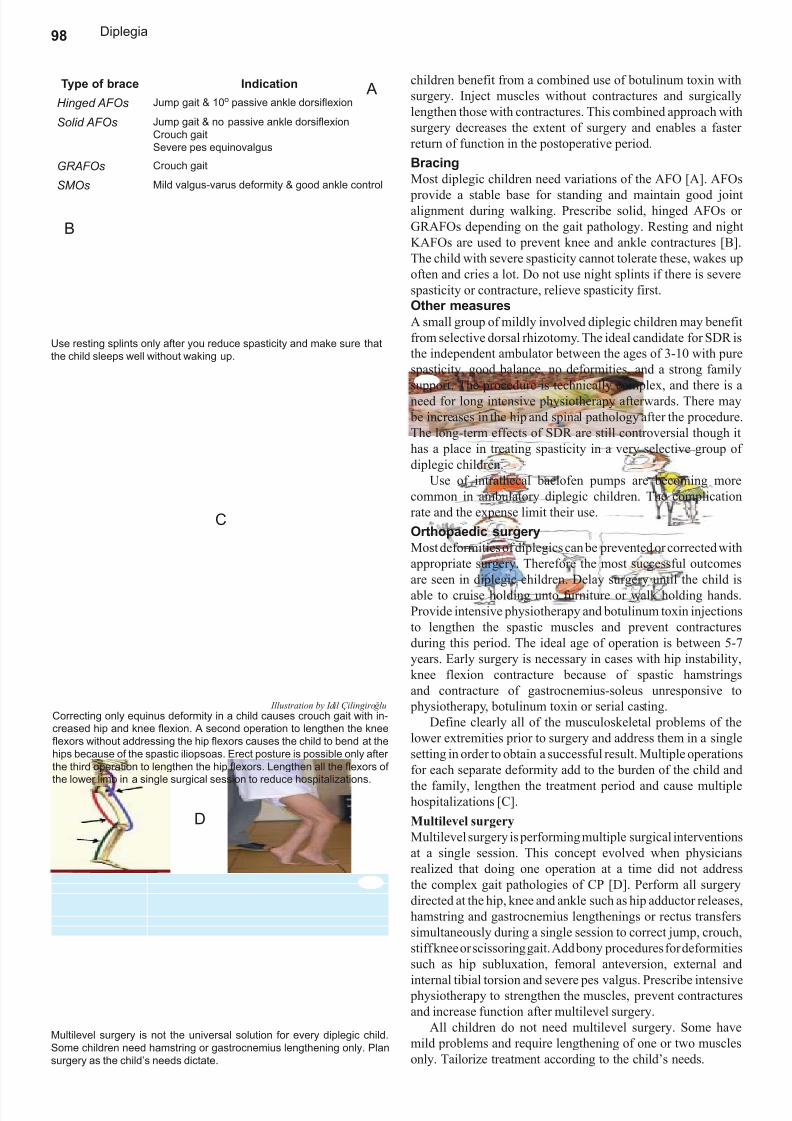
8/9/2019 Guía de ayuda para la parálisis cerebral.pdf
http://slidepdf.com/reader/full/guia-de-ayuda-para-la-paralisis-cerebralpdf 98/143
98 Diplegia
children benefit from a combined use of botulinum toxin withsurgery. Inject muscles without contractures and surgicallylengthen those with contractures. This combined approach withsurgery decreases the extent of surgery and enables a fasterreturn of function in the postoperative period.
Bracing
Most diplegic children need variations of the AFO [A]. AFOs
provide a stable base for standing and maintain good jointalignment during walking. Prescribe solid, hinged AFOs orGRAFOs depending on the gait pathology. Resting and nightKAFOs are used to prevent knee and ankle contractures [B].The child with severe spasticity cannot tolerate these, wakes upoften and cries a lot. Do not use night splints if there is severespasticity or contracture, relieve spasticity first.Other measures
A small group of mildly involved diplegic children may benefitfrom selective dorsal rhizotomy. The ideal candidate for SDR isthe independent ambulator between the ages of 3-10 with purespasticity, good balance, no deformities, and a strong familysupport. The procedure is technically complex, and there is aneed for long intensive physiotherapy afterwards. There may be increases in the hip and spinal pathology after the procedure.The long-term effects of SDR are still controversial though ithas a place in treating spasticity in a very selective group ofdiplegic children.
Use of intrathecal baclofen pumps are becoming morecommon in ambulatory diplegic children. The complicationrate and the expense limit their use.
Orthopaedic surgery
Most deformities of diplegics can be prevented or corrected withappropriate surgery. Therefore the most successful outcomesare seen in diplegic children. Delay surgery until the child isable to cruise holding unto furniture or walk holding hands.Provide intensive physiotherapy and botulinum toxin injectionsto lengthen the spastic muscles and prevent contracturesduring this period. The ideal age of operation is between 5-7years. Early surgery is necessary in cases with hip instability,knee flexion contracture because of spastic hamstringsand contracture of gastrocnemius-soleus unresponsive to physiotherapy, botulinum toxin or serial casting.
Define clearly all of the musculoskeletal problems of thelower extremities prior to surgery and address them in a singlesetting in order to obtain a successful result. Multiple operationsfor each separate deformity add to the burden of the child and
the family, lengthen the treatment period and cause multiplehospitalizations [C].
Multilevel surgery
Multilevel surgery is performing multiple surgical interventionsat a single session. This concept evolved when physiciansrealized that doing one operation at a time did not addressthe complex gait pathologies of CP [D]. Perform all surgerydirected at the hip, knee and ankle such as hip adductor releases,hamstring and gastrocnemius lengthenings or rectus transferssimultaneously during a single session to correct jump, crouch,stiff knee or scissoring gait. Add bony procedures for deformitiessuch as hip subluxation, femoral anteversion, external and
internal tibial torsion and severe pes valgus. Prescribe intensive physiotherapy to strengthen the muscles, prevent contracturesand increase function after multilevel surgery.
All children do not need multilevel surgery. Some havemild problems and require lengthening of one or two musclesonly. Tailorize treatment according to the child’s needs.
Type of brace Indication
Hinged AFOs Jump gait & 10o passive ankle dorsiflexion
Solid AFOs Jump gait & no passive ankle dorsiflexion
Crouch gait
Severe pes equinovalgus
GRAFOs Crouch gait
SMOs Mild valgus-varus deformity & good ankle control
Correcting only equinus deformity in a child causes crouch gait with in-creased hip and knee flexion. A second operation to lengthen the knee
flexors without addressing the hip flexors causes the child to bend at the
hips because of the spastic iliopsoas. Erect posture is possible only after
the third operation to lengthen the hip flexors. Lengthen all the flexors ofthe lower limb in a single surgical session to reduce hospitalizations.
D
D
C
C
A
Multilevel surgery is not the universal solution for every diplegic child.
Some children need hamstring or gastrocnemius lengthening only. Plan
surgery as the child’s needs dictate.
Use resting splints only after you reduce spasticity and make sure that
the child sleeps well without waking up.
B
B
Illustration by Idil Çilingiroğlu
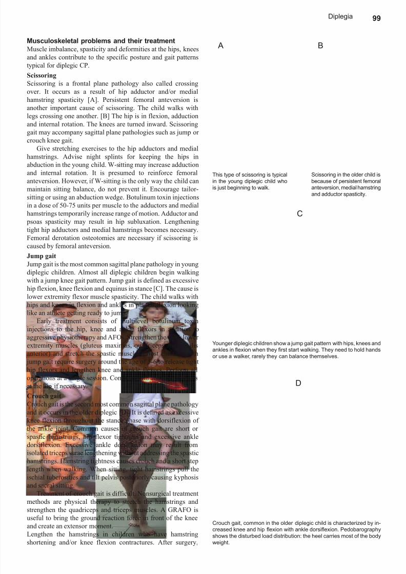
8/9/2019 Guía de ayuda para la parálisis cerebral.pdf
http://slidepdf.com/reader/full/guia-de-ayuda-para-la-paralisis-cerebralpdf 99/143
99Diplegia
Musculoskeletal problems and their treatment
Muscle imbalance, spasticity and deformities at the hips, kneesand ankles contribute to the specific posture and gait patternstypical for diplegic CP.
Scissoring
Scissoring is a frontal plane pathology also called crossingover. It occurs as a result of hip adductor and/or medial
hamstring spasticity [A]. Persistent femoral anteversion isanother important cause of scissoring. The child walks withlegs crossing one another. [B] The hip is in flexion, adductionand internal rotation. The knees are turned inward. Scissoringgait may accompany sagittal plane pathologies such as jump orcrouch knee gait.
Give stretching exercises to the hip adductors and medialhamstrings. Advise night splints for keeping the hips inabduction in the young child. W-sitting may increase adductionand internal rotation. It is presumed to reinforce femoralanteversion. However, if W-sitting is the only way the child canmaintain sitting balance, do not prevent it. Encourage tailor-sitting or using an abduction wedge. Botulinum toxin injectionsin a dose of 50-75 units per muscle to the adductors and medialhamstrings temporarily increase range of motion. Adductor and psoas spasticity may result in hip subluxation. Lengtheningtight hip adductors and medial hamstrings becomes necessary.Femoral derotation osteotomies are necessary if scissoring iscaused by femoral anteversion.
Jump gait
Jump gait is the most common sagittal plane pathology in youngdiplegic children. Almost all diplegic children begin walkingwith a jump knee gait pattern. Jump gait is defined as excessivehip flexion, knee flexion and equinus in stance [C]. The cause islower extremity flexor muscle spasticity. The child walks withhips and knees in flexion and ankles in plantar flexion lookinglike an athlete getting ready to jump.
Early treatment consists of multilevel botulinum toxininjections to the hip, knee and ankle flexors in addition toaggressive physiotherapy and AFOs. Strengthen the weak lowerextremity muscles (gluteus maximus, quadriceps and tibialisanterior) and stretch the spastic muscles. Most children with jump gait require surgery around the age of 5-6 to release tighthip flexors and lengthen knee and ankle flexors. Perform alloperations at a single session. Combine with adductor releasesat the hip if necessary.
Crouch gait
Crouch gait is the second most common sagittal plane pathologyand it occurs in the older diplegic [D]. It is defined as excessiveknee flexion throughout the stance phase with dorsiflexion ofthe ankle joint. Common causes of crouch gait are short orspastic hamstrings, hip flexor tightness and excessive ankledorsiflexion. Excessive ankle dorsiflexion may result fromisolated triceps surae lengthening without addressing the spastichamstrings. Hamstring tightness causes crouch and a short steplength when walking. When sitting, tight hamstrings pull theischial tuberosities and tilt pelvis posteriorly causing kyphosisand sacral sitting.
Treatment of crouch gait is difficult. Nonsurgical treatment
methods are physical therapy to stretch the hamstrings andstrengthen the quadriceps and triceps muscles. A GRAFO isuseful to bring the ground reaction force in front of the kneeand create an extensor moment.Lengthen the hamstrings in children who have hamstringshortening and/or knee flexion contractures. After surgery,
Scissoring in the older child is
because of persistent femoralanteversion, medial hamstring
and adductor spasticity.
Younger diplegic children show a jump gait pattern with hips, knees andankles in flexion when they first start walking. They need to hold hands
or use a walker, rarely they can balance themselves.
D
D
C
C
B
B A
Crouch gait, common in the older diplegic child is characterized by in-creased knee and hip flexion with ankle dorsiflexion. Pedobarography
shows the disturbed load distribution: the heel carries most of the body
weight.
This type of scissoring is typicalin the young diplegic child who
is just beginning to walk.
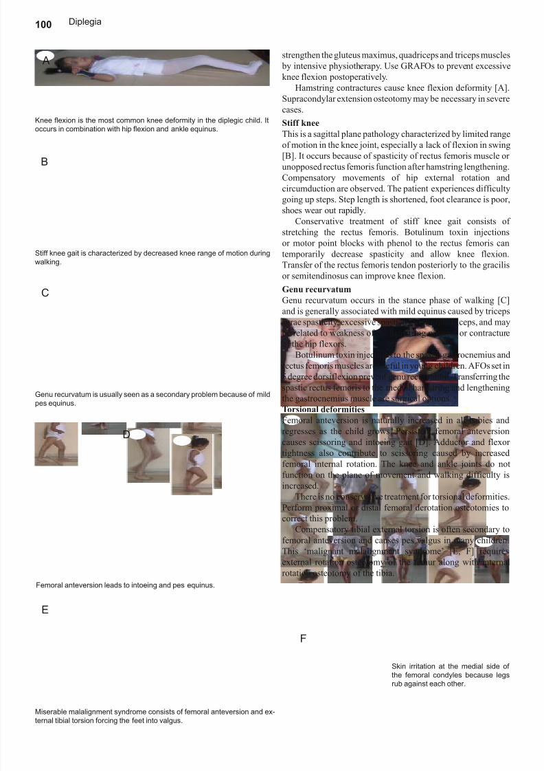
8/9/2019 Guía de ayuda para la parálisis cerebral.pdf
http://slidepdf.com/reader/full/guia-de-ayuda-para-la-paralisis-cerebralpdf 100/143
100 Diplegia
strengthen the gluteus maximus, quadriceps and triceps muscles by intensive physiotherapy. Use GRAFOs to prevent excessiveknee flexion postoperatively.
Hamstring contractures cause knee flexion deformity [A].Supracondylar extension osteotomy may be necessary in severecases.
Stiff knee
This is a sagittal plane pathology characterized by limited rangeof motion in the knee joint, especially a lack of flexion in swing[B]. It occurs because of spasticity of rectus femoris muscle orunopposed rectus femoris function after hamstring lengthening.Compensatory movements of hip external rotation andcircumduction are observed. The patient experiences difficultygoing up steps. Step length is shortened, foot clearance is poor,shoes wear out rapidly.
Conservative treatment of stiff knee gait consists ofstretching the rectus femoris. Botulinum toxin injectionsor motor point blocks with phenol to the rectus femoris cantemporarily decrease spasticity and allow knee flexion.Transfer of the rectus femoris tendon posteriorly to the gracilisor semitendinosus can improve knee flexion.
Genu recurvatum
Genu recurvatum occurs in the stance phase of walking [C]and is generally associated with mild equinus caused by tricepssurae spasticity, excessive spasticity in the quadriceps, and may be related to weakness of the hamstring muscles or contractureof the hip flexors.
Botulinum toxin injections to the spastic gastrocnemius andrectus femoris muscles are useful in young children. AFOs set in5 degree dorsiflexion prevent genu recurvatum. Transferring thespastic rectus femoris to the medial hamstring and lengtheningthe gastrocnemius muscle are surgical options.Torsional deformities
Femoral anteversion is naturally increased in all babies andregresses as the child grows. Persistent femoral anteversioncauses scissoring and intoeing gait [D]. Adductor and flexortightness also contribute to scissoring caused by increasedfemoral internal rotation. The knee and ankle joints do notfunction on the plane of movement and walking difficulty isincreased.
There is no conservative treatment for torsional deformities.Perform proximal or distal femoral derotation osteotomies tocorrect this problem.
Compensatory tibial external torsion is often secondary to
femoral anteversion and causes pes valgus in many children.This ‘malignant malalignment syndrome’ [E, F] requiresexternal rotation osteotomy of the femur along with internalrotation osteotomy of the tibia.
F
F
Miserable malalignment syndrome consists of femoral anteversion and ex-
ternal tibial torsion forcing the feet into valgus.
Genu recurvatum is usually seen as a secondary problem because of mild
pes equinus.
C
C
B
B
A
Knee flexion is the most common knee deformity in the diplegic child. Itoccurs in combination with hip flexion and ankle equinus.
Stiff knee gait is characterized by decreased knee range of motion during
walking.
Femoral anteversion leads to intoeing and pes equinus.
D
D
E
E
Skin irritation at the medial side of
the femoral condyles because legsrub against each other.
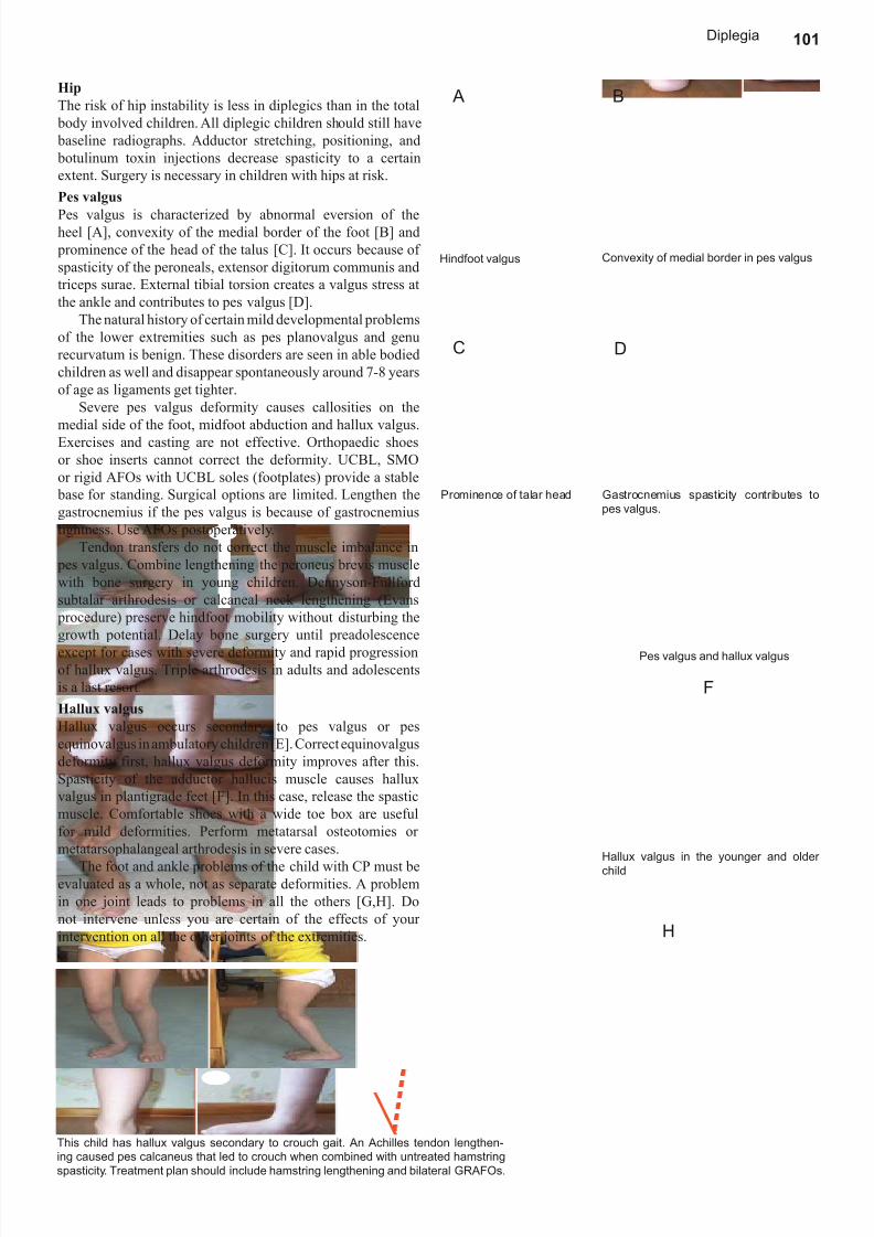
8/9/2019 Guía de ayuda para la parálisis cerebral.pdf
http://slidepdf.com/reader/full/guia-de-ayuda-para-la-paralisis-cerebralpdf 101/143
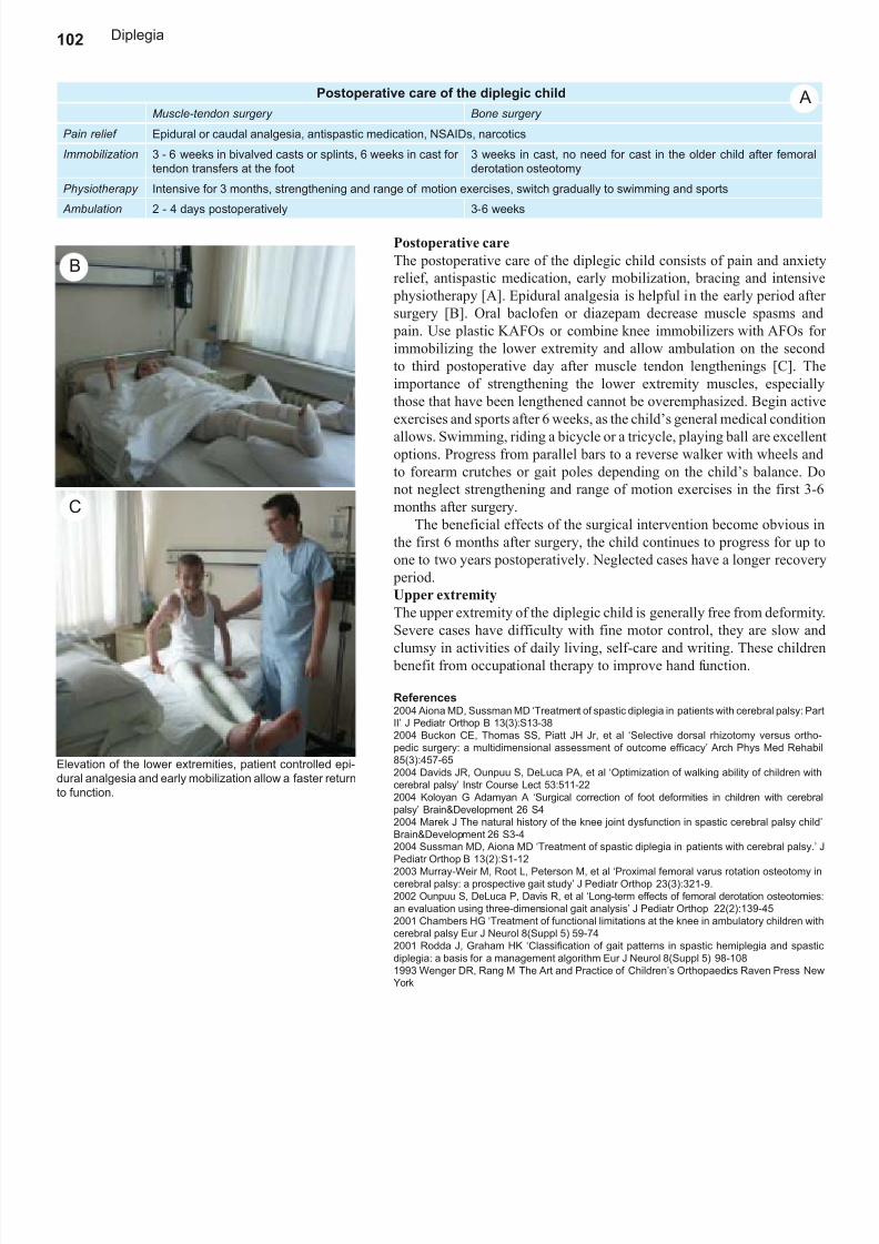
8/9/2019 Guía de ayuda para la parálisis cerebral.pdf
http://slidepdf.com/reader/full/guia-de-ayuda-para-la-paralisis-cerebralpdf 102/143
102 Diplegia
Postoperative care
The postoperative care of the diplegic child consists of pain and anxietyrelief, antispastic medication, early mobilization, bracing and intensive physiotherapy [A]. Epidural analgesia is helpful in the early period aftersurgery [B]. Oral baclofen or diazepam decrease muscle spasms and pain. Use plastic KAFOs or combine knee immobilizers with AFOs forimmobilizing the lower extremity and allow ambulation on the secondto third postoperative day after muscle tendon lengthenings [C]. Theimportance of strengthening the lower extremity muscles, especiallythose that have been lengthened cannot be overemphasized. Begin activeexercises and sports after 6 weeks, as the child’s general medical condition
allows. Swimming, riding a bicycle or a tricycle, playing ball are excellentoptions. Progress from parallel bars to a reverse walker with wheels andto forearm crutches or gait poles depending on the child’s balance. Donot neglect strengthening and range of motion exercises in the first 3-6months after surgery.
The beneficial effects of the surgical intervention become obvious inthe first 6 months after surgery, the child continues to progress for up toone to two years postoperatively. Neglected cases have a longer recovery period.Upper extremity
The upper extremity of the diplegic child is generally free from deformity.Severe cases have difficulty with fine motor control, they are slow and
clumsy in activities of daily living, self-care and writing. These children benefit from occupational therapy to improve hand function.
References2004 Aiona MD, Sussman MD ‘Treatment of spastic diplegia in patients with cerebral palsy: PartII’ J Pediatr Orthop B 13(3):S13-38
2004 Buckon CE, Thomas SS, Piatt JH Jr, et al ‘Selective dorsal rhizotomy versus ortho-
pedic surgery: a multidimensional assessment of outcome efficacy’ Arch Phys Med Rehabil
85(3):457-65
2004 Davids JR, Ounpuu S, DeLuca PA, et al ‘Optimization of walking ability of children with
cerebral palsy’ Instr Course Lect 53:511-22
2004 Koloyan G Adamyan A ‘Surgical correction of foot deformities in children with cerebral
palsy’ Brain&Development 26 S4
2004 Marek J The natural history of the knee joint dysfunction in spastic cerebral palsy child’
Brain&Development 26 S3-42004 Sussman MD, Aiona MD ‘Treatment of spastic diplegia in patients with cerebral palsy.’ J
Pediatr Orthop B 13(2):S1-12
2003 Murray-Weir M, Root L, Peterson M, et al ‘Proximal femoral varus rotation osteotomy incerebral palsy: a prospective gait study’ J Pediatr Orthop 23(3):321-9.
2002 Ounpuu S, DeLuca P, Davis R, et al ‘Long-term effects of femoral derotation osteotomies:
an evaluation using three-dimensional gait analysis’ J Pediatr Orthop 22(2):139-45
2001 Chambers HG ‘Treatment of functional limitations at the knee in ambulatory children with
cerebral palsy Eur J Neurol 8(Suppl 5) 59-74
2001 Rodda J, Graham HK ‘Classification of gait patterns in spastic hemiplegia and spastic
diplegia: a basis for a management algorithm Eur J Neurol 8(Suppl 5) 98-108
1993 Wenger DR, Rang M The Art and Practice of Children’s Orthopaedics Raven Press NewYork
Postoperative care of the diplegic child
Muscle-tendon surgery Bone surgery
Pain relief Epidural or caudal analgesia, antispastic medication, NSAIDs, narcotics
Immobilization 3 - 6 weeks in bivalved casts or splints, 6 weeks in cast for
tendon transfers at the foot
3 weeks in cast, no need for cast in the older child after femoral
derotation osteotomy
Physiotherapy Intensive for 3 months, strengthening and range of motion exercises, switch gradually to swimming and sports
Ambulation 2 - 4 days postoperatively 3-6 weeks
Elevation of the lower extremities, patient controlled epi-
dural analgesia and early mobilization allow a faster return
to function.
C
C
B
B
A
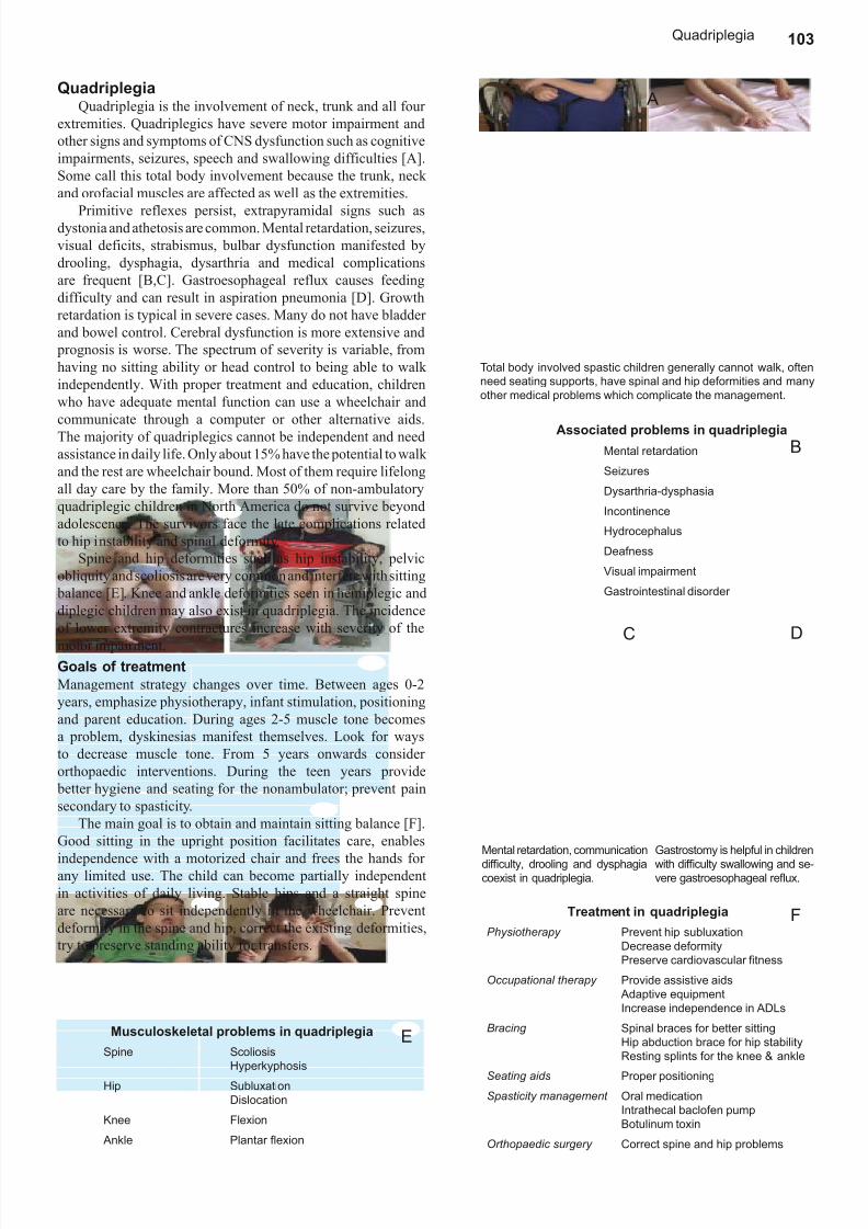
8/9/2019 Guía de ayuda para la parálisis cerebral.pdf
http://slidepdf.com/reader/full/guia-de-ayuda-para-la-paralisis-cerebralpdf 103/143
103Quadriplegia
QuadriplegiaQuadriplegia is the involvement of neck, trunk and all four
extremities. Quadriplegics have severe motor impairment andother signs and symptoms of CNS dysfunction such as cognitiveimpairments, seizures, speech and swallowing difficulties [A].Some call this total body involvement because the trunk, neckand orofacial muscles are affected as well as the extremities.
Primitive reflexes persist, extrapyramidal signs such asdystonia and athetosis are common. Mental retardation, seizures,visual deficits, strabismus, bulbar dysfunction manifested bydrooling, dysphagia, dysarthria and medical complicationsare frequent [B,C]. Gastroesophageal reflux causes feedingdifficulty and can result in aspiration pneumonia [D]. Growthretardation is typical in severe cases. Many do not have bladderand bowel control. Cerebral dysfunction is more extensive and prognosis is worse. The spectrum of severity is variable, fromhaving no sitting ability or head control to being able to walkindependently. With proper treatment and education, childrenwho have adequate mental function can use a wheelchair andcommunicate through a computer or other alternative aids.
The majority of quadriplegics cannot be independent and needassistance in daily life. Only about 15% have the potential to walkand the rest are wheelchair bound. Most of them require lifelongall day care by the family. More than 50% of non-ambulatoryquadriplegic children in North America do not survive beyondadolescence. The survivors face the late complications relatedto hip instability and spinal deformity.
Spine and hip deformities such as hip instability, pelvicobliquity and scoliosis are very common and interfere with sitting balance [E]. Knee and ankle deformities seen in hemiplegic anddiplegic children may also exist in quadriplegia. The incidenceof lower extremity contractures increase with severity of the
motor impairment.Goals of treatment
Management strategy changes over time. Between ages 0-2years, emphasize physiotherapy, infant stimulation, positioningand parent education. During ages 2-5 muscle tone becomesa problem, dyskinesias manifest themselves. Look for waysto decrease muscle tone. From 5 years onwards considerorthopaedic interventions. During the teen years provide better hygiene and seating for the nonambulator; prevent painsecondary to spasticity.
The main goal is to obtain and maintain sitting balance [F].Good sitting in the upright position facilitates care, enables
independence with a motorized chair and frees the hands forany limited use. The child can become partially independentin activities of daily living. Stable hips and a straight spineare necessary to sit independently in the wheelchair. Preventdeformity in the spine and hip, correct the existing deformities,try to preserve standing ability for transfers.
Associated problems in quadriplegiaMental retardation
Seizures
Dysarthria-dysphasia
Incontinence
Hydrocephalus
Deafness
Visual impairment
Gastrointestinal disorder
Total body involved spastic children generally cannot walk, oftenneed seating supports, have spinal and hip deformities and many
other medical problems which complicate the management.
Musculoskeletal problems in quadriplegia
Spine Scoliosis
Hyperkyphosis
Hip Subluxation
Dislocation
Knee Flexion
Ankle Plantar flexion
Treatment in quadriplegia
Physiotherapy Prevent hip subluxation
Decrease deformity
Preserve cardiovascular fitness
Occupational therapy Provide assistive aids
Adaptive equipment
Increase independence in ADLs
Bracing Spinal braces for better sitting
Hip abduction brace for hip stability
Resting splints for the knee & ankle
Seating aids Proper positioning
Spasticity management Oral medication
Intrathecal baclofen pump
Botulinum toxin
Orthopaedic surgery Correct spine and hip problems
F
F
E
E
D
DC
B
B
A
Mental retardation, communication
difficulty, drooling and dysphagiacoexist in quadriplegia.
Gastrostomy is helpful in children
with difficulty swallowing and se-vere gastroesophageal reflux.
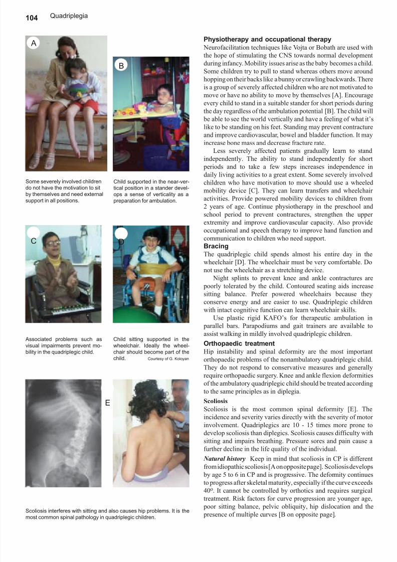
8/9/2019 Guía de ayuda para la parálisis cerebral.pdf
http://slidepdf.com/reader/full/guia-de-ayuda-para-la-paralisis-cerebralpdf 104/143
104 Quadriplegia
D
DC
B
B
APhysiotherapy and occupational therapy
Neurofacilitation techniques like Vojta or Bobath are used withthe hope of stimulating the CNS towards normal developmentduring infancy. Mobility issues arise as the baby becomes a child.Some children try to pull to stand whereas others move aroundhopping on their backs like a bunny or crawling backwards. Thereis a group of severely affected children who are not motivated to
move or have no ability to move by themselves [A]. Encourageevery child to stand in a suitable stander for short periods duringthe day regardless of the ambulation potential [B]. The child will be able to see the world vertically and have a feeling of what it’slike to be standing on his feet. Standing may prevent contractureand improve cardiovascular, bowel and bladder function. It mayincrease bone mass and decrease fracture rate.
Less severely affected patients gradually learn to standindependently. The ability to stand independently for short periods and to take a few steps increases independence indaily living activities to a great extent. Some severely involvedchildren who have motivation to move should use a wheeledmobility device [C]. They can learn transfers and wheelchair
activities. Provide powered mobility devices to children from2 years of age. Continue physiotherapy in the preschool andschool period to prevent contractures, strengthen the upperextremity and improve cardiovascular capacity. Also provideoccupational and speech therapy to improve hand function andcommunication to children who need support.Bracing
The quadriplegic child spends almost his entire day in thewheelchair [D]. The wheelchair must be very comfortable. Donot use the wheelchair as a stretching device.
Night splints to prevent knee and ankle contractures are poorly tolerated by the child. Contoured seating aids increase
sitting balance. Prefer powered wheelchairs because theyconserve energy and are easier to use. Quadriplegic childrenwith intact cognitive function can learn wheelchair skills.
Use plastic rigid KAFO’s for therapeutic ambulation in parallel bars. Parapodiums and gait trainers are available toassist walking in mildly involved quadriplegic children.
Orthopaedic treatment
Hip instability and spinal deformity are the most importantorthopaedic problems of the nonambulatory quadriplegic child.They do not respond to conservative measures and generallyrequire orthopaedic surgery. Knee and ankle flexion deformitiesof the ambulatory quadriplegic child should be treated according
to the same principles as in diplegia.Scoliosis
Scoliosis is the most common spinal deformity [E]. Theincidence and severity varies directly with the severity of motorinvolvement. Quadriplegics are 10 - 15 times more prone todevelop scoliosis than diplegics. Scoliosis causes difficulty withsitting and impairs breathing. Pressure sores and pain cause afurther decline in the life quality of the individual.
Natural history Keep in mind that scoliosis in CP is differentfrom idiopathic scoliosis [A on opposite page]. Scoliosis develops by age 5 to 6 in CP and is progressive. The deformity continuesto progress after skeletal maturity, especially if the curve exceeds
40o
. It cannot be controlled by orthotics and requires surgicaltreatment. Risk factors for curve progression are younger age, poor sitting balance, pelvic obliquity, hip dislocation and the presence of multiple curves [B on opposite page].
Some severely involved childrendo not have the motivation to sit
by themselves and need externalsupport in all positions.
Child sitting supported in the
wheelchair. Ideally the wheel-
chair should become part of thechild. Courtesy of G. Koloyan
Associated problems such as
visual impairments prevent mo-
bility in the quadriplegic child.
Scoliosis interferes with sitting and also causes hip problems. It is the
most common spinal pathology in quadriplegic children.
Child supported in the near-ver-tical position in a stander devel-
ops a sense of verticality as apreparation for ambulation.
E
E
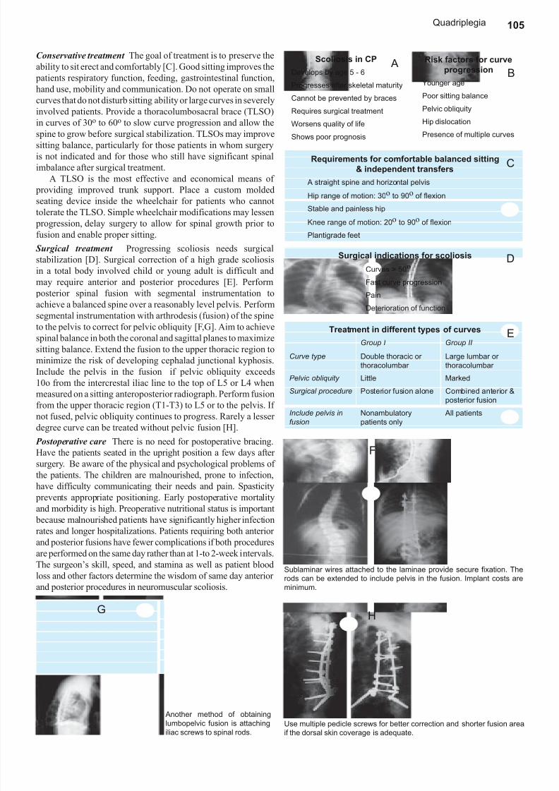
8/9/2019 Guía de ayuda para la parálisis cerebral.pdf
http://slidepdf.com/reader/full/guia-de-ayuda-para-la-paralisis-cerebralpdf 105/143
105Quadriplegia
Risk factors for curve
progression
Younger age
Poor sitting balance
Pelvic obliquity
Hip dislocation
Presence of multiple curves
Scoliosis in CP
Develops by age 5 - 6
Progresses after skeletal maturity
Cannot be prevented by braces
Requires surgical treatment
Worsens quality of life
Shows poor prognosis
Conservative treatment The goal of treatment is to preserve theability to sit erect and comfortably [C]. Good sitting improves the patients respiratory function, feeding, gastrointestinal function,hand use, mobility and communication. Do not operate on smallcurves that do not disturb sitting ability or large curves in severelyinvolved patients. Provide a thoracolumbosacral brace (TLSO)in curves of 30o to 60o to slow curve progression and allow the
spine to grow before surgical stabilization. TLSOs may improvesitting balance, particularly for those patients in whom surgeryis not indicated and for those who still have significant spinalimbalance after surgical treatment.
A TLSO is the most effective and economical means of providing improved trunk support. Place a custom moldedseating device inside the wheelchair for patients who cannottolerate the TLSO. Simple wheelchair modifications may lessen progression, delay surgery to allow for spinal growth prior tofusion and enable proper sitting.
Surgical treatment Progressing scoliosis needs surgicalstabilization [D]. Surgical correction of a high grade scoliosisin a total body involved child or young adult is difficult andmay require anterior and posterior procedures [E]. Perform posterior spinal fusion with segmental instrumentation toachieve a balanced spine over a reasonably level pelvis. Performsegmental instrumentation with arthrodesis (fusion) of the spineto the pelvis to correct for pelvic obliquity [F,G]. Aim to achievespinal balance in both the coronal and sagittal planes to maximizesitting balance. Extend the fusion to the upper thoracic region tominimize the risk of developing cephalad junctional kyphosis.Include the pelvis in the fusion if pelvic obliquity exceeds10o from the intercrestal iliac line to the top of L5 or L4 whenmeasured on a sitting anteroposterior radiograph. Perform fusionfrom the upper thoracic region (T1-T3) to L5 or to the pelvis. If
not fused, pelvic obliquity continues to progress. Rarely a lesserdegree curve can be treated without pelvic fusion [H].
Postoperative care There is no need for postoperative bracing.Have the patients seated in the upright position a few days aftersurgery. Be aware of the physical and psychological problems ofthe patients. The children are malnourished, prone to infection,have difficulty communicating their needs and pain. Spasticity prevents appropriate positioning. Early postoperative mortalityand morbidity is high. Preoperative nutritional status is important because malnourished patients have significantly higher infectionrates and longer hospitalizations. Patients requiring both anteriorand posterior fusions have fewer complications if both procedures
are performed on the same day rather than at 1-to 2-week intervals.The surgeon’s skill, speed, and stamina as well as patient bloodloss and other factors determine the wisdom of same day anteriorand posterior procedures in neuromuscular scoliosis.
Requirements for comfortable balanced sitting
& independent transfers
A straight spine and horizontal pelvis
Hip range of motion: 30o to 90o of flexion
Stable and painless hip
Knee range of motion: 20o to 90o of flexion
Plantigrade feet
Surgical indications for scoliosis
Curves > 50o
Fast curve progression
Pain
Deterioration of function
Treatment in different types of curves
Group I Group II
Curve type Double thoracic or
thoracolumbar
Large lumbar or
thoracolumbar
Pelvic obliquity Little Marked
Surgical procedure Posterior fusion alone Combined anterior &
posterior fusion
Include pelvis infusion Nonambulatorypatients only All patients
D
D
C
C
B
B
A
E
E
Use multiple pedicle screws for better correction and shorter fusion areaif the dorsal skin coverage is adequate.
Sublaminar wires attached to the laminae provide secure fixation. Therods can be extended to include pelvis in the fusion. Implant costs are
minimum.
Another method of obtaining
lumbopelvic fusion is attaching
iliac screws to spinal rods.
F
F
G
GH
H
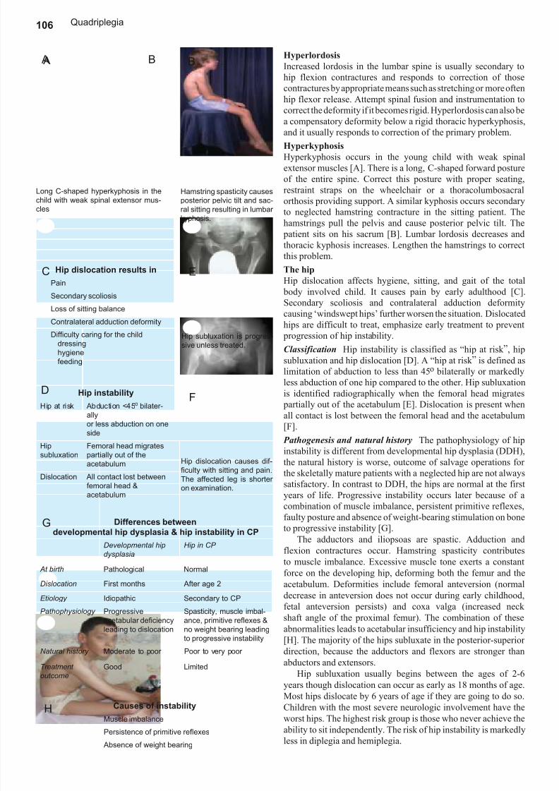
8/9/2019 Guía de ayuda para la parálisis cerebral.pdf
http://slidepdf.com/reader/full/guia-de-ayuda-para-la-paralisis-cerebralpdf 106/143
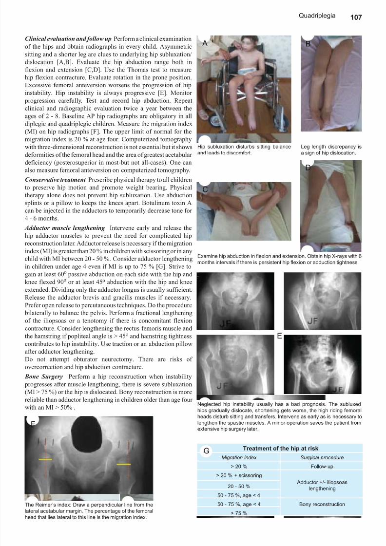
8/9/2019 Guía de ayuda para la parálisis cerebral.pdf
http://slidepdf.com/reader/full/guia-de-ayuda-para-la-paralisis-cerebralpdf 107/143
107Quadriplegia
Treatment of the hip at risk
Migration index Surgical procedure
> 20 % Follow-up
> 20 % + scissoring
Adductor +/- iliopsoas
lengthening20 - 50 %
50 - 75 %, age < 4
50 - 75 %, age < 4 Bony reconstruction
> 75 %
Clinical evaluation and follow up Perform a clinical examinationof the hips and obtain radiographs in every child. Asymmetricsitting and a shorter leg are clues to underlying hip subluxation/dislocation [A,B]. Evaluate the hip abduction range both inflexion and extension [C,D]. Use the Thomas test to measurehip flexion contracture. Evaluate rotation in the prone position.Excessive femoral anteversion worsens the progression of hip
instability. Hip instability is always progressive [E]. Monitor progression carefully. Test and record hip abduction. Repeatclinical and radiographic evaluation twice a year between theages of 2 - 8. Baseline AP hip radiographs are obligatory in alldiplegic and quadriplegic children. Measure the migration index(MI) on hip radiographs [F]. The upper limit of normal for themigration index is 20 % at age four. Computerized tomographywith three-dimensional reconstruction is not essential but it showsdeformities of the femoral head and the area of greatest acetabulardeficiency (posterosuperior in most-but not all-cases). One canalso measure femoral anteversion on computerized tomography.
Conservative treatment Prescribe physical therapy to all childrento preserve hip motion and promote weight bearing. Physicaltherapy alone does not prevent hip subluxation. Use abductionsplints or a pillow to keeps the knees apart. Botulinum toxin Acan be injected in the adductors to temporarily decrease tone for4 - 6 months.
Adductor muscle lengthening Intervene early and release thehip adductor muscles to prevent the need for complicated hipreconstruction later. Adductor release is necessary if the migrationindex (MI) is greater than 20 % in children with scissoring or in anychild with MI between 20 - 50 %. Consider adductor lengtheningin children under age 4 even if MI is up to 75 % [G]. Strive togain at least 60o passive abduction on each side with the hip andknee flexed 90o or at least 45o abduction with the hip and knee
extended. Dividing only the adductor longus is usually sufficient.Release the adductor brevis and gracilis muscles if necessary.Prefer open release to percutaneous techniques. Do the procedure bilaterally to balance the pelvis. Perform a fractional lengtheningof the iliopsoas or a tenotomy if there is concomitant flexioncontracture. Consider lengthening the rectus femoris muscle andthe hamstring if popliteal angle is > 45o and hamstring tightnesscontributes to hip instability. Use traction or an abduction pillowafter adductor lengthening.Do not attempt obturator neurectomy. There are risks ofovercorrection and hip abduction contracture.
Bone Surgery Perform a hip reconstruction when instability
progresses after muscle lengthening, there is severe subluxation(MI > 75 %) or the hip is dislocated. Bony reconstruction is morereliable than adductor lengthening in children older than age fourwith an MI > 50% .
F
F
E
E
D
D
C
C
B
B A
G
G
The Reimer’s index: Draw a perpendicular line from the
lateral acetabular margin. The percentage of the femoral
head that lies lateral to this line is the migration index.
Neglected hip instability usually has a bad prognosis. The subluxed
hips gradually dislocate, shortening gets worse, the high riding femoral
heads disturb sitting and transfers. Intervene as early as is necessary to
lengthen the spastic muscles. A minor operation saves the patient fromextensive hip surgery later.
Examine hip abduction in flexion and extension. Obtain hip X-rays with 6
months intervals if there is persistent hip flexion or adduction tightness.
Hip subluxation disturbs sitting balance
and leads to discomfort.
Leg length discrepancy is
a sign of hip dislocation.
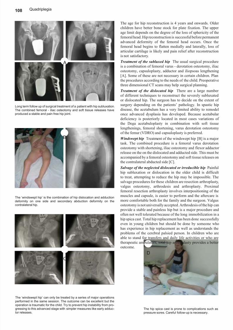
8/9/2019 Guía de ayuda para la parálisis cerebral.pdf
http://slidepdf.com/reader/full/guia-de-ayuda-para-la-paralisis-cerebralpdf 108/143
108 Quadriplegia
D
D
C
C
B
B
A
The age for hip reconstruction is 4 years and onwards. Olderchildren have better bone stock for plate fixation. The upperage limit depends on the degree of the loss of sphericity of thefemoral head. Hip reconstruction is successful before permanentadvanced deformity of the femoral head occurs. Once thefemoral head begins to flatten medially and laterally, loss ofarticular cartilage is likely and pain relief after reconstruction
is not satisfactory.Treatment of the subluxed hip The usual surgical procedureis a combination of femoral varus - derotation osteotomy, iliacosteotomy, capsuloplasty, adductor and iliopsoas lengthening[A]. Some of these are not necessary in certain children. Planthe procedures according to the needs of the child. Preoperativethree dimensional CT scans may help surgical planning.
Treatment of the dislocated hip There are a large numberof different techniques to reconstruct the severely subluxatedor dislocated hip. The surgeon has to decide on the extent ofsurgery depending on the patients’ pathology. In spastic hipdisease, the acetabulum has a very limited ability to remodel
once advanced dysplasia has developed. Because acetabulardeficiency is posteriorly located in most cases variations ofthe Dega acetabuloplasty in combination with soft tissuelengthenings, femoral shortening, varus derotation osteotomyof the femur (VDRO) and capsuloplasty is preferred.
Windswept hip Treatment of the windswept hip [B] is a majortask. The combined procedure is a femoral varus derotationosteotomy with shortening, iliac osteotomy and flexor adductorrelease on the on the dislocated and adducted side. This must beaccompanied by a femoral osteotomy and soft tissue releases onthe contralateral abducted side [C].
Salvage of the neglected dislocated or irreducible hip Painful
hip subluxation or dislocation in the older child is difficultto treat, attempting to reduce the hip may be impossible. Thesalvage procedures for these children are resection arthroplasty,valgus osteotomy, arthrodesis and arthroplasty. Proximalfemoral resection arthroplasty involves interpositioning of themuscles and capsule, is easier to perform and the aftercare ismore comfortable both for the family and the surgeon. Valgusosteotomy is not universally accepted. Arthrodesis of the hip can provide a stable and painless hip but is a major procedure andoften not well tolerated because of the long immobilization in ahip spica cast. Total hip replacement has been done successfullyeven in young children but should be done by someone whohas experience in hip replacement as well as understands the problems of the cerebral palsied person. In children who areable to stand for transfers and daily life activities or who aretherapeutic ambulators, total hip arthroplasty provides a betteroutcome.
Long term follow up of surgical treatment of a patient with hip subluxation.
The combined femoral - iliac osteotomy and soft tissue releases haveproduced a stable and pain free hip joint.
The ‘windswept hip’ is the combination of hip dislocation and adduction
deformity on one side and secondary abduction deformity on thecontralateral hip.
The ‘windswept hip’ can only be treated by a series of major operations
performed in the same session. The outcome can be excellent but the
operation is traumatic for the child. Try to prevent hip instability from pro-gressing to this advanced stage with simpler measures like early adduc-
tor releases.The hip spica cast is prone to complications such as
pressure sores. Careful follow-up is necessary.
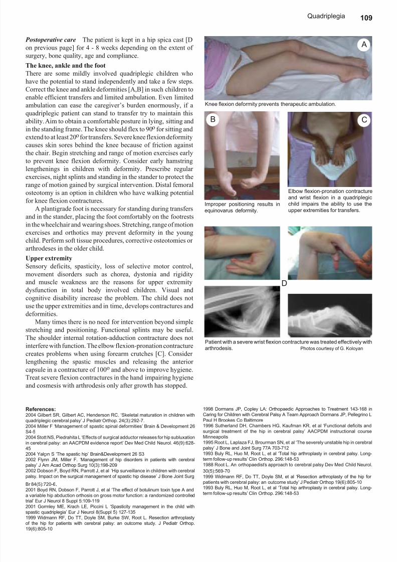
8/9/2019 Guía de ayuda para la parálisis cerebral.pdf
http://slidepdf.com/reader/full/guia-de-ayuda-para-la-paralisis-cerebralpdf 109/143
109Quadriplegia
Postoperative care The patient is kept in a hip spica cast [Don previous page] for 4 - 8 weeks depending on the extent ofsurgery, bone quality, age and compliance.
The knee, ankle and the foot
There are some mildly involved quadriplegic children whohave the potential to stand independently and take a few steps.Correct the knee and ankle deformities [A,B] in such children to
enable efficient transfers and limited ambulation. Even limitedambulation can ease the caregiver’s burden enormously, if aquadriplegic patient can stand to transfer try to maintain thisability. Aim to obtain a comfortable posture in lying, sitting andin the standing frame. The knee should flex to 90o for sitting andextend to at least 20o for transfers. Severe knee flexion deformitycauses skin sores behind the knee because of friction againstthe chair. Begin stretching and range of motion exercises earlyto prevent knee flexion deformity. Consider early hamstringlengthenings in children with deformity. Prescribe regularexercises, night splints and standing in the stander to protect therange of motion gained by surgical intervention. Distal femoralosteotomy is an option in children who have walking potentialfor knee flexion contractures.
A plantigrade foot is necessary for standing during transfersand in the stander, placing the foot comfortably on the footrestsin the wheelchair and wearing shoes. Stretching, range of motionexercises and orthotics may prevent deformity in the youngchild. Perform soft tissue procedures, corrective osteotomies orarthrodeses in the older child.
Upper extremity
Sensory deficits, spasticity, loss of selective motor control,movement disorders such as chorea, dystonia and rigidityand muscle weakness are the reasons for upper extremitydysfunction in total body involved children. Visual and
cognitive disability increase the problem. The child does notuse the upper extremities and in time, develops contractures anddeformities.
Many times there is no need for intervention beyond simplestretching and positioning. Functional splints may be useful.The shoulder internal rotation-adduction contracture does notinterfere with function. The elbow flexion-pronation contracturecreates problems when using forearm crutches [C]. Considerlengthening the spastic muscles and releasing the anteriorcapsule in a contracture of 100o and above to improve hygiene.Treat severe flexion contractures in the hand impairing hygieneand cosmesis with arthrodesis only after growth has stopped.
Elbow flexion-pronation contracture
and wrist flexion in a quadriplegicchild impairs the ability to use the
upper extremities for transfers.
Knee flexion deformity prevents therapeutic ambulation.
A
B
B C
C
D
D
Improper positioning results in
equinovarus deformity.
References:2004 Gilbert SR, Gilbert AC, Henderson RC. ‘Skeletal maturation in children with
quadriplegic cerebral palsy’ J Pediatr Orthop. 24(3):292-7.
2004 Miller F ‘Management of spastic spinal deformities’ Brain & Development 26S4-5
2004 Stott NS, Piedrahita L ‘Effects of surgical adductor releases for hip subluxation
in cerebral palsy: an AACPDM evidence report’ Dev Med Child Neurol. 46(9):628-45
2004 Yalçın S ‘The spastic hip’ Brain&Development 26 S3
2002 Flynn JM, Miller F. ‘Management of hip disorders in patients with cerebralpalsy’ J Am Acad Orthop Surg 10(3):198-209
2002 Dobson F, Boyd RN, Parrott J, et al ‘Hip surveillance in children with cerebral
palsy. Impact on the surgical management of spastic hip disease’ J Bone Joint Surg
Br 84(5):720-6.2001 Boyd RN, Dobson F, Parrott J, et al ‘The effect of botulinum toxin type A and
a variable hip abduction orthosis on gross motor function: a randomized controlledtrial’ Eur J Neurol 8 Suppl 5:109-119
2001 Gormley ME, Krach LE, Piccini L ‘Spasticity management in the child with
spastic quadriplegia’ Eur J Neurol 8(Suppl 5) 127-1351999 Widmann RF, Do TT, Doyle SM, Burke SW, Root L. Resection arthroplasty
of the hip for patients with cerebral palsy: an outcome study. J Pediatr Orthop.
19(6):805-10
1998 Dormans JP, Copley LA: Orthopaedic Approaches to Treatment 143-168 in
Caring for Children with Cerebral Palsy A Team Approach Dormans JP, Pellegrino L
Paul H Brookes Co Baltimore1996 Sutherland DH, Chambers HG, Kaufman KR, et al ‘Functional deficits and
surgical treatment of the hip in cerebral palsy’ AACPDM instructional course
Minneapolis1995 Root L, Laplaza FJ, Brourman SN, et al ‘The severely unstable hip in cerebral
palsy’ J Bone and Joint Surg 77A 703-7121993 Buly RL, Huo M, Root L, et al ‘Total hip arthroplasty in cerebral palsy. Long-
term follow-up results’ Clin Orthop. 296:148-53
1988 Root L. An orthopaedist’s approach to cerebral palsy Dev Med Child Neurol.
30(5):569-70
1999 Widmann RF, Do TT, Doyle SM, et al ‘Resection arthroplasty of the hip for
patients with cerebral palsy: an outcome study’ J Pediatr Orthop 19(6):805-101993 Buly RL, Huo M, Root L, et al ‘Total hip arthroplasty in cerebral palsy. Long-
term follow-up results’ Clin Orthop. 296:148-53
Patient with a severe wrist flexion contracture was treated effectively witharthrodesis. Photos courtesy of G. Koloyan

8/9/2019 Guía de ayuda para la parálisis cerebral.pdf
http://slidepdf.com/reader/full/guia-de-ayuda-para-la-paralisis-cerebralpdf 110/143
110 Dyskinesia
DyskinesiaAthetosis, dystonia and chorea are the main movementdisorders seen in dyskinetic children [A]. These children areinitially hypotonic. As they get older, muscle tone begins tofluctuate. Involuntary movements occur when the child triesto move. Sometimes there is also movement at rest. When thechild is totally relaxed in the supine position or asleep, there is
full range of motion and decreased muscle tone. When the childwakes up or is excited, he becomes rigid. Lack of coordinationis even more prominent during strenuous activities. Thedyskinetic child spends excessive energy because of continuousuncontrolled movements. Abnormal contractions of manymuscles occurring with the slightest voluntary motion increasethe energy demand considerably.
Classification
Dyskinetic patients are subdivided into two groups [B]. The firstand most common group are hyperkinetic or choreo-athetoidchildren. They show purposeless, often massive involuntarymovements. The initiation of a movement of one extremity
leads to movement of other muscle groups. Rapid, randomand jerky movements are called chorea and slow writhingmovements are called athetosis. They increase when the childis excited or frightened.
The second group are dystonic children [C]. They manifestabnormal shifts of general muscle tone induced by movement.When the child tries to move, there is a co-contraction ofagonist and antagonist muscles leading to an abnormal postureof one or more parts of the body. These abnormal and distorted postures occur in a stereotyped pattern. The trunk and neck arerigid. As in all types of dyskinetic CP, the contractions in theflexor and extensor muscles of the extremities increase withvoluntary movement and disappear during sleep.
Dyskinesia may accompany spasticity in a group oftotal body involved children [D]. Athetosis is common incombination with spastic diplegia.
Associated features
Mental status is generally not impaired [E]. There iscommunication difficulty because of oromotor dysfunctionand most of these children are unable to talk. Spasticity oforopharyngeal muscles impair feeding. Growth retardation anda decreased capacity to gain weight are characteristic.
Movement problems in dyskinesia
Athetosis Involuntary, slow writhing movements of the hands
feet face or tongue
Chorea Multiple rapid jerky movements usually of the hands
and feet.
Dystonia Muscle tone is greatly increased. There are slow
torsional contractions which increase with attempts
at voluntary movement and result in abnormal pos-turing. Dystonia is localized more to the trunk and
proximal extremities.
Classification
Choreo-athetoid Dystonic
Hyperkinetic Rigid
Purposeless
involuntary movements
Co-contraction of agonist & antagonists
Severe dystonia interfering with sitting and position-
ing may respond to medical treatment only.
Involuntary contraction of hand muscles prevents ef-fective use of the extremity.
E
E
D
D
C
C
B
B
A
Continuous repetitive muscle activity causes involuntary movements in all four extremities and the trunk in this 10 year old child. These movements
increase when she wants to accomplish a certain task. Communication is difficult in spite of her normal mental function.
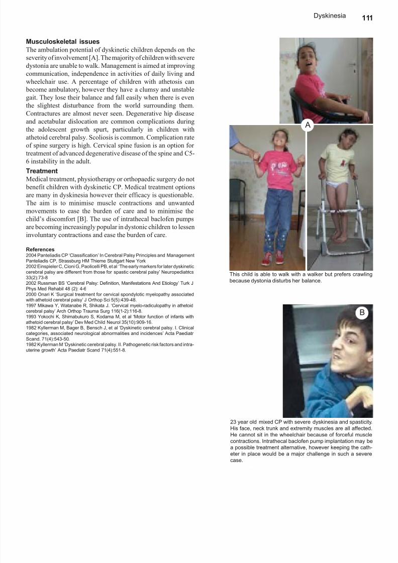
8/9/2019 Guía de ayuda para la parálisis cerebral.pdf
http://slidepdf.com/reader/full/guia-de-ayuda-para-la-paralisis-cerebralpdf 111/143
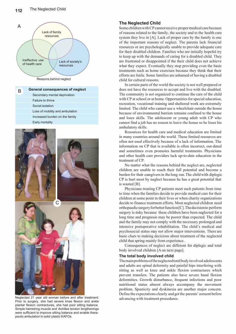
8/9/2019 Guía de ayuda para la parálisis cerebral.pdf
http://slidepdf.com/reader/full/guia-de-ayuda-para-la-paralisis-cerebralpdf 112/143
112 The Neglected Child
The Neglected ChildSome children with CP cannot receive proper medical care becauseof reasons related to the family, the society and to the health caresystem they live in [A]. Lack of proper care by the family is oneof the important reasons of neglect. The parents lack financialresources or are psychologically unable to provide adequate carefor their disabled children. Families who are initially hopeful tryto keep up with the demands of caring for a disabled child. Theyare frustrated or disappointed if the their child does not achievewhat they expect. Eventually they stop providing even the basictreatments such as home exercises because they think that theirefforts are futile. Some families are ashamed of having a disabledchild for cultural reasons.
In certain parts of the world the society is not well prepared ordoes not have the resources to accept and live with the disabled.The community is not organized to continue the care of the childwith CP at school or at home. Opportunities for special education,recreation, vocational training and sheltered work are extremelylimited. The child who cannot use a wheelchair outside the house because of environmental barriers remains confined to the house
and loses skills. The adolescent or young adult with CP whocannot find a job has no reason to leave the house so he loses hisambulatory skills.
Resources for health care and medical education are limitedin many countries around the world. These limited resources areoften not used effectively because of a lack of information. Theinformation on CP that is available is often incorrect, out-datedand sometimes even promotes harmful treatments. Physiciansand other health care providers lack up-to-date education in thetreatment of CP.
No matter what the reasons behind the neglect are, neglectedchildren are unable to reach their full potential and become a
burden for their caregivers in the long run. The child with diplegicCP is hurt most by neglect because he has a great potential thatis wasted [B].
Physicians treating CP patients meet such patients from timeto time when the families decide to provide medical care for theirchildren at some point in their lives or when charity organizationsdecide to finance treatment efforts. Most neglected children needorthopaedic surgery for better function [C]. The decision to performsurgery is risky because these children have been neglected for along time and prognosis may be poorer than expected. The childand the family may not comply with the necessary prolonged andintensive postoperative rehabilitation. The child’s medical and psychosocial status may not allow major interventions. There are basic clues to making decisions about treatment of the neglectedchild that spring mainly from experience.
Consequences of neglect are different for diplegic and total body involved children [A on next page].
The total body involved child
The main problems of the neglected total body involved adolescentsand adults are spinal deformity and painful hips interfering withsitting as well as knee and ankle flexion contractures which prevent transfers. The patients also have severe hand flexiondeformities. Growth disturbance, frequent infections and poornutritional status almost always accompany the movement problem. Spasticity and dyskinesia are another major concern.
Define the expectations clearly and get the parents’ consent beforeadvancing with treatment procedures.Neglected 21 year old woman before and after treatment:
Prior to surgery, she had severe knee flexion and ankleplantar flexion contractures, she had poor sitting balance.
Simple hamstring muscle and Achilles tendon lengthenings
were sufficient to improve sitting balance and enable thera-
peutic ambulation in solid plastic KAFOs.
General consequences of neglect
Secondary mental deprivation
Failure to thrive
Social isolation
Loss of mobility and ambulation
Increased burden on the family
Early mortality
B
B
A
C
C
Lack of family
resources
Ineffective use
of health careLack of society’s
resources
Reasons behind neglect
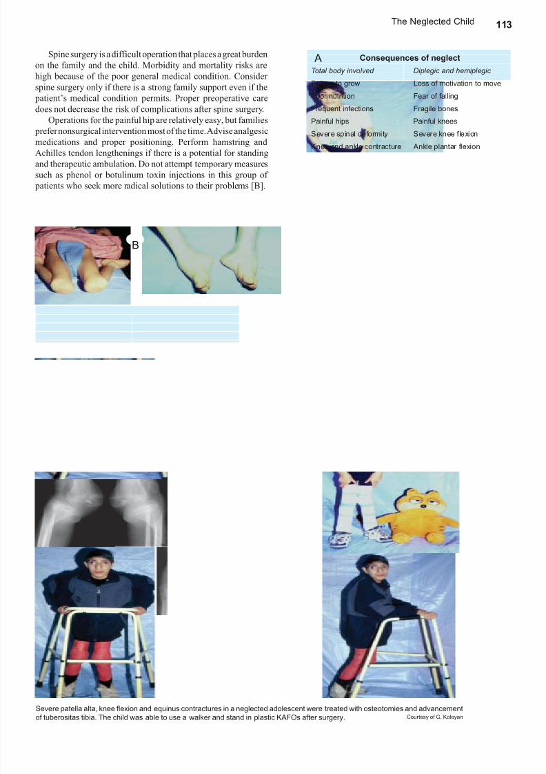
8/9/2019 Guía de ayuda para la parálisis cerebral.pdf
http://slidepdf.com/reader/full/guia-de-ayuda-para-la-paralisis-cerebralpdf 113/143
113The Neglected Child
B
B
Consequences of neglect
Total body involved Diplegic and hemiplegic
Failure to grow Loss of motivation to move
Poor nutrition Fear of falling
Frequent infections Fragile bones
Painful hips Painful knees
Severe spinal deformity Severe knee flexionKnee and ankle contracture Ankle plantar flexion
Spine surgery is a difficult operation that places a great burdenon the family and the child. Morbidity and mortality risks arehigh because of the poor general medical condition. Considerspine surgery only if there is a strong family support even if the patient’s medical condition permits. Proper preoperative caredoes not decrease the risk of complications after spine surgery.
Operations for the painful hip are relatively easy, but families
prefer nonsurgical intervention most of the time. Advise analgesicmedications and proper positioning. Perform hamstring andAchilles tendon lengthenings if there is a potential for standingand therapeutic ambulation. Do not attempt temporary measuressuch as phenol or botulinum toxin injections in this group of patients who seek more radical solutions to their problems [B].
A
Severe patella alta, knee flexion and equinus contractures in a neglected adolescent were treated with osteotomies and advancement
of tuberositas tibia. The child was able to use a walker and stand in plastic KAFOs after surgery. Courtesy of G. Koloyan
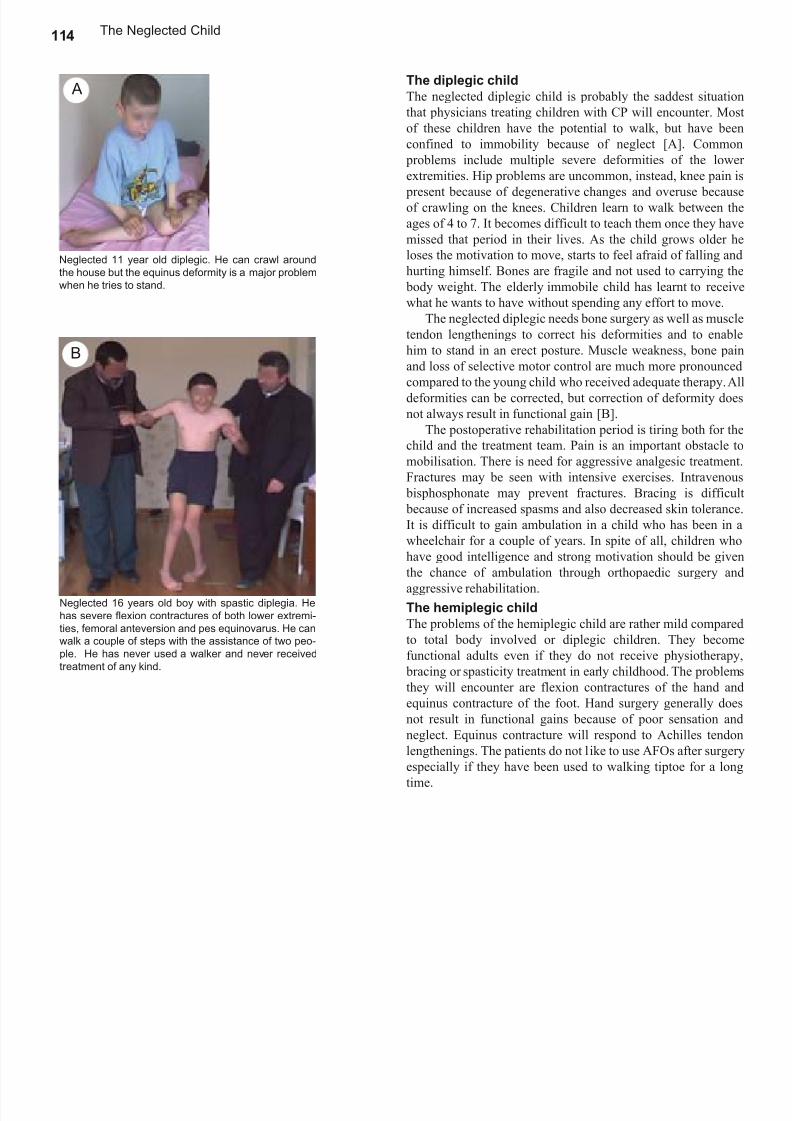
8/9/2019 Guía de ayuda para la parálisis cerebral.pdf
http://slidepdf.com/reader/full/guia-de-ayuda-para-la-paralisis-cerebralpdf 114/143
114 The Neglected Child
The diplegic child
The neglected diplegic child is probably the saddest situationthat physicians treating children with CP will encounter. Mostof these children have the potential to walk, but have beenconfined to immobility because of neglect [A]. Common problems include multiple severe deformities of the lowerextremities. Hip problems are uncommon, instead, knee pain is
present because of degenerative changes and overuse becauseof crawling on the knees. Children learn to walk between theages of 4 to 7. It becomes difficult to teach them once they havemissed that period in their lives. As the child grows older heloses the motivation to move, starts to feel afraid of falling andhurting himself. Bones are fragile and not used to carrying the body weight. The elderly immobile child has learnt to receivewhat he wants to have without spending any effort to move.
The neglected diplegic needs bone surgery as well as muscletendon lengthenings to correct his deformities and to enablehim to stand in an erect posture. Muscle weakness, bone painand loss of selective motor control are much more pronouncedcompared to the young child who received adequate therapy. All
deformities can be corrected, but correction of deformity doesnot always result in functional gain [B].
The postoperative rehabilitation period is tiring both for thechild and the treatment team. Pain is an important obstacle tomobilisation. There is need for aggressive analgesic treatment.Fractures may be seen with intensive exercises. Intravenous bisphosphonate may prevent fractures. Bracing is difficult because of increased spasms and also decreased skin tolerance.It is difficult to gain ambulation in a child who has been in awheelchair for a couple of years. In spite of all, children whohave good intelligence and strong motivation should be giventhe chance of ambulation through orthopaedic surgery and
aggressive rehabilitation.The hemiplegic child
The problems of the hemiplegic child are rather mild comparedto total body involved or diplegic children. They becomefunctional adults even if they do not receive physiotherapy, bracing or spasticity treatment in early childhood. The problemsthey will encounter are flexion contractures of the hand andequinus contracture of the foot. Hand surgery generally doesnot result in functional gains because of poor sensation andneglect. Equinus contracture will respond to Achilles tendonlengthenings. The patients do not like to use AFOs after surgeryespecially if they have been used to walking tiptoe for a long
time.
Neglected 11 year old diplegic. He can crawl around
the house but the equinus deformity is a major problem
when he tries to stand.
Neglected 16 years old boy with spastic diplegia. He
has severe flexion contractures of both lower extremi-
ties, femoral anteversion and pes equinovarus. He canwalk a couple of steps with the assistance of two peo-
ple. He has never used a walker and never received
treatment of any kind.
B
B
A
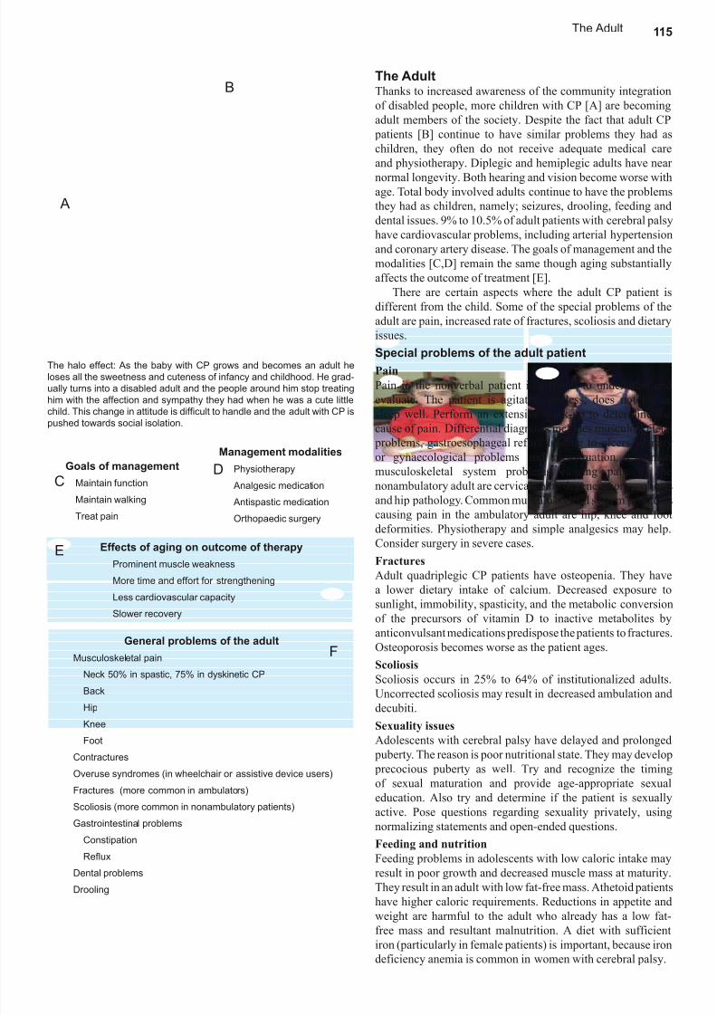
8/9/2019 Guía de ayuda para la parálisis cerebral.pdf
http://slidepdf.com/reader/full/guia-de-ayuda-para-la-paralisis-cerebralpdf 115/143
115The Adult
General problems of the adult
Musculoskeletal pain
Neck 50% in spastic, 75% in dyskinetic CP
Back
HipKnee
Foot
Contractures
Overuse syndromes (in wheelchair or assistive device users)
Fractures (more common in ambulators)
Scoliosis (more common in nonambulatory patients)
Gastrointestinal problems
Constipation
Reflux
Dental problems
Drooling
The AdultThanks to increased awareness of the community integrationof disabled people, more children with CP [A] are becomingadult members of the society. Despite the fact that adult CP patients [B] continue to have similar problems they had aschildren, they often do not receive adequate medical careand physiotherapy. Diplegic and hemiplegic adults have near
normal longevity. Both hearing and vision become worse withage. Total body involved adults continue to have the problemsthey had as children, namely; seizures, drooling, feeding anddental issues. 9% to 10.5% of adult patients with cerebral palsyhave cardiovascular problems, including arterial hypertensionand coronary artery disease. The goals of management and themodalities [C,D] remain the same though aging substantiallyaffects the outcome of treatment [E].
There are certain aspects where the adult CP patient isdifferent from the child. Some of the special problems of theadult are pain, increased rate of fractures, scoliosis and dietaryissues.
Special problems of the adult patientPain
Pain in the nonverbal patient is difficult to understand andevaluate. The patient is agitated, restless, does not eat orsleep well. Perform an extensive work-up to determine thecause of pain. Differential diagnosis includes musculoskeletal problems, gastroesophageal reflux leading to ulcers, urinaryor gynaecological problems and menstruation. Commonmusculoskeletal system problems causing pain in thenonambulatory adult are cervical spine degeneration, scoliosisand hip pathology. Common musculoskeletal system problemscausing pain in the ambulatory adult are hip, knee and foot
deformities. Physiotherapy and simple analgesics may help.Consider surgery in severe cases.
Fractures
Adult quadriplegic CP patients have osteopenia. They havea lower dietary intake of calcium. Decreased exposure tosunlight, immobility, spasticity, and the metabolic conversionof the precursors of vitamin D to inactive metabolites byanticonvulsant medications predispose the patients to fractures.Osteoporosis becomes worse as the patient ages.
Scoliosis
Scoliosis occurs in 25% to 64% of institutionalized adults.Uncorrected scoliosis may result in decreased ambulation and
decubiti.Sexuality issues
Adolescents with cerebral palsy have delayed and prolonged puberty. The reason is poor nutritional state. They may develop precocious puberty as well. Try and recognize the timingof sexual maturation and provide age-appropriate sexualeducation. Also try and determine if the patient is sexuallyactive. Pose questions regarding sexuality privately, usingnormalizing statements and open-ended questions.
Feeding and nutrition
Feeding problems in adolescents with low caloric intake mayresult in poor growth and decreased muscle mass at maturity.
They result in an adult with low fat-free mass. Athetoid patientshave higher caloric requirements. Reductions in appetite andweight are harmful to the adult who already has a low fat-free mass and resultant malnutrition. A diet with sufficientiron (particularly in female patients) is important, because irondeficiency anemia is common in women with cerebral palsy.
The halo effect: As the baby with CP grows and becomes an adult he
loses all the sweetness and cuteness of infancy and childhood. He grad-
ually turns into a disabled adult and the people around him stop treating
him with the affection and sympathy they had when he was a cute little
child. This change in attitude is difficult to handle and the adult with CP is
pushed towards social isolation.
Management modalities
Physiotherapy
Analgesic medication
Antispastic medication
Orthopaedic surgery
Goals of management
Maintain function
Maintain walking
Treat pain
Effects of aging on outcome of therapy
Prominent muscle weakness
More time and effort for strengthening
Less cardiovascular capacity
Slower recovery
B
B
A
E
E
D
DC
C
F
F
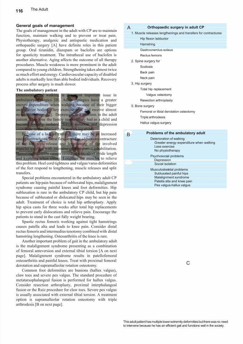
8/9/2019 Guía de ayuda para la parálisis cerebral.pdf
http://slidepdf.com/reader/full/guia-de-ayuda-para-la-paralisis-cerebralpdf 116/143
116 The Adult
Orthopaedic surgery in adult CP
1. Muscle releases lengthenings and transfers for contractures
Hip flexor /adductor
Hamstring
Gastrocnemius-soleus
Rectus femoris
2. Spine surgery for
Scoliosis
Back pain
Neck pain
3. Hip surgery
Total hip replacement
Valgus osteotomy
Resection arthroplasty
5. Bone surgery
Femoral or tibial derotation osteotomy
Triple arthrodesis
Hallux valgus surgery
General goals of management
The goals of management in the adult with CP are to maintainfunction, maintain walking and to prevent or treat pain.Physiotherapy, analgesic and antispastic medication andorthopaedic surgery [A] have definite roles in this patientgroup. Oral tizanidin, diazepam or baclofen are optionsfor spasticity treatment. The intrathecal use of baclofen is
another alternative. Aging affects the outcome of all therapy procedures. Muscle weakness is more prominent in the adultcompared to young children. Strengthening takes almost twiceas much effort and energy. Cardiovascular capacity of disabledadults is markedly less than able bodied individuals. Recovery process after surgery is much slower.
The ambulatory patient
Deterioration of walking is the most important issue inambulatory diplegics [B]. Adult diplegics have a greaterenergy expenditure when walking because of their biggerand heavier bodies. They exercise less, and receive almostno physiotherapy [C]. Depression is a problem in the adult
patient. They lose the family support they had as a child and become socially isolated. Social isolation and depressioncontribute to the deterioration in walking ability.
Because of a lack of exercise there may be an increasedrate of contractures. Treat flexion and/or adduction contractureof the hip with release and lengthening of the involvedmuscles together with intensive post-operative rehabilitation.Hamstring tightness causes crouched gait, short stride lengthand kyphosis when sitting. Lengthen the muscles to relievethis problem. Heel cord tightness and valgus/varus deformitiesof the feet respond to lengthening, muscle releases and splittransfers.
Special problems encountered in the ambulatory adult CP patients are hip pain because of subluxated hips, malalignmentsyndrome causing painful knees and foot deformities. Hipsubluxation is rare in the ambulatory CP child, but hip pain because of subluxated or dislocated hips may be seen in theadult. Treatment of choice is total hip arthroplasty. Applyhip spica casts for three weeks after total hip replacementsto prevent early dislocations and relieve pain. Encourage the patients to stand in the cast fully weight bearing.
Spastic rectus femoris working against tight hamstringscauses patella alta and leads to knee pain. Consider distalrectus femoris and intermedius tenotomy combined with distalhamstring lengthening. Osteoarthritis of the knee is rare.
Another important problem of gait in the ambulatory adultis the malalignment syndrome presenting as a combinationof femoral anteversion and external tibial torsion [A on next page]. Malalignment syndrome results in patellofemoralosteoarthritis and painful knees. Treat with proximal femoralderotation and supramalleolar rotation osteotomy.
Common foot deformities are bunions (hallux valgus),claw toes and severe pes valgus. The standard procedure ofmetatarsophalangeal fusion is performed for hallux valgus.Consider resection arthroplasty, proximal interphalangealfusion or the Ruiz procedure for claw toes. Severe pes valgusis usually associated with external tibial torsion. A treatment
option is supramalleolar rotation osteotomy with triplearthrodesis [B on next page].
Problems of the ambulatory adult
Deterioration of walking
Greater energy expenditure when walking
Less exercise
No physiotherapy
Psychosocial problems
Depression
Social isolation
Musculoskeletal problems
Subluxated painful hips
Malalignment syndrome
Patella alta and knee pain Pes valgus-hallux valgus
This adult patient has multiple lower extremity deformities but there was no need
to intervene because he has an efficient gait and functions well in the society.
C
C
B
B
A
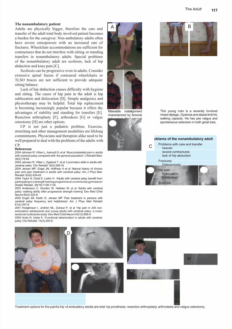
8/9/2019 Guía de ayuda para la parálisis cerebral.pdf
http://slidepdf.com/reader/full/guia-de-ayuda-para-la-paralisis-cerebralpdf 117/143
117The Adult
This young man is a severely involved
mixed diplegic. Dystonia and ataxia limit hiswalking capacity. He has pes valgus and
spontaneous extension in both great toes.
The nonambulatory patient
Adults are physically bigger, therefore the care andtransfer of the adult total body involved patient becomesa burden for the caregiver. Non-ambulatory adults oftenhave severe osteoporosis with an increased rate offractures. Wheelchair accommodations are sufficient forcontractures that do not interfere with sitting or standing
transfers in nonambulatory adults. Special problemsof the nonambulatory adult are scoliosis, lack of hipabduction and knee pain [C].
Scoliosis can be progressive even in adults. Considerextensive spinal fusion if contoured wheelchairs orTLSO braces are not sufficient to provide adequatesitting balance.
Lack of hip abduction causes difficulty with hygieneand sitting. The cause of hip pain in the adult is hipsubluxation and dislocation [D]. Simple analgesics and physiotherapy may be helpful. Total hip replacementis becoming increasingly popular because it offers theadvantages of stability and standing for transfers [E].Resection arthroplasty [F], arthrodesis [G] or valgusosteotomy [H] are other options.
CP is not just a pediatric problem. Exercise,stretching and other management modalities are lifelongcommitments. Physicians and therapists alike need to bewell prepared to deal with the problems of the adults withCP.References2004 Jahnsen R, Villien L, Aamodt G, et al ‘Musculoskeletal pain in adults
with cerebral palsy compared with the general population’ J Rehabil Med.36(2):78-84
2004 Jahnsen R, Villien L, Egeland T, et al ‘Locomotion skills in adults with
cerebral palsy’ Clin Rehabil 18(3):309-162004 Jensen MP, Engel JM, Hoffman A et al ‘Natural history of chronic
pain and pain treatment in adults with cerebral palsy’ Am J Phys MedRehabil. 83(6):439-45
2004 Taylor N, Dodd K, Larkin H. ‘Adults with cerebral palsy benefit from
participating in a strength training programme at a community gymnasium’Disabil Rehabil. 26(19):1128-1134.
2003 Andersson C, Grooten W, Hellsten M, et al ‘Adults with cerebral
palsy: walking ability after progressive strength training’ Dev Med ChildNeurol 45(4):220-8.
2002 Engel JM, Kartin D, Jensen MP ‘Pain treatment in persons with
cerebral palsy frequency and helpfulness’ Am J Phys Med Rehabil81(4):291-6
2001 Hodgkinson I, Jindrich ML, Duhaut P, et al ‘Hip pain in 234 non-
ambulatory adolescents and young adults with cerebral palsy: a cross-sectional multicentre study’ Dev Med Child Neurol 43(12):806-8
2000 Ando N, Ueda S. ‘Functional deterioration in adults with cerebralpalsy’ Clin Rehabil. 14(3):300-6.
Problems of the nonambulatory adult
Problems with care and transfer
heavier
severe contractures
lack of hip abduction
Fractures
Osteoporosis
Hip pain
Subluxation Dislocation
Scoliosis
Miserable malalignment
characterized by femoralanteversion, tibial external
rotation and planovalgus.
C
C
B
B A
Treatment options for the painful hip of ambulatory adults are total hip prosthesis, resection arthroplasty, arthrodesis and valgus osteotomy.
F
F
E
E
D
D
H
H
G
G
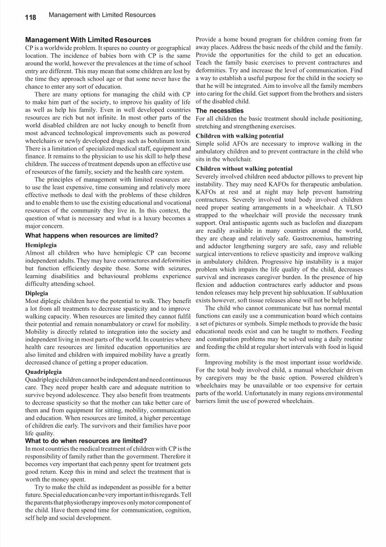
8/9/2019 Guía de ayuda para la parálisis cerebral.pdf
http://slidepdf.com/reader/full/guia-de-ayuda-para-la-paralisis-cerebralpdf 118/143
118 Management with Limited Resources
Management With Limited ResourcesCP is a worldwide problem. It spares no country or geographicallocation. The incidence of babies born with CP is the samearound the world, however the prevalences at the time of schoolentry are different. This may mean that some children are lost bythe time they approach school age or that some never have thechance to enter any sort of education.
There are many options for managing the child with CPto make him part of the society, to improve his quality of lifeas well as help his family. Even in well developed countriesresources are rich but not infinite. In most other parts of theworld disabled children are not lucky enough to benefit frommost advanced technological improvements such as poweredwheelchairs or newly developed drugs such as botulinum toxin.There is a limitation of specialized medical staff, equipment andfinance. It remains to the physician to use his skill to help thesechildren. The success of treatment depends upon an effective useof resources of the family, society and the health care system.
The principles of management with limited resources areto use the least expensive, time consuming and relatively moreeffective methods to deal with the problems of these childrenand to enable them to use the existing educational and vocationalresources of the community they live in. In this context, thequestion of what is necessary and what is a luxury becomes amajor concern.
What happens when resources are limited?
Hemiplegia
Almost all children who have hemiplegic CP can becomeindependent adults. They may have contractures and deformities but function efficiently despite these. Some with seizures,learning disabilities and behavioural problems experiencedifficulty attending school.
Diplegia
Most diplegic children have the potential to walk. They benefita lot from all treatments to decrease spasticity and to improvewalking capacity. When resources are limited they cannot fulfiltheir potential and remain nonambulatory or crawl for mobility.Mobility is directly related to integration into the society andindependent living in most parts of the world. In countries wherehealth care resources are limited education opportunities arealso limited and children with impaired mobility have a greatlydecreased chance of getting a proper education.
Quadriplegia
Quadriplegic children cannot be independent and need continuouscare. They need proper health care and adequate nutrition tosurvive beyond adolescence. They also benefit from treatmentsto decrease spasticity so that the mother can take better care ofthem and from equipment for sitting, mobility, communicationand education. When resources are limited, a higher percentageof children die early. The survivors and their families have poorlife quality.What to do when resources are limited?
In most countries the medical treatment of children with CP is theresponsibility of family rather than the government. Therefore it becomes very important that each penny spent for treatment getsgood return. Keep this in mind and select the treatment that is
worth the money spent.Try to make the child as independent as possible for a better
future. Special education can be very important in this regards. Tellthe parents that physiotherapy improves only motor component ofthe child. Have them spend time for communication, cognition,self help and social development.
Provide a home bound program for children coming from faraway places. Address the basic needs of the child and the family.Provide the opportunities for the child to get an education.Teach the family basic exercises to prevent contractures anddeformities. Try and increase the level of communication. Finda way to establish a useful purpose for the child in the society sothat he will be integrated. Aim to involve all the family members
into caring for the child. Get support from the brothers and sistersof the disabled child.
The necessities
For all children the basic treatment should include positioning,stretching and strengthening exercises.
Children with walking potential
Simple solid AFOs are necessary to improve walking in theambulatory children and to prevent contracture in the child whosits in the wheelchair.
Children without walking potential
Severely involved children need abductor pillows to prevent hipinstability. They may need KAFOs for therapeutic ambulation.
KAFOs at rest and at night may help prevent hamstringcontractures. Severely involved total body involved childrenneed proper seating arrangements in a wheelchair. A TLSOstrapped to the wheelchair will provide the necessary trunksupport. Oral antispastic agents such as baclofen and diazepamare readily available in many countries around the world,they are cheap and relatively safe. Gastrocnemius, hamstringand adductor lengthening surgery are safe, easy and reliablesurgical interventions to relieve spasticity and improve walkingin ambulatory children. Progressive hip instability is a major problem which impairs the life quality of the child, decreasessurvival and increases caregiver burden. In the presence of hip
flexion and adduction contractures early adductor and psoastendon releases may help prevent hip subluxation. If subluxationexists however, soft tissue releases alone will not be helpful.
The child who cannot communicate but has normal mentalfunctions can easily use a communication board which containsa set of pictures or symbols. Simple methods to provide the basiceducational needs exist and can be taught to mothers. Feedingand constipation problems may be solved using a daily routineand feeding the child at regular short intervals with food in liquidform.
Improving mobility is the most important issue worldwide.For the total body involved child, a manual wheelchair driven by caregivers may be the basic option. Powered children’s
wheelchairs may be unavailable or too expensive for certain parts of the world. Unfortunately in many regions environmental barriers limit the use of powered wheelchairs.
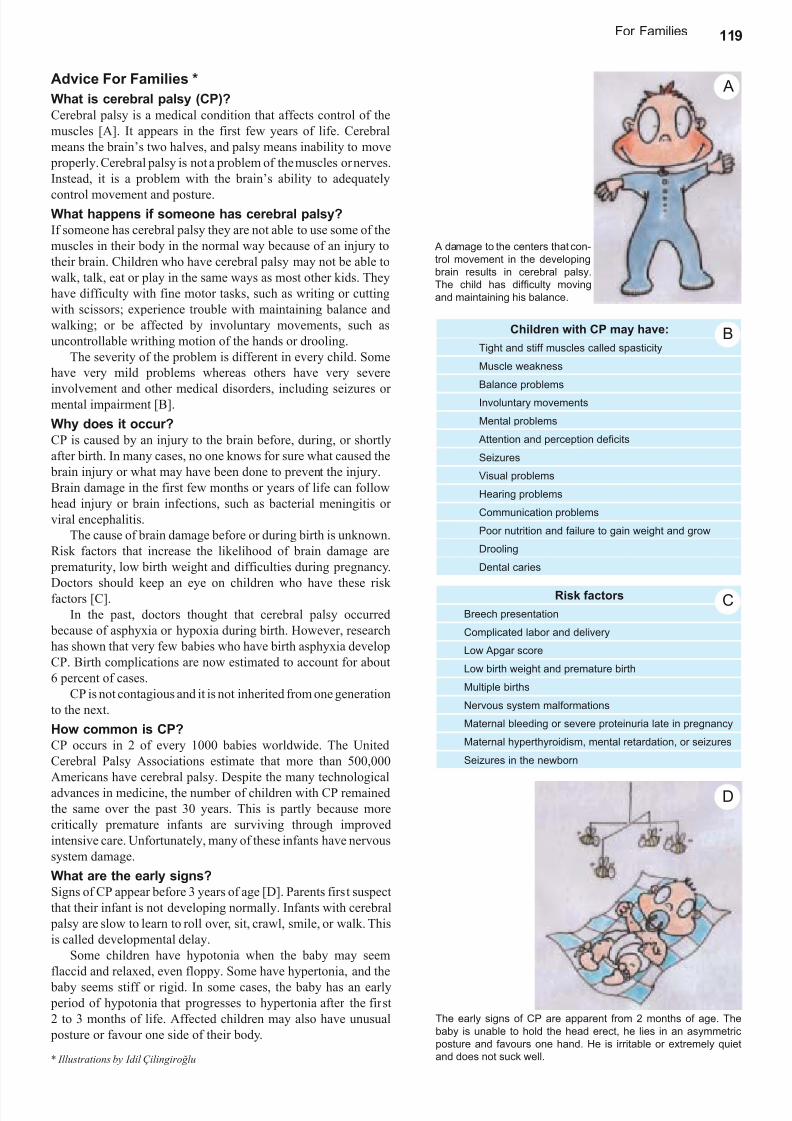
8/9/2019 Guía de ayuda para la parálisis cerebral.pdf
http://slidepdf.com/reader/full/guia-de-ayuda-para-la-paralisis-cerebralpdf 119/143
119For Families
Advice For Families *
What is cerebral palsy (CP)?
Cerebral palsy is a medical condition that affects control of the
muscles [A]. It appears in the first few years of life. Cerebral
means the brain’s two halves, and palsy means inability to move
properly. Cerebral palsy is not a problem of the muscles or nerves.
Instead, it is a problem with the brain’s ability to adequately
control movement and posture.What happens if someone has cerebral palsy?
If someone has cerebral palsy they are not able to use some of the
muscles in their body in the normal way because of an injury to
their brain. Children who have cerebral palsy may not be able to
walk, talk, eat or play in the same ways as most other kids. They
have difficulty with fine motor tasks, such as writing or cutting
with scissors; experience trouble with maintaining balance and
walking; or be affected by involuntary movements, such as
uncontrollable writhing motion of the hands or drooling.
The severity of the problem is different in every child. Some
have very mild problems whereas others have very severe
involvement and other medical disorders, including seizures ormental impairment [B].
Why does it occur?
CP is caused by an injury to the brain before, during, or shortly
after birth. In many cases, no one knows for sure what caused the
brain injury or what may have been done to prevent the injury.
Brain damage in the first few months or years of life can follow
head injury or brain infections, such as bacterial meningitis or
viral encephalitis.
The cause of brain damage before or during birth is unknown.
Risk factors that increase the likelihood of brain damage are
prematurity, low birth weight and difficulties during pregnancy.
Doctors should keep an eye on children who have these riskfactors [C].
In the past, doctors thought that cerebral palsy occurred
because of asphyxia or hypoxia during birth. However, research
has shown that very few babies who have birth asphyxia develop
CP. Birth complications are now estimated to account for about
6 percent of cases.
CP is not contagious and it is not inherited from one generation
to the next.
How common is CP?
CP occurs in 2 of every 1000 babies worldwide. The United
Cerebral Palsy Associations estimate that more than 500,000
Americans have cerebral palsy. Despite the many technological
advances in medicine, the number of children with CP remained
the same over the past 30 years. This is partly because more
critically premature infants are surviving through improved
intensive care. Unfortunately, many of these infants have nervous
system damage.
What are the early signs?
Signs of CP appear before 3 years of age [D]. Parents first suspect
that their infant is not developing normally. Infants with cerebral
palsy are slow to learn to roll over, sit, crawl, smile, or walk. This
is called developmental delay.
Some children have hypotonia when the baby may seem
flaccid and relaxed, even floppy. Some have hypertonia, and the
baby seems stiff or rigid. In some cases, the baby has an early
period of hypotonia that progresses to hypertonia after the first
2 to 3 months of life. Affected children may also have unusual
posture or favour one side of their body.
Children with CP may have:
Tight and stiff muscles called spasticity
Muscle weakness
Balance problems
Involuntary movements
Mental problems
Attention and perception deficits
Seizures
Visual problems
Hearing problems
Communication problems
Poor nutrition and failure to gain weight and grow
Drooling
Dental caries
Risk factors
Breech presentation
Complicated labor and delivery
Low Apgar score
Low birth weight and premature birth
Multiple births
Nervous system malformations
Maternal bleeding or severe proteinuria late in pregnancy
Maternal hyperthyroidism, mental retardation, or seizures
Seizures in the newborn
DD
CC
B
A
A damage to the centers that con-
trol movement in the developing
brain results in cerebral palsy.
The child has difficulty moving
and maintaining his balance.
The early signs of CP are apparent from 2 months of age. The
baby is unable to hold the head erect, he lies in an asymmetric
posture and favours one hand. He is irritable or extremely quiet
and does not suck well.* Illustrations by Idil Çilingiroğlu
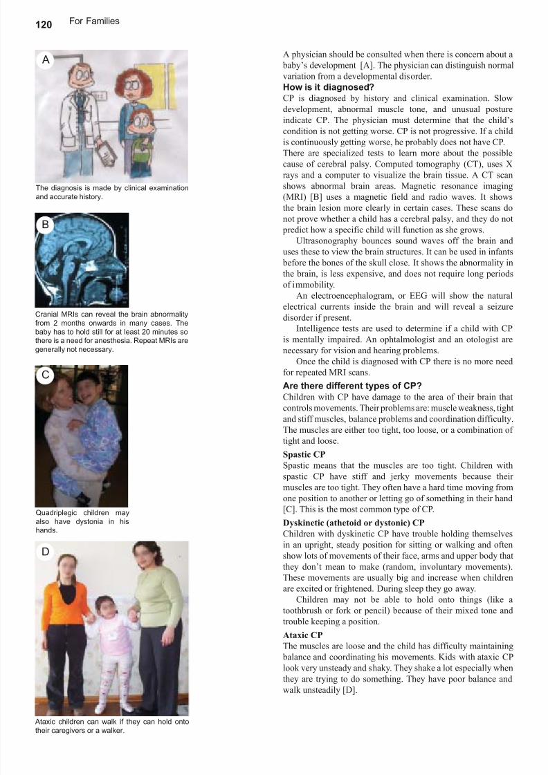
8/9/2019 Guía de ayuda para la parálisis cerebral.pdf
http://slidepdf.com/reader/full/guia-de-ayuda-para-la-paralisis-cerebralpdf 120/143
120 For Families
DD
CC
B
AA physician should be consulted when there is concern about a
baby’s development [A]. The physician can distinguish normal
variation from a developmental disorder.
How is it diagnosed?
CP is diagnosed by history and clinical examination. Slow
development, abnormal muscle tone, and unusual posture
indicate CP. The physician must determine that the child’s
condition is not getting worse. CP is not progressive. If a childis continuously getting worse, he probably does not have CP.
There are specialized tests to learn more about the possible
cause of cerebral palsy. Computed tomography (CT), uses X
rays and a computer to visualize the brain tissue. A CT scan
shows abnormal brain areas. Magnetic resonance imaging
(MRI) [B] uses a magnetic field and radio waves. It shows
the brain lesion more clearly in certain cases. These scans do
not prove whether a child has a cerebral palsy, and they do not
predict how a specific child will function as she grows.
Ultrasonography bounces sound waves off the brain and
uses these to view the brain structures. It can be used in infants
before the bones of the skull close. It shows the abnormality in
the brain, is less expensive, and does not require long periodsof immobility.
An electroencephalogram, or EEG will show the natural
electrical currents inside the brain and will reveal a seizure
disorder if present.
Intelligence tests are used to determine if a child with CP
is mentally impaired. An ophtalmologist and an otologist are
necessary for vision and hearing problems.
Once the child is diagnosed with CP there is no more need
for repeated MRI scans.
Are there different types of CP?
Children with CP have damage to the area of their brain that
controls movements. Their problems are: muscle weakness, tightand stiff muscles, balance problems and coordination difficulty.
The muscles are either too tight, too loose, or a combination of
tight and loose.
Spastic CP
Spastic means that the muscles are too tight. Children with
spastic CP have stiff and jerky movements because their
muscles are too tight. They often have a hard time moving from
one position to another or letting go of something in their hand
[C]. This is the most common type of CP.
Dyskinetic (athetoid or dystonic) CP
Children with dyskinetic CP have trouble holding themselves
in an upright, steady position for sitting or walking and oftenshow lots of movements of their face, arms and upper body that
they don’t mean to make (random, involuntary movements).
These movements are usually big and increase when children
are excited or frightened. During sleep they go away.
Children may not be able to hold onto things (like a
toothbrush or fork or pencil) because of their mixed tone and
trouble keeping a position.
Ataxic CP
The muscles are loose and the child has difficulty maintaining
balance and coordinating his movements. Kids with ataxic CP
look very unsteady and shaky. They shake a lot especially when
they are trying to do something. They have poor balance andwalk unsteadily [D].
The diagnosis is made by clinical examination
and accurate history.
Cranial MRIs can reveal the brain abnormality
from 2 months onwards in many cases. The
baby has to hold still for at least 20 minutes so
there is a need for anesthesia. Repeat MRIs are
generally not necessary.
Quadriplegic children may
also have dystonia in his
hands.
Ataxic children can walk if they can hold onto
their caregivers or a walker.
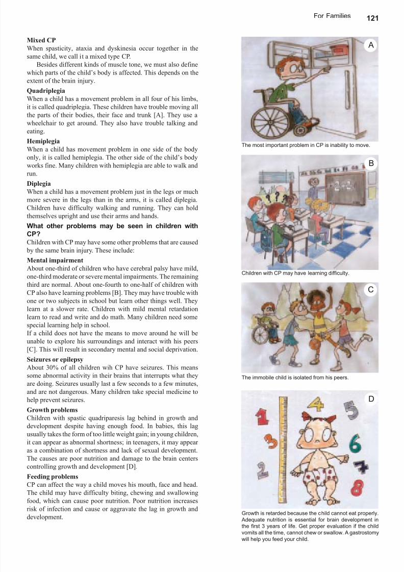
8/9/2019 Guía de ayuda para la parálisis cerebral.pdf
http://slidepdf.com/reader/full/guia-de-ayuda-para-la-paralisis-cerebralpdf 121/143
121For Families
Mixed CP
When spasticity, ataxia and dyskinesia occur together in the
same child, we call it a mixed type CP.
Besides different kinds of muscle tone, we must also define
which parts of the child’s body is affected. This depends on the
extent of the brain injury.
Quadriplegia
When a child has a movement problem in all four of his limbs,it is called quadriplegia. These children have trouble moving all
the parts of their bodies, their face and trunk [A]. They use a
wheelchair to get around. They also have trouble talking and
eating.
Hemiplegia
When a child has movement problem in one side of the body
only, it is called hemiplegia. The other side of the child’s body
works fine. Many children with hemiplegia are able to walk and
run.
Diplegia
When a child has a movement problem just in the legs or much
more severe in the legs than in the arms, it is called diplegia.Children have difficulty walking and running. They can hold
themselves upright and use their arms and hands.
What other problems may be seen in children with
CP?
Children with CP may have some other problems that are caused
by the same brain injury. These include:
Mental impairment
About one-third of children who have cerebral palsy have mild,
one-third moderate or severe mental impairments. The remaining
third are normal. About one-fourth to one-half of children with
CP also have learning problems [B]. They may have trouble with
one or two subjects in school but learn other things well. Theylearn at a slower rate. Children with mild mental retardation
learn to read and write and do math. Many children need some
special learning help in school.
If a child does not have the means to move around he will be
unable to explore his surroundings and interact with his peers
[C]. This will result in secondary mental and social deprivation.
Seizures or epilepsy
About 30% of all children wih CP have seizures. This means
some abnormal activity in their brains that interrupts what they
are doing. Seizures usually last a few seconds to a few minutes,
and are not dangerous. Many children take special medicine to
help prevent seizures.Growth problems
Children with spastic quadriparesis lag behind in growth and
development despite having enough food. In babies, this lag
usually takes the form of too little weight gain; in young children,
it can appear as abnormal shortness; in teenagers, it may appear
as a combination of shortness and lack of sexual development.
The causes are poor nutrition and damage to the brain centers
controlling growth and development [D].
Feeding problems
CP can affect the way a child moves his mouth, face and head.
The child may have difficulty biting, chewing and swallowing
food, which can cause poor nutrition. Poor nutrition increasesrisk of infection and cause or aggravate the lag in growth and
development.
DD
CC
B
A
The most important problem in CP is inability to move.
The immobile child is isolated from his peers.
Children with CP may have learning difficulty.
Growth is retarded because the child cannot eat properly.
Adequate nutrition is essential for brain development in
the first 3 years of life. Get proper evaluation if the child
vomits all the time, cannot chew or swallow. A gastrostomy
will help you feed your child.
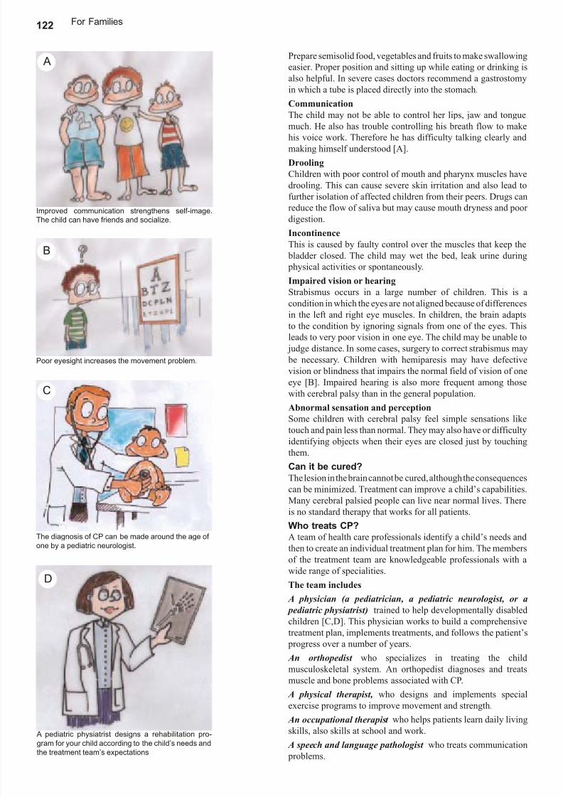
8/9/2019 Guía de ayuda para la parálisis cerebral.pdf
http://slidepdf.com/reader/full/guia-de-ayuda-para-la-paralisis-cerebralpdf 122/143
122 For Families
DD
CC
B
APrepare semisolid food, vegetables and fruits to make swallowing
easier. Proper position and sitting up while eating or drinking is
also helpful. In severe cases doctors recommend a gastrostomy
in which a tube is placed directly into the stomach.
Communication
The child may not be able to control her lips, jaw and tongue
much. He also has trouble controlling his breath flow to make
his voice work. Therefore he has difficulty talking clearly andmaking himself understood [A].
Drooling
Children with poor control of mouth and pharynx muscles have
drooling. This can cause severe skin irritation and also lead to
further isolation of affected children from their peers. Drugs can
reduce the flow of saliva but may cause mouth dryness and poor
digestion.
Incontinence
This is caused by faulty control over the muscles that keep the
bladder closed. The child may wet the bed, leak urine during
physical activities or spontaneously.
Impaired vision or hearingStrabismus occurs in a large number of children. This is a
condition in which the eyes are not aligned because of differences
in the left and right eye muscles. In children, the brain adapts
to the condition by ignoring signals from one of the eyes. This
leads to very poor vision in one eye. The child may be unable to
judge distance. In some cases, surgery to correct strabismus may
be necessary. Children with hemiparesis may have defective
vision or blindness that impairs the normal field of vision of one
eye [B]. Impaired hearing is also more frequent among those
with cerebral palsy than in the general population.
Abnormal sensation and perception
Some children with cerebral palsy feel simple sensations liketouch and pain less than normal. They may also have or difficulty
identifying objects when their eyes are closed just by touching
them.
Can it be cured?
The lesion in the brain cannot be cured, although the consequences
can be minimized. Treatment can improve a child’s capabilities.
Many cerebral palsied people can live near normal lives. There
is no standard therapy that works for all patients.
Who treats CP?
A team of health care professionals identify a child’s needs and
then to create an individual treatment plan for him. The members
of the treatment team are knowledgeable professionals with awide range of specialities.
The team includes
A physician (a pediatrician, a pediatric neurologist, or a
pediatric physiatrist) trained to help developmentally disabled
children [C,D]. This physician works to build a comprehensive
treatment plan, implements treatments, and follows the patient’s
progress over a number of years.
An orthopedist who specializes in treating the child
musculoskeletal system. An orthopedist diagnoses and treats
muscle and bone problems associated with CP.
A physical therapist, who designs and implements specialexercise programs to improve movement and strength.
An occupational therapist who helps patients learn daily living
skills, also skills at school and work.
A speech and language pathologist who treats communication
problems.
Poor eyesight increases the movement problem.
Improved communication strengthens self-image.
The child can have friends and socialize.
The diagnosis of CP can be made around the age of
one by a pediatric neurologist.
A pediatric physiatrist designs a rehabilitation pro-
gram for your child according to the child’s needs and
the treatment team’s expectations
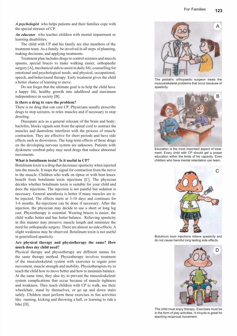
8/9/2019 Guía de ayuda para la parálisis cerebral.pdf
http://slidepdf.com/reader/full/guia-de-ayuda-para-la-paralisis-cerebralpdf 123/143
123For Families
A psychologist who helps patients and their families cope with
the special stresses of CP.
An educator who teaches children with mental impairment or
learning disabilities.
The child with CP and his family are also members of the
treatment team. As a family, be involved in all steps of planning,
making decisions, and applying treatments.
Treatment plan includes drugs to control seizures and musclespasms, special braces to make walking easier, orthopaedic
surgery [A], mechanical aids to assist in daily life, counselling for
emotional and psychological needs, and physical, occupational,
speech, and behavioural therapy. Early treatment gives the child
a better chance of learning to move.
Do not forget that the ultimate goal is to help the child have
a happy life, healthy growth into adulthood and maximum
independence in society [B].
Is there a drug to cure the problem?
There is no drug that can cure CP. Physicians usually prescribe
drugs to stop seizures, to relax muscles and if necessary to stop
drooling.Diazepam acts as a general relaxant of the brain and body;
baclofen, blocks signals sent from the spinal cord to contract the
muscles and dantrolene interferes with the process of muscle
contraction. They are effective for short periods and have side
effects such as drowsiness. The long-term effects of these drugs
on the developing nervous system are unknown. Patients with
dyskinetic cerebral palsy may need drugs that reduce abnormal
movements.
What is botulinum toxin? Is it useful in CP?
Botulinum toxin is a drug that decreases spasticity when injected
into the muscle. It stops the signal for contraction from the nerve
to the muscle. Children who walk on tiptoe or with bent knees benefit from botulinum toxin injections [C]. The physician
decides whether botulinum toxin is suitable for your child and
does the injections. The injection is not painful but sedation is
necessary. General anesthesia is better if many muscles are to
be injected. The effects starts at 3-10 days and continues for
3-6 months. Re-injections can be done if necessary. After the
injection, the physician may decide to use a short or long leg
cast. Physiotherapy is essential. Wearing braces is easier, the
child walks better and has better balance. Relieving spasticity
in this manner may preserve muscle length and minimize the
need for orthopaedic surgery. There are almost no side-effects. A
slight weakness may be observed. Botulinum toxin is not useful
in generalized spasticity.
Are physical therapy and physiotherapy the same? How
much does my child need?
Physical therapy and physiotherapy are different names for
the same therapy method. Physiotherapy involves treatment
of the musculoskeletal system with exercises to regain joint
movement, muscle strength and mobility. Physiotherapists try to
teach the child how to move better and how to maintain balance.
At the same time, they also try to prevent the musculoskeletal
system complications that occur because of muscle tightness
and weakness. They teach children with CP to walk, use their
wheelchair, stand by themselves, or go up and down stairs
safely. Children must perform these exercises in fun activitieslike running, kicking and throwing a ball, or learning to ride a
bike [D].
The pediatric orthopaedic surgeon treats the
musculoskeletal problems that occur because of
spasticity.
Education is the most important aspect of treat-
ment. Every child with CP should get a proper
education within the limits of his capacity. Even
children who have mental retardation can learn.
Botulinum toxin injections relieve spasticity anddo not cause harmful long lasting side effects.
The child must enjoy therapy. Exercises must be
in the form of play activities. A tricycle is great for
teaching reciprocal movement.
DD
CC
B
A
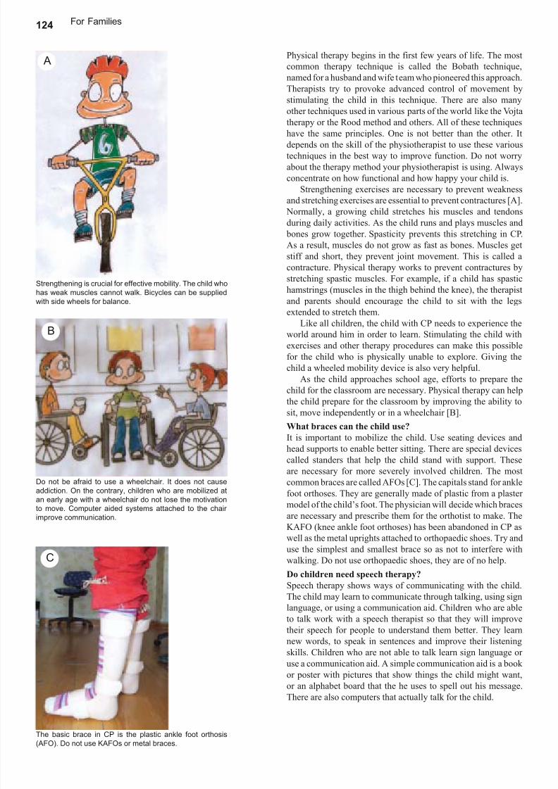
8/9/2019 Guía de ayuda para la parálisis cerebral.pdf
http://slidepdf.com/reader/full/guia-de-ayuda-para-la-paralisis-cerebralpdf 124/143
124 For Families
Physical therapy begins in the first few years of life. The most
common therapy technique is called the Bobath technique,
named for a husband and wife team who pioneered this approach.
Therapists try to provoke advanced control of movement by
stimulating the child in this technique. There are also many
other techniques used in various parts of the world like the Vojta
therapy or the Rood method and others. All of these techniques
have the same principles. One is not better than the other. Itdepends on the skill of the physiotherapist to use these various
techniques in the best way to improve function. Do not worry
about the therapy method your physiotherapist is using. Always
concentrate on how functional and how happy your child is.
Strengthening exercises are necessary to prevent weakness
and stretching exercises are essential to prevent contractures [A].
Normally, a growing child stretches his muscles and tendons
during daily activities. As the child runs and plays muscles and
bones grow together. Spasticity prevents this stretching in CP.
As a result, muscles do not grow as fast as bones. Muscles get
stiff and short, they prevent joint movement. This is called a
contracture. Physical therapy works to prevent contractures by
stretching spastic muscles. For example, if a child has spastichamstrings (muscles in the thigh behind the knee), the therapist
and parents should encourage the child to sit with the legs
extended to stretch them.
Like all children, the child with CP needs to experience the
world around him in order to learn. Stimulating the child with
exercises and other therapy procedures can make this possible
for the child who is physically unable to explore. Giving the
child a wheeled mobility device is also very helpful.
As the child approaches school age, efforts to prepare the
child for the classroom are necessary. Physical therapy can help
the child prepare for the classroom by improving the ability to
sit, move independently or in a wheelchair [B].What braces can the child use?
It is important to mobilize the child. Use seating devices and
head supports to enable better sitting. There are special devices
called standers that help the child stand with support. These
are necessary for more severely involved children. The most
common braces are called AFOs [C]. The capitals stand for ankle
foot orthoses. They are generally made of plastic from a plaster
model of the child’s foot. The physician will decide which braces
are necessary and prescribe them for the orthotist to make. The
KAFO (knee ankle foot orthoses) has been abandoned in CP as
well as the metal uprights attached to orthopaedic shoes. Try and
use the simplest and smallest brace so as not to interfere withwalking. Do not use orthopaedic shoes, they are of no help.
Do children need speech therapy?
Speech therapy shows ways of communicating with the child.
The child may learn to communicate through talking, using sign
language, or using a communication aid. Children who are able
to talk work with a speech therapist so that they will improve
their speech for people to understand them better. They learn
new words, to speak in sentences and improve their listening
skills. Children who are not able to talk learn sign language or
use a communication aid. A simple communication aid is a book
or poster with pictures that show things the child might want,
or an alphabet board that the he uses to spell out his message.
There are also computers that actually talk for the child.
Strengthening is crucial for effective mobility. The child who
has weak muscles cannot walk. Bicycles can be supplied
with side wheels for balance.
The basic brace in CP is the plastic ankle foot orthosis
(AFO). Do not use KAFOs or metal braces.
Do not be afraid to use a wheelchair. It does not cause
addiction. On the contrary, children who are mobilized at
an early age with a wheelchair do not lose the motivation
to move. Computer aided systems attached to the chair
improve communication.
B
A
CC
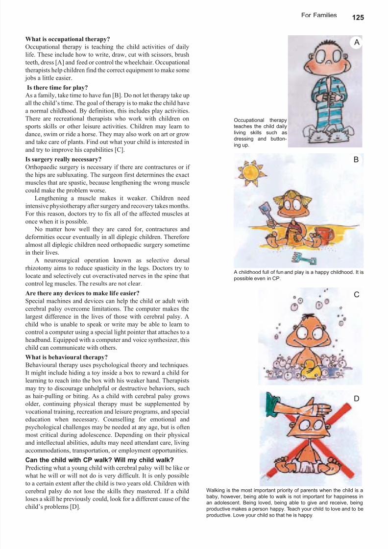
8/9/2019 Guía de ayuda para la parálisis cerebral.pdf
http://slidepdf.com/reader/full/guia-de-ayuda-para-la-paralisis-cerebralpdf 125/143
125For Families
What is occupational therapy?
Occupational therapy is teaching the child activities of daily
life. These include how to write, draw, cut with scissors, brush
teeth, dress [A] and feed or control the wheelchair. Occupational
therapists help children find the correct equipment to make some
jobs a little easier.
Is there time for play?
As a family, take time to have fun [B]. Do not let therapy take upall the child’s time. The goal of therapy is to make the child have
a normal childhood. By definition, this includes play activities.
There are recreational therapists who work with children on
sports skills or other leisure activities. Children may learn to
dance, swim or ride a horse. They may also work on art or grow
and take care of plants. Find out what your child is interested in
and try to improve his capabilities [C].
Is surgery really necessary?
Orthopaedic surgery is necessary if there are contractures or if
the hips are subluxating. The surgeon first determines the exact
muscles that are spastic, because lengthening the wrong muscle
could make the problem worse.Lengthening a muscle makes it weaker. Children need
intensive physiotherapy after surgery and recovery takes months.
For this reason, doctors try to fix all of the affected muscles at
once when it is possible.
No matter how well they are cared for, contractures and
deformities occur eventually in all diplegic children. Therefore
almost all diplegic children need orthopaedic surgery sometime
in their lives.
A neurosurgical operation known as selective dorsal
rhizotomy aims to reduce spasticity in the legs. Doctors try to
locate and selectively cut overactivated nerves in the spine that
control leg muscles. The results are not clear.
Are there any devices to make life easier?
Special machines and devices can help the child or adult with
cerebral palsy overcome limitations. The computer makes the
largest difference in the lives of those with cerebral palsy. A
child who is unable to speak or write may be able to learn to
control a computer using a special light pointer that attaches to a
headband. Equipped with a computer and voice synthesizer, this
child can communicate with others.
What is behavioural therapy?
Behavioural therapy uses psychological theory and techniques.
It might include hiding a toy inside a box to reward a child for
learning to reach into the box with his weaker hand. Therapists
may try to discourage unhelpful or destructive behaviors, such
as hair-pulling or biting. As a child with cerebral palsy grows
older, continuing physical therapy must be supplemented by
vocational training, recreation and leisure programs, and special
education when necessary. Counselling for emotional and
psychological challenges may be needed at any age, but is often
most critical during adolescence. Depending on their physical
and intellectual abilities, adults may need attendant care, living
accommodations, transportation, or employment opportunities.
Can the child with CP walk? Will my child walk?
Predicting what a young child with cerebral palsy will be like or
what he will or will not do is very difficult. It is only possible
to a certain extent after the child is two years old. Children with
cerebral palsy do not lose the skills they mastered. If a child
loses a skill he previously could, look for a different cause of the
child’s problems [D].
Occupational therapy
teaches the child daily
living skills such as
dressing and button-
ing up.
A childhood full of fun and play is a happy childhood. It is
possible even in CP.
Walking is the most important priority of parents when the child is a
baby, however, being able to walk is not important for happiness in
an adolescent. Being loved, being able to give and receive, being
productive makes a person happy. Teach your child to love and to be
productive. Love your child so that he is happy.
A
B
CC
DD
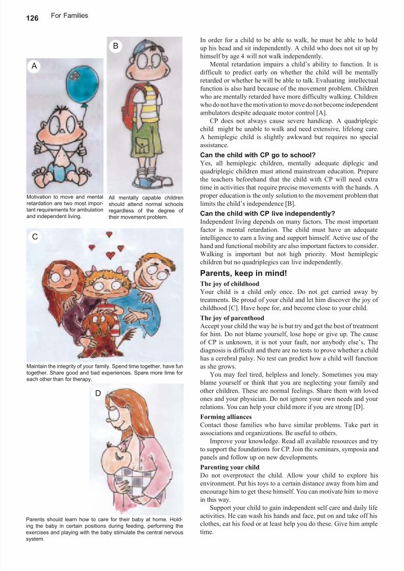
8/9/2019 Guía de ayuda para la parálisis cerebral.pdf
http://slidepdf.com/reader/full/guia-de-ayuda-para-la-paralisis-cerebralpdf 126/143
126 For Families
In order for a child to be able to walk, he must be able to hold
up his head and sit independently. A child who does not sit up by
himself by age 4 will not walk independently.
Mental retardation impairs a child’s ability to function. It is
difficult to predict early on whether the child will be mentally
retarded or whether he will be able to talk. Evaluating intellectual
function is also hard because of the movement problem. Children
who are mentally retarded have more difficulty walking. Childrenwho do not have the motivation to move do not become independent
ambulators despite adequate motor control [A].
CP does not always cause severe handicap. A quadriplegic
child might be unable to walk and need extensive, lifelong care.
A hemiplegic child is slightly awkward but requires no special
assistance.
Can the child with CP go to school?
Yes, all hemiplegic children, mentally adequate diplegic and
quadriplegic children must attend mainstream education. Prepare
the teachers beforehand that the child with CP will need extra
time in activities that require precise movements with the hands. A
proper education is the only solution to the movement problem that
limits the child’s independence [B].
Can the child with CP live independently?
Independent living depends on many factors. The most important
factor is mental retardation. The child must have an adequate
intelligence to earn a living and support himself. Active use of the
hand and functional mobility are also important factors to consider.
Walking is important but not high priority. Most hemiplegic
children but no quadriplegics can live independently.
Parents, keep in mind!
The joy of childhood
Your child is a child only once. Do not get carried away by
treatments. Be proud of your child and let him discover the joy ofchildhood [C]. Have hope for, and become close to your child.
The joy of parenthood
Accept your child the way he is but try and get the best of treatment
for him. Do not blame yourself, lose hope or give up. The cause
of CP is unknown, it is not your fault, nor anybody else’s. The
diagnosis is difficult and there are no tests to prove whether a child
has a cerebral palsy. No test can predict how a child will function
as she grows.
You may feel tired, helpless and lonely. Sometimes you may
blame yourself or think that you are neglecting your family and
other children. These are normal feelings. Share them with loved
ones and your physician. Do not ignore your own needs and yourrelations. You can help your child more if you are strong [D].
Forming alliances
Contact those families who have similar problems. Take part in
associations and organizations. Be useful to others.
Improve your knowledge. Read all available resources and try
to support the foundations for CP. Join the seminars, symposia and
panels and follow up on new developments.
Parenting your child
Do not overprotect the child. Allow your child to explore his
environment. Put his toys to a certain distance away from him and
encourage him to get these himself. You can motivate him to move
in this way.Support your child to gain independent self care and daily life
activities. He can wash his hands and face, put on and take off his
clothes, eat his food or at least help you do these. Give him ample
time.
Parents should learn how to care for their baby at home. Hold-
ing the baby in certain positions during feeding, performing the
exercises and playing with the baby stimulate the central nervous
system.
Motivation to move and mental
retardation are two most impor-
tant requirements for ambulation
and independent living.
All mentally capable childrenshould attend normal schools
regardless of the degree of
their movement problem.
Maintain the integrity of your family. Spend time together, have fun
together. Share good and bad experiences. Spare more time for
each other than for therapy.
A
B
CC
DD
8/9/2019 Guía de ayuda para la parálisis cerebral.pdf
http://slidepdf.com/reader/full/guia-de-ayuda-para-la-paralisis-cerebralpdf 127/143
127For Families
Infancy When your child is just a baby, learn how to hold, feed
and exercise him. Infancy is a time of uncertainties. Do not be
discouraged and keep loving your baby.
Toddler When your child is a toddler support his body so that
he can use his hands in active play. Encourage him to move
by himself. Get him wheeled devices [A] so that he can move
around the house and explore his surroundings. Never believe
those who say the child should crawl before he can walk or thathis spine will bend if you make him sit. See for yourself what
your child is capable of doing and support him if he wants to do
that.
Schooling Never neglect the child’s education. Defend your
child’s right to get the best education possible [B]. Special
education is possible if your child cannot enter mainstream
education. Do not allow anything to disturb his school and
academic life. Walking does not make a person a human being,
an active productive mind does.
Adolescence Provide psychological support to your child during
adolescence. Try and be his friend [C]. Encourage him in the
things he wants to do. Allow him to make his own decisions.Guide him gently in his relationships with his surroundings.
Children need other children. Help your child make friends.
Interaction with the health care providers
Know your child well. Define your needs and express these
openly to the treatment team.
Learn and perform the exercises that your physiotherapist
teaches you. Ask once more if you do not understand or have
difficulty performing.
Know your demands from the health care providers and from
the society in general at every point in your child’s life and be
ready to voice them clearly. Remember that nobody can know
what your child needs better than you or him [D]. Problems canonly be solved if they are defined clearly and made known. The
community will respond to your efforts.
Continuous care
Focus on your child’s strengths, assets and interests rather than
his weaknesses. People excel in the things they like doing. If
your child wants to do something, support him. Praise your
child frequently for his accomplishments. Do not dwell on his
weaknesses. It is much easier to improve his strong points than
his weaknesses. Remember, your child does not need to walk to
be happy. He needs your love and attention.
Mobility is essential for the toddler
who needs to explore to learn and
develop skills. Sports is better than
all sorts of physiotherapy for the ado-
lescent at school.
Throughout the various therapy pro-
cedures and the anxiety of rearing achild with CP, do not miss the joy of
being a father or a mother. Remem-
ber the joy of parenthood.
When buying clothes look for:
Finger rings or other large zipper pulls
Flat and nonskid shoes
Raglan sleeves
Front openings
Short backs
Nonrestrictive elastic
Velcro
Leg zippers
Hook and loop closures
Flat-felled seams
RoomyCut Longer in the seat to allow for sitting
Cut shorter in the legs and arms and shorter shirts that don’t
get caught in wheels.
Knit fabrics
Table modified from the website: http://geocities.com/aneecp/clothing.htm
Talking To Your Doctor
Here are some tips for communicating more effectively with the physi-
cian so that the visit to the doctor will be more productive.
Make a list of your concerns. Look to the list as you talk so you will
not forget anything.
Try to arrive 10-15 minutes before the appointment time even if yourdoctor never runs on time.
Have someone with you to help you listen and for emotional support
if needed.
Be conscious of the doctor’s time. If you are doing well 10-15 minutes
is all that is necessary.
Use the medications as prescribed. Report effects of the medications
accurately. Have a list of the medications and dosages with you all
the time.
Do not tell the doctor what to do. Work with your doctor. You are a
team.
If you do not understand what the doctor says ask him/her to repeat
it.
Do not accept ambiguous answers.
Be impressed with physicians who send you to get a second opinion,
or admit that they do not have all the answers.
THANK the doctor for his/her time.
Educate yourself about CP.
DD
A
CC
B
E
E
8/9/2019 Guía de ayuda para la parálisis cerebral.pdf
http://slidepdf.com/reader/full/guia-de-ayuda-para-la-paralisis-cerebralpdf 128/143
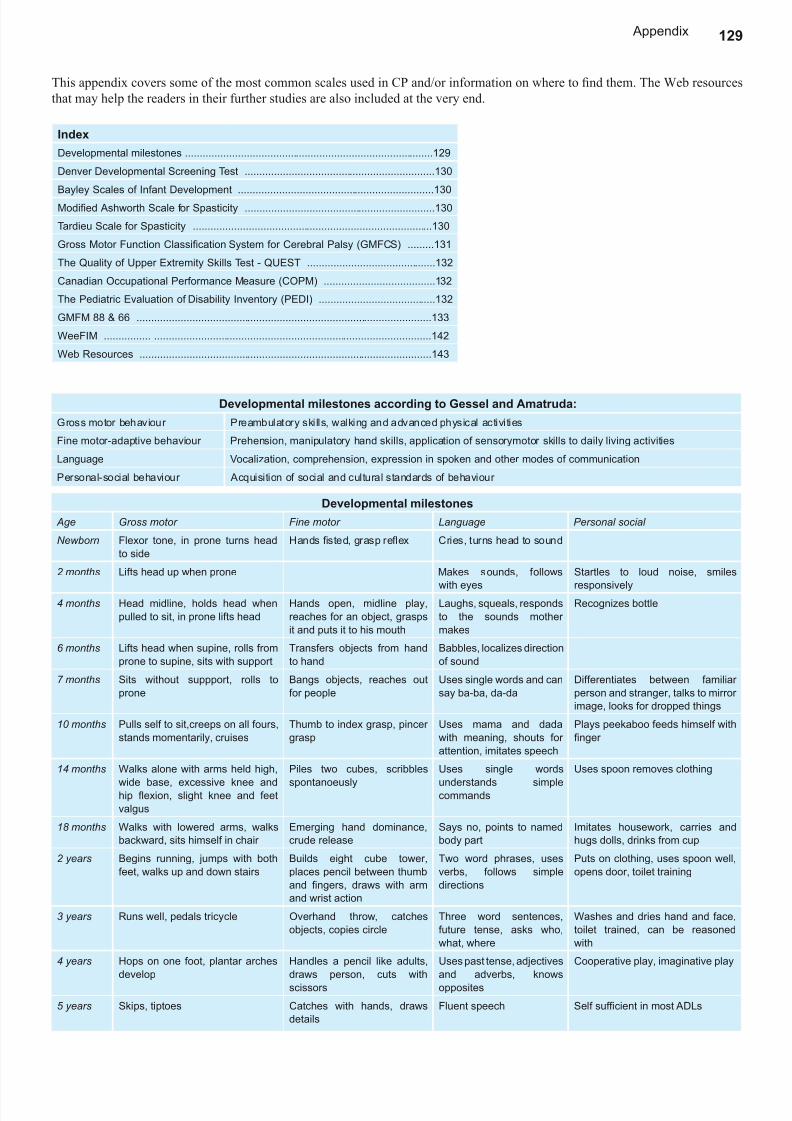
8/9/2019 Guía de ayuda para la parálisis cerebral.pdf
http://slidepdf.com/reader/full/guia-de-ayuda-para-la-paralisis-cerebralpdf 129/143
129 Appendix
Developmental milestones according to Gessel and Amatruda:
Gross motor behaviour Preambulatory skills, walking and advanced physical activities
Fine motor-adaptive behaviour Prehension, manipulatory hand skills, application of sensorymotor skills to daily living activities
Language Vocalization, comprehension, expression in spoken and other modes of communication
Personal-social behaviour Acquisition of social and cultural standards of behaviour
Developmental milestones
Age Gross motor Fine motor Language Personal social
Newborn Flexor tone, in prone turns head
to side
Hands fisted, grasp reflex Cries, turns head to sound
2 months Lifts head up when prone Makes sounds, follows
with eyes
Startles to loud noise, smiles
responsively4 months Head midline, holds head when
pulled to sit, in prone lifts head
Hands open, midline play,
reaches for an object, grasps
it and puts it to his mouth
Laughs, squeals, responds
to the sounds mother
makes
Recognizes bottle
6 months Lifts head when supine, rolls from
prone to supine, sits with support
Transfers objects from hand
to hand
Babbles, localizes direction
of sound
7 months Sits without suppport, rolls to
prone
Bangs objects, reaches out
for people
Uses single words and can
say ba-ba, da-da
Differentiates between familiar
person and stranger, talks to mirror
image, looks for dropped things
10 months Pulls self to sit,creeps on all fours,
stands momentarily, cruises
Thumb to index grasp, pincer
grasp
Uses mama and dada
with meaning, shouts for
attention, imitates speech
Plays peekaboo feeds himself with
finger
14 months Walks alone with arms held high,
wide base, excessive knee andhip flexion, slight knee and feet
valgus
Piles two cubes, scribbles
spontanoeusly
Uses single words
understands simplecommands
Uses spoon removes clothing
18 months Walks with lowered arms, walks
backward, sits himself in chair
Emerging hand dominance,
crude release
Says no, points to named
body part
Imitates housework, carries and
hugs dolls, drinks from cup
2 years Begins running, jumps with both
feet, walks up and down stairs
Builds eight cube tower,
places pencil between thumb
and fingers, draws with arm
and wrist action
Two word phrases, uses
verbs, follows simple
directions
Puts on clothing, uses spoon well,
opens door, toilet training
3 years Runs well, pedals tricycle Overhand throw, catches
objects, copies circle
Three word sentences,
future tense, asks who,
what, where
Washes and dries hand and face,
toilet trained, can be reasoned
with
4 years Hops on one foot, plantar arches
develop
Handles a pencil like adults,
draws person, cuts with
scissors
Uses past tense, adjectives
and adverbs, knows
opposites
Cooperative play, imaginative play
5 years Skips, tiptoes Catches with hands, draws
details
Fluent speech Self sufficient in most ADLs
This appendix covers some of the most common scales used in CP and/or information on where to find them. The Web resources
that may help the readers in their further studies are also included at the very end.
Index
Developmental milestones .....................................................................................129
Denver Developmental Screening Test .................................................................130
Bayley Scales of Infant Development ...................................................................130
Modified Ashworth Scale for Spasticity .................................................................130
Tardieu Scale for Spasticity ..................................................................................130
Gross Motor Function Classification System for Cerebral Palsy (GMFCS) .........131
The Quality of Upper Extremity Skills Test - QUEST ............................................132
Canadian Occupational Performance Measure (COPM) ......................................132
The Pediatric Evaluation of Disability Inventory (PEDI) ........................................132
GMFM 88 & 66 .....................................................................................................133
WeeFIM ................ ...............................................................................................142
Web Resources ....................................................................................................143
8/9/2019 Guía de ayuda para la parálisis cerebral.pdf
http://slidepdf.com/reader/full/guia-de-ayuda-para-la-paralisis-cerebralpdf 130/143
130 Appendix
Tardieu ScaleVelocity of stretch
V1 As slow as possible (slower than the rate of the natural drop of the limb segment under gravity)
V2 Speed of the limb segment falling under gravity
V3 As fast as possible (faster than the rate of the natural drop of the limb segment under gravity)
Grading Tardieu scale
Quality of muscle reaction (X)
0 No resistance throughout the course of the passive movement
1 Slight resistance throughout the course of the passive movement
2 Clear catch at precise angle, interrupting the passive movement, followed by release
3 Fatiguable clonus (less than 10 s when maintaining the pressure) occurring at a precise angle, followed by release
4 Unfatiguable clonus (less than 10 s when maintaining the pressure) occurring at a precise angle
Angle of muscle action (V) measured relative to the position of minimal stretch of the muscle (corresponding to angle zero) for all joints except hip
where it is relative to the resting anatomical position
Developmental Tests
These tests describe the development of the child in various functional stages.
Denver Development Screening Test
This test evaluates the developmental deficits in infants and young children from age 1 month to 6 years in the areas of global motor
function, language, fine-motor adaptation and social contact.
The Denver Developmental Screening Test (DDST) II is administered to children between birth and six years of age. It can screen
children who are apparently normal for possible problems and monitor children who have high risk because of past history such as
perinatal difficulties. It is not an IQ test nor will it predict what the level of the child’s future intelligence and ability will be. Do notuse the Denver II for diagnosis. The Denver II tests the child on twenty simple tasks and on 4 different domains. Personal - social
measures the child’s ability to get along with people and to take care of himself / herself. Fine Motor Adaptive test identifies the
child’s ability to see and to use his hands to pick up objects and to draw. Language tests determine the child’s ability to hear, follow
direction and to speak. Gross Motor identifies the child’s ability to sit, walk and jump.
Further Reading:
Denver II Training Manual, Second Edition Revised 1992
Bayley Scales of Infant Development
This test evaluates cognition, language, social behaviour and motor functions in children from 1 to 42 months old. The purpose of the
Bayley Scales of Infant Development is to diagnose developmental delay. The test takes approximately 45 minutes. The examiner
gives a series of stimuli to which the child responds. The Mental Scales assess memory, learning, problem-solving ability, and verbal
communication skills. The Motor Scales evaluate sitting and standing, gross motor skills and fine motor skills. The Infant Behavior
Record (IBR) assesses the child’s social and emotional development through a standardized description of his or her behaviourduring the testing session. Scores are measured against norms for each of the 14 different age groups. The Bayley scales determine
whether a child is developing normally and provide for early diagnosis and intervention in cases of developmental delay.
The Modified Ashworth scale
0 No increase in muscle tone
1 Slight increase in tone with a catch and release or minimal resistance at end of range
2 As 1 but with minimal resistance through range following catch3 More marked increase tone through ROM
4 Considerable increase in tone, passive movement difficult.
5 Affected part rigid
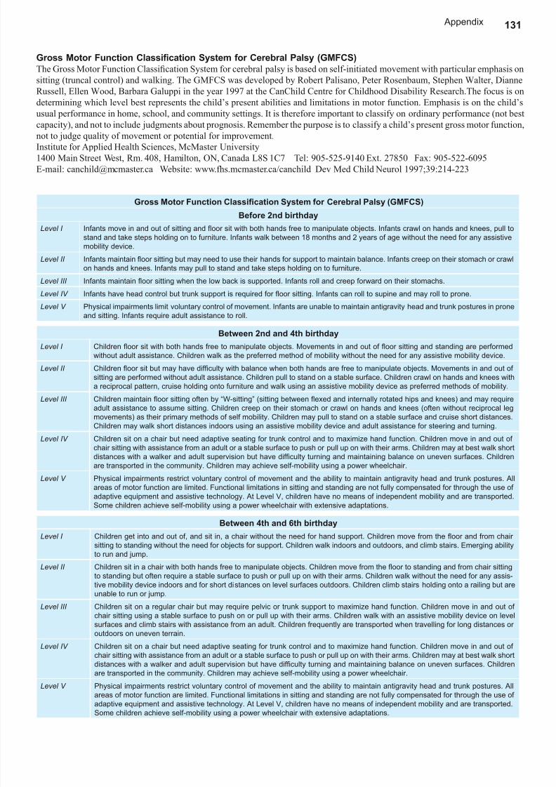
8/9/2019 Guía de ayuda para la parálisis cerebral.pdf
http://slidepdf.com/reader/full/guia-de-ayuda-para-la-paralisis-cerebralpdf 131/143
131 Appendix
Gross Motor Function Classification System for Cerebral Palsy (GMFCS)
The Gross Motor Function Classification System for cerebral palsy is based on self-initiated movement with particular emphasis on
sitting (truncal control) and walking. The GMFCS was developed by Robert Palisano, Peter Rosenbaum, Stephen Walter, Dianne
Russell, Ellen Wood, Barbara Galuppi in the year 1997 at the CanChild Centre for Childhood Disability Research.The focus is on
determining which level best represents the child’s present abilities and limitations in motor function. Emphasis is on the child’s
usual performance in home, school, and community settings. It is therefore important to classify on ordinary performance (not best
capacity), and not to include judgments about prognosis. Remember the purpose is to classify a child’s present gross motor function,
not to judge quality of movement or potential for improvement.Institute for Applied Health Sciences, McMaster University
1400 Main Street West, Rm. 408, Hamilton, ON, Canada L8S 1C7 Tel: 905-525-9140 Ext. 27850 Fax: 905-522-6095
E-mail: [email protected] Website: www.fhs.mcmaster.ca/canchild Dev Med Child Neurol 1997;39:214-223
Gross Motor Function Classification System for Cerebral Palsy (GMFCS)
Before 2nd birthday
Level I Infants move in and out of sitting and floor sit with both hands free to manipulate objects. Infants crawl on hands and knees, pull to
stand and take steps holding on to furniture. Infants walk between 18 months and 2 years of age without the need for any assistive
mobility device.
Level II Infants maintain floor sitting but may need to use their hands for support to maintain balance. Infants creep on their stomach or crawl
on hands and knees. Infants may pull to stand and take steps holding on to furniture.
Level III Infants maintain floor sitting when the low back is supported. Infants roll and creep forward on their stomachs.
Level IV Infants have head control but trunk support is required for floor sitting. Infants can roll to supine and may roll to prone.
Level V Physical impairments limit voluntary control of movement. Infants are unable to maintain antigravity head and trunk postures in prone
and sitting. Infants require adult assistance to roll.
Between 2nd and 4th birthday
Level I Children floor sit with both hands free to manipulate objects. Movements in and out of floor sitting and standing are performed
without adult assistance. Children walk as the preferred method of mobility without the need for any assistive mobility device.
Level II Children floor sit but may have difficulty with balance when both hands are free to manipulate objects. Movements in and out of
sitting are performed without adult assistance. Children pull to stand on a stable surface. Children crawl on hands and knees with
a reciprocal pattern, cruise holding onto furniture and walk using an assistive mobility device as preferred methods of mobility.
Level III Children maintain floor sitting often by “W-sitting” (sitting between flexed and internally rotated hips and knees) and may require
adult assistance to assume sitting. Children creep on their stomach or crawl on hands and knees (often without reciprocal leg
movements) as their primary methods of self mobility. Children may pull to stand on a stable surface and cruise short distances.
Children may walk short distances indoors using an assistive mobility device and adult assistance for steering and turning.
Level IV Children sit on a chair but need adaptive seating for trunk control and to maximize hand function. Children move in and out of
chair sitting with assistance from an adult or a stable surface to push or pull up on with their arms. Children may at best walk short
distances with a walker and adult supervision but have difficulty turning and maintaining balance on uneven surfaces. Children
are transported in the community. Children may achieve self-mobility using a power wheelchair.
Level V Physical impairments restrict voluntary control of movement and the ability to maintain antigravity head and trunk postures. All
areas of motor function are limited. Functional limitations in sitting and standing are not fully compensated for through the use of
adaptive equipment and assistive technology. At Level V, children have no means of independent mobility and are transported.
Some children achieve self-mobility using a power wheelchair with extensive adaptations.
Between 4th and 6th birthday
Level I Children get into and out of, and sit in, a chair without the need for hand support. Children move from the floor and from chair
sitting to standing without the need for objects for support. Children walk indoors and outdoors, and climb stairs. Emerging ability
to run and jump.
Level II Children sit in a chair with both hands free to manipulate objects. Children move from the floor to standing and from chair sitting
to standing but often require a stable surface to push or pull up on with their arms. Children walk without the need for any assis-
tive mobility device indoors and for short distances on level surfaces outdoors. Children climb stairs holding onto a railing but are
unable to run or jump.
Level III Children sit on a regular chair but may require pelvic or trunk support to maximize hand function. Children move in and out of
chair sitting using a stable surface to push on or pull up with their arms. Children walk with an assistive mobility device on level
surfaces and climb stairs with assistance from an adult. Children frequently are transported when travelling for long distances or
outdoors on uneven terrain.
Level IV Children sit on a chair but need adaptive seating for trunk control and to maximize hand function. Children move in and out of
chair sitting with assistance from an adult or a stable surface to push or pull up on with their arms. Children may at best walk short
distances with a walker and adult supervision but have difficulty turning and maintaining balance on uneven surfaces. Children
are transported in the community. Children may achieve self-mobility using a power wheelchair.
Level V Physical impairments restrict voluntary control of movement and the ability to maintain antigravity head and trunk postures. Allareas of motor function are limited. Functional limitations in sitting and standing are not fully compensated for through the use of
adaptive equipment and assistive technology. At Level V, children have no means of independent mobility and are transported.
Some children achieve self-mobility using a power wheelchair with extensive adaptations.
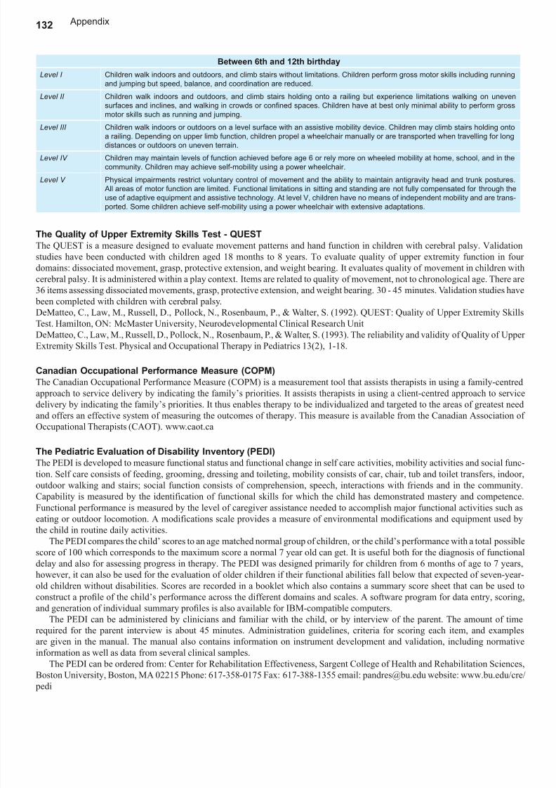
8/9/2019 Guía de ayuda para la parálisis cerebral.pdf
http://slidepdf.com/reader/full/guia-de-ayuda-para-la-paralisis-cerebralpdf 132/143
132 Appendix
Between 6th and 12th birthday
Level I Children walk indoors and outdoors, and climb stairs without limitations. Children perform gross motor skills including running
and jumping but speed, balance, and coordination are reduced.
Level II Children walk indoors and outdoors, and climb stairs holding onto a railing but experience limitations walking on uneven
surfaces and inclines, and walking in crowds or confined spaces. Children have at best only minimal ability to perform gross
motor skills such as running and jumping.
Level III Children walk indoors or outdoors on a level surface with an assistive mobility device. Children may climb stairs holding onto
a railing. Depending on upper limb function, children propel a wheelchair manually or are transported when travelling for longdistances or outdoors on uneven terrain.
Level IV Children may maintain levels of function achieved before age 6 or rely more on wheeled mobility at home, school, and in the
community. Children may achieve self-mobility using a power wheelchair.
Level V Physical impairments restrict voluntary control of movement and the ability to maintain antigravity head and trunk postures.
All areas of motor function are limited. Functional limitations in sitting and standing are not fully compensated for through the
use of adaptive equipment and assistive technology. At level V, children have no means of independent mobility and are trans-
ported. Some children achieve self-mobility using a power wheelchair with extensive adaptations.
The Quality of Upper Extremity Skills Test - QUEST
The QUEST is a measure designed to evaluate movement patterns and hand function in children with cerebral palsy. Validation
studies have been conducted with children aged 18 months to 8 years. To evaluate quality of upper extremity function in four
domains: dissociated movement, grasp, protective extension, and weight bearing. It evaluates quality of movement in children with
cerebral palsy. It is administered within a play context. Items are related to quality of movement, not to chronological age. There are36 items assessing dissociated movements, grasp, protective extension, and weight bearing. 30 - 45 minutes. Validation studies have
been completed with children with cerebral palsy.
DeMatteo, C., Law, M., Russell, D., Pollock, N., Rosenbaum, P., & Walter, S. (1992). QUEST: Quality of Upper Extremity Skills
Test. Hamilton, ON: McMaster University, Neurodevelopmental Clinical Research Unit
DeMatteo, C., Law, M., Russell, D., Pollock, N., Rosenbaum, P., & Walter, S. (1993). The reliability and validity of Quality of Upper
Extremity Skills Test. Physical and Occupational Therapy in Pediatrics 13(2), 1-18.
Canadian Occupational Performance Measure (COPM)
The Canadian Occupational Performance Measure (COPM) is a measurement tool that assists therapists in using a family-centred
approach to service delivery by indicating the family’s priorities. It assists therapists in using a client-centred approach to service
delivery by indicating the family’s priorities. It thus enables therapy to be individualized and targeted to the areas of greatest need
and offers an effective system of measuring the outcomes of therapy. This measure is available from the Canadian Association ofOccupational Therapists (CAOT). www.caot.ca
The Pediatric Evaluation of Disability Inventory (PEDI)
The PEDI is developed to measure functional status and functional change in self care activities, mobility activities and social func-
tion. Self care consists of feeding, grooming, dressing and toileting, mobility consists of car, chair, tub and toilet transfers, indoor,
outdoor walking and stairs; social function consists of comprehension, speech, interactions with friends and in the community.
Capability is measured by the identification of functional skills for which the child has demonstrated mastery and competence.
Functional performance is measured by the level of caregiver assistance needed to accomplish major functional activities such as
eating or outdoor locomotion. A modifications scale provides a measure of environmental modifications and equipment used by
the child in routine daily activities.
The PEDI compares the child’ scores to an age matched normal group of children, or the child’s performance with a total possible
score of 100 which corresponds to the maximum score a normal 7 year old can get. It is useful both for the diagnosis of functionaldelay and also for assessing progress in therapy. The PEDI was designed primarily for children from 6 months of age to 7 years,
however, it can also be used for the evaluation of older children if their functional abilities fall below that expected of seven-year-
old children without disabilities. Scores are recorded in a booklet which also contains a summary score sheet that can be used to
construct a profile of the child’s performance across the different domains and scales. A software program for data entry, scoring,
and generation of individual summary profiles is also available for IBM-compatible computers.
The PEDI can be administered by clinicians and familiar with the child, or by interview of the parent. The amount of time
required for the parent interview is about 45 minutes. Administration guidelines, criteria for scoring each item, and examples
are given in the manual. The manual also contains information on instrument development and validation, including normative
information as well as data from several clinical samples.
The PEDI can be ordered from: Center for Rehabilitation Effectiveness, Sargent College of Health and Rehabilitation Sciences,
Boston University, Boston, MA 02215 Phone: 617-358-0175 Fax: 617-388-1355 email: [email protected] website: www.bu.edu/cre/
pedi
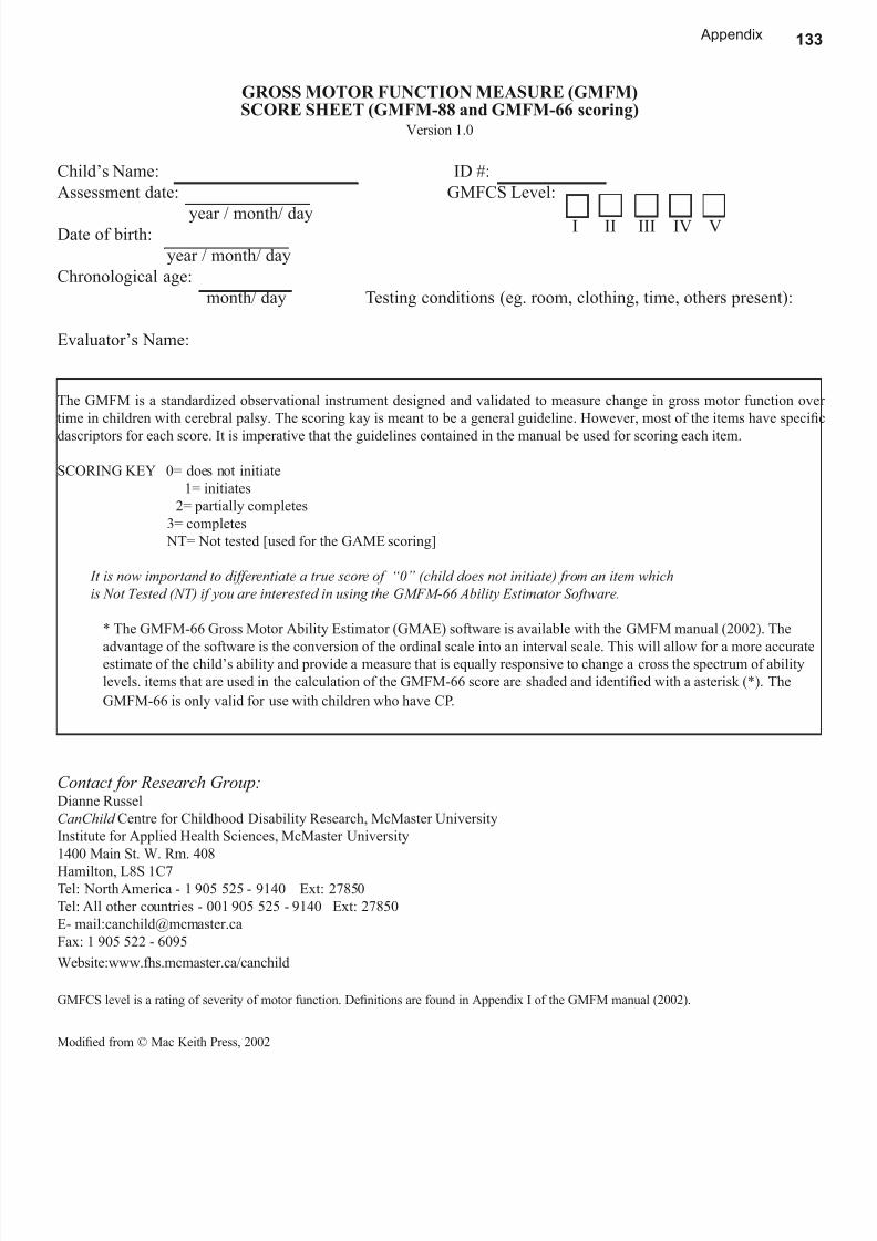
8/9/2019 Guía de ayuda para la parálisis cerebral.pdf
http://slidepdf.com/reader/full/guia-de-ayuda-para-la-paralisis-cerebralpdf 133/143
133 Appendix
GROSS MOTOR FUNCTION MEASURE (GMFM)SCORE SHEET (GMFM-88 and GMFM-66 scoring)
Version 1.0
Child’s Name: ID #:
Assessment date: GMFCS Level:
year / month/ dayDate of birth:
year / month/ day
Chronological age:
month/ day Testing conditions (eg. room, clothing, time, others present):
Evaluator’s Name:
The GMFM is a standardized observational instrument designed and validated to measure change in gross motor function over
time in children with cerebral palsy. The scoring kay is meant to be a general guideline. However, most of the items have specific
dascriptors for each score. It is imperative that the guidelines contained in the manual be used for scoring each item.
SCORING KEY 0= does not initiate
1= initiates
2= partially completes
3= completes
NT= Not tested [used for the GAME scoring]
It is now importand to differentiate a true score of “0” (child does not initiate) from an item which
is Not Tested (NT) if you are interested in using the GMFM-66 Ability Estimator Software.
* The GMFM-66 Gross Motor Ability Estimator (GMAE) software is available with the GMFM manual (2002). The
advantage of the software is the conversion of the ordinal scale into an interval scale. This will allow for a more accurateestimate of the child’s ability and provide a measure that is equally responsive to change a cross the spectrum of ability
levels. items that are used in the calculation of the GMFM-66 score are shaded and identified with a asterisk (*). The
GMFM-66 is only valid for use with children who have CP.
Contact for Research Group:Dianne Russel
CanChild Centre for Childhood Disability Research, McMaster University
Institute for Applied Health Sciences, McMaster University
1400 Main St. W. Rm. 408
Hamilton, L8S 1C7Tel: North America - 1 905 525 - 9140 Ext: 27850
Tel: All other countries - 001 905 525 - 9140 Ext: 27850
E- mail:[email protected]
Fax: 1 905 522 - 6095
Website:www.fhs.mcmaster.ca/canchild
GMFCS level is a rating of severity of motor function. Definitions are found in Appendix I of the GMFM manual (2002).
Modified from © Mac Keith Press, 2002
I II III IV V
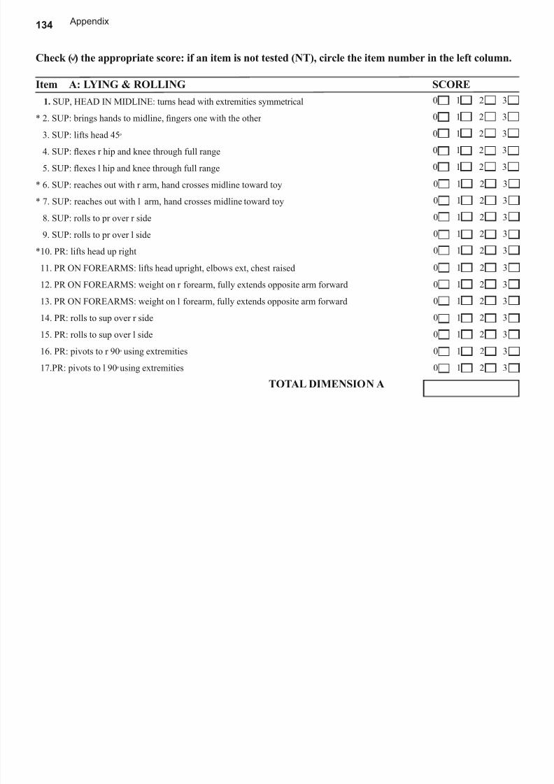
8/9/2019 Guía de ayuda para la parálisis cerebral.pdf
http://slidepdf.com/reader/full/guia-de-ayuda-para-la-paralisis-cerebralpdf 134/143
134 Appendix
Check ( ) the appropriate score: if an item is not tested (NT), circle the item number in the left column.
Item A: LYING & ROLLING SCORE
1. SUP, HEAD IN MIDLINE: turns head with extremities symmetrical
* 2. SUP: brings hands to midline, fingers one with the other
3. SUP: lifts head 45o
4. SUP: flexes r hip and knee through full range
5. SUP: flexes l hip and knee through full range
* 6. SUP: reaches out with r arm, hand crosses midline toward toy
* 7. SUP: reaches out with l arm, hand crosses midline toward toy
8. SUP: rolls to pr over r side
9. SUP: rolls to pr over l side
*10. PR: lifts head up right
11. PR ON FOREARMS: lifts head upright, elbows ext, chest raised
12. PR ON FOREARMS: weight on r forearm, fully extends opposite arm forward
13. PR ON FOREARMS: weight on l forearm, fully extends opposite arm forward
14. PR: rolls to sup over r side
15. PR: rolls to sup over l side
16. PR: pivots to r 90o using extremities
17.PR: pivots to l 90o using extremities
TOTAL DIMENSION A
0 1 2 3
0 1 2 3
0 1 2 30 1 2 3
0 1 2 3
0 1 2 3
0 1 2 3
0 1 2 3
0 1 2 3
0 1 2 3
0 1 2 3
0 1 2 3
0 1 2 3
0 1 2 3
0 1 2 3
0 1 2 3
0 1 2 3
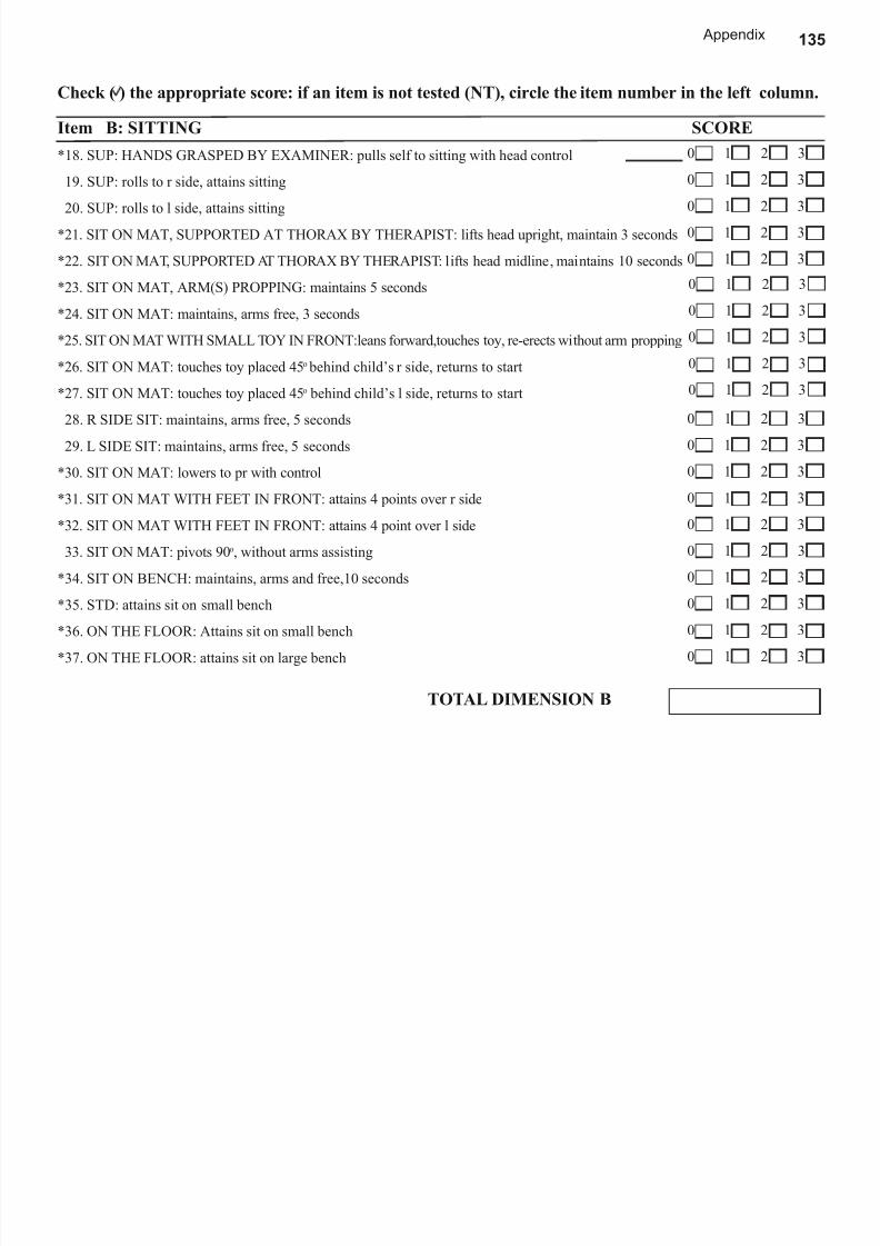
8/9/2019 Guía de ayuda para la parálisis cerebral.pdf
http://slidepdf.com/reader/full/guia-de-ayuda-para-la-paralisis-cerebralpdf 135/143
135 Appendix
Check ( ) the appropriate score: if an item is not tested (NT), circle the item number in the left column.
Item B: SITTING SCORE
*18. SUP: HANDS GRASPED BY EXAMINER: pulls self to sitting with head control
19. SUP: rolls to r side, attains sitting
20. SUP: rolls to l side, attains sitting
*21. SIT ON MAT, SUPPORTED AT THORAX BY THERAPIST: lifts head upright, maintain 3 seconds
*22. SIT ON MAT, SUPPORTED AT THORAX BY THERAPIST: lifts head midline, maintains 10 seconds
*23. SIT ON MAT, ARM(S) PROPPING: maintains 5 seconds
*24. SIT ON MAT: maintains, arms free, 3 seconds
*25. SIT ON MAT WITH SMALL TOY IN FRONT:leans forward,touches toy, re-erects without arm propping
*26. SIT ON MAT: touches toy placed 45o behind child’s r side, returns to start
*27. SIT ON MAT: touches toy placed 45o behind child’s l side, returns to start
28. R SIDE SIT: maintains, arms free, 5 seconds
29. L SIDE SIT: maintains, arms free, 5 seconds
*30. SIT ON MAT: lowers to pr with control
*31. SIT ON MAT WITH FEET IN FRONT: attains 4 points over r side
*32. SIT ON MAT WITH FEET IN FRONT: attains 4 point over l side
33. SIT ON MAT: pivots 90o, without arms assisting
*34. SIT ON BENCH: maintains, arms and free,10 seconds
*35. STD: attains sit on small bench
*36. ON THE FLOOR: Attains sit on small bench
*37. ON THE FLOOR: attains sit on large bench
TOTAL DIMENSION B
0 1 2 3
0 1 2 3
0 1 2 3
0 1 2 3
0 1 2 3
0 1 2 3
0 1 2 3
0 1 2 3
0 1 2 3
0 1 2 3
0 1 2 3
0 1 2 3
0 1 2 3
0 1 2 3
0 1 2 3
0 1 2 3
0 1 2 3
0 1 2 3
0 1 2 3
0 1 2 3
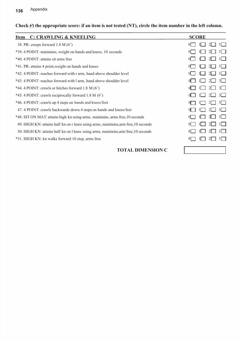
8/9/2019 Guía de ayuda para la parálisis cerebral.pdf
http://slidepdf.com/reader/full/guia-de-ayuda-para-la-paralisis-cerebralpdf 136/143
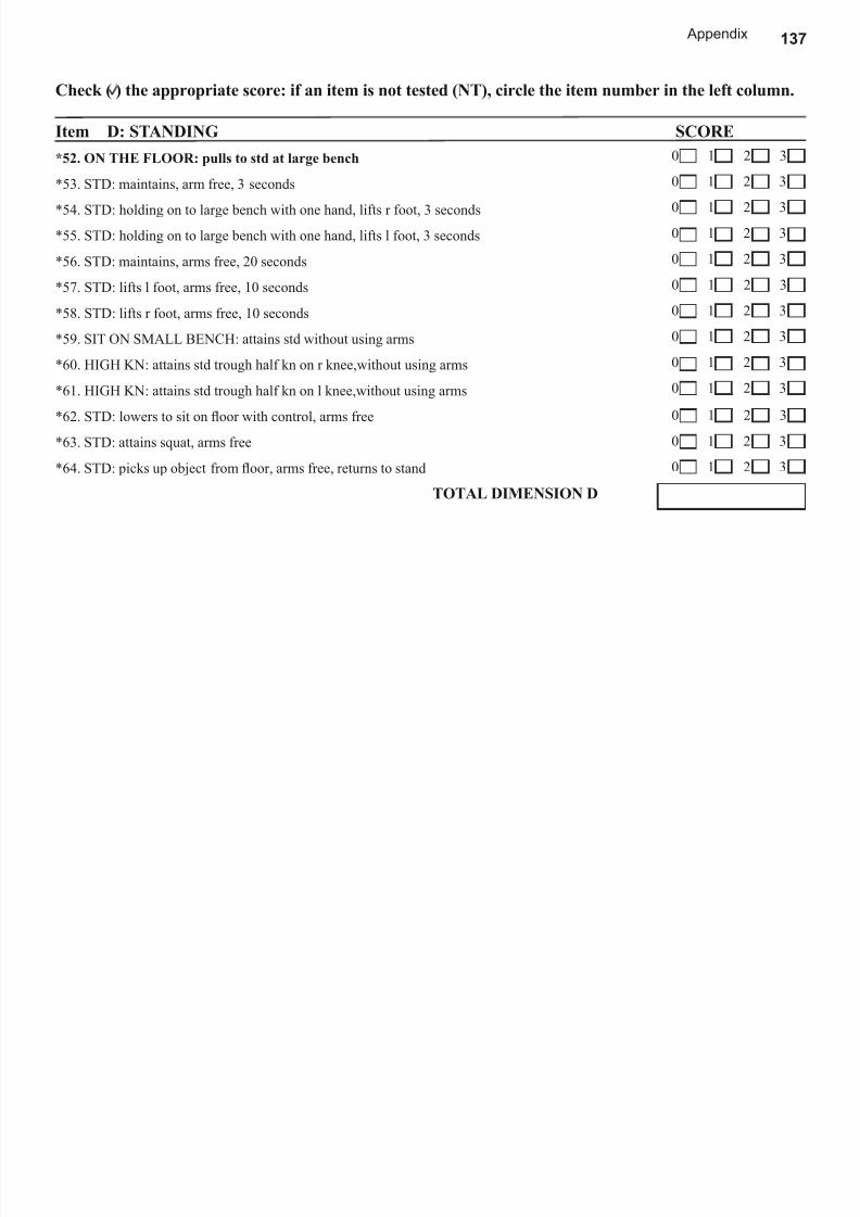
8/9/2019 Guía de ayuda para la parálisis cerebral.pdf
http://slidepdf.com/reader/full/guia-de-ayuda-para-la-paralisis-cerebralpdf 137/143
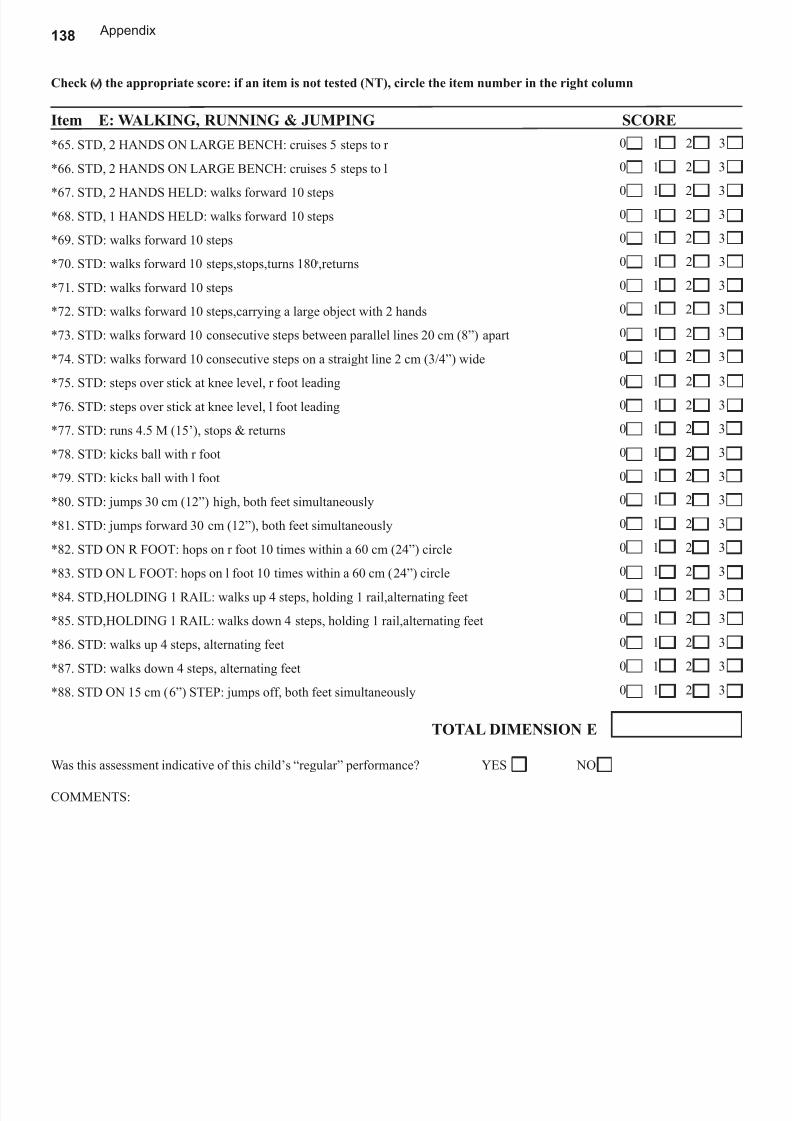
8/9/2019 Guía de ayuda para la parálisis cerebral.pdf
http://slidepdf.com/reader/full/guia-de-ayuda-para-la-paralisis-cerebralpdf 138/143
138 Appendix
Check ( ) the appropriate score: if an item is not tested (NT), circle the item number in the right column
Item E: WALKING, RUNNING & JUMPING SCORE
*65. STD, 2 HANDS ON LARGE BENCH: cruises 5 steps to r
*66. STD, 2 HANDS ON LARGE BENCH: cruises 5 steps to l
*67. STD, 2 HANDS HELD: walks forward 10 steps*68. STD, 1 HANDS HELD: walks forward 10 steps
*69. STD: walks forward 10 steps
*70. STD: walks forward 10 steps,stops,turns 180o,returns
*71. STD: walks forward 10 steps
*72. STD: walks forward 10 steps,carrying a large object with 2 hands
*73. STD: walks forward 10 consecutive steps between parallel lines 20 cm (8”) apart
*74. STD: walks forward 10 consecutive steps on a straight line 2 cm (3/4”) wide
*75. STD: steps over stick at knee level, r foot leading
*76. STD: steps over stick at knee level, l foot leading
*77. STD: runs 4.5 M (15’), stops & returns
*78. STD: kicks ball with r foot
*79. STD: kicks ball with l foot
*80. STD: jumps 30 cm (12”) high, both feet simultaneously
*81. STD: jumps forward 30 cm (12”), both feet simultaneously
*82. STD ON R FOOT: hops on r foot 10 times within a 60 cm (24”) circle
*83. STD ON L FOOT: hops on l foot 10 times within a 60 cm (24”) circle
*84. STD,HOLDING 1 RAIL: walks up 4 steps, holding 1 rail,alternating feet
*85. STD,HOLDING 1 RAIL: walks down 4 steps, holding 1 rail,alternating feet
*86. STD: walks up 4 steps, alternating feet
*87. STD: walks down 4 steps, alternating feet
*88. STD ON 15 cm (6”) STEP: jumps off, both feet simultaneously
TOTAL DIMENSION E
Was this assessment indicative of this child’s “regular” performance? YES NO
COMMENTS:
0 1 2 3
0 1 2 3
0 1 2 3
0 1 2 3
0 1 2 3
0 1 2 3
0 1 2 3
0 1 2 3
0 1 2 3
0 1 2 3
0 1 2 3
0 1 2 3
0 1 2 3
0 1 2 3
0 1 2 3
0 1 2 3
0 1 2 3
0 1 2 30 1 2 3
0 1 2 3
0 1 2 3
0 1 2 3
0 1 2 3
0 1 2 3
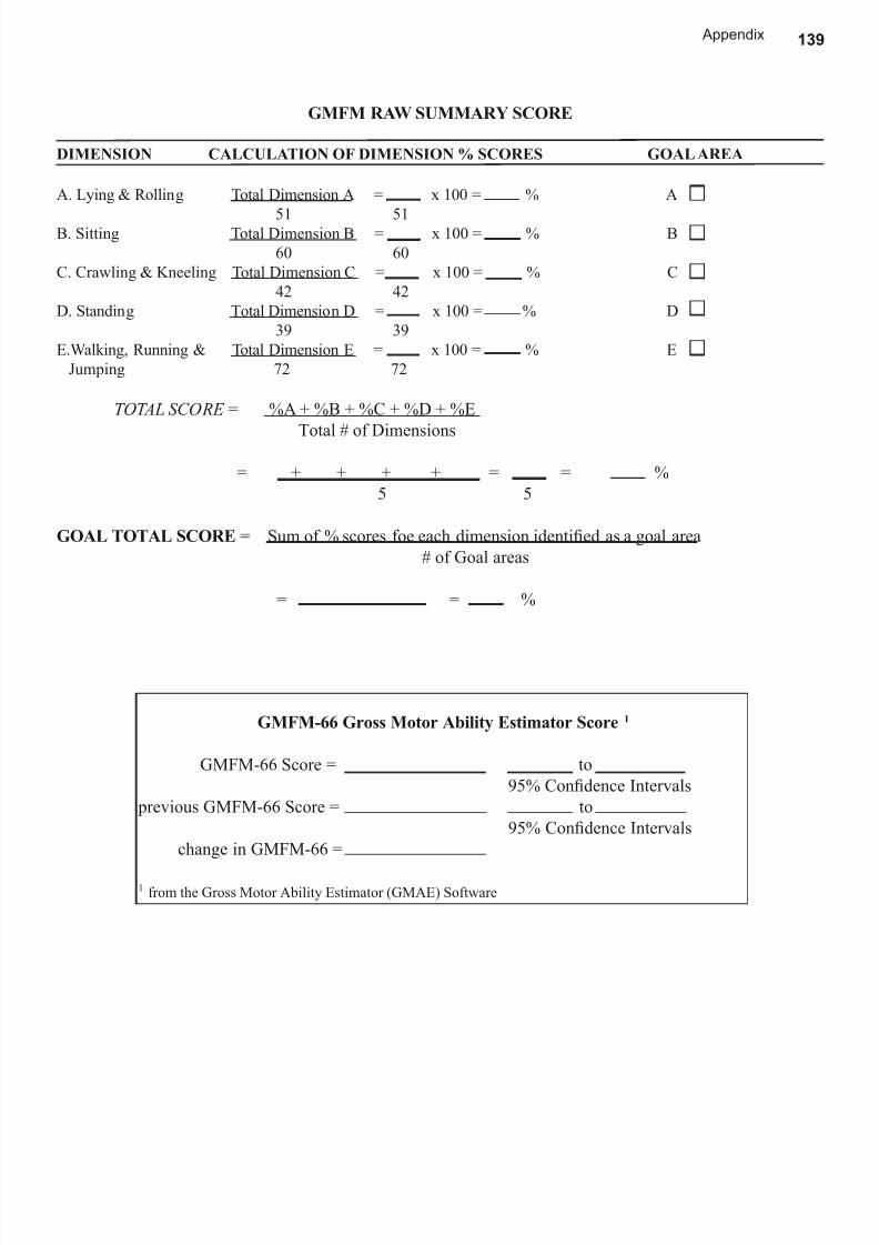
8/9/2019 Guía de ayuda para la parálisis cerebral.pdf
http://slidepdf.com/reader/full/guia-de-ayuda-para-la-paralisis-cerebralpdf 139/143
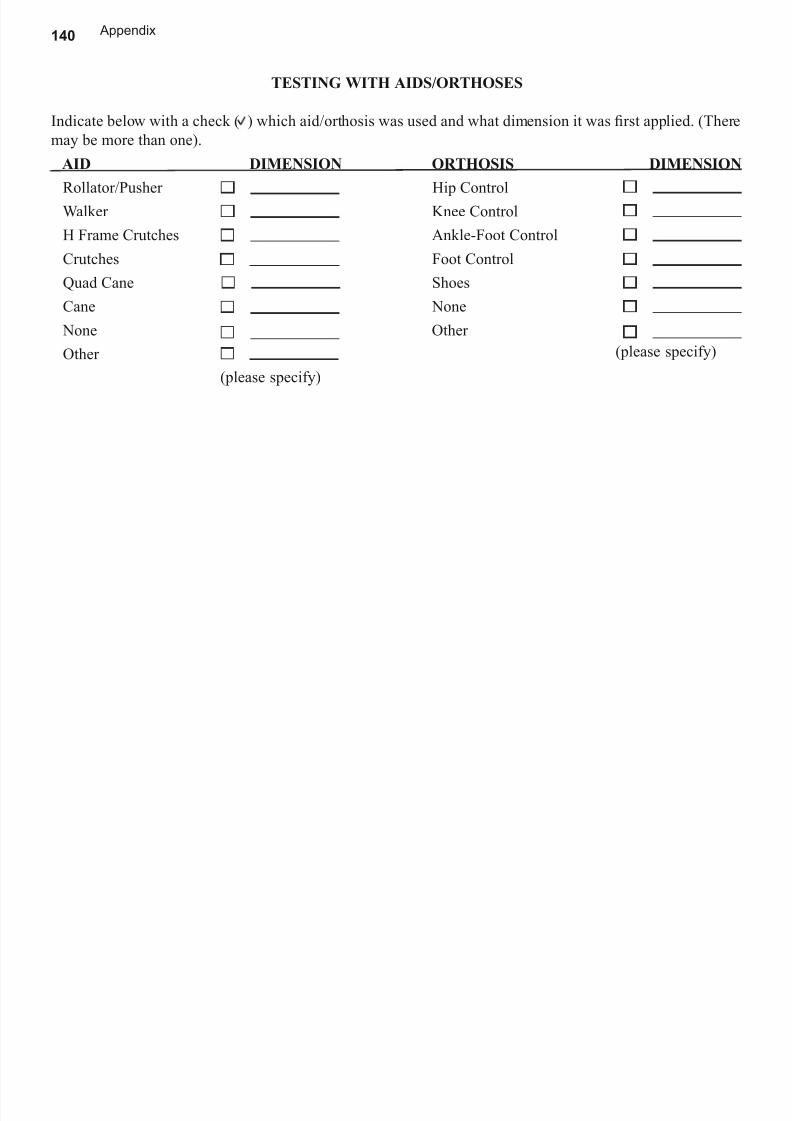
8/9/2019 Guía de ayuda para la parálisis cerebral.pdf
http://slidepdf.com/reader/full/guia-de-ayuda-para-la-paralisis-cerebralpdf 140/143
140 Appendix
TESTING WITH AIDS/ORTHOSES
Indicate below with a check ( ) which aid/orthosis was used and what dimension it was first applied. (There
may be more than one).
AID DIMENSION ORTHOSIS DIMENSION
Rollator/Pusher Hip Control
Walker Knee Control
H Frame Crutches Ankle-Foot Control
Crutches Foot Control
Quad Cane Shoes
Cane None
None Other
Other
(please specify)
(please specify)

8/9/2019 Guía de ayuda para la parálisis cerebral.pdf
http://slidepdf.com/reader/full/guia-de-ayuda-para-la-paralisis-cerebralpdf 141/143
141 Appendix
RAW SUMMARY SCORE USING AIDS/ORTHOSES
DIMENSION CALCULATION OF DIMENSION % SCORES GOAL AREA
F. Lying & Rolling Total Dimension A = x 100= % A
51 51
G. Sitting Total Dimension B = x 100= % B
60 60
H. Crawling & Kneeling Total Dimension C = x100= % C
42 42
I. Standing Total Dimension D = x100= % D
39 39
J. Walking, Running & Total Dimension E = x 100= % EJumping 72 72
TOTAL SCORE = %A + %B + %C + %D + %E
Total # of Dimensions
= + + + + = = %
5 5
GOAL TOTAL SCORE = Sum of % scores foe each dimension identified as a goal area
# of Goal areas
= = %
GMFM-66 Gross Motor Ability Estimator Score 1
GMFM-66 Score = to
95% Confidence Intervals previous GMFM-66 Score = to
95% Confidence Intervals
change in GMFM-66 =
1 from the Gross Motor Ability Estimator (GMAE) Software
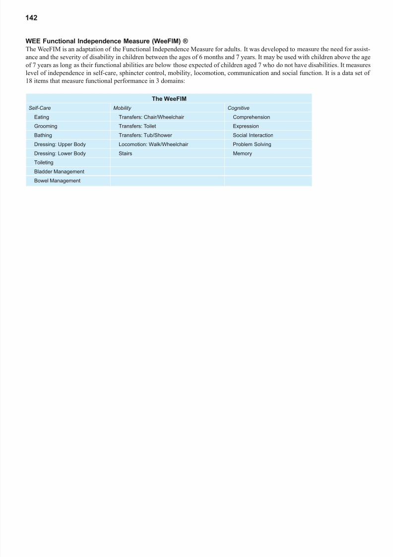
8/9/2019 Guía de ayuda para la parálisis cerebral.pdf
http://slidepdf.com/reader/full/guia-de-ayuda-para-la-paralisis-cerebralpdf 142/143
142 Appendix
WEE Functional Independence Measure (WeeFIM) ®
The WeeFIM is an adaptation of the Functional Independence Measure for adults. It was developed to measure the need for assist-
ance and the severity of disability in children between the ages of 6 months and 7 years. It may be used with children above the age
of 7 years as long as their functional abilities are below those expected of children aged 7 who do not have disabilities. It measures
level of independence in self-care, sphincter control, mobility, locomotion, communication and social function. It is a data set of
18 items that measure functional performance in 3 domains:
The WeeFIM
Self-Care Mobility Cognitive
Eating Transfers: Chair/Wheelchair Comprehension
Grooming Transfers: Toilet Expression
Bathing Transfers: Tub/Shower Social Interaction
Dressing: Upper Body Locomotion: Walk/Wheelchair Problem Solving
Dressing: Lower Body Stairs Memory
Toileting
Bladder Management
Bowel Management
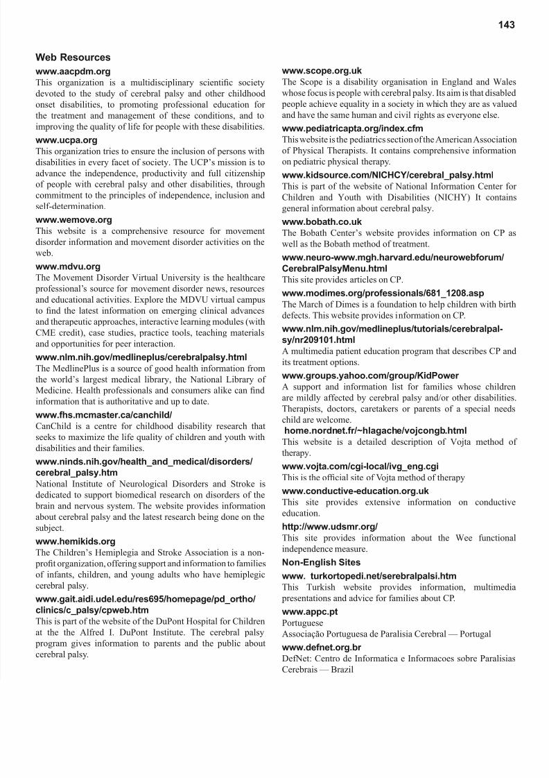
8/9/2019 Guía de ayuda para la parálisis cerebral.pdf
http://slidepdf.com/reader/full/guia-de-ayuda-para-la-paralisis-cerebralpdf 143/143
143 Appendix
Web Resources
www.aacpdm.org
This organization is a multidisciplinary scientific society
devoted to the study of cerebral palsy and other childhood
onset disabilities, to promoting professional education for
the treatment and management of these conditions, and to
improving the quality of life for people with these disabilities.
www.ucpa.org
This organization tries to ensure the inclusion of persons with
disabilities in every facet of society. The UCP’s mission is to
advance the independence, productivity and full citizenship
of people with cerebral palsy and other disabilities, through
commitment to the principles of independence, inclusion and
self-determination.
www.wemove.org
This website is a comprehensive resource for movement
disorder information and movement disorder activities on the
web.
www.mdvu.org
The Movement Disorder Virtual University is the healthcare
professional’s source for movement disorder news, resources
and educational activities. Explore the MDVU virtual campus
to find the latest information on emerging clinical advances
and therapeutic approaches, interactive learning modules (with
CME credit), case studies, practice tools, teaching materials
and opportunities for peer interaction.
www.nlm.nih.gov/medlineplus/cerebralpalsy.html
The MedlinePlus is a source of good health information from
the world’s largest medical library, the National Library of
Medicine. Health professionals and consumers alike can find
information that is authoritative and up to date.
www.fhs.mcmaster.ca/canchild/
CanChild is a centre for childhood disability research that
www.scope.org.uk
The Scope is a disability organisation in England and Wales
whose focus is people with cerebral palsy. Its aim is that disabled
people achieve equality in a society in which they are as valued
and have the same human and civil rights as everyone else.
www.pediatricapta.org/index.cfm
This website is the pediatrics section of the American Association
of Physical Therapists. It contains comprehensive information
on pediatric physical therapy.
www.kidsource.com/NICHCY/cerebral_palsy.html
This is part of the website of National Information Center for
Children and Youth with Disabilities (NICHY) It contains
general information about cerebral palsy.
www.bobath.co.uk
The Bobath Center’s website provides information on CP as
well as the Bobath method of treatment.
www.neuro-www.mgh.harvard.edu/neurowebforum/
CerebralPalsyMenu.htmlThis site provides articles on CP.
www.modimes.org/professionals/681_1208.asp
The March of Dimes is a foundation to help children with birth
defects. This website provides information on CP.
www.nlm.nih.gov/medlineplus/tutorials/cerebralpal-
sy/nr209101.html
A multimedia patient education program that describes CP and
its treatment options.
www.groups.yahoo.com/group/KidPower
A support and information list for families whose children
are mildly affected by cerebral palsy and/or other disabilities.
Therapists, doctors, caretakers or parents of a special needschild are welcome.
home nordnet fr/~hlagache/vojcongb html