guideline for nurses on assisting with intubation … · our lady’s children’s hospital,...
TRANSCRIPT
GUIDELINE FOR NURSES ON ASSISTING WITH INTUBATION AND EXTUBATION OF INFANTS AND CHILDREN
Version Number V3
Date of Issue May 2016
Reference Number NAIEIC-05-2016-ETRC-V3
Review Interval 3 yearly
Approved By
Name: Fionnuala O’Neill
Title: Nurse Practice Coordinator
Signature: Date: May 2016
Authorised By
Name: Rachel Kenna
Title: Director of Nursing
Signature: Date: May 2016
Author/s
Name: Eileen Tiernan
Title: Clinical Coordinator Graduate Diploma
Critical Care Nursing (Children), PICUs.
Name: Rosemary Clerkin Title: Clinical Nurse Facilitator, Theatres
Acknowledgement: Andrew Pendred, Audio-
visual for images
(Document reviewed by Consultant Anaesthetists,
Intensivists, Pharmacist and Resuscitation
Committee)
Location of Copies On Hospital Intranet and locally in department
Document Review History
Review Date Reviewed By Signature
2019
Document Change History
Change to Document Reason for Change
Our Lady’s Children’s Hospital, Crumlin
Document Name: GUIDELINE FOR NURSES ON ASSISTING WITH INTUBATION AND EXTUBATION OF INFANTS AND CHILDREN
Reference Number: NAIEIC-05-2016-ETRC-V3 Version Number: V3
Date of Issue: May 2016 Page 2 of 33
Department of Anaesthetics and Recovery
Contents Page
1. Intubation
a) Introduction
b) Assisting with the Intubation of an Infant / Child
3
3
8
2. Cuffed Endotracheal Tube – Checking Cuff Pressure
13
3. Extubation of an Infant /Children
a) Suspected Accidental / Unplanned Extubation (UE)
14
19
3. Care of an Infant / Child with a Nasopharyngeal Tube
20
5. Appendices
Appendix I: Endotracheal Tubes
Appendix II: Equipment for Intubation
Appendix III: Securing a Nasal Endotracheal Tube (ETT)
using the Modified Melbourne Strapping Technique
22
23
26
6. References
32
Our Lady’s Children’s Hospital, Crumlin
Document Name: GUIDELINE FOR NURSES ON ASSISTING WITH INTUBATION AND EXTUBATION OF INFANTS AND CHILDREN
Reference Number: NAIEIC-05-2016-ETRC-V3 Version Number: V3
Date of Issue: May 2016 Page 3 of 33
Department of Anaesthetics and Recovery
1. a) Intubation - Introduction
Intubation is the placement of an endotracheal tube in the trachea and is the gold standard and
method of choice for establishment and maintenance of an airway (Chethan and Hughes 2008).
Indications for Intubation / Ventilation
Maintenance of patent airway / upper airway obstruction
Worsening Respiratory Distress / Respiratory Failure
Prolonged apnoea
Inadequate ventilation
Worsening hypoxia, despite oxygen therapy
Elective Intubation, i.e. following neonatal surgery, cardiac surgery or prior to general
anaesthesia
Trauma i.e. facial injuries
Neurological i.e. raised intracranial pressure (ICP), deteriorating Glasgow Coma Scale (GCS)
i.e. < 8 with no gag reflex
Inhalation Burns
Resuscitation (NSCNN 2005, BCH 2012, Hazinski 2013).
Equipment (Appendix I)
Cardiac monitor with audible QRS tone
Oxygen saturation monitor
Blood pressure monitoring of patient
Rebreathing circuit (bag), mask (appropriate size) and oxygen source
Airway appropriate size
Appropriate size ETT, one 0.5mm smaller and 0.5mm larger (internal diameter measurement mm ET tube
Appropriate sized laryngoscope i.e. o Straight blade (Miller)
Preterm Infant – Size 0
Infants - Size 0-1
Small Child - Size 1 or 2 o Curved blade (Mackintosh)
Child
Infant/ Child (<12kgs) Size 1
Child (< 22kgs) Size 2
Large Child (< 30kgs) Size 3
Adolescent Size 3-4 NB: (Attach blade and check light and have spare light, blades and batteries to hand)
Wall suction with yankauer
Suction catheters, appropriate size
Magill’s forceps appropriate size
Gauze and K-Y Jelly
Nasogastric tube prn
Elastoplast tape (cut in trouser legs x 2) and 3rd piece with ‘eye hole’ slit
Embroidery cotton (6 strand)
Duoderm
Cavilon ™ Swabs
Ventilator with appropriate settings checked by anaesthetist
Stethoscope
Scissors
Trolley or clear surface for equipment Optional:
Introducer / Stylet
Artery forceps
Gum elastic bougie (older child)
End tidal CO2 detector
SOAPME (Quick Guide)
Suction (Yankeur)
Oxygen and rebreathing / bag-mask circuit
Airways (ETT, LMA, Guedel, NPA)
Positioning and Personnel (e.g. Shoulder roll, role allocation)
Medication and Monitoring (Consider atropine for neonatal intubation, Saturation pulse volume on)
Equipment –i.e. Ventilator working (Evans 2001, BCH 2012)
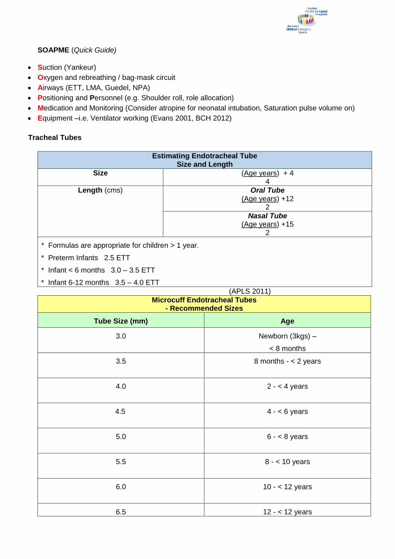
Tracheal Tubes
Estimating Endotracheal Tube Size and Length
Size (Age years) + 4 4
Length (cms)
Oral Tube (Age years) +12
2
Nasal Tube (Age years) +15
2
* Formulas are appropriate for children > 1 year.
* Preterm Infants 2.5 ETT
* Infant < 6 months 3.0 – 3.5 ETT
* Infant 6-12 months 3.5 – 4.0 ETT
(APLS 2011)
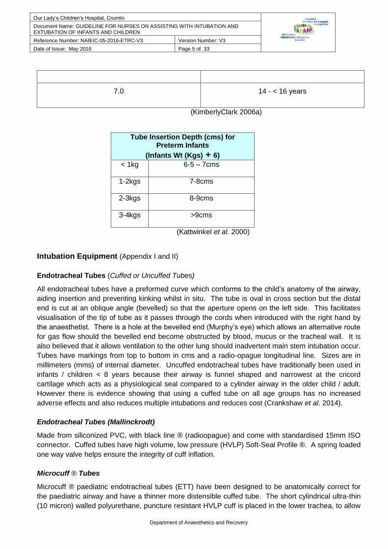
Microcuff Endotracheal Tubes - Recommended Sizes
Tube Size (mm) Age
3.0
Newborn (3kgs) –
< 8 months
3.5 8 months - < 2 years
4.0 2 - < 4 years
4.5 4 - < 6 years
5.0 6 - < 8 years
5.5 8 - < 10 years
6.0 10 - < 12 years
6.5 12 - < 12 years
Our Lady’s Children’s Hospital, Crumlin
Document Name: GUIDELINE FOR NURSES ON ASSISTING WITH INTUBATION AND EXTUBATION OF INFANTS AND CHILDREN
Reference Number: NAIEIC-05-2016-ETRC-V3 Version Number: V3
Date of Issue: May 2016 Page 5 of 33
Department of Anaesthetics and Recovery
7.0 14 - < 16 years
(KimberlyClark 2006a)
Tube Insertion Depth (cms) for Preterm Infants
(Infants Wt (Kgs) + 6)
< 1kg 6-5 – 7cms
1-2kgs 7-8cms
2-3kgs 8-9cms
3-4kgs >9cms
(Kattwinkel et al. 2000)
Intubation Equipment (Appendix I and II)
Endotracheal Tubes (Cuffed or Uncuffed Tubes)
All endotracheal tubes have a preformed curve which conforms to the child’s anatomy of the airway,
aiding insertion and preventing kinking whilst in situ. The tube is oval in cross section but the distal
end is cut at an oblique angle (bevelled) so that the aperture opens on the left side. This facilitates
visualisation of the tip of tube as it passes through the cords when introduced with the right hand by
the anaesthetist. There is a hole at the bevelled end (Murphy’s eye) which allows an alternative route
for gas flow should the bevelled end become obstructed by blood, mucus or the tracheal wall. It is
also believed that it allows ventilation to the other lung should inadvertent main stem intubation occur.
Tubes have markings from top to bottom in cms and a radio-opague longitudinal line. Sizes are in
millimeters (mms) of internal diameter. Uncuffed endotracheal tubes have traditionally been used in
infants / children < 8 years because their airway is funnel shaped and narrowest at the cricord
cartilage which acts as a physiological seal compared to a cylinder airway in the older child / adult.
However there is evidence showing that using a cuffed tube on all age groups has no increased
adverse effects and also reduces multiple intubations and reduces cost (Crankshaw et al. 2014).
Endotracheal Tubes (Mallinckrodt)
Made from siliconized PVC, with black line ® (radioopague) and come with standardised 15mm ISO
connector. Cuffed tubes have high volume, low pressure (HVLP) Soft-Seal Profile ®. A spring loaded
one way valve helps ensure the integrity of cuff inflation.
Microcuff ® Tubes
Microcuff ® paediatric endotracheal tubes (ETT) have been designed to be anatomically correct for
the paediatric airway and have a thinner more distensible cuffed tube. The short cylindrical ultra-thin
(10 micron) walled polyurethane, puncture resistant HVLP cuff is placed in the lower trachea, to allow

Our Lady’s Children’s Hospital, Crumlin
Document Name: GUIDELINE FOR NURSES ON ASSISTING WITH INTUBATION AND EXTUBATION OF INFANTS AND CHILDREN
Reference Number: NAIEIC-05-2016-ETRC-V3 Version Number: V3
Date of Issue: May 2016 Page 6 of 33
Department of Anaesthetics and Recovery
expansion of cuff for ‘tracheal sealing’ instead of ‘cricoid sealing’. They are capable of sealing at very
low pressures (< 20 cms of water), average 11 cms of water compared to PVC ETT, which is almost
half the pressure of conventional cuffed ETT. The cuff also fills the gap between tracheal wall and
tube without folds and channels, clinging and draping to the wet mucosa almost like ‘clingfilm’ at lower
pressures compared to PVC. There is maximum reduced airleak, improving the efficacy of
ventilation. Anatomically based depth markings with 4 pre-glottic markings, ensures a cuff-free
subglottic area, thus reducing the risk to the pressure sensitive larynx and the development of
subglottic stenosis. There is also sufficient margin for preventing inadvertent tracheal extubation or
endobronchial intubation. Microcuff tubes also reduce the risk of aspiration, need for re-intubations
and lower pollution of environment and staff by gases. The microcuff endotracheal tube may be used
by anaesthetists for specific situations i.e. facial / inhalation burns (Kimberley Clark 2006a, 2006b).
Larngoscopes
This is a rigid instrument that the anaesthetist uses to examine the larynx and facilitate endotracheal
intubation. It consists of the handle containing the battery and the blade which is used to move
tongue and soft tissues aside to ensure a good view of the larynx. At the blade tip is an incandescent
bulb, which turns on when blade and handle are attached together and locked into a 90 degree angle.
Straight Blade – Usually used for infants and enables anterior laryngeal placement. Epiglottis
can be lifted to view vocal cords. However it can cause vagal stimulation resulting in
bradycardia or larngospasm.
Curved Blade – Usually used in children > 1 year. Its curved blade flange is large enabling
better control of the tongue and is able to move the epiglottis forward by lifting from the front.
There is less need to exert leverage on the child’s upper teeth which reduces the risk of dental
damage. The tip of the blade is inserted into the vallecula (mucosal pocket) at base of tongue,
anterior to the epiglottis and moved forward. Vocal cords can be visualised with less vagal
stimulation as the mucosa of vallecula is innervated by the glossopharyngeal nerve.
Magill Forceps
Used to grasp the endotracheal tube, especially when inserting through the nose and pass it through
the vocal cords.
Intubation Adjuncts
Introducer / Stylet
This is a long bendable rod which can be inserted into the endotracheal tube and used to help
facilitate a difficult intubation. It is placed into the endotracheal tube before the procedure. Care is
necessary to ensure that the tip doesn’t protrude beyond the end or side holes of the endotracheal
tube during the procedure to avoid trauma to the tissues. Following successful placement of the tube
the stylet is removed.
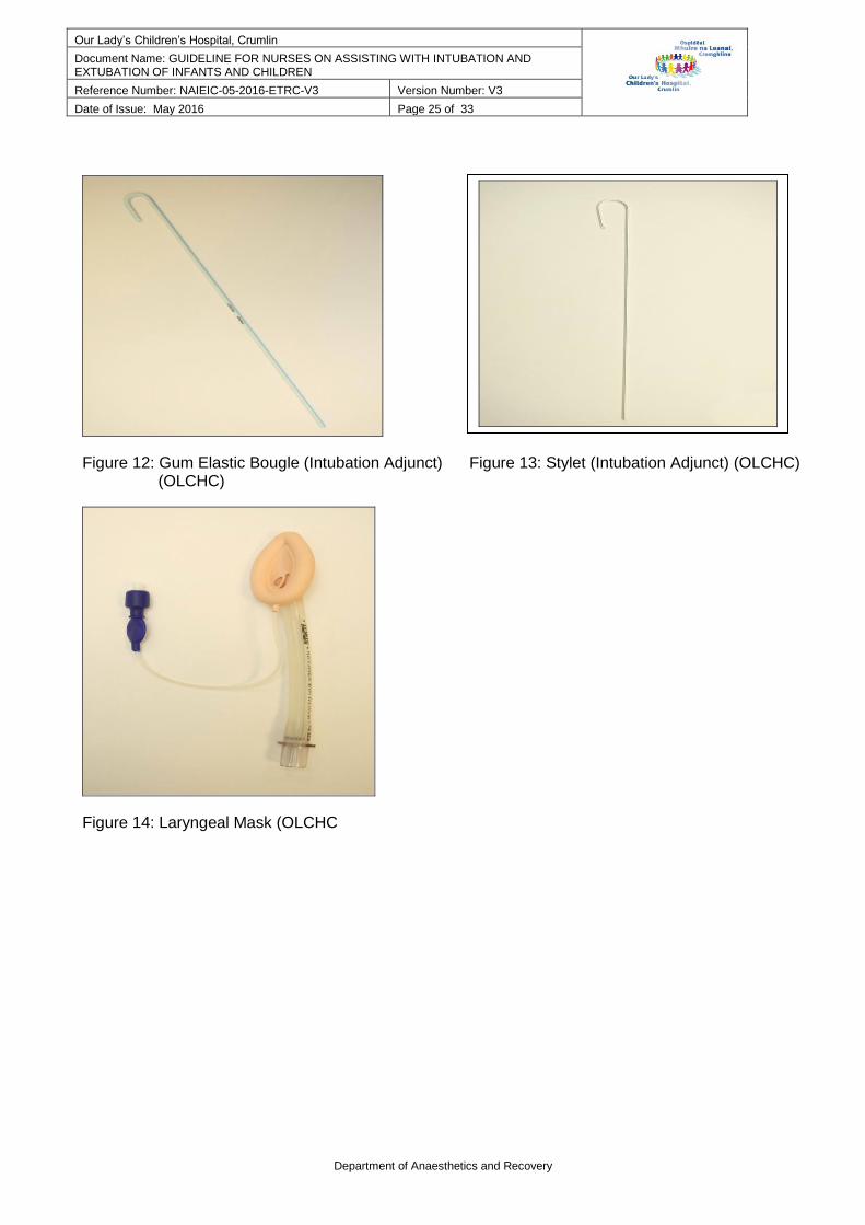
Bougie
This is a straight semi-rigid stylet type device with a bent tip that can be used to facilitate intubation. It
is carefully advanced through the cords into the larynx until the tip enters the main stem bronchus.

Our Lady’s Children’s Hospital, Crumlin
Document Name: GUIDELINE FOR NURSES ON ASSISTING WITH INTUBATION AND EXTUBATION OF INFANTS AND CHILDREN
Reference Number: NAIEIC-05-2016-ETRC-V3 Version Number: V3
Date of Issue: May 2016 Page 7 of 33
Department of Anaesthetics and Recovery
The endotracheal tube is then threaded into the larynx. Once correct placement has been achieved
the bougie is removed.
Laryngeal Mask Airway
The laryngeal mask airway (LMA) is a supra-glottic airway device. It consists of tube PVC (single
use) or silicone (usually reusable). It fills the gap between face mask and endotracheal tube. In
children a smaller size is used which makes it easy to position. However it may give a false sense of
security because the mask is also easy to dislodge, therefore vigilance is essential. It may be used in
the PICU in an emergency when failed intubation has occurred, where it helps establish and maintain
the child’s airway. Positive pressure ventilation can be applied. The LMA however does not protect
against aspiration and is contraindicated where there is a risk of regurgitation.
(Chethan and Hughes 2008, Fine 2008, Gerber 2008a,2008b, Maxwell 2008, National Maternity
Hospital 2008, APLS 2011, Mitchell and Patel 2011, University of Virginia 2011).
Mask
Size
Patient Size Maximum
Cuff Volume
(Air)
1 Neonates / Infants < 5 kgs Up to 4 mls
1 ½ Infants 5-10 kgs Up to 7 mls
2 Children 10-20 kgs Up to 10 mls
2 ½ Children 20-30 kgs Up to 14 mls
3 Children 30-50 kgs Up to 20 mls
4 Adults 50 -70 kgs Up to 30 mls
5 Adults 70-100 kgs Up to 40 mls
Table 1: LMA Quick Guide (LMA 2010)
Medication for Intubation (Given by anaesthetist)
Atropine (0.01mg – 0.02 mg / kg). ( Usually minimum dose of 0.1 mg/kg to prevent paradoxical bradycardia)
0.9% Normal Saline flush (dated and timed)
Induction Agents
Thiopentone (3 – 5mgs / kg)
Ketamine (2mgs / kg)
Propofol (2-3mgs / kg) (Over 3 years, ensure no egg allergy) (Ghanta et al 2007)
Muscle Relaxants
Suxamethonium (1.5mg – 2mgs /kg)
Pancuronium / Vercuronium (0.1 – 0.3mg /kg)
Atracurium besylate (0.5mg / kg)
Our Lady’s Children’s Hospital, Crumlin
Document Name: GUIDELINE FOR NURSES ON ASSISTING WITH INTUBATION AND EXTUBATION OF INFANTS AND CHILDREN
Reference Number: NAIEIC-05-2016-ETRC-V3 Version Number: V3
Date of Issue: May 2016 Page 8 of 33
Department of Anaesthetics and Recovery
Rapid Induction Pack (Available in ward fridges for emergency use)
Suxamethonium (50mgs / ml) 2ml ampoule
Thiopentone (25mgs/ ml) 20 ml ampoule
Ketamine (10mg / ml) 20 ml vial
Pancuronium (2mg / ml) 2 ml ampoule x 2
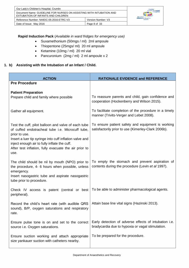
1. b) Assisting with the Intubation of an Infant / Child.
ACTION
RATIONALE EVIDENCE and REFERENCE
Pre Procedure
Patient Preparation
Prepare child and family where possible
Gather all equipment.
Test the cuff, pilot balloon and valve of each tube
of cuffed endotracheal tube i.e. Microcuff tube,
prior to use.
Insert a luer tip syringe into cuff inflation valve and
inject enough air to fully inflate the cuff.
After test inflation, fully evacuate the air prior to
use.
The child should be nil by mouth (NPO) prior to
the procedure, 4- 6 hours when possible, unless
emergency.
Insert nasogastric tube and aspirate nasogastric
tube prior to procedure.
Check IV access is patent (central or best
peripheral).
Record the child’s heart rate (with audible QRS
sound), B/P, oxygen saturations and respiratory
rate.
Ensure pulse tone is on and set to the correct
source i.e. Oxygen saturations.
Ensure suction working and attach appropriate
size yankauer suction with catheters nearby.
To reassure parents and child, gain confidence and
cooperation (Hockenberry and Wilson 2015).
To facilitate completion of the procedure in a timely
manner (Trivits-Verger and Lebet 2008).
To ensure patient safety and equipment is working
satisfactorily prior to use (Kimerley-Clark 2006b).
To empty the stomach and prevent aspiration of
contents during the procedure (Levin et al 1997).
To be able to administer pharmacological agents.
Attain base line vital signs (Hazinski 2013).
Early detection of adverse effects of intubation i.e.
bradycardia due to hypoxia or vagal stimulation.
To be prepared for the procedure.

Our Lady’s Children’s Hospital, Crumlin
Document Name: GUIDELINE FOR NURSES ON ASSISTING WITH INTUBATION AND EXTUBATION OF INFANTS AND CHILDREN
Reference Number: NAIEIC-05-2016-ETRC-V3 Version Number: V3
Date of Issue: May 2016 Page 9 of 33
Department of Anaesthetics and Recovery
In the neonate or preterm infant ensure infant is
kept warm i.e. radiant warmer, giraffe incubator.
Put on personal protective equipment (PPE)
following risk assessment i.e. apron, goggles etc.
Wash hands. ANTT Level 3.
Procedure
Rapid Sequence Induction (RSI) is usually
performed with any patient considered at risk of
regurgitating their stomach contents. Muscle
relaxant and sedative is administered in rapid
sequence.
Confirm with Anaesthetist that necessary
equipment is readily available prior to procedure.
Ensure infant / child is supine with head at side of
incubator/ radiant warmer or the top of the cot /
bed.
The anaesthetist stands at the child’s head which
is positioned midline with minimal extension of the
neck (infant neutral and child in sniffing position).
A roll may be placed under shoulders.
NB: Avoid:
- Hyperextending or rotating the neck.
- Flexing the head towards the chin.
Anaesthetist will manually ventilate the child with
100% oxygen, using a rebreathing bag, mask,
valve for a minimum of 3 minutes, prior to
intubation.
NB: Maintenance of oxygenation is a priority
during the procedure
Procedure should be limited to maximum of 20
seconds.
Ensure yankauer and suction available to hand at
infant / child’s shoulder to allow easy access for
the anaesthetist.
Cricoid Pressure
The anaesthetist may request the nurse to apply
cricoid pressure (Sellick’s manoeuvre).
To protect against cold stress and its detrimental
effects (Karlsen 2014).
Standard precautions (OLHSC 2007, OLCHC 2010,
2011)
The aim is to minimise the risk of gastric aspiration
during intubation. It also facilitates airway
visualisation through muscle relaxation, control of
agitation and prevention of involuntary reflexes i.e.
gag reflex (Gardiner and Grindrod 2005, Zelicof-paul
et al. 2005, Hazinski 2013).
To ensure patient safety during the procedure.
Opens airway fully and aligns the trachea which
allows visualisation of glottis and larynx (APLS 2011,
NSCNN 2005, National Maternity Hospital 2000).
The glottis will be raised above the line of sight and
narrow the trachea and under flexing will cause the
posterioir pharynx to be viewed but prevent direct
visualisation of the glottis (National Maternity
Hospital 2008).
Fill the reserve lung volume with oxygen rather than
room air to minimise the risk of desaturation or
bradycardia (Levin et al 1997).
To prevent hypoxia (APLS 2011, Henderson 2011).
Bradycardia and oxygen desaturations can occur
within 55 seconds (Bottor 2009).
Pharyngeal suction may be required to ensure good
visualisation of the cords.
Cricoid pressure compresses the cricoid cartilage
against the cervical vertebrae, closing the
Our Lady’s Children’s Hospital, Crumlin
Document Name: GUIDELINE FOR NURSES ON ASSISTING WITH INTUBATION AND EXTUBATION OF INFANTS AND CHILDREN
Reference Number: NAIEIC-05-2016-ETRC-V3 Version Number: V3
Date of Issue: May 2016 Page 10 of 33
Department of Anaesthetics and Recovery
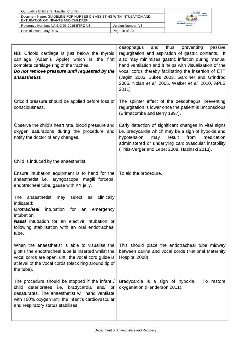
NB: Cricoid cartilage is just below the thyroid
cartilage (Adam’s Apple) which is the first
complete cartilage ring of the trachea.
Do not remove pressure until requested by the
anaesthetist.
Cricoid pressure should be applied before loss of
consciousness.
Observe the child’s heart rate, blood pressure and
oxygen saturations during the procedure and
notify the doctor of any changes.
Child is induced by the anaesthetist.
Ensure intubation equipment is to hand for the
anaesthetist i.e. laryngoscope, magill forceps,
endotracheal tube, gauze with KY jelly.
The anaesthetist may select as clinically
indicated:
Orotracheal intubation for an emergency
intubation
Nasal intubation for an elective intubation or
following stabilisation with an oral endotracheal
tube.
When the anaesthetist is able to visualise the
glottis the endotracheal tube is inserted whilst the
vocal cords are open, until the vocal cord guide is
at level of the vocal cords (black ring around tip of
the tube).
The procedure should be stopped if the infant /
child deteriorates i.e. bradycardia and/ or
desaturates. The anaesthetist will hand ventilate
with 100% oxygen until the infant’s cardiovascular
and respiratory status stabilises.
oesophagus and thus preventing passive
regurgitation and aspiration of gastric contents. It
also may minimises gastric inflation during manual
hand ventilation and it helps with visualisation of the
vocal cords thereby facilitating the insertion of ETT
(Jagim 2003, Jukes 2003, Gardiner and Grindrod
2005, Nolan et al. 2005, Walker et al. 2010, APLS
2011)
The splinter effect of the oesophagus, preventing
regurgitation is lower once the patient is unconscious
(Brimacombe and Berry 1997).
Early detection of significant changes in vital signs
i.e. bradycardia which may be a sign of hypoxia and
hypotension may result from medication
administered or underlying cardiovascular instability
(Tritis-Verger and Lebet 2008, Hazinski 2013).
To aid the procedure.
This should place the endotracheal tube midway
between carina and vocal cords (National Maternity
Hospital 2008).
Bradycardia is a sign of hypoxia. To restore
oxygenation (Henderson 2011).
Our Lady’s Children’s Hospital, Crumlin
Document Name: GUIDELINE FOR NURSES ON ASSISTING WITH INTUBATION AND EXTUBATION OF INFANTS AND CHILDREN
Reference Number: NAIEIC-05-2016-ETRC-V3 Version Number: V3
Date of Issue: May 2016 Page 11 of 33
Department of Anaesthetics and Recovery
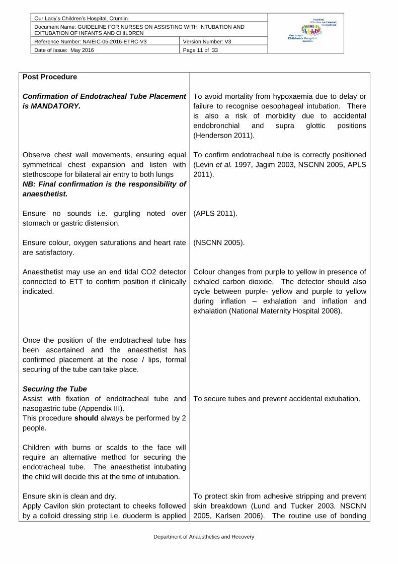
Post Procedure
Confirmation of Endotracheal Tube Placement
is MANDATORY.
Observe chest wall movements, ensuring equal
symmetrical chest expansion and listen with
stethoscope for bilateral air entry to both lungs
NB: Final confirmation is the responsibility of
anaesthetist.
Ensure no sounds i.e. gurgling noted over
stomach or gastric distension.
Ensure colour, oxygen saturations and heart rate
are satisfactory.
Anaesthetist may use an end tidal CO2 detector
connected to ETT to confirm position if clinically
indicated.
Once the position of the endotracheal tube has
been ascertained and the anaesthetist has
confirmed placement at the nose / lips, formal
securing of the tube can take place.
Securing the Tube
Assist with fixation of endotracheal tube and
nasogastric tube (Appendix III).
This procedure should always be performed by 2
people.
Children with burns or scalds to the face will
require an alternative method for securing the
endotracheal tube. The anaesthetist intubating
the child will decide this at the time of intubation.
Ensure skin is clean and dry.
Apply Cavilon skin protectant to cheeks followed
by a colloid dressing strip i.e. duoderm is applied
To avoid mortality from hypoxaemia due to delay or
failure to recognise oesophageal intubation. There
is also a risk of morbidity due to accidental
endobronchial and supra glottic positions
(Henderson 2011).
To confirm endotracheal tube is correctly positioned
(Levin et al. 1997, Jagim 2003, NSCNN 2005, APLS
2011).
(APLS 2011).
(NSCNN 2005).
Colour changes from purple to yellow in presence of
exhaled carbon dioxide. The detector should also
cycle between purple- yellow and purple to yellow
during inflation – exhalation and inflation and
exhalation (National Maternity Hospital 2008).
To secure tubes and prevent accidental extubation.
To protect skin from adhesive stripping and prevent
skin breakdown (Lund and Tucker 2003, NSCNN
2005, Karlsen 2006). The routine use of bonding
Our Lady’s Children’s Hospital, Crumlin
Document Name: GUIDELINE FOR NURSES ON ASSISTING WITH INTUBATION AND EXTUBATION OF INFANTS AND CHILDREN
Reference Number: NAIEIC-05-2016-ETRC-V3 Version Number: V3
Date of Issue: May 2016 Page 12 of 33
Department of Anaesthetics and Recovery
to cheeks as clinically indicated.
Cut 2 pieces of elastoplast approx. 10 – 15 cms
long into “trouser legs” as clinically indicated. Also
a third piece of Elastoplast with ‘eye-hole’ (slit) in
the middle.
Follow procedure for strapping as per pictorial
guideline (Appendix 1).
NB: The ETT should be secure from both (right
and left) directions of the face.
Organise retaping of the ETT by anaesthetist if
the ETT position is incorrect on chest x-ray or if
the tapes become wet and loose i.e. (move >
0.5cms)
Following Securing of Endotracheal Tube.
Suction the infant /child post intubation.
Obtain blood gas post intubation
Ensure chest x-ray is requested by anaesthetist
and obtained post intubation with head in midline
position.
When infant is stable the external part of the tube
may be shortened if necessary by the
anaesthetist.
Air Leak
The endotracheal tube is checked for presence of
air leak at 25-30 cms water using manometer, by
the anaesthetist:
Appropriate tube size – a small airleak is
present when child is hand ventilated.
Tube too small – Significant air leak
present.
agents i.e. Compound Tincture of Benzoin is not
recommended in neonates as it results in epidermal
stripping because the epidermis becomes stronger
than the cohesion of epidermis to dermis when it is
removed (Lund and Tucker 2003). It is also not
recommended in adults due to: drying of the skin;
skin occluded and impaired function and local
irritation / allergy especially in atopic patients (Lund
and Tucker 2003, Scardamaglia et al. 2003).
The tape may be placed onto transparent glossy
paper prior to cutting as this facilitates straight
cutting and the tape peels easily away for use.
(ALSG 2008).
To maintain satisfactory ventilation and prevent
unplanned extubation which may result in a life
threatening event. The infant < 3 months is more at
risk of an unplanned extubation due to shorter
trachea.
To maintain a clear airway.
Evaluate respiratory status.
To verify endotracheal tube position approximately
1-2cms above the carina and correct placement of
nasogastric tube (NSCNN 2005).
This reduces dead space and prevents kinking of the
tube (National Maternity Hospital 2008).
A tube that is too small may result in a large airleak
which may contribute to under ventilation. This may
be compensated for by increasing the tidal volume
on the ventilator. However reintubation with a larger
tube size may be required.
An excessively large tube may contribute to the
development of laryngeal oedema and sub glottis
stenosis (Hazinski 2013).
Our Lady’s Children’s Hospital, Crumlin
Document Name: GUIDELINE FOR NURSES ON ASSISTING WITH INTUBATION AND EXTUBATION OF INFANTS AND CHILDREN
Reference Number: NAIEIC-05-2016-ETRC-V3 Version Number: V3
Date of Issue: May 2016 Page 13 of 33
Department of Anaesthetics and Recovery
Tube too large – Air leak not detected
until inspiratory pressure > 30cms water.
Remove personal protective equipment (PPE)
and wash hands.
Document procedure in the nursing notes
including size and type of endotracheal tube and
length to nose or lips.
Also insertion of nasogastric tube as appropriate.
Ensure that all medication has been documented
in the drug kardex by the anaesthetist.
Discuss with the anaesthetist, the ventilation plan
for the child i.e. ventilation mode / settings and
need for sedation / analgesia.
If intubation occurred on a ward organise safe
transfer to PICU.
To reduce cross infection. (OLHSC 2007, OLCHC
2011).
To maintain accountability and ensure continuity of
care (NMBI 2015a).
To ensure safe and effective management of the
patient post intubation.
2. Cuffed Endotracheal Tube: Checking Cuff Pressure
Nursing Considerations with Cuffed Endotracheal Tubes
NB: Always discuss with consultant anaesthetist regarding specific patient requirements and inflation
pressures for cuffed endotracheal tube.
ACTION
RATIONALE EVIDENCE and REFERENCE
Following endotracheal intubation an air leak
should be present around the ETT at 20 cms
water (positive) airway pressure with the cuff not
inflated.
Cuff should be carefully inflated using a cuff
pressure manometer or syringe until air leak is
no longer present. (Attach syringe to valve port).
Cuff pressure should not exceed 20 cms water.
If no air leak is detected the tube may be too large
and the anaesthetist may consider changing the
tube (Kimberley-Clark 2006b, Microcuff 2011)
(Kimberley-Clark 2006b, Microcuff 2011)
Our Lady’s Children’s Hospital, Crumlin
Document Name: GUIDELINE FOR NURSES ON ASSISTING WITH INTUBATION AND EXTUBATION OF INFANTS AND CHILDREN
Reference Number: NAIEIC-05-2016-ETRC-V3 Version Number: V3
Date of Issue: May 2016 Page 14 of 33
Department of Anaesthetics and Recovery
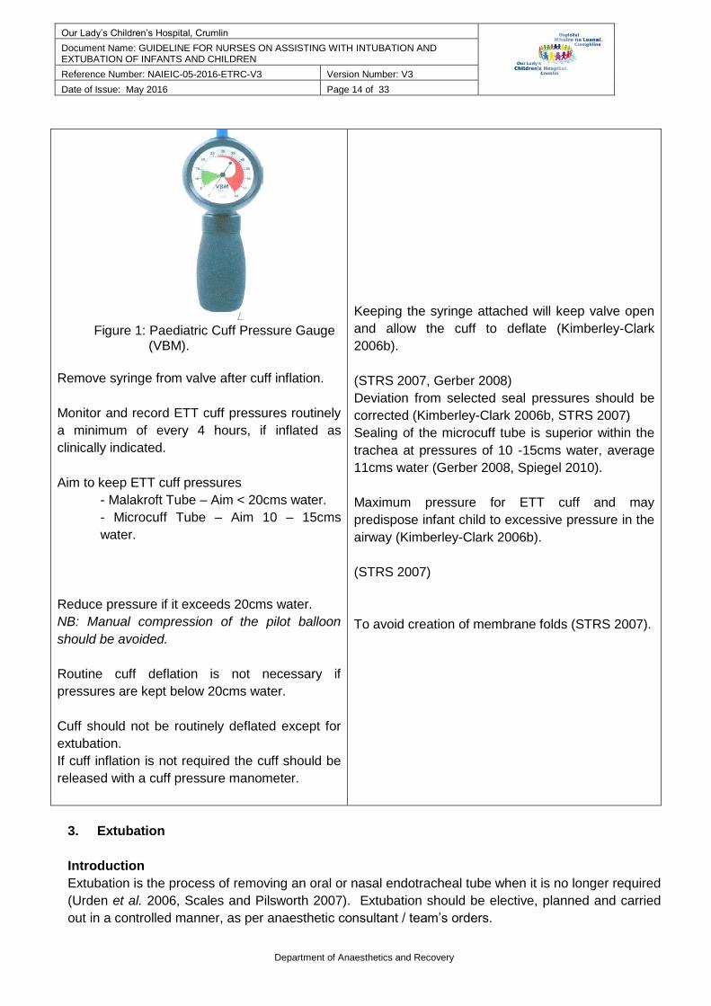
Figure 1: Paediatric Cuff Pressure Gauge (VBM).
Remove syringe from valve after cuff inflation.
Monitor and record ETT cuff pressures routinely
a minimum of every 4 hours, if inflated as
clinically indicated.
Aim to keep ETT cuff pressures
- Malakroft Tube – Aim < 20cms water.
- Microcuff Tube – Aim 10 – 15cms
water.
Reduce pressure if it exceeds 20cms water.
NB: Manual compression of the pilot balloon
should be avoided.
Routine cuff deflation is not necessary if
pressures are kept below 20cms water.
Cuff should not be routinely deflated except for
extubation.
If cuff inflation is not required the cuff should be
released with a cuff pressure manometer.
Keeping the syringe attached will keep valve open
and allow the cuff to deflate (Kimberley-Clark
2006b).
(STRS 2007, Gerber 2008)
Deviation from selected seal pressures should be
corrected (Kimberley-Clark 2006b, STRS 2007)
Sealing of the microcuff tube is superior within the
trachea at pressures of 10 -15cms water, average
11cms water (Gerber 2008, Spiegel 2010).
Maximum pressure for ETT cuff and may
predispose infant child to excessive pressure in the
airway (Kimberley-Clark 2006b).
(STRS 2007)
To avoid creation of membrane folds (STRS 2007).
3. Extubation
Introduction
Extubation is the process of removing an oral or nasal endotracheal tube when it is no longer required
(Urden et al. 2006, Scales and Pilsworth 2007). Extubation should be elective, planned and carried
out in a controlled manner, as per anaesthetic consultant / team’s orders.
Our Lady’s Children’s Hospital, Crumlin
Document Name: GUIDELINE FOR NURSES ON ASSISTING WITH INTUBATION AND EXTUBATION OF INFANTS AND CHILDREN
Reference Number: NAIEIC-05-2016-ETRC-V3 Version Number: V3
Date of Issue: May 2016 Page 15 of 33
Department of Anaesthetics and Recovery
Indications and Readiness for Extubation
Child is not in respiratory distress with a good respiratory pattern.
Minimal ventilation i.e. pressure support / CPAP 5 is required
Satisfactory blood gas and oxygen saturations with oxygen requirements <40%
Child is haemodynamically stable, peripherally warm and well perfused
Apyrexial
Heart rate normal / upper limits
Chest clear with minimal secretions
Child is awake and responsive and requiring minimal or no sedation
Cough, swallow and gag reflexes present in child without a disability
Satisfactory nutrition with adequate respiratory muscle strength and ventilator drive
Child should have an audible leak at 20cms water prior to extubation.
NB: If no leak present Dexamethasone I.V. 6 hourly is charted for 24 hours prior to extubation to
reduce the incidence of post extubation stidor (De 2004, Lancaster 2007, Scales and Pilsworth
2007, Wratney and Chieifetz, 2007, Karmarkar and Varshney 2008).
Equipment
Rebreathing circuit and mask (checked)
Airway (appropriate size)
Suction and catheters (checked)
Yankauer
Oxygen cannula / tube
Bubble humidifier
Duoderm tape
Plaster removal swabs i.e. Appeel™
Tegaderm dressing
Scissors
5 / 10 ml syringe
Stethoscope
Emergency intubation trolley nearby
Nebuliser circuit and mask
Racemic Epinephrine
Non-sterile gloves (Scales and Pilsworth 2007)
ACTION RATIONALE, EVIDENCE and REFERENCE
Pre Procedure
Prepare child and family.
It is best for extubation to take place early in the
morning if possible.
To reassure parents and child, gain confidence and
cooperation (Hockenberry and Wilson 2015).
There is more nursing and medical support
available during the day post extubation to ensure
timely intervention if necessary. This is important if
the child’s condition deteriorates, gets into
Our Lady’s Children’s Hospital, Crumlin
Document Name: GUIDELINE FOR NURSES ON ASSISTING WITH INTUBATION AND EXTUBATION OF INFANTS AND CHILDREN
Reference Number: NAIEIC-05-2016-ETRC-V3 Version Number: V3
Date of Issue: May 2016 Page 16 of 33
Department of Anaesthetics and Recovery
Ensure physiotherapy is carried out prior to
extubation, if requested by medical staff.
Withhold feeds 2-4 hours prior to extubation.
Prior to procedure aspirate nasogastric tube.
NB: The neonate may require maintenance fluids
whilst nil by mouth.
Wean sedation as appropriate i.e. morphine
infusion decreased to maximum 20 mcg/ kg/ hour.
Ensure suction and oxygen equipment are
functioning properly.
An appropriate size bag, face mask and airway
should be available at the child’s bedside.
Establish and set up oxygen therapy for post
extubation.
Ensure all necessary equipment is available
including intubation equipment.
Wash hands using an aseptic non-touch
technique (ANNT) level 3.
Put on personal protective equipment (PPE) i.e.
goggles, glasses, apron, and gloves.
Monitor child’s vital signs and oxygen saturation.
Procedure
Inform anaesthetist immediately prior to
procedure.
respiratory distress / failure and requires
reintubating or non-invasive ventilation (Lancaster
2007, Scales and Pilsworth 2007).
To decrease the risk of aspiration during the
procedure and ensure the stomach is empty should
reintubation be required (De 2005, Parker 2007,
Hazinski 2013).
The neonate is more at risk of hypoglycaemia.
To ensure the child will not have respiratory
depression due to sedation on extubation.
To ensure patient safety (Hazinski 2013).
Airway problems are more common during
extubation and recovery compared to intubation
(Mitchell 2011).
Most patients require 5-10% more inspired oxygen
post extubation.
To ensure the procedure is completed smoothly in a
safe and coordinated manner (Cull 1999, Dougherty
and Lister 2015).
To prevent cross infection (Infection Control
Association 2002, OLHSC 2007, OLCHC 2010)
There is a high risk of droplet contamination of the
eyes during the procedure as the child may cough /
expectorate (Lancaster 2007).
To ensure timely intervention should child’s condition
deteriorate.
If reintubation is required, staff need to be prepared.
Anaesthetist needs to be on the Paediatric Intensive
Care Unit (PICU) and available to reintubate if the
Our Lady’s Children’s Hospital, Crumlin
Document Name: GUIDELINE FOR NURSES ON ASSISTING WITH INTUBATION AND EXTUBATION OF INFANTS AND CHILDREN
Reference Number: NAIEIC-05-2016-ETRC-V3 Version Number: V3
Date of Issue: May 2016 Page 17 of 33
Department of Anaesthetics and Recovery
The procedure is performed by the anaesthetist
assisted by nurse or 2 nurses, one of which is a
senior PICU / recovery nurse who is deemed
competent in the procedure and is working within
her scope of practice.
Elevate head of bed for infants or sit an older
child in a comfortable upright position.
Suction child prior to the procedure.
Ensure infant / child awake, with stable vital signs
and good motor responses
Explain procedure to child as appropriate and
reassure.
Deflate the cuff using a 5-10 ml syringe, if a
cuffed endotracheal tube is in situ.
Gently remove tapes from the child’s face using
the appropriate adhesive remover.
Secure nasogastric tube to child’s forehead.
Affix face mask to rebreathing circuit, in
preparation for post extubation.
Support and encourage child as appropriate.
Encourage deep breathing and coughing during
the procedure as appropriate.
Give suction support prior to extubation.
Remove the endotracheal tube in a swift motion
on expiration whilst suctioning. An older child
child fails extubation (Lancaster 2007, Hazinski
2013).
To ensure a safe and coordinated extubation by
competent practitioners (NMBI 2015b).
To facilitate spontaneous breathing, diaphragmatic
expansion, maximises lung expansion and the work
of breathing is decreased as gravity assists in lung
expansion. Facilitates an effective cough (Lancaster
2007, Karmarkar and Varshney 2008).
To clear the airway.
To ensure patient is ready for extubation.
To relieve anxiety and gain cooperation.
This frees the endotracheal tube from child’s face for
easy removal (Trivits-Verger and Lebet 2008).
To provide reassurance and support for the child
prior to and during the procedure (Trivits-Verger and
Lebet 2008).
To ensure oxygenation is optimised and secretions
are cleared (Lancaster 2007).
Tracheal extubation has been demonstrated to
greatly impair oxygenation (Karmarker and Varshney
2008).
To free tube and remove any remaining secretions.
Procedure is carried out on expiration when glottis is
Our Lady’s Children’s Hospital, Crumlin
Document Name: GUIDELINE FOR NURSES ON ASSISTING WITH INTUBATION AND EXTUBATION OF INFANTS AND CHILDREN
Reference Number: NAIEIC-05-2016-ETRC-V3 Version Number: V3
Date of Issue: May 2016 Page 18 of 33
Department of Anaesthetics and Recovery
may be able to do this themselves with direction
as appropriate.
NB: Do not stop or reinsert the tube during the
removal.
Apply oxygen via re breathing circuit and mask.
Suction the oropharynx under direct vision as
clinically indicated.
Assess respiratory status for signs of respiratory
distress, desaturation and stridor.
Note if there is an audible stridor report to
anaesthetist immediately and document.
Post Procedure
Secure supplementary oxygen therapy via nasal
cannula as clinically indicated.
Minimal handling of patient initially.
Observe child for:
- drowsiness / lethargy
- restlessness
Close observation and continual reassessment of
respiratory status over 24 hours (even following
transfer out of PICU to the ward).
o Monitor heart rate, respiratory rate and
oxygen saturation.
o Observe for signs of respiratory distress:
intercostal, sternal and subcostal
recession: tachypnoea; nasal flaring;
tracheal tug; dyspnoea; pale / grey /
mottled colour.
o Arterial or venous blood gas post
fully open to prevent laryngospasm and trauma
(Karmarkar and Varshney 2008, Hodd et al 2010).
Extubation may result in vocal cord or tissue damage
or cause laryngeal spasm which may occude the
patents airway (Lancaster 2007).
To prevent Hypoxemia (Trivits-Verger and Lebet
2008).
To maintain a clear airway and prevent secretion
accumulation. Minimise aspirate of oral content with
first breath after extubation (Parker 2007, Trivits-
Verger and Lebet 2008, Mitchell 2011).
There is a risk of trauma t the soft tissues if suction
blindly (Mitchell 2011).
Evaluate if patient can breathe without endotracheal
tube.
A stridor may indicate an obstructed airway and
require timely intervention.
To minimise metabolic requirements and prevent
hypoxia (Hazinski 2013). Maximum 2 litres oxygen
via nasal cannula.
To evaluate respiratory status and early detection of
upper airway obstruction or respiratory distress
(Levin et al. 1997).
NB: A drowsy child may be hypercapnoeic (high
pCO2) whilst a restless child may be hypoxic.
To ensure timely intervention should the child’s
condition deteriorates.
To establish if child is able to maintain adequate
Our Lady’s Children’s Hospital, Crumlin
Document Name: GUIDELINE FOR NURSES ON ASSISTING WITH INTUBATION AND EXTUBATION OF INFANTS AND CHILDREN
Reference Number: NAIEIC-05-2016-ETRC-V3 Version Number: V3
Date of Issue: May 2016 Page 19 of 33
Department of Anaesthetics and Recovery
extubation if access available (1 hour post
extubation and repeated depending on the
child’s condition while in the PICU).
o Chest x-ray if ordered.
The infant should be placed on an apnoea
monitor (MR10) as clinically indicated.
Observe for post extubation distress i.e. croupy
cough with inspiratory stridor.
o If present administer Racemic Epinephrine
nebulised as prescribed.
Encourage child to cough and deep breath,
suction as necessary.
Ensure chest physiotherapy is performed as
requested.
Recommence feeds once respiratory status is
stable, 2-4 hours post extubation.
Dispose of clinical waste appropriately.
Remove personal protective equipment (PPE)
and wash hands.
Document procedure and child’s response in the
nursing notes.
pulmonary perfusion and exchange of gases
(Lancaster 2007).
Early detection of further respiratory problems i.e.
collapse / atelectasis.
This may be due to laryngospasm, airway
obstruction or laryngeal oedema. 1mm subglottic
oedema in the infant de-creases cross-sectional by
35% (Karmarkar and Varshney 2008). To decrease
vasoconstriction and bronchodilation and thereby
treat upper airway oedema and stridor (Levin et al
1997, Karmarkar and Varshney 2008, Trivits-Verger
and Lebet 2008).
To promote hyperinflation of the lungs and helps
remove secretions (Trivits-Verger and Lebet 2008).
This allows the pharyngeal sensation to return to
normal and reduce the risk of reintubation (Parker
2007).
To promote safety and prevent cross infection
(OLCHC 2008).
To prevent cross infection (Infection Control
Association 2002, OLHSC 2007, OLCHC 2010)
Maintains continuity of care and accountability
through accurate recording of medical and nursing
intervention. (NMBI 2015a).
3. a) Accidental / Unplanned Extubation
Unplanned extubation (UE) is a serious airway complication in PICU, which may result in adverse patient
deterioration leading to a potentially life threatening event. It is considered a quality of care indicator in the
Paediatric Intensive Care Unit.
Recognition
Actual witnessed ETT removal by the infant / child.
Increased respiratory effort / respiratory distress
Our Lady’s Children’s Hospital, Crumlin
Document Name: GUIDELINE FOR NURSES ON ASSISTING WITH INTUBATION AND EXTUBATION OF INFANTS AND CHILDREN
Reference Number: NAIEIC-05-2016-ETRC-V3 Version Number: V3
Date of Issue: May 2016 Page 20 of 33
Department of Anaesthetics and Recovery
Desaturation, deterioration in patients’ central colour
Decreased chest movement
Reduced air entry
Any audible patient sound i.e. any sound at all – cry, whimper etc.
Bradycardia
Suspected Unplanned Extubation (This is an emergency)
Call for assistance / alarm
2nd nurse
o Double bleeps anaesthetist bleep 652 (PICU 2) or bleep 468 (PICU 1)
o Emergency Number 2222 if clinically indicated
Connect rebreathe circuit to ETT
Manually ventilate patient for 2 breaths
Second nurse listens and observes chest movement for equal and bilateral air entry
Listen over stomach to exclude oesophageal intubation
If in doubt take it out
(Decision is made in conjunction with most senior nurse present i.e. shift leader)
Confirmed Unplanned Extubation
Contact anaesthetist immediately (as above)
Swiftly remove endotracheal tube (deflate cuff if cuffed tube)
Maintain Airway, Breathing and Circulation (ABC)
Place infant / child flat on back, (infant neutral position / child sniffing position) to open airway
Apply appropriate size mask to the rebreathing circuit and manually ventilate with 100%
oxygen
Observe chest movement for chest rise
If no chest rise, reposition airway and ensure no leak around mask, until chest rise is observed
Observe infant / child’s colour, oxygen saturation and heart rate
Prepare equipment for re-intubation and ensure availability of reintubation medication
Aspirate stomach contents
Reassess sedation level
Assess patients own respiratory effort as may not need to be manually ventilate if nearly ready
for extubation i.e. on CPAP
NB: An incident and UE audit form must be completed once the child’s condition is stable.
Ensure 5-15 minute vital signs have been documented during any clinical deterioration as
clinically indicated
4. Care of an Infant with a Nasopharyngeal Tube
Nursing Considerations with a Nasopharyngeal Tube A nasopharyngeal tube may be used to deliver nasopharyngeal CPAP as an elective procedure post
extubation in an infant < 6 months. This will assist the infant who has respiratory distress by reducing the
work of breathing and minimise the need for reintubation.
Our Lady’s Children’s Hospital, Crumlin
Document Name: GUIDELINE FOR NURSES ON ASSISTING WITH INTUBATION AND EXTUBATION OF INFANTS AND CHILDREN
Reference Number: NAIEIC-05-2016-ETRC-V3 Version Number: V3
Date of Issue: May 2016 Page 21 of 33
Department of Anaesthetics and Recovery
ACTION RATIONALE, EVIDENCE and REFERENCE
Size
The length of tube is estimated by measuring
from the tip of the infant’s nostril to the tragus of
the ear and taped at the nostril. Anaesthetist will
make final decision regarding length.
A Malakroft endotracheal tube (ETT) is usually
used and shortened to correct length.
Size of nasopharyngeal tube is same as for ETT.
Length measured from flange of the infants’
nostril to the tragus of the ear.
Secured in place as for ETT (Appendix III)
The tube is lubricated alongside of tube prior to
insertion using K.Y jelly.
Safety
A spare approximately cut nasopharyngeal tube is
kept by the infants’ cot / radiant warmer.
An oxygen face mask is attached to the
rebreathing circuit.
Monitoring of Infant
Close observation of the infant for signs of
respiratory distress / failure.
Ensure apnoea, oxygen saturation and respiratory
monitoring is on including alarms.
The infant will be able to vocalise.
Regular suctioning of the nasopharyngeal tube.
Ensure infant is receiving humidity from a Fisher
Paykel Humidifier set at 37 celius.
To ensure correct placement (Resuscitation Council
UK 2008, APLS 2011).
(Resuscitation Council UK 2008, APLS 2011).
To aid insertion and prevent trauma to nasal mucosa
which is very vascular.
To be prepared for emergency reinsertion of a new
nasopharyngeal tube, if tube becomes blocked.
NB: This can be done by the nurse
The Nasopharygneal tube needs to be removed
should it block and be replaced. Airway suctioned
and hand ventilation as clinically indicated.
Early detection of any respiratory deterioration and
to ensure timely intervention.
To ensure patient safety.
The nasopharyngeal tube does not pass the vocal
cords.
To maintain patent airway. Nasopharyngeal tubes
are prone to blocking.
Optimise humidity.
Our Lady’s Children’s Hospital, Crumlin
Document Name: GUIDELINE FOR NURSES ON ASSISTING WITH INTUBATION AND EXTUBATION OF INFANTS AND CHILDREN
Reference Number: NAIEIC-05-2016-ETRC-V3 Version Number: V3
Date of Issue: May 2016 Page 22 of 33
Department of Anaesthetics and Recovery
5. APPENDICES
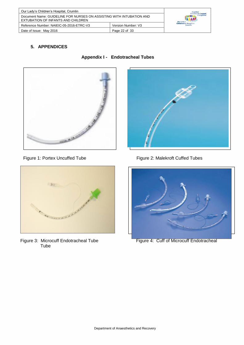
Appendix I - Endotracheal Tubes
Figure 1: Portex Uncuffed Tube Figure 2: Malekroft Cuffed Tubes
Figure 3: Microcuff Endotracheal Tube Figure 4: Cuff of Microcuff Endotracheal Tube
Our Lady’s Children’s Hospital, Crumlin
Document Name: GUIDELINE FOR NURSES ON ASSISTING WITH INTUBATION AND EXTUBATION OF INFANTS AND CHILDREN
Reference Number: NAIEIC-05-2016-ETRC-V3 Version Number: V3
Date of Issue: May 2016 Page 23 of 33
Department of Anaesthetics and Recovery
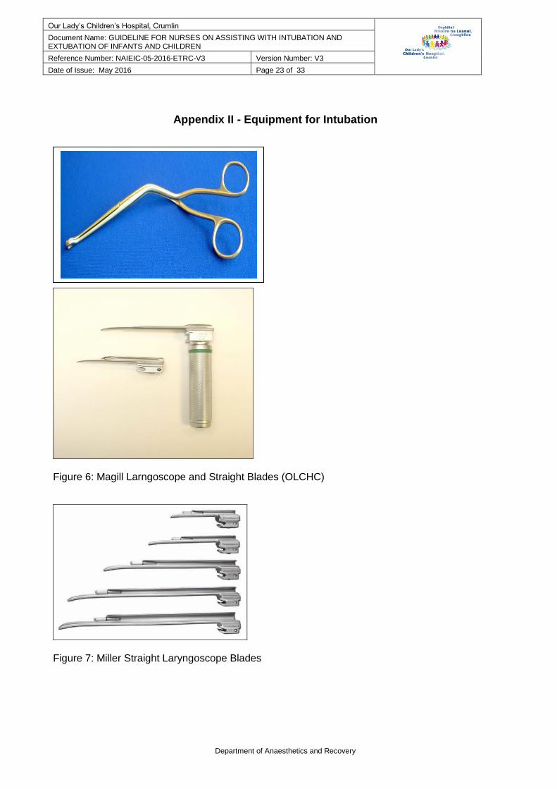
Appendix II - Equipment for Intubation
Figure 5: Magill Forcep
Figure 6: Magill Larngoscope and Straight Blades (OLCHC)
Figure 7: Miller Straight Laryngoscope Blades

Our Lady’s Children’s Hospital, Crumlin
Document Name: GUIDELINE FOR NURSES ON ASSISTING WITH INTUBATION AND EXTUBATION OF INFANTS AND CHILDREN
Reference Number: NAIEIC-05-2016-ETRC-V3 Version Number: V3
Date of Issue: May 2016 Page 24 of 33
Department of Anaesthetics and Recovery
Figure 8: Mackintosh Larngoscope and Blade (OLCHC)
Figure 10: Duoderm Extra Thin
Figure 11: Cavilon Wipes
Our Lady’s Children’s Hospital, Crumlin
Document Name: GUIDELINE FOR NURSES ON ASSISTING WITH INTUBATION AND EXTUBATION OF INFANTS AND CHILDREN
Reference Number: NAIEIC-05-2016-ETRC-V3 Version Number: V3
Date of Issue: May 2016 Page 25 of 33
Department of Anaesthetics and Recovery
Figure 12: Gum Elastic Bougle (Intubation Adjunct) Figure 13: Stylet (Intubation Adjunct) (OLCHC) (OLCHC)
Figure 14: Laryngeal Mask (OLCHC
Our Lady’s Children’s Hospital, Crumlin
Document Name: GUIDELINE FOR NURSES ON ASSISTING WITH INTUBATION AND EXTUBATION OF INFANTS AND CHILDREN
Reference Number: NAIEIC-05-2016-ETRC-V3 Version Number: V3
Date of Issue: May 2016 Page 26 of 33
Department of Anaesthetics and Recovery
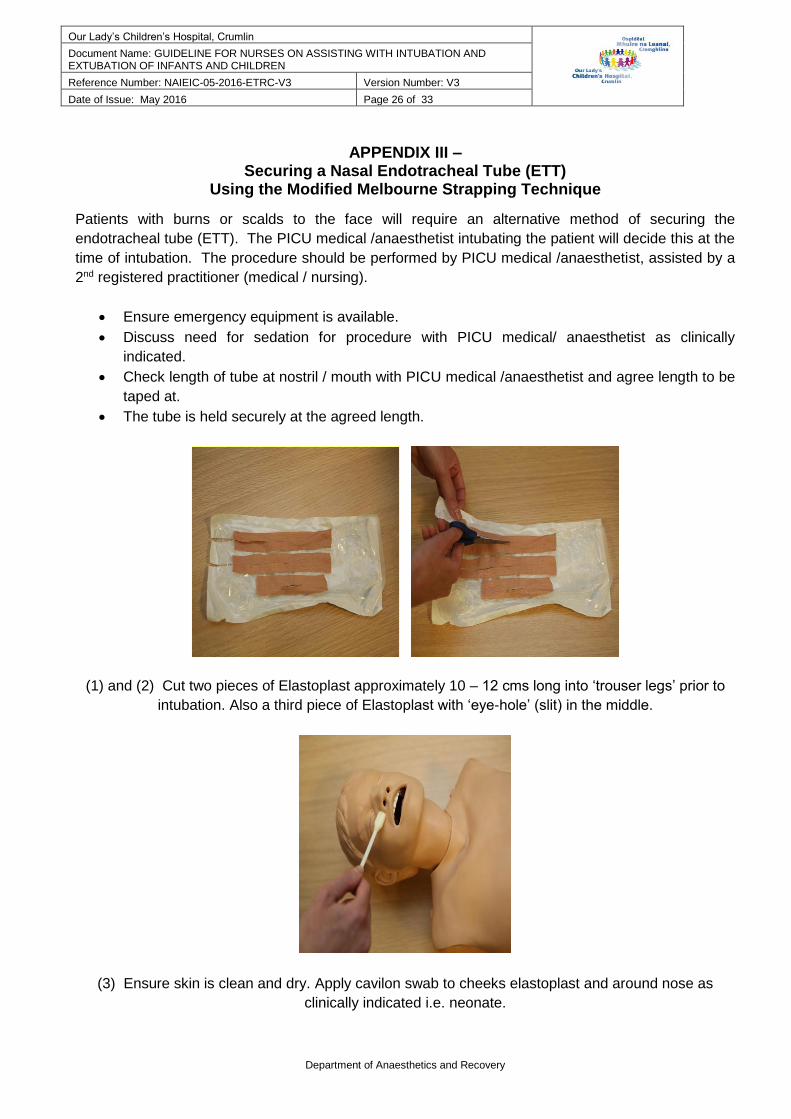
APPENDIX III – Securing a Nasal Endotracheal Tube (ETT)
Using the Modified Melbourne Strapping Technique
Patients with burns or scalds to the face will require an alternative method of securing the
endotracheal tube (ETT). The PICU medical /anaesthetist intubating the patient will decide this at the
time of intubation. The procedure should be performed by PICU medical /anaesthetist, assisted by a
2nd registered practitioner (medical / nursing).
Ensure emergency equipment is available.
Discuss need for sedation for procedure with PICU medical/ anaesthetist as clinically
indicated.
Check length of tube at nostril / mouth with PICU medical /anaesthetist and agree length to be
taped at.
The tube is held securely at the agreed length.
(1) and (2) Cut two pieces of Elastoplast approximately 10 – 12 cms long into ‘trouser legs’ prior to
intubation. Also a third piece of Elastoplast with ‘eye-hole’ (slit) in the middle.
(3) Ensure skin is clean and dry. Apply cavilon swab to cheeks elastoplast and around nose as
clinically indicated i.e. neonate.
Our Lady’s Children’s Hospital, Crumlin
Document Name: GUIDELINE FOR NURSES ON ASSISTING WITH INTUBATION AND EXTUBATION OF INFANTS AND CHILDREN
Reference Number: NAIEIC-05-2016-ETRC-V3 Version Number: V3
Date of Issue: May 2016 Page 27 of 33
Department of Anaesthetics and Recovery
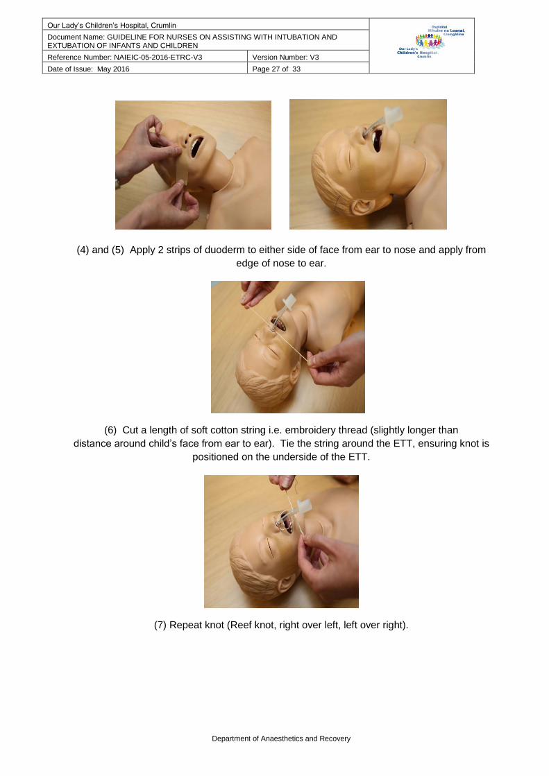
(4) and (5) Apply 2 strips of duoderm to either side of face from ear to nose and apply from
edge of nose to ear.
(6) Cut a length of soft cotton string i.e. embroidery thread (slightly longer than
distance around child’s face from ear to ear). Tie the string around the ETT, ensuring knot is
positioned on the underside of the ETT.
(7) Repeat knot (Reef knot, right over left, left over right).
Our Lady’s Children’s Hospital, Crumlin
Document Name: GUIDELINE FOR NURSES ON ASSISTING WITH INTUBATION AND EXTUBATION OF INFANTS AND CHILDREN
Reference Number: NAIEIC-05-2016-ETRC-V3 Version Number: V3
Date of Issue: May 2016 Page 28 of 33
Department of Anaesthetics and Recovery
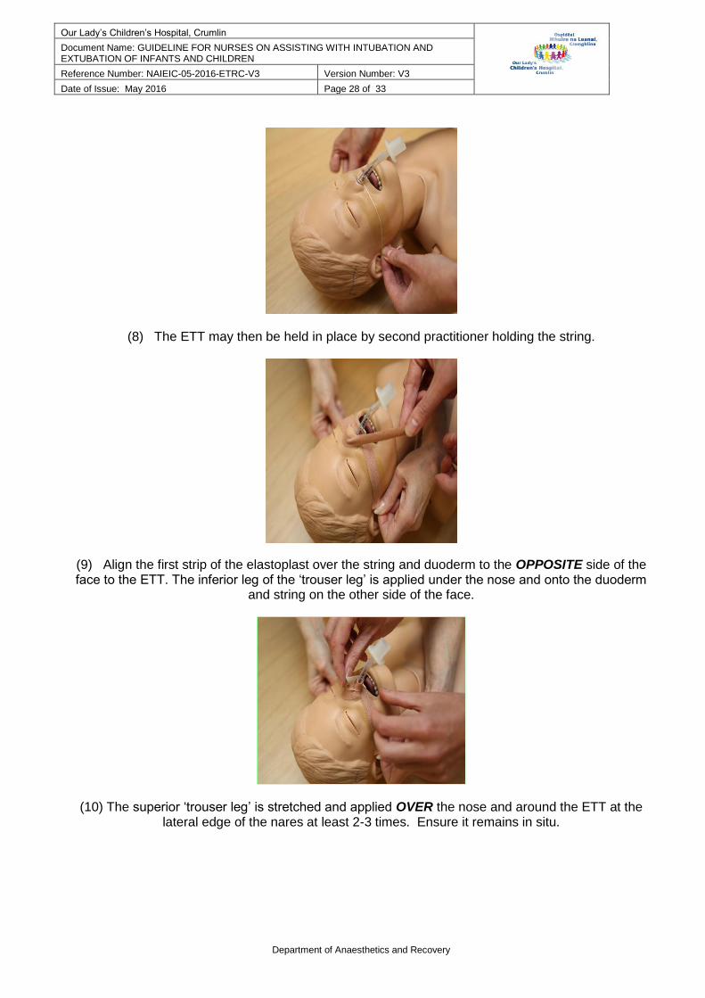
(8) The ETT may then be held in place by second practitioner holding the string.
(9) Align the first strip of the elastoplast over the string and duoderm to the OPPOSITE side of the face to the ETT. The inferior leg of the ‘trouser leg’ is applied under the nose and onto the duoderm
and string on the other side of the face.
(10) The superior ‘trouser leg’ is stretched and applied OVER the nose and around the ETT at the lateral edge of the nares at least 2-3 times. Ensure it remains in situ.
Our Lady’s Children’s Hospital, Crumlin
Document Name: GUIDELINE FOR NURSES ON ASSISTING WITH INTUBATION AND EXTUBATION OF INFANTS AND CHILDREN
Reference Number: NAIEIC-05-2016-ETRC-V3 Version Number: V3
Date of Issue: May 2016 Page 29 of 33
Department of Anaesthetics and Recovery
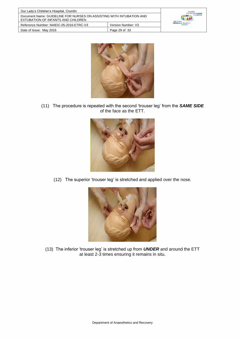
(11) The procedure is repeated with the second ‘trouser leg’ from the SAME SIDE of the face as the ETT.
(12) The superior ‘trouser leg’ is stretched and applied over the nose.
(13) The inferior ‘trouser leg’ is stretched up from UNDER and around the ETT at least 2-3 times ensuring it remains in situ.
Our Lady’s Children’s Hospital, Crumlin
Document Name: GUIDELINE FOR NURSES ON ASSISTING WITH INTUBATION AND EXTUBATION OF INFANTS AND CHILDREN
Reference Number: NAIEIC-05-2016-ETRC-V3 Version Number: V3
Date of Issue: May 2016 Page 30 of 33
Department of Anaesthetics and Recovery
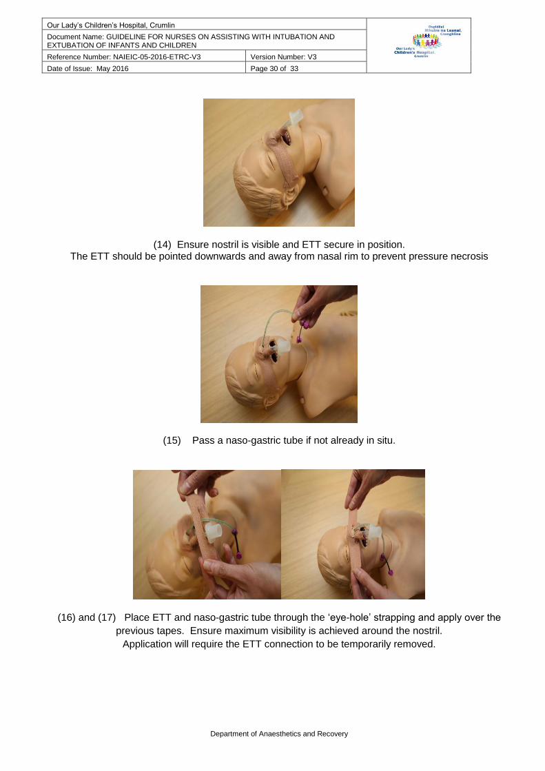
(14) Ensure nostril is visible and ETT secure in position. The ETT should be pointed downwards and away from nasal rim to prevent pressure necrosis
(15) Pass a naso-gastric tube if not already in situ.
(16) and (17) Place ETT and naso-gastric tube through the ‘eye-hole’ strapping and apply over the
previous tapes. Ensure maximum visibility is achieved around the nostril.
Application will require the ETT connection to be temporarily removed.
Our Lady’s Children’s Hospital, Crumlin
Document Name: GUIDELINE FOR NURSES ON ASSISTING WITH INTUBATION AND EXTUBATION OF INFANTS AND CHILDREN
Reference Number: NAIEIC-05-2016-ETRC-V3 Version Number: V3
Date of Issue: May 2016 Page 31 of 33
Department of Anaesthetics and Recovery
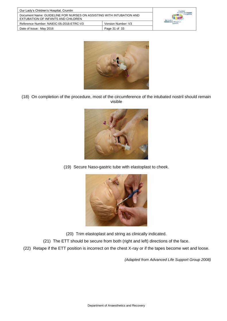
(18) On completion of the procedure, most of the circumference of the intubated nostril should remain visible
(19) Secure Naso-gastric tube with elastoplast to cheek.
(20) Trim elastoplast and string as clinically indicated.
(21) The ETT should be secure from both (right and left) directions of the face.
(22) Retape if the ETT position is incorrect on the chest X-ray or if the tapes become wet and loose.
(Adapted from Advanced Life Support Group 2008)
Our Lady’s Children’s Hospital, Crumlin
Document Name: GUIDELINE FOR NURSES ON ASSISTING WITH INTUBATION AND EXTUBATION OF INFANTS AND CHILDREN
Reference Number: NAIEIC-05-2016-ETRC-V3 Version Number: V3
Date of Issue: May 2016 Page 32 of 33
Department of Anaesthetics and Recovery
6. References
Advanced Life Support Group (2011) Advanced Paediatric Life Support, The Practical Approach, 5th Edition. Blackwell Wiley Publishing: Chichester. Brimacombe, J.R. and Berry, A.M. (1997) Cricoid pressure. Canadian Journal of Anaesthesia, 44(4), 414-425. Botter, L.T. (2009) Rapid sequence intubation in the neonate. Advances in Neonatal Care 9(3), 111-117. Chethan, D.B. and Hughes, R.C. (2008) Tracheal intubation, tracheal tubes and laryngeal mask airways. Journal of preoperative Practice 18(3) 88-90, 92-94. Crankshaw D, McViety J, and Entwistle M. (2014) A review of cuffed vs uncuffed endotracheal tubes in children. Pediatric Anesthesia and Critical Care Journal, 2(2):70-73 Cull, C. (1999) Extubation in ICU: enhancing the nursing role. Professional Nurse 14(9), 618-621. Davies, M.W. and Davis, P.G. (2010) Nebulized Raemic Epinephrine for Extubation of Newborn Infants (review). Cochrane Database of Systemic Reviews 2002 Issue 1. Art No. CD000506 10.1002/14651858 CD000506. Available online: www.cochrane.org/reviews/enlab000506.html (Accessed 16th August 2011). De, D. (2004) Clinical skills: a care plan approach to nurse-led extubation. British Journal of Nursing 13(18), 1086-1090. Dougherty, L. and Lister, S.E. (2015) (eds) The Royal Marsden Hospital, Manual of Clinical Nursing Procedures,9th Edition. Wiley Blackwell : Chichester. Eastern Perinatal Network (2010) Clinical Procedure: Nasotracheal Intubation. Eastern Perinatal Network. Available online: www.neonatal.org.uk/documents/5101.pdf ( Accessed 27th July 2011). Evans, T. and Carroll, P. (2001) Rapid Sequence Intubation. American Journal of Nursing, 17: 16-20. Fine, G.F. (2008) Problems with endotracheal tubes, cuffed vs. uncuffed. In: New Advances in Pediatric Ventilation: Revolutionizing the Management of Pediatric intubation with Cuffed tubes. Kimberley Clark Healthcare 6-9. Gerber, A.C. (2008a) Microcuff pediatric tubes: now anesthesiologists can use cuffed endotracheal tubes in children. In: New Advances in Pediatric Ventilation: Revolutionizing the Management of Pediatric intubation with Cuffed tubes. Kimberley Clark Healthcare 10-15. Gerber, A.C. (2008b) Cuffed tubes for infants and children in anaesthesia and intensive care: Why we should change to cuffed tubes in pediatric airway management. Journal of Paediatric Respiratory and Critical Care, 4(4), 3-9. Ghanta, S. Abdel-Latif, M. Lui, K. Ravindranathan, H. Awad, J. and Oei, J. (2007) Profolol compared with morphine, atropine and suxamethonium regimen as induction agents for neonatal endotracheal intubation: a randomised controlled trial. Paediatric 119, 1248-1255. Hazinski, M.F. (2013) Care of the Critically Ill Child. Mosby: St Louis. Henderson, J. (2011) Tracheal intubation: direct larnynosopy. In: Calder, I. and Pearce, A. (eds) Core Topics in Airway Management, 2nd Edition. Cambridge University Press: Cambridge, 110-120. Hockenberry, M.J. and Wilson, D. (eds) (2014) Wong’s Nursing Care of Infants and Children, 9th Edition. Mosby: St Louis. Hodd, J. Doyle, A. Carter, J. Albarran, J. and Young, P. (2010) Extubation in intensive care units in the UK: an online survey. Nursing in Critical Care 15 (6), 281-284.

Our Lady’s Children’s Hospital, Crumlin
Document Name: GUIDELINE FOR NURSES ON ASSISTING WITH INTUBATION AND EXTUBATION OF INFANTS AND CHILDREN
Reference Number: NAIEIC-05-2016-ETRC-V3 Version Number: V3
Date of Issue: May 2016 Page 33 of 33
Department of Anaesthetics and Recovery
Infection Control Nurses Association (2002) Hand Contamination Guidelines, 2nd Edition. ICNA, Fitwise Bathgate. Jagim, M. (2003) Airway management. American Journal of Nursing 103(100, 32-35. Jukes G. (2003) Endotracheal Intubation. Nursing Times. Available online: www.nursingtimes.net/nursing-practice-clinical-research/endotracheal-intubation. (Accessed 21st February 2012).
Khine, H.H. Corddry, D.H. kettrick, R.G. martin, T.M. McCloskey, J.J. Rose, J.B. Theroux, M.C. and Zagnoev, M. (1997) Comparison of cuffed and uncuffed endotracheal tubes in young children during general anesthesia.
Anesthesiology, 86: 627-631. Karlsen K.A. (2014) The STABLE ® Program. Pre-transport / Post Resuscitation Stabilization Care of Sick Infants, Guidelines for Neonatal Healthcare Providers: Learner / Provider Manual, 6th Edition. American Academy of Pediatrics: Salt Lake City. Karmarkar, S. and Varshney, S. (2008) Tracheal extubation. Continuing Education in Anaesthesia, Critical Care and Pain 8(6), 214-220. Kattwinkel, J. Denson, S. Zaichkin, J. and Niermeyer, S. (2000) Textbook of Neonatal Resuscitation, 4th Edition. American Heart Association and American Academy of Pediatrics: Elk Grove Village Illinois Kimberley-Clark (2006a) Pediatric. Endotracheal Tube: Finally a Cuffed ET Tube Designed for the Pediatric Anatomy. Kimberley-Clark. Available online: www.kchealthcare.com/microcuff (Accessed 24th August 2011). Kimberley-Clark (2006b) Paediatric Endotracheal Tube. Kimberley-Clark. Available online: www.kchealthcare.com/microcuff (Accessed 24th August 2011). Lancaster, L. (2007) Extubation after cardiac surgery: A practical guide. British Journal of Cardiac Nursing 2(6), 265-270. Levin, D.L. and Morriss, F.C. (eds) (1997) Essentials of Pediatric Intensive Care, 2nd Edition. Churchill Livingstone. LMA (2010) LMA Quick Reference Guide. Available online: www.lmana.com/files/supreme_quick-reference-card.pdf?PHPSESSID=7145dofe38bef2676cebdIfe8 (Accessed 19th July 2015). Lund, C.H. and Tucker, J.A. (2003) Adhesion and newborn skin. In: Hoath, S.B. and Maibach, H.I. Neonatal Skin Structure and Function. Deckker Inc: New York, 299-322. Maxwell, L.G. (2008) Endotracheal tube use in children: history as retext for current teaching. In: New Advances in Pediatric Ventilation: Revolutionizing the Management of Pediatric intubation with Cuffed tubes. Kimberley Clark Healthcare 1- 5. Microcuff (2011) The Microcuff ® Paediatric Concept. Microcuff: Weinheim, Germany. Available online: www.microsoft.com (Accessed 4th October 2011). Mitchell, V. (2011) Extubation. In: Calder, I. and Pearce, (eds) A. Core Topics in Airway Management, 2nd Edition. Cambridge University Press: Cambridge, 158-168. Mitchell, V. and Patel, A. (2011) Tracheal tubes, tracheostomy tubes. In: Calder, I. and Pearce, A. (eds) Core Topics in Airway Management, 2nd Edition. Cambridge University Press: Cambridge, 91-103. NMBI (2015a) Recording Clinical Practice, 2nd Edition. Nursing and Midwifery Board of Ireland: Dublin. NMBI (2015b) Scope of Nursing and Midwifery Practice Framework, 2nd Edition. Nursing and Midwifery Board of Ireland: Dublin.
Our Lady’s Children’s Hospital, Crumlin
Document Name: GUIDELINE FOR NURSES ON ASSISTING WITH INTUBATION AND EXTUBATION OF INFANTS AND CHILDREN
Reference Number: NAIEIC-05-2016-ETRC-V3 Version Number: V3
Date of Issue: May 2016 Page 34 of 33
Department of Anaesthetics and Recovery
National Maternity Hospital (2008) Neonatal Endotracheal Intubation. National Maternity Hospital: Dublin. Norfolk, Suffolk and Cambridgeshire Neonatal Network (NSCNN) (2005) Clinical Procedure: Endotracheal Intubation. NSCNN. Available online: www.neonatal.org.uk/document/3263.pdf ( Accessed 28th July 2011). OLHSC (2007) Standard Precautions. Our Lady’s Hospital for Sick Children’s: Dublin. OLHSC (2008) Infection Control Policy. Our Lady’s Hospital for Sick Children’s: Dublin. OLCHC (2010) Guidelines on Hand Hygiene. Our Lady’s Children’s Hospital Crumlin: Dublin. OLCHC (2011) Aseptic Non Touch Technique. Our Lady’s Children’s Hospital Crumlin: Dublin. Parker, M.M. (2007) Tips on extubation in mechanically ventilated children: close observation is the key to successful extubation. Journal of Respiratory Diseases 28(5), 203-207. Resuscitation Council UK (2008) Paediatric Immediate Life Support. Resuscitation Council UK: London. Scales, K. and Pilsworth, J. (2007) A practical guide to extubation. Nursing Standard 22(2), 44-48. Scardamaglia, L. Nixon, R. and Fewings, J. (2003) Compound Tincture of Benzoin: a common contact allergen? Australian Journal of Dermatology 44(3), 180-184. South Thames Retrieval Service (STRS) (2007) Securing Oral Endotracheal Tubes. South Thames Retrieval Service, London. Available online: www.strs.nhs.uk/resources/pdf/guidelines/oralETT.pdf (Accessed 4th October 2011). Spiegel, J.E. (2010) Endotracheal Tube Cuffs: design and Function. Anaesthesiology News.51-58. Available online: www.anesthesiologynews.com (Accessed 4th October 2011). Trivits-Verger, J. and Lebet, R.M. (2008) ACCN Procedure Manual for Pediatric Acute and Critical Care. Saunders Ellsevier: Philadelphia. University of Virginia (2011) Airway Management: Understanding Equipment. University of Virginia Available online: www.healthstem.virginia.edu/Internet/Anaesthesiology-Elective/airway/equip (Accessed 18th August 2011).
Walker, R. W. M. Ravi, R. and Haylett, K. (2010) Effects of cricord force on airway calibre in children: a bronchoscopic assessment. British Journal of Anaesthesia, 104 (1): 71–4 Wratney, A.T. and Chieifetz, I.M. (2007) AARC Clinicqal Practice Guideline. Removal of the Endotracheal Tube. Respiratory 52(1), 81-93. Available online: www.rcjournal.com/cpgs/pdf/removal_of_endotracheal_tube.pdf (Accessed 12th August 2011). Zelicof-Paul, A. Smith-Lockridge, A. Schnadower, D. Tyler, S. Levin, S. Roskind, C. and Dayan, P. (2005) Controversies in rapid sequence intubation in children. Current Opinions in Pediatrics 17, 355-362.
Disclaimer 2016, Our Lady’s Children’s Hospital Crumlin, Dublin 12. All rights reserved. No part of this publication may be reproduced, stored in a retrieval system or transmitted in any form or by any means without the prior written permission of the copyright holder. Every effort has been made to ensure that the information provided is accurate and in accord with standards accepted at the time of printing.