guidelines for the clinical management of snake bites in
TRANSCRIPT

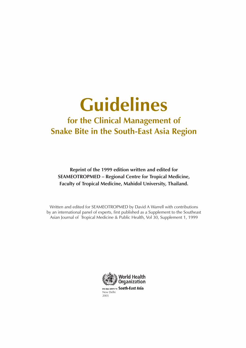
Guidelinesfor the Clinical Management ofSnake bites in the South-East Asia Region
Spectacled CobraSaw Scaled Viper
Common KraitCommon Krait
Russells ViperRussells Viper
Written and edited for SEAMEOTROPMED by David A Warrell with contributionsby an international panel of experts, first published as a Supplement to the Southeast
Asian Journal of Tropical Medicine & Public Health, Vol 30, Supplement 1, 1999
New Delhi2005
Guidelines
Reprint of the 1999 edition written and edited forSEAMEOTROPMED – Regional Centre for Tropical Medicine,Faculty of Tropical Medicine, Mahidol University, Thailand.
for the Clinical Management ofSnake Bite in the South-East Asia Region
iiGuidelines for the Clinical Managementof Snake bite in the South-East Asia Region
© World Health Organization 2005
Publications of the World Health Organization enjoy copyright protection in accordance with theprovisions of Protocol 2 of the Universal Copyright Convention. For rights of reproduction ortranslation, in part or in toto, of publications issued by the WHO Regional Office for South-EastAsia, application should be made to the Regional Office for South-East Asia, World Health House,Indraprastha Estate, New Delhi 110002, India.
The designations employed and the presentation of material in this publication do not imply theexpression of any opinion whatsoever on the part of the Secretariat of the World Health Organizationconcerning the legal status of any country, territory, city or area or of its authorities, or concerningthe delimitation of its frontiers or boundaries.
iiiGuidelines for the Clinical Management
of Snake bite in the South-East Asia Region
Contents
Preface ............................................................................................... v
1. Introduction .................................................................................. 11.1 Venomous snakes of South-East Asia ..................................................... 11.2 Snake venoms ...................................................................................... 81.3 How common are snake bites? ............................................................. 91.4 How do snake bites happen? .............................................................. 111.5 How can snake bites be avoided? ....................................................... 11
2. Symptoms and Signs of Snake Bite ............................................. 132.1 When venom has not been injected ................................................... 132.2 When venom has been injected ......................................................... 132.3 Clinical pattern of envenoming by snakes in South-East Asia ................ 142.4 Clinical syndromes of snake bite in South-East Asia ............................. 182.5 Long term complications (sequelae) of snake bite ................................ 19
3. Symptoms and Signs of Cobra-spit Ophthalmia ........................ 21
4. Management of Snake Bites in South-East Asia .......................... 234.1 First aid treatment ............................................................................... 234.2 Transport to hospital ........................................................................... 264.3 Treatment in the dispensary or hospital ............................................... 264.4 Detailed clinical assessment and species diagnosis ............................... 274.5 Investigations/laboratory tests .............................................................. 304.6 Antivenom treatment .......................................................................... 324.7 Supportive/ancillary treatment ............................................................ 404.8 Treatment of the bitten part ................................................................ 464.9 Rehabilitation ..................................................................................... 47
ivGuidelines for the Clinical Managementof Snake bite in the South-East Asia Region
5. Management of Cobra Spit Ophthalmia..................................... 49
6. Conclusions and Main Recommendations ................................. 51
7. Further Reading .......................................................................... 55
Annex1. Algorithm: Antivenom treatment of snakebite cases .................................... 61
2. Algorithm: Differentiating major asian snakespecies by clincal symdrome ....................................................................... 62
3. Antivenoms for treatment of bites by South-East Asian snakes ...................... 63
4. Measurement of Central Venous Pressure ................................................... 66
5. Measurement of intracompartmental pressure intensely swollen snake-bitten limbs .............................................................. 67
vGuidelines for the Clinical Management
of Snake bite in the South-East Asia Region
Preface
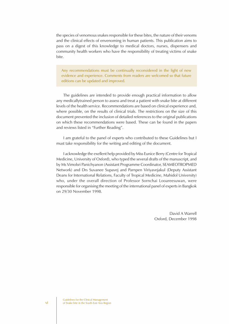
The geographical area specifically covered by this publication extends from Pakistanand the rest of the Indian subcontinent in the west through to the Philippines andIndonesia in the east, excluding Tibet, China, Taiwan, Korea, Japan, the eastern islandsof Indonesia and New Guinea and Australia (Figure 1, inside of front cover).
In many parts of this region, snake bite is a familiar occupational hazard offarmers, plantation workers and others, resulting in tens of thousands of deaths eachyear and innumerable cases of chronic physical handicap. Much is now known about
Figure 1: Map of Asia showing the area specifically covered by the guidelines
viGuidelines for the Clinical Managementof Snake bite in the South-East Asia Region
the species of venomous snakes responsible for these bites, the nature of their venomsand the clinical effects of envenoming in human patients. This publication aims topass on a digest of this knowledge to medical doctors, nurses, dispensers andcommunity health workers who have the responsibility of treating victims of snakebite.
Any recommendations must be continually reconsidered in the light of newevidence and experience. Comments from readers are welcomed so that futureeditions can be updated and improved.
The guidelines are intended to provide enough practical information to allowany medicallytrained person to assess and treat a patient with snake bite at differentlevels of the health service. Recommendations are based on clinical experience and,where possible, on the results of clinical trials. The restrictions on the size of thisdocument prevented the inclusion of detailed references to the original publicationson which these recommendations were based. These can be found in the papersand reviews listed in “Further Reading”.
I am grateful to the panel of experts who contributed to these Guidelines but Imust take responsibility for the writing and editing of the document.
I acknowledge the exellent help provided by Miss Eunice Berry (Centre for TropicalMedicine, University of Oxford), who typed the several drafts of the manuscript, andby Ms Vimolsri Panichyanon (Assistant Programme Coordinator, SEAMEOTROPMEDNetwork) and Drs Suvanee Supavej and Parnpen Viriyavejakul (Deputy AssistantDeans for International Relations, Faculty of Tropical Medicine, Mahidol University)who, under the overall direction of Professor Sornchai Looareesuwan, wereresponsible for organising the meeting of the international panel of experts in Bangkokon 29/30 November 1998.
David A WarrellOxford, December 1998
viiGuidelines for the Clinical Management
of Snake bite in the South-East Asia Region
Nepal*Bishnu Bahadur BhetwalBijalpura - 2 V.D.C.P.O. - BijalpuraDist - MahottariNepal
IndiaKirpal S ChughKothi No 601, Sector 18BChandigarh - 160 018India
Papua New GuineaDavid G LallooNuffield Dept Clinical Medicine,University of OxfordJohn Radcliffe HospitalHeadingtonOxford OX3 9DUUK
ThailandSornchai LooareesuwanSEAMEOTROPMED Regional Centrefor Tropical MedicineFaculty of Tropical Medicine
Names and Addresses of theInternational Panel of Experts
who Contributed to the Guidelines
Mahidol University420/6 Rajvithi RoadBangkok 10400Thailand
MyanmarMay-Mya-Win)Renal and Dialysing UnitsTingangyun Sanpya HospitalYangonMyanmar
Sri LankaLena SjöströmTherapeutic Antibodies LtdClinical Operations (UK)14-15 Newbury StreetLondon EC1A 7HUUK
Sri Lanka, ThailandR David G TheakstonAlistair Reid Venom Research UnitLiverpool School of Tropical MedicinePembroke PlaceLiverpool L3 5QAUK
viiiGuidelines for the Clinical Managementof Snake bite in the South-East Asia Region
Myanmar, Sri Lanka,Thailand, Viet NamDavid A WarrellCentre for Tropical MedicineUniversity of OxfordJohn Radcliffe HospitalHeadingtonOxford OX3 9DUUK
Philippines, ThailandGeorge WattDept of MedicineAFRIMS315/6 Rajvithi RoadBangkok 10400Thailand
AustraliaJulian WhiteState Toxinology ServicesPoisons Information CentreAdelaide Children’s HospitalKing William StreetNorth AdelaideSA 5006Australia
*indicates countries of clinical experiencewith snake bite patients in theSouth-East Asian region.
1Guidelines for the Clinical Management
of Snake bite in the South-East Asia Region
1.1 Venomous snakes of South-East Asia
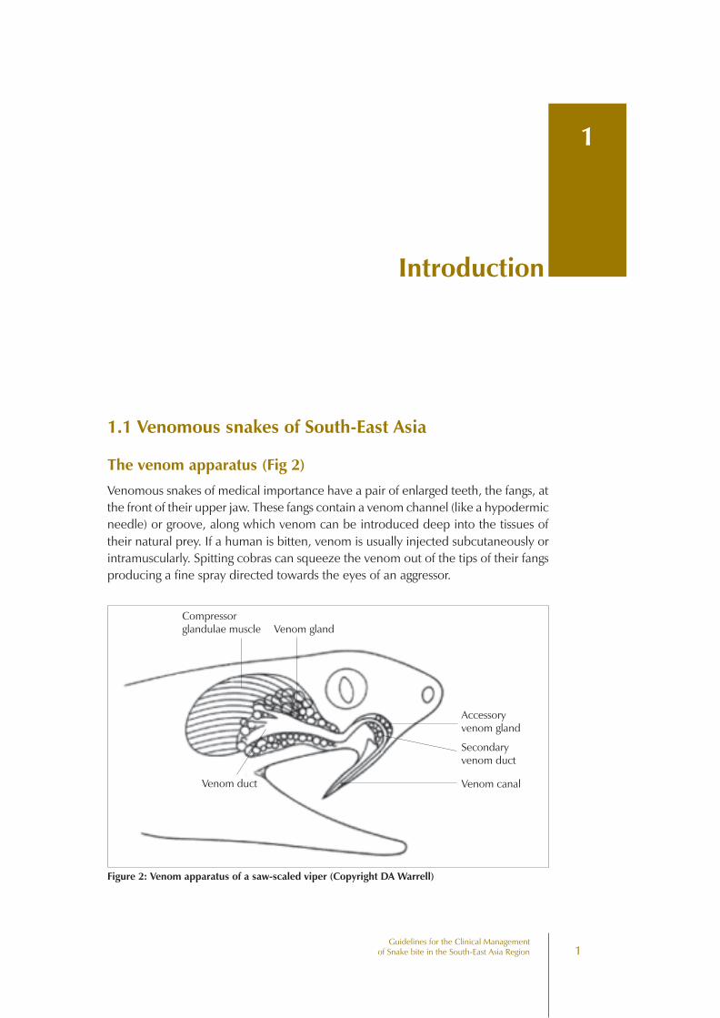
The venom apparatus (Fig 2)
Venomous snakes of medical importance have a pair of enlarged teeth, the fangs, atthe front of their upper jaw. These fangs contain a venom channel (like a hypodermicneedle) or groove, along which venom can be introduced deep into the tissues oftheir natural prey. If a human is bitten, venom is usually injected subcutaneously orintramuscularly. Spitting cobras can squeeze the venom out of the tips of their fangsproducing a fine spray directed towards the eyes of an aggressor.
Introduction
1
Compressorglandulae muscle Venom gland
Accessoryvenom gland
Secondaryvenom duct
Venom canalVenom duct
Figure 2: Venom apparatus of a saw-scaled viper (Copyright DA Warrell)
2Guidelines for the Clinical Managementof Snake bite in the South-East Asia Region
Classification
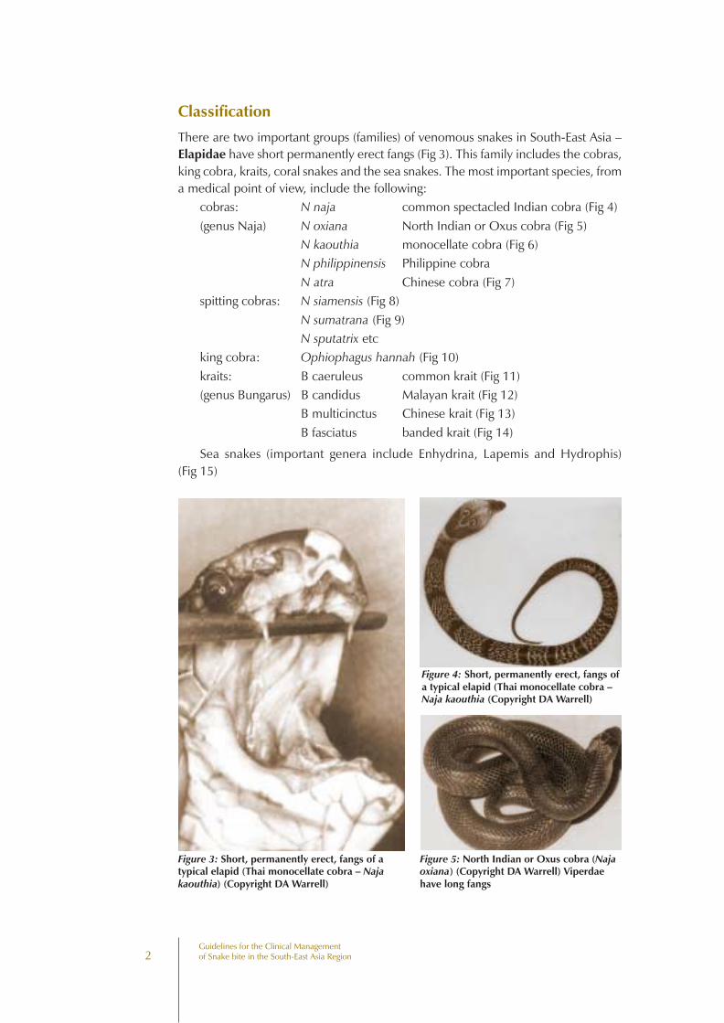
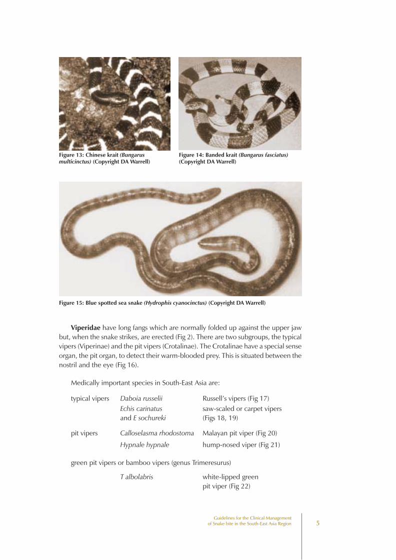
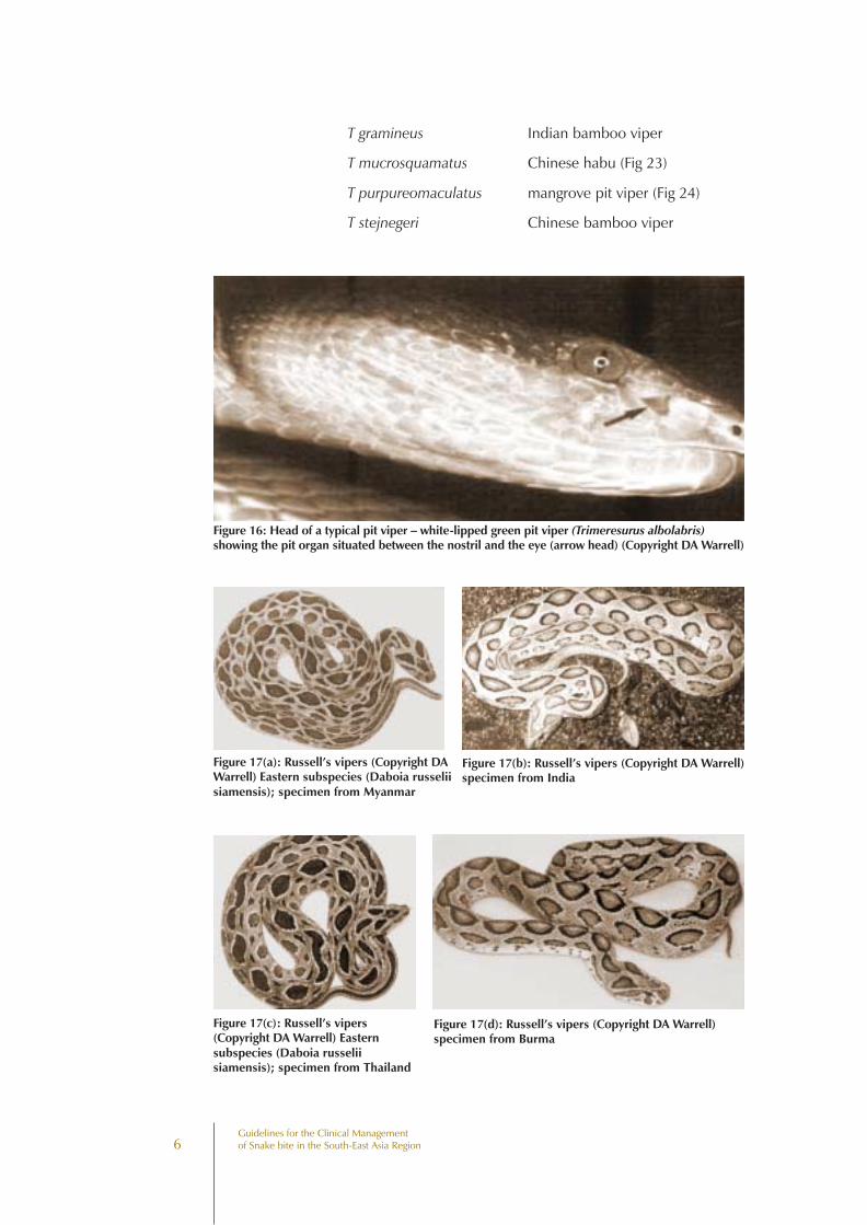
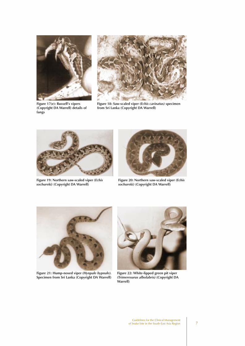
There are two important groups (families) of venomous snakes in South-East Asia –Elapidae have short permanently erect fangs (Fig 3). This family includes the cobras,king cobra, kraits, coral snakes and the sea snakes. The most important species, froma medical point of view, include the following:
cobras: N naja common spectacled Indian cobra (Fig 4)(genus Naja) N oxiana North Indian or Oxus cobra (Fig 5)
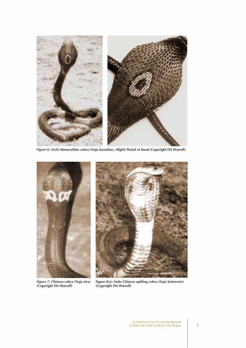
N kaouthia monocellate cobra (Fig 6)N philippinensis Philippine cobraN atra Chinese cobra (Fig 7)
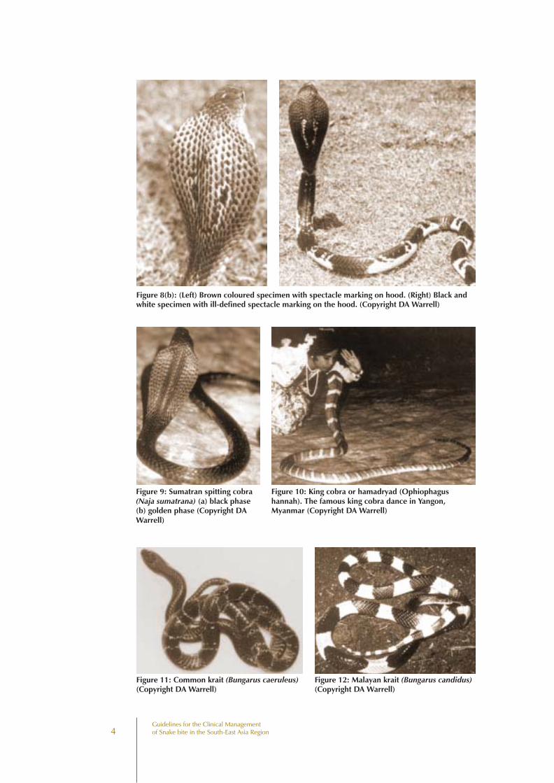
spitting cobras: N siamensis (Fig 8)N sumatrana (Fig 9)N sputatrix etc
king cobra: Ophiophagus hannah (Fig 10)kraits: B caeruleus common krait (Fig 11)(genus Bungarus) B candidus Malayan krait (Fig 12)
B multicinctus Chinese krait (Fig 13)B fasciatus banded krait (Fig 14)
Sea snakes (important genera include Enhydrina, Lapemis and Hydrophis)(Fig 15)
Figure 3: Short, permanently erect, fangs of atypical elapid (Thai monocellate cobra – Najakaouthia) (Copyright DA Warrell)
Figure 4: Short, permanently erect, fangs ofa typical elapid (Thai monocellate cobra –Naja kaouthia (Copyright DA Warrell)
Figure 5: North Indian or Oxus cobra (Najaoxiana) (Copyright DA Warrell) Viperdaehave long fangs
3Guidelines for the Clinical Management
of Snake bite in the South-East Asia Region
Figure 6: (Left) Monocellate cobra (Naja kaouthia), (Right) Detail of hood (Copyright DA Warrell)
Figure 7: Chinese cobra (Naja atra)(Copyright DA Warrell)
Figure 8(a): Indo-Chinese spitting cobra (Naja Saimensis)(Copyright DA Warrell)
4Guidelines for the Clinical Managementof Snake bite in the South-East Asia Region
Figure 8(b): (Left) Brown coloured specimen with spectacle marking on hood. (Right) Black andwhite specimen with ill-defined spectacle marking on the hood. (Copyright DA Warrell)
Figure 9: Sumatran spitting cobra(Naja sumatrana) (a) black phase(b) golden phase (Copyright DAWarrell)
Figure 10: King cobra or hamadryad (Ophiophagushannah). The famous king cobra dance in Yangon,Myanmar (Copyright DA Warrell)
Figure 11: Common krait (Bungarus caeruleus)(Copyright DA Warrell)
Figure 12: Malayan krait (Bungarus candidus)(Copyright DA Warrell)
5Guidelines for the Clinical Management
of Snake bite in the South-East Asia Region
Figure 13: Chinese krait (Bungarusmulticinctus) (Copyright DA Warrell)
Figure 14: Banded krait (Bungarus fasciatus)(Copyright DA Warrell)
Figure 15: Blue spotted sea snake (Hydrophis cyanocinctus) (Copyright DA Warrell)
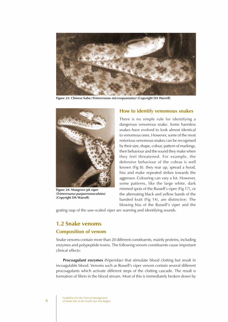
Viperidae have long fangs which are normally folded up against the upper jawbut, when the snake strikes, are erected (Fig 2). There are two subgroups, the typicalvipers (Viperinae) and the pit vipers (Crotalinae). The Crotalinae have a special senseorgan, the pit organ, to detect their warm-blooded prey. This is situated between thenostril and the eye (Fig 16).
Medically important species in South-East Asia are:
typical vipers Daboia russelii Russell’s vipers (Fig 17)Echis carinatus saw-scaled or carpet vipersand E sochureki (Figs 18, 19)
pit vipers Calloselasma rhodostoma Malayan pit viper (Fig 20)
Hypnale hypnale hump-nosed viper (Fig 21)
green pit vipers or bamboo vipers (genus Trimeresurus)
T albolabris white-lipped greenpit viper (Fig 22)
6Guidelines for the Clinical Managementof Snake bite in the South-East Asia Region
T gramineus Indian bamboo viper
T mucrosquamatus Chinese habu (Fig 23)
T purpureomaculatus mangrove pit viper (Fig 24)
T stejnegeri Chinese bamboo viper
Figure 16: Head of a typical pit viper – white-lipped green pit viper (Trimeresurus albolabris)showing the pit organ situated between the nostril and the eye (arrow head) (Copyright DA Warrell)
Figure 17(a): Russell’s vipers (Copyright DAWarrell) Eastern subspecies (Daboia russeliisiamensis); specimen from Myanmar
Figure 17(b): Russell’s vipers (Copyright DA Warrell)specimen from India
Figure 17(c): Russell’s vipers(Copyright DA Warrell) Easternsubspecies (Daboia russeliisiamensis); specimen from Thailand
Figure 17(d): Russell’s vipers (Copyright DA Warrell)specimen from Burma
7Guidelines for the Clinical Management
of Snake bite in the South-East Asia Region
Figure 17(e): Russell’s vipers(Copyright DA Warrell) details offangs
Figure 18: Saw-scaled viper (Echis carinatus) specimenfrom Sri Lanka (Copyright DA Warrell)
Figure 19: Northern saw-scaled viper (Echissochureki) (Copyright DA Warrell)
Figure 20: Northern saw-scaled viper (Echissochureki) (Copyright DA Warrell)
Figure 21: Hump-nosed viper (Hynpale hypnale).Specimen from Sri Lanka (Copyright DA Warrell)
Figure 22: White-lipped green pit viper(Trimeresurus albolabris) (Copyright DAWarrell)
8Guidelines for the Clinical Managementof Snake bite in the South-East Asia Region
How to identify venomous snakes
There is no simple rule for identifying adangerous venomous snake. Some harmlesssnakes have evolved to look almost identicalto venomous ones. However, some of the mostnotorious venomous snakes can be recognisedby their size, shape, colour, pattern of markings,their behaviour and the sound they make whenthey feel threatened. For example, thedefensive behaviour of the cobras is wellknown (Fig 8): they rear up, spread a hood,hiss and make repeated strikes towards theaggressor. Colouring can vary a lot. However,some patterns, like the large white, darkrimmed spots of the Russell’s viper (Fig 17), orthe alternating black and yellow bands of thebanded krait (Fig 14), are distinctive. Theblowing hiss of the Russell’s viper and the
grating rasp of the saw-scaled viper are warning and identifying sounds.
1.2 Snake venomsComposition of venom
Snake venoms contain more than 20 different constituents, mainly proteins, includingenzymes and polypeptide toxins. The following venom constituents cause importantclinical effects:
Procoagulant enzymes (Viperidae) that stimulate blood clotting but result inincoagulable blood. Venoms such as Russell’s viper venom contain several differentprocoagulants which activate different steps of the clotting cascade. The result isformation of fibrin in the blood stream. Most of this is immediately broken down by
Figure 23: Chinese habu (Trimeresurus microsquamatus) (Copyright DA Warrell)
Figure 24: Mangrove pit viper(Trimeresurus purpureomaculatus)(Copyright DA Warrell)
9Guidelines for the Clinical Management
of Snake bite in the South-East Asia Region
the body’s own fibrinolytic system. Eventually, and sometimes within 30 minutes ofthe bite, the levels of clotting factors have been so depleted (“consumptioncoagulopathy”) that the blood will not clot.
Haemorrhagins (zinc metalloproteinases) that damage the endothelial lining ofblood vessel walls causing spontaneous systemic haemorrhage.
Cytolytic or necrotic toxins - these digestive hydrolases (proteolytic enzymesand phospholipases A) polypeptide toxins and other factors increase permeabilityresulting in local swelling. They may also destroy cell membranes and tissues.
Haemolytic and myolytic phospholipases A2 - these enzymes damage cellmembranes, endothelium, skeletal muscle, nerve and red blood cells.
Pre-synaptic neurotoxins (Elapidae and some Viperidae) - these arephospholipases A2 that damage nerve endings, initially releasing acetylcholinetransmitter, then interfering with release.
Post-synaptic neurotoxins (Elapidae) - these polypeptides compete withacetylcholine for receptors in the neuromuscular junction and lead to curare-likeparalysis.
Quantity of venom injected at a bite
This is very variable, depending on the species and size of the snake, the mechanicalefficiency of the bite, whether one or two fangs penetrated the skin and whetherthere were repeated strikes. The snake may be able to control whether or not venomis injected. For whatever reason, a proportion of bites by venomous snakes do notresult in the injection of sufficient venom to cause clinical effects. About 50% of bitesby Malayan pit vipers and Russell’s vipers, 30% of bites by cobras and 5-10% of bitesby saw-scaled vipers do not result in any symptoms or signs of envenoming. Snakesdo not exhaust their store of venom, even after several strikes, and they are no lessvenomous after eating their prey.
Although large snakes tend to inject more venom than smaller specimens of thesame species, the venom of smaller, younger vipers may be richer in some dangerouscomponents, such as those affecting haemostasis.
1.3 How common are snake bites?It is difficult to answer this question because many snake bites and even deaths fromsnake bite are not recorded. One reason is that many snake bite victims are treatednot in hospitals but by traditional healers.
Bites by small snakes should not be ignored or dismissed. They should be takenjust as seriously as bites by large snakes of the same species.
10Guidelines for the Clinical Managementof Snake bite in the South-East Asia Region
Bangladesh – a survey of 10% of the country in 1988-9 revealed 764 bites with168 deaths in one year. Cobra bites (34% of all bites) caused a case fatality of 40%.
Bhutan – (no data available)
Cambodia – (no data available)
India – estimates in the region of 200,000 bites and 15-20,000 snake bite deathsper year, originally made in the last century, are still quoted. No reliable nationalstatistics are available. In 1981, a thousand deaths were reported in MaharashtraState. In the Burdwan district of West Bengal 29,489 people were bitten in one yearwith 1,301 deaths. It is estimated that between 35,000 and 50,000 people die ofsnake bite each year among India’s population of 980 million.
Indonesia – no reliable data are available from this vast archipelago. Snake bitesand deaths are reported from some islands, eg Komodo, but fewer than 20 deathsare registered each year.
Lao DPR – (no data available)
Malaysia – bites are common, especially in northwest peninsular Malaysia, butthere are few deaths.
Myanmar (Burma) – snake bites and snake bite deaths have been reliably reportedfrom colonial times. Russell’s vipers are responsible for 90% of cases. In 1991, therewere 14,000 bites with 1,000 deaths and in 1997, 8,000 bites with 500 deaths.Under-reporting is estimated at 12%. There are peaks of incidence in May and Junein urban areas and during the rice harvest in October to December in rural areas.
Nepal – there are estimated to be at least 20,000 snake bites with about 200deaths in hospitals each year, mainly in the Terai region. One survey suggested asmany as 1,000 deaths per year. Among 16 fatalities recorded at one rural clinicduring a monsoon season, 15 had died on their way to seek medical care.
Pakistan – there are an estimated 20,000 snake bite deaths each year
Philippines – there are no reliable estimates of mortality among the many islandsof thearchipelago. Figures of 200-300 deaths each year have been suggested. Onlycobras cause fatal envenoming, their usual victims being rice farmers.
Sri Lanka – epidemiological studies in Anuradhapura showed that only two-thirds of cases of fatal snake bite were being reported to the Government Agent
To remedy the deficiency in reliable snake bite data, it is strongly recommendedthat snake bites should be made a specific notifiable disease in all countries in theSouth East Asian region.
11Guidelines for the Clinical Management
of Snake bite in the South-East Asia Region
Statistical Branch. However, the Registrar General received reports of more than 800deaths from bites and stings by venomous animals and insects in the late 1970s andthe true annual incidence of snake bite fatalities may exceed 1,000.
Thailand – between 1985 and 1989, the number of reported snake bite casesincreased from 3,377 to 6,038 per year, reflecting increased diligence in reportingrather than a true increase in snake bites; the number of deaths ranged from 81 to183 (average 141) per year. In 1991 there were 1,469 reported bites with five deaths,in 1992, 6,733 bites with 19 deaths and, in 1994, 8,486 bites with eight deaths.Deaths reported in hospital returns were only 11% of the number recorded by thePublic Health Authorities. In a national survey of dead snakes brought to hospital bythe people they had bitten, 70% of the snakes were venomous species, the mostcommonly brought species being Malayan pit viper (Calloselasma rhodostoma) 38%,white-lipped green pit viper (Trimeresurus albolabris) 27%, Russell’s viper (Daboiarusselii siamensis) 14%, Indo-Chinese spitting cobra (Naja siamensis) 10% andmonocellate cobra (N kaouthia) 7%. In an analysis of 46 fatal cases in which thesnake had been reliably identified, Malayan kraits (Bungarus candidus) and Malayanpit vipers were each responsible for 13 cases, monocellate cobras for 12 and Russell’svipers for seven deaths.
Viet Nam – there are an estimated 30,000 bites per year. Among 430 rubberplantation workers bitten by Malayan pit vipers between 1993 and 1998, the casefatality was 22%, but only a minority had received antivenom treatment. Fishermenare still occasionally killed by sea snakes but rarely reach hospitals.
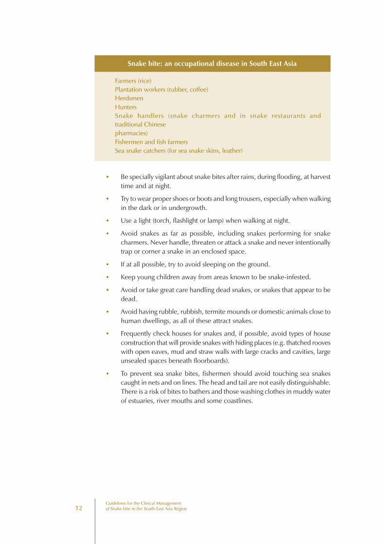
1.4 How do snake bites happen?In South-East Asia, snake bite is an occupational hazard of rice farmers; rubber,coffee and other plantation workers; fishermen and those who handle snakes. Mostsnake bites happen when the snake is trodden on, either in the dark or in undergrowth,by someone who is bare-footed or wearing only sandals. The snake may be pickedup, unintentionally in a handful of foliage or intentionally by someone who is tryingto show off. Some bites occur when the snake (usually a krait) comes in to the homeat night in search of its prey (other snakes, lizards, frogs, mice) and someone sleepingon the floor rolls over onto the snake in their sleep. Not all snake bites happen inrural areas. For example, in some large cities, such as Jammu in India, people whosleep in small huts (jhuggies) are frequently bitten by kraits.
1.5 How can snake bites be avoided?Snake bite is an occupational hazard that is very difficult to avoid completely. However,attention to the following recommendations might reduce the number of accidents.
• Education! Know your local snakes, know the sort of places where they liketo live and hide, know at what times of year, at what times of day/night orin what kinds of weather they are most likely to be active.
12Guidelines for the Clinical Managementof Snake bite in the South-East Asia Region
• Be specially vigilant about snake bites after rains, during flooding, at harvesttime and at night.
• Try to wear proper shoes or boots and long trousers, especially when walkingin the dark or in undergrowth.
• Use a light (torch, flashlight or lamp) when walking at night.
• Avoid snakes as far as possible, including snakes performing for snakecharmers. Never handle, threaten or attack a snake and never intentionallytrap or corner a snake in an enclosed space.
• If at all possible, try to avoid sleeping on the ground.
• Keep young children away from areas known to be snake-infested.
• Avoid or take great care handling dead snakes, or snakes that appear to bedead.
• Avoid having rubble, rubbish, termite mounds or domestic animals close tohuman dwellings, as all of these attract snakes.
• Frequently check houses for snakes and, if possible, avoid types of houseconstruction that will provide snakes with hiding places (e.g. thatched rooveswith open eaves, mud and straw walls with large cracks and cavities, largeunsealed spaces beneath floorboards).
• To prevent sea snake bites, fishermen should avoid touching sea snakescaught in nets and on lines. The head and tail are not easily distinguishable.There is a risk of bites to bathers and those washing clothes in muddy waterof estuaries, river mouths and some coastlines.
Snake bite: an occupational disease in South East Asia
Farmers (rice)Plantation workers (rubber, coffee)HerdsmenHuntersSnake handlers (snake charmers and in snake restaurants andtraditional Chinesepharmacies)Fishermen and fish farmersSea snake catchers (for sea snake skins, leather)
13Guidelines for the Clinical Management
of Snake bite in the South-East Asia Region
2.1 When venom has not been injectedSome people who are bitten by snakes or suspect or imagine that they have beenbitten, may develop quite striking symptoms and signs, even when no venom hasbeen injected. This results from an understandable fear of the consequences of a realvenomous bite. Anxious people may overbreathe so that they develop pins andneedles of the extremities, stiffness or tetany of their hands and feet and dizziness.Others may develop vasovagal shock after the bite or suspected bite – faintness andcollapse with profound slowing of the heart. Others may become highly agitated andirrational and may develop a wide range of misleading symptoms. Another source ofsymptoms and signs not caused by snake venom is first aid and traditional treatments.Constricting bands or tourniquets may cause pain, swelling and congestion. Ingestedherbal remedies may cause vomiting. Instillation of irritant plant juices into the eyesmay cause conjunctivitis. Forcible insufflation of oils into the respiratory tract maylead to aspiration pneumonia, bronchospasm, ruptured ear drums and pneumothorax.Incisions, cauterisation, immersion in scalding liquid and heating over a fire canresult in devastating injuries.
2.2 When venom has been injected
Early symptoms and signs
Following the immediate pain of mechanical penetration of the skin by the snake’sfangs, there may be increasing local pain (burning, bursting, throbbing) at the site ofthe bite, local swelling that gradually extends proximally up the bitten limb andtender, painful enlargement of the regional lymph nodes draining the site of the bite(in the groin – femoral or inguinal, following bites in the lower limb; at the elbow(epitrochlear) or in the axilla following bites in the upper limb). However, bites bykraits, sea snakes and Philippine cobras may be virtually painless and may cause
Symptoms andSigns of Snake Bite
2
14Guidelines for the Clinical Managementof Snake bite in the South-East Asia Region
negligible local swelling. Someone who is sleeping may not even wake up whenbitten by a krait and there may be no detectable fang marks or signs of localenvenoming.
2.3 Clinical pattern of envenoming by snakesin South-East AsiaSymptoms and signs vary according to the species of snake responsible for the biteand the amount of venom injected. Sometimes the identity of the biting snake canbe confirmed by examining the dead snake; it may be strongly suspected from thepatient’s description or the circumstances of the bite or from knowledge of the clinicaleffects of the venom of that species. This information will enable the doctor to choosean appropriate antivenom, anticipate the likely complications and therefore takeappropriate action. If the biting species is unknown, recognition of the emergingpattern of symptoms, signs and results of laboratory tests (“the clinical syndrome”),may suggest which species was responsible.
Local symptoms and signs in the bitten part
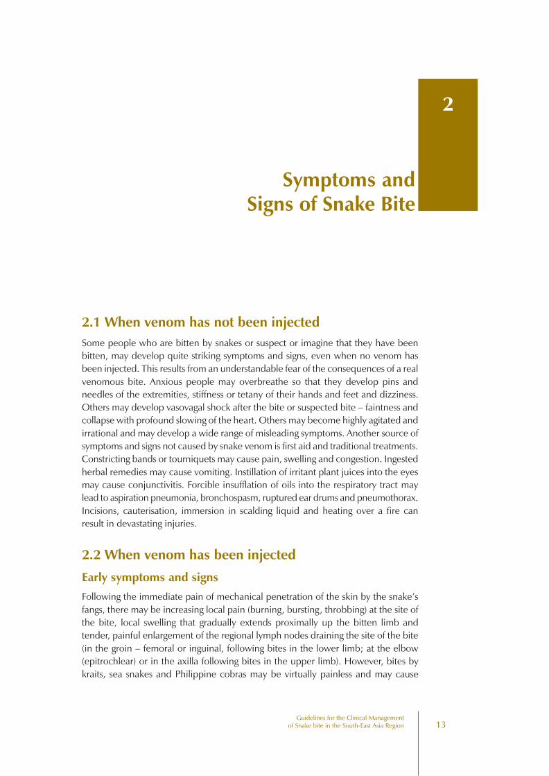
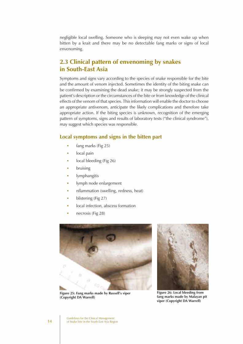
• fang marks (Fig 25)
• local pain
• local bleeding (Fig 26)
• bruising
• lymphangitis
• lymph node enlargement
• nflammation (swelling, redness, heat)
• blistering (Fig 27)
• local infection, abscess formation
• necrosis (Fig 28)
Figure 25: Fang marks made by Russell’s viper(Copyright DA Warrell)
Figure 26: Local bleeding fromfang marks made by Malayan pitviper (Copyright DA Warrell)
15Guidelines for the Clinical Management
of Snake bite in the South-East Asia Region
Figure 28: Tissue necrosis following a bite by a Malayan pit viper (Copyright DA Warrell)
Figure 27 Local swelling and blistering (a) with bruising, following a bite by a Malayan pit viper(Copyright DA Warrell), (Bottom) Local swelling and blistering (b) with early necrosis following abite by a monocellate cobra (Naja kaouthia) (Copyright DA Warrell)
Figure 28(a): Tissue necrosis following a bite by an Indochinese spitting cobra (Naja siamensis)(Copyright Sornchai Looareesuwan)
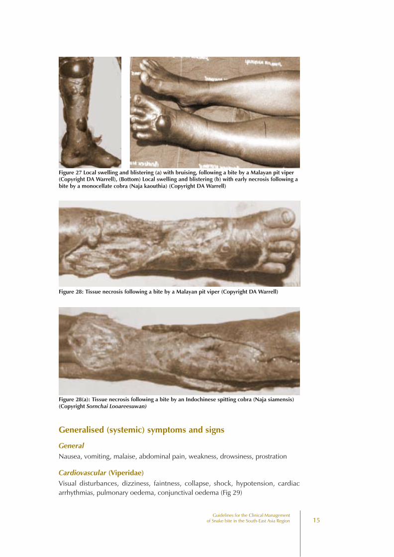
Generalised (systemic) symptoms and signs
GeneralNausea, vomiting, malaise, abdominal pain, weakness, drowsiness, prostration
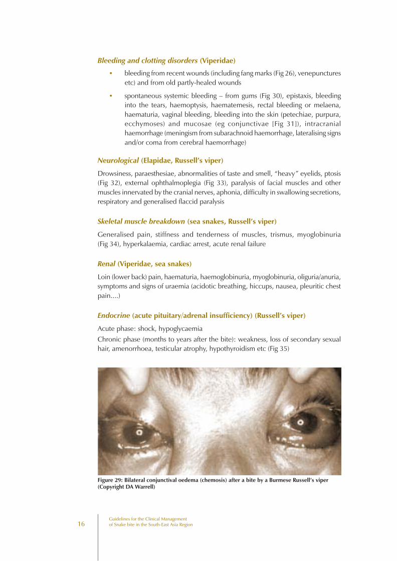
Cardiovascular (Viperidae)Visual disturbances, dizziness, faintness, collapse, shock, hypotension, cardiacarrhythmias, pulmonary oedema, conjunctival oedema (Fig 29)
16Guidelines for the Clinical Managementof Snake bite in the South-East Asia Region
Bleeding and clotting disorders (Viperidae)
• bleeding from recent wounds (including fang marks (Fig 26), venepuncturesetc) and from old partly-healed wounds
• spontaneous systemic bleeding – from gums (Fig 30), epistaxis, bleedinginto the tears, haemoptysis, haematemesis, rectal bleeding or melaena,haematuria, vaginal bleeding, bleeding into the skin (petechiae, purpura,ecchymoses) and mucosae (eg conjunctivae [Fig 31]), intracranialhaemorrhage (meningism from subarachnoid haemorrhage, lateralising signsand/or coma from cerebral haemorrhage)
Neurological (Elapidae, Russell’s viper)
Drowsiness, paraesthesiae, abnormalities of taste and smell, “heavy” eyelids, ptosis(Fig 32), external ophthalmoplegia (Fig 33), paralysis of facial muscles and othermuscles innervated by the cranial nerves, aphonia, difficulty in swallowing secretions,respiratory and generalised flaccid paralysis
Skeletal muscle breakdown (sea snakes, Russell’s viper)
Generalised pain, stiffness and tenderness of muscles, trismus, myoglobinuria(Fig 34), hyperkalaemia, cardiac arrest, acute renal failure
Renal (Viperidae, sea snakes)
Loin (lower back) pain, haematuria, haemoglobinuria, myoglobinuria, oliguria/anuria,symptoms and signs of uraemia (acidotic breathing, hiccups, nausea, pleuritic chestpain....)
Endocrine (acute pituitary/adrenal insufficiency) (Russell’s viper)
Acute phase: shock, hypoglycaemiaChronic phase (months to years after the bite): weakness, loss of secondary sexualhair, amenorrhoea, testicular atrophy, hypothyroidism etc (Fig 35)
Figure 29: Bilateral conjunctival oedema (chemosis) after a bite by a Burmese Russell’s viper(Copyright DA Warrell)
17Guidelines for the Clinical Management
of Snake bite in the South-East Asia Region
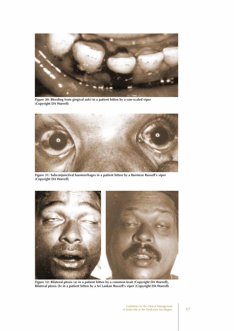
Figure 30: Bleeding from gingival sulci in a patient bitten by a saw-scaled viper(Copyright DA Warrell)
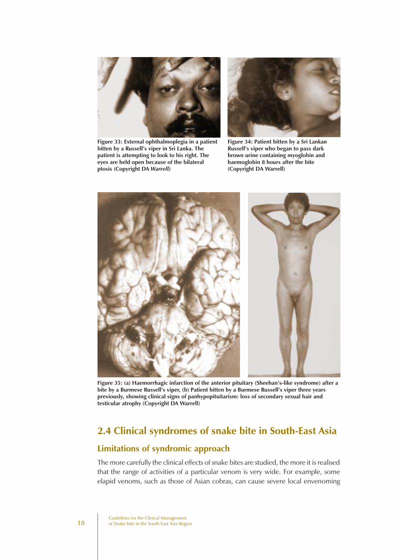
Figure 31: Subconjunctival haemorrhages in a patient bitten by a Burmese Russell’s viper(Copyright DA Warrell)
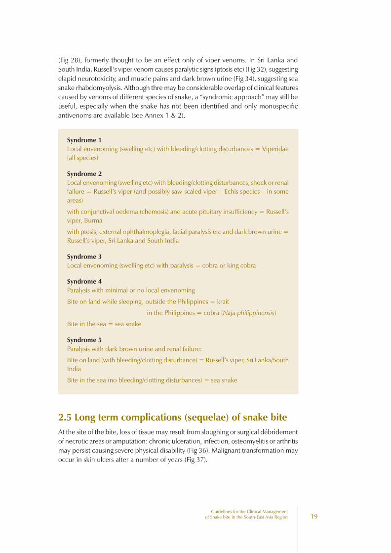
Figure 32: Bilateral ptosis (a) in a patient bitten by a common krait (Copyright DA Warrell),Bilateral ptosis (b) in a patient bitten by a Sri Lankan Russell’s viper (Copyright DA Warrell)
18Guidelines for the Clinical Managementof Snake bite in the South-East Asia Region
Figure 33: External ophthalmoplegia in a patientbitten by a Russell’s viper in Sri Lanka. Thepatient is attempting to look to his right. Theeyes are held open because of the bilateralptosis (Copyright DA Warrell)
Figure 34: Patient bitten by a Sri LankanRussell’s viper who began to pass darkbrown urine containing myoglobin andhaemoglobin 8 hours after the bite(Copyright DA Warrell)
Figure 35: (a) Haemorrhagic infarction of the anterior pituitary (Sheehan’s-like syndrome) after abite by a Burmese Russell’s viper, (b) Patient bitten by a Burmese Russell’s viper three yearspreviously, showing clinical signs of panhypopituitarism: loss of secondary sexual hair andtesticular atrophy (Copyright DA Warrell)
2.4 Clinical syndromes of snake bite in South-East Asia
Limitations of syndromic approach
The more carefully the clinical effects of snake bites are studied, the more it is realisedthat the range of activities of a particular venom is very wide. For example, someelapid venoms, such as those of Asian cobras, can cause severe local envenoming
19Guidelines for the Clinical Management
of Snake bite in the South-East Asia Region
(Fig 28), formerly thought to be an effect only of viper venoms. In Sri Lanka andSouth India, Russell’s viper venom causes paralytic signs (ptosis etc) (Fig 32), suggestingelapid neurotoxicity, and muscle pains and dark brown urine (Fig 34), suggesting seasnake rhabdomyolysis. Although thre may be considerable overlap of clinical featurescaused by venoms of different species of snake, a “syndromic approach” may still beuseful, especially when the snake has not been identified and only monospecificantivenoms are available (see Annex 1 & 2).
Syndrome 1Local envenoming (swelling etc) with bleeding/clotting disturbances = Viperidae(all species)
Syndrome 2Local envenoming (swelling etc) with bleeding/clotting disturbances, shock or renalfailure = Russell’s viper (and possibly saw-scaled viper – Echis species – in someareas)
with conjunctival oedema (chemosis) and acute pituitary insufficiency = Russell’sviper, Burma
with ptosis, external ophthalmoplegia, facial paralysis etc and dark brown urine =Russell’s viper, Sri Lanka and South India
Syndrome 3Local envenoming (swelling etc) with paralysis = cobra or king cobra
Syndrome 4Paralysis with minimal or no local envenoming
Bite on land while sleeping, outside the Philippines = krait
in the Philippines = cobra (Naja philippinensis)
Bite in the sea = sea snake
Syndrome 5Paralysis with dark brown urine and renal failure:
Bite on land (with bleeding/clotting disturbance) = Russell’s viper, Sri Lanka/SouthIndia
Bite in the sea (no bleeding/clotting disturbances) = sea snake
2.5 Long term complications (sequelae) of snake biteAt the site of the bite, loss of tissue may result from sloughing or surgical débridementof necrotic areas or amputation: chronic ulceration, infection, osteomyelitis or arthritismay persist causing severe physical disability (Fig 36). Malignant transformation mayoccur in skin ulcers after a number of years (Fig 37).
20Guidelines for the Clinical Managementof Snake bite in the South-East Asia Region
Chronic renal failure occurs afterbilateral cortical necrosis (Russell’sviper bites) and chronicpanhypopituitarism or diabetesinsipidus after Russell’s viper bites inMyanmar and South India (Fig 35b).Chronic neurological deficit is seenin the few patients who surviveintracranial haemorrhages(Viperidae).
Figure 36: (a) and (b) Chronic physical handicap resulting from necrotic envenoming by Malayanpit vipers (Copyright DA Warrell)
Figure 37: Squamous cell carcinoma developing atthe site of a chronic skin ulcer with osteomyelitis 8years after a bite by a Malayan pit viper (CopyrightDA Warrell)
21Guidelines for the Clinical Management
of Snake bite in the South-East Asia Region
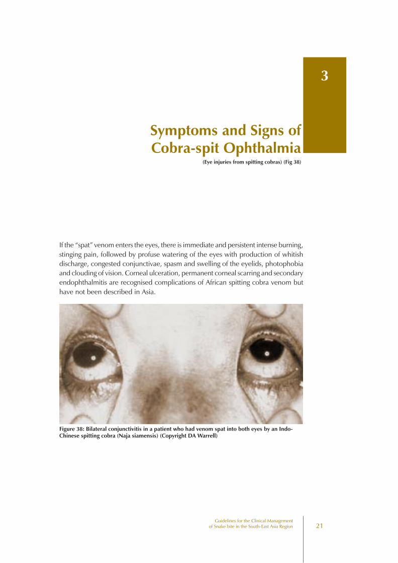
If the “spat” venom enters the eyes, there is immediate and persistent intense burning,stinging pain, followed by profuse watering of the eyes with production of whitishdischarge, congested conjunctivae, spasm and swelling of the eyelids, photophobiaand clouding of vision. Corneal ulceration, permanent corneal scarring and secondaryendophthalmitis are recognised complications of African spitting cobra venom buthave not been described in Asia.
Symptoms and Signs ofCobra-spit Ophthalmia
3
(Eye injuries from spitting cobras) (Fig 38)
Figure 38: Bilateral conjunctivitis in a patient who had venom spat into both eyes by an Indo-Chinese spitting cobra (Naja siamensis) (Copyright DA Warrell)

22Guidelines for the Clinical Managementof Snake bite in the South-East Asia Region
23Guidelines for the Clinical Management
of Snake bite in the South-East Asia Region
Management of SnakeBites in South-East Asia
4
The following steps or stages are often involved
Management of snake bite
••••• First aid treatment
••••• Transport to hospital
••••• Rapid clinical assessment and resuscitation
••••• Detailed clinical assessment and species diagnosis
••••• Investigations/laboratory tests
••••• Antivenom treatment
••••• Observation of the response to antivenom: decision about the need for furtherdose(s) of antivenom
••••• Supportive/ancillary treatment
••••• Treatment of the bitten part
••••• Rehabilitation
••••• Treatment of chronic complications
4.1 First aid treatmentFirst aid treatment is carried out immediately or very soon after the bite, before thepatient reaches a dispensary or hospital. It can be performed by the snake bite victimhimself/herself or by anyone else who is present.
24Guidelines for the Clinical Managementof Snake bite in the South-East Asia Region
Aims of first aid
••••• attempt to retard systemic absorption of venom
••••• preserve life and prevent complications before the patient can receive medicalcare (at a dispensary or hospital)
••••• control distressing or dangerous early symptoms of envenoming
••••• arrange the transport of the patient to a place where they can receive medicalcare (4.2)
••••• Above all, do no harm!
Unfortunately, most of the traditional, popular, available and affordable first aidmethods have proved to be useless or even frankly dangerous. These methods include:making local incisions or pricks/punctures (“tattooing”) at the site of the bite or in thebitten limb, attempts to suck the venom out of the wound, use of (black) snakestones, tying tight bands (tourniquets) around the limb, electric shock, topicalinstillation or application of chemicals, herbs or ice packs.
Local people may have great confidence in traditional (herbal) treatments, butthey must not be allowed to delay medical treatment or to do harm.
Most traditional first aid methods should be discouraged:They do more harm than good !
Recommended first aid methods
••••• Reassure the victim who may be very anxious
••••• Immobilise the bitten limb with a splint or sling (any movement or muscularcontraction increases absorption of venom into the bloodstream and lymphatics)
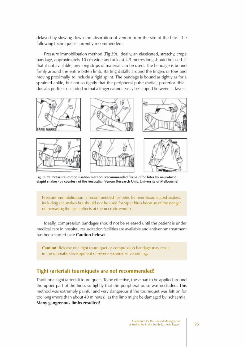
••••• Consider pressure-immobilisation (Fig 39) for some elapid bites
••••• Avoid any interference with the bite wound as this may introduce infection,increase absorption of the venom and increase local bleeding
As far as the snake is concerned – do not attempt to kill it as this may be dangerous.However, if the snake has already been killed, it should be taken to the dispensary orhospital with the patient in case it can be identified. However, do not handle thesnake with your bare hands as even a severed head can bite!
The special danger of rapidly developing paralytic envenoming afterbites by some elapid snakes: use of pressure-immobilisation
Bites by cobras, king cobras, kraits or sea snakes may lead, on rare occasions, to therapid development of life-threatening respiratory paralysis. This paralysis might be
25Guidelines for the Clinical Management
of Snake bite in the South-East Asia Region
delayed by slowing down the absorption of venom from the site of the bite. Thefollowing technique is currently recommended:
Pressure immobilisation method (Fig 39). Ideally, an elasticated, stretchy, crepebandage, approximately 10 cm wide and at least 4.5 metres long should be used. Ifthat it not available, any long strips of material can be used. The bandage is boundfirmly around the entire bitten limb, starting distally around the fingers or toes andmoving proximally, to include a rigid splint. The bandage is bound as tightly as for asprained ankle, but not so tightly that the peripheral pulse (radial, posterior tibial,dorsalis pedis) is occluded or that a finger cannot easily be slipped between its layers.
Pressure immobilisation is recommended for bites by neurotoxic elapid snakes,including sea snakes but should not be used for viper bites because of the dangerof increasing the local effects of the necrotic venom.
Ideally, compression bandages should not be released until the patient is undermedical care in hospital, resuscitation facilities are available and antivenom treatmenthas been started (see Caution below).
Caution: Release of a tight tourniquet or compression bandage may resultin the dramatic development of severe systemic envenoming.
Tight (arterial) tourniquets are not recommended!
Traditional tight (arterial) tourniquets. To be effective, these had to be applied aroundthe upper part of the limb, so tightly that the peripheral pulse was occluded. Thismethod was extremely painful and very dangerous if the tourniquet was left on fortoo long (more than about 40 minutes), as the limb might be damaged by ischaemia.Many gangrenous limbs resulted!
Figure 39: Pressure immobilisation method. Recommended first-aid for bites by neurotoxicelapid snakes (by courtesy of the Australian Venom Research Unit, University of Melbourne)
26Guidelines for the Clinical Managementof Snake bite in the South-East Asia Region
Viper and cobra bites
The pressure-immobilisation method as described above will increaseintracompartmental pressure and, by localising the venom, might be expected toincrease the locally-necrotic effects of viper venoms and some cobra venoms.
Pressure bandaging is not recommended for bites by vipers and cobras whosevenoms cause local necrosis.
The use of a local compression pad applied over the wound, without pressurebandaging of the entire bitten limb, has produced promising results in Myanmar anddeserves further study.
Arterial tourniquets are not recommended
4.2 Transport to hospitalThe patient must be transported to a place where they can receive medical care(dispensary or hospital) as quickly, but as safely and comfortably as possible. Anymovement, but especially movement of the bitten limb, must be reduced to anabsolute minimum to avoid increasing the systemic absorption of venom. Any muscularcontraction will increase this spread of venom from the site of the bite. A stretcher,bicycle, cart, horse, motor vehicle, train or boat should be used, or the patient shouldbe carried.
4.3 Treatment in the dispensary or hospital
Rapid clinical assessment and resuscitation
Cardiopulmonary resuscitation may be needed, including administration of oxygenand establishment of intravenous access. Airway, respiratory movements (Breathing)and arterial pulse (Circulation) must be checked immediately. The level ofconsciousness must be assessed.
The following are examples of clinical situations in which snake bite victimsmight require urgent resuscitation:
• Profound hypotension and shock resulting from direct cardiovascular effectsof the venom or secondary effects such as hypovolaemia or haemorrhagicshock.
• Terminal respiratory failure from progressive neurotoxic envenoming thathas led to paralysis of the respiratory muscles.
• Sudden deterioration or rapid development of severe systemic envenomingfollowing the release of a tight tourniquet or compression bandage (seeCaution above).
27Guidelines for the Clinical Management
of Snake bite in the South-East Asia Region
• Cardiac arrest precipitated by hyperkalaemia resulting from skeletal musclebreakdown (rhabdomyolysis) after sea snake bite.
• Late results of severe envenoming such as renal failure and septicaemiacomplicating local necrosis.
4.4 Detailed clinical assessment and species diagnosis
History
A precise history of the circumstances of the bite and the progression of local andsystemic symptoms and signs is very important. Three useful initial questions are:
“In what part of your body have you been bitten?”
The doctor can see immediately evidence that the patient has been bitten by asnake (eg fang marks) and the nature and extent of signs of local envenoming.
“When were you bitten?”
Assessment of the severity of envenoming depends on how long ago the patient wasbitten. If the patient has arrived at the hospital soon after the bite, there may be fewsymptoms and signs even though a large amount of venom may have been injected.
“Where is the snake that bit you?”
If the snake has been killed and brought, its correct identification can be very helpful.If it is obviously a harmless species (or not a snake at all!), the patient can be quicklyreassured and discharged from hospital.
Early clues that a patient has severe envenoming:
••••• Snake identified as a very dangerous one
••••• Rapid early extension of local swelling from the site of the bite
••••• Early tender enlargement of local lymph nodes, indicating spread of venom inthe lymphatic system
••••• Early systemic symptoms: collapse (hypotension, shock), nausea, vomiting,diarrhoea, severe headache, “heaviness” of the eyelids, inappropriate(pathological) drowsiness or early ptosis/ophthalmoplegia
••••• Early spontaneous systemic bleeding
••••• Passage of dark brown urine
Patients who become defibrinogenated or thrombocytopenic may begin to bleedfrom old, partially-healed wounds as well as bleeding persistently from the fang marks.
The patient should be asked how much urine has been passed since the bite andwhether it was a normal colour.
28Guidelines for the Clinical Managementof Snake bite in the South-East Asia Region
An important early symptom of sea snake envenoming that may develop as soonas 30 minutes after the bite is generalised pain, tenderness and stiffness of musclesand trismus.
Physical examination
This should start with careful assessment of the site of the bite and signs of localenvenoming.
Examination of the bitten part
The extent of swelling, which is usually also the extent of tenderness to palpation,should be recorded. Lymph nodes draining the limb should be palpated and overlyingecchymoses and lymphangitic lines noted.
A bitten limb may be tensely oedematous, cold, immobile and with impalpablearterial pulses. These appearances may suggest intravascular thrombosis, which isexceptionally rare after snake bite, or a compartmental syndrome, which isuncommon. If possible, intracompartmental pressure should be measured (seeAnnex 5) and the blood flow and patency of arteries and veins assessed (eg by dopplerultrasound).
Early signs of necrosis may include blistering, demarcated darkening (easilyconfused with bruising) or paleness of the skin, loss of sensation and a smell ofputrefaction (rotting flesh).
General examination
Measure the blood pressure (sitting up and lying to detect a postural drop indicativeof hypovolaemia) and heart rate. Examine the skin and mucous membranes forevidence of petechiae, purpura, ecchymoses and, in the conjunctivae, chemosis.Thoroughly examine the gingival sulci, using a torch and tongue depressor, as thesemay show the earliest evidence of spontaneous systemic bleeding. Examine the nosefor epistaxis. Abdominal tenderness may suggest gastrointestinal or retroperitonealbleeding. Loin (low back) pain and tenderness suggests acute renal ischaemia (Russell’sviper bites). Intracranial haemorrhage is suggested by lateralising neurological signs,asymmetrical pupils, convulsions or impaired consciousness (in the absence ofrespiratory or circulatory failure).
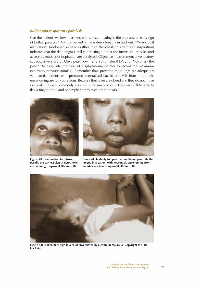
Neurotoxic envenoming
To exclude early neurotoxic envenoming, ask the patient to look up and observewhether the upper lids retract fully (Fig 40). Test eye movements for evidence ofearly external ophthalmoplegia (Fig 33). Check the size and reaction of the pupils.Ask the patient to open their mouth wide and protrude their tongue; early restrictionin mouth opening may indicate trismus (sea snake envenoming) or more often paralysisof pterygoid muscles (Fig 41). Check other muscles innervated by the cranial nerves(facial muscles, tongue, gag reflex etc). The muscles flexing the neck may be paralysed,giving the “broken neck sign” (Fig 42).
29Guidelines for the Clinical Management
of Snake bite in the South-East Asia Region
Bulbar and respiratory paralysis
Can the patient swallow or are secretions accumulating in the pharynx, an early signof bulbar paralysis? Ask the patient to take deep breaths in and out. “Paradoxicalrespiration” (abdomen expands rather than the chest on attempted inspiration)indicates that the diaphragm is still contracting but that the intercostal muscles andaccessory muscles of inspiration are paralysed. Objective measurement of ventilatorycapacity is very useful. Use a peak flow metre, spirometer (FEV1 and FVC) or ask thepatient to blow into the tube of a sphygmomanometer to record the maximumexpiratory pressure (mmHg). Remember that, provided their lungs are adequatelyventilated, patients with profound generalised flaccid paralysis from neurotoxicenvenoming are fully conscious. Because their eyes are closed and they do not moveor speak, they are commonly assumed to be unconscious. They may still be able toflex a finger or toe and so simple communication is possible.
Figure 40: Examination for ptosis,usually the earliest sign of neurotoxicenvenoming (Copyright DA Warrell)
Figure 42: Broken neck sign in a child envenomed by a cobra in Malaysia (Copyright the lateHA Reid)
Figure 41: Inability to open the mouth and protrude thetongue in a patient with neurotoxic envenoming fromthe Malayan krait (Copyright DA Warrell)
30Guidelines for the Clinical Managementof Snake bite in the South-East Asia Region
Do not assume that patients have irreversible brain damage because they areareflexic, unresponsive to painful stimuli, or have fixed dilated pupils.
Generalised rhabdomyolysis
In victims of envenoming by sea snakes and Russell’s vipers in Sri Lanka and SouthIndia, muscles, especially of the neck, trunk and proximal part of the limbs, maybecome tender and painful on active or passive movement and later may becomeparalysed. In sea snake bite there is pseudotrismus that can be overcome by sustainedpressure on the lower jaw. Myoglobinuria may be evident 3 hours after the bite.
Examination of pregnant women
There will be concern about fetal distress (revealed by fetal bradycardia), vaginalbleeding and threatened abortion. Monitoring of uterine contractions and fetal heartrate is useful. Lactating women who have been bitten by snakes should be encouragedto continue breast feeding.
Species diagnosis
If the dead snake has been brought, it can be identified. Otherwise, the speciesresponsible can be inferred indirectly form the patient’s description of the snake andthe clinical syndrome of symptoms and signs (see above and Annex 1 & 2). This isspecially important in Thailand where only monospecific antivenoms are available.
4.5 Investigations/laboratory tests
20 minute whole blood clotting test (20WBCT)
This very useful and informative bedside test requires very little skill and only onepiece of apparatus - a new, clean, dry, glass vessel (tube or bottle).
20 minute whole blood clotting test (20WBCT)
••••• Place a few mls of freshly sampled venous blood in a small glass vessel
••••• Leave undisturbed for 20 minutes at ambient temperature
••••• Tip the vessel once
••••• If the blood is still liquid (unclotted) and runs out, the patient has hypo-fibrinogenaemia (“incoagulable blood”) as a result of venom-inducedconsumption coagulopathy
••••• In the South East Asian region, incoagulable blood is diagnostic of a viper biteand rules out an elapid bite
••••• Warning! If the vessel used for the test is not made of ordinary glass, or if ithas been used before and cleaned with detergent, its wall may not stimulateclotting of the blood sample in the usual way and test will be invalid
••••• If there is any doubt, repeat the test in duplicate, including a “control” (bloodfrom a healthy person)
31Guidelines for the Clinical Management
of Snake bite in the South-East Asia Region
Other tests
Haemoglobin concentration/haematocrit: a transient increase indicateshaemoconcentration resulting from a generalised increase in capillary permeability(eg in Russell’s viper bite). More often, there is a decrease reflecting blood loss or, inthe case of Indian and Sri Lankan Russell’s viper bite, intravascular haemolysis.
Platelet count: this may be decreased in victims of viper bites.
White blood cell count: an early neutrophil leucocytosis is evidence of systemicenvenoming from any species.
Blood film: fragmented red cells (“helmet cell”, schistocytes) are seen whenthere is microangiopathic haemolysis.
Plasma/serum may be pinkish or brownish if there is gross haemoglobinaemiaor myoglobinaemia.
Biochemical abnormalities: aminotransferases and muscle enzymes (creatinekinase, aldolase etc) will be elevated if there is severe local damage or, particularly, ifthere is generalised muscle damage (Sri Lankan and South Indian Russell’s viperbites, sea snake bites). Mild hepatic dysfunction is reflected in slight increases inother serum enzymes. Bilirubin is elevated following massive extravasation of blood.Creatinine, urea or blood urea nitrogen levels are raised in the renal failure of Russell’sviper and saw-scaled viper bites and sea snake bites. Early hyperkalaemia may beseen following extensive rhabdomyolysis in sea snake bites. Bicarbonate will be lowin metabolic acidosis (eg enal failure).
Arterial blood gases and pH may show evidence of respiratory failure (neurotoxicenvenoming) and acidaemia (respiratory or metabolic acidosis).
Warning: arterial puncture is contraindicated in patients with haemostaticabnormalities (Viperidae)
Desaturation: arterial oxygen desaturation can be assessed non-invasively inpatients with respiratory failure or shock using a finger oximeter.
Urine examination: the urine should be tested by dipsticks for blood/haemoglobin/myoglobin. Standard dipsticks do not distinguish blood, haemoglobinand myoglobin. Haemoglobin and myoglobin can be separated by immunoassaysbut there is no easy or reliable test. Microscopy will confirm whether there areerythrocytes in the urine. Red cell casts indicate glomerular bleeding. Massiveproteinuria is an early sign of the generalised increase in capillary permeability inRussell’s viper envenoming.
32Guidelines for the Clinical Managementof Snake bite in the South-East Asia Region
4.6 Antivenom treatment
Antivenom is the only specific antidote to snake venom. A most important decisionin the management of a snake bite victim is whether or not to give antivenom.
What is antivenom?
Antivenom is immunoglobulin (usually the enzyme refined F(ab)2 fragment of IgG)purified from the serum or plasma of a horse or sheep that has been immunised withthe venoms of one or more species of snake. “Specific” antivenom, implies that theantivenom has been raised against the venom of the snake that has bitten the patientand that it can therefore be expected to contain specific antibody that will neutralisethat particular venom. Monovalent or monospecific antivenom neutralises the venomof only one species of snake. Polyvalent or polyspecific antivenom neutralises thevenoms of several different species of snakes, usually the most important species,from a medical point of view, in a particular geographical area. For example, Haffkine,Kasauli, Serum Institute of India and Bengal “polyvalent anti-snake venom serum” israised in horses using the venoms of t he four most important venomous snakes inIndia (Indian cobra, Naja naja; Indian krait, Bungarus caeruleus; Russell’s viper, Daboiarusselii; saw-scaled viper, Echis carinatus). Antibodies raised against the venom ofonespecies may have cross-neutralising activity against other venoms, usually from closelyrelated species. This is known as paraspecific activity. For example, the manufacturersof Haffkine polyvalent anti-snake venom serum claim that this antivenom alsoneutralises venoms of two Trimeresurus species.
Indications for antivenom treatment (see also Annex 1 & 2)
Antivenom treatment carries a risk of severe adverse reactions and in most countriesit is costly and may be in limited supply. It should therefore be used only in patientsin whom the benefits of antivenom treatment are considered to exceed the risks.
Indications for antivenom vary in different countries.
Inappropriate use of antivenom
In some parts of the world, antivenom is given to any patient claiming to have beenbitten by a snake, irrespective of symptoms or signs of envenoming. Sometimes thelocal community are so frightened of snake bite that they compel the doctor to giveantivenom against medical advice. These practices should be strongly discouragedas they expose patients who may not need treatment to the risks of antivenomreactions; they also waste valuable and scarce stocks of antivenom.
33Guidelines for the Clinical Management
of Snake bite in the South-East Asia Region
Indications for antivenom
Antivenom treatment is recommended if and when a patient with proven orsuspected snake develops one or more of the following signs
Systemic envenoming
••••• Haemostatic abnormalities: spontaneous systemic bleeding (clinical),coagulopathy (20WBCT or other laboratory) or thrombocytopenia (<100 x109/litre) (laboratory)
••••• Neurotoxic signs: ptosis, external ophthalmoplegia, paralysis etc (clinical)
••••• Cardiovascular abnormalities: hypotension, shock, cardiac arrhythmia (clinical),abnormal ECG
••••• Acute renal failure: oliguria/anuria (clinical), rising blood creatinine/ urea(laboratory)
••••• (Haemoglobin-/myoglobin-uria:) dark brown urine (clinical), urine dipsticks, otherevidence of intravascular haemolysis or generalised rhabdomyolysis (muscleaches and pains, hyperkalaemia) (clinical, laboratory)
••••• Supporting laboratory evidence of systemic envenoming (see 4.5, page 30)
Local envenoming
••••• Local swelling involving more than half of the bitten limb (in the absence of atourniquet) Swelling after bites on the digits (toes and especially fingers)
••••• Rapid extension of swelling (for example beyond the wrist or ankle within afew hours of bites on the hands or feet)
••••• Development of an enlarged tender lymph node draining the bitten limb
How long after the bite can antivenom be expected to beeffective?
Antivenom treatment should be given as soon as it is indicated. It may reverse systemicenvenoming even when this has persisted for several days or, in the case of haemostaticabnormalities, for two or more weeks. However, when there are signs of localenvenoming, without systemic envenoming, antivenom will be effective only if itcan be given within the first few hours after the bite.
Prediction of antivenom reactions
Skin and conjunctival “hypersensitivity” tests may reveal IgE mediated Type Ihypersensitivity to horse or sheep proteins but do not predict the large majority ofearly (anaphylactic) or late (serum sickness type) antivenom reactions. Since theymay delay treatment and can in themselves be sensitizing, these tests should notbe used.
34Guidelines for the Clinical Managementof Snake bite in the South-East Asia Region
Contraindications to antivenom
There is no absolute contraindication to antivenom treatment, but patients whohave reacted to horse (equine) or sheep (ovine) serum in the past (for example aftertreatment with equine anti-tetanus serum, equine anti-rabies serum or equine orovine antivenom) and those with a strong history of atopic diseases (especially severeasthma) should be given antivenom only if they have signs of systemic envenoming.
Prophylaxis in high risk patients
In the absence of any prophylactic regimen that has proved effective in clinical trials,these high risk patients may be pre-treated empirically with subcutaneous epinephrine(adrenaline), intravenous antihistamines (both anti-H1, such as promethazine orchloramphenicol; and anti- H2, such as cimetidine or ranitidine) and corticosteroid.In asthmatic patients, prophylactic use of an inhaled adrenergic ∃ 2 agonist such assalbutamol may prevent bronchospasm.
Selection of antivenom
Antivenom should be given only if its stated range of specificity includes the speciesknown or thought to have been responsible for the bite. Liquid antivenoms that havebecome opaque should not be used as precipitation of protein indicates loss ofactivity and an increased risk of reactions.
Expiry dates quoted by manufacturers are often very conservative. Provided thatantivenom has been properly stored, it can be expected to retain useful activity formany months after the stated “expiry date”.
If the biting species is known, the ideal treatment is with a monospecific/monovalent antivenom, as this involves administration of a lower dose of antivenomprotein than with a polyspecific/ polyvalent antivenoms. Polyspecific/polyvalentantivenoms are preferred in many countries because of the difficulty in identifyingspecies responsible for bites. Polyspecific antivenoms can be just as effective asmonospecific ones, but since they contain specific antibodies against several differentvenoms, a larger dose of antivenom protein must be administered to neutralise aparticular venom.
Administration of antivenom
• Epinephrine (adrenaline) should always be drawn up in readiness beforeantivenom is administered.
• Antivenom should be given by the intravenous route whenever possible.
Freeze-dried (lyophilised) antivenoms are reconstituted, usually with 10 ml of sterilewater for injection per ampoule. The freeze-dried protein may be difficult to dissolve.Two methods of administration are recommended:
35Guidelines for the Clinical Management
of Snake bite in the South-East Asia Region
(1) Intravenous “push” injection: reconstituted freeze-dried antivenom or neatliquid antivenom is given by slow intravenous injection (not more than 2ml/minute). This method has the advantage that the doctor/nurse/dispensergiving the antivenom must remain with the patient during the time whensome early reactions may develop. It is also economical, saving the use ofintravenous fluids, giving sets, cannulae etc.
(2) Intravenous infusion: reconstituted freeze-dried or neat liquid antivenom isdiluted in approximately 5-10 ml of isotonic fluid per kg body weight (ie250-500 ml of isotonic saline or 5% dextrose in the case of an adult patient)and is infused at a constant rate over a period of about one hour.
Local administration of antivenom at the site of the bite is notrecommended!
Although this route may seem rational, it should not be used as it is extremely painful,may increase intracompartmental pressure and has not been shown to be effective.
Intramuscular injection of antivenom
Antivenoms are large molecules (F(ab )2 fragments or sometimes whole IgG) which,after intramuscular injection, are absorbed slowly via lymphatics. Bioavailability ispoor, especially after intragluteal injection and blood levels of antivenom never reachthose achieved rapidly by intravenous administration. Other disadvantages are thepain of injection of large volumes of antivenom and the risk of haematoma formationin patients with haemostatic abnormalities.
Antivenom must never be given by the intramuscular route if it could be givenintravenously.
Situations in which intramuscular administration might be considered :
• at a peripheral first aid station, before a patient with obvious envenoming isput in an ambulance for a journey to hospital that may last several hours;
• on an expedition exploring a remote area very far from medical care;
• when intravenous access has proved impossible.
Although the risk of antivenom reactions is less with intramuscular than intravenousadministration, epinephrine (adrenaline) must be readily available. Patients must beclosely observed for at least one hour after starting intravenous antivenomadministration, so that early anaphylactic antivenom reactions can be detected andtreated early with epinephrine (adrenaline).
Under these unusual circumstances, the dose of antivenom should be dividedbetween a number of sites in the upper anterolateral region of both thighs. A maximumof 5-10 ml should be given at each site by deep intramuscular injection followed bymassage to aid absorption. Local bleeding and haematoma formation is a problem inpatients with incoagulable blood.
36Guidelines for the Clinical Managementof Snake bite in the South-East Asia Region
Finding enough muscle mass to contain such large volumes of antivenom isparticularly difficult in children.
Antivenom should never be injected into the gluteal region (upper outer quadrantof the buttock) as absorption is exceptionally slow and unreliable and there isalways the danger of sciatic nerve damage when the injection is given by aninexperienced operator.
Dose of antivenom
Snakes inject the same dose of venom into children and adults. Children musttherefore be given exactly the same dose of antivenom as adults.
Manufacturers’ recommendations are usually based on inappropriate animal tests inwhich venom and antivenom are incubated before being injected into the test animal.The recommended dose is often the amount of antivenom required to neutralise theaverage venom yield when captive snakes are milked of their venom. In practice, thechoice of an initial dose of antivenom is usually empirical.
Antivenom manufacturers, health institutions and medical research organisationsshould encourage and promote the proper clinical testing of antivenoms as withother therapeutic agents. This is the only reliable guide to the initial dose (andsafety) of an antivenom.
Since the neutralising power of antivenoms varies from batch to batch, the resultsof a particular clinical trial may soon become obsolete if the manufacturers changethe strength of the antivenom.
Antivenom reactions
A proportion of patients, usually more than 20%, develop a reaction either early(within a few hours) or late (5 days or more) after being given antivenom.
Early anaphylactic reactions: usually within 10-180 minutes of starting antivenom,the patient begins to itch (often over the scalp) and develops urticaria, dry cough,fever, nausea, vomiting, abdominal colic, diarrhoea and tachycardia. A minority ofthese patients may develop severe life-threatening anaphylaxis: hypotension,bronchospasm and angio-oedema. Fatal reactions have probably been under-reportedas death after snake bite is usually attributed to the venom.
In most cases, these reactions are not truly “allergic”. They are not IgE-mediatedtype I hypersensitivity reactions to horse or sheep proteins as there is no evidence ofspecific IgE, either by skin testing or radioallergosorbent tests (RAST). Complementactivation by IgG aggregates or residual Fc fragments or direct stimulation of mastcells or basophils by antivenom protein are more likely mechanisms for these reactions.
37Guidelines for the Clinical Management
of Snake bite in the South-East Asia Region
Pyrogenic (endotoxin) reactions usually develop 1-2 hours after treatment.Symptoms include shaking chills (rigors), fever, vasodilatation and a fall in bloodpressure. Febrile convulsions may be precipitated in children. These reactions arecaused by pyrogen contamination during the manufacturing process. They arecommonly reported.
Late (serum sickness type) reactions develop 1-12 (mean 7) days after treatment.Clinical features include fever, nausea, vomiting, diarrhoea, itching, recurrent urticaria,arthralgia, myalgia, lymphadenopathy, periarticular swellings, mononeuritis multiplex,proteinuria with immune complex nephritis and rarely encephalopathy. Patients whosuffer early reactions and are treated with antihistamines and corticosteroid are lesslikely to develop late reactions.
Treatment of early anaphylactic and pyrogenic antivenomreactions
Epinephrine (adrenaline) is given intramuscularly (into the deltoid muscle or theupper lateral thigh) in an initial dose of 0.5 mg for adults, 0.01 mg/kg body weight forchildren. Severe, life-threatening anaphylaxis can evolve very rapidly and soepinephrine (adrenaline) should be given at the very first sign of a reaction, evenwhen only a few spots of urticaria have appeared or at the start of itching, tachycardiaor restlessness. The dose can be repeated every 5-10 minutes if the patient’s conditionis deteriorating.
At the earliest sign of a reaction:
••••• Antivenom administration must be temporarily suspended
••••• Epinephrine (adrenaline) (0.1% solution, 1 in 1,000, 1 mg/ml) is the effectivetreatment for early anaphylactic and pyrogenic antivenom reactions
Additional treatment
After epinephrine (adrenaline), an anti H1 antihistamine such as chlorpheniraminemaleate (adults 10 mg, children 0.2 mg/kg by intravenous injection over a few minutes)should be given followed by intravenous hydrocortisone (adults 100 mg, children2 mg/kg body weight). The corticosteroid is unlikely to act for several hours, but mayprevent recurrent anaphylaxis.
There is increasing evidence that anti H2 antihistamines such as cimetidine orranitidine have a role in the treatment of severe anaphylaxis. Both drugs are given,diluted in 20 ml isotonic saline, by slow intravenous injection (over 2 minutes).
Doses: cimetidine – adults 200 mg, children 4 mg/kg;ranitidine – adults 50 mg, children 1 mg/kg.
In pyrogenic reactions the patient must also be cooled physically and withantipyretics (for example paracetamol by mouth or suppository). Intravenous fluidsshould be given to correct hypovolaemia.
38Guidelines for the Clinical Managementof Snake bite in the South-East Asia Region
Treatment of late (serum sickness) reactions
Late (serum sickness) reactions usually respond to a 5-day course of oral antihistamine.Patients who fail to respond in 24-48 hours should be given a 5-day course ofprednisolone.
Doses: Chlorpheniramine: adults 2 mg six hourly, children 0.25 mg/kg /dayin divided doses
Prednisolone: adults 5 mg six hourly, children 0.7 mg/kg/day in divided doses for5-7 days
Observation of the response to antivenom
If an adequate dose of appropriate antivenom has been administered, the followingresponses may be seen.
• General: the patient feels better. Nausea, headache and generalised achesand pains may disappear very quickly. This may be partly attributable to aplacebo effect.
• Spontaneous systemic bleeding (eg from the gums) usually stops within 15-30 minutes.
• Blood coagulability (as measured by 20WBCT) is usually restored in 3-9hours. Bleeding from new and partly healed wounds usually stops muchsooner than this.
• In shocked patients, blood pressure may increase within the first 30-60minutes and arrhythmias such as sinus bradycardia may resolve.
• Neurotoxic envenoming of the post-synaptic type (cobra bites) may beginto improve as early as 30 minutes after antivenom, but usually take severalhours. Envenoming with presynaptic toxins (kraits and sea snakes) is unlikelyto respond in this way.
• Active haemolysis and rhabdomyolysis may cease within a few hours andthe urine returns to its normal colour.
Recurrence of systemic envenoming
In patients envenomed by vipers, after an initial response to antivenom (cessation ofbleeding, restoration of blood coagulability), signs of systemic envenoming may recurwithin 24-48 hours.
This is attributable to:
(1) continuing absorption of venom from the “depot” at the site of the bite,perhaps assisted by improved blood supply following correction of shock,hypovolaemia etc, after elimination of antivenom (range of elimination half-lives: IgG 45 hours; F(ab)2 80-100 hours; Fab 12-18 hours);
(2) a redistribution of venom from the tissues into the vascular space, as theresult of antivenom treatment.
Recurrent neurotoxic envenoming after treatment of cobra bite has also beendescribed.
39Guidelines for the Clinical Management
of Snake bite in the South-East Asia Region
Criteria for repeating the initial dose of antivenom
Criteria for giving more antivenom
••••• Persistence or recurrence of blood incoagulability after 6 hr of bleeding after1-2 hr
••••• Deteriorating neurotoxic or cardiovascular signs after 1-2 hr
If the blood remains incoagulable (as measured by 20WBCT) six hours after theinitial dose of antivenom, the same dose should be repeated. This is based on theobservation that, if a large dose of antivenom (more than enough to neutralise thevenom procoagulant enzymes) is given initially, the time taken for the liver to restorecoagulable levels of fibrinogen and other clotting factors is 3-9 hours.
In patients who continue to bleed briskly, the dose of antivenom should berepeated within 1-2 hours.
In case of deteriorating neurotoxicity or cardiovascular signs, the initial doseof antivenom should be repeated after 1-2 hours, and full supportive treatment mustbe considered.
Conservative treatment when no antivenom is available
This will be the situation in many parts of the region, where supplies of antivenomrun out or where the bite is known to have been inflicted by a species against whosevenom there is no available specific antivenom (for example for bites by the Malayankrait (Bungarus candidus), coral snakes - genera Calliophis and Maticora), sea snakes,the mangrove/shore pit viper T purpureomaculatus and the mountain pit viper Ovophismonticola.
The following conservative measures are suggested:
Neurotoxic envenoming with respiratory paralysis: assisted ventilation. Thishas proved effective, and has been followed by complete recovery, even after beingmaintained for periods of more than one month. Manual ventilation (anaestheticbag) by relays of doctors, medical students, relatives and nurses has been effectivewhere no mechanical ventilator was available. Anticholinesterases should always betried (see below Trial of anticholinesterase, p 41).
Haemostatic abnormalities – strict bed rest to avoid even minor trauma;transfusion of clotting factors and platelets; ideally, fresh frozen plasma andcryoprecipitate with platelet concentrates or, if these are not available, fresh wholeblood. Intramuscular injections should be avoided.
Shock, myocardial damage: hypovolaemia should be corrected with colloid/crystalloids, controlled by observation of the central venous pressure. Ancillary pressor
40Guidelines for the Clinical Managementof Snake bite in the South-East Asia Region
drugs (dopamine or epinephrine-adrenaline) may also be needed. Patients withhypotension associated with bradycardia should be treated with atropine.
Renal failure: conservative treatment or dialysis (see Oliguria and renal failure,page 42).
Dark brown urine (myoglobinuria or haemoglobinuria): correct hypovolaemiaand acidosis and consider a single infusion of mannitol (see Prevention of renal damagein patients with myoglobinuria or haemoglobinuria, page 45 ).
Severe local envenoming: local necrosis, intracompartmental syndromes andeven thrombosis of major vessels is more likely in patients who cannot be treatedwith antivenom. Surgical intervention may be needed but the risks of surgery in apatient with consumption coagulopathy, thrombocytopenia and enhanced fibrinolysismust be balanced against the lifethreatening complications of local envenoming.Prophylactic broad spectrum antimicrobial treatment is justified (see Bacterialinfections, page 46).
4.7 Supportive/ancillary treatmentAntivenom treatment can be expected to neutralise free circulating venom, preventprogression of envenoming and allow recovery. However, these processes take timeand the severely envenomed patient may require life support systems such as assistedventilation and renal dialysis until the severely damaged organs and tissues have hadtime to recover.
Dangers of venepuncture in patients with haemostaticabnormalities
In patients with incoagulable blood, any injection (subcutaneous, intramuscular) and,particularly venepuncture, carries a risk of persistent bleeding and haematomaformation. Arterial puncture is contraindicated in such patients.
Repeated venepuncture can be avoided by using an indwelling cannula and three-way tap system. When blood coagulability has been restored, the dead space shouldbe filled with heparinised saline, but beware! If this is not flushed out before bloodsampling, misleading results will be obtained in clotting tests, including the 20WBCT.
In patients with coagulopathy, sites of venous access and placement of intravenouscannulae or catheters should be chosen where haemostasis by external pressure ismost likely to be effective, eg the antecubital fossa. If possible, avoid jugular, subclavianand femoral vein puncture. A pressure pad must be applied at the site of anyvenepuncture.
Neurotoxic envenoming
Antivenom treatment alone cannot be relied upon to save the life of a patient withbulbar and respiratory paralysis.
41Guidelines for the Clinical Management
of Snake bite in the South-East Asia Region
Death may result from aspiration, airway obstruction or respiratory failure. Aclear airway must be maintained. Once there is loss of gag reflex and pooling ofsecretions in the pharynx, failure of the cough reflex or respiratory distress, a cuffedendotracheal tube should be inserted. If this is impossible for any reason, atracheostomy should be performed and a snugly-fitting or cuffed tracheostomy tubeinserted.
Although artificial ventilation was first suggested for neurotoxic envenoming 125years ago, patients continue to die of asphyxiation because some doctors believethat antivenom is sufficient treatment.
Anticholinesterase drugs have a variable, but potentially very useful effect inpatients with neurotoxic envenoming, especially those bitten by cobras.
A trial of anticholinesterase (eg “Tensilon test”) should be performed in everypatient with neurotoxic envenoming, as it would be in any patient with suspectedmyasthenia gravis.
Trial of anticholinesterase
Anticholinesterase (eg “Tensilon”/edrophonium) test
••••• Baseline observations
••••• Give atropine intravenously
••••• Give anticholinesterase drug
••••• Observe effect
••••• If positive, institute regular atropine and (long acting) anticholinesterase
Ideally, a short acting anticholinesterase, such as edrophonium (“Tensilon”), shouldbe used. Baseline observations or measurements are made against which to assessthe effectiveness of the anticholinesterase. Atropine sulphate (adults 0.6 mg, children50 µg/kg body weight) is given by intravenous injection (to prevent the undesirablemuscarinic effects of acetylcholine such as increased secretions, sweating, bradycardiaand colic) followed immediately by edrophonium chloride (adults 10 mg, children0.25 mg/kg body weight) given intravenously over 3 or 4 minutes. The patient isobserved over the next 10-20 minutes for signs of improved neuromusculartransmission. Ptosis may disappear (Fig 43) and ventilatory capacity (peak flow, FEV1or maximum expiratory pressure) may improve.
If edrophonium chloride is not available, any other anticholinesterases(neostigmine – “Prostigmine”, distigmine, pyridostigmine, ambenomium) can be usedfor this assessment but a longer period of observation will be needed (up to 1 hour).
Patients who respond convincingly can be maintained on a longer-actinganticholinesterase such as neostigmine methylsulphate combined with atropine.
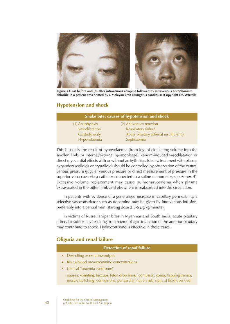
42Guidelines for the Clinical Managementof Snake bite in the South-East Asia Region
Hypotension and shock
Snake bite: causes of hypotension and shock
(1) Anaphylaxis (2) Antivenom reactionVasodilatation Respiratory failureCardiotoxicity Acute pituitary adrenal insufficiencyHypovolaemia Septicaemia
This is usually the result of hypovolaemia (from loss of circulating volume into theswollen limb, or internal/external haemorrhage), venom-induced vasodilatation ordirect myocardial effects with or without arrhythmias. Ideally, treatment with plasmaexpanders (colloids or crystalloid) should be controlled by observation of the centralvenous pressure (jugular venous pressure or direct measurement of pressure in thesuperior vena cava via a catheter connected to a saline manometer, see Annex 4).Excessive volume replacement may cause pulmonaryoedema when plasmaextravasated in the bitten limb and elsewhere is reabsorbed into the circulation.
In patients with evidence of a generalised increase in capillary permeability, aselective vasoconstrictor such as dopamine may be given by intravenous infusion,preferably into a central vein (starting dose 2.5-5 µg/kg/minute).
In victims of Russell’s viper bites in Myanmar and South India, acute pituitaryadrenal insufficiency resulting from haemorrhagic infarction of the anterior pituitarymay contribute to shock. Hydrocortisone is effective in these cases.
Oliguria and renal failure
Detection of renal failure
••••• Dwindling or no urine output
••••• Rising blood urea/creatinine concentrations
••••• Clinical “uraemia syndrome”
nausea, vomiting, hiccups, fetor, drowsiness, confusion, coma, flapping tremor,muscle twitching, convulsions, pericardial friction rub, signs of fluid overload
Figure 43: (a) before and (b) after intravenous atropine followed by intravenous edrophoniumchloride in a patient envenomed by a Malayan krait (Bungarus candidus) (Copyright DA Warrell)
43Guidelines for the Clinical Management
of Snake bite in the South-East Asia Region
In patients with any of these features, the following should be monitored
• pulse rate
• blood pressure, lying and sitting, to detect postural hypotension
• respiratory rate
• temperature
• height of jugular venous pulse
• auscultation of lung bases for crepitations
Oliguric phase of renal failure
Most, but not all, patients with acute renal failure are oliguric, defined as a urineoutput of less than 400 ml/day or less than 20 ml/hour. Conservative managementmay tide the patient over, avoiding the need for dialysis. If the patient is hypovolaemic,indicated by supine or postural hypotension, empty neck veins, sunken eyeballs, lossof skin turgor and dryness of mucosae, proceed as follows:
(1) Establish intravenous access
(2) Insert a urethral catheter (full sterile precautions!)
(3) Determine the central venous pressure. This can be achieved either byobserving the vertical height of the jugular venous pulsation above the sternalangle with the patient propped up on pillows at 45o; or by directmeasurement of central venous (superior vena caval) pressure through along catheter preferably inserted at the antecubital fossa (see Annex 4). Thecatheter is connected to a saline manometer, the 0 point of which must beplaced at the same level as the right atrium (that is, at the sternal anglewhen the patient is propped up at 45o). In someone who is obviously volume-depleted, resuscitation should start immediately, and not be delayed until acentral venous line has been inserted.
(4) Fluid challenge: depending on the initial state of hydration/dehydration,an adult patient can be given two litres of isotonic saline over one hour or,until the jugular venous pressure/central venous pressure has risen to 8-10cm above the sternal angle (with the patient propped up at 45o). The patientmust be closely observed while this is being done. The fluid challenge mustbe stopped immediately if pulmonary oedema develops. If the urine outputdoes not improve, try furosamide challenge.
(5) Furosamide (frusemide) challenge: 100 mg of furosamide is injected slowly(4-5 mg/minute). If this does not induce a urine output of 40 ml/hour, givea second dose of furosamide, 200 mg. If urine output does not improve, trymannitol challenge.
(6) Mannitol challenge: 200 ml of 20% mannitol may be infused intravenouslyover 20 minutes but this must not be repeated as there is a danger ofinducing dangerous fluid and electrolyte imbalance. An improvement inurine output to more than 40 ml/hr or more than 1 litre/day is consideredsatisfactory.
(7) Conservative management: If the urine output does not improve, despitethese challenges, no further diuretics should be given and fluid intake should
44Guidelines for the Clinical Managementof Snake bite in the South-East Asia Region
be restricted to a total of the previous day’s output plus “insensible losses”(500-1000 ml/day). If possible, the patient should be referred to a renalunit. The diet should be bland, high in calories (1700/day), low in protein(less than 40g/day), low in potassium (avoid fruit, fruit juices and potassium-containing drugs) and low in salt. Infections will cause tissue breakdownand increase urea levels. They should be prevented or treated promptlywith non-nephrotoxic antibiotics (ie avoid aminoglycosides such asgentamicin).
(8) Biochemical monitoring: Serum potassium, urea, creatinine and, if possible,pH, bicarbonate, calcium and phosphate should be monitored frequently.If this is not possible the electrocardiogram (ECG) should be examined forevidence of hyperkalaemia, especially following bites by sea snakes, or SriLankan or South Indian Russell’s vipers or if the patient is passing darkbrown urine, indicating rhabdomyolysis or intravascular haemolysis.
ECG evidence of hyperkalaemia: tall peaked T waves, prolonged P-Rinterval, absent P waves, wide QRS complexes.
Emergency treatment of hyperkalaemia(serum potassium >6.5 mmol/l or ECG changes)
••••• give 10 ml of 10% calcium gluconate intravenously over 2 minutes(with ECG monitoring if possible) repeated up to three times
••••• give 50 ml of 50% dextrose with 10 units of soluble insulin intravenously
••••• sodium bicarbonate (40 ml of 8.4%) by slow intravenous infusion anda â2 agonist aerosol by inhaler (eg salbutamol – “Ventolin” 5-10 mg)may also be used
These emergency treatments will control hyperkalaemia for 3-6 hoursonly. If the patient is hypotensive and profoundly acidotic (deep sighing“Kussmaul” respirations, very low plasma bicarbonate concentration or verylow pH - <7.10), 40 ml of 8.4% sodium bicarbonate (1 mmol/ml) may beinfused intravenously over 30 minutes. If this leads to circulatoryimprovement, the dose can be repeated.
Caution: Intravenous bicarbonate may precipitate profoundhypocalcaemia and fitting, especially in patients with rhabdomyolysis.
(9) Dialysis
Indications for dialysis
••••• Clinical uraemia
••••• Fluid overload
••••• Blood biochemistry – one or more of the followingcreatinine >6 mg/dl (500 µmol/l)urea >200 mg/dl (400 mmol/l)potassium >7 mmol/l (or hyperkalaemic ECG changes)symptomatic acidosis
45Guidelines for the Clinical Management
of Snake bite in the South-East Asia Region
Prevention of renal damage in patients with myoglobinuria orhaemoglobinuria
To minimise the risk of renal damage from excretedmyoglobin and/or haemoglobin:
••••• correct hypovolaemia (see above) and maintain saline diuresis (if possible)
••••• correct severe acidosis with bicarbonate (see above)
••••• give a single infusion of mannitol (200 ml of 20% solution over 20 minutes)
Diuretic phase of renal failure
Urine output increases following the period of anuria. The patient may becomepolyuric and volume depleted so that salt and water must be carefully replaced.Hypokalaemia may develop, in which case a diet rich in potassium (fruit and fruitjuices) is recommended.
Renal recovery phase
The diuretic phase may last for months after Russell’s viper bite. In Myanmar andSouth India, hypopituitarism may complicate recovery of Russell’s viper bite victims.Corticosteroid, fluid and electrolyte replacement may be needed in these patients.
Persisting renal dysfunction
In Myanmar, persistent tubular degenerative changes were observed in Russell’s viperbite victims who showed continuing albuminuria, hypertension and nocturia for upto 11 months after the bite, despite apparent recovery in renal function. In India, 20-25% of patients referred to renal units with acute renal failure following Russell’sviper bite suffered oliguria for more than 4 weeks suggesting the possibility of bilateralrenal cortical necrosis. This can be confirmed by renal biopsy or contrast enhancedCT scans of the kidneys. Patients with patchy cortical necrosis show delayed andpartial recovery of renal function but those with diffuse cortical necrosis requireregular maintenance dialysis and eventual renal transplantation.
Haemostatic disturbances
Bleeding and clotting disturbances usually respond satisfactorily to treatment withspecific antivenom, but the dose may need to be repeated several times, at six hourlyintervals, before blood coagulability (assessed by the 20WBCT) is finally andpermanently restored.
In exceptional circumstances, such as severe bleeding or imminent urgent surgery,once specific antivenom has been given to neutralise venom procoagulants and otherantihaemostatic toxins, restoration of coagulability and platelet function can beaccelerated by giving fresh frozen plasma, cryoprecipitate (fibrinogen, factor VIII),fresh whole blood or platelet concentrates.
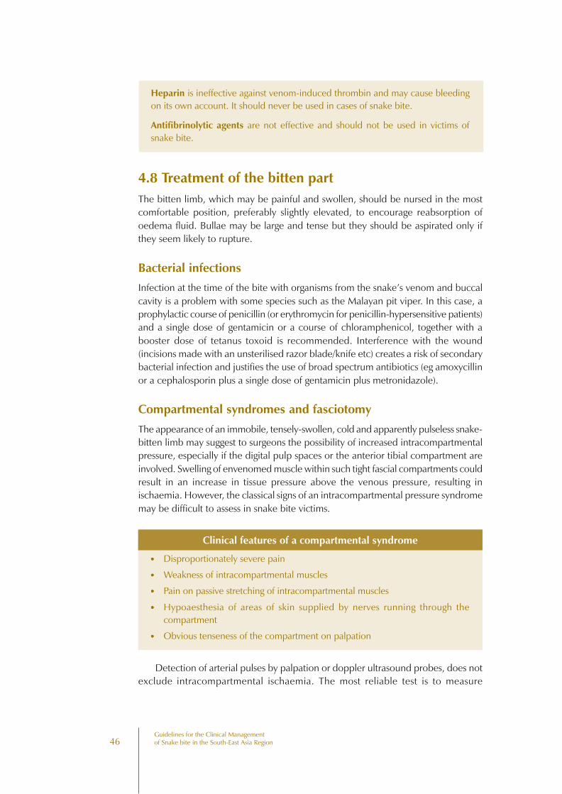
46Guidelines for the Clinical Managementof Snake bite in the South-East Asia Region
Heparin is ineffective against venom-induced thrombin and may cause bleedingon its own account. It should never be used in cases of snake bite.
Antifibrinolytic agents are not effective and should not be used in victims ofsnake bite.
4.8 Treatment of the bitten partThe bitten limb, which may be painful and swollen, should be nursed in the mostcomfortable position, preferably slightly elevated, to encourage reabsorption ofoedema fluid. Bullae may be large and tense but they should be aspirated only ifthey seem likely to rupture.
Bacterial infections
Infection at the time of the bite with organisms from the snake’s venom and buccalcavity is a problem with some species such as the Malayan pit viper. In this case, aprophylactic course of penicillin (or erythromycin for penicillin-hypersensitive patients)and a single dose of gentamicin or a course of chloramphenicol, together with abooster dose of tetanus toxoid is recommended. Interference with the wound(incisions made with an unsterilised razor blade/knife etc) creates a risk of secondarybacterial infection and justifies the use of broad spectrum antibiotics (eg amoxycillinor a cephalosporin plus a single dose of gentamicin plus metronidazole).
Compartmental syndromes and fasciotomy
The appearance of an immobile, tensely-swollen, cold and apparently pulseless snake-bitten limb may suggest to surgeons the possibility of increased intracompartmentalpressure, especially if the digital pulp spaces or the anterior tibial compartment areinvolved. Swelling of envenomed muscle within such tight fascial compartments couldresult in an increase in tissue pressure above the venous pressure, resulting inischaemia. However, the classical signs of an intracompartmental pressure syndromemay be difficult to assess in snake bite victims.
Clinical features of a compartmental syndrome
••••• Disproportionately severe pain
••••• Weakness of intracompartmental muscles
••••• Pain on passive stretching of intracompartmental muscles
••••• Hypoaesthesia of areas of skin supplied by nerves running through thecompartment
••••• Obvious tenseness of the compartment on palpation
Detection of arterial pulses by palpation or doppler ultrasound probes, does notexclude intracompartmental ischaemia. The most reliable test is to measure
47Guidelines for the Clinical Management
of Snake bite in the South-East Asia Region
Figure 44: Disastrous results of unnecessary fasciotomy in snake bite victims (a) profusebleeding in a patient with mild local envenoming but severe coagulopathy following a bite by agreen pit viper (Trimeresurus albolabris) (Copyright Sornchai Looareesuwan). Disastrous resultsof unnecessary fasciotomy in snake bite victims (b) Persistent bleeding for 10 days, resulting inhaemorrhagic shock despite transfusion of 20 unites of blood, in a victim of Malayan pit viperbite in whom fasciotomy was performed before adequate antivenom treatment had been given tocorrect the coagulopathy (Copyright DA Warrell). (Right) Disastrous results of unnecessaryfasciotomy in snake bite victims (c) Residual skin loss and exposure of tendons followingfasciotomy for mild local envenoming in a patient bitten by a green pit viper (Trimeresurusalbolabris) (Copyright Sornchai Looareesuwan)
intracompartmental pressure directly through a cannula introduced into thecompartment and connected to a pressure transducer or manometer (Annex 5). Inorthopaedic practice, intracompartmental pressures exceeding 40 mmHg (less inchildren) may carry a risk of ischaemic necrosis (eg Volkmann’s ischaemia or anteriortibial compartment syndrome). However, fasciotomy should not be contemplateduntil haemostatic abnormalities have been corrected, otherwise the patient maybleed to death (Fig 44). Animal studies have suggested that muscle sufficientlyenvenomed and swollen to cause intracompartmental syndromes, may already beirreversibly damaged by the direct effects of the venom. Early treatment withantivenom remains the best way of preventing irreversible muscle damage.
Criteria for fasciotomy in snake-bitten limbs
Haemostatic abnormalities have been corrected (antivenom with or without clottingfactors)••••• clinical evidence of an intracompartmental syndrome
••••• intracompartmental pressure >40 mmHg (in adults)
4.9 RehabilitationRestoration of normal function in the bitten part after the patient has been dischargedfrom hospital is not usually supervised. Conventional physiotherapy may wellaccelerate this process. In patients with severe local envenoming, the limb should bemaintained in a functional position. For example, in the leg, equinus deformity ofthe ankle should be prevented by application of a back slab.

48Guidelines for the Clinical Managementof Snake bite in the South-East Asia Region
49Guidelines for the Clinical Management
of Snake bite in the South-East Asia Region
Management of CobraSpit Ophthalmia
5
First aid consists of irrigating the affected eyes and other mucous membranes withliberal quantities of water or any other available bland liquid. Instillation of 0.5%adrenaline drops relieves pain and inflammation. In view of the risk of corneal abrasion,fluorescein staining or slit lamp examination is essential. Otherwise, topicalantimicrobials (tetracycline or chloramphenicol) should be applied to preventendophthalmitis or blinding corneal opacities. Some ophthalmologists recommendthe use of a dressing pad to close the eye.
The instillation of diluted antivenom may cause local irritation and is of uncertainbenefit. It is not recommended.

50Guidelines for the Clinical Managementof Snake bite in the South-East Asia Region
51Guidelines for the Clinical Management
of Snake bite in the South-East Asia Region
Conclusions and MainRecommendations
6
1. It is clear that in many parts of the South East Asian region, snake bite is animportant medical emergency and cause of hospital admission. It results in thedeath or chronic disability of many active younger people, especially thoseinvolved in farming and plantation work. However, the true scale of mortalityand acute and chronic morbidity from snake bite remains uncertain because ofinadequate reporting in almost every part of the region.
To remedy this deficiency, it is strongly recommended that snake bite should bemade a specific notifiable disease in all countries in the South East Asian region.
2. Snake bite is an occupational disease of farmers, plantation workers, herdsmen,fishermen and other food producers. It is therefore a medical problem that hasimportant implications for the nutrition and economy of the countries where itoccurs commonly.
It is recommended that snake bite should be formally recognised as an importantoccupational disease in the South East Asian region.
3. Despite its importance, there have been fewer proper clinical studies of snakebite than of almost any other tropical disease. Snake bites probably cause moredeaths in the region than do Entamoeba histolytica infections but only a smallfraction of the research investment in amoebiasis has been devoted to the studyof snake bite.
It is recommended that governments, academic institutions, pharmaceutical,agricultural and other industries and other funding bodies, should actively encourageand sponsor properly designed clinical studies of all aspects of snake bite.
52Guidelines for the Clinical Managementof Snake bite in the South-East Asia Region
4. Some ministries of health in the region have begun to organise training of doctorsand other medical workers in the clinical management of snake bite patients.However, medical personnel throughout the region would benefit from moreformal instruction on all aspects of the subject. This should include theidentification of medically-important species of snake, clinical diagnosis and theappropriate use of antivenoms and ancillary treatments.
It is recommended that education and training on snake bite should be includedin the curriculum of medical schools and should be addressed specifically throughthe organisation of special training courses and other educational events.
5. Community education on snake bite is outside the terms of eference of thispublication. However, it is clear that this is an essential component of anycommunity programme for prevention of snake bite.
Community education about venomous snakes and snake bite is stronglyrecommended as the method most likely to succeed in preventing bites.
6. Most of the familiar methods for first-aid treatment of snake bite, both westernand “traditional/herbal”, have been found to result in more harm (risk) thangood (benefit). Their use should be discouraged and they should never be allowedto delay the movement of the patient to medical care at the hospital or dispensary.
Recommended first-aid methods emphasise reassurance, immobilisation of thebitten limb and movement of the patient to a place where they can receive medicalcare as soon as possible.
7. Diagnosis of the species of snake responsible for the bite is important for optimalclinical management. This may be achieved by identifying the dead snake or byinference from the “clinical syndrome” of envenoming.
A syndromic approach should be developed for diagnosing the species responsiblefor snake bites in different parts of the region.
8. Antivenom is the only effective antidote for snake venom. However, it is usuallyexpensive and in short supply and its use carries the risk of potentially dangerousreactions.
• It is recommended that antivenom should be used only in patients in whomthe benefits of treatment are considered to exceed the risks. Indications forantivenom include signs of systemic and/or severe local envenoming.
• Skin/conjunctival hypersensitivity testing does not reliably predict early or lateantivenom reactions and is not recommended.
53Guidelines for the Clinical Management
of Snake bite in the South-East Asia Region
• It is recommended that whenever possible antivenom should be given by slowintravenous injection or infusion.
• Epinephrine (adrenaline) should always be drawn up in readiness in case of anearly anaphylactic antivenom reaction.
• Subcutaneous epinephrine (adernaline) may reduce the incidence of earlyantivenom reactions if given immediately before the start of antivenomtreatment.
9.
When no antivenom is available, judicious conservative treatment can in manycases save the life of the patient.
10.
In the case of neurotoxic envenoming with bulbar and respiratory paralysis,antivenom alone cannot be relied upon to prevent early death from asphyxiation.Artificial ventilation is essential in such cases.
11.
Conservative management and, in some cases, dialysis, is an effective supportivetreatment for acute renal failure in victims of Russell’s viper, saw-scaled viper andsea snake bites.
12.
Fasciotomy should not be carried out in snake bite patients unless or untilhaemostatic abnormalities have been corrected, clinical features of anintracompartmental syndrome are present and a high intracompartmental pressurehas been confirmed by direct measurement.

54Guidelines for the Clinical Managementof Snake bite in the South-East Asia Region
55Guidelines for the Clinical Management
of Snake bite in the South-East Asia Region
Further Reading
7
Bhat RN (1974). Viperine snake bite poisoning in Jammu. J Indian Med Assoc 63:383-392.
Bhetwal BB, O’Shea M, Warrell DA (1998). Snakes and snake bite in Nepal. TropicalDoctor 28: 193-5.
Bon C, Goyffon M (1996). Envenomings and their treatments. Editions FondationMarcel Mérieux, Lyon.
Bücherl W, Buckley EE & Deulofeu V (eds) (1968, 1971). Venomous animals andtheir venoms. Vols 1 and 2. Academic Press, New York.
Chugh KS (1989). Snake-bite-induced acute renal failure in India. Kidney International35: 891-907.
Gans C & Gans KA (eds) (1978). Biology of the reptilia. Vol 8. Academic Press, London.
Gopalakrishnakone P (ed) (1994). Sea snake toxinology. National University ofSingapore Press.
Gopalakrishnakone P & Chou LM (eds) (1990). Snakes of medical importance (Asia-Pacific region). National University of Singapore Press.
Ho M et al (1986). Clinical significance of venom antigen levels in patients envenomedby the Malayan pit viper (Calloselasma rhodostoma). American J Trop Med Hyg 34:579-587.
Ho M et al (1990). Pharmacokinetics of three commercial antivenoms in patientsenvenomed by the Malayan pit viper (Calloselasma rhodostoma) in Thailand. AmericanJ Trop Med Hyg 42: 260-66.
56Guidelines for the Clinical Managementof Snake bite in the South-East Asia Region
Hutton RA et al (1990). Arboreal green pit vipers (genus Trimeresurus) of South EastAsia: bites by T albolabris and T macrops in Thailand and a review of the literature.Transactions Roy Soc Trop Med Hyg 84: 866-874.
Junghanss T & Bodio M (1995). Notfal-Handbuch Gifttiere. Diagnose-Terapie-Biologie.Georg Thieme Verlag, Stuttgart.
Lee C-Y (ed) (1979). Snake venoms. Handbook of experimental pharmacology. Vol52. Springer-Verlag, Berlin.
Looareesuwan S, Viravan C, Warrell DA (1988). Factors contributing to fatal snakebite in the rural tropics: analysis of 46 cases in Thailand. Trans Roy Soc Trop Med Hyg82: 930-4.
Malasit P et al (1986). Prediction, prevention and mechanism of early (anaphylactic)antivenom reactions in victims of snake bites. British Medical Journal 292: 17-20.
Matsen FA (1980). Compartmental syndromes. New York: Grune & Stratton. MayMya Win (1996). Snake bite control for primary health care providers. 1st edition.WHO Snake Bite control Project, Myanmar.
Myint-Lwin et al (1985). Bites by Russell’s viper (Vipera russelli siamensis) in Burma:haemostatic, vascular and renal disturbances in response to treatment. Lancet ii:1259-64.
Phillips RE et al (1988). Paralysis, rhabdomyolysis and haemolysis caused by bites ofRussell’s viper (Vipera russelli pulchella) in Sri Lanka: failure of Indian (Haffkine)antivenom. Quarterly Journal Medicine 68: 691-716.
Reid HA, Thean PC, Chan KE, Baharom AR (1963). Clinical effects of bites by Malayanviper (Ancistrodon rhodostoma). Lancet i: 617-21.
Reid HA (1964). Cobra bites. BMJ 2: 540-545.
Reid HA (1968). Symptomatology, pathology and treatment of land snake bite inIndia and South East Asia. In: Venomous Animals and their Venoms [Bücherl W,Buckley EE & Deulofeu V (eds)], Academic Press, New York, pp 611-642.
Reid HA (1975). Epidemiology of sea snake bites. J Trop Med Hyg 78: 106-113.
Reid HA, Chan KE & Thean PC (1963). Prolonged coagulation defect (defibrinationsyndrome) in Malayan viper bite. Lancet i: 621-626.
Reid HA & Lim KJ (1957). Sea snake bite. A survey of fishing villages in northwestMalaya. BMJ 2: 1266-1272.
Reid HA, Thean PC & Martin WJ (1963). Specific antivenene and prednisone inviper bite poisoning: controlled trial. BMJ 2: 1378-1380.
57Guidelines for the Clinical Management
of Snake bite in the South-East Asia Region
Saini RK et al (1986). Snake bite poisoning presenting as early morning neuroparalyticsymptoms in jhuggi dwellers. J Assoc Physns India 34: 415-417.
Sano-Martins IS et al (1994). Reliability of the simple 20 minute whole blood clottingtest (WBCT20) as an indicator of low plasma fibrinogen concentration in patientsenvenomed by Bothrops snakes. Toxicon 32: 1045-1050.
Sitprija V, Boonpucknavig V (1979). Snake venoms and nephrotoxicity. In: Lee C-Y(ed). Snake venoms. Handbook of Experimental Pharmacology 52: 997-1018.
Sutherland SK, Coulter AR & Harris RD (1979). Rationalisation of first-aid measuresfor elapid snake bite. Lancet i: 183-186.
Swaroop S & Grab B (1954). Snake bite mortality in the world. Bull World HealthOrg 10: 35-76.
Than-Than et al (1987). Evolution of coagulation abnormalities following Russell’sviper bite in Burma. British J Haematology 65: 193-198.
Than-Than et al (1988). Haemostatic disturbances in patients bitten by Russell’s viper(Vipera russelli siamensis) in Burma. British J Haematology 69: 513-520.
Than-Than et al (1989). Contribution of focal haemorrhage and microvascular fibrindeposition to fatal envenoming by Russell’s viper (Vipera russelli siamensis) in Burma.Acta Tropica, Basel 46: 23-38.
Theakston RDG et al (1990). Bacteriological studies of the venom and mouth cavitiesof wild Malayan pit vipers (Calloselasma rhodostoma) in southern Thailand. TransRoy Soc Trop Med Hyg 84: 875-879.
Theakston RDG & Warrell DA (1991). Antivenoms: a list of hyperimmune seracurrently available for the treatment of envenoming by bites and stings. Toxicon 29:1419-70.
Theakston RDG et al (1990). Envenoming by the common krait (Bungarus caeruleus)and Sri Lankan cobra (Naja naja naja): efficacy and complications of theray withHaffkine antivenom. Transactions Roy Soc Trop Med Hyg 84: 301-308.
Thein-Than et al (1991). Development of renal function abnormalities followingRussell’s viper (Vipera russelli siamensis) bite in Myanmar. Trans Roy Soc Trop MedHyg 85: 404-409.
Thorpe RS, Wüster W, Malhotra A (eds) (1997). Venomous snakes. Ecology, evolutionand snake bite. Symposia of the Zoological Society of London No 70, ClarendonPress, Oxford.
Tin-Nu-Swe et al (1993). Renal ischaemia, transient glomerular leak and acute renaltubular damage in patients envenomed by Russell’s vipers (Daboia russelii siamensis)in Myanmar. Trans Roy Soc Trop Med Hyg 87: 678-681.
58Guidelines for the Clinical Managementof Snake bite in the South-East Asia Region
Tin-Myint et al (1991). Bites by the king cobra (Ophiophagus hannah) in Myanamar:successful treatment of severe neurotoxic envenoming. QJM 80: 751-762.
Tun-Pe et al (1987). Acute and chronic pituitary failure resembling Sheehan’s syndromefollowing bites by Russell’s viper in Burma. Lancet ii: 763-7.
Tun-Pe et al (1991). Bites by Russell’s viper (Daboia russelii siamensis) in Myanmar:effect of snake’s length and recent feeding on venom antigenaemia and severity ofenvenoming. Trans Roy Soc Trop Med Hyg 85: 804-8.
Tun-Pe et al (1995). Local compression pads as a first-aid measure for victims of bitesby Russell’s viper (Daboia russelii siamensis) in Myanmar. Trans Roy Soc Trop MedHyg 89: 293-295.
Viravan C et al (1986). ELISA-confirmation of acute and past envenoming by themonocellate Thai cobra (Naja kaouthia). American J Trop Med Hyg 35: 173-181.
Viravan C et al (1992). A national hospital-based survey of snakes responsible forbites in Thailand. Transactions Roy Soc Trop Med Hyg 86: 100-106.
Warrell DA, Arnett C (1976). The importance of bites by the saw-scaled or carpetviper (Echis carinatus). Epidemiological studies in Nigeria and a review of the worldliterature. Acta Tropica Basel 33: 307-341.
Warrell DA et al (1977). Poisoning by bites of the saw-scaled or carpet viper (Echiscarinatus) in Nigeria. Quart J Med 46: 33-62.
Warrell DA et al (1986). Randomised comparative trial of three monospecificantivenoms for bites by the Malayan pit viper (Calloselasma rhodostoma) in southernThailand: clinical and laboratory correlations. American J Trop Med Hyg 35: 1235-1247.
Warrell DA (1986). Tropical snake bite: clinical studies in South-East Asia. In: HarrisJB (ed). Natural Toxins. Animal, plant and microbial. Clarendon Press, Oxford pp 25-45.
Warrell DA et al (1983). Severe neurotoxic envenoming by the Malayan krait (Bungaruscandidus [Linnaeus]): response to antivenom and anticholinesterase. BMJ 286: 678-680.
Warrell DA (1989). Russell’s viper: biology, venom and treatment of bites. Trans RoySoc Trop Med Hyg 83: 732-40.
Warrell DA (1990). Treatment of snake bite in the Asia-Pacific Region: a personalview. In: Gopalakrishnakone P, Chou LM (eds). Snakes of medical importance(Asia-Pacific region). National University of Singapore Press, pp 641-670.
Warrell DA (1992). The global problem of snake bite: its prevention and treatment.In: Recent Advances in Toxinology Research [Gopalakrishnakone P, Tan CK (eds)],National University of Singapore, Vol 1, pp 121-153.
59Guidelines for the Clinical Management
of Snake bite in the South-East Asia Region
Warrell DA (1995). Clinical toxicology of snake bite in Asia. In: Clinical Toxicology ofAnimal Venoms and Poisons (Meier J & White J [eds]), CRC Press, Boca Raton, pp493-594.
Watt G et al (1986). Positive response to edrophonium in patients with neurotoxicenvenoming by cobras (Naja naja philippinensis): a placebo-controlled study. NewEngl J Med 315: 1444-1448.
Watt G et al (1987). Bites by the Philippine cobra (Naja naja philippinensis): animportant cause of death among rice farmers. Am J Trop Med Hyg 37(3): 636-639.
Watt G et al (1988). Tourniquet application after cobra bite: delay in the onset ofneurotoxicity and the dangers of sudden release. American J Trop Med & Hyg 38:618-622.
Watt G et al (1989). Comparison of tensilon® and antivenom for the treatment ofcobra-bite paralysis. Trans Roy Soc Trop Med Hyg 83: 570-3.
Wüster W et al (1997). Redescription of Naja siamensis (Serpentes: Elapidae), awidely overlooked spitting cobra from South East Asia: geographic variation, medicalimportance and designation of neotype. J Zool Lond 243: 771-88.

60Guidelines for the Clinical Managementof Snake bite in the South-East Asia Region
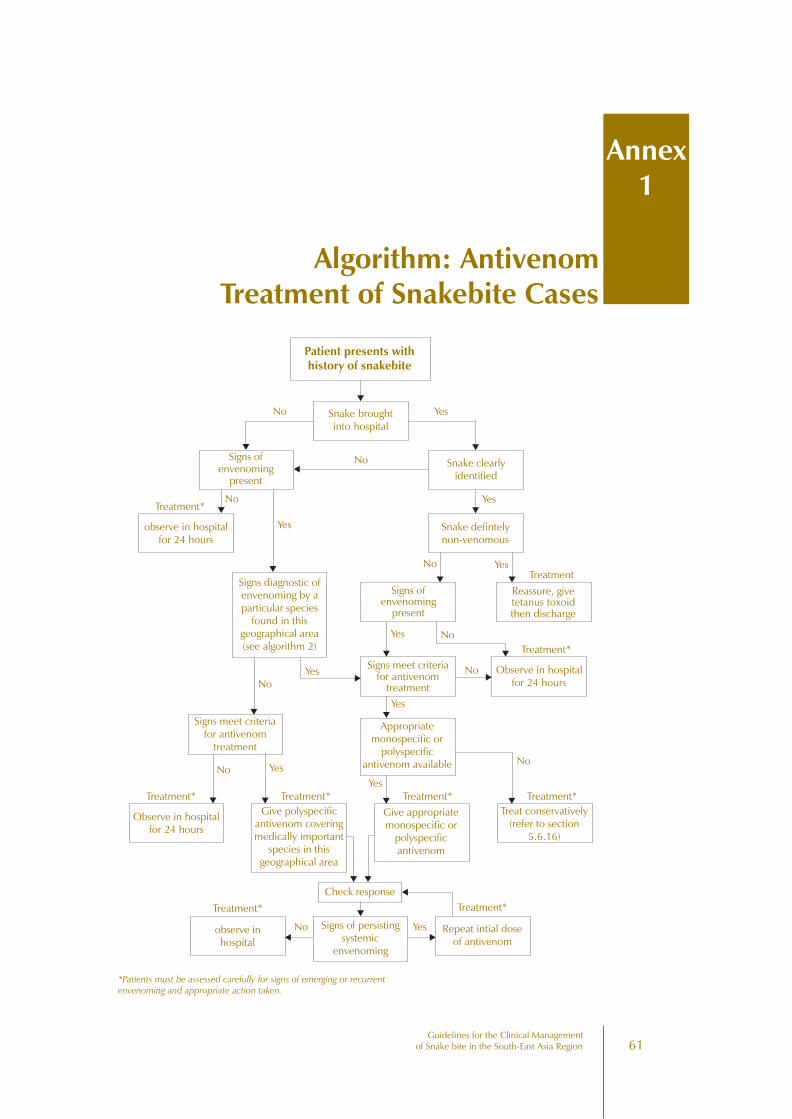
61Guidelines for the Clinical Management
of Snake bite in the South-East Asia Region
Algorithm: AntivenomTreatment of Snakebite Cases
Annex1
Patient presents withhistory of snakebite
Snake broughtinto hospital
Signs ofenvenoming
present
Snake clearlyidentified
observe in hospitalfor 24 hours
Treatment*
Snake defintelynon-venomous
Signs diagnostic ofenvenoming by aparticular species
found in thisgeographical area(see algorithm 2)
Signs ofenvenoming
present
Reassure, givetetanus toxoidthen discharge
Signs meet criteriafor antivenom
treatment
Observe in hospitalfor 24 hours
Signs meet criteriafor antivenom
treatment
Appropriatemonospecific or
polyspecificantivenom available
Treatment*
Give polyspecificantivenom coveringmedically important
species in thisgeographical area
Give appropriatemonospecific or
polyspecificantivenom
Treat conservatively(refer to section
5.6.16)
Check response
Treatment*
observe inhospital
Signs of persistingsystemic
envenoming
Repeat intial doseof antivenom
Observe in hospitalfor 24 hours
Treatment* Treatment* Treatment* Treatment*
Treatment
No Yes
No
Yes
No Yes
YesNo
NoYes
No Yes
Yes No
No
Yes
Yes
No
No Yes
Treatment*
*Patients must be assessed carefully for signs of emerging or recurrentenvenoming and appropriate action taken.
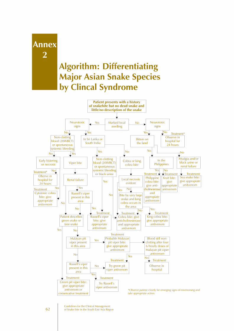
62Guidelines for the Clinical Managementof Snake bite in the South-East Asia Region
Algorithm: DifferentiatingMajor Asian Snake Speciesby Clincal Syndrome
Annex2
Patient presents with a historyof snakebite but no dead snake and
little/no description of the snake
Marked localswelling
NoYesNeurotoxicsigns
Neurotoxicsigns
Non-clottingblood (20WBCT)or spontaneous
systemic bleeding
In Sri Lanka orSouth India
Bitten onthe land
Observe inhospital for24 hours
Early bisteringor necrosis
Oberve inhospital for24 hours
Cytotoxic cobrabite: give
appropriateantivenom
Viper bite
Renal failure
Non-clottingblood (20WBCT)or spontaneous
systemic bleedingor black urine
Cobra or kingcobra bite
In thePhilippines
Myalgia and/orblack urine orrenal failure
Russell’s viperpresent in this
area
Local necrosisevident
Philippinecobra bite:give anti-
cholinesteraseand
appropiateantivenom
Sea snake bite:give appropiate
anitvenom
Krait bite:give
appropriateantivenom
Patient describesgreen snake or
tree snake
Russell’s viperbite: give
appropriateantivenom
Cobra bite: giveanticholinesteraseand appropriate
antivenom
King cobra bite:give appropriate
antivenom
Bite by very largesnake and kingcobra occurs in
the area
Malayan pitviper presentin this area
Probable Malayanpit viper bite:
give appropriateantivenom
Blood still non-clotting after four6 hourly doses ofMalayan pit viper
antivenom
Russell’s viperpresent in this
area
Try green pitviper antivenom
Observe inhospital
Try Russell’sviper antivenom
Green pit viper bite:give appropriate
antivenom orconservative treatment
No Yes Yes No
No YesYes No
No
Yes
No
Yes
YesNo
Yes No
Yes No
Yes No
YesNo
Treatment*
Treatment*
Treatment
TreatmentTreatmentTreatment
TreatmentTreatmentTreatment
Treatment
Treatment
TreatmentTreatment
NoYes
No
Yes
Treatment
No
NoYes
Yes
*Observe patient closely for emerging signs of envenoming andtake appropriate action.
Yes
No
63Guidelines for the Clinical Management
of Snake bite in the South-East Asia Region
1. ChinaShanghai Institute of Biological Products, Ministry of Health, 1262 Yan An Road (W),Shanghai 200052, China (Tel ++ 8621-62803189; Fax ++ 8621-62801807).Contact: Ms Minzhi Lu, Manager, International Affairs & Trade Department(Tel ++ 8621-62805234)
(liquid antivenoms, 10-15 ml/ampoule)
• Agkistrodon acutus antivenin (purified) (= Deinagkistrodon acutus, found inNorth Viet Nam). Recommended dose 8,000 IU (= 4 ampoules)
• “Agkistrodon halys” antivenin (purified) (said to be active against venoms ofTrimeresurus mucrosquamatus and T stejnegeri). Recommended dose 6,000IU (= 1 ampoule)
• Bungarus multicinctus antivenin (purified) (said to be effective against thevenom of Ophiophagus hannah). Recommended dose for bites by bothspecies 10,000 IU (= 1.25 ampoules)
• “Naja naja” antivenom (purified) (= Naja atra). Recommended dose 2,000IU (= 2 ampoules)
2. GermanyKnoll AG, Postfach 21 08 05, 67008 Ludwigshafen, Germany (Tel ++ 49621-5892688; Fax ++ 49621-5893707). Contact: Mr Lok, Managing Director
(liquid antivenom, 10 ml/ampoule)
• Cobra monospecific antivenom (Naja naja sputatrix = Malaysian N sumatrana)
3. India(a) Bengal Chemicals & Pharmaceuticals, 6 Ganesh Chunder Avenue, Calcutta
(Fax ++91 33 2257697)(liquid antivenom)
• Polyvalent (Bungarus caeruleus, Naja naja, Vipera russelli, Echis carinatus)
Antivenoms for Treatment ofBites by South-East Asian Snakes
(List by Country of Manufacture)
Annex3
64Guidelines for the Clinical Managementof Snake bite in the South-East Asia Region
(b) Central Research Institute (Simla Hills), 173 204 (HP) Kasauli (Tel ++ 91-17932060; Fax ++ 91-179272049)
(liquid and lyophilised antivenoms, 10 ml/ampoule)
• Polyvalent (B caeruleus, N naja, V russelli, E carinatus)
(c) Haffkine Biopharmaceutical Company Ltd, Acharya Donde Marg, Parel, Bombay400012 (Tel ++ 91-224129320 and 234129224; Fax ++ 91 41 68578; Telex11.71427 HBPC IN)(lyopholised antivenoms, 10 ml/ampoule)
• Polyvalent anti-snake venom serum (B caeruleus, E carinatus, N naja, V russelli)
(d) King’s Institute of Preventive Medicine, Guindi, Madras NA5
• Polyvalent
(e) Serum Institute of India Ltd, 212/2 Hadapsar, Pune-411 028 (Tel ++ 91-212672016; Fax ++ 91-212672040; Telex 145-7317 SERA IN, 145-7216 SEALIN) Contact: Dr SS Jadhav, Executive Director (QA)
(lyopholized antivenoms)
• Polyvalent (B caeruleus, N naja, V russelli, E carinatus)
• Bivalent (E carinatus, V russelli)
4. IndonesiaPerum Bio Farma (Pasteur Institute), Jl Pasteur 28, Post Box 1136, Bandung 40161(Tel ++ 6222-83755; Fax ++ 6222-210299; Telex 28432 BIOFAR IA)
(liquid antivenom, 5 ml/ampoule)
• Polyvalent antivenom serum (Calloselasma rhodostoma, B fasciatus, Nsputatrix)
5. IranState Serum & Vaccine Institute, Razi Hessarek, bP 656, Teheran(Tel ++ 98 2221 2005)(liquid antivenoms, 10 ml/ampoule)
• Polyvalent snake antivenom (equine) (said to neutralise the venoms of twoSouth-East Asian species – Naja oxiana and Echis carinatus (probably E sochureki),Vipera lebetina (= Macrovipera lebetina) and Pseudocerastes persicus
6. Myanmar (Burma)Myanmar Pharmaceutical Factory, Yangon(lyophilized and liquid antivenoms, 10 ml/ampoule)
• Viper antivenom (V russelli)
• Cobra antivenom (N kaouthia)
65Guidelines for the Clinical Management
of Snake bite in the South-East Asia Region
7. PakistanNational Institute of Health, Biological Production Division, Islamabad (Tel ++ 9251-240946; Fax ++ 9251-20797; Telex 5811-NAIB-PK) Contact: Shahid Akhtar(liquid and lyophilized antivenoms, 10 ml/ampoule)
• Polyvalent anti-snake venom serum (B caeruleus, E carinatus, N naja, Vlebetina, V russelli)
8. PhilippinesBiologicals Production Service, Dept of Health, Los Baños, Laguna(liquid antivenom)
• Philippine cobra antivenin (Naja philippinensis)
9. TaiwanNational Institute of Preventive Medicine, 161 Kun-Yang Street, Nan-Kang, Taipei,ROC 11513 (Tel ++ 8862-7859215; Fax ++ 8862-7853944). Contact: Dr Gong-Ren Wang, Director(lyophilised antivenoms, 10 ml/ampoule)
• Bungarus multicinctus and N atra bivalent antivenom
• Trimeresurus muquosquamatus and Trimeresurus grammineus (= T stejnegeri)bivalent antivenom
• Agkistrodon acutus (= Deinagkistrodon acutus) antivenom
10. ThailandThe Thai Red Cross Society, Queen Saovabha Memorial Institute, 1871 Rama VIRoad, Bangkok 10330 (Tel ++ 662-2520161-4; Fax ++ 662-2540212; Telex 82535THRESCO TH)(freeze dried monovalent antivenoms, 10 ml/ampoule)
• Cobra antivenom
• King cobra antivenin
• Banded krait antivenin
• Russell’s viper antivenin
• Malayan pit viper antivenin
• Green pit viper antivenin
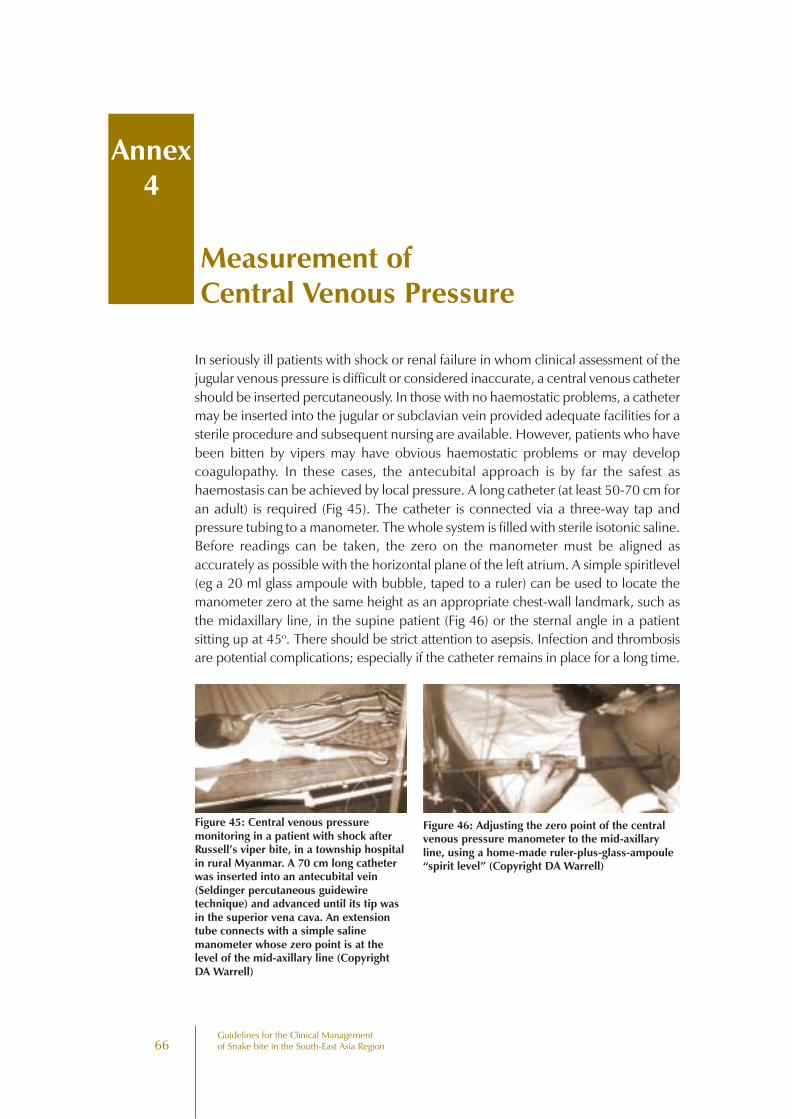
66Guidelines for the Clinical Managementof Snake bite in the South-East Asia Region
Measurement ofCentral Venous Pressure
Annex4
In seriously ill patients with shock or renal failure in whom clinical assessment of thejugular venous pressure is difficult or considered inaccurate, a central venous cathetershould be inserted percutaneously. In those with no haemostatic problems, a cathetermay be inserted into the jugular or subclavian vein provided adequate facilities for asterile procedure and subsequent nursing are available. However, patients who havebeen bitten by vipers may have obvious haemostatic problems or may developcoagulopathy. In these cases, the antecubital approach is by far the safest ashaemostasis can be achieved by local pressure. A long catheter (at least 50-70 cm foran adult) is required (Fig 45). The catheter is connected via a three-way tap andpressure tubing to a manometer. The whole system is filled with sterile isotonic saline.Before readings can be taken, the zero on the manometer must be aligned asaccurately as possible with the horizontal plane of the left atrium. A simple spiritlevel(eg a 20 ml glass ampoule with bubble, taped to a ruler) can be used to locate themanometer zero at the same height as an appropriate chest-wall landmark, such asthe midaxillary line, in the supine patient (Fig 46) or the sternal angle in a patientsitting up at 45o. There should be strict attention to asepsis. Infection and thrombosisare potential complications; especially if the catheter remains in place for a long time.
Figure 45: Central venous pressuremonitoring in a patient with shock afterRussell’s viper bite, in a township hospitalin rural Myanmar. A 70 cm long catheterwas inserted into an antecubital vein(Seldinger percutaneous guidewiretechnique) and advanced until its tip wasin the superior vena cava. An extensiontube connects with a simple salinemanometer whose zero point is at thelevel of the mid-axillary line (CopyrightDA Warrell)
Figure 46: Adjusting the zero point of the centralvenous pressure manometer to the mid-axillaryline, using a home-made ruler-plus-glass-ampoule“spirit level” (Copyright DA Warrell)
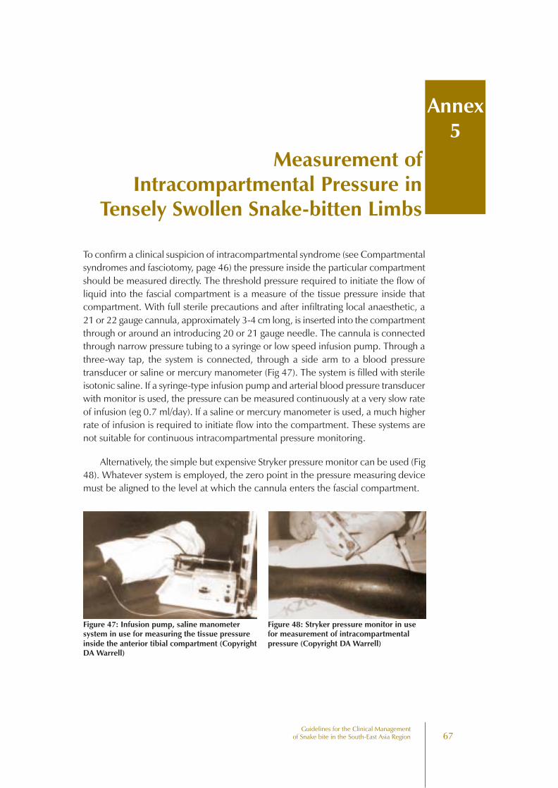
67Guidelines for the Clinical Management
of Snake bite in the South-East Asia Region
To confirm a clinical suspicion of intracompartmental syndrome (see Compartmentalsyndromes and fasciotomy, page 46) the pressure inside the particular compartmentshould be measured directly. The threshold pressure required to initiate the flow ofliquid into the fascial compartment is a measure of the tissue pressure inside thatcompartment. With full sterile precautions and after infiltrating local anaesthetic, a21 or 22 gauge cannula, approximately 3-4 cm long, is inserted into the compartmentthrough or around an introducing 20 or 21 gauge needle. The cannula is connectedthrough narrow pressure tubing to a syringe or low speed infusion pump. Through athree-way tap, the system is connected, through a side arm to a blood pressuretransducer or saline or mercury manometer (Fig 47). The system is filled with sterileisotonic saline. If a syringe-type infusion pump and arterial blood pressure transducerwith monitor is used, the pressure can be measured continuously at a very slow rateof infusion (eg 0.7 ml/day). If a saline or mercury manometer is used, a much higherrate of infusion is required to initiate flow into the compartment. These systems arenot suitable for continuous intracompartmental pressure monitoring.
Alternatively, the simple but expensive Stryker pressure monitor can be used (Fig48). Whatever system is employed, the zero point in the pressure measuring devicemust be aligned to the level at which the cannula enters the fascial compartment.
Measurement ofIntracompartmental Pressure in
Tensely Swollen Snake-bitten Limbs
Annex5
Figure 47: Infusion pump, saline manometersystem in use for measuring the tissue pressureinside the anterior tibial compartment (CopyrightDA Warrell)
Figure 48: Stryker pressure monitor in usefor measurement of intracompartmentalpressure (Copyright DA Warrell)
Guidelinesfor the Clinical Management ofSnake bites in the South-East Asia Region
Spectacled CobraSaw Scaled Viper
Common KraitCommon Krait
Russells ViperRussells Viper