guidelines for the management of snakebites
TRANSCRIPT

102
CTDT
10.5005/jp-journals-10055-0048
REVIEW ARTICLE
Guidelines for the Management of Snakebites1Gurinder Mohan, 2Arvinderpal Singh, 3Trimaan Singh
ABSTRACT
India is estimated to have the highest snakebite mortality in the world. World Health Organization (WHO) places the number of bites to be 83,000 per annum with 11,000 deaths.1 Most of the fatalities are due to the victim not reaching the hospital in time where definite treatment can be administered. In addition, community is also not well informed about the occupational risks and simple measures which can prevent the bite. It continues to adopt harmful first-aid practices, such as tourniquets, cutting, and suction. Studies reveal that primary care doctors do not treat snakebite patients mainly due to lack of confidence.2 At the secondary and the tertiary care level, multiple protocols are being followed for polyvalent anti-snake venom (ASV) administration, predominantly based on western textbooks.
Keywords: Anti-snake venom, First aid, Snakebite.
How to cite this article: Mohan G, Singh A, Singh T. Guidelines for the Management of Snakebites. Curr Trends Diagn Treat 2018;2(2):102-108.
Source of support: Self
Conflict of interest: None
SNAKES OF INDIA
There are about 236 species of snakes in India, most of which are nonpoisonous. Their bites, apart from causing panic reaction and local injury, do not harm the patients. However, there are 13 known species that are poisonous, of these, four, namely common cobra (Naja naja), Russell’s viper (Daboia russelii), saw-scaled viper (Echis carinatus), and common krait (Bungarus caeruleus), are highly veno- mous and believed to be responsible for most of the poisonous bites in India.3 However, this assumption of great four has led to non-identification of other poisonous species which are going unnoticed and leading to deaths. The recent discovery of the hump-nosed viper, capable of causing life-threatening symptoms, has demonstrated this. This nonrecognition has led the ASV manufacturers
to produce antivenom only against these four species.4 Thus, there is a need to abandon the old concept of “The Big Four” in order to determine all the medically significant species in India.
Snake venoms comprises >100 different proteins: enzymes, nonenzymatic polypeptide toxins, and nontoxic proteins. Enzymes are digestive hydrolases, hyaluroni-dase (spreading factor), yellow l-amino acid oxidases, phospholipases A2, and peptidases. Snake venom metallo- proteases (SVMPs) damage basement membranes, causing endothelial cell damage and spontaneous systemic bleeding. Procoagulant enzymes are thrombin-like, split-ting fibrinogen, or activators of factors V, X, prothrombin, and other clotting factors, causing disseminated intra- vascular coagulation (DIC), consumption coagulopathy, and incoagulable blood. Phospholipases A2 damage mito-chondria, red blood cells, leucocytes, platelets, peripheral nerve endings, skeletal muscle, vascular endothelium, and other membranes. It produces presynaptic neurotoxic activity, cardiotoxicity, myotoxicity, tissue necrosis, hypo-tension, hemolysis, anticoagulation, hemorrhage, plasma leakage (edema formation), and autopharmacological release of histamine and other autacoids. Polypeptide postsynaptic (α) neurotoxins bind to acetylcholine recep-tors at the motor endplate. Presynaptic (β) neurotoxins are phospholipases that damage nerve endings irreparably. The composition and antigenicity of snake venoms vary greatly between and within species, maturity, seasonally, between sexes, and throughout the geographical range. Thus, envenoming by a particular species in one part of its geographical range may not be responsive to antivenom raised against venom from the same species in another area. “Dry bites”: about 50% of venomous snakebites do not result in envenoming.
CLINICAL PRESENTATION
General: Fear and anxiety are very common and can lead to panic and trauma related to falls due to severe anxiety.Local envenoming:
• Local pain at the site of the bite (krait bites are usually painless),
• Local swelling spreading proximally,• Tenderness,• Painful swelling of regional lymph nodes draining
bite site,
1Professor and Head, 2Associate Professor, 3Senior Resident1,3Department of Medicine, Sri Guru Ram Das Institute of Medical Sciences & Research, Jalandhar, Punjab, India2Department of Anaesthesia, Sri Guru Ram Das Institute of Medical Sciences & Research, Jalandhar, Punjab, India
Corresponding Author: Trimaan Singh, Senior Resident, Department of Medicine, Sri Guru Ram Das Institute of Medical Sciences & Research, Jalandhar, Punjab, India, Phone: 9855358989, e-mail: [email protected]

Guidelines for the Management of Snakebites
AMEI's Current Trends in Diagnosis & Treatment, July-December 2018;2(2):102-108 103
CTDT
• Other signs: fang marks, persistent local bleeding,bruising, lymphangitis, inflammation (swelling,redness, heat), blistering (blebs, bullae, vesicles), infec-tion, abscess formation, necrosis (Fig. 1).
Systemic envenoming: nausea, vomiting, malaise, abdom-inal pain, weakness, drowsiness, prostration.
Cardiovascular (Viperidae):
• Hypotension, syncope, collapse, shock,• Cardiac arrhythmias,• Myocardial damage.
Generalized increase in capillary permeability:
• Facial, periorbital, conjunctival edema (chemosis),• Pleural and pericardial effusions, pulmonary edema,
Fig. 1: Signs of local envenomation (Copyright DA Warrell)
Fig 2: Intracranial bleed (Copyright Dr Suvarna Patil)
• Massive albuminuria,• Hemoconcentration.
Bleeding and clotting disorders (Viperidae):
• Local traumatic bleeding from recent and partlyhealed wounds and venepuncture sites;
• Spontaneous systemic bleeding (gums, epistaxis, hem-optysis, meningism from subarachnoid hemorrhage,lateralizing signs, and/or coma from cerebral hemor-rhage/thrombosis), hematemesis, rectal bleeding ormelena, hematuria, vaginal bleeding, subconjuncti-val hemorrhages, skin petechiae, purpura, discoidhemorrhages, ecchymoses (Fig. 2).
Neurological (Elapidae, Viperidae, e.g., Russell’s viper D. russelii, and Gloydius species) (Fig. 3):
Gurinder Mohan et al.
104
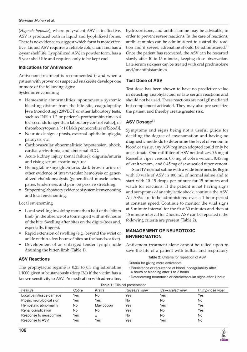
Fig. 5: Conjunctivitis associated with spat of venom (Copyright DA Warrell)
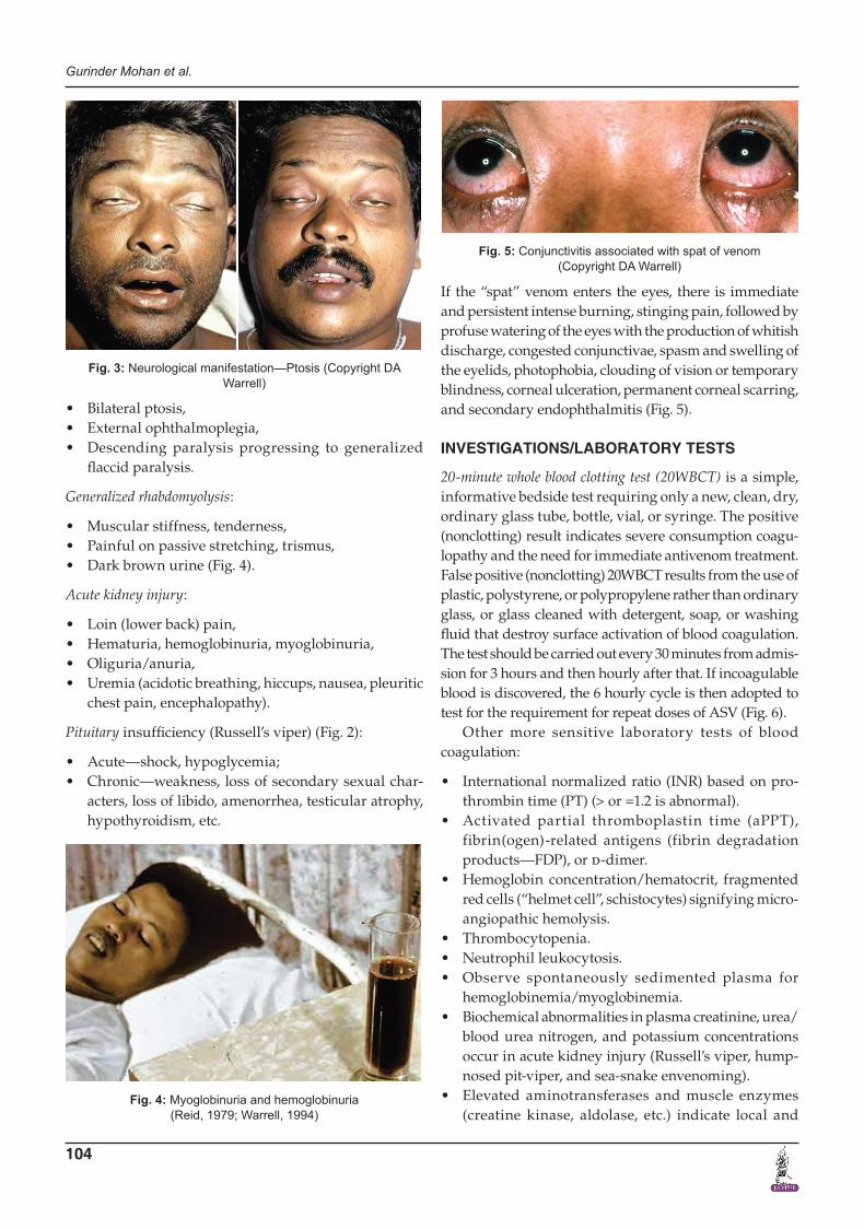
Fig. 3: Neurological manifestation—Ptosis (Copyright DA Warrell)
• Bilateral ptosis,• External ophthalmoplegia,• Descending paralysis progressing to generalized
flaccid paralysis.
Generalized rhabdomyolysis:
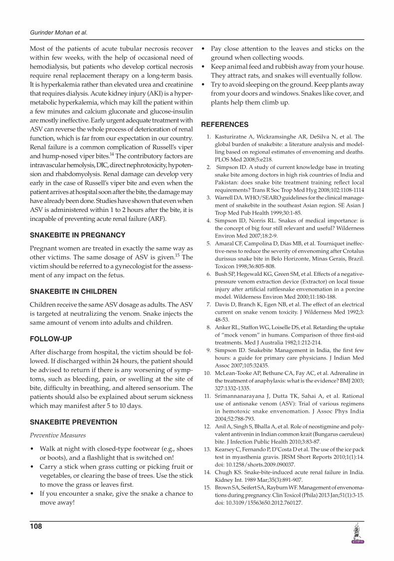
• Muscular stiffness, tenderness,• Painful on passive stretching, trismus,• Dark brown urine (Fig. 4).
Acute kidney injury:
• Loin (lower back) pain,• Hematuria, hemoglobinuria, myoglobinuria,• Oliguria/anuria,• Uremia (acidotic breathing, hiccups, nausea, pleuritic
chest pain, encephalopathy).
Pituitary insufficiency (Russell’s viper) (Fig. 2):
• Acute—shock, hypoglycemia;• Chronic—weakness, loss of secondary sexual char-
acters, loss of libido, amenorrhea, testicular atrophy,hypothyroidism, etc.
If the “spat” venom enters the eyes, there is immediate and persistent intense burning, stinging pain, followed by profuse watering of the eyes with the production of whitish discharge, congested conjunctivae, spasm and swelling of the eyelids, photophobia, clouding of vision or temporary blindness, corneal ulceration, permanent corneal scarring, and secondary endophthalmitis (Fig. 5).
INVESTIGATIONS/LABORATORY TESTS
20-minute whole blood clotting test (20WBCT) is a simple,informative bedside test requiring only a new, clean, dry, ordinary glass tube, bottle, vial, or syringe. The positive(nonclotting) result indicates severe consumption coagu-lopathy and the need for immediate antivenom treatment. False positive (nonclotting) 20WBCT results from the use of plastic, polystyrene, or polypropylene rather than ordinaryglass, or glass cleaned with detergent, soap, or washingfluid that destroy surface activation of blood coagulation.The test should be carried out every 30 minutes from admis-sion for 3 hours and then hourly after that. If incoagulableblood is discovered, the 6 hourly cycle is then adopted totest for the requirement for repeat doses of ASV (Fig. 6).
Other more sensitive laboratory tests of blood coagulation:
• International normalized ratio (INR) based on pro-thrombin time (PT) (> or =1.2 is abnormal).
• Activated partial thromboplastin time (aPPT),fibrin(ogen)-related antigens (fibrin degradationproducts—FDP), or d-dimer.
• Hemoglobin concentration/hematocrit, fragmentedred cells (“helmet cell”, schistocytes) signifying micro-angiopathic hemolysis.
• Thrombocytopenia.• Neutrophil leukocytosis.• Observe spontaneously sedimented plasma for
hemoglobinemia/myoglobinemia.• Biochemical abnormalities in plasma creatinine, urea/
blood urea nitrogen, and potassium concentrationsoccur in acute kidney injury (Russell’s viper, hump-nosed pit-viper, and sea-snake envenoming).
• Elevated aminotransferases and muscle enzymes(creatine kinase, aldolase, etc.) indicate local and
Fig. 4: Myoglobinuria and hemoglobinuria (Reid, 1979; Warrell, 1994)

Guidelines for the Management of Snakebites
AMEI's Current Trends in Diagnosis & Treatment, July-December 2018;2(2):102-108 105
CTDT
generalized muscle damage (sea snakes, some kraits, some Australasian Elapidae, and Russell’s viper bites).
• Hyponatremia is associated with krait bites.• Urine examination: dipsticks test for blood hemo-
globin, myoglobin, and proteinuria.• Urine microscopy to detect erythrocytes and red cell
casts, indicating glomerular bleeding and eosino-philia suggesting acute interstitial nephritis.
Other investigations:
• Chest radiography for detecting pulmonary edema,hemorrhages, infarcts, pleural effusions, and secondarybronchopneumonia.
• Ultrasound for assessing local envenoming, deepvein thrombosis, pleural and pericardial effusion,and bleeding.
• Echocardiography for myocardial dysfunction.• Computed tomography (CT) and magnetic resonance
imaging (MRI) for intracranial and spinal hemor-rhages and infarcts and osteomyelitis at the bite site.
• ECG for arrhythmias, myocardial damage, and evi-dence of hyperkalemia.
Also, the single breath counting test is done in suspected bites with signs of neurological involvement and the same is repeated at 15 minute interval over the first 2 hours.
MANAGEMENT PROTOCOL (AS PER RECENT WHO GUIDELINES 2016)
First-Aid Treatment8
The first aid being currently recommended is based on the mnemonic.“CARRY NO R.I.G.H.T.”It consists of the following:
CARRY = Do not allow victim to walk even for a short distance; just carry him in any form, especially when bite is at leg.
No = Tourniquet5; No-electrotherapy7; No-cutting; No-pressure immobilization; No-sucking of venom6
R = Reassure the patient. About 70% of all snakebites are from nonvenomous species. Only 50% of bites by venomous species actually envenomate the patient.
I = Immobilize in the same way as a fractured limb. Use bandages or cloth to hold the splints, not to block the blood supply or apply pressure. Do not apply any compression in the form of tight ligatures, they do not work and can be dangerous!
GH = Get to hospital immediately. Traditional rem-edies have NO PROVEN benefit in treating snakebite.
T = Tell the doctor of any systemic symptoms that manifest on the way to hospital.
Traditional methods should be discarded.
Pain
Snakebite can often cause severe pain at the bite site. This can normally be treated with painkillers, such as paracetamol. Aspirin and nonsteroidal anti-inflammatory drugs (NSAIDs) should not be used, as they can exacerbate bleeding. This can be particularly dangerous in a patient already having coagulopathy. Mild opiates, such as tramadol 50 mg, can be used for good pain relief.
Handling Tourniquets
Care must be taken when removing tight tourniquets tied by the victim. Sudden removal can lead to a massive surge of venom leading to neurological paralysis and hypoten-sion due to vasodilation. Before removal of the tourniquet, test for the presence of a pulse distal to the tourniquet. Be prepared to handle complications, such as sudden respiratory distress or hypotension. If the tourniquet has occluded the distal pulse, then a blood pressure cuff can be applied to reduce the pressure slowly.9
ASV
ASV is the mainstay of treatment. In India, polyvalent ASV, i.e., effective against all the four common species: Russell’s viper, common cobra, common krait, and saw-scaled viper, and no monovalent ASVs are available. There are known species, such as the hump-nosed viper
Fig. 6: 20-minute whole blood clotting test (20WBCT)
Gurinder Mohan et al.
106
(Hypnale hypnale), where polyvalent ASV is ineffective. ASV is produced both in liquid and lyophilized forms. There is no evidence to suggest which form is more effec-tive. Liquid ASV requires a reliable cold chain and has a 2-year shelf life. Lyophilized ASV, in powder form, has a 5-year shelf life and requires only to be kept cool.
Indications for Antivenom
Antivenom treatment is recommended if and when a patient with proven or suspected snakebite develops one or more of the following signs:Systemic envenoming
• Hemostatic abnormalities: spontaneous systemicbleeding distant from the bite site, coagulopathy[+ve (nonclotting) 20WBCT or other laboratory tests,such as INR > 1.2 or patient’s prothrombin time > 4to 5 seconds longer than laboratory control value], orthrombocytopenia [< 1.0 lakh per microliter of blood)].
• Neurotoxic signs: ptosis, external ophthalmoplegia,paralysis, etc.
• Cardiovascular abnormalities: hypotension, shock,cardiac arrhythmia, and abnormal ECG.
• Acute kidney injury (renal failure): oliguria/anuriaand rising serum creatinine/urea.
• Hemoglobin-/myoglobinuria: dark brown urine orother evidence of intravascular hemolysis or gener-alized rhabdomyolysis (generalized muscle aches,pains, tenderness, and pain on passive stretching.
• Supporting laboratory evidence of systemic envenomingand local envenoming.
Local envenoming
• Local swelling involving more than half of the bittenlimb (in the absence of a tourniquet) within 48 hoursof the bite. Swelling after bites on the digits (toes and, especially, fingers).
• Rapid extension of swelling (e.g., beyond the wrist orankle within a few hours of bites on the hands or feet).
• Development of an enlarged tender lymph nodedraining the bitten limb (Table 1).
ASV Reactions
The prophylactic regime is 0.25 to 0.3 mg adrenaline 1:1000 given subcutaneously (deep IM) if the victim has a known sensitivity to ASV. Premedication with adrenaline,
hydrocortisone, and antihistamine may be advisable, in order to prevent severe reactions. In the case of reactions, antihistaminics can be administered to control the reac-tion and if severe, adrenaline should be administered.10 Once the patient has recovered, the ASV can be restarted slowly after 10 to 15 minutes, keeping close observation. Late serum sickness can be treated with oral prednisolone and/or antihistaminics.
Test Dose of ASV
Test dose has been shown to have no predictive value in detecting anaphylactoid or late serum reactions and should not be used. These reactions are not IgE mediated but complement activated. They may also pre-sensitize the patient and thereby create greater risk.
ASV Dosage11
Symptoms and signs being not a useful guide for deciding the degree of envenomation and having no diagnostic methods to determine the level of venom in blood or tissue, any ASV regimen adopted could only be an estimate. One milliliter of ASV neutralizes 0.6 mg of Russell’s viper venom, 0.6 mg of cobra venom, 0.45 mg of krait venom, and 0.45 mg of saw-scaled viper venom.
Start IV normal saline with a wide bore needle. Begin with 10 vials of ASV in 100 mL of normal saline and to start with 10–15 drops per minute for 15 minutes and watch for reactions. If the patient is not having signs and symptoms of anaphylactic shock, continue the ASV. All ASVs are to be administered over a 1 hour period at constant speed. Continue to monitor the vital signs at 5 minute interval for the first 30 minutes and then at 15 minute interval for 2 hours. ASV can be repeated if the following criteria are present (Table 2).
MANAGEMENT OF NEUROTOXIC ENVENOMATION
Antivenom treatment alone cannot be relied upon to save the life of a patient with bulbar and respiratory
Table 1: Clinical presentationFeature Cobra Kraits Russell’s viper Saw-scaled viper Hump-nose viperLocal pain/tissue damage Yes No Yes Yes YesPtosis, neurological sign Yes Yes No No NoHemostatic abnormality No May occour Yes Yes YesRenal complication No No Yes No YesResponse to neostigmine Yes ± No No NoResponse to ASV Yes Yes Yes Yes No
Table 2: Criteria for repetition of ASVCriteria for giving more antivenom• Persistence or recurrence of blood incoagulability after 6 hours or bleeding after 1 to 2 hours• Deteriorating neurotoxic or cardiovascular signs after 1 hour

Guidelines for the Management of Snakebites
AMEI's Current Trends in Diagnosis & Treatment, July-December 2018;2(2):102-108 107
CTDT
paralysis. Death may result from aspiration, airway obstruction, or respiratory failure. A clear airway must be maintained. Once there is loss of gag reflex and pooling of secretions in the pharynx, failure of the cough reflex, or respiratory distress, a cuffed endotracheal, laryngeal mask airway, or supraglottic airway should be inserted. If this is impossible for any reason, or the need for prolonged ventilation is anticipated, a trache-ostomy should be performed and cuffed tracheostomy tube should be inserted.
Neostigmine is an anticholinesterase that prolongs the life of acetylcholine and can, therefore, reverse respiratory failure and neurotoxic symptoms. It is particularly effective for postsynaptic neurotoxins, such as those of the cobra. There is some doubt over its usefulness against presynaptic neurotoxin, such as those of the krait and Russell’s viper.
NEOSTIGMINE TEST12
This test involves the administration of 1.5 to 2 mg of neostigmine IM together with 0.6 mg of atropine IV. The patient should be closely observed for 1 hour to decide whether neostigmine is effective. The following measures are useful objective methods to asses this:
• Single breath count• mm of iris uncovered (amount covered by the
descending eye lid)• Inter incisor distance (distance between upper and
lower incisors)• Length of time upwards gaze can be maintained.• FEV1 (forced expiratory volume (in 1 second)) or
forced vital capacity (FVC), if available
If the victim responds to the neostigmine test, then con-tinue with 0.5 mg of neostigmine IM half hourly plus 0.6 mg atropine IV for five doses and then 2 to 12 hourly according to recovery. If there is no improvement in symptoms after 1 hour, neostigmine should be stopped.
Patients who respond convincingly can be maintained on neostigmine methylsulfate, 0.5 to 2.5 mg every half hour up to 10 mg/24 hours maximum for adults or 0.01 to 0.04 mg/kg every 2 to 4 hours for children by intramuscu-lar, intravenous, or subcutaneous injection together with atropine to block muscarinic side effects. Patients able to swallow tablets may be maintained on atropine 0.6 mg twice each day, neostigmine 15 mg four times each day, or pyridostigmine 60 mg four times each day.
Ice Test (Fig. 7)
The “ice pack test”13 as a “possible alternative to the Tensilon test” is recovery of ptosis in the one eye after applying an ice pad that inhibits intrinsic cholinesterase in a patient with postsynaptic blockade of neuromuscular
transmission. In this, digital pressure is applied to the frontalis muscle to avoid its influencing upper eyelid retraction. An ice-filled plastic glove or frozen ice pack is then gently applied to one eyelid for 2 minutes, after which the palpebral fissures of both eyes are immediately remeasured. A more-than-2 mm difference in palpebral fissure between the cooled and control eyelids might be considered a positive result.
RENAL FAILURE IN SNAKEBITE
The acute renal failure which occurs due to snakebite is multifactorial:
• Severe and persistent hypotension leading to acutetubular necrosis.
• Rhabdomyolysis and myoglobinuria.• DIC.• Vasculitis.• Acute diffuse interstitial nephritis.• Extracapillary proliferative glomerulonephritis.
Fig. 7: Ice test for checking neurotoxicity (Delaney Y, Khooshabeh R, Benjamin L, 2002)
Gurinder Mohan et al.
108
Most of the patients of acute tubular necrosis recover within few weeks, with the help of occasional need of hemodialysis, but patients who develop cortical necrosis require renal replacement therapy on a long-term basis. It is hyperkalemia rather than elevated urea and creatinine that requires dialysis. Acute kidney injury (AKI) is a hyper-metabolic hyperkalemia, which may kill the patient within a few minutes and calcium gluconate and glucose-insulin are mostly ineffective. Early urgent adequate treatment with ASV can reverse the whole process of deterioration of renal function, which is far from our expectation in our country. Renal failure is a common complication of Russell’s viper and hump-nosed viper bites.14 The contributory factors are intravascular hemolysis, DIC, direct nephrotoxicity, hypoten-sion and rhabdomyolysis. Renal damage can develop very early in the case of Russell’s viper bite and even when the patient arrives at hospital soon after the bite, the damage may have already been done. Studies have shown that even when ASV is administered within 1 to 2 hours after the bite, it is incapable of preventing acute renal failure (ARF).
SNAKEBITE IN PREGNANCY
Pregnant women are treated in exactly the same way as other victims. The same dosage of ASV is given.15 The victim should be referred to a gynecologist for the assess-ment of any impact on the fetus.
SNAKEBITE IN CHILDREN
Children receive the same ASV dosage as adults. The ASV is targeted at neutralizing the venom. Snake injects the same amount of venom into adults and children.
FOLLOW-UP
After discharge from hospital, the victim should be fol-lowed. If discharged within 24 hours, the patient should be advised to return if there is any worsening of symp-toms, such as bleeding, pain, or swelling at the site of bite, difficulty in breathing, and altered sensorium. The patients should also be explained about serum sickness which may manifest after 5 to 10 days.
SNAKEBITE PREVENTION
Preventive Measures
• Walk at night with closed-type footwear (e.g., shoesor boots), and a flashlight that is switched on!
• Carry a stick when grass cutting or picking fruit orvegetables, or clearing the base of trees. Use the stickto move the grass or leaves first.
• If you encounter a snake, give the snake a chance tomove away!
• Pay close attention to the leaves and sticks on theground when collecting woods.
• Keep animal feed and rubbish away from your house.They attract rats, and snakes will eventually follow.
• Try to avoid sleeping on the ground. Keep plants away from your doors and windows. Snakes like cover, and plants help them climb up.
REFERENCES
1. Kasturiratne A, Wickramsinghe AR, DeSilva N, et al. Theglobal burden of snakebite: a literature analysis and model-ling based on regional estimates of envenoming and deaths.PLOS Med 2008;5:e218.
2. Simpson ID. A study of current knowledge base in treatingsnake bite among doctors in high risk countries of India andPakistan: does snake bite treatment training reflect localrequirements? Trans R Soc Trop Med Hyg 2008;102:1108-1114
3. Warrell DA. WHO/SEARO guidelines for the clinical manage-ment of snakebite in the southeast Asian region. SE Asian JTrop Med Pub Health 1999;30:1-85.
4. Simpson ID, Norris RL. Snakes of medical importance: isthe concept of big four still relevant and useful? WildernessEnviron Med 2007;18:2-9.
5. Amaral CF, Campolina D, Dias MB, et al. Tourniquet ineffec-tive-ness to reduce the severity of envenoming after Crotalus durissus snake bite in Belo Horizonte, Minas Gerais, Brazil.Toxicon 1998;36:805-808.
6. Bush SP, Hegewald KG, Green SM, et al. Effects of a negative-pressure venom extraction device (Extractor) on local tissueinjury after artificial rattlesnake envenomation in a porcinemodel. Wilderness Environ Med 2000;11:180-188.
7. Davis D, Branch K, Egen NB, et al. The effect of an electricalcurrent on snake venom toxicity. J Wilderness Med 1992;3:48-53.
8. Anker RL, Staffon WG, Loiselle DS, et al. Retarding the uptakeof “mock venom” in humans. Comparison of three first-aidtreatments. Med J Australia 1982;1:212-214.
9. Simpson ID. Snakebite Management in India, the first fewhours: a guide for primary care physicians. J Indian MedAssoc 2007;105:32435.
10. McLean-Tooke AP, Bethune CA, Fay AC, et al. Adrenaline in the treatment of anaphylaxis: what is the evidence? BMJ 2003; 327:1332-1335.
11. Srimannanarayana J, Dutta TK, Sahai A, et al. Rationaluse of antisnake venom (ASV): Trial of various regimensin hemotoxic snake envenomation. J Assoc Phys India2004;52:788-793.
12. Anil A, Singh S, Bhalla A, et al. Role of neostigmine and poly-valent antivenin in Indian common krait (Bungarus caeruleus) bite. J Infection Public Health 2010;3:83-87.
13. Kearsey C, Fernando P, D’Costa D et al. The use of the ice packtest in myasthenia gravis. JRSM Short Reports 2010;1(1):14.doi: 10.1258/shorts.2009.090037.
14. Chugh KS. Snake-bite-induced acute renal failure in India.Kidney Int. 1989 Mar;35(3):891-907.
15. Brown SA, Seifert SA, Rayburn WF. Management of envenoma-tions during pregnancy. Clin Toxicol (Phila) 2013 Jan;51(1):3-15. doi: 10.3109/15563650.2012.760127.