guillain–barré and miller fisher syndromes—new diagnostic classification
TRANSCRIPT
NATURE REVIEWS | NEUROLOGY ADVANCE ONLINE PUBLICATION | 1
IntroductionGuillain–Barré syndrome (GBS) is the broad term used to describe a number of related acute autoimmune neuropathies, although the term is also used more specifically to define patients with peripheral polyneuropathy affecting all four limbs with or without cranial nerve involvement. GBS was first recognized in 1916 by Guillain, Barré and Strohl, who described two patients who developed acute areflexic paralysis in association with raised protein levels in cerebrospinal fluid (CSF), but no increased cell content.1 By 1938, Guillain had recognized various forms of GBS and proposed a clinical classification that took into account four presentations: the lower form, the spinal and midbrain form, the midbrain form, and poly radiculo neuropathy with impaired mentation.2
Almost 20 years later, in his seminal paper, Miller Fisher described three patients with “an unusual variant of acute idiopathic polyneuritis (syndrome of ophthalmoplegia, ataxia and areflexia),” which bore
resemblance to the midbrain form proposed by Guillain.2,3 Contemporaneously, Bickerstaff described eight patients who presented with ophthalmoplegia, ataxia and hypersomnolence, similar to—yet distinct from—GBS polyradiculoneuropathy with impaired mentation.4
Clinical diagnostic criteria for GBS were introduced in 1978 following an increase in incidence after the swine flu vaccination programme, and these criteria were later reaffirmed.5,6 The criteria were devised to enable nonneurologists to recognize GBS and were intentionally restrictive, requiring the presence of universal limb areflexia or hyporeflexia. However, with the identification of several new phenotypes in the past 30 years, the conceptual framework of GBS has become increasingly complex. For example, in 1986, Ropper described patients who developed rapidly progressive oropharyngeal, neck and shoulder weakness that mimicked the descending paralysis seen in botulism.7 In 1994, the same author also described some patients with areflexic paraparesis7,8 and others with acute progression of facial diplegia and numbness in the extremities;8 he speculated that these
three conditions were localized subtypes of GBS, which he called pharyngeal–cervical– brachial weakness, the paraparetic variant, and bifacial weakness with paraesthesias, respectively. These studies led, in 1990, to proposed diagnostic criteria for pure motor GBS, pure sensory GBS, MFS, several localized subtypes of GBS (including pharyngeal–cervical– brachial weakness and paraparetic GBS) and pure pandysautonomia.9
A further revision was prompted by results from nerve conduction studies. Classification criteria published in 2001 by a GBS consensus group based in the Netherlands described several subtypes of GBS: sensorimotor GBS, pure motor GBS, MFS and a bulbar form.10 Nerve conduction studies enabled the group to further subclassify sensorimotor GBS into either acute inflammatory demyelinating polyneuro pathy (AIDP) or acute motor–sensory axonal neuropathy, and pure motor GBS into acute motor demyelinating neuropathy or acute motor axonal neuropathy (AMAN). Criteria outlined by the Brighton Collaboration GBS working group in 2011 have also used nerve conduction studies to identify patients with vaccinationrelated GBS or MFS.11
The primary aim of this Perspectives article from the GBS Classification Group (Box 1) is to present clinical criteria to enable neurologists and nonneurologists to diagnose GBS and all its variants using a simple yet allinclusive classification sys tem. While some variants are rare (for example, acute ptosis and acute mydriasis) and might never be encountered by many physi cians, others (for example, pharyngeal– cervical–brachial weakness) might be more common than previously thought, having been frequently misdiagnosed as myasthenia gravis, botulism or brainstem stroke.12 We also consider the classification of GBS, MFS and their subtypes. Rather than broadly categorizing each subtype as an axonal or demyelinating neuropathy, we propose new diagnostic criteria based on an inclusive set of clinical features. To avoid confusion, we use the terms ‘classic GBS’ to describe patients who present with acute flaccid paralysis of all four limbs, and ‘GBS subtypes’ to collectively describe the localized forms of GBS. Similarly, we use ‘classic MFS’ to
OPINION
Guillain–Barré and Miller Fisher syndromes—new diagnostic classificationBenjamin R. Wakerley, Antonino Uncini and Nobuhiro Yuki for the GBS Classification Group
Abstract | Guillain–Barré syndrome (GBS) and its variant, Miller Fisher syndrome (MFS), exist as several clinical subtypes with different neurological features and presentations. Although the typical clinical features of GBS and MFS are well recognized, current classification systems do not comprehensively describe the full spectrum of either syndrome. In this Perspectives article, GBS and MFS are classified on the basis of current understanding of the common pathophysiological profiles of each disease phenotype. GBS is subclassified into classic and localized forms (for example, pharyngeal–cervical–brachial weakness and bifacial weakness with paraesthesias), and MFS is divided into incomplete (for example, acute ophthalmoparesis, acute ataxic neuropathy) and CNS subtypes (Bickerstaff brainstem encephalitis). Diagnostic criteria based on clinical characteristics are suggested for each condition. We believe this approach to be more inclusive than existing systems, and argue that it could facilitate early clinical diagnosis and initiation of appropriate immunotherapy.
Wakerley, B. R. et al. Nat. Rev. Neurol. advance online publication 29 July 2014; doi:10.1038/nrneurol.2014.138
Competing interestsThe authors declare no competing interests.
PERSPECTIVES
© 2014 Macmillan Publishers Limited. All rights reserved
2 | ADVANCE ONLINE PUBLICATION www.nature.com/nrneurol
describe patients with acute ophthalmoplegia and ataxia, whereas the ‘MFS subtypes’ encompass both moreextensive (that is, with additional features, such as hyper somnolence) and lessextensive (incomplete) forms of MFS.
Nosological considerationsThough phenotypically different, GBS and MFS subtypes share a number of clinical features including the presence of antecedent infection, a monophasic disease course, areflexia, distal paraesthesias, CSF
albuminocytological dissociation, and nerve conduction abnormalities.3,4,7,8,13 In addition, some patients have overlapping or sequential diagnoses: some patients with MFS develop tetraplegia during the clinical course of illness,14 one patient with pharyngeal–cervical–brachial weakness was reported to develop leg weakness during the progressive phase of the illness,15 and another patient in the recovery phase of classic GBS showed only pharyngeal– cervical–brachial weakness.16 These factors support the classification of these various
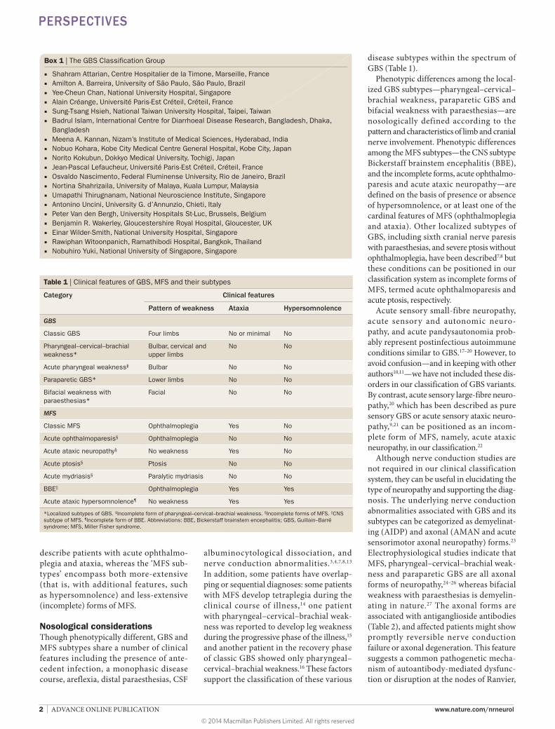
disease subtypes within the spectrum of GBS (Table 1).
Phenotypic differences among the localized GBS subtypes—pharyngeal– cervical–brachial weakness, paraparetic GBS and bifacial weakness with paraesthesias—are nosologically defined according to the pattern and characteristics of limb and cranial nerve involvement. Phenotypic differences among the MFS subtypes—the CNS subtype Bickerstaff brainstem encephalitis (BBE), and the incomplete forms, acute ophthalmoparesis and acute ataxic neuropathy—are defined on the basis of presence or absence of hyper somno lence, or at least one of the cardinal features of MFS (ophthalmo plegia and ataxia). Other localized subtypes of GBS, including sixth cranial nerve paresis with paraesthesias, and severe ptosis without ophthalmoplegia, have been described7,8 but these conditions can be positioned in our classification system as incomplete forms of MFS, termed acute ophthalmoparesis and acute ptosis, respectively.
Acute sensory smallfibre neuropathy, acute sensory and autonomic neuropathy, and acute pandysautonomia probably represent postinfectious autoimmune conditions similar to GBS.17–20 However, to avoid confusion—and in keeping with other authors10,11—we have not included these disorders in our classification of GBS variants. By contrast, acute sensory largefibre neuropathy,20 which has been described as pure sensory GBS or acute sensory ataxic neuropathy,9,21 can be positioned as an incomplete form of MFS, namely, acute ataxic neuropathy, in our classification.22
Although nerve conduction studies are not required in our clinical classification system, they can be useful in elucidating the type of neuropathy and supporting the diagnosis. The underlying nerve conduction abnormalities associated with GBS and its subtypes can be categorized as demyelinating (AIDP) and axonal (AMAN and acute sensorimotor axonal neuropathy) forms.23 Electrophysiological studies indicate that MFS, pharyngeal–cervical–brachial weakness and paraparetic GBS are all axonal forms of neuropathy,24–26 whereas bifacial weakness with paraesthesias is demyelinating in nature.27 The axonal forms are associated with antiganglioside anti bodies (Table 2), and affected patients might show promptly reversible nerve conduction failure or axonal degeneration. This feature suggests a common pathogenetic mechanism of autoantibodymediated dysfunction or disruption at the nodes of Ranvier,
Box 1 | The GBS Classification Group
■ Shahram Attarian, Centre Hospitalier de la Timone, Marseille, France ■ Amilton A. Barreira, University of São Paulo, São Paulo, Brazil ■ Yee-Cheun Chan, National University Hospital, Singapore ■ Alain Créange, Université Paris-Est Créteil, Créteil, France ■ Sung-Tsang Hsieh, National Taiwan University Hospital, Taipei, Taiwan ■ Badrul Islam, International Centre for Diarrhoeal Disease Research, Bangladesh, Dhaka,
Bangladesh ■ Meena A. Kannan, Nizam’s Institute of Medical Sciences, Hyderabad, India ■ Nobuo Kohara, Kobe City Medical Centre General Hospital, Kobe City, Japan ■ Norito Kokubun, Dokkyo Medical University, Tochigi, Japan ■ Jean-Pascal Lefaucheur, Université Paris-Est Créteil, Créteil, France ■ Osvaldo Nascimento, Federal Fluminense University, Rio de Janeiro, Brazil ■ Nortina Shahrizaila, University of Malaya, Kuala Lumpur, Malaysia ■ Umapathi Thirugnanam, National Neuroscience Institute, Singapore ■ Antonino Uncini, University G. d’Annunzio, Chieti, Italy ■ Peter Van den Bergh, University Hospitals St-Luc, Brussels, Belgium ■ Benjamin R. Wakerley, Gloucestershire Royal Hospital, Gloucester, UK ■ Einar Wilder-Smith, National University Hospital, Singapore ■ Rawiphan Witoonpanich, Ramathibodi Hospital, Bangkok, Thailand ■ Nobuhiro Yuki, National University of Singapore, Singapore
Table 1 | Clinical features of GBS, MFS and their subtypes
Category Clinical features
Pattern of weakness Ataxia Hypersomnolence
GBS
Classic GBS Four limbs No or minimal No
Pharyngeal–cervical–brachial weakness*
Bulbar, cervical and upper limbs
No No
Acute pharyngeal weakness‡ Bulbar No No
Paraparetic GBS* Lower limbs No No
Bifacial weakness with paraesthesias*
Facial No No
MFS
Classic MFS Ophthalmoplegia Yes No
Acute ophthalmoparesis§ Ophthalmoplegia No No
Acute ataxic neuropathy§ No weakness Yes No
Acute ptosis§ Ptosis No No
Acute mydriasis§ Paralytic mydriasis No No
BBE|| Ophthalmoplegia Yes Yes
Acute ataxic hypersomnolence¶ No weakness Yes Yes
*Localized subtypes of GBS. ‡Incomplete form of pharyngeal–cervical–brachial weakness. §Incomplete forms of MFS. ||CNS subtype of MFS. ¶Incomplete form of BBE. Abbreviations: BBE, Bickerstaff brainstem encephalitis; GBS, Guillain–Barré syndrome; MFS, Miller Fisher syndrome.
PERSPECTIVES
© 2014 Macmillan Publishers Limited. All rights reserved
NATURE REVIEWS | NEUROLOGY ADVANCE ONLINE PUBLICATION | 3
resulting in a continuum of nerve pathologies from transitory conduction failure to axonal degeneration.23,26,28
We believe that the updated classification system we present enables most forms of GBS to be accurately defined as discrete or overlapping syndromes on the basis of their clinical features.
Clinical–serological relationshipOur classification system does not stipulate antiganglioside antibody testing, although the results can be informative. The discovery
of antiGQ1b antibodies in patients with MFS and BBE provided important evidence that these disorders formed part of the same disease spectrum,29 and a comparative study revealed that antiGQ1b antibodies were present in 83% of patients with MFS and 68% of patients with BBE.30 GQ1b is strongly expressed in the oculo motor, trochlear and abducens nerves, as well as muscle spindles; thus, antiGQ1b antibodies are thought to cause both ophthalmoplegia and cerebellarlike ataxia.31,32 In addition, GQ1b is possibly expressed in the reticular formation, and
antiGQ1b antibodies might, therefore, explain the decreased level of consciousness seen in patients with BBE.30 Acute ophthalmoparesis is also associated with the presence of antiGQ1b antibodies, and some patients later develop bilateral facial or bulbar weakness.22 Neither ptosis nor mydriasis is a cardinal feature of MFS, but patients often present with these signs.33 Isolated ptosis and mydriasis, caused by weakness of the levator palpebrae superioris and iris sphincter muscles, respectively, have also been reported in association with
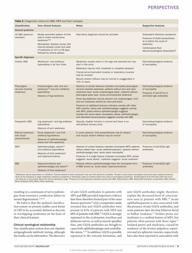
Table 2 | Diagnostic criteria for GBS, MFS and their subtypes
Classification Core clinical features Notes Supportive features
General syndrome
All GBS spectrum disorders
Mostly symmetric pattern of limb and/or motor cranial-nerve weakness*‡§
Monophasic disease course with interval between onset and nadir of weakness of 12 h to 28 days, followed by clinical plateau
Alternative diagnosis should be excluded Antecedent infectious symptoms||
Presence of distal paraesthesia at or before the onset of weakness
Cerebrospinal fluid albuminocytological dissociation¶
Specific diagnoses
Classic GBS Weakness* and areflexia/hyporeflexia in all four limbs
Weakness usually starts in the legs and ascends but may start in the arms
Weakness may be mild, moderate or complete paralysis
Cranial-nerve-innervated muscles or respiratory muscles may be involved
Muscle stretch reflexes may be normal or exaggerated in 10% of cases
Electrophysiological evidence of neuropathy
Pharyngeal–cervical–brachial weakness
Oropharyngeal, neck and arm weakness*‡ and arm areflexia/hyporeflexia
Absence of leg weakness
Absence of certain features indicates incomplete pharyngeal–cervical–brachial weakness: patients without arm and neck weakness have ‘acute oropharyngeal palsy’; patients without pharyngeal palsy have ‘acute cervicobrachial weakness’
Some leg weakness may be present, but oropharyngeal, neck and arm weakness should be more prominent
Presence of additional features indicates overlap with other GBS variants: ataxia with ophthalmoplegia suggests overlap with MFS; ataxia without ophthalmoplegia suggests overlap with acute ataxic neuropathy; ataxia, ophthalmoplegia and disturbed consciousness suggests overlap with BBE
Electrophysiological evidence of neuropathy
Presence of anti-GT1a or anti-GQ1b IgG antibodies
Paraparetic GBS Leg weakness* and leg areflexia/hyporeflexia
Absence of arm weakness
Typically, bladder function is normal and there is no well-defined sensory level
Electrophysiological evidence of neuropathy
Bifacial weakness with distal paraesthesias
Facial weakness* and limb areflexia/hyporeflexia
Absence of ophthalmoplegia, ataxia and limb weakness
In some patients, limb paraesthesias may be absent and muscle stretch reflexes may be normal
Electrophysiological evidence of neuropathy
MFS Ophthalmoplegia, ataxia*‡ and areflexia/hyporeflexia
Absence of limb weakness# and hypersomnolence
Absence of certain features indicates incomplete MFS: patients without ataxia have ‘acute ophthalmoparesis’; patients without ophthalmoplegia have ‘acute ataxic neuropathy’
Presence of a single feature indicates incomplete MFS: ptosis suggests ‘acute ptosis’; mydriasis suggests ‘acute mydriasis’
Presence of anti-GQ1b IgG antibodies
BBE Hypersomnolence and ophthalmoplegia and ataxia*‡
Absence of limb weakness#
Patients without ophthalmoplegia have the incomplete form of BBE known as ‘acute ataxic hypersomnolence’
Presence of anti-GQ1b IgG antibodies
*Weakness may be asymmetric or unilateral. ‡Clinical severity of each component may vary from partial to complete. §Except in acute ataxic neuropathy and acute ataxic hypersomnolence. ||Such as the presence of upper respiratory infectious symptoms or diarrhoea 3 days to 6 weeks before the onset of neurological symptoms. ¶Cerebrospinal fluid with total white cell count <50 cells per μl and protein above the normal laboratory range. #Presence of limb weakness indicates overlap with GBS. Abbreviations: BBE, Bickerstaff brainstem encephalitis; GBS, Guillain–Barré syndrome; MFS, Miller Fisher syndrome.
PERSPECTIVES
© 2014 Macmillan Publishers Limited. All rights reserved
4 | ADVANCE ONLINE PUBLICATION www.nature.com/nrneurol
antiGQ1b antibodies, and these features might represent a very incomplete form of MFS.34,35
Socalled ataxic GBS is characterized by profound cerebellarlike ataxia in the absence of both a Romberg sign and ophthalmoplegia.13 Patients with ataxic GBS also carry antiGQ1b IgG antibodies, which supports its classification as an incomplete form of MFS.36 Acute sensory ataxic neuropathy is characterized by profound sensory ataxia in the absence of ophthalmoplegia, but with a Romberg sign.21 The nosological position of acute sensory ataxic neuropathy became clear when affected individuals were compared with patients who had ataxic GBS.33 AntiGQ1b antibodies were found in 18% of patients with acute sensory ataxic neuropathy, and in 65% of patients with ataxic GBS. AntiGD1b IgG antibodies without GQ1b crossreactivity were detected in 35% of the patients with acute sensory ataxic neuropathy and 14% of those with ataxic GBS. These findings suggest that the conditions are not distinct but, rather, variants within a continuous spectrum. Cerebellarlike ataxia has also been described in patients with MFS, and is thought to be caused by selective dysfunction of muscle spindle afferents mediated by antiGQ1b anti bodies.3,37 To avoid confusion, ataxic GBS and acute sensory ataxic neuropathy are classified in this article as incomplete forms of MFS, and are collectively referred to as acute ataxic neuropathy.33,38,39
GT1a is more densely expressed than GQ1b in human glossopharyngeal and vagal nerves.40 Patients with pharyngeal–cervical–brachial weakness often carry antiGT1a IgG antibodies, some of which might crossreact with GQ1b.41 Although the serological profiles of patients with pharyngeal– cervical–brachial weakness show con siderable overlap with those of patients with MFS, we believe that pharyngeal– cervical– brachial weakness is best placed as a subtype of GBS rather than MFS. By definition, patients with pharyngeal– cervical–brachial weakness display arm weakness, whereas those with MFS do not, and pharyngeal– cervical– brachial weakness should, therefore, be considered a localized subtype of GBS. Patients with acute oropharyngeal palsy have antiGT1a and antiGQ1b anti bodies,42 supporting the classification of this condition as an incomplete form of pharyngeal– cervical–brachial weakness. By contrast, AMAN is associated with antiGM1 or antiGD1a IgG antibodies,43 and the discovery of antiGD1a antibodies in a patient with
paraparetic GBS whose nerve conduction results indicated axonal neuro pathy suggested that paraparetic GBS is a localized form of AMAN.25
Peripheral versus central pathologyMany neurologists view GBS as a disease that only affects the peripheral nerves, but this assertion is not always true: some patients display exaggerated deep tendon reflexes, either transiently or throughout the course of their illness.44,45 The early formal criteria for GBS5 required hyporeflexia or areflexia, but the researcher who developed these criteria later recognized that some patients with features otherwise typical of GBS also displayed “extensor plantar responses, and illdefined sensory levels,” indicating possible CNS involvement.6 Although application of these criteria has enabled most patients with GBS to be identified, some individuals have undoubtedly been misdiagnosed in the past.
The risk of misdiagnosis was first highlighted in a report that described three North American men who each developed rapidly progressive tetraparesis after a gastrointestinal illness, but were not diagnosed as having GBS owing to the presence of normal to brisk deep tendon reflexes.46 Similarly, acute pure motor neuropathy in four patients who carried antiGM1 IgG antibodies was not initially diagnosed as GBS because of the preservation of reflexes, yet nerve conduction studies later confirmed AMAN.47 The presence of hyperreflexia resulted in AMAN being initially misdiagnosed as postinfectious myelitis in a patient with progressive weakness in all four limbs, although nerve conduction studies later revealed the correct diagnosis.48
A study of 213 patients with GBS identified 23 patients (10%) who demonstrated normal or brisk reflexes during the clinical course of illness.45 Among these individuals, tendon reflexes were normal in eight patients and exaggerated in three patients throughout the course of illness. The remaining 12 patients exhibited exaggerated reflexes at some stage, which later returned to normal. Interestingly, patients with GBS who had preserved deep tendon reflexes more frequently presented with pure motor limb weakness, and were more likely to have antiGM1 or antiGD1a antibodies, as well as neurophysio logical features consistent with AMAN, than were patients with reduced reflexes. A similar rate (9%) of normal or exaggerated reflexes was observed in a study of 494 patients with GBS in the Netherlands, and most of these
patients showed evidence of a mild, pure motor neuro pathy.49 Some clini cians have suggested that such individuals should be categ orized as having a ‘hyper reflexic variant’ of GBS,50 but we believe that this distinction is unnecessary. The localization and underlying mechanism of hyper reflexia in such individuals remain unknown, although disruption of intramedullary inhibitory interneurons, which could occur if anti ganglio side antibodies crossed the blood–brain barrier, has been postulated.48
The earliest descriptions of BBE and MFS suggested overlap between the two syndromes; for example, half of Bickerstaff ’s patients had hyporeflexia or areflexia,4 and one of Miller Fisher’s three patients experienced drowsiness.3 This overlap led to widespread conjecture about whether the aetiologies of these disorders were central or peripheral. In a large sample of patients with BBE, CNS pathology was observed on brain MRI in 11%, and abnormal EEG recordings were obtained in 57%.30 Though only 2% of patients with MFS showed abnormalities on MRI, 25% of patients had aberrant EEG activity,30 which indicated that some patients with MFS—despite having no impairment of consciousness—had evidence of CNS dysfunction. A complementary overlap between BBE and MFS exists with regard to PNS pathology: 74% of patients with MFS demonstrated absence of the soleus Hreflex in peripheral nerve testing, as did three of four patients with BBE. Together, these results suggest that MFS and BBE lie within a clinical spectrum of variable involvement of the PNS and CNS.
Diagnosis and classificationWe believe that the diagnosis of GBS, MFS and their subtypes can be made clinically in the majority of patients, and that current diagnostic criteria are too rigid and overly reliant on laboratory data.6,10,11 Nerve conduction studies and CSF analysis are often inconclusive in the early stages of disease and, therefore, should not delay diagnosis and treatment if GBS or its variants are suspected on clinical grounds. CSF albuminocytological dissociation is absent within the first week of symptom onset in more than half of patients with GBS.51 In approximately 40% of patients, nerve conduction studies performed within the first week can suggest a diagnosis of neuropathy without fulfilling the criteria for one of the specific electrophysiological subtypes.49 Moreover, on the basis of serial nerve conduction study recordings, the electrophysiological classification of
PERSPECTIVES
© 2014 Macmillan Publishers Limited. All rights reserved
NATURE REVIEWS | NEUROLOGY ADVANCE ONLINE PUBLICATION | 5
GBS has been shown to change in 24–38% of patients , making the diagnosis of the precise subtype an a posteri ori process.23,52 Furthermore, neurophysiological examination is not readily available in some parts of the world. Antiganglioside antibody testing is useful, but obtaining results takes time and, therefore, should not be relied on for diagnosis.
Striking an optimal balance between diagnostic criteria that are neither too inclusive nor too restrictive remains important for clinical practice. Two decades ago, the American Academy of Neurology outlined strict diagnostic criteria (specificity 100%, sensitivity 46%) for chronic inflammatory demyelinating polyneuropathy (CIDP).53 These criteria have since been replaced by slightly less specific (96%) but much more sensitive (81%) criteria that enable the recognition of atypical presentations, for which immunotherapy would have otherwise been delayed or withheld.54,55 We believe that a similar reappraisal of the current diagnostic and classification criteria for GBS is warranted to avoid underdiagnosis and delayed treatment, especially in patients with atypical forms or those with normal or exaggerated deep tendon reflexes.
The diagnostic criteria that we present in Table 2 facilitate the classification of GBS, MFS and their subtypes. Weakness, which may affect the limbs or the territories served by motor cranial nerves, is a core feature that is present in almost all subtypes, the main exceptions being acute ataxic neuropathy and acute ataxic hypersomnolence (Table 1). Weakness is usually symmetric; however, unilateral ophthalmoplegia with or without unilateral ataxia (and, rarely, with unilateral limb weakness) has been reported in association with antiganglioside antibodies.11,56–58
The other core feature is that the clinical course should be monophasic. The time interval between the onset and nadir of neurological symptoms ranges from 12 h to 28 days, and is followed by a clinical plateau. In a study published in 2014, 97% of patients with GBS reached symptom nadir within 4 weeks.49 We regard treatmentrelated clinical fluctuations occurring within 8 weeks after the start of immunotherapy as part of the monophasic course, and patients exhibiting these fluctuations should be differentiated from patients with acuteonset CIDP.59 The latter diagnosis should be considered in patients who deteriorate after 8 weeks from initial onset, or when three or more treatment related fluctuations occur.
Recurrent GBS is seen in less than 10% of patients, but subsequent episodes seem to become more severe and occur at shorter time intervals as time goes on.60,61 A history of upper respiratory infections or diarrhoea 3 days to 6 weeks before the onset of GBS is common and supports the diagnosis.
GBS can be diagnosed in patients who present with bilateral flaccid limb weakness. Frequently, patients with GBS also experience distal limb numbness, paraesthesia or pain, any of which can even be their initial symptom.62 Weakness associated with GBS is often described as ascending, and might involve respiratory muscles and cranial nerves. Up to 10% of patients might display normal or exaggerated reflexes throughout the disease course.45,49 In patients with suspected GBS who present with preserved or exaggerated reflexes, repeated nerve conduction studies are essential, as evidence of peripheral neuropathy can confirm the diagnosis.
The key clinical finding in patients with pharyngeal–cervical–brachial weakness is oropharyngeal, neck and arm weakness associated with areflexia.12 The majority also experience some sensory disturbance in the arms, although sensory impairment was not included in the original description of this GBS subtype.7 The consistency and severity of leg weakness varies within and between patients, but generally should not be more prominent than arm weakness. Incomplete forms of pharyngeal– cervical–brachial weakness have also been described. For example, isolated bulbar palsy results in acute pharyngeal weakness,42 but can also progress to complete pharyngeal–cervical–brachial weakness during the course of the illness.63 Along with such transitions, the presence of antiGT1a or antiGQ1b IgG antibodies further supports the clinical diagnosis of pharyngeal– cervical–brachial weakness (or one of its incomplete forms).24,41
Patients with paraparetic GBS develop flaccid leg weakness, but have normal neuro logical findings in the upper limbs.7,8 The diagnosis is supported by evidence of axonaltype neuropathy on nerve conduction studies, and the presence of antiganglioside antibodies.25
Bilateral facial weakness in the absence of limb weakness can be sufficient to warrant a diagnosis of bifacial weakness with paraesthesias, although sensory disturbance in the limbs also occurs in the majority of individuals.8,27 Diagnosis is supported by demyelinating features on nerve conduction studies in the limbs.
Patients with MFS present with ophthalmoplegia, ataxia and areflexia,3 and those who additionally experience hypersomnolence have BBE, the CNS subtype of MFS.4,64 Although none of the patients originally described by Bickerstaff displayed hyperreflexia, some researchers have suggested that the presence of hyperreflexia is sufficient to diagnose BBE even in the absence of hypersomnolence, because both features indicate CNS involvement.65 However, given that some patients with GBS also display hyperreflexia,45 our classification system categorizes such patients as having MFS rather than BBE. Incomplete forms of MFS include acute ataxic neuropathy, which can be diagnosed in the absence of ophthalmoplegia,33 and acute ophthalmo paresis, which can be diagnosed in the absence of ataxia.22 Very incomplete forms of MFS show only isolated ptosis or mydriasis.34,35 Incomplete BBE, known as acute ataxic hypersomnolence, can be the diagnosis in patients who have evidence of ataxia and hypersomnolence in the absence of ophthal moplegia.66 The presence of antiGQ1b or antiGD1b IgG antibodies can confirm the clinical diagnosis of these MFS subtypes.
The possibility of overlap between GBS, MFS and their subtypes warrants brief discussion. Patients with MFS or BBE who develop limb weakness can be diagnosed as having overlap with GBS.14,65 Leg weakness can develop in some patients with pharyngeal–cervical–brachial weakness. If it occurs early and is severe,16 this represents fulminant pharyngeal–cervical–brachial weakness, rather than overlap with GBS. As outlined in the original description,7 patients with pharyngeal–cervical– brachial weakness could present with ophthalmoplegia, whereas those patients with pharyngeal–cervical– brachial weakness presenting with additional ophthalmo plegia and ataxia in the absence of tetraparesis have overlapping MFS.
The frequency of different subtypes within the GBS–MFS spectrum has not been examined in detail, and is likely to vary in different parts of the world. One prospective study of more than 250 patients diagnosed with GBS at a single hospital in the USA found the following frequencies: MFS 5%, pharyngeal–cervical–brachial weakness 3%, paraparetic GBS 2%, and bifacial weakness with paraesthesias 1%.9 A retrospective singlehospital study conducted in Taiwan found the following frequencies among 43 patients: MFS 7%, BBE 7%, pharyngeal– cervical–brachial weakness 5%, and polyneuritis cranialis 5%.67 In a study from
PERSPECTIVES
© 2014 Macmillan Publishers Limited. All rights reserved
6 | ADVANCE ONLINE PUBLICATION www.nature.com/nrneurol
the Netherlands that applied the Brighton crit eria11 to diagnose approximately 500 patients with GBS, after exclusion of those with MFS and BBE, weakness was restricted to the legs in 6% of patients, which suggests a diagnosis of paraparetic GBS, and to the arms in 1% of patients, in keeping with pharyngeal–cervical–brachial weakness.49
Differential diagnosesNumerous conditions can mimic GBS, MFS and their subtypes. In this section, we highlight the most important of these differential diagnoses—diseases that also cause rapidly progressive and often symmetric paresis (Box 2).68
In patients who present with weakness and exaggerated reflexes, a central cause should always be excluded before the weakness is
attributed to GBS. Conversely, patients with extensive necrotizing myelo pathy can lose their deep tendon reflexes. Spinal cord or, in some patients, brainstem pathology should be excluded early with MRI, especially if a ‘sensory level’—a point on the body below which sensation is reduced or absent—is present, or if weakness develops abruptly in association with urinary retention. Although urinary dysfunction may develop in over onequarter of patients with GBS,69 typically it is not an early feature.
Isolated sensory symptoms in the presence of normal deep tendon reflexes might be misdiagnosed as a conversion or dissociative disorder in some patients with GBS, especially early in the disease course. Such patients should be advised to return to the clinic if they experience disease progression. By contrast, functional weakness is usually sudden in onset (less than 12 h into the disease course) and asymmetric. When present, motor and sensory physical signs are inconsistent or incongruent with any GBS subtype. In the absence of sensory deficits, differential diagnoses for GBS include periodic paralysis, myasthenia gravis, botulism, poliomyelitis, and acute myopathy of any cause.
The main differential diagnoses for both MFS and pharyngeal–cervical–brachial weakness are brainstem strokes, myasthenia gravis and botulism. In patients with reduced levels of consciousness, Wernicke encephalopathy and brainstem encephalitis should be excluded before considering BBE. In patients with isolated or multiple cranial neuropathies—especially when these are asymmetric—other inflammatory, neoplastic and infectious aetiologies must be excluded.
In the early stages, paraparetic GBS can be difficult to distinguish from other more common conditions, such as lumbosacral plexopathy or cauda equina syndrome. MRI with gadolinium contrast of the lumbo sacral region is, therefore, mandatory to exclude an infiltrative or compressive cause of the paraparesis. In patients with diabetic neuropathy or idiopathic lumbosacral plexopathy, onset is typically asymmetric, with continued progression and bilateral involvement beyond 1 month. Cytomegalovirus infection in the context of HIVpositive patients might also cause painful lumbosacral plexopathy, and this infection is usually associated with urinary retention.
Careful history taking and clinical examination should enable clinicians to differentiate bifacial weakness with paraesthesias
from bilateral Bell palsy in the majority of individuals. Typically, patients with bilateral Bell palsy do not have distal paraesthesias70 and are likely to present with other features, including mastoid pain and hyperacusis; these patients are less likely to recover fully than are patients with bifacial weakness with paraesthesias.27 Laboratory data can also differentiate patients with Bell palsy (who do not demonstrate CSF albuminocytological dissociation or evidence of demyelination in their limbs) from individuals who have bifacial weakness with paraesthesias. However, rapidly progressive isolated bilateral facial weakness may also be present in patients with sarcoidosis or Lyme disease.
ConclusionsIn this article, we classify GBS, MFS and their subtypes according to their clinical features. The appreciation that these conditions form a continuum wherein discrete, complete and incomplete forms of disease exist (and sometimes overlap) remains the most important concept for defining atypical presentations of these disorders and overlap syndromes. The validity of this classification system still needs to be tested in large cohorts of patients by independent assessors in different parts of the world, and we accept that further refinements may be necessary. Moreover, this classification system should be discussed in the context of guidelines for the management of GBS published by the European Federation of Neurological Societies and the Peripheral Nerve Society. The use of this classification system could enable important data to be collected on the frequency of different subtypes of GBS and MFS in different countries, as well as the natural course of these illnesses and their responses to treatment. We envisage that this approach would help to identify the most commonly encountered differential diagnoses for each variant, and contribute to the development of standardized management protocols.
Department of Neurology, Gloucestershire Royal Hospital, Great Western Road, Gloucester GL1 3NN, UK (B.R.W.). Department of Neuroscience, Imaging and Clinical Sciences, University G. d’Annunzio, Via dei Vestini 31, Chieti 66013, Italy (A.U.). Departments of Medicine and Physiology, Yong Loo Lin School of Medicine, National University of Singapore, Unit 09‑01, Centre for Translational Medicine, 14 Medical Drive, Singapore 117599, Singapore (N.Y.). Correspondence to: N.Y. [email protected]
Box 2 | Differential diagnoses
GBS ■ Acute spinal cord disease ■ Carcinomatous or lymphomatous
meningitis ■ Myasthenia gravis ■ Critical illness neuropathy ■ Thiamine deficiency ■ Periodic paralysis ■ Corticosteroid-induced myopathy ■ Toxins (such as neurotoxic shellfish
poisoning) ■ Acute hypophosphataemia ■ Prolonged use of neuromuscular junction
blocking drugs ■ Tick paralysis ■ West Nile poliomyelitis ■ Acute intermittent porphyria ■ Functional paralysis
MFS, BBE, and pharyngeal–cervical–brachial weakness ■ Brainstem stroke ■ Myasthenia gravis ■ Wernicke encephalopathy ■ Botulism ■ Brainstem encephalitis ■ Diphtheria ■ Tick paralysis
Paraparetic GBS ■ Lumbosacral plexopathy ■ Diabetic neuropathy ■ Neoplasms ■ Inflammatory conditions (such as
sarcoidosis) ■ Infections (such as cytomegalovirus,
Lyme disease) ■ Lesions of the cauda equina
Bifacial weakness with paraesthesias ■ Lyme disease ■ Sarcoidosis
Abbreviations: BBE, Bickerstaff brainstem encephalitis; GBS, Guillain–Barré syndrome; MFS, Miller Fisher syndrome.
PERSPECTIVES
© 2014 Macmillan Publishers Limited. All rights reserved
NATURE REVIEWS | NEUROLOGY ADVANCE ONLINE PUBLICATION | 7
1. Guillain, G., Barré, J. A. & Strohl, A. Sur un syndrome de radiculonévrite avec hyperalbuminose du liquide céphalo-rachidien sans réaction cellulaire: remarques sur les caractères cliniques et graphiques des réflexes tendineux [French]. Bull. Mem. Hop. Paris 40, 1462–1470 (1916).
2. Guillain, G. Les polyradiculonévrites avec dissociation alumbinocytologique et á evolution favorable (syndrome de Guillain et Barré) [French]. J. Belge Neurol. Psychiatr. 38, 323–329 (1938).
3. Fisher, M. An unusual variant of acute idiopathic polyneuritis (syndrome of ophthalmoplegia, ataxia and areflexia). N. Engl. J. Med. 255, 57–65 (1956).
4. Bickerstaff, E. R. Brain-stem encephalitis: further observations on a grave syndrome with benign prognosis. Br. Med. J. 1, 1384–1387 (1957).
5. Asbury, A. K., Arnason, B. G., Karp, H. R. & McFarlin, D. E. Criteria for diagnosis of Guillain–Barré syndrome. Ann. Neurol. 3, 565–566 (1978).
6. Asbury, A. K. & Cornblath, D. R. Assessment of current diagnostic criteria for Guillain–Barré syndrome. Ann. Neurol. 27 (Suppl.), S21–S24 (1990).
7. Ropper, A. H. Unusual clinical variants and signs in Guillain–Barré syndrome. Arch. Neurol. 43, 1150–1152 (1986).
8. Ropper, A. H. Further regional variants of acute immune polyneuropathy: bifacial weakness or sixth nerve paresis with paresthesias, lumbar polyradiculopathy, and ataxia with pharyngeal–cervical–brachial weakness. Arch. Neurol. 51, 671–675 (1994).
9. Ropper, A. H., Wijdicks, E. F. M. & Truax, B. Guillain–Barré Syndrome (F. A. Davis, 1990).
10. van der Meché, F. G. et al. Diagnostic and classification criteria for the Guillain–Barré syndrome. Eur. Neurol. 45, 133–139 (2001).
11. Sejvar, J. J. et al. Guillain–Barré syndrome and Fisher syndrome: case definitions and guidelines for collection, analysis, and presentation of immunization safety data. Vaccine 29, 599–612 (2011).
12. Wakerley, B. R. & Yuki, N. Pharyngeal–cervical–brachial variant of Guillain–Barré syndrome. J. Neurol. Neurosurg. Psychiatry 85, 339–344 (2013).
13. Richter, R. B. The ataxic form of polyradiculoneuritis (Landry–Guillain–Barré syndrome): clinical and pathologic observations. J. Neuropathol. Exp. Neurol. 21, 171–184 (1962).
14. Kimoto, K. et al. Relationship of bacterial strains to clinical syndromes of Campylobacter-associated neuropathies. Neurology 67, 1837–1843 (2006).
15. Mizoguchi, K. et al. Two species of antiganglioside antibodies in a patient with a pharyngeal–cervical–brachial variant of Guillain–Barré syndrome. J. Neurol. Neurosurg. Psychiatry 57, 1121–1123 (1994).
16. Miura, Y., Susuki, K., Yuki, N., Ayabe, M. & Shoji, H. Guillain–Barré syndrome presenting pharyngeal–cervical–brachial weakness in the recovery phase. Eur. Neurol. 48, 53–54 (2002).
17. Koike, H. et al. Clinicopathological features of acute autonomic and sensory neuropathy. Brain 133, 2881–2896 (2010).
18. Koike, H. et al. The spectrum of clinicopathological features in pure autonomic neuropathy. J. Neurol. 259, 2067–2075 (2012).
19. Ohyama, K. et al. Autonomic manifestations in acute sensory ataxic neuropathy: a case report. Auton. Neurosci. 179, 155–158 (2013).
20. Uncini, A. & Yuki, N. Sensory Guillain–Barré syndrome and related disorders: an attempt at systematization. Muscle Nerve 45, 464–470 (2012).
21. Pan, C. L., Yuki, N., Koga, M., Chiang, M. C. & Hsieh, S. T. Acute sensory ataxic neuropathy associated with monospecific anti-GD1b IgG antibody. Neurology 57, 1316–1318 (2001).
22. Yuki, N., Odaka, M. & Hirata, K. Acute ophthalmoparesis (without ataxia) associated with anti-GQ1b IgG antibody: clinical features. Ophthalmology 108, 196–200 (2001).
23. Kokubun, N. et al. Conduction block in acute motor axonal neuropathy. Brain 133, 2897–2908 (2010).
24. Arai, M., Susuki, K. & Koga, M. Axonal pharyngeal–cervical–brachial variant of Guillain–Barré syndrome without anti-GT1a IgG antibody. Muscle Nerve 28, 246–250 (2003).
25. Nagashima, T. et al. Paraparetic Guillain–Barré syndrome: extending the axonal spectrum. Neurol. Clin. Neurosci. 1, 224–226 (2013).
26. Umapathi, T., Tan, E. Y., Kokubun, N., Verma, K. & Yuki, N. Non-demyelinating, reversible conduction failure in Fisher syndrome and related disorders. J. Neurol. Neurosurg. Psychiatry 83, 941–948 (2012).
27. Susuki, K., Koga, M., Hirata, K., Isogai, E. & Yuki, N. A Guillain–Barré syndrome variant with prominent facial diplegia. J. Neurol. 256, 1899–1905 (2009).
28. Uncini, A., Susuki, K. & Yuki, N. Nodo-paranodopathy: beyond the demyelinating and axonal classification in anti-ganglioside antibody-mediated neuropathies. Clin. Neurophysiol. 124, 1928–1934 (2013).
29. Shahrizaila, N. & Yuki, N. Bickerstaff brainstem encephalitis and Fisher syndrome: anti-GQ1b antibody syndrome. J. Neurol. Neurosurg. Psychiatry 84, 576–583 (2013).
30. Ito, M. et al. Bickerstaff’s brainstem encephalitis and Fisher syndrome form a continuous spectrum: clinical analysis of 581 cases. J. Neurol. 255, 674–682 (2008).
31. Chiba, A., Kusunoki, S., Obata, H., Machinami, R. & Kanazawa, I. Serum anti-GQ1b IgG antibody is associated with ophthalmoplegia in Miller Fisher syndrome and Guillain–Barré syndrome: clinical and immunohistochemical studies. Neurology 43, 1911–1917 (1993).
32. Liu, J. X., Willison, H. J. & Pedrosa-Domellof, F. Immunolocalization of GQ1b and related gangliosides in human extraocular neuromuscular junctions and muscle spindles. Invest. Ophthalmol. Vis. Sci. 50, 3226–3232 (2009).
33. Ito, M., Matsuno, K., Sakumoto, Y., Hirata, K. & Yuki, N. Ataxic Guillain–Barré syndrome and acute sensory ataxic neuropathy form a continuous spectrum. J. Neurol. Neurosurg. Psychiatry 82, 294–299 (2011).
34. Jindal, G., Parmar, V. R. & Gupta, V. K. Isolated ptosis as acute ophthalmoplegia without ataxia, positive for anti-GQ1b immunoglobulin G. Pediatr. Neurol. 41, 451–452 (2009).
35. Yuki, N., Koga, M. & Hirata, K. Isolated internal ophthalmoplegia associated with immunoglobulin G anti-GQ1b antibody. Neurology 51, 1515–1516 (1998).
36. Yuki, N., Susuki, K. & Hirata, K. Ataxic Guillain–Barré syndrome with anti-GQ1b antibody: relation to Miller Fisher syndrome. Neurology 54, 1851–1853 (2000).
37. Yuki, N. & Uncini, A. Acute and chronic ataxic neuropathies with disialosyl antibodies: a continuous clinical spectrum and a common
pathophysiological mechanism. Muscle Nerve 49, 629–635 (2014).
38. Notturno, F., Caporale, C. M. & Uncini, A. Acute sensory ataxic neuropathy with antibodies to GD1b and GQ1b gangliosides and prompt recovery. Muscle Nerve 37, 265–268 (2008).
39. Willison, H. J., Almemar, A., Veitch, J. & Thrush, D. Acute ataxic neuropathy with cross-reactive antibodies to GD1b and GD3 gangliosides. Neurology 44, 2395–2397 (1994).
40. Koga, M., Yoshino, H., Morimatsu, M. & Yuki, N. Anti-GT1a IgG in Guillain–Barré syndrome. J. Neurol. Neurosurg. Psychiatry 72, 767–771 (2002).
41. Nagashima, T., Koga, M., Odaka, M., Hirata, K. & Yuki, N. Continuous spectrum of pharyngeal–cervical–brachial variant of Guillain–Barré syndrome. Arch. Neurol. 64, 1519–1523 (2007).
42. O’Leary, C. P. et al. Acute oropharyngeal palsy is associated with antibodies to GQ1b and GT1a gangliosides. J. Neurol. Neurosurg. Psychiatry 61, 649–651 (1996).
43. Ogawara, K. et al. Axonal Guillain–Barré syndrome: relation to anti-ganglioside antibodies and Campylobacter jejuni infection in Japan. Ann. Neurol. 48, 624–631 (2000).
44. McKhann, G. M. et al. Acute motor axonal neuropathy: a frequent cause of acute flaccid paralysis in China. Ann. Neurol. 33, 333–342 (1993).
45. Yuki, N. et al. Guillain–Barré syndrome associated with normal or exaggerated tendon reflexes. J. Neurol. 259, 1181–1190 (2012).
46. Jackson, C. E., Barohn, R. J. & Mendell, J. R. Acute paralytic syndrome in three American men: comparison with Chinese cases. Arch. Neurol. 50, 732–735 (1993).
47. Yuki, N. & Hirata, K. Preserved tendon reflexes in Campylobacter neuropathy. Ann. Neurol. 43, 546–547 (1998).
48. Kuwabara, S. et al. Hyperreflexia in axonal Guillain–Barré syndrome subsequent to Campylobacter jejuni enteritis. J. Neurol. Sci. 199, 89–92 (2002).
49. Fokke, C. et al. Diagnosis of Guillain–Barré syndrome and validation of Brighton criteria. Brain 137, 33–43 (2014).
50. Baheti, N. N., Manuel, D., Shinde, P. D., Radhakrishnan, A. & Nair, M. Hyperreflexic Guillain–Barré syndrome. Ann. Indian Acad. Neurol. 13, 305–307 (2010).
51. Nishimoto, Y., Odaka, M., Hirata, K. & Yuki, N. Usefulness of anti-GQ1b IgG antibody testing in Fisher syndrome compared with cerebrospinal fluid examination. J. Neuroimmunol. 148, 200–205 (2004).
52. Uncini, A., Manzoli, C., Notturno, F. & Capasso, M. Pitfalls in electrodiagnosis of Guillain–Barré syndrome subtypes. J. Neurol. Neurosurg. Psychiatry 81, 1157–1163 (2010).
53. An ad hoc subcommittee of the American Academy of Neurology AIDS Task Force. Research criteria for diagnosis of chronic inflammatory demyelinating polyneuropathy (CIDP). Neurology 41, 617–618 (1991).
54. Joint Task Force of the EFNS and the PNS. European Federation of Neurological Societies/Peripheral Nerve Society Guideline on management of chronic inflammatory demyelinating polyradiculoneuropathy: report of a joint task force of the European Federation of Neurological Societies and the Peripheral Nerve Society—first revision. J. Peripher. Nerv. Syst. 15, 1–9 (2010).
PERSPECTIVES
© 2014 Macmillan Publishers Limited. All rights reserved
8 | ADVANCE ONLINE PUBLICATION www.nature.com/nrneurol
55. Rajabally, Y. A., Nicolas, G., Pieret, F., Bouche, P. & Van den Bergh, P. Y. Validity of diagnostic criteria for chronic inflammatory demyelinating polyneuropathy: a multicentre European study. J. Neurol. Neurosurg. Psychiatry 80, 1364–1368 (2009).
56. Ichikawa, H. et al. Unilateral oculomotor nerve palsy associated with anti-GQ1b IgG antibody. Neurology 59, 957–958 (2002).
57. Logullo, F., Manicone, M., Di Bella, P. & Provinciali, L. Asymmetric Guillain–Barré syndrome. Neurol. Sci. 27, 355–359 (2006).
58. Susuki, K., Yuki, N., Muramatsu, M. & Hirata, K. Unilateral ophthalmoparesis and limb ataxia associated with anti-GQ1b IgG antibody. J. Neurol. 247, 652–653 (2000).
59. Ruts, L., van Koningsveld, R. & van Doorn, P. A. Distinguishing acute-onset CIDP from Guillain–Barré syndrome with treatment related fluctuations. Neurology 65, 138–140 (2005).
60. Kuitwaard, K., van Koningsveld, R., Ruts, L., Jacobs, B. C. & van Doorn, P. A. Recurrent Guillain–Barré syndrome. J. Neurol. Neurosurg. Psychiatry 80, 56–59 (2009).
61. Mossberg, N. et al. The recurrent Guillain–Barré syndrome: a long-term population-based study. Acta Neurol. Scand. 126, 154–161 (2012).
62. Ropper, A. H. The Guillain–Barré syndrome. N. Engl. J. Med. 326, 1130–1136 (1992).
63. Nagashima, T., Odaka, M., Koga, M., Yuki, N. & Hirata, K. A case of pharyngeal–cervical–brachial weakness suggestive of the nosological continuity between acute oropharyngeal palsy [Japanese]. Rinsho Shinkeigaku 42, 523–526 (2002).
64. Bickerstaff, E. R. & Cloake, P. C. Mesencephalitis and rhombencephalitis. Br. Med. J. 2, 77–81 (1951).
65. Odaka, M. et al. Bickerstaff’s brainstem encephalitis: clinical features of 62 cases and a subgroup associated with Guillain–Barré syndrome. Brain 126, 2279–2290 (2003).
66. Wakerley, B. R., Soon, D., Chan, Y. C. & Yuki, N. Atypical Bickerstaff brainstem encephalitis: ataxic hypersomnolence without ophthalmoplegia. J. Neurol. Neurosurg. Psychiatry 84, 1206–1207 (2013).
67. Lin, J. J. et al. Clinical variants of Guillain–Barré syndrome in children. Pediatr. Neurol. 47, 91–96 (2012).
68. Ropper, A. H. & Samuels, M. A. Adam’s and Victor’s Principle of Neurology 9th edn 1251–1325 (McGraw–Hill, 2009).
69. Sakakibara, R. et al. Prevalence and mechanism of bladder dysfunction in Guillain–Barré syndrome. Neurourol. Urodyn. 28, 432–437 (2009).
70. Yanagihara, N., Mori, H., Kozawa, T., Nakamura, K. & Kita, M. Bell’s palsy. Nonrecurrent v recurrent and unilateral v bilateral. Arch. Otolaryngol. 110, 374–377 (1984).
AcknowledgementsThe authors are grateful to James Sejvar for his suggestions and critical reading of the manuscript. The authors’ work was supported by the Singapore National Medical Research Council (Individual Research Grant 10nov086 and Clinician Science Award 047/2012 to N.Y.) and Yong Loo Lin School of Medicine, Singapore (R-172-000-264-733).
Author contributionsB.R.W. and N.Y. researched data for the article and contributed equally to writing the article. B.R.W., A.U., N.Y. and all members of the GBS Classification Group made substantial contributions to discussions of the content, and review and/or editing of the manuscript before submission.
PERSPECTIVES
© 2014 Macmillan Publishers Limited. All rights reserved