gynaecology cancer awareness
TRANSCRIPT

GYNAECOLOGY CANCER AWARENESS
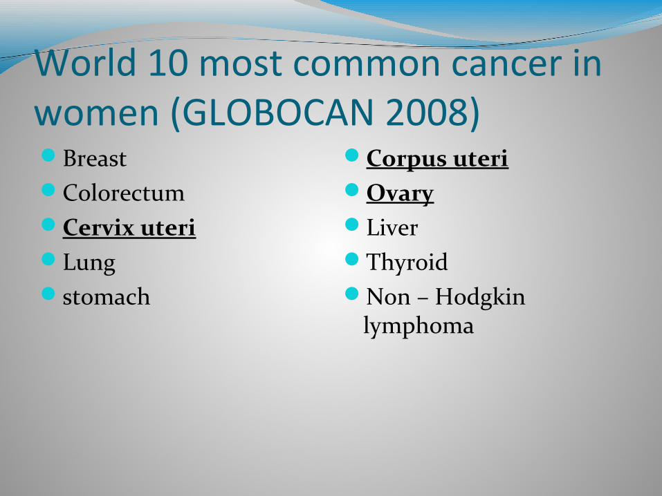
World 10 most common cancer in women (GLOBOCAN 2008)BreastColorectumCervix uteriLung stomach
Corpus uteriOvaryLiverThyroidNon – Hodgkin
lymphoma
Cervical CancerThird most common cancer in women.8.8 % of all female cancer.Responsible for a total of 275,000 deaths; 58% were
reported from Asia.
Uterine Cancer6th most common women cancer.More than 90 % of patients with uterine cancer are
more than 50 years old.Cancer of the uterus has a much more favourable
prognosis than ovarian and cervical cancer with 5 – year survival rates of around 80 – 90 % in developed countries and 70 % in developing countries.
Ovarian CancerCancer of the ovary is one of the most lethal
gynaecological malignancies due to late presentation, poor respond to treatment and high recurrent rate.
3.7 % of all women cancer; with 4.2 % of all cancer deaths in women.

Risk factorsCan be considered as sexually transmitted disease.Early age of sexual activity predisposes to cervical
cancer. Relative risk is 2.5 if the age of the first sexual exposure is < 18 years old.
Women with hx of HPV infection is also at a higher risk. Women with multiple sexual partners have a relative risk of 2.8 if the no. of partners > 4.
Multiparous women are at a higher risk. Women in lower socio-economic class are at a higher risk.

Women who are married to a man with multiple sexual partners are at a higher risk.
Cigarette smoking has been identified as a significant risk factor by 2 – 5 folds.
Patients who are immunodeficient are at a higher risk.
In-utero exposure to DES.Women with uncircumcised sexual partner may be at
higher risk.
PreventionThere are 3 modalities of cervical cancer prevention:1. Primary prevention:- prevention of HPV infection either through sexual
abstinence, healthy lifestyle and HPV vaccination.2. Secondary prevention:- detection and treatment of precancerous state
through screening.3. Tertiary prevention:- detection and treatment of an early stage of cancer.
1. HPV vaccination HPV is the primary aetiology of cervical cancer.Following the limitations of secondary prevention: - 2` prevention does not prevent HPV infection or
pre-invasive lesions of the cervix.- lesion that has rapid progression rate may not be
detected in time.- higher chances of missing the lesions in the cervical
canal.- limited sensitivity and specificity.- high cost and labour intensive.
Oncogenic types of HPV are estimated to cause almost 100% of cervical cancer.
2 potential benefits of HPV vaccination:- prevention of HPV-related precancerous and
cancerous lesions.- prevention of HPV-related benign lesions.
Currently 2 types of prophylactic HPV vaccines available in the market:
- quadrivalent HPV vaccine against HPV 16,18,6,11- bivalent HPV vaccine against HPV 16 and 18
2. Screening of cervical cancerScreening of precancerous lesions and invasive
cervical cancer is secondary prevention.It stops the progression of disease once it has already
started.Comprehensive analysis of data by International
Agency of Research on Cancer (IARC) showed:- well organised screening programs were effective in
reducing cervical cancer incidence and mortality.
- the greatest reduction in the cervical cancer incidence was in women aged between 30 and 49 years, for whom the focus of screening was the most intense.
Target age range of a screening program was a more important determinant of risk reduction than frequency of screening, within the defined age range.
Screening interval every 2 – 3 years have been found to be as effective as annual screening.
The protective effect of 5 years screening interval in the organised screening program was still high (> 80 %).
Screening every 2 years is approximately 50% more expensive than screening every 3 years.
Risk of developing invasive cervical cancer is 3 – 10 x greater in women who have not been screened. Risk also increases with long duration following the last normal screening test.
Type of screening tests1. cytology2. visual inspection3. HPV testing4. others
1. Cytological screening2 types:1. conventional cytology2. liquid – based cytology
Conventional cytology:- Pap smear screening program significantly reduced
the incidence and mortality of cervical cancer.- in UK, since the introduction of the organised pap
smear screening program in 1988, the incidence and mortality rate of cervical cancer have fallen by more than 50%.
- Pap smear is highly specific in detecting invasive cervical cancer and HGSIL, but less specific in low grade lesions.
- pap smear is not very sensitive. Low sensitivity was attributed to poor sample collection, incorrect slide preparation and lab interpretation errors.
To reduce the false negative, sample from the endocervix has to be taken. Cytobrush can obtain more cell than Ayre’s spatula and cotton tip applicator.
Liquid – based cytology:- introduced to increase the sensitivity and specificity
of cervical screening.- more positive results with LBC.- large reduction in unsatisfactory smear in LBC
group.- per test cost of LBC is higher.
2. Visual inspectionDirect visualisation of the cervix after application of
either acetic acid 3-5% or Lugol’s iodine.A result is obtained immediately and treatment can
be performed at the same time.
3. HPV testingHPV DNA is identified in almost all 99.7% verified
cases of cervical cancer worldwide.4 potential roles of HPV testing:1. triage of women with equivocal cytology2. following up of patient after treatment of CIN3. HPV testing in “see and treat” approach4. HPV testing as primary screening test
4. OthersOptoelectronic device - for optical and electrical
assessment of the cervix to detect any abnormal epithelium.
New technologies:- HPV genotyping- HPV mRNA- HPV viral load- HPV integration
Presentation Asymptomatic at an early stage.Abnormal vaginal bleeding = speculum exam.Most common - post coital bleeding.Passing out blood stained mucus.Pelvic pain.Copious foul smelling PV d/c.Constitutional symptoms.
Pelvic pain, urinary problems and constitutional symptoms – strong indication of advance stage disease.
Pyometra – if the d/s has obstructed the uterine outflow tract. Presents with pelvic pain, purulent vaginal d/c and fever.
Invasion posteriorly may cause tenesmus and PR bleeding.
late stage – SOB, bone pain, severe headache, neurological problems, LOA and LOW.
Lymphatic obstruction may result in lower limb lymphoedema.
Venous outflow obstruction can lead to DVT.

Type of endometrial cancerBroadly classified into 2 main categories:Type I endometrial cancer:- is related to exposure to unopposed endogenous or
exogenous oestrogen.- frequently arise on a background of atypical
hyperplasia.- have an association with obesity, nulliparity, insulin
resistance and hyperoestrogenism.- usually well differentiated and have a better
prognosis.
Type II:- has no association with predisposing factors.- example – uterine papillary serous, clear cell
carcinoma.- patients tend to be elderly and thin.- have a poorer prognosis.
80% of endometrial cancer are type I.
Risk factorsProlonged exposure to oestrogen.High level of endogenous oestrogen is often seen in
obese women, patients with PCOS and oestrogen producing tumour.
50% of patients has BMI > 25 kg/m2.In premenopausal women, obesity causes insulin
resistance, ovarian androgen excess, anovulation and chronic unopposed oestrogen.
DM and HPT – risk factors independent of their association with obesity.
The use of COCP for 1 year decreased the risk of endometrial cancers by more than 40%.
Women on combined HRT have also been found to have lower risk.
About 5% of endometrial cancer is hereditary, a/w a hereditary non-polyposis colon cancer syndrome (HNPCC).

Smoking is said to reduce the risk.Pregnancy has a natural protective effect because of
high production of progesterone by placenta.
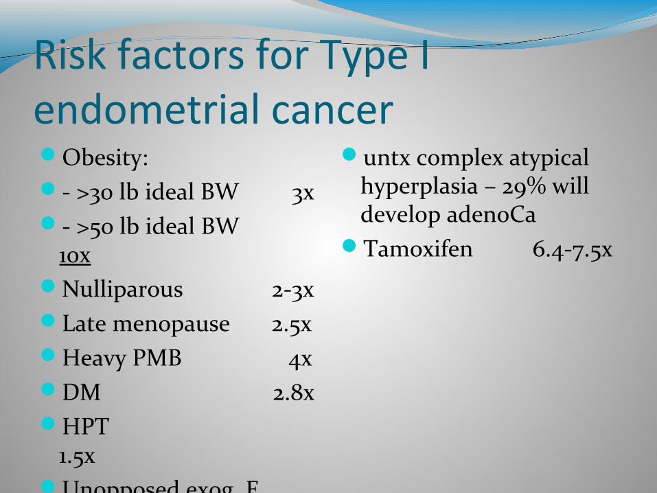
Risk factors for Type I endometrial cancerObesity:- >30 lb ideal BW 3x- >50 lb ideal BW
10xNulliparous 2-3xLate menopause 2.5xHeavy PMB 4xDM 2.8xHPT
1.5xUnopposed exog. E
9.5x
untx complex atypical hyperplasia – 29% will develop adenoCa
Tamoxifen 6.4-7.5x
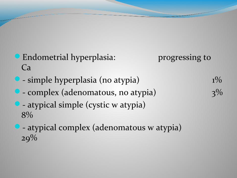
Endometrial hyperplasia: progressing to Ca
- simple hyperplasia (no atypia) 1%- complex (adenomatous, no atypia) 3%- atypical simple (cystic w atypia)
8%- atypical complex (adenomatous w atypia)
29%

Prevention Can be prevented by treating the precursor lesions
either by hormonal or surgical treatment.Adding a progestogen for at least 10 - 12 days per cycle
in HRT.Women with amenorrhoea or oligomenorrhoea
(including women with PCOS) should be treated with cyclical progestogen, to avoid prolonged exposure of the endometrium to a high level of oestrogen. Withdrawal bleeding of 3 monthly is sufficient to prevent endometrial pathology.
Suggestions of a protective effect of high phyto-oestrogen consumption among post menopausal women.
The use of IUCD and tubal ligation have also been associated with a lower risk.
Weight reduction and physical activities to achieve ideal body weight, can reverse the effect of prolonged exposure to a high level of oestrogen.
Pregnancy is a physiological protection, due to a high level of of progesterone produced b the placenta.
The use of COCP for 1 year decreased the risk by > 40%. Also known to reduce the incidence of ovarian cancer.
There is no effective screening method for endometrial cancer.
For available methods - accuracy in asymtomatic population is limited.
In women presenting with PMB, TVS showing ET of 5mm or less and negative endometrial sampling, have nearly 100% negative predictive value.
Presentation Majority of patients with endometrial cancer are
postmenopausal women.Most common presenting Sx is PMB.Abnormal PV d/c.abnormal PV bleeding in 80% (including PMB and
irregular menses in premenopausal women).Pressure Sx due to uterine enlargement.Sx indicating extrauterine spread.

Ovarian cancer
Epithelial cancer is infrequent below 40 years old.Peak rate is at 70 - 74 years age group with the
incidence of 57/100,000 population.Ovarian cancer at the older age group carries a poorer
prognosis.
Risk factors1. Reproductive factors:Parous women have 30 - 60% less risk compared to
nulliparous.The birth of the first child reduces the risk by 45%.
Every further pregnancy reduces the risk by 15%.Breast feeding reduces the risk.Tubal ligation is also associated with lower risk.Ovulation induction agent increases the risk by 2-3 x,
if taken for > 12 ovulatory cycles.
OCP reduces the risk by 30 - 60 %.History of breast cancer increases the risk.Early menache and late menopause are risk factors.Slightly increased risk with HRT.
2. Genetic factors:Lifetime risk in general population is 1.6%.Increased to 4% in women with a first degree relative
affected.Increased to 7% when 2 first degree relatives are
affected.5-10% of all epithelial ovarian ca results from
hereditary predisposition.
The frequency of BRCA 1 mutation is about 1 in 800.62% of patients with epithelial ovarian cancer have a
mutation of p53.
3. Environmental factors:Incidence highest in industrialised countries except
Japan.Incidence lowest in under developed and developing
countries.High intake of meat and animal fat diet increases the
risk.Low fat diet may reduce the risk in postmenopausal
women. Obesity has been attributed to risk of ovarian cancer.
Prevention OCP (chemoprevention) has the highest protective
effect against ovarian cancer.Taking OCP for at least 5 years reduces the relative
risk of by 50%.Tubal ligation and hysterectomy reduces the risk.Prophylactic oopherectomy is often performed
together with hysterectomy for benign conditions.

Prophylactic oopherectomy to women with BRCA1 and BRCA2 mutations after the age of 30 - 35 years, reduces the risk by 80%.
For women with BRCA1 and BRCA2, recommended to do TVS and CA125 levels every 6 - 12 months, in women age 25 - 35 years old.
Genetic testing for BRCA1 and BRCA2.

Screening There is no effective screening method.
Presentation 75 - 85% of patients already has peritoneal spread at
the time of the diagnosis.Most common presentation:- abdominal discomfort- abdominal pain and distension- GIT symptoms e.g. nausea, dyspepsia, constipation.
Diagnosis of early stage cancer is usually made by abdominal and pelvic examination.
In premenopausal women, functional cyst is rarely > 8cm, and will resolve after 2-3 months without treatment.
Adnexal mass in pre-menarchal and post menopausal women has a higher likely hood of being malignant.