h journal of cytology & histology - omics publishing group · pdf file3department of...
TRANSCRIPT

Volume 6 • Issue 2 • 1000315J Cytol HistolISSN: 2157-7099 JCH, an open access journal
Research Article Open Access
Taha, et al., J Cytol Histol 2015, 6:2 DOI: 10.4172/2157-7099.1000315
Research Article Open Access
Effect of Moringa oleifera Leaves on Diclofenac Sodium Induced Hepatic Injury in Albino Rats: Ultrastructural and Immunohistochemical StudiesNevine R Taha1, Samar O Rabah2, Soad A Shaker3 and Maysoon M Mograby4
1Department of Biological sciences, faculty of sciences, King Abdulazis University, Saudi Arabia and on leaves from Faculty of medicine ,Cairo university 2Department of Biological sciences , Faculty of Sciences, King Abdulazis University, Saudi Arabia3Department of Histology and Anatomy. Faculty of Medicine, King Abdulazis University, Saudi Arabia4Faculty of Sciences, King Abdulazis University, Saudi Arabia
*Corresponding author: Nevine Refaat Taha, Department of Biologicalsciences, faculty of sciences, King Abdulazis University, Saudi Arabia and onleaves from Faculty of medicine ,Cairo university, King Abdulazis University,Saudi Arabia, Tel: 966508722708; E-mail: [email protected]
Received January 04, 2015; Accepted February 16, 2015; Published February 18, 2015.
Citation: Taha NR, Rabah SO, Shaker SA, Mograby MM (2015) Effect of Moringa oleifera Leaves on Diclofenac Sodium Induced Hepatic Injury in Albino Rats: Ultrastructural and Immunohistochemical Studies. J Cytol Histol 6: 315. doi:10.4172/2157-7099.1000315
Copyright: © 2015 Taha NR, et al. This is an open-access article distributed under the terms of the Creative Commons Attribution License, which permits unrestricted use, distribution, and reproduction in any medium, provided the original author and source are credited.
Abstract
Objective: This study was conducted to investigate the possible hepatoprotective role of Moringa oleifera leaves extract known by its phenolic antioxidant components against acute hepatic injury induced by different doses of Diclofenac sodium (DIC) in albino rats.
Materials and methods: Ninety male albino rats of 200-250 gm were sorted into 9 groups each with 10 rats: group1serves as control received orally normal saline, group 2 received a dose of 8 mg/kg DIC orally for 15 days, groups 4, 6, 8 received (50, 100, 150 mg/kg) DIC orally for 3 days, groups 3,5,7,9 received orally Moringa oleifera leaves extract (500 mg/kg) before the different DIC doses. At the end of the experiments, blood was collected for assessment of liver functions, pieces from right lobe of the liver were sectioned and stained for light and ultrastructural studies.
Results: Biochemical results showed significant alteration in liver functions tests which coincide with severity of hepatocellular damage indicating acute hepatotoxicity. Histopathological changes include: dilatation and congestion of central veins and blood sinusoids. Apoptotic cells, mononuclear cells infiltration, periportal fibrous tissue deposition and focal areas of fatty degeneration (microvesicular steatosis) were observed. Ultrastructurally, there were degenerated cytoplasmic organelles (rER and mitochondria), prominent Von Kupffer cells and Ito cells. Such hepatotoxicity showed strong improvement when Moringa oleifera extract was administrated with DIC.
Conclusion: The present study proved that acute hepatocellular damage induced by DIC was dose dependent which looked to be trigerred by mitochondrial dysfunction, and it was potentially improved by the administration of Moringa oleifera leaves extract.
Keywords: Diclofenac sodium; Moringa oleifera; Von kupffer cells;Ito cells
Abbreviations: DIC: Diclofenac; CV: Central Vein; PV: PortalVein; Mof: Moringa oleifera; rER: Rough Endoplasmic Reticulum; VK: Von Kupffer Cell
Introduction Hepatotoxicity is an adverse drug reaction associated with
nonsteroidal anti-inflammatory drug (NSAIDs) use. Although its occurrence is less common compared to other (NSAIDs)-related complications, hepatotoxicity is identified as the common cause for withdrawal of some NSAIDs [1].
Diclofenac (DIC), is a nonsteroidal anti-inflammatory drug (NSAID), which has analgesic and anti-inflammatory effects and widely used for treatment of a variety of rheumatoid disorders [2]. DIC causes a rare but potentially severe liver injury in humans [3], significant hepatotoxicity were seen in 1-5 per 100.000 patients consuming this drug [4].
It is one of the most common drugs associated with idiosyncratic hepatic injury as the insult is mild, gradual, building up over time and ultimately reaching a threshold after which massive injury occurs [5]. This hepatotoxicity of DIC was also documented in experimental animal studies [6], so the same drug can cause idiosyncratic hepatotoxicity in humans while producing overt liver injury in animal models at very high doses [7] and therefore dependent on the duration of exposure and dose [8]. Idiosyncratic drug-induced liver injury (DILI) is a major
clinical problem estimated incidences range from 0.001% for severe cases (acute liver failure) with certain drugs to >1% for milder cases (increases in plasma aminotransferases activity) in other groups of drugs [9].
The mechanism of DIC induced hepatotoxicity is controversial. Beside direct drug intrinsic toxicity, it also involves bioactivation to reactive intermediates metabolites 4-OH and 5-OH diclofenac following its metabolism by CYP2C9 and CYP3A4 enzymes. As a consequence, enhanced superoxide production increased intracellular calcium Ca2+ results in lethal cell injury [10]. The production of oxidative stress generation by peroxidase-catalysed reaction [11], mitochondrial injury (uncoupling of oxidative phosphorylation and decrease ATP synthesis) [12,13], and immune-mediated mechanisms [10] have been also suggested to play a role in diclofenac-mediated liver toxicity.
Journal of Cytology & HistologyJour
nal o
f Cytology &Histology
ISSN: 2157-7099

Page 2 of 8
Volume 6 • Issue 2 • 1000315J Cytol HistolISSN: 2157-7099 JCH, an open access journal
Citation: Taha NR, Rabah SO, Shaker SA, Mograby MM (2015) Effect of Moringa oleifera Leaves on Diclofenac Sodium Induced Hepatic Injury in Albino Rats: Ultrastructural and Immunohistochemical Studies. J Cytol Histol 6: 315. doi:10.4172/2157-7099.1000315
Many findings indicate that DIC metabolites are capable of causing apoptosis of hepatocytes [14]. This was identified to be related to the ability to cause oxidative stress that is followed by mitochondrial permeability transition (MPT). MPT was found to cause leakage of cytochrome c and other apoptotic components from mitochondria into the cytosol, leading to activation of caspases cascade that ends in apoptosis of hepatocytes [15].
Reactive oxygen species (ROS) on the other hand, play an important role in fibrogenesis, throughout activation of Kupffer cells by damaged hepatocytes membrane, metabolites of toxic agents and infiltrating inflammatory cells. The activated Kupffer cells release a number of agents such as transforming growth factor-α (TGF-α), platelet-derived growth factor (PDGF), and tumor necrosis factor-α (TNF-α) and ROS [16]. These factors act on the hepatic stellate cells (HSC, fat-storing cells, or Ito cells), that are localized in the parasinusoidal space (space of Disse) undergo morphological transition to myofibroblast-like cells with subsequent excessive production of extracellular matrix (ECM) components [17].
As mitochondrial dysfunction has been involved in DIC induced liver injury, mitochondrial fatty acid β-oxidation became impaired causing steatosis [18] that leads to lipid peroxidation, whose reactive products malonaldehyde (MDA) and 4-hydroxy-nonenal (HNE) damage the respiratory chain and mtDNA [19] and stimulate collagen synthesis by Ito cells [20].
Although several natural extraction from plants have been shown to protect against chemically induced liver toxicity, a consensus on the protective effects of natural substances for treatment of DIC induced hepatic toxicity has not yet been reached.
Herbal medicines derived from plant extract are being increasingly utilized to treat a variety of clinical diseases including liver diseases [21].
Various researchers have established hepatoprotective activity of Silymarin a natural medicine against several toxins as carbon tetrachloride, ethanol and acetaminophen [22], recently [23] proved the effectiveness of Livina a polyherbal formulation against DIC hepatotoxicity.
Moringa oleifera (Mof) is one of the most widely distributed and naturalized among the species of family Moringacae and is called “Miracle vegetable” because of it is a medicinal and functional food. Its leaves act as a good source of natural antioxidants due to the presence of various compounds as ascorbic acid, flavenoids, phenolics and carotenoids [24], the leaves have antihypercholestorelemic action [25], antihypertensive [26] or to treat diabetes mellitus [27]. Reports have also described the plant to be highly potent anti-inflammatory agent [28] and antitumour activity [29].
Keeping the above information in view, the present study was designed to demonstrate the possible protective role of Moringa oleifera leaves extract known by its phenolic antioxidants components against oxidative stress mediated by DIC administration and could serve as free radicals inhibitors.
Materials and MethodsChemicals
Diclofenac (Diclofenac Sodium) was purchased from AL-Nahdy pharmaceuticals (Jeddah). Each tablet contains a concentration of 100 mg of Diclofenac (DIC), tablets were grinded so the calculated doses for each rat can be weighed.
Moringa oleifera Lam (Mof) powder from herbal leaves were purchased from Herbs Paranaque, Metro Manila, Philippines.
AST (aspartate transaminase), ALT (alanine transaminase), GGT (gamma-glutamyl transpeptidase) and ALP (alkaline phosphatase) kits were obtained from Siemens Healthcare Diagnostics Ltd.
Experimental animals
Ninety Male albino Wistar rats (200-250 g) were obtained from the animal resources division of King Fahd Medical Research Center. The rats were housed at 22 ± 3°C and relative humidity of 44%-55% with a 12 h dark/light cycle and were provided with standard laboratory feed and water ad libitum. The use of experimental animals in this study was conducted under the guidance of the basic standards in the care and use of laboratory animals, which has been prepared and published by the National Institutes of Health. The study protocol has been approved by the Research Ethics committee at King Abdulaziz University.
Preparation of Mof hydroalcoholic extract
According to the study of [30], a known quantity of the dried Mof powder ground material (300 gm) was subjected to soxhlet extraction using 80% hydroalcoholic solvent (80% ethanol: 20% distilled water). The final extract was filtrated and then the solvent allowed to evaporate under reduced pressure vacuum system using a rotary evaporator to maintain semisolid material. Finally the semisolid extract was transferred to a Gamma 2-20 freeze dryer, drying lasts for 48 hrs at -20°C to yield solid extract with then stored at 4°C until further use.
Experimental design
Animals were sorted into 9 groups each with 10 rats: group1serves as control received orally normal saline, group2 received therapeutic dose) 8 mg/kg DIC orally for 15 days [2-31] and was calculated by extrapolating the therapeutic dose of humans to rat by referring to the table reported by Paget and Barnes [32], and is equivalent to human daily therapeutic dose used in clinical fields. Groups 4,6,8 received high doses (50, 100, 150 mg/kg) DIC orally for 3 days according to the previous hepatotoxic study of DIC in rodents [2], groups 3,5,7,9 received orally hydroethanolic Moringa oleifera leaves extract (500 mg/kg) 1 hour before the different aforementioned DIC doses [33-35]. At the end of the experimental duration, rats were euthanized and collection of blood from intra-orbital sinus was done from all groups for analysis of liver functions alanine aminotransferase (ALT), aspartate aminotransferase (AST), γ-glutamyltransferase (GGT), alkaline phosphatase (ALP) and bilirubin as well as total proteins and albumin using the corresponding diagnostic kits (Siemens) for each biological marker according to the manufacturer’s instructions. Sampling, reagent delivery, mixing and processing are automatically performed by the Dimension Vista® System Flex® reagent cartridge. For details of this processing refer to Dimension Vista® Operator’s Guide Cat.NO.K2041.
Histological methods
Light microscopic study: The right lobe of liver was dissected, cut into small pieces (2 × 2 mm) washed by normal saline, fixed in 10% neutral buffered formaldehyde, dehydrated in ascending grades of alcohol, cleared in xylol and embedded in paraffin. Sections of 5um thick were cut and stained with the Hematoxylin and Eosin (HandE) and Masson’s trichrome stain (MT).
Ultrastructural study: small pieces (1 µm) were fixed in glutaraldehyde 3% in cocodylate buffer (pH=7.4), dehydrated and

Page 3 of 8
Volume 6 • Issue 2 • 1000315J Cytol HistolISSN: 2157-7099 JCH, an open access journal
Citation: Taha NR, Rabah SO, Shaker SA, Mograby MM (2015) Effect of Moringa oleifera Leaves on Diclofenac Sodium Induced Hepatic Injury in Albino Rats: Ultrastructural and Immunohistochemical Studies. J Cytol Histol 6: 315. doi:10.4172/2157-7099.1000315
embedded in epoxy-resin. Semithin sections (3 µm) thickness were cut and stained with toluidine blue. The selected ultrathin sections were cut, mounted onto copper grids, stained with uranyl acetate and subsequently with lead citrate and examined under a transmission electron microscope (FEI Morgagni 268, Japan).
Immunohistochemical study: Immunohistochemical stains were performed on neutral-buffered, formalin-fixed, paraffin-embedded tissue sections (5 μm). The standard immunohistochemistry staining procedure was performed as described previously [36]. Briefly, deparaffinisation was performed using xylene and ethanol. The antigens were retrieved by boiling the tissue slides with 0.01 M citric buffer in a microwave for 5 min. Hydrogen peroxide was used to quench the endogenous peroxidase activity. After blocking with 10% serum-Tris buffer for 20 min at room temperature, the sections were incubated with the primary antibody at room temperature for 120 min. Corresponding biotinylated conjugated secondary antibody from the Dako staining system was used. Slides stained with secondary antibody only were used as negative controls. The nuclei were counterstained with haematoxylin.
For the immunostaining, the labeling intensity (area percentage) of sinusoidal cells exhibit positive Desmin expression were measured in 10 fields (40X objective lens and 10X ocular lens) in each rat tissue liver using pro- image plus media cypernetics U.S.A program.
Statistics analysis
The results were analyzed using the SPSS software package for Windows version 20. Data were expressed as mean ± standard deviation. Differences between groups were assessed by using one way ANOVA, LSD test. The differences showing a level of P<0.05 was considered to be statistically significant.
Results Biochemical results (Liver function tests)
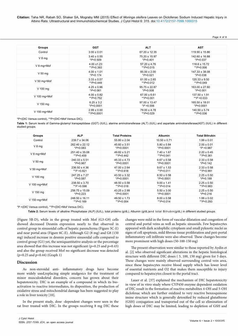
Serum GGT level was significantly increased in animals receiving 150 mg of DIC compared to control group (P=0.0001). Concerning serum levels of ALT, there was significant increase in experimental groups receiving DIC (50 mg, 100 mg and 150 mg) compared to control (P=0.021, P=0.008 and P=0.006, respectively) but significant decrease in ALT serum levels was observed after receiving Moringa extract in animals receiving the same dose of DIC (8 mg, 50 mg, 100 mg, 150 mg) (P=0.015, P=0.012, P=0.001, P=0.029, respectively). Serum levels of AST in all experimental groups receiving different doses of DIC (8 mg, 50 mg, 100 mg and 150 mg) were significantly increased compared to control (P=0.037, P=0.038, P=0.001, P=0.0001, respectively) but there was significant decrease in AST serum levels after receiving Moringa extract in animals receiving the same dose of DIC (8 mg, 50 mg, 100 mg, 150 mg) (P=0.006. P=0.049, P=0.033, P=0.036, respectively) (Table 1).
Total proteins serum levels showed significant decreased in experimental groups receiving different doses of DIC (8 mg, 50 mg, 100 mg and 150 mg) compared to control (P=0.0001 for all) but significant decrease in total protein serum levels after receiving Moringa extract in animals receiving the same dose of DIC only in doses 100 mg and 150 mg (P=0.018 and P=0.004, respectively). Serum levels of albumin in all experimental groups receiving different doses of DIC (8 mg, 50 mg, 100 mg and 150 mg) were significantly increased compared to control (P=0.0001 for all) but significant decrease in albumin serum levels after receiving Moringa extract in animals receiving the same dose of DIC (8 mg, 50 mg, 100 mg, 150 mg) (P=0.041. P=0.011, P=0.014, P=0.014, respectively) (Table 2).
Histological results
Light microscopic results: Sections of control rat liver showed ill defined liver lobules identified only by the presence of central ( CV) and peripheral located portal areas contain portal vein (PV), branches of hepatic artery (HA) and bile ducts (BD). Hepatocytes are arranged in cords separated by thin walled blood sinusoids. They have acidophilic cytoplasm with rounded, vesicular and central nuclei (Figure 1A).
In Group 2 (8 mg) mild changes in the form of sinusoidal dilatation with prominence of few Von Kuppfer cells were observed, hepatocytes showed no changes except few cells exhibit karyomegaly (large nuclei) while others show signs of apoptosis (dark pyknotic nucleus and acidophilic cytoplasm) (Figure 1B).
Increase doses of DIC G8 (150 mg) result in more histological changes. Lipid droplets of various sizes were observed in hepatocytes surrounding the central vein (microvesicular steatosis), while more cells showed signs of apoptosis (Figure 1E). Focal areas of cell necrosis with loss of normal lobular architecture were obvious (Figure 1D).
Concerning G4 and G6 (50-100 mg) the same histopathological changes were similar to those observed in G8 but to a lesser extent (not shown here).
Regarding the portal area DIC induced dilatation and congestion of portal vessels, bile ducts proliferation and mononuclear cells infiltration which were mild in G2 (8 mg) (Figure 2B) and became more pronounced in G8 (150 mg) (Figure 2D).
Administration of Moringa oleifera extract showed improvement of the previous histopathological changes induced by different doses of DIC such as no signs of apoptosis in most of hepatocytes, no sinusoidal congestion or fatty infiltration but still dilatation of central vein was detected in some lobules (Figure 1F). Portal area also looked more or less normal with less dilatation and congestion of portal vein (Figure 2E).
Electron microscopic results: Control liver parenchyma showed normal hepatocytes with rounded, euchromatic nucleus, numerous mitochondria and regularly arranged rER tubules (Figure 3A-B).
Compared to control the ultrastructural changes of liver tissue of DIC treated animals confirmed what was seen in light microscopy such as: Dilatation and congestion of blood sinusoids in G2 (8 mg) (Figure 3C). Hepatocytes of this group showed swollen mitochondria with dense matrix and ill defined cristae, in addition to dilatation of rER tubules (Figure 3D) and prominence of parasinusoidal Ito cells (Figure 4C).
In G8 (150 mg) more changes were observed such as focal areas of degenerated cytoplasmic organelles leaving cytoplasmic vacuoles with fine granular residual contents (Figure 3F).
The ameliorative ultrastructural changes observed in groups received Mof (G3, G9) compared to DIC treated groups were: the hepatocytes almost restore their normal appearance with less degenerative cytoplasmic organelles: mitochondria and rER (Figure 3E) but still some hepatocytes showed lipid droplets (Figure 3G) or microvesicular steatosis in (Figure 4B) and prominent Von Kupffer cell (Figure 4D).
Immunohistochemical results: In control group G1 few peri-sinusoidal cells showed positive Desmin expression (Figure 5A).
In group G2 (8 mg) and G8 (150 mg) obvious increase in desmin positive sinusoidal cells compared to control group was observed
Page 4 of 8
Volume 6 • Issue 2 • 1000315J Cytol HistolISSN: 2157-7099 JCH, an open access journal
Citation: Taha NR, Rabah SO, Shaker SA, Mograby MM (2015) Effect of Moringa oleifera Leaves on Diclofenac Sodium Induced Hepatic Injury in Albino Rats: Ultrastructural and Immunohistochemical Studies. J Cytol Histol 6: 315. doi:10.4172/2157-7099.1000315
(Figure 5B-D), while in the group treated with Mof (G3-G9) cells showed decreased Desmin expression nearly to that observed in control group in sinusoidal cells of hepatic parenchyma (Figure 5C-E) and near portal area (Figure 6C-E). Although G2 (8 mg) and G8 (150 mg) induced increase in desmin positive sinusoidal cells compared to control group (G1) yet, the semiquantitative analysis or the percentage area showed that this increase was not significant (p=0.25 and p=0.43) and also the group received Mof no significant decrease was detected (p=0.25 and p=0.44) (Graph 1)
DiscussionAs non-steroidal anti- inflammatory drugs have become
more widely used,replacing simple analgesics for the treatment of minor musculoskeletal disorders, concern has grown about their hepatotoxicity. DIC is an example of a compound in which its bio-activation to reactive intermediates, its disposition, the production of oxidative stress and mitochondrial damage has been suggested to play a role in liver toxicity [10].
In the present study, dose -dependent changes were seen in the rat liver treated with DIC. In the groups receiving 8 mg DIC these
changes were mild in the form of vascular dilatation and congestion of central and portal veins as well as hepatic sinusoids. Few hepatocytes appeared with dark acidophilic cytoplasm and small pyknotic nuclei as signs of cell apoptosis, mild fibrous tissue proliferation and peri-portal inflammatory cell infiltrate were also observed. These changes become more prominent with high doses (50-100-150 mg)
The present observation were similar to those reported by Aydin et al. [2] who observed significant alterations in the hepatic histological structure with different DIC doses ( 5, 100, 150 mg) given for 5 days. These changes were mainly observed surrounding central vein area, since these hepatocytes receive blood supply which has lower level of essential nutrients and O2 that makes them susceptible to injury compared to hepatocytes closest to the portal tract.
Lauer et al. [37] explained the mechanism of DIC hepatotoxicity in view of in vitro study where CYP450 enzyme dependent oxidation of DIC result in the formation of reactive metabolites 4-OH and 5-OH diclofenac which are further oxidized to very reactive benzoquinone imine structure which is generally detoxified by reduced glutathione (GSH) conjugation and transported out of the cell so elimination of high doses of DIC may be limited, leading to depletion of GSH and
Groups GGT ALT ASTControl 3.00 ± 0.01 67.20 ± 12.35 119.90 ± 15.86
V 8 mg 3.40 ± 0.55*P=0.509
75.20 ± 15.97*P=0.401
142.60 ± 16.88*P=0.037
V 8 mg+Mof 4.00 ±1.23**P=0.393
57.20 ± 4.76**P=0.015
118.6 ± 15.72**P=0.006
V 50 mg 4.00 ± 1.01*P=0.174
85.00 ± 2.00*P=0.021
147.33 ± 38.08*P=0.038
V 50 mg+Mof 3.33 ± 0.57**P=0.449
61.00 ± 2.65**P=0.012
120.33 ± 9.50**P=0.049
V 100 mg 4.25 ± 0.96*P=0.061
95.75 ± 22.87*P=0.008
163.00 ± 27.60*P=0.001
V 100 mg+Mof 4.00 ± 0.82**P=0.752
67.00 ± 6.61**P=0.001
137.50 ± 1.91**P =0.033
V 150 mg 6.25 ± 3.2*P=0.0001
97.00 ± 13.47*P =0.006
165.50 ± 18.01*P=0.0001
V 150 mg+Mof 2.99 ± 0.00**P=0.0001
79.00 ± 4.78**P=0.029
140.50 ± 5.74**P=0.036
*P=(DIC Versus control), **P=(DIC+Mof Versus DIC).Table 1: Serum levels of Gamma-glutamyl transpeptidase (GGT) (IU/L), alanine aminotransferase (ALT) (IU/L) and aspartate aminotransferase(AST) (IU/L) in different studied groups.
Groups ALP Total Proteins Albumin Total BilirubinControl 239.7 ± 34.06 55.80 ± 2.04 10.50 ± 0.71 1.99 ± 0.01
V 8 mg 262.40 ± 22.12*P=0.083
45.60 ± 3.51*P=0.0001
5.80 ± 0.84*P=0.0001
2.00 ± 0.01*P =0.987
V 8 mg+Mof 251.40 ± 35.08**P=0.459
49.82 ± 5.21**P=0.442
7.40 ± 1.67**P=0.041
2.20 ± 0.45**P=0.361
V 50 mg 240.33 ± 5.51 *P=0.967
45.33 ± 4.73*P=0.0001
6.67 ± 0.58*P=0.0001
2.33 ± 0.58*P=0.142
V 50 mg+Mof 236.50 ± 4.36**P =0.821
47.00 ± 2.64**P=0.618
9.67 ± 1.53 **P=0.011
2.33 ± 0.58**P=0.991
V 100 mg 247.25 ± 7.27*P=0.587
43.50 ± 3.32*P=0.0001
6.50 ± 0.58*P=0.0001
2.25 ± 0.50*P=0.199
V 100 mg+Mof 238.50 ± 3.70**P =0.598
48.50 ± 0.58**P=0.018
9.00 ± 1.41 **P=0.014
2.25 ± 0.50**P=0.983
V 150 mg 256.75 ± 15.09*P=0.223
43.25 ± 2.99*P=0.0001
5.50 ± 3.00*P=0.0001
2.25 ± 0.50*P=0.216
V 150 mg+Mof 248.50 ± 16.11**P=0.169
49.50 ± 1.73**P=0.004
8.00 ± 0.58**P=0.014
1.99 ± 0.02**P=0.293
*P =(DIC Versus control), **P=(DIC+Mof Versus DIC).Table 2: Serum levels of alkaline Phosphatase (ALP) (IU/L), total proteins (g/dL), Albumin (g/dL)and total Bi l rubin(g/dL) in different studied groups.

Page 5 of 8
Volume 6 • Issue 2 • 1000315J Cytol HistolISSN: 2157-7099 JCH, an open access journal
Citation: Taha NR, Rabah SO, Shaker SA, Mograby MM (2015) Effect of Moringa oleifera Leaves on Diclofenac Sodium Induced Hepatic Injury in Albino Rats: Ultrastructural and Immunohistochemical Studies. J Cytol Histol 6: 315. doi:10.4172/2157-7099.1000315
thereby reducing the cellular defense against oxidative stress causing liver insult.
In addition to the generation of reactive metabolites, DIC hepatotoxicity involved mitochondrial injury. In current study ultrastructure morphological alterations of mitochondrial shape (swelling, pleomorphic, destruction of their cristae and condensation of its matrix) were observed. This is in line with the study of Moorthy et al. [31] who attributed this damage to the oxidative stress followed by mitochondrial permeability transition (MPT). MPT was found to cause leakage of cytochrome c and other proteins leading to apoptosis [12]. Apoptosis and necrosis depend on how severely impaired mitochondria become, this is in turn is a function of the severity of oxidative stress and whether or not the cell is sensitized to the immune system [38].
The increased deposition of tiny lipid droplets (microvesicularsteatosis) observed in hepatocytes of animals receiving high dose of DIC were described also by Servidio et al. [39] where they attributed it to mitochondrial dysfunction and altered oxidative phosphorylation and fatty β oxidation. Increased concentrations of intracellular fatty acids may be directly toxic to hepatocytes or lead to oxidative stress [40].
With high doses progression of hepatocellular damage, increased deposition of collagen fibers around portal tracts, central vein and perisinusoidal were obvious
In the present study, immunohistochemistry for desmin expression in rat livers was done to evaluate if different doses of DIC induced fibrosis in liver parenchyma especially in periportal area. Previous literature pointed to expression of Desmin by lobular Ito cells which
accumulate in septae of fibrotic rat livers and were strongly desmin positive [41].
Rockey et al. [42] reported that perisinusoidal desmin positive cells were found to be numerous in areas of fibrosis and suggested that these cells are derived from lipocytes (fat-storing cells, Ito cells) after their activation and transformation to myofibroblasts producing excessive extracellular matrix (ECM) components during toxic liver injury.
Current study showed high expression of desmin in both therapeutic (8 mg) and high doses (150 mg) of treated animals denoting that DIC can also be involved in liver fibrosis following hepatocellular injury and activation of Von Kuppfer cells [17]. Von Kuppfer cells were reported by Roberts et al. [43] to activate Ito cells by releasing an array of inflammatory and growth control mediators as well as reactive oxygen species.
In animal groups treated by Moringa oleifera extract, Mof succeeded to decrease Desmin expression although the reduction wasn’t statistically significant and most probably related to the antioxidant activity exerted by the herb extract, which protect the liver from progression of fibrogenesis by inhibiting Ito cells activation [23] A previous study of Boigk et al. [44] elucidated the role of antioxidants such as Silymarin in decreasing the accumulation of hepatic collagen in advanced biliary cirrhosis.
The biochemical results in the current study showed significant elevation in the serum levels of AST, ALT and GGT in a dose
Figure 1: Sections from Rat liver at the region of Central vein stained by H&E showing (A) Control group (G1) with normal central vein (CV), hepatocytes cords (black arrows) separated by thin walled blood sinusoids (white arrows). (B) G2 (8 mg) showing numerous apoptotic hepatocytes (thin black arrows) prominent sinusoidal Von kupffer cells (white arrows). (C) G3 (8 mg+Mof) Normal hepatocytes (arrows) prominent Von kupffer cells are still present but less compared to G2 (white arrows). (D) G8 (150 mg) showing in the same hepatic lobule focal areas of degenerated hepatocytes with loss of lobular architecture (white star), aggregation of apoptoic cells (black arrows) and many congested blood sinusoids (arrowheads). (E) another sample showing congestion of central vein with nearby hepatocytes (black star) containing many lipid droplets (microvesicular steatosis). (F) G9 (150 mg+Mof) Hepatocytes looked almost normal few cells showed signs of apoptosis (black arrows), no steatosis (black star).
Figure 2: Sections from rat liver near portal areas showing: (A) Control with normal portal triad portal vein(PV) hepatic artery (A) and bile duct (BD) hepatocytes showed normal architecture regarding arrangement of cytoplasm and nuclear appearance. (B) G2 (8 mg): mild changes with more binucleated cells (white arrows). Von kupffer cells are prominent (black arrows). (C) G3 (8 mg+Mof): hepatocytes appeared to possess more vesicular nuclei (white arrows) still Von kupffer cells appeared prominent ( black arrows). (D) G 8 (150 mg): Portal area showed congestion of blood vessels (stars) and bile duct proliferation ( black arrows). (E) G9 (150 mg+Mof ) : potential improvement was observed still congestion of portal vein (star) and mild microvesicular steateosis (black arrow).

Page 6 of 8
Volume 6 • Issue 2 • 1000315J Cytol HistolISSN: 2157-7099 JCH, an open access journal
Citation: Taha NR, Rabah SO, Shaker SA, Mograby MM (2015) Effect of Moringa oleifera Leaves on Diclofenac Sodium Induced Hepatic Injury in Albino Rats: Ultrastructural and Immunohistochemical Studies. J Cytol Histol 6: 315. doi:10.4172/2157-7099.1000315
dependent manner which improves after the intake of Moringa leaves extract, while the ALP showed no significant increase, as the ALP is an indicator of cholestatic liver dysfunction which was not detected in present study. These results are supported by those previously reported clinically by Dufour et al. [45] that injury to the liver whether acute or chronic results in an increase in serum concentrations of aminotransferases (AST and ALT), both aminotransferases are highly concentrated in the liver,
Recently, Darbar et al. [23] showed that administration of DIC caused a dramatic elevation in serum AST, ALT and GGT indicating subchronic hepatotoxicity with severe damage to hepatic tissue membranes during DIC intoxication and the release of these enzymes into the circulation.
Decreased in total proteins and albumin levels were noticed in the present study and was previously explained by Ponsoda et al. [46] to be result from toxic metabolite of DIC that cause alteration inmitochondrial function and depletion of ATP, thus inhibiting hepatic anabolic processes namely gluconeogenesis and protein synthesis.
In the present study, it was observed that co-administration of Mof with Diclofenac efficiently reduced the DIC induced elevation of serum AST, ALT and GGT in a dose dependent manner indicating a promising hepatoprotective activity of Mof as it is suggested by Fakurazi et al. [47] that Mof may preserve the structural integrity of hepatocytes membranes and subsequently preventing enzyme leakage into plasma.
Figure 3: Photographs of TEM sections showing: (A) Control liver parenchyma: Hepatocytes (H) with rounded, euchromatic nucleus (N) separated by blood sinusoids (S). (B) The cells showed numerous normal shaped mitochondria of various sizes (white arrows) as well as clusters of (rER) tubules (stars). (C) G2 (8 mg): dilated blood sinusoids (S). (D) enlarged pleomorphic mitochondria (white arrows) and dilated rER tubules (black arrows) the nucleus (N) showed slight increase in peripheral chromatin and elaborating microvilli project in the space of Disse (stars). (E) G3 (8 mg+Mof) showing hepatocyte with almost normal cell organelles, rER (black arrows) and mitochondria (white arrows) .The nucleus (N) showed patches of heterochromatin. (F) G8 (150 mg) : showing focal areas of degenerated cytoplasmic organelles leaving vacuoles. They contain fine granular or cellular debris and myelin figures (black arrows). There are numerous pleomorphic enlarged or deformed mitochondria (white arrows). (G) G9 (150 mg+Mof) almost normal hepatocytes structure with normally appeared mitochondria and rER tubules still few lipid droplets were detected (arrowheads). the nucleus ( N) showed slight condensation of peripheral chromatin.
Figure 4: TEM sections of Liver sinusoidal area bordering hepatocytes showing the sinusoidal cells: (A-C) Prominent Ito cell with lipid droplet ( white arrow) and Von Kupffer cells (black arrows) in G8 receiving DIC (150 mg) while in (B-D) group treated with Mof G9 (150 mg+Mof) still Ito cell (white arrow) in (B) and Von Kupffer cells (black arrow) in (D) were obvious and the adjacent hepatocyte (H) showed microvesicular steatosis (black star) in (B).
Figure 5: Sections in rat liver stained immunohistochemistry for desmin showing: A-G1 (control group) few positive sinusoidal cells (black arrows) near central vein (CV) region. B- G2 (8mg/kg) increased number and staining intensity of desmin positive cells (black arrows) in hepatic sinusoids compared to control . C-G3 (8mg +Mof) decrease in desmin positive cells (black arrows). D- G8 (150mg/kg) showing also increase in desmin positive cells (black arrows). E- G9 (150 mg/kg +Mof) showing decrease in desmin positive cells

Page 7 of 8
Volume 6 • Issue 2 • 1000315J Cytol HistolISSN: 2157-7099 JCH, an open access journal
Citation: Taha NR, Rabah SO, Shaker SA, Mograby MM (2015) Effect of Moringa oleifera Leaves on Diclofenac Sodium Induced Hepatic Injury in Albino Rats: Ultrastructural and Immunohistochemical Studies. J Cytol Histol 6: 315. doi:10.4172/2157-7099.1000315
Figure 6: Sections in rat liver stained immunohistochemistry for desmin near portal area showing: increase in desmin positive cells in both (B) low dose G2 (8 mg/kg) and high dose (D) G8 (150 mg/kg) (black arrows) compared to G1 (control) (A). Perivascular tissue also showed positive staining (white arrows). Administration of Mof (G3 and G9) result in decrease of Desmin positive cells Figs (C and E) compared to treated rats receiving low and high doses of DIC (G2 and G8) .
0
1
2
3
4
5
6
7
Control V 8mg V 150mg V 8mg+Mof V 150mg+Mof
Area
per
cent
Desmin expression
p=0.43
p=0.25
p=0.44
p=0.25
Graph 1: Area percentage of Desmin immunoexpression in the studied groups, values is means ± SD (p<0.05).
Previous benefits of using Moringa oleifera in experimental studies concerning hepatoprotective effect were reported in literature. Rakesh et al. [48] showed the effectiveness of Moringa oleifera leaves extract against carbon tetrachloride induced liver damage. Paliwal et al. [33] suggested a chemopreventive potential of Moringa extract against chemical carcinogens DMBA (synthetic polyaromatic hydrocarbon7,12-dimethyl benz(a)ancrathene induced hepatic carcinogenicity.
ConclusionThe present study proved that hepatocellular damage induced
by DIC was mild with low therapeutic dose compared to the severe damage of high doses and this injury was triggered by mitochondrial dysfunction, decrease ATP synthesis, oxidative stress and it was almost
improved based on the histological and biochemical results by the administration of Moringa oleifera leaves extract before the intake of DIC. In view of such results Mof could be considered as a potential source of natural antioxidant agent that encourages its use in future medicinal field.
Acknowledgements
This paper was funded by the Deanship of King AbdulAzis City for Science and technology under grant number A-S-10-0009. The authors therefore acknowledge with thanks the deanship for his financial support.
References1. Teoh NC, Farrell GC (2003) Hepatotoxicity associated with non-steroidal anti-
inflammatory drugs. Clin Liver Dis 7: 401-413.
2. Aydin G, Gokcimen A, Cicek E, Gokalp O (2003) Histopathologic changes in liver and renal tissue induced by different does in liver and renal tissue induced by different doses of diclofenac sodium in rats. Turk J Vet Ani Sci 27: 1131-1140.
3. Hackstein H, Mohl W, Püschel W, Stallmach A, Zeitz M (1998) [Diclofenac-associated acute cholestatis hepatitis]. Z Gastroenterol 36: 385-389.
4. Tolman KG (1998) Hepatotoxicity of non-narcotic analgesics. Am J Med 105: 13S-19S.
5. Daly AK, Aithal GP, Leathart JS, Swainsbury RA (2007) Genetic susceptibility to diclofenac-induced hepatotoxicity: contribution of UGT2B7, CYP2C8, and ABCC2 genotypes. Gastroenterology 132: 272-281.
6. Amin A, Hamza AA (2005) Oxidative stress mediates drug-induced hepatotoxicity in rats: a possible role of DNA fragmentation. Toxicology 208: 367-375.
7. Amacher DE (1998) Serum transaminase elevations as indicators of hepatic injury following the administration of drugs. Regul Toxicol Pharmacol 27: 119-130.
8. Uetrecht JP (1999) New concepts in immunology relevant to idiosyncratic drug reactions: the “Danger Hypothesis” and innate immune system. Chem Res Toxicol 12: 387-395.
9. Watkins PB, Seeff LB (2006) Drug-induced liver injury: summary of a single topic clinical research conference. Hepatology 43: 618-631.
10. Lim MS, Lim PK, Gupta R, Boelsterli A (2006) Critical role of free cytosolic calcium, but not uncoupling in mitochondrial permeability transition and cell death induced by diclofenac oxidative metabolites in immortalized human hepatocytes. Toxicol Applied Pharmacol 217: 322-331.
11. Cantoni L, Valaperta R, Ponsoda X, Castell JV, Barelli D, et al. (2003) Induction of hepatic heme oxygenase-1 by diclofenac in rodents: role of oxidative stress and cytochrome P-450 activity. J Hepatol 38: 776-783.
12. Masubuchi Y, Nakayama S, Horie T (2002) Role of mitochondrial permeability transition in diclofenac-induced hepatocyte injury in rats. Hepatology 35: 544-551.
13. Siu WP, Pun PB, Latchoumycandane C, Boelsterli UA (2008) Bax-mediate mitochondrial outer membrane permeabilization (MOMP), distinct from the mitochondrial permeability transition, is a key mechanism in diclofenac-induced hepatocyte injury: multiple protective roles of cyclosporin A. Toxicol Appl Pharmacol 227: 451-461.
14. Gómez-Lechón MJ, Ponsoda X, O’Connor E, Donato T, Castell JV, et al. (2003) Diclofenac induces apoptosis in hepatocytes by alteration of mitochondrial function and generation of ROS. Biochem Pharmacol 66: 2155-2167.
15. Sokol RJ, Straka MS, Dahl R, Devereaux MW, Yerushalmi B, et al. (2001) Role of oxidant stress in the permeability transition induced in rat hepatic mitochondria by hydrophobic bile acids. Pediatr Res 49: 519-531.
16. Adachi T, Togashi H, Suzuki A, Kasai S, Ito J, et al. (2005) NAD(P)H oxidase plays a crucial role in PDGF-induced proliferation of hepatic stellate cells. Hepatology 41: 1272-1281.
17. Wu J, Zern MA (2000) Hepatic stellate cells: a target for the treatment of liver fibrosis. J Gastroenterol 35: 665-672.
18. Fromenty B, Pessayre D (1995) Inhibition of mitochondrial beta-oxidation as a mechanism of hepatotoxicity. Pharmacol Ther 67: 101-154.
19. Pessayre D, Fromenty B (2005) NASH: a mitochondrial disease. J Hepatol 42: 928-940.

Page 8 of 8
Volume 6 • Issue 2 • 1000315J Cytol HistolISSN: 2157-7099 JCH, an open access journal
Citation: Taha NR, Rabah SO, Shaker SA, Mograby MM (2015) Effect of Moringa oleifera Leaves on Diclofenac Sodium Induced Hepatic Injury in Albino Rats: Ultrastructural and Immunohistochemical Studies. J Cytol Histol 6: 315. doi:10.4172/2157-7099.1000315
20. Pessayre D, Feldmann G, Haouzi D, Fau D et al. (2000) Hepatocyte apoptosis triggered by natural substances (cytokines, other endogenous substances and foreign toxins). In: Cameron RG, Feuer G (eds.) Apoptosis and its modulationby drugs. Handbook Exp Pharmacol. Springer, Heidelberg 142: 59-108.
21. Chattopadhyay RR (2003) Possible mechanism of hepatoprotective activity ofAzadirachta indica leaf extract: part II. J Ethnopharmacol 89: 217-219.
22. Pradhan SC, Girish C (2006) Hepatoprotective herbal drug, silymarin fromexperimental pharmacology to clinical medicine. Indian J Med Res 124: 491-504.
23. Darbar S, Bhattacharya A, Chattopadhyay S (2010) Ameliorative effect of Livina, a polyherbal preparation on Diclofenac-induced liver injury: A comparison withSilymarin. Journal of pharmacy research 3: 2794-2798.Journal of pharmacyresearch 3: 2794-2798.
24. Pari L, Kuramac M, Kosinska A, Rybarczyk A (2007) Antioxidant activity of thecrude extracts of drumstick tree (Moringa Oleifera lam ) and sweet bromweed(scoparia dulcisI.), leaves. Pol J Food Nutr Sci 57: 203-208.
25. Ghasi S, Nwobodo E, Ofili JO (2000) Hypocholesterolemic effects of crude extract of leaf of Moringa oleifera Lam in high-fat diet fed wistar rats. JEthnopharmacol 69: 21-25.
26. Faizi S, Siddiqui BS, Saleem R, Aftab K, Shaheen F, et al. (1998) Hypotensive constituents from the pods of Moringa oleifera. Planta Med 64: 225-228.
27. Kar A, Choudhary BK, Bandyopadhyay NG (2003) Comparative evaluation ofhypoglycaemic activity of some Indian medicinal plants in alloxan diabetic rats. J Ethnopharmacol 84: 105-108.
28. Ezeamuzle IC, Ambedederomo AW, Shode FO, Ekwebelem SC (1996) Anti-inflammatory effects of Moringa oleifera root extract. Int J Pharmacogn 34: 207-212.
29. Murakami A, Kitazonz Y, Jiwajinda S, Koshimizu K (1998) Niaziminin, athio carbamate from the leaves of Moringa oleifera, holds a strict structuralrequirement for inhibition of tumor promotor-induced Epstein-Barr virusactivation. Planta Med 64: 319-323.
30. Bharali R, Tabassum J, Azad, MH (2003) Chemomodulatory Effect of MoringaOleifera, Lam, on Hepatic Carcinogen Metabolising Enzymes, AntioxidantParameters and Skin Papillomagenesis in Mice, Asian Pacific J Cancer Prev 4: 131-139.
31. Moorthy M, Fakurazi S, Ithin H (2008) Morphological alterations in Mitochondria following diclofenac and Ibuprofen administration Pakistan Journal of Biological Sciences 11: 1901-1908.
32. Paget GE, Barnes JM (1964) In: Evaluation of drug activities: pharmacometrics Ed. DR Laurence and Al Bacharach. London & New York.
33. Paliwal R, Sharma V, Pracheta S, Sharma SH (2011) Hepatoprotectiveand antioxidant potential of Moringa Oleifera pods against DMBA- inducedhepatocarcinogenesis in male mice 3: 128-138.
34. Buraimoh AA, Bako IG, Ibrahim FB (2011) Hepatoprotective effect of Ethanolic leave extract of Moringa oleifera on the histology of Paracetamol induced liverdamage in Wistar rats International journal of animal and Veterinary Advances 3: 10-13.
35. Taha NR, Amin HA, Sultan AA (2015) The protective effect of Moringa oleifera leaves against cyclophosphamide-induced urinary bladder toxicity in rats.Tissue Cell 47: 94-104.
36. Knittel T, Kobold D, Pisgaglia F, Neubauer K, et al. (1999) Localisationof liver myofibroblasts and hepatic stellate cells in normal and diseased rat livers: distinct roles of (myo-)fibroblast subpopulations in hepatic tissue repair. Histochem Cell Biol 112: 387-401.
37. Lauer B, Tuschl G, Kling M, Mueller SO (2009) Species-specific toxicity of diclofenac and troglitazone in primary human and rat hepatocytes. Chem BiolInteract 179: 17-24.
38. Leist M, Single B, Castoldi AF, Kunhele S (1997) Intracellular adenosinetriphosphate (ATP) concentration: a switch in the decision between apoptosisand necrosis. J Exp Med 185: 1481486.
39. Servidio G, Bellan F, Vendemiale G, Altomare E (2011) Mitochondrialdysfunction in nonalcoholic steatohepatitis. Expert Rev Gastroenterol Hepatol5: 233-244.
40. Wanless IR, Shiota K (2004) The pathogenesis of nonalcoholic steatohepatitisand other fatty liver diseases: a four-step model including the role of lipidrelease and hepatic venular obstruction in the progression to cirrhosis. SeminLiver Dis 24: 99-106.
41. Ballardani G, Fallani M, Biagini G, Bianchi FB (1988) Desmin and actin in theidentification of Ito cells and in monitoring their evolution to myofibroblasts in experimental liver fibrosis. Virchows Arch B cell Pathol Incl Mol Pathol 56: 45-49.
42. Rockey DC, Boyles JK, Gabbiani G, Friedman SL (1992) Rat hepaticlipocytes express smooth muscle actin upon activation in vivo and in culture. JSubmicrosc Cytol Pathol 24: 193-203.
43. Roberts RA, Ganey PE, Ju C, Kamendulis LM, Rusyn I, et al. (2007) Role ofthe Kupffer cell in mediating hepatic toxicity and carcinogenesis. Toxicol Sci96: 2-15.
44. Boigk G, Stroedter L, Herbst H, Waldschmidt J (1997) Silymarin retardscollagen accumulation in early and advanced biliary fibrosis secondary to complete bile duct obliteration in rats. Hepatology 26: 643-649.
45. Dufour DR, Lott JA, Nolte FS, Gretch DR, Koff RS, et al. (2000) Diagnosis and monitoring of hepatic injury. I. Performance characteristics of laboratory tests.Clin Chem 46: 2027-2049.
46. Ponsoda X, Bort R, Jover R, Go´mez-Lecho´n MJ (1995) Molecular mechanism of diclofenac hepatotoxicity: association of cell injury with oxidative metabolism and decrease in ATP levels. Toxicol In Vitro 9: 439-444.
47. Fakurazi S, Hiruszah I, Nanthini U (2008) Moringa Oleifera Lam preventsacetaminophen liver injury through restoration of glutathione level. Food andchemical toxicology 46: 2611-2615.
48. Rakesh S, Singh V (2010) In vivo antioxidant activity of Moringa Oleifera leafand pod extracts against carbon tetrachloride induced liver damage in albinomice. Journal of chemical and pharmaceutical Research 2: 275-283.