haematuria and - mackayurology.com · hemorrhagic fever, scarlet fever, filariasis connective...
TRANSCRIPT

HAEMATURIA and it’s management
Dr Sanjeev Bandi MBBS.,FRCSI, FRACS(Urology)
Consultant Urologist
Mackay Urology
Definition:
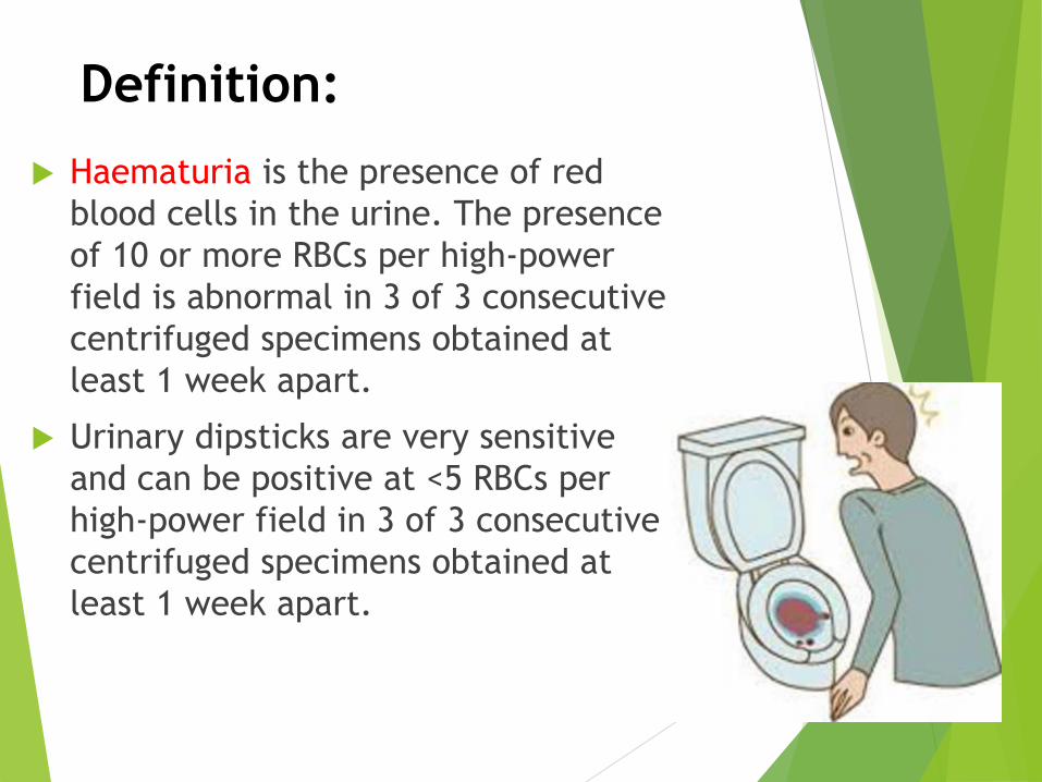
Haematuria is the presence of red
blood cells in the urine. The presence
of 10 or more RBCs per high-power
field is abnormal in 3 of 3 consecutive
centrifuged specimens obtained at
least 1 week apart.
Urinary dipsticks are very sensitive
and can be positive at <5 RBCs per
high-power field in 3 of 3 consecutive
centrifuged specimens obtained at
least 1 week apart.

Types:According to the amount of RBC in the urine, hematuria can be
classified as:
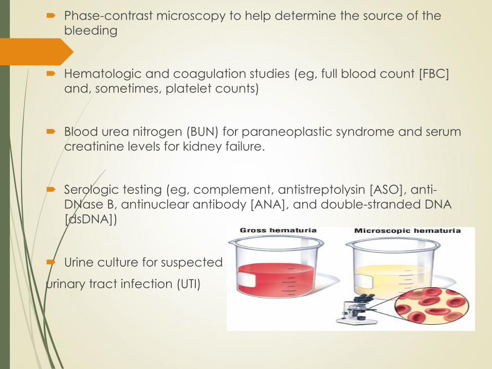
Gross (ie,overtly bloody, smoky, or tea-colored urine)
Microscopic > 10 RBC”s /HPF
According to Timing (when it occurs during urination):
Early (initial) haematuria: Urethral origin, distal to external
Sphincter
Terminal haematuria: Bladder neck or prostate origin
Diffuse (total) haematuria: Source is in the bladder or upper
urinary tract
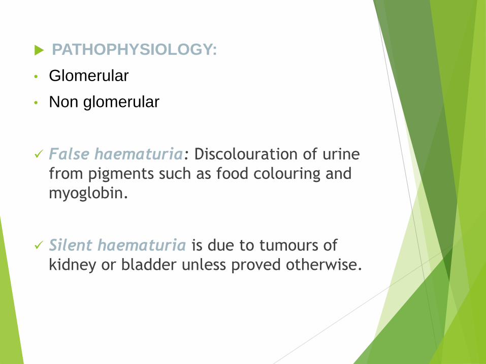
PATHOPHYSIOLOGY:
• Glomerular
• Non glomerular
False haematuria: Discolouration of urine
from pigments such as food colouring and
myoglobin.
Silent haematuria is due to tumours of
kidney or bladder unless proved otherwise.

AETIOLOGY
Diseases of the urinary system – the
most common cause
Glomerular
Interstitial
Uroepithelium
Vascular
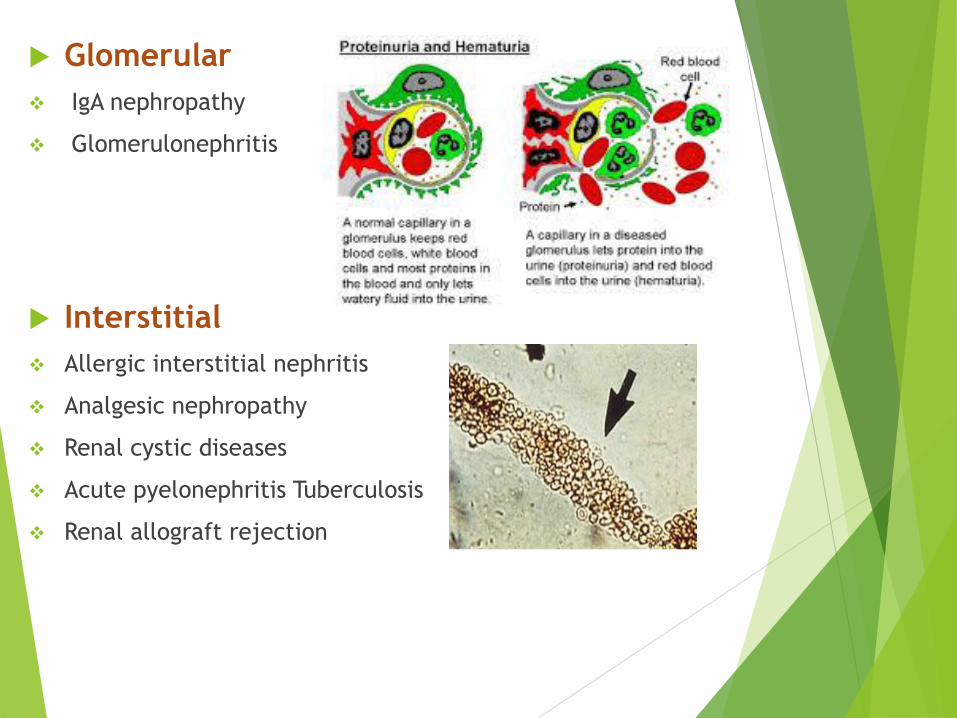
Glomerular
IgA nephropathy
Glomerulonephritis
Interstitial
Allergic interstitial nephritis
Analgesic nephropathy
Renal cystic diseases
Acute pyelonephritis Tuberculosis
Renal allograft rejection

Uroepithelium
Malignancy
Trauma
Papillary Necrosis
Cystitis/Urethritis/Prostatitis
Parasitic Diseases (Schistosomiasis)
Stones
Vascular
Arterial emboli or thrombosis
Arteriovenous fistulae
Renal vein thrombosis

System disorders (less common):
Haematological disorders--aplastic anemia, leukemia
hemophilia\,ITP (idiopathic thrombocytopenic purpura)
Infection--infective endocarditis,Septicemia,epidemic
hemorrhagic fever, scarlet fever, Filariasis
Connective tissue diseases--SLE, polyarteritis nodosa
Cardiovascular diseases--hypertensive nephropathy,
chronic heart failure - renal artery sclerosis.
Endocrine and metabolism diseases-- gout - diabetes
mellitus

Diseases of adjacent organs
to urinary tractAppendicitis
carcinoma of the rectum
carcinoma of the colon
uterocervical cancer
Drug and chemical agents anticoagulation
Cyclophosphamide, rifampin, sulfonamide, phenytoin,
Miscellaneousexercise induced hematuria

CAUSES:
DIFFERENTIAL DIAGNOSIS
• Polluted urine: menstruation
• Drug and food: Rifampicin, Nitrofurantoin, sulfonamides,
adriamycin.
• Porphyria: porphyrin in urine (+)
• Hemoglobinuria (hemolysis)
• Myoglobinuria
Signs and symptoms
The first step in the evaluation of haematuria consists of a detailed history
and a thorough physical examination.
Efforts should be made to distinguish glomerular causes from
extraglomerular one:
Passage of clots in urine suggests an extraglomerular cause
Fever, abdominal pain, dysuria, frequency, and recent enuresis in
older children may point to a urinary tract infection as the cause
Recent trauma to the abdomen may be indicative of hydronephrosis
Early-morning periorbital puffiness, weight gain, oliguria, dark-colored
urine, and edema or hypertension suggest a glomerular cause,
Hematuria due to glomerular causes is painless
Recent throat or skin infection may suggest post infectious
glomerulonephritis

Joint pains, skin rashes, and prolonged fever in adolescents suggest
a collagen vascular disorder(Rheumatoid arthritis, Systemic lupus
erythematosus)
Skin rashes and arthritis can occur in Henoch-Schönlein
purpura and systemic lupus erythematosus
Information regarding exercise, menstruation, recent bladder
catheterization, intake of certain drugs or toxic substances, or
passage of a calculus may also assist in the differential diagnosis.
A family history that is suggestive of Alport syndrome, collagen
vascular diseases, urolithiasis, or polycystic kidney disease is
important
Alport syndrome is a genetic condition characterized by kidney
disease, hearing loss, and eye abnormalities. People with Alport
syndrome experience progressive loss of kidney function. Almost all
affected individuals have blood in their urine (haematuria), which
indicates abnormal functioning of the kidneys.

Physical examination
Measurement of the blood pressure and volume
status is especially important when glomerulonephritis is a consideration.
Evaluation for the presence of periorbital puffiness or peripheral edema
Detailed skin examination to look for purpura.
Abdominal examination to look for palpable mass reveals a renal tumor or hydronephrosis may exist,
A palpable bladder after voiding indicates obstruction or retention
A bruit over the kidney suggests a vascular cause.
Always check for extrarenal manifestations and co morbid
conditions.
Check for other sites of bleeding. PR examination should not be
missed. to diagnose prostatitis, prostate cancer, epididymitis,
meatal stenosis, and other structural causes of hematuria .
Inspect external genitalia in male for trauma.
Atrial fibrillation raises the possibility of renal embolic infarction,
especially if the patient has flank pain
Costovertebral angle tenderness is also suggestive of
pyelonephritis, nephrolithiasis, or ureteropelvic junction
obstruction.
Detailed ophthalmologic evaluation (in familial hematuria)
Diagnosis
The laboratory tests ordered for the evaluation of hematuria must be
based on the clinical history and the physical examination. Tests that
may be helpful include the following:
Urinalysis with careful microscopic review of the urine sample
Urine dip strip analysis it is the most commonly used method of testing
the urine for blood is the urine test strip or dipstick, which utilizes the
peroxidase-like activity of hemoglobin to generate a color change.
False-positive tests may occur in the setting of myoglobinuria or hemoglobinuria, confirmed by the absence of RBCs on microscopic
examination.
Phase-contrast microscopy to help determine the source of the
bleeding
Hematologic and coagulation studies (eg, full blood count [FBC]
and, sometimes, platelet counts)
Blood urea nitrogen (BUN) for paraneoplastic syndrome and serum
creatinine levels for kidney failure.
Serologic testing (eg, complement, antistreptolysin [ASO], anti-
DNase B, antinuclear antibody [ANA], and double-stranded DNA
[dsDNA])
Urine culture for suspected
urinary tract infection (UTI)
Cx Bladder Cancer Test
Research from Pacific Edge1 shows
that messenger RNA (mRNA) levels of
specific biomarkers are present at
higher levels of concentration in
patient urine samples that are
positive for bladder cancer than in
patients who are negative for the disease.
1 Holyoake et al: Development of a multiplex RNA urine test for the
detection and stratification of transitional cell carcinoma of the bladder, Clin Cancer Res 2008; 14(3): 742-749
Cxbladder Biomarker Gene Descriptions
MDK: Blood vessel growth and cell migrationPrincipally involved in cell proliferation, migration and angiogenesis in cancer cells.
HOXA13: Cell differentiationPrincipally involved in cell differentiation and the morphogenesis and differentiation of the genitourinary tracts.
CDC2 (CDK1): Cell divisionCyclin dependent kinase. Essential to mitotic cell cycle: cell proliferation.
IGFBP5: Programmed cell deathActs as an anti-apoptotic gene.
CXCR2: InflammationMediates neutrophil migration to sites of inflammation. Moderates non-malignant inflammation(False Positives).
Cx Bladder Cancer Test
A small sample (5 mL) of mid
stream urine is required for the
test. The sample undergoes a
precise set of processes to
extract and purify the mRNA
present in the patient urine sample.
Cx Bladder Cancer Test
The purified RNA is then quantified by a
technique called Reverse Transcription
quantitative Polymerase Chain Reaction or
RT-qPCR. RT-qPCR first involves the conversion
of RNA to DNA and then the amplification of
that DNA by millions of fold, regulated by a
repetitive cycle of temperature adjustment.
The resulting millions of copies of DNA are
detected by a probe whose fluorescence is
directly proportional to the number of copies of DNA present.
Cx Bladder Cancer Test
In the Cxbladder test, each of the five
biomarkers of interest to us is quantified
by a different probe and the relationship
between the individual biomarkers is
determined by a mathematical
equation. The calculated outcome
provides a measure of the probability of
the presence of urothelial carcinoma (UC).

Cx Bladder Cancer Test
How Cxbladder can be used in your practice:
Replace the need for other urine-based tests in primary workup.
Complement cystoscopy for bladder cancer detection.
Detect urothelial tumors not visible by cystoscopy.
Replace the need for CT / IVP in primary workup in some
instances.
Improve patient compliance with accurate, non-invasive testing.

Pacific Edge Named in
TIN100 Top 10 List
We are delighted to be named in the top 10 Hot
Emerging Companies in the TIN100 2016 report,
alongside amazing people from Vend, PushPay,
ARANZ Medical and FlintFox, to name a few. TIN
Managing Director, Greg Shanahan, said: "These ten
companies almost doubled the record performance
of the companies on last year's list with a continued
revenue growth of over $61 million (or 138%). "All ten
companies are exciting examples of how technology
is disrupting the way we do business, from the
development of wireless power systems to new remote
methods for diagnosing cancer," Mr. Shanahan
added. http://bit.ly/2emSWLc

Imaging studies
The following may be helpful:
Renal and bladder ultrasonography
Voiding cystourethrography
CT urography: now replaces IVU.
MRI.
Retrograde pyelography.
Renal biopsy: in nephrological cases
Cystoscopy

Kidney biopsy is rarely indicated:
Significant proteinuria
Abnormal renal function
Recurrent persistent hematuria
Serologic abnormalities (abnormal complement, ANA, or
dsDNA levels)
Recurrent gross hematuria
A family history of end-stage renal disease
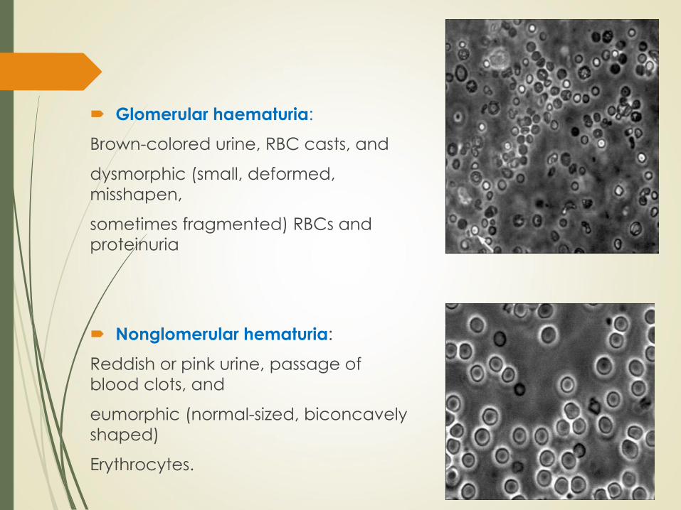
Glomerular haematuria:
Brown-colored urine, RBC casts, and
dysmorphic (small, deformed,
misshapen,
sometimes fragmented) RBCs and
proteinuria
Nonglomerular hematuria:
Reddish or pink urine, passage of
blood clots, and
eumorphic (normal-sized, biconcavely
shaped)
Erythrocytes.

Management:
Haematuria is a sign and not itself a
disease; thus, therapy should be directed
at the process causing it
Asymptomatic (isolated) haematuria
generally does not require treatment.
In conditions associated with abnormal
clinical, laboratory, or imaging studies,
treatment may be necessary, as
appropriate, with the primary diagnosis

Surgical intervention may be necessary with certain
anatomic abnormalities (eg, ureteropelvic junction
obstruction, tumor, or significant urolithiasis)
Dietary modification is usually not indicated, except for
children who may tend to develop hypertension or
edema as a result of the primary disease process (eg,
nephritis)
Patients with persistent microscopic haematuria should be
monitored every 6-12 months for the appearance of signs
or symptoms indicative of progressive renal disease

MORTALITY/MORBIDITY
• IN GENERAL, CHILDREN WITH ISOLATED ASYMPTOMATIC
MICROSCOPIC HAEMATURIA TEND TO DO WELL,
• WHEREAS THOSE WITH ASSOCIATED FINDINGS
(EG, HYPERTENSION, PROTEINURIA, ABNORMAL SERUM
CREATININE LEVELS) ARE MORE LIKELY TO HAVE SERIOUS
PROBLEMS.
• SINCE HAEMATURIA IS THE END RESULT OF VARIOUS PROCESSES,
THE MORBIDITY AND MORTALITY RATES OF THE CONDITION
DEPEND ON THE PRIMARY PROCESS THAT INITIATED IT.
RACE:
• THE INCIDENCE OF HAEMATURIA IN SPECIFIC RACIAL GROUPS IS DETERMINED BY THE PRIMARY CAUSE.
• FOR EXAMPLE, IDIOPATHIC HYPERCALCIURIA IS INFREQUENT IN BLACK AND ASIAN CHILDREN,
• BUT RELATIVELY COMMON IN WHITES. CONVERSELY, HAEMATURIA CAUSED BY SICKLE CELL DISEASE IS MORE COMMON IN BLACKS THAN IN WHITES.

SEX:
• SEX MAY PREDISPOSE A CHILD TO SPECIFIC
DISEASES THAT MANIFEST AS HAEMATURIA.
• FOR EXAMPLE, THE SEX-LINKED FORM
OF ALPORT SYNDROME HAS A MALE
PREPONDERANCE,
• WHEREAS LUPUS NEPHRITIS IS MORE COMMON
IN ADOLESCENT GIRLS

AGE:
• PREVALENCE OF CERTAIN CONDITIONS VARIES WITH AGE.
• FOR INSTANCE, WILMS TUMORS ARE MORE FREQUENT IN
CHILDREN OF PRESCHOOL AGE,
• WHEREAS ACUTE POSTINFECTIOUS GLOMERULONEPHRITIS IS
MORE FREQUENT IN THE SCHOOL-AGED POPULATION.
• IN ADULTS, HEMATURIA IS OFTEN A SIGN OF MALIGNANCY OF
THE GENITOURINARY TRACT (EG, RENAL CELL CARCINOMA,
BLADDER TUMORS, PROSTATIC TUMORS). THESE CONDITIONS
ARE RARE IN CHILDREN.

THANK YOU