haemogram profile of the mediterranean flour moth ... · pdf fileis available on hematology of...
TRANSCRIPT

ISJ 10: 128-140, 2013 ISSN 1824-307X
RESEARCH REPORT Circulating hemocytes of Mediterranean flour moth, Ephestia kuehniella Zell. (Lep: Pyralidae) and their response to thermal stress V Ghasemi1, S Moharramipour1, JJ Sendi2 1Department of Entomology, Faculty of Agriculture, Tarbiat Modares University, Tehran, Iran 2Department of Plant Protection, Faculty of Agricultural Sciences, University of Guilan, Rasht, Iran
Accepted October 27, 2013
Abstract The hemocytes of Ephestia kuehniella Zell. are arranged in five main classes: prohemocytes
(PRs), plasmatocytes (PLs), granulocytes (GRs), oenocytoids (OEs) and spherulocytes (SPs). Two other morphotypes of hemocytes namely vermicytes (VEs) and podocytes (POs) were also distinguished in hemolymph of this species. Hemogram was studied in five developmental stages of the insect including early IVth, late IVth, early Vth and late Vth instar larvae as well as prepupa. Results indicated that total hemocyte count (THC) increased gradually with increasing in developmental stages and reached their maximal level in prepupa. Also, total cell numbers in the late IVth and Vth instar larvae and prepupa were significantly higher than that in the early IVth and Vth instar larvae. An increasing trend was found in hemolymph volume (HV) and number of hemocytes in circulation (HIC) with increasing in developmental stages of tested insect. PLs and GRs were the two abundant morphotypes of hemocytes in most stages. Mitotic index (MI) of hemocytes was found to be high in the early part of each stage than in the later larval stage and in prepupa. Heat and chill stresses were also assessed on hemocytes of 2-days old Vth instar larvae. The findings revealed that high temperature (40 °C) caused a significant increase in THC principally PLs, OEs and the MI along with a drastic reduction in number of GRs and HV. In contrast, chilling (4 °C) led to a significant reduction in THC, proportion of PLs and MI with increase in counts of OEs. Changes in the cytological features of the hemocytes of incubated larvae are also discussed. Key Words: Ephestia kuehniella; hemocytes; hemogram; heat and chill stress; cytological features
Introduction
Innate immune system of insects is classified
as cellular and humoral responses. Cellular immunity is hemocyte-mediated reactions like phagocytosis, nodule formation and encapsulation (Lackie, 1988; Strand and Pech 1995; Gillespie et al., 1997; Irving et al., 2005). Insect hemogram and their peculiar hemocyte combination in each stage are important as a case for development of environmental adaptability (Sharma et al., 2008) since some vital physiological functions are performed by hemocytes. An exact analysis of insect hemogram and their hemocyte population dynamics throughout developmental stages should involve the simultaneous approach to a series of parameters including total hemocyte count (THC), ___________________________________________________________________________
Corresponding author: Saeid Moharramipour Department of Entomology, Faculty of Agriculture Tarbiat Modares University P. O. Box 14115-336, Tehran, Iran E-mail: [email protected]
differential hemocyte count (DHC), hemolymph volume (HV), absolute number of hemocytes in circulation (HIC), mitosis index (MI), maximum daily production (MDP) of hemocytes through mitosis occurred in circulation and so on (Arnold and Hinks 1976; Salt, 1968; Shapiro, 1979). It is proved that insect hematological parameters are affected by biotic factors i.e. pathogens, nematodes and parasitoids and abiotic ones i.e. age, eclosion, sex, pesticides and starvation (Sanjayan et al., 1996; Sharma et al., 2008). Like other abiotic factors, changes in temperature may also affect the hemocytic immune responses of insects.
T here are several articles demonstrating the effects of temperature on hematology in lepidopterous insects. For example, Pandey et al. (2010) showed that heat stress caused a significant variation in hemocytic immune responses of tropical tasar silkworm, Antheraea mylitta (Drury). In another study on Danaus chrysippus larvae, it was demonstrated that chilling caused a decline in the number of hemocytes, however, heating elicited an
128
A B C
D E F
G H I
J K
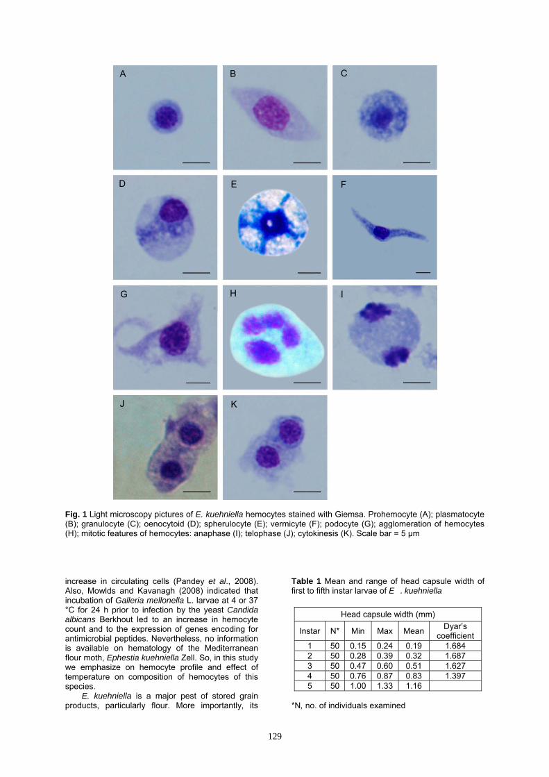
Fig. 1 Light microscopy pictures of E. kuehniella hemocytes stained with Giemsa. Prohemocyte (A); plasmatocyte (B); granulocyte (C); oenocytoid (D); spherulocyte (E); vermicyte (F); podocyte (G); agglomeration of hemocytes (H); mitotic features of hemocytes: anaphase (I); telophase (J); cytokinesis (K). Scale bar = 5 µm increase in circulating cells (Pandey et al., 2008). Also, Mowlds and Kavanagh (2008) indicated that incubation of Galleria mellonella L. larvae at 4 or 37 °C for 24 h prior to infection by the yeast Candida albicans Berkhout led to an increase in hemocyte count and to the expression of genes encoding for antimicrobial peptides. Nevertheless, no information is available on hematology of the Mediterranean flour moth, Ephestia kuehniella Zell. So, in this study we emphasize on hemocyte profile and effect of temperature on composition of hemocytes of this species.
E. kuehniella is a major pest of stored grain products, particularly flour. More importantly, its
Table 1 Mean and range of head capsule width of first to fifth instar larvae of E . kuehniella
Head capsule width (mm)
Instar N* Min Max Mean Dyar’s coefficient
1 50 0.15 0.24 0.19 1.684 2 50 0.28 0.39 0.32 1.687 3 50 0.47 0.60 0.51 1.627 4 50 0.76 0.87 0.83 1.397 5 50 1.00 1.33 1.16
*N, no. of individuals examined
129
A B C
D E F
G H I
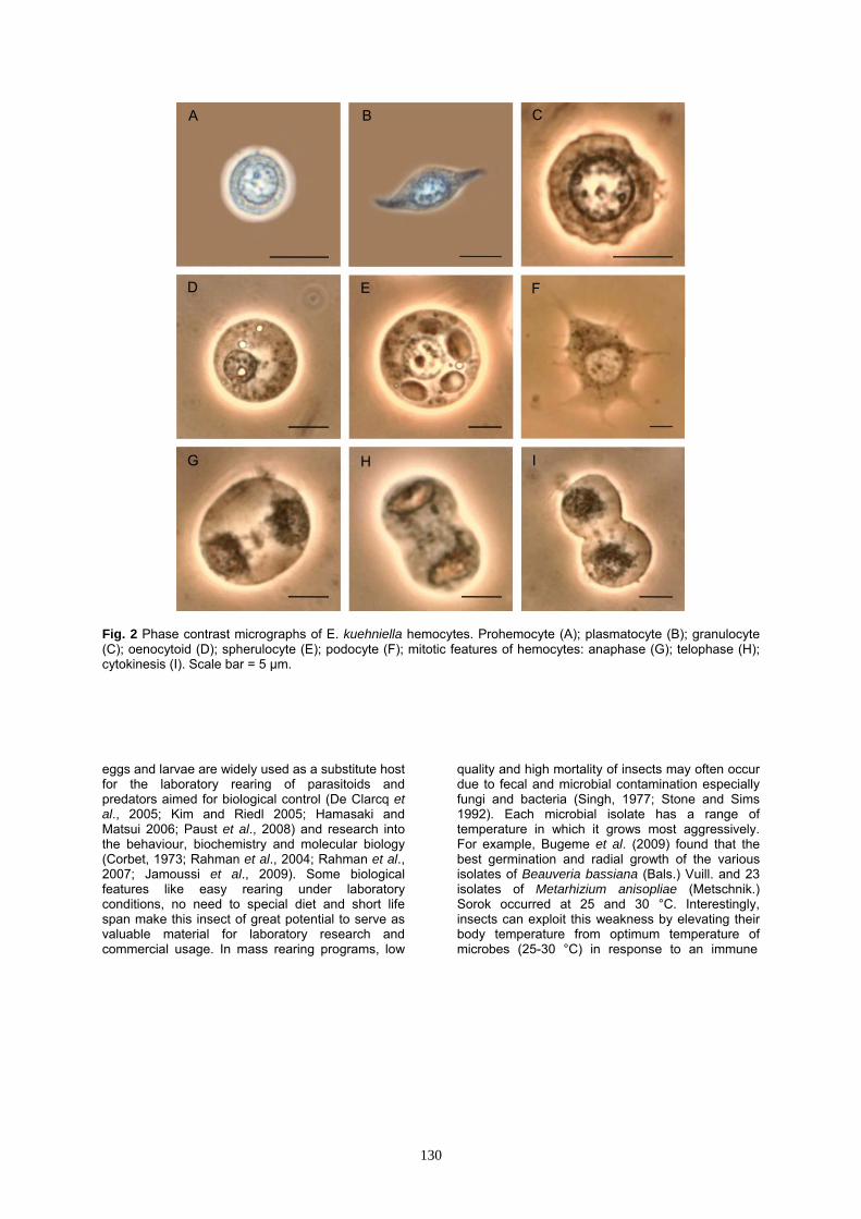
Fig. 2 Phase contrast micrographs of E. kuehniella hemocytes. Prohemocyte (A); plasmatocyte (B); granulocyte (C); oenocytoid (D); spherulocyte (E); podocyte (F); mitotic features of hemocytes: anaphase (G); telophase (H); cytokinesis (I). Scale bar = 5 µm. eggs and larvae are widely used as a substitute host for the laboratory rearing of parasitoids and predators aimed for biological control (De Clarcq et al., 2005; Kim and Riedl 2005; Hamasaki and Matsui 2006; Paust et al., 2008) and research into the behaviour, biochemistry and molecular biology (Corbet, 1973; Rahman et al., 2004; Rahman et al., 2007; Jamoussi et al., 2009). Some biological features like easy rearing under laboratory conditions, no need to special diet and short life span make this insect of great potential to serve as valuable material for laboratory research and commercial usage. In mass rearing programs, low
quality and high mortality of insects may often occur due to fecal and microbial contamination especially fungi and bacteria (Singh, 1977; Stone and Sims 1992). Each microbial isolate has a range of temperature in which it grows most aggressively. For example, Bugeme et al. (2009) found that the best germination and radial growth of the various isolates of Beauveria bassiana (Bals.) Vuill. and 23 isolates of Metarhizium anisopliae (Metschnik.) Sorok occurred at 25 and 30 °C. Interestingly, insects can exploit this weakness by elevating their body temperature from optimum temperature of microbes (25-30 °C) in response to an immune
130

Fig. 3 Scanning electron microscopy of E . kuehniella hemocytes. Prohemocyte (A); plasmatocyte (B); granulocyte (C); oenocytoid (D); spherulocyte (E); vermicyte (F). Scale bar = 5 µm. attack through thermoregulatory behaviour. In this case, Mostafa et al. (2005) reported that temperature and relative humidity had significant effects on cellular defense response of E. kuehniella larvae fed Bacillus thuringiensis Berliner. Some earlier studies in arthropods have also shown that they respond to microbial infection and parasitism by altering their thermoregulatory behaviour and developing a behavioral fever (Kluger et al., 1996). Increased body temperature above normal levels through selection of warmer habitats has been shown to favor host survival in a number of host–pathogen associations (Bronstein and Conner 1984; Louis et al., 1986; Boorstein and Ewald 1987; Watson et al., 1993; Inglis et al., 1996; Adamo,
1998; Karban, 1998; Elliot et al., 2002). The above statements indicate the importance of temperature on immune responses of insects to microbes.
This work represents a starting point in the study of hemocyte production in E. kuehniella and the importance of fever behaviour during immune challenges. The first aim of presented research is morphological characterization of circulating hemocytes. As larval instars are used for laboratory rearing of parasitoids, the hemocyte composition was determined in various developmental stages of this species. Moreover, the changes in hemogram and hemocyte population of two-days old fifth instar larvae were evaluated after heating and chilling stress.
Table 2 Micrometric measurements of hemocytes of E .. kuehniella
Size (µm)
Cytoplasm Nucleus
Cell morphotype* Width (mean ± SE) Length (mean ± SE) Width (mean ± SE) Length (mean ± SE)
PR 4.8-7.7 (6.2 ± 0.18) 5.0-7.8 (6.3 ± 0.18) 3.1-5.3 (4.0 ± 0.15) 3.2-5.3 (4.0 ± 0.15)
PL 4.6-9.5 (7.3 ± 0.26) 10.1-18.2 (14.4 ± 0.47) 2.5-6.9 (5.5 ± 0.27) 3.6-8.1 (6.4 ± 0.27)
GR 7.5-9.4 (8.8 ± 0.19) 8.7-11.3 (9.5 ± 0.23) 3.6-5.0 (4.5 ± 0.12) 3.8-5.1 (4.7 ± 0.10)
OE 6.7-17.7 (9.8 ± 1.00) 7.1-19.4 (11.0 ± 1.11) 2.7-6.3 (3.7 ± 0.32) 2.9-6.5 (3.8 ± 0.32)
SP 8.5-16.5 (14.2 ± 0.98) 10.5-17.9 (15.8 ± 0.85) 3.7-7.2 (4.7 ± 0.30) 3.6-7.4 (4.9 ± 0.31)
VE 4.0-8.0 (4.9 ± 0.38) 23.0-34.4 (27.0 ± 1.06) 2.9-5.1 (3.8 ± 0.25) 4.0-8.0 (5.4 ± 0.45)
PO 3.4-5.1 (4.3 ± 0.15) 17.3-23.3 (20.9 ± 0.68) 3.0-4.2 (3.5 ± 0.13) 3.2-4.5 (3.8 ± 0.14)
*PR, prohemocyte; PL, plasmatocyte; GR, granulocyte; OE, oenocytoid; SP, spherulocyte; VE, vermicyte; PO, podocyte (n=30)
131

E EE
B**
A**
C**
D*
cns bcns bcns b ab ab a
0
5
10
15
20
25
30
35
40
45
20000
30000
40000
50000
60000
70000
80000
90000
0 10 20 30 40 50 60
Tem
pera
ture
(°C)
THC
(cel
ls/m
m3 )
Time (h)
THC treatment THC control Temperature (°C)A
Fig. 4 Effect of heating (A) and chilling (B) on total hemocyte count of E . kuehniella larvae at different time intervals. Each data point is the mean ± SE of at least 10 individual. Different letters indicate significant differences between groups (ANOVA, p < 0.05). Significant differences versus the respective controls were marked by **p< 0.01, *p < 0.05 and ns Non significant at p < 0.05, Student's t-test. Materials and Methods Experimental insects
Eggs of Ephestia kuehniella were provided by the Insectarium of Scientific and Industrial Research Organization of Iran. Each 300 eggs were reared in a plastic container (25 cm length, 15 cm width, and 10 cm height) half filled with mixture of equal amounts of wheat flour and bran. Powdered yeast (5 g) was also added to each jar. The rearing conditions were 26±1 °C, 75 % relative humidity in darkness.
Different larval instars of this moth were determined in accordance with the Dyar’s law through the measurement of the head capsule width (Dyar, 1890). Also, body weight of different
developmental stages of E. kuehniella was measured.
Hematological assays were carried out on the following developmental stages of the insect: L1: two-days old IVth instar larvae (2 days after ecdysis into the forth instar), L2: five-days old IVth instar larvae (2 days before ecdysis into the fifth instar), L3: two-days old Vth instar larvae (2 days after ecdysis into the fifth instar), L4: six-days old Vth instar larvae (2 days before prepupa stage) and L5: one-day old prepupa (Pp).
Hemolymph collection
The hemolymph was collected from subjected insect by amputating the foreleg with a pair of fine scissors sterilized in 70 % ethanol.
A AAB
C
B
A Adns dns
cd** cd**
bc** b**
a**
0
5
10
15
20
25
30
35
40
45
20000
25000
30000
35000
40000
45000
0 10 20 30 40 50 60
Tem
pera
ture
(°C)
THC
(cel
ls/m
m3 )
Time (h)
B
132

Table 3 Body weight, hemolymph volume (HV), total hemocyte count (THC), hemocytes in circulation (HIC) and daily hemocyte production by mitosis (MDP) in each developmental stage studied of E . kuehniella
Phase Body weight (mg) HV (μl) THC (No. of
cells×104/mm3) HIC (No. of cells×104)
MDP (No. of cells×103 /day)
L1 8.15 ± 0.19e 1.05 ± 0.06d 1.31 ± 3.00×10²e 1.38 ± 0.08×10⁴d 1.09 ± 0.65×10²c
L2 12.22 ± 0.37d 1.44 ± 0.07d 1.94 ± 8.24×10²d 2.82 ± 0.20×10⁴d 0.52 ± 0.39×10²c
L3 19.77 ± 0.44c 3.81 ± 0.23c 3.18 ± 9.44×10²c 1.21 ± 0.94×10⁴c 11.37 ± 8.83×10²a
L4 27.18 ± 0.29a 6.04 ± 0.24b 4.36 ± 7.46×10²b 2.64 ± 1.23×10⁴b 6.21 ± 2.90×10²b
L5 22.58 ± 0.37b 6.90 ± 0.22a 5.64 ± 12.0×10²a 3.83 ± 1.20×10⁴a 6.53 ± 2.01×10²b
Means with the same letters in each column are not significantly different at p < 0.05, Tukey's test. Values are expressed as mean ± SE (n = 50). Hemocyte profile Light microscopy studies
The hemocyte monolayers were prepared on clean glass slides and placed at room temperature for 20 min till dry. Then, the hemocytes were stained with Giemsa (diluted 7 times with distilled water and filtered before use) for 25 min, differentiated in saturated solution of lithium carbonate for 30 seconds and then washed in distilled water. After drying, permanent microscopy slides were prepared using Canada balsam. Hemocytes were observed under a light microscope (Olympus BX51) and photographed with a DP 72 camera system powered with DIC Nomarski. Phase contrast microscopy studies
Five µl of fresh hemolymph was mixed with an equal amount of anticoagulant buffer (NaOH, 98 mM; NaCl, 186 mM; EDTA, 17 mM; Citric acid, 41 mM; pH 4.5) on clean glass slides and covered with a cover slip. Since hemocytes begin to deteriorate after bleeding, a fresh preparation was made every 8-10 min. The samples were examined under the phase contrast microscope (Olympus BX51) and images were taken using a Dino-eye AM 7023 B camera. Scanning electron microscopy (SEM)
For SEM studies, 5 µl of fresh hemolymph was mixed with an equal amount of anticoagulant buffer on clean, grease-free 1 cm2 microscope slides and smears were made by drawing a second slide across the first one at a 45° angle. The smears were allowed to air-dry at room temperature and were directly taken in 2.5 % glutaraldehyde in phosphate buffer saline (PBS) (0.1 M, pH 7.2) for 1 h, washed three times in PBS, post-fixed for 1.5 h with osmium tetra oxide (OsO4) (1% in PBS), re-washed in PBS for three times, dehydrated in graded ethanol solutions in distilled water and finally dried with Hexamethyldisilazane (HMDS) for 10 min. The slides were mounted on specimen stubs, coated with gold in a spin coater and finally were observed under FESEM (Hitachi-S4160). All steps were carried out at room temperature. Microscopy classification of the hemocytes was done based on
identification key for hemocyte types set by Gupta (1979).
Hemogram studies Total hemocyte count (THC)
For THC, 2 μl aliquot of hemolymph was mixed immediately in an Eppendorf tube with 98 μl of Tyson solution (NaCl2.72 mM; Na2SO48.96 mM; glycerol 43.68 mM; methyl violet 0.061 mM). THC was conducted with a standard Neubauer hemocytometer. The cells were counted using a light microscope and number of total hemocytes per cubic millimeter (mm3) was calculated using the following formula of Jones (1962):
Hemocytes in x 1mm2 x Dilution x Depth factor of chamber
No. of squares counted where dilution = 50 times, depth factor of the chamber = 10 (constant) and No. of squares counted = 5. Differential hemocyte count (DHC)
For DHC, permanent microscopy slides were prepared according to the method used in light microscopy studies. Differential counting of hemocyte morphotypes was performed by classifying 200 cells per smear (Arnold and Hinks 1976). The various stages of mitotic division were distinguished based on the method of Amaral et al. (2010) and number of hemocytes undergoing mitotic division was recorded in each 200-cell of a smear. Measurements of cell and nucleus lengths and widths were made with a digital objective lens of light microscope (Olympus BX51). A minimum of 30 cells of each morphotype were measured. Hemolymph volume (HV)
Hemolymph volume was directly measured through collecting whole hemolymph of larvae using Hamilton syringe.
133

Hemocytes in circulation (HIC) The total number of free hemocytes in
circulation for any developmental stages is hypothetically derived from the formula THC × HV.
Maximum daily production (MDP) of hemocytes through mitosis occurred in circulation
This hematological parameter is hypothetically determined as (MI × HIC × 24) / 1000. Thermal regimes
Three thermal treatments were utilized in this work. Two-days old Vth instar larvae weighing 20 ± 1 mg were used during the experiment. All larvae were placed inside a programmable, refrigerated test chamber, model MK53 (Binder, Tuttlingen, Germany). To assess the effect of heat shocks on THC, HV and DHC of subjected larvae, the test chamber was scheduled to heat gradually at a constant rate of 0.05 °C/min starting from 27 °C to 40 °C. After 12 h exposing to 40 °C, larvae were cooled to 27 °C at the same rate of heating and were maintained at 27 °C for 24 h. For chilling assay the same trend was used and larvae were exposed to 4 °C for 12 h. Control larvae were stored at 27 °C for whole of the experiment course. THC and HV were determined in subsequent series of time. DHC and MI were determined just after 12 h exposure of larvae to the thermal regimes. Also, the possible cytological abnormalities of hemocytes were investigated after 12 h exposure of larvae to heat and chill stress and appropriate images were acquired with a DP 72 camera system powered with DIC Nomarski. Statistical analysis
Means were analyzed using the SPSS program version 16.0 for analysis of variance (ANOVA) and the means were grouped using Tukey’s test (P<0.05). Also, statistical comparison between treatment and control at each time point was performed using Student’s t-test. Results
The life cycle of E. kuehniella includes five
stages: egg, larva, prepupa, pupa and adult. Also
according to Dyar’s law, five distinct instar larvae were identified (Table 1). Hemocyte profile
Five main hemocyte morphotypes were recognized in the hemolymph of E. kuehniella, i.e. prohemocytes, plasmatocytes, granulocytes, oenocytoids and sphrulocytes. Prohemocytes (PRs)
PRs are usually the smallest hemocytes found in the hemolymph, round or oval cells with variable sizes (4.8-7.7 μm wide and 5-7.8 μm long) (Figs 1A, 2A, 3A and Table 2). The nucleus is large, round, usually centrally located and almost filling the most part of cytoplasm. Nuclear size is variable (3.1-5.3 μm wide and 3.2-5.3 μm long) (Table 2). A thin peripheral layer of homogeneous cytoplasm surrounds the nucleus.
Plasmatocytes (PLs)
PLs are small to large, polymorphic cells with variable sizes (4.6-9.5 μm wide and 10.1-18.2 μm long) (Figs 1B, 2B, 3B and Table 2). The cytoplasm is abundant and generally agranular or slightly granular. The cytoplasmic membrane may have filopodia. The nucleus may be round or elongate and is generally centrally located. Nuclear size is variable (2.5-6.9 μm wide and 3.6-8.1 μm long) (Table 2). Scattered chromatin masses may be present in nucleus of these cells. Granulocytes (GRs)
These are small to large, spherical or oval cells of variable sizes (7.5-9.4 μm wide and 8.7-11.3 μm long) (Figs 1C, 2C, 3C and Table 2). The nucleus is rounded and is centrally located with variable sizes (3.6-5 μm wide and 3.8-5.1 μm long) (Table 2). The cytoplasm is characteristically granular and the cell membrane is usually articulated. Oenocytoids (OEs)
OEs are small to large, oval or spherical cells with widely variable sizes (6.7-17.7 μm wide and 7.1-19.4 μm long) and shapes (Figs. 1D, 2D, 3D and Table 2). The plasma membrane is generally
Table 4 Differential hemocyte count (DHC) in each developmental stage studied of E . kuehniella
% Hemocyte morphotype (Mean ± SE) Phase PR* PL GR OE SP MI
L1 9.16 ± 0.64b 61.86 ± 3.11bc 16.36 ± 1.27ab 2.46 ± 0.33b 10.16 ± 1.55a 3.30 ± 0.60a
L2 3.18 ± 0.19d 75.05 ± 1.14a 11.32 ± 1.21b 5.14 ± 0.53b 5.31 ± 0.65b 0.78 ± 0.34b
L3 15.74 ± 0.69a 53.34 ± 1.75c 22.44 ± 1.99a 2.24 ± 0.40b 6.24 ± 0.78ab 3.90 ± 0.67a
L4 5.48 ± 0.84c 62.64 ± 4.18abc 17.63 ± 2.88ab 10.15 ± 1.12a 4.10 ± 0.95b 0.98 ± 0.27b
L5 3.76 ± 0.48cd 69.26 ± 2.48ab 11.56 ± 2.27b 5.06 ± 1.71b 10.36 ± 1.04a 0.70 ± 0.25b
* (PR, prohemocyte; PL, plasmatocyte; GR, granulocyte; OE, oenocytoid; SP, spherulocyte; MI, mitotic index). Means with the same letters in each column are not significantly different at p < 0.05, Tukey's test. Values are expressed as mean ± SE (n=10).
134

A
B
Fig. 5 Effect of heating (A) and chilling (B) on hemolymph volume of E . kuehniella larvae at different time intervals. Each data point is the mean ± SE of at least 10 individual. Different letters indicate significant differences between groups (ANOVA, p < 0.05). Significant differences versus the respective controls were marked by **p < 0.01, *p < 0.05 and ns Non significant at p < 0.05, Student's t-test. without micropapillae, filopodia or other irregular processes. The cytoplasm is slightly granular. The nucleus is small, round or elongate and generally eccentrically located (2.7-6.3 μm wide and 2.9-6.5 μm long) (Table 2). Spherulocytes (SPs)
These are small to large, ovoid or round cells with variable sizes (8.5-16.5 μm wide and 10.5-17.9 μm long) and usually larger than other hemocytes identified in E. kuehniella. (Figs. 1E, 2E, 3E and Table 2). The nucleus is generally small, round, central (3.7-7.2 μm wide and 3.6-7.4 μm long)
(Table 2) and generally obscured by the intracytoplasmic spherules that are characteristic of these cells. The number of the spherules varies from 4-6.
Two other morphotypes were also distinguished: Vermicytes (VEs)
VEs are extremely elongated cells (4-8 μm wide and 23-34.4 μm long) with slightly granular or agranular cytoplasm (Figs. 1F, 3F and Table 2). The nucleus may be located centrally or eccentrically (2.9-5.1 μm wide and 4-8 μm long) (Table 2).
135

Table 5 Changes in differential hemocyte count (DHC) of two-days old Vth instar larvae of E . kuehniella 12 h after treatment under different temperature regimes
% Hemocyte morphotype (Mean ± SE)
Temperature (°C) PR* PL GR OE SP MI
27 °C (Control) 15.68 ± 0.65a 51.78 ± 1.42b 23.28 ± 1.27a 3.18 ± 0.43b 6.08 ± 0.33a 4.10 ± 0.73b
40 °C (Heating) 15.18 ± 1.55a 61.28 ± 0.79a 11.68 ± 1.20b 4.68 ± 0.51a 7.18 ± 0.33a 9.00 ± 0.59a
4 °C (Chilling) 13.46 ± 0.95a 43.16 ± 2.21c 29.26 ± 2.83a 6.06 ± 0.51a 8.06 ± 0.94a 0.40 ± 0.18c
* (PR, prohemocyte; PL, plasmatocyte; GR, granulocyte; OE, oenocytoid; SP, spherulocyte; MI, mitotic index). Means with the same letters in each column are not significantly different at p < 0.05, Tukey's test. Podocytes (POs)
POs are very large in size (3.4-5.1 μm wide and 17.3-23.3 μm long), extremely flattened PL-like cells with several cytoplasmic extensions (Figs 1G, 2F and Table 2). The nucleus is generally large and centrally located and may appear punctuate (3-4.2 μm wide and 3.2-4.5 μm long) (Table 2).
An agglomeration of hemocytes (Fig. 1H) was also observed in some profiles. Some stages of mitotic division including anaphase (Figs. 1I and 2G), telophase (Figs 1J and 2H) followed by cytokinesis (Figs 1K and 2I) to produce two identical daughter cells were also characterized in hemocytes of E. kuehniella.
Hemogram
There is a gradual increase in body weight during larval stages, with maximum in L5, followed by significant decline in prepupa (F = 491.03, df = 4, p = 0.000) (Table 3). In general, total hemocyte population of E. kuehniella showed an increasing trend with increase in larval instars (F = 424.75, df = 4, p = 0.000). It was also found that THC in the late stages (L2 and L4) and prepupa was significantly higher than that in the early stages (L1 and L3) (Table 3).
An increasing trend was found in HV of tested insect with increase in developmental stages (F = 198.21, df = 4, P = 0.000) (Table 3).
Along with increase in THC and HV, a statistically significant increase in HIC was observed (F = 330.63, df = 4, p = 0.000) (Table 3). Also, among different developmental stages of E. kuehniella, the highest level of hemocyte production through mitosis was observed in L1 and L3 because of maximum mitotic activity in these two stages (Table 3).
The differential hemocyte profile during the different developmental stages of E. kuehniella is provided in Table 4. PLs and GRs were the most abundant hemocytes followed by the SPs, OEs and PRs in all studied stages. We also observed significant variations of DHC between early and later part of each stage. Generally, it is found that proportion of PRs, GRs, OEs, SPs (except in prepupa) and also MI in the early of each instar was significantly higher than that in the late part and
prepupa. In contrast, PLs were found to be in their maximum proportion at the end of each instar and also prepupa (Table 4). Thermal regimes
The results of the effects of thermal stress on THC and HV of 2-days old Vth instar larvae of E. kuehniella are presented in Figs. 4 and 5. As it can be seen, the larvae exposed to 40 °C for 12 h showed a drastic increase in THC from 3.18×10⁴ to 7.03×10⁴ cells/mm3 (Fig. 4A). Interestingly, the increase in total number of hemocytes continued even 6 hours after returning of larvae to normal temperature (27 °C) reaching 8.21×10⁴ cell/mm 3
(Fig. 4A). Also, HV significantly decreased from 3.81 μl to the lowest value of 2.1 μl after exposing the larvae to heat stress for 12 h (Fig. 5A). In contrast, chilling for 12 h caused a significant reduction in THC of subjected larvae from 3.12×10⁴ to 2.25×10⁴ cells/mm3 (Fig. 4B). When cold exposed larvae were returned to normal temperature (27 °C), total number of hemocytes gradually increased and reached to 3.03×10⁴ cells/mm3 by increasing the incubation time (Fig. 4B). No significant differences were observed in HV of control and cold acclimated larvae at various time series (Fig. 5B).
According to the results of thermal stress on DHC, variations in the proportion of different hemocytes were significant among three temperature treatments (Table 5). While PLs and OEs percentages increased in heat-exposed larvae, a significant reduction in the counts of GRs was recorded. In chilled larvae, on the other hand, OEs rose in their number with a significant decline in PLs. Also, a little increase in GRs was observed but it was not statistically significant. No significant differences were observed in the proportion of PRs and SPs under three temperature regimes. Low temperature resulted in a decrease in mitotically dividing cells of E. kuehniella larvae but in contrast, high temperature caused a significant increase in MI (Table 5). Effect of thermal stress on cell-structure of hemocytes
Heating for 12 h led to vacuolization in the cytoplasm of PLs (Fig. 6A) and increase in number
136

Fig. 6 Cell-structure abnormality in larvae exposed to heat stress. Vacuolization in cytoplasm of plasmatocytes (A) and rounded plasmatocytes loosing filipods (B) after 12 h exposing of larvae to 40 °C. Scale bar = 10 µm. of rounded PLs without filipods (Fig. 6B). No considerable changes have been found in the cytological features of hemocytes of incubated larvae at 4 °C. Discussion
The present study provides detailed information
of hemocyte profile and hemogram of the Mediterranean flour moth, E. kuehniella. Strand and Pech (1995) divided the lepidopteran hemocytes into five classes on the basis of morphology, i.e. prohemocytes, plasmatocytes, granu locytes, oenocytoids and spherulocytes. Similar results were also reported for other lepidopterous larvae, such as Plutella xylostella (Huang et al., 2010), Ectomoyelois ceratoniae (Khosravi et al., 2012), Hyphantria cunea (Ajamhassani et al., 2013), Bombyx mori (Tan et al., 2013). We also found all of these five morphotypes of hemocytes in E. kuehniella larva in this study.
Also, our results indicated that plasmatocytes and granulocytes are the most abundant hemocytes in the hemolymph of E. kuehniella. Prior studies have shown that plasmatocytes and granulocytes were responsible for cellular immune responses in many lepidopteran insect larvae, such as G. mellonella (Tojo et al., 2000) and Manduca sexta L. (Ling and Yu, 2006) and together usually comprise more than 50 % of the hemocytes in circulation (Lackie, 1988; Ratcliffe, 1993).
According to the results of cell measurements, it is shown that each cell morphotype is of a varied size. We also found various forms of each particular hemocyte in our tested insect. As shown in the results, two other morphotypes of hemocytes termed vermicytes and podocytes were distinguished in the hemolymph of E. kuehniella. These two morphotypes have not been recognized as distinct types in electron microscopic studies so
far, primarily because ultrastructurally they appear similar to plasmatocytes (Gupta, 1997). The origin of vermicytes and podocytes is unknown, but it is conceivable that they are derived from plasmatocytes and are considered as variant forms of plasmatocytes (Gupta, 1997). This variation in the form and size has brought many problems in the classification of hemocytes because the insect blood cells are highly pleomorphic, and the particular form they present at any one time seems to depend on the age, developmental stage, nutritional state and species of insect as well as on the methods of collection and examination used by the investigator (Jones, 1962; Lai-Fook and Neuwirth 1972).
The maintenance of circulating hemocytes in larval Lepidoptera has been attributed to both the release of hemocytes from hematopoietic organs and the mitosis of hemocytes in the circulatory system (Gardiner and Strand 2000). The levels of mitotic activity in circulating hemocytes rarely exceed 1 % in almost all cases (Jones, 1967a; Jones and Lin 1968; Jones, 1967b), but it is shown that in E. kuehniella this activity varies with developmental stage of insect. The mitotic index of larvae in L1 and L3 reached 3.3 % and 3.9 %, while the dividing cells observed in L2, L4 and prepupa were 0.78%, 0.98% and 0.7%, respectively. In a parallel study on Oncopeltus fasciatus Dallas, Feir and McClain (1968) found that the MI was very low immediately after ecdysis into the fifth instar. They noted that mitotic activity began to rise at 23 hour post-ecdysis, reached its peak (4.06 %) in the 30-hr group, remained high until 74 hour, and then declined during the remainder of the stadium. These findings agree with the reports of Sanjayan et al. (1996), who observed different values of the mitotic index in final instar and adult insects of Spilostethus hospes Fab.
Mitotic activity has been most consistently reported in the prohemocytes and hemocyte
137

differentiation studies in some insect species revealed that prohemocytes are the stem cells from which the other cells arise (Gupta, 1991). In case of the silkworm, B. mori, it is proved that approximately 43 % of prohemocytes differentiate into plasmatocytes, granulocytes and spherulocytes (Yamashita and Iwabuchi, 2001). The population of prohemocytes in L1 and L3 is significantly higher than that in L2 and L4 and prepupa. The rapid decline in their counts coinciding with the increase in plasmatocytes and partially oenocytoids in the later part of larval instar suggests that their differentiation is probably toward the formation of plasmatocytes and oenocytoids at that time.
The results indicated that cell population increased during larval development of E. kuhniella, and reached a peak during the prepupal period. Also, THC was in its high value at the late stage of each larval instar than the early stage. It has been frequently reported that THC varies with the developmental stage and physiological condition of the insect. Patton and Flint (1959) noted that counts from Periplaneta decreased at the molt and later increased post molt. Also, Crossley (1975) proved that hemopoiesis in insects is under endocrine control. In this case, Akai and Sato (1971) showed that the increase of ecdysteroids in hemolymph of B. mori would be resulted in more hemocyte production and release from the hematopoietic organs. So, it could be concluded that changes in THC during the developmental stages of E. kuehniella would be probably attributed to the variations in ecdysteroids and juvenile hormones (JH) titers.
In many insect species, fluctuations in the number of hemocytes are influenced by the release of hemocytes from the hemapoietic organ and attachment of the cells to internal tissues (Tu et al., 2002; Okazaki et al., 2006). The number of hemocytes in circulation can change rapidly in response to stress, wounding or infection (Gillespie et al., 2000; Mowlds and Kavanagh 2008). As low and high temperatures are a source of stress for insects, it is possible that the number of hemocytes was directly altered by the change in temperature. In this aspect, results of present study clearly indicated that compared to control temperature, heating caused a significant increase in THC and in contrast, those larvae incubated at 4 °C showed a significant reduction in THC. So, it seems that exposing the insects to high temperatures can increase the environmental fitness of larvae through a similar mechanism to thermoregulatory behaviour by increasing total number of hemocytes especially PLs. There are several reasons to justify these changes in THC. Cell cycle can be divided in three periods: interphase consisting G1 (pre-synthetic interphase), S phase (DNA synthetic interphase) and G2 (post-synthetic interphase), mitosis phase and the final phase, cytokinesis, where the new cell is completely divided. In many cases it was found that the time taken for cells to divide decreases in response to an increase in temperature (Francis and Barlow 1988). Moore et al. (1997) stated that temperature reduction from 37 °C to 30 °C caused a rapid decrease in the percentage of cells in S phase and accumulation of cells in G1. In another study, Bloemkolk et al. (1992) showed that maximal cell
density was observed at 37 °C. Lower temperatures, in contrast, caused cells to stay longer in the G1 phase of the cell cycle. In parallel, our results showed that compared to chilling, heating caused a 2.2 folds increase in mitotic rate of hemocytes. So, it is concluded that increase in THC of E. kuehniella larvae exposed to high temperature may be attributed to more mitotic rate of hemocytes and more importantly increased hemocyte proliferation of hemopoietic organs.
It was found that an inverse relationship exists between the THC and the HV after incubation of larvae at 40 °C. Thus, it seems that another possible reason for the increase in THC is due to loss of body fluid as a result of desiccation. Also, release of hemocytes attached to the internal organs of heat exposed larvae into the hemolymph circulation can be another reason of increase in THC. On the other hand, the low hemocyte counts in chilled larvae found to be primarily attributable to adherence to respected larval tissues. Some authors indicated that low counts from unfixed larvae in heat may have been caused by increased clumping and hence, unavailability of circulating hemocytes. We could not find any significant relationship between THC and HV in chilled larvae.
Our results indicated that heating led to cell structure changes in PLs. There are several evidences regarding the activation of hemocytes which are characterized by expanded filipods during immune challenge (Kwon and Kim 2007). However, in our experiment rounded PLs without filipods were increased in response to heating condition. This phenomenon was occurred by heating without pathogenic infection. Therefore, it could be expected that pathogenic infection would be the main reason for expanding filipods in PLs in response to fever. Further investigations are needed to be done to elucidate this kind of cell response. Conclusion
In this study, we determined the morphological
characteristics of hemocytes of E. kuehniella and changes in hemocyte composition during the larval development that were essential for further understanding of cellular responses of this insect to environmental and pathological stresses. Our findings revealed the impact of thermal stress on THC, DHC and HV of E. kuehniella. However, further investigations may have to be performed to determine if changes in hematological properties of thermal treated larvae could affect their cell mediated immune responses. It might then be important to conduct next experiments on the effect of thermal shocks upon hemocyte composition and different immune responses of E. kuehniella after infection with various pathogens. References Adamo SA. The specificity of behavioral fever in the
cricket Acheta domesticus. J. Parasitol. 84(3): 529-533, 1998.
Ajamhassani M, Jalali Sendi J, Zibaee A, Askary H, Farsi MJ. Immunoliogical responses of Hyphantria cunea (Drury) (Lepidoptera: Arctiidae) to entomopathogenic fungi,
138

Beauveria bassiana (Bals.-Criy) and Isaria farinosae (Holmsk.) FR. J. Plant Prot. Res. 53(2): 110-118, 2013.
Amaral IMR, Neto JFM, Pereira GB, Franco MB, Beletti ME, Kerr WE, Bonetti AM, Ueira-Vieira C. Circulating hemocytes from larvae of Melipona scutellaris (Hymenoptera, Apidae, Meliponini): Cell types and their role in phagocytosis. Micron 41: 123-129, 2010.
Arnold JW, Hinks CF. Haemopoiesis in Lepidoptera. I. The multiplication of circulating haemocytes. Can. J. Zool. 54(6): 1003-1012, 1976.
Bloemkolk JW, Gray MR, Merchant F, Mosmann TR. Effect of temperature on hybridoma cell cycle and MAb production. Biotechnol. Bioeng. 40(3): 427-431, 1992.
Boorstein SM, Ewald PW. Costs and benefits of behavioral fever in Melanoplus sanguinipes infected by Nosema acridophagus. Physiol. Zool. 60: 586–595, 1987.
Bronstein SM, Conner WE. Endotoxin-induced behavioural fever in the Madagascar cockroach, Gromphadorhina portentosa. J. Insect Physiol. 30: 327-330, 1984.
Bugeme DM, Maniania NK, Knapp M, Boga HI. Effect of temperature on virulence of Beauveria bassiana and Metarhizium anisopliae isolates to Tetranychus evansi. Exp. Appl. Acarol. 46: 275-285, 2008.
Corbet SA. Oviposition pheromone in larval mandibular glands of Ephestia kuehniella. Nature 243: 537-538, 1973.
Crossley AC. The cytophysiology of insect blood. Adv. Insect Physiol. 11: 117-221, 1975.
De Clarcq P, Bonte M, Van Speybroeck K, Bolckmans K, Deforce K. Development and reproduction of Adalia bipunctata (Coleoptera: Coccinellidae) on eggs of Ephestia kuehniella (Lepidoptera: Pyralidae) and pollen. Pest Manag. Sci. 61: 1129-1132, 2005.
Dyar HG. The number of molts of lepidopterous larvae. Psyche 5: 420-422, 1890.
Elliot SL, Blanford S, Thomas MB. Host-pathogen interactions in a varying environment: temperature, behavioural fever and fitness. Proc. Roy. Soc. B: Biol. Sci. 269(1500): 1599-1607, 2002.Feir D, McClain E. Mitotic activity of the circulating heniocytes of the large milkweed bug, Oncopeltus fasciatus. Ann. Entomol. Soc. Am. 61: 413-416, 1968.
Francis D, Barlow PW. Temperature and the cell cycle. Symp. Soc. Exp. Biol. 42: 181-201, 1988.
Gardiner EMM, Strand MR. Hematopoiesis in larval Pseudoplusia includens and Spodoptera frugiperda. Arch. Insect Biochem. Physiol. 43: 147-164, 2000.
Gillespie JP, Kanost MR, Trenczek T. Biological mediators of insect immunity. Annu. Rev. Entomol. 42: 611-643, 1997.
Gillespie JP, Burnett C, Charnley AK. The immune response of the desert locust Schistocerca gregaria during mycosis of the entomopathogenic fungus, Metarhizium anisopliae var acridum. J. Insect Physiol. 46: 429-437, 2000.
Gupta AP. Insect Hemocytes. Development, forms, functions and techniques. Cambridge University Press, Cambridge, UK, 1979.
Gupta AP. Insect immunocytes and other hemocytes: roles in cellular and humoral immunity. In: Gupta AP (ed.), Immunology of Insects and Other Arthropods. CRC Press, Boca Raton, pp 19-118, 1991.
Hamasaki K, Matsui M. Development and reproduction of an aphidophagous coccinellid, Propylea japonica (Thunberg) (Coleoptera: Coccinellidae), reared on an alternative diet, Ephestia kuehniella Zeller (Lepidoptera: Pyralidae) eggs. Appl. Entomol. Zool. 41: 233-237, 2006.
Huang F, Yang Y, Shi M, Li J, Chen Z, Chen F, et al. Ultrastructural and functional characterization of circulating hemocytes from Plutella xylostella larva: Cell types and their role in phagocytosis. Tissue Cell 42: 360-364, 2010.
Inglis GD, Johnson DL, Goettel MS. Effects of temperature and thermoregulation on mycosis by Beauveria bassiana in grasshoppers. Biol. Control 7: 131-139, 1996.
Irving P, Ubeda JM, Doucet D, Troxler L, Lagueux M, Zachary D, Hoffmann JA, Hetru C, Meister M. New insights into Drosophila larval haemocyte functions through genome-wide analysis. Cell Microbiol. 7: 335-350, 2005.
Jamoussi K, Sellami S, Abdelkefi-Mesrati L, Givaudan A, Jaoua S. Heterologous expression of Bacillus thuringiensis vegetative insecticidal protein-encoding gene vip3LB in Photorhabdus temperate strain K122 and oral toxicity against the Lepidoptera Ephestia kuehniella and Spodoptera littoralis. Mol. Biochem. 43: 97-103, 2009.
Jones JC. Current concepts concerning insect hemocytes. Am. Zool. 2: 209-246, 1962.
Jones JC. Changes in the haemocyte picture of Galleria mellonella (Linnaeus). Biol. Bull. 132: 211-221, 1967a.
Jones JC. Normal differential counts of haemocytes in relation to ecdysis and feeding in Rhodnius. J. Insect Physiol. 13:: 1133-1141, 1967b.
Jones JC, Liu DΡ. Α quantitative study of mitotic divisions of haemocytes of Galleria mellonella larvae. J. Insect Physiol. 14: 1055-1061, 1968.
Karban R. Caterpillar basking behavior and nonlethal parasitism by tachinid flies. J. Insect Behav. 11: 713–723, 1998.
Khosravi R, Jalali Sendi J, Ghasemi V. Identification of hemocytes in carob moth, Ectomoyelois ceratoniae Zeller (Lepidoptera: Pyralidae) larvae. Plant Pests Res. 2: 29-39, 2012.
Kim DS, Riedl H. Effect of temperature on development and fecundity of the predaceous plant bug Deraeocoris brevis reared on Ephestia kuehniella eggs. Biocontrol 50: 881-897, 2005.
Kluger MJ, Kozak W, Conn CA, Leon LR, Soszynski D. The adaptive value of fever. Infect. Dis. Clin. North Am. 10: 1-20, 1996.
Kwon S, Kim Y. Immunosupressive action of pyriproxyfen, a juvenile hormone analoge, enhances pathogencity of Bacillus thuringiensis
139

subsp. Kurstaki against diamondback moth, Plutella xylostella (Lepidoptera: Yponomuetidae). Biol. Control 42: 72-76, 2007. Lackie AM. Haemocyte behavior. Adv. Insect Physiol. 21: 85-178, 1988.
Lai-Fook J, Neuwirth M. The importance of methods of fixation in the study of insect blood cells. Can. J. Zool. 50: 1011-1013, 1972.
Ling E, Yu XQ. Hemocytes from the tobacco hornworm Manduca sexta have distinct functions in phagocytosis of foreign particles and self dead cells. Dev. Comp. Immunol. 30: 301-309, 2006.
Louis C, Jourdan M, Cabanac M. Behavioral fever and therapy in a rickettsia-infected Orthoptera. Am. J. Physiol. 250: 991-995, 1986.
Moore A, Mercer J, Dutina G, Donahue CJ, Bauer KD, Mather JP, et al. Effects of temperature shift on cell cycle, apoptosis and nucleotide pools in CHO cell batch cultures. Cytotechnology 23: 47-54, 1997.
Mowlds P, Kavanagh K. Effect of pre-incubation temperature on susceptibility of Galleria mellonella larvae to infection by Candida albicans. Mycopathologia 165: 5-12, 2008.
Okazaki T, Okudaira N, Iwabuchi K, Fugo H, Nagai T. Apoptosis and adhesion of hemocytes during molting stage of silkworm, Bombyx mori. Zool. Sci. 23: 299-304, 2006.
Pandey JP, Tiwari RK, Kumar D. Reduction in haemocyte mediated immune response in Danais chrysippus following treatment with neem-based insecticides. J. Entomol. 5: 200-206, 2008.
Pandey JP, Mishra PK, Kumar D, Singh BMK, Prasad BC. Effect of temperature on hemocytic immune responses of tropical tasar silkworm, Antheraea mylitta D. Res. J. Immunol. 3: 169-177, 2010.
Patton RL, Flint RA. The variation in the blood-cell count of Periplaneta americana (L.) during a molt. Ann. Entomol. Soc. Am. 52: 240-242, 1959.
Paust A, Reichmuth C, Buttner C, Prozell S, Adler C, Scholler M. Spatial effects on competition between the larval parasitoids Habrobracon hebetor (Say) (Hymenoptera: Braconidae) and Venturia canescens (Gravenhorst) (Hymenoptera: Ichneumonidae) parasitising the Mediterranean flour moth, Ephestia kuehniella Zeller (Lepidoptera: Pyralidae). Mitt. Deutsch. Ges. Allg. Angew. Entomol. (DGaaE) 16: 291-294, 2008.
Rahman MM, Roberts HL, Schmidt O. The development of the endoparasitoid Venturia canescens in Bt-tolerant, immune induced larvae of the flour mothEphestia kuehniella. J. Invertebr. pathol. 87: 129-131, 2004.
Rahman MM, Roberts HL, Schmidt O. Factors affecting growth in the koinobiont endoparasitoid Venturia canescens in the flour
moth Ephestia kuehniella. J. Insect physiol. 53: 463-467, 2007.
Ratcliffe NA. Cellular defense responses of insects: Unresolved problems. In: Beckage NE, Thompson SN, Federici BA (eds). Parasites and Pathogens of Insects, vol. I. Academic Press, San Diego, CA, pp 267-304, 1993.
Sanjayan KP, Ravikumar T, Albert S. Changes in the haemocyte profile of Spilostethus hospes (Fab) (Heteroptera: Lygaeidae) in relation to eclosion, sex and mating. J. Biosci. 21: 781-788, 1996.
Salt G. The resistance of insect parasitoids to the defence reactions of their hosts. Biol. Rev. Camb. Philos. Soc. 43: 200-232, 1968.
Shapiro M. Changes in hemocyte populations. In: Gupta AP (ed.), Insect hemocytes, Cambridge University Press, Cambridge, pp 475-524, 1979.
Sharma PR, Sharma OP, Saxena BP. Effect of sweet flag rhizome oil (Acorus calamus) on hemogram and ultrastructure of hemocytes of the tobacco armyworm, Spodoptera litura (Lepidoptera: Noctuidae). Micron 39: 544-551, 2008.
Singh P. Artificial diets for insects, mites, and spiders, New York, USA, 1977.
Stone T, Sims S. Insect rearing and the development of bioengineered crops. In: Anderson TE, Lepa N (eds), Advances in insects rearing for research and pest anagement, Oxford, Westview Press/Oxford and IBH Publishing, UK, pp 33-40, 1992.
Strand MR, Pech LL. Immunological basis for compatibility in parasitoid-host relationships. Annu. Rev. Entomol. 40: 31-56, 1995.
Tan J, Xu M, Zhang K, Wang X, Chen S, Li T, Xiang Z, Cui H. Characterization of hemocytes proliferation in larval silkworm, Bombyx mori. J. Insect Physiol. 59: 595-603, 2013.
Tojo S, Naganuma F, Arakawa K, Yokoo S. Involvement of both granular cells and plasmatocytes in phagocytic reactions in the greater wax moth, Galleria mellonella. J. Insect Physiol. 46: 1129-1135, 2000.
Tu ZL, Kobayashi Y, Kiguchi K, Watanabe H, Yamamoto K. Effects of heavy-ion radiosurgery on the hemopoietic function of the silkworm Bombyx mori. J. Radiat. Res. 43: 269-275, 2002.
Watson DW, Mullens BA, Petersen JJ. Behavioral fever response of Musca domestica (Diptera: Muscidae) to infection by Entomophthora muscae (Zygomycetes: Entomophthorales). J. Invertebr. Pathol. 61: 10-16, 1993.
Yamashita M, Iwabuchi K. Bombyx mori prohemocyte division and differentiation in individual microcultures. J. Insect Physiol. 47: 325-331, 2001.
140