hairy root induction in helicteres isora l. and … article hairy root induction in helicteres isora...
TRANSCRIPT
ORIGINAL ARTICLE
Hairy Root Induction in Helicteres isora L. and Productionof Diosgenin in Hairy Roots
Vinay Kumar • Dnyanada Desai • Varsha Shriram
Received: 10 March 2014 / Accepted: 23 March 2014 / Published online: 9 April 2014
� The Author(s) 2014. This article is published with open access at Springerlink.com
Abstract Mature seeds of Helicteres isora L. were collected from seven geographical locations of Maharashtra and Goa
(India) and evaluated for diosgenin (a bioactive steroidal sapogenin of prime importance) extraction and quantification.
Chemotypic variations were evidenced with diosgenin quantity ranging from 33 lg g-1 seeds (Osmanabad forests) to
138 lg g-1 (Khopoli region). Nodal and leaf explants from in vitro-raised seedlings were used for callus and Agrobac-
terium-mediated transformation, respectively. Compact, hard, whitish-green callus (2.65 g explant-1) was obtained on
MS ? 13.32 lM BAP ? 2.32 lM Kin after 30 days of inoculation. Various parameters including types of explant and
Agrobacterium strain, culture density, duration of infection and various medium compositions were optimized for hairy
root production. A. rhizogenes strain ATCC-15834 successfully induced hairy roots from leaf explants (1 cm2) with 42 %
efficiency. Transgenic status of the roots was confirmed by PCR using rolB and VirD specific primers. Hairy roots showed
an ability to synthesize diosgenin. Diosgenin yield was increased *8 times in hairy roots and *5 times in callus than the
seeds of wild plants. Enhanced diosgenin content was associated with proline accumulation in hairy roots. This is the first
report on induction of hairy roots in H. isora.
Keywords Diosgenin � Helicteres isora � Hairy roots
1 Introduction
Diosgenin is a naturally occurring highly bioactive steroidal
sapogenin belonging to the group of triterpenes and heavily
used in pharmaceutical industry for commercial steroid
production [1–3]. Diosgenin is used as an intermediate in the
synthesis of corticosteroids, sex hormones, oral contracep-
tives as well as other steroids via hemisynthesis [4, 5]. In
spite of the introduction of new precursors like solasodine,
hecogenin and tigogenin for steroidal drug synthesis, dios-
genin remains the major precursor. Diosgenin is used in
traditional medicine as an anti-hypercholesterolemia, anti-
hypertriacylglycerolemia, anti-diabetes and anti-hypergly-
cemia agent [6–8]. Diosgenin has been reported to show
anti-proliferative and pro-apoptotic actions as a chemopre-
ventive and therapeutic agent against various cancers [2, 9]
and to prevent cardiovascular diseases [10]. Diosgenin also
play an important role in control of cholesterol metabolism
[11] and has shown estrogenic effects on mammary epi-
thelium of ovariectomized mouse [12].
Dioscorea species are traditionally dominant source of
diosgenin and related steroidal saponins [13]. However,
overexploitation has led to the rapid depletion of these
species and Dioscorea deltoidea, the richest source of
V. Kumar (&) � D. Desai
Department of Biotechnology, Modern College, Ganeshkhind,
Pune 411 016, India
e-mail: [email protected];
V. Shriram
Department of Botany, Prof. Ramkrishna More College, Akurdi,
Pune 411 044, India
123
Nat. Prod. Bioprospect. (2014) 4:107–112
DOI 10.1007/s13659-014-0011-9
diosgenin has subsequently become an endangered species,
besides sharp shortage of diosgenin in pharmaceutical
industry [13, 14]. In India, commercial production of ste-
roidal drugs is totally based on diosgenin [13]; however,
the annual production (30 ton) is well short of required
(150 ton) and therefore relies on imports, which empha-
sizes the need to enhance diosgenin production. Therefore,
it is crucial to search new and alternative sources of dios-
genin and to develop strategies for enhanced production
without harming the plant species [15].
Helicteres isora L. (Sterculiaceae), commonly known as
East Indian Screw Tree has been reported as a source of
diosgenin and a major advantage this plant holds is that
unlike many other sources, diosgenin is not admixed with
other steroidal sapogenins in this plant and therefore makes
it easy to isolate it in pure form [16]. One more advantage
lies in the abundant availability of H. isora throughout
India. However, the diosgenin content in this plant has
been reported low and needs considerable enhancement
prior to its use as a commercial source of diosgenin [16].
All these facts make H. isora a preferred candidate for
exploration as a source of diosgenin and use of plant cell
cultures and genetic transformation approaches for its
stimulated production.
We are reporting herein, for the first time, screening of
H. isora seeds of different geographic locations in Western
Ghats regions of Maharashtra and Goa (India) for dios-
genin production, identification of elite chemotype,
induction of hairy roots and their evaluation for diosgenin
production.
2 Results and Discussion
2.1 Selection of H. isora Chemotype with Highest
Diosgenin
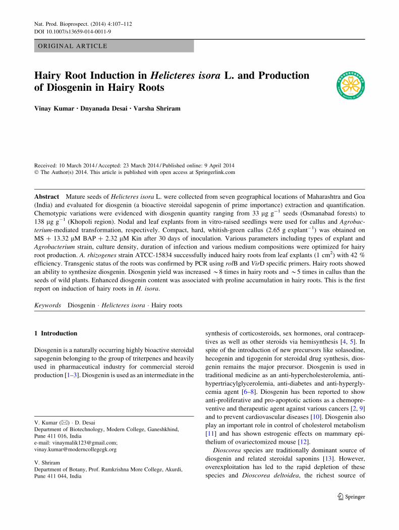
Notable eco-geographical variations were evidenced in
terms of diosgenin content extracted from seeds of H. isora
growing in seven regions of Maharashtra and Goa (Fig. 1).
Amongst all the samples, the seeds from Khopoli region
plants exhibited highest diosgenin (138 lg g-1 seeds)
content followed by Goa and Nandurbar, whereas the
samples collected from the forests in Osmanabad district
showed lowest diosgenin (33 lg g-1 seeds). The seeds
from Khopoli region were therefore selected and used for
further experiments. This is the first attempt to assess
whether genetic and environmental factors influenced the
diosgenin content in H. isora seeds collected from different
geographical locations of Maharashtra and Goa. Previ-
ously, Taylor et al. [17] reported significant eco-geo-
graphical variations with respect to diosgenin content in
Fenugreek accessions from Canada.
2.2 Callus Production
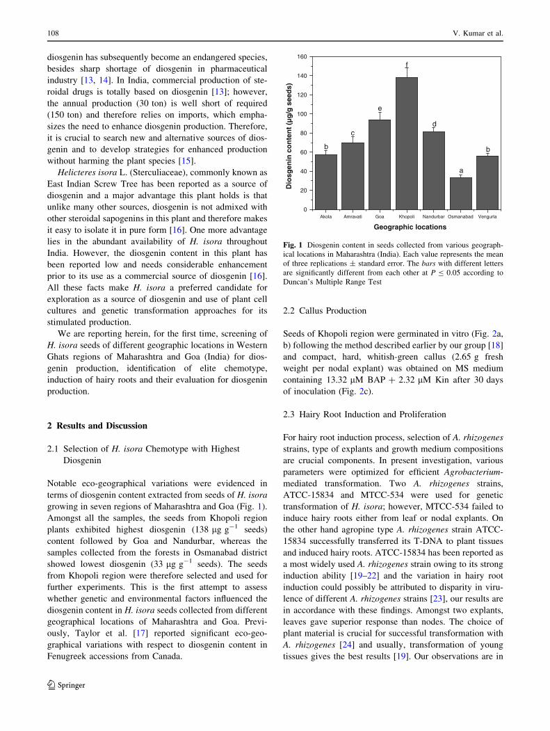
Seeds of Khopoli region were germinated in vitro (Fig. 2a,
b) following the method described earlier by our group [18]
and compact, hard, whitish-green callus (2.65 g fresh
weight per nodal explant) was obtained on MS medium
containing 13.32 lM BAP ? 2.32 lM Kin after 30 days
of inoculation (Fig. 2c).
2.3 Hairy Root Induction and Proliferation
For hairy root induction process, selection of A. rhizogenes
strains, type of explants and growth medium compositions
are crucial components. In present investigation, various
parameters were optimized for efficient Agrobacterium-
mediated transformation. Two A. rhizogenes strains,
ATCC-15834 and MTCC-534 were used for genetic
transformation of H. isora; however, MTCC-534 failed to
induce hairy roots either from leaf or nodal explants. On
the other hand agropine type A. rhizogenes strain ATCC-
15834 successfully transferred its T-DNA to plant tissues
and induced hairy roots. ATCC-15834 has been reported as
a most widely used A. rhizogenes strain owing to its strong
induction ability [19–22] and the variation in hairy root
induction could possibly be attributed to disparity in viru-
lence of different A. rhizogenes strains [23], our results are
in accordance with these findings. Amongst two explants,
leaves gave superior response than nodes. The choice of
plant material is crucial for successful transformation with
A. rhizogenes [24] and usually, transformation of young
tissues gives the best results [19]. Our observations are in
Akola Amravati Goa Khopoli Nandurbar Osmanabad Vengurla0
20
40
60
80
100
120
140
160
f
e
dc
b b
a
Dio
sgen
in c
on
ten
t (µ
g/g
see
ds)
Geographic locations
Fig. 1 Diosgenin content in seeds collected from various geograph-
ical locations in Maharashtra (India). Each value represents the mean
of three replications ± standard error. The bars with different letters
are significantly different from each other at P B 0.05 according to
Duncan’s Multiple Range Test
108 V. Kumar et al.
123
conformity of these findings and third and fourth young
leaves from apices were found most responsive for trans-
formation. The optimal time for bacterial infection of
leaves was observed to be 40 min. The optimal bacterial
density for transformation was found to be at 0.8 OD. Two
days co-cultivation period was found best for H. isora
transformation (detailed data not shown). Hairy roots
started appearing on the cutting-edges of leaves and at the
point of injection on the leaf fragments (Fig. 2d), but not
on nodal explants, after 3 weeks of inoculation on MS
medium containing 250 mg L-1 cefotaxime and 30 g L-1
sucrose in dark conditions with 42 % frequency. The
growth medium is considered as crucial component of
hairy root induction and growth [21]. Various modifica-
tions in MS basal medium- i) reducing sucrose content
from 30 to 20 g L-1; (ii) excluding MS vitamins and
including B5 vitamins; (iii) reducing basal strength to half
(�-MS); (iv) reducing basal strength to one-fourth (�-MS)
were tried for optimal hairy root production. Amongst
those, �-MS was found best for hairy root growth, which
further proliferated upon their subculturing onto the same
medium and incubated at 16 h photoperiod with
25 lmol m-2 s-1 light intensity (Fig. 2e, f). Hairy root
cultures were identified on the basis of their morphology
Fig. 2 a Pods collected from various locations (i: Akola; ii:
Amravati; iii: Goa; iv: Khopoli; v: Nandurbar; vi: Osmanabad and
vii: Vengurla). b In vitro germinated seedlings. c 1 Month old callus
of H. isora obtained on MS ? 13.32 lM BAP ? 2.32 lM Kin. d–
f Induction and proliferation of hairy roots from leaf explants
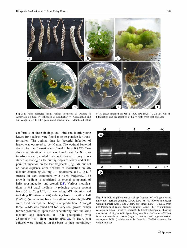
Fig. 3 a PCR amplification of 423 bp fragment of rolB gene using
hairy root derived genomic DNA. Lane M 100–500 bp molecular
weight marker; Lane 1 and 2 hairy root lines; Lane -C DNA from
non-transformed roots (negative control); Lane ?C Agrobacterium
rhizogenes DNA (positive control). b Electrophoregram showing
absence of VirD gene (438 bp) in hairy root lines 1–5, lane -C DNA
from non-transformed roots (negative control), ?C: Agrobacterium
rhizogenes DNA (positive control), Lane M 100–500 bp molecular
weight marker
Diosgenin Production in H. isora Hairy Roots 109
123
and their genetic nature was further confirmed using PCR
amplification of rolB gene in the hairy root lines using gene
specific forward and reverse primers. Two hairy root lines
were selected for PCR confirmation, whereas A. rhizogenes
served as positive control and DNA from non-transformed
seedling roots served as negative control. Both the lines
showed presence of 423 bp rolB amplified products
(Fig. 3a, lane 1 and 2), indicating the integration of T-DNA
of A. rhizogenes in H. isora. This gene has been widely
used and advocated for confirmation of A. rhizogenes-
mediated transformation in hairy roots of various plant
species [19–21, 24]. PCR analysis was also carried using
VirD gene-specific primers to check the contamination of
hairy roots with residual Agrobacterium. Results (Fig. 3b
lanes 1–5) clearly revealed the absence of contamination of
hairy roots with Agrobacterium strain. VirD gene was not
detected in non-transformed root (Fig. 3b lane -c).
2.4 Diosgenin Production by Callus and Hairy Root
Cultures
Diosgenin was detected from crude extracts of seeds of
wild plants growing in Khopoli region and compared with
the leaves and non-transformed roots of in vitro germinated
seedlings, 1 month old callus and 6 weeks old hairy root
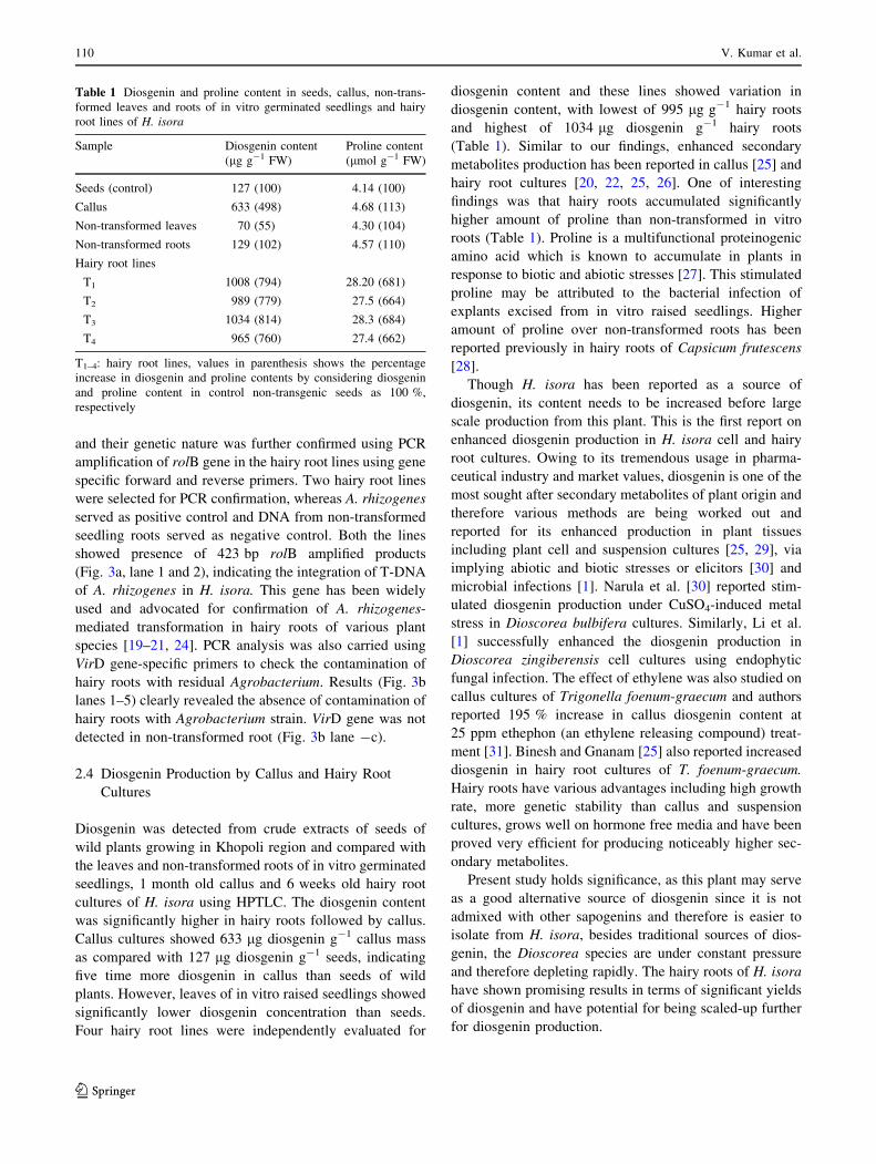
cultures of H. isora using HPTLC. The diosgenin content
was significantly higher in hairy roots followed by callus.
Callus cultures showed 633 lg diosgenin g-1 callus mass
as compared with 127 lg diosgenin g-1 seeds, indicating
five time more diosgenin in callus than seeds of wild
plants. However, leaves of in vitro raised seedlings showed
significantly lower diosgenin concentration than seeds.
Four hairy root lines were independently evaluated for
diosgenin content and these lines showed variation in
diosgenin content, with lowest of 995 lg g-1 hairy roots
and highest of 1034 lg diosgenin g-1 hairy roots
(Table 1). Similar to our findings, enhanced secondary
metabolites production has been reported in callus [25] and
hairy root cultures [20, 22, 25, 26]. One of interesting
findings was that hairy roots accumulated significantly
higher amount of proline than non-transformed in vitro
roots (Table 1). Proline is a multifunctional proteinogenic
amino acid which is known to accumulate in plants in
response to biotic and abiotic stresses [27]. This stimulated
proline may be attributed to the bacterial infection of
explants excised from in vitro raised seedlings. Higher
amount of proline over non-transformed roots has been
reported previously in hairy roots of Capsicum frutescens
[28].
Though H. isora has been reported as a source of
diosgenin, its content needs to be increased before large
scale production from this plant. This is the first report on
enhanced diosgenin production in H. isora cell and hairy
root cultures. Owing to its tremendous usage in pharma-
ceutical industry and market values, diosgenin is one of the
most sought after secondary metabolites of plant origin and
therefore various methods are being worked out and
reported for its enhanced production in plant tissues
including plant cell and suspension cultures [25, 29], via
implying abiotic and biotic stresses or elicitors [30] and
microbial infections [1]. Narula et al. [30] reported stim-
ulated diosgenin production under CuSO4-induced metal
stress in Dioscorea bulbifera cultures. Similarly, Li et al.
[1] successfully enhanced the diosgenin production in
Dioscorea zingiberensis cell cultures using endophytic
fungal infection. The effect of ethylene was also studied on
callus cultures of Trigonella foenum-graecum and authors
reported 195 % increase in callus diosgenin content at
25 ppm ethephon (an ethylene releasing compound) treat-
ment [31]. Binesh and Gnanam [25] also reported increased
diosgenin in hairy root cultures of T. foenum-graecum.
Hairy roots have various advantages including high growth
rate, more genetic stability than callus and suspension
cultures, grows well on hormone free media and have been
proved very efficient for producing noticeably higher sec-
ondary metabolites.
Present study holds significance, as this plant may serve
as a good alternative source of diosgenin since it is not
admixed with other sapogenins and therefore is easier to
isolate from H. isora, besides traditional sources of dios-
genin, the Dioscorea species are under constant pressure
and therefore depleting rapidly. The hairy roots of H. isora
have shown promising results in terms of significant yields
of diosgenin and have potential for being scaled-up further
for diosgenin production.
Table 1 Diosgenin and proline content in seeds, callus, non-trans-
formed leaves and roots of in vitro germinated seedlings and hairy
root lines of H. isora
Sample Diosgenin content
(lg g-1 FW)
Proline content
(lmol g-1 FW)
Seeds (control) 127 (100) 4.14 (100)
Callus 633 (498) 4.68 (113)
Non-transformed leaves 70 (55) 4.30 (104)
Non-transformed roots 129 (102) 4.57 (110)
Hairy root lines
T1 1008 (794) 28.20 (681)
T2 989 (779) 27.5 (664)
T3 1034 (814) 28.3 (684)
T4 965 (760) 27.4 (662)
T1–4: hairy root lines, values in parenthesis shows the percentage
increase in diosgenin and proline contents by considering diosgenin
and proline content in control non-transgenic seeds as 100 %,
respectively
110 V. Kumar et al.
123
3 Experimental Section
3.1 Plant Material
Mature pods of H. isora, growing in forest areas of seven
different geographic regions in Maharashtra including
Akola, Amaravati, Khopoli, Nandurbar, Osmanabad,
Vengurla and Goa (Fig. 2a) were collected and samples
were authenticated at Anantrao Pawar College, Pune and
specimen voucher (No. APCP/21/2012-13) was submitted.
Seeds from the mature pods were separated and used for
further study.
3.2 Extraction and Quantification of Diosgenin
Mature seeds (2 g) separated from pods were finely pow-
dered and extracted with 120 mL of methanol for 5–6 h
followed by concentration in vacuo at 40 �C. 25 mL of
15 % methanolic-HCl was added and reflux for 2 h. Acidic
solvent was removed under vacuum to dryness. Residue
was extracted three times with chloroform and moisture
was removed from extract using anhydrous sodium sulfate.
Finally solvent was removed completely to get final
extract. The callus and hairy root extracts were also pre-
pared in same manner.
For preliminary screening, diosgenin content was esti-
mated spectrophotometrically from the seeds collected
from all the seven geographical locations to select elite
chemotype with higher diosgenin content as described,
based on color reaction of diosgenin with perchloric acid
and by referring to a standard curve of diosgenin (Sigma-
Aldrich, Germany) [32]. Diosgenin quantification of crude
extracts wild type seeds, callus and hairy roots obtained
from Khopoli seeds was carried out on a high perfor-
mance thin layer chromatography (HPTLC) system (CA-
MAG, Switzerland) comprising of a Linomat-5 applicator
and CAMAG TLC densitometer scanner analysis. Sta-
tionary phase was silica gel 60 F256, 20 9 10 cm TLC
plate (Merck), toluene:chloroform:ethyl acetate (8:8:2)
was used as mobile phase and anisaldehyde as a devel-
oping reagent. Chromatographs were evaluated by soft-
ware (winCATS planar chromatography manager) to
detect the presence of and quantify diosgenin against the
standard.
3.3 Callus Production
Seeds of Khopoli region plants were germinated in vitro as
described earlier [18]. Briefly, seeds were treated with
concentrated H2SO4 for 4–5 min to break seed coat fol-
lowed by washing with 1.0 % Tween-20 for 5 min and
three rinses with distilled water. Surface sterilized seeds
were germinated on MS medium [33] containing 30 g L-1
sucrose, 8 g L-1 agar (HiMedia, Mumbai, India) and pH
5.8. Cultures were maintained at 25 ± 2 �C in dark for
4 days and then transferred to 16 h day-1 photoperiod at
25 lmol m-2 s-1 light intensity. Nodal explants
(1–1.2 cm) excised from 3 to 4 weeks old in vitro raised
seedlings were used for callus formation on MS medium
supplemented with 13.32 lM BAP ? 2.32 lM Kin.
3.4 Establishment of Hairy Root Cultures
Agrobacterium rhizogenes strains ATCC-15834 (NCIM,
NCL, Pune) and MTCC-534 (IMTECH, Chandigarh) were
grown for 48 h in yeast extract mannitol (YEM) medium.
Leaf (1 cm2) explants and nodal segments from in vitro
raised plants were either immersed or punctured with
hypodermic needles attached to a syringe containing
overnight grown bacterial suspensions of both Agrobacte-
rium strains separately (OD600 = 0.5–0.8) for 10–50 min
with continuous shaking and were then dried with sterilized
Whatman No. 1 filter papers to remove excess bacteria and
co-cultivated on solidified hormone free MS media for
1–5 days in dark conditions. One hundred explants were
used per experiment and each experiment was carried out
in triplicate. The explants were given a wash with
250 mg L-1 cefotaxime to kill the residual Agrobacterium
and the cultures were then transferred to MS medium
containing 250 mg L-1 cefotaxime, 30 g L-1 sucrose and
8 g L-1 agar for further growth in complete darkness
25 ± 2 �C for 3 weeks. After this, the hairy root lines were
maintained by subculture of 3–5 cm long pieces of roots on
the same medium on 16 h day-1 photoperiod at
25 lmol m-2 s-1 light intensity.
3.5 PCR Confirmation of Hairy Roots
Genomic DNA was extracted using CTAB method [34]
from young hairy root lines separately, as well as control
(non-transformed roots of in vitro germinated seedling),
while the Ri-plasmid of A. rhizogenes was used as a
positive control [35]. Integration of the rolB gene into the
plant genome was confirmed by PCR analysis [24]. The
specific primers for extending 423 bp fragment of nucle-
otides rolB gene were designed and their homology
checked through the BLAST search. The primers consisted
of nucleotides with the sequence of rolB1: 50-GCTCTTG
CAGTGCTAGATTT-30 and rolB2: 50-GAAGGTGCAAG
CTACCTCTC-30 for the forward and reverse, respectively
[24]. PCR analysis was carried out using a GeneAmp PCR
System (Applied Biosystems, USA) to confirm the pre-
sence of a 423 bp rolB gene fragment in the putative
transformants. Amplification conditions for rolB gene were
1 cycle at 95 �C for 5 min followed by 35 cycles of
amplification (95 �C for 30 s, 56 �C for 30 s, 72 �C for
Diosgenin Production in H. isora Hairy Roots 111
123
1 min) followed by a final extension step at 72 �C for
10 min. To confirm the transgenics free from any con-
tamination with residual Agrobacterium, VirD gene spe-
cific primers, forward 50-ATGTCGCAAGGCAGTAAGCC
C-30 and reverse 50-GGAGTCTTTCAGCATGGAGCAA-
30 were used and amplification conditions were 1 cycle at
94 �C for 2 min followed by 35 cycles of amplification
(45 s at 94 �C, 40 s at 55 �C and 45 s at 72 �C) and 1 cycle
at 72 �C for 10 min to give a 438 bp product [36]. The
electrophoresis of the PCR products was performed on 2 %
agarose gel spiked with ethidium bromide. The gel was
visualized under UV light and photographed using GelDoc
XR documentation system (BIO-Rad, USA).
3.6 Estimation of Proline from Callus and Hairy Roots
Samples (500 mg) were homogenized in 5 mL 3 % sul-
fosalicylic acid using a mortar and pestle and centrifuged at
12,0009g for 20 min. Two mL aliquot of supernatant was
mixed with equal volumes of glacial acetic acid and acid
ninhydrin reagent followed by boiling of reaction mixture
in water bath at 100 �C for 30 min. After cooling the
reaction mixture, 6 mL toluene was added and after thor-
ough mixing, the chromophore containing toluene was
separated and absorbance was recorded at 520 nm against
toluene blank [37].
Acknowledgments This work was financially supported by a grant
from University Grants Commission, New Delhi in the form a major
research project [F. No. 41-521/2012 (SR)] to VK. Authors also
acknowledge the facilities used for this work at Modern College
created under DST-FIST Program.
Conflict of interest The authors declare no conflict of interest.
Open Access This article is distributed under the terms of the
Creative Commons Attribution License which permits any use, dis-
tribution, and reproduction in any medium, provided the original
author(s) and the source are credited.
References
1. P. Li, Z. Mao, J. Lou, Y. Li, Y. Mou, S. Lu, Y. Peng, L. Zhou,
Molecules 16, 10631–10641 (2011)
2. M.J. Liu, Z. Wang, Y. Ju, R.N. Wong, Q.Y. Wu, Cancer Che-
mother. Pharmacol. 55, 79–90 (2005)
3. K. Patel, M. Gadewar, V. Tahilyani, D.K. Patel, Nat. Prod.
Bioprospect. 2, 46–52 (2012)
4. J. Nino, D.A. Jimenez, O.M. Mosquera, Y.M. Correa, J. Braz.
Chem. Soc. 18, 1073–1076 (2007)
5. H.J. Shah, S.S. Lele, J. Anal. Bioanal. Tech. 3, 141 (2012)
6. M.A. Juarez-Oropeza, J.C. Diaz-Zagoya, J.L. Rabinowitz, Int.
J. Biochem. 19, 679–683 (1987)
7. M.A. McAnuff, W.W. Harding, F.O. Omoruyi, H. Jacobs, E.Y.
Morrison, H.N. Asemota, Food Chem. Toxicol. 43, 1667–1672
(2005)
8. I.S. Son, J.H. Kim, H.Y. Sohn, K.H. Son, J.S. Kim, C.S. Kwon,
Biosci. Biotechnol. Biochem. 71, 3063–3071 (2007)
9. J. Raju, R. Mehta, Nutr. Cancer 61, 27–35 (2009)
10. G. Gong, Y. Qin, W. Huang, S. Zhou, X. Wu, X. Yang, Y. Zhao,
D. Li, Chem. Biol. Interact. 184, 366–375 (2010)
11. I.D. Roman, A. Thewles, R. Coleman, Biochem. Biophys. Acta
1255, 77–81 (1995)
12. Aradhana, A.R. Rao, R.K. Kale, Indian J. Exp. Biol. 30, 367–370
(1992)
13. H.C. Chaturvedi, M. Jain, N.R. Kidwai, Indian J. Exp. Biol. 45,
937–948 (2007)
14. P. Li, Y. Mou, S. Lu, W. Sun, J. Lou, C. Yin, L. Zhou, Afr.
J. Pharm. Pharmacol. 6, 1186–1193 (2012)
15. B. Chapagain, Z. Wiesman, Afr. J. Biotechnol. 4, 1209–1213
(2005)
16. B. Barik, A.K. Dey, P.C. Das, Indian J. Chem. 20B, 938 (1981)
17. W.G. Taylor, H.J. Zulyniak, K.W. Richards, S.N. Acharya, S.
Bitman, J.L. Elder, J. Agric. Food Chem. 50, 5994–5997 (2002)
18. V. Shriram, V. Kumar, M.G. Shitole, In vitro cell DEV. Biol.-
Plant 44, 186-193 (2008)
19. V. Bonhomme, D. Laurain-Mattar, J. Lacoux, M.A. Fliniaux, A.
Jacquin-Dubreuil, J. Biotechnol. 81, 151–158 (2000)
20. S. Dhakulkar, T.R. Ganapathi, S. Bhargava, V.A. Bapat, Plant
Sci. 169, 812–818 (2005)
21. R.P. Chandran, V.P. Potty, Indian J. Biotechnol. 7, 122–128
(2008)
22. E. Kochan, M. Wasicela, M. Sienkiewicz, In Vitro Cell Dev.
Biol. - Plant, 49, 24-29 (2013)
23. J.R. Porter, Crit. Rev. Plant Sci. 10, 387–421 (1999)
24. B. Glab, T. Furmanek, M. Miklaszewska, A. Banas, A. Krolicka,
Acta Physiol. Plant. 35, 2137–2145 (2013)
25. A. Binesh, R. Gnanam, Adv Biotech. 9, 33–40 (2009)
26. F. Medina-Bolivar, J. Condori, A.M. Rimando, J. Hubstenberger,
K. Shelton, S.F. O’Keefe, S. Bennett, M.C. Dolan, Phytochem-
istry 68, 1992–2003 (2007)
27. L. Szabados, A. Savoure, Trends Plant Sci. 15, 89–97 (2009)
28. S. Sekiguchi, T. Yamakawa, T. Kodama, S.S. Smith, M.M.
Yeoman, Plant Biotechnol. 16, 153–158 (1999)
29. M. Amir, M. Mujeeb, A. Sabih, S. Ahmad, A. Ahmad, W.A.
Siddiqui, Planta Med. 76, 35 (2010)
30. A. Narula, S. Kumar, P.S. Srivastava, Plant Cell Rep. 24,
250–254 (2005)
31. R. Oncina, J.A. Del Rio, P. Gomez, A. Ortuno, Food Chem. 76,
475–479 (2002)
32. S.C. Slack, W.J. Mader, Anal. Chem. 33, 625–627 (1961)
33. T. Murashige, F.A. Skoog, Physiol, Plant 15, 473–497 (1962)
34. J.J. Doyle, J.L. Doyle, Phytochem. Bull. 19, 11–15 (1987)
35. B. Wang, G. Zhang, L. Zhu, L. Chen, Y. Zhang, Colloids Surf. B
53, 101–104 (2006)
36. N.A. Chashmi, M. Sharifi, M. Yousefzadi, M. Behmanesh, H.
Rezadoost, A. Cardillo, J. Palazon, Med. Chem. Res. 22, 745–752
(2013)
37. L.S. Bates, R.P. Waldran, I.D. Teare, Plant Soil 39, 205–208
(1973)
112 V. Kumar et al.
123