hamartomatous lesion at the base of tongue: a rare entity
TRANSCRIPT
IP Journal of Diagnostic Pathology and Oncology 2021;6(3):223–225
Content available at: https://www.ipinnovative.com/open-access-journals
IP Journal of Diagnostic Pathology and Oncology
Journal homepage: https://www.jdpo.org/
Case Report
Hamartomatous lesion at the base of tongue: A rare entity
Ankush Blaggan1, Neelam Gupta1,*, Vikas Dubey1, Mehak Kashyap1, Nechal Kaur1
1Dept. of Pathology, Maharishi Markandeshwar Medical College and Hospital, Kumarhatti, Solan, Himachal Pradesh, India
A R T I C L E I N F O
Article history:Received 19-08-2021Accepted 24-08-2021Available online 13-09-2021
Keywords:Hamartomatous
A B S T R A C T
Hamartomas are benign proliferative lesions with no metastatic properties. The hamartoma of the tongueis usually rare. It is usually difficult to diagnose grossly. But can be diagnosed definitely with help ofhistopathological examination. Our case study is on a 5-year-old female who was asymptomatic andpresented with cystic swelling at the base of the tongue which was initially diagnosed as a cyst but wasdiagnosed histopathologically as a hamartoma.
This is an Open Access (OA) journal, and articles are distributed under the terms of the Creative CommonsAttribution-NonCommercial-ShareAlike 4.0 License, which allows others to remix, tweak, and build uponthe work non-commercially, as long as appropriate credit is given and the new creations are licensed underthe identical terms.
For reprints contact: [email protected]
1. Introduction
In 1904, Albrecht was the first one to use the term”hamartoma”.1–3 Hamartomas are not true neoplasm but areprimary non-neoplastic malformations or inborn errors oftissue development, characterized by an abnormal mixtureof native tissues of a part of the body with an excess of oneor more of the cellular components. These are commonlyseen in respiratory and digestive organs, such as the lungs,pancreas, and liver.4
Histologically the tongue tumors in children can beclassified as solid tumors, excretory cysts, lymphovascularmasses, and reactive epithelial lesions.5 Unlike trueneoplasms, they do not exhibit any unchecked monoclonaltissue proliferation.
2. Case Report
This report describes a 5-year-old girl who presented witha sublingual swelling which was initially diagnosed asa cyst for which she underwent surgical excision. Theaffected person had no other medical issues. The diagnosisof hamartoma was made after histopathological analysis or
* Corresponding author.E-mail address: [email protected] (N. Gupta).
examination. There have been no signs of recurrence. Thisreport summarizes the relevant literature.
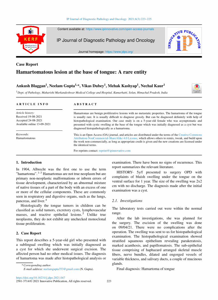
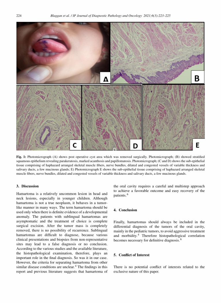
HISTORY- 5y/f presented to surgery OPD withcomplaints of bluish swelling under the tongue on theventral surface for 1 year. The size of the swelling was 2x2cm with no discharge. The diagnosis made after the initialexamination was a cyst.
2.1. Investigations
The laboratory tests carried out were within the normalrange.
After the lab investigations, she was planned forthe surgery. The excision of the swelling was doneon 09/04/21. There were no complications after theoperation. The swelling was sent to us for histopathologicalexamination. The histopathological examination showedstratified squamous epithelium revealing parakeratosis,marked acanthosis, and papillomatosis. The sub-epithelialtissue comprising of haphazard arranged skeletal musclefibers, nerve bundles, dilated and engorged vessels ofvariable thickness, and salivary ducts, a couple of mucinousglands.
Final diagnosis: Hamartoma of tongue
https://doi.org/10.18231/j.jdpo.2021.0472581-3714/© 2021 Innovative Publication, All rights reserved. 223
224 Blaggan et al. / IP Journal of Diagnostic Pathology and Oncology 2021;6(3):223–225
Fig. 1: Photomicrograph (A) shows post operative cyst area which was removed surgically. Photomicrograph; (B) showed stratifiedsquamous epithelium revealing parakeratosis, marked acanthosis and papillomatosis. Photomicrograph; (C and D) shows the sub-epithelialtissue comprising of haphazard arranged skeletal muscle fibers, nerve bundles, dilated and congested vessels of variable thickness andsalivary ducts, a few mucinous glands; E) Photomicrograph E shows the sub-epithelial tissue comprising of haphazard arranged skeletalmuscle fibers, nerve bundles, dilated and congested vessels of variable thickness and salivary ducts, a few mucinous glands.
3. Discussion
Hamartoma is a relatively uncommon lesion in head andneck lesions, especially in younger children. Althoughhamartoma is not a true neoplasm, it behaves in a tumor-like manner in many ways. The term hamartoma should beused only when there is definite evidence of a developmentalanomaly. The patients with sublingual hamartomas areasymptomatic and the treatment of choice is completesurgical excision. After the tumor mass is completelyremoved, there is no possibility of recurrence. Sublingualhamartomas are difficult to diagnose, because variousclinical presentations and biopsies from non-representativesites may lead to a false diagnosis or no conclusion.According to the various studies and the available literature,the histopathological examination, therefore, plays animportant role in the final diagnosis. So was it in our case.However, the criteria for separating hamartoma from othersimilar disease conditions are unclear.4 The findings in thisreport and previous literature suggests that hamartoma of
the oral cavity requires a careful and multistep approachto achieve a favorable outcome and easy recovery of thepatients.6
4. Conclusion
Finally, hamartomas should always be included in thedifferential diagnosis of the tumors of the oral cavity,mainly in the pediatric tumors, to avoid aggressive treatmentand morbidity.4 Therefore histopathological correlationbecomes necessary for definitive diagnosis.6
5. Conflict of Interest
There is no potential conflict of interests related to theexclusive nature of this paper.

Blaggan et al. / IP Journal of Diagnostic Pathology and Oncology 2021;6(3):223–225 225
6. Source of Funding
No financial support was received for the work on thismanuscript.
References1. Elameen SM. Hamartoma of the base of the tongue. J Otolaryngol ENT
Res. 2015;2(5):1–3.2. Vashishth A, Mathur NN, Choudhary SR, Khanna G. Giant vascular
hamartoma of the tongue. Malays J Med Sci. 2014;21(2):74–81.3. Patil S, Rao RS, Majumdar B. Hamartomas of the oral cavity. J Int Soc
Prev Community Dent. 2015;5(5):347–53.4. Jain RK. Hamartoma of the head and neck. Indian J Otolaryngol Head
Neck Surg. 1999;51(4):76–8.5. Kreiger PA, Ernst LM, Elden LM, Kazahaya K, Alawi F, Russo PA,
et al. Hamartomatous tongue lesions in children. Am J Surg Pathol.2007;31(8):1186–90.
6. Li J, Mao C, Ma L, Zhou X. Giant sublingual hamartoma with medialcleft tongue: a case report and literature review. J Int Med Res.2020;48(8):300060520942089. doi:10.1177/0300060520942089.
Author biography
Ankush Blaggan, Post Graduate Student
Neelam Gupta, Professor and HOD
Vikas Dubey, Assistant Professor
Mehak Kashyap, Post Graduate Student
Nechal Kaur, Post Graduate Student
Cite this article: Blaggan A, Gupta N, Dubey V, Kashyap M, Kaur N.Hamartomatous lesion at the base of tongue: A rare entity. IP J DiagnPathol Oncol 2021;6(3):223-225.