hazard assessment report summary and conclusions · pdf filehazard assessment report summary...
TRANSCRIPT

1
Application A603 Supporting Document 1 HAZARD ASSESSMENT REPORT Summary and conclusions The toxicological database for erythrosine is extensive and adequate to establish a suitable health standard for regulatory purposes. The current acceptable daily intake (ADI) for erythrosine, as established in 1990 by the Joint FAO/WHO Committee on Food Additives is 0.1 mg/kg bw/day. As part of the hazard assessment, FSANZ considered information published in the medical literature on intolerance reactions to erythrosine. An extensive search of the medical database revealed only a few clinical studies on the potential role of erythrosine in intolerance reactions. The studies investigated the effect of erythrosine on a number of clinical patients with various symptoms. The patients were challenged with various doses of erythrosine, from 1 mg up to 30 mg. In some of the studies, symptoms were reported with the higher doses of erythrosine, many times higher than the ADI. Although it is not possible to estimate, based on the available evidence, the prevalence of intolerance reactions to erythrosine in the general population, it is unlikely to be common. As erythrosine is poorly absorbed from the gastrointestinal tract, the exposure and, therefore the potential for intolerance reactions resulting from the small amounts of erythrosine in the diet, would be very low. FSANZ has evaluated a range of supplementary studies published since the last comprehensive toxicological evaluation of erythrosine by JECFA in 1990. The studies covered metabolism, reproduction and developmental toxicity, genotoxicity, in addition to a range of other studies. The toxicity profile of erythrosine is well-defined. It is poorly absorbed from the digestive tract in both rats and humans and distributes almost entirely to the liver, where it is excreted unchanged in the bile. Erythrosine has low acute oral toxicity, does not cause reproductive or developmental toxicity, and the weight-of-evidence indicates that it is unlikely to be genotoxic. In both humans and rats, repeated ingestion results in elevated serum thyroid stimulating hormone (TSH) levels. In humans, at doses above 1.0 mg/kg bw/day this is associated with increased serum iodine, while in rats, there is compelling evidence that this is due to the inhibition of the peripheral metabolism of thyroxine (T4) to tri-iodothyronine (T3) in the liver at and above doses of 2.5 mg/kg bw. Erythrosine does not directly act on the thyroid gland in either species. The weight-of-evidence indicates that erythrosine is not carcinogenic, however, benign thyroid tumours have been observed at very high doses (>2500 mg/kg bw/day) in a minority of long-term feeding studies in rats. It is most likely that the occurrence of these tumours was secondary to the compound’s hormonal effects and is not relevant to humans based on well-recognised interspecies differences in thyroid physiology. Based on a consideration of all of the available studies, including the supplementary ones published since 1990 when JECFA last considered the toxicity of erythrosine, FSANZ is unable to find a basis to amend the ADI of 0.1 mg/kg bw/day established by JECFA.

2
This evaluation re-affirms the ADI established by JECFA in 1990. Therefore, an ADI of 0.1 mg/kg bw/day is appropriate for dietary risk assessment purposes. 1. Intolerance reactions to erythrosine 1.1 Background and methodology Erythrosine is a colouring agent permitted for use in food and in medicines for oral use in many countries around the world, including Australia and New Zealand. Erythrosine is also used in dentistry to stain and visualise plaque. An electronic literature search was conducted using the PubMed database. The terms used were: “Erythrosine AND Intolerance”, “Erythrosine” AND “Adverse Effects”, “Hypersensitivity” AND “Erythrosine”, “Food Colouring Agents” AND “Hypersensitivity”. Publications which relate to health effects/ medical symptoms of erythrosine in humans were identified. 1.2 Literature review Mikkelsen et al (1978) reported a clinical study of 56 patients suffering from urticaria and/or angioneurotic oedema. The main purpose of the study was to determine the role of annatto – a widely used natural food colour – in provoking symptoms in those patients. The patients had been suffering from urticaria or angioedema for more than four weeks. No information was provided on the age/ gender or weight of patients. Initially, the patients were given an elimination diet for at least three weeks and all non-vital drugs were suspended. The diet consisted of food free of artificial colours and preservatives and also free of ingredients identified by the authors as ‘well known to cause urticaria, such as strawberries, shellfish, and coffee’. In most of the patients, challenge was performed when the patient was free of symptoms. The authors stated that it was possible to test some of the patients who were not completely free of symptoms after 3 weeks’ diet, because this group of patients had attacks periodically or decreasing symptoms during the diet. The patients were tested with various substances including food colours, a preservative (sodium benzoate), aspirin and the natural food colour annatto extract. A table presented in the paper shows the results for patients tested with doses of ten substances. In a group of 56 patients tested with annatto, fifteen (27%) reacted. Fifty patients were tested with erythrosine at 1 mg and 10 mg. The results indicate that six patients (12%) were positive. The paper does not identify which dose (1 mg or 10 mg) triggered the reaction. The results, as presented in the published paper, do not include a negative control substance (placebo) in the testing of patients; and the potential role of drug suspension in those patients showing a positive reaction was not addressed by the authors. Weber et al (1979) reported on their study to determine incidence of bronchoconstriction due to chemicals in a small population with moderately severe bronchial asthma. Forty five patients were selected on the basis of perennial wheezing of moderate severity requiring maintenance use of bronchodilators, particularly if the self-reported history suggested wheezing was precipitated by aspirin or food containing large amounts of colouring agents or preservatives. Sixteen of the patients were male ranging in age from 17 to 62 years of age; and 29 were female ranging in age from 15 to 62 years of age. The patients underwent open challenge (where both the researchers and participants know which treatment is being administered; the opposite of blind challenge). However, the patients did not know the dose they received at any time. The challenge was conducted over several days and in variable order with various chemicals including a mix of non-azo colours containing brilliant blue, erythrosine and indigotin at 5, 10 mg each. With the exception of
3
one of the chemicals, acetyl salicylic acid, all positive open challenges were repeated under double-blind conditions at a later date. At the onset of the study, lower doses were used for testing. After the first 13 patients, the upper dose was raised to 20 mg. Of 42 patients who were tested with 20 mg erythrosine, two were positive in an open challenge and only one of those two patients was positive in a double-blind placebo controlled challenge. A mix of azo colours (ponceau, amaranth and sunset yellow) was also used in the study with similar results. The authors concluded that, in this group of patients, following challenge with azo or non-azo colour mix at the specified doses, bronchoconstriction was uncommon. Ibero et al (1982) evaluated intolerance to food colours, preservatives and salicylates in children with clinical symptoms suggestive of adverse reactions to food. Twenty four children with symptoms including: atopic dermatitis, recurrent urticaria and/or angioedema– associated with spasmodic cough in five patients; gastrointestinal syndrome with chronic diarrhoea and abdominal pain (with negative gastroenterologic and parasitologic studies). The age of these children varied between 18 and 153 months (1.5 – 12 years). After a number of tests ruled out allergic disease, the patients were submitted to testing with food additives. The patients were put on a 48 hour-diet which excluded colours, benzoates and salicylates. The test substances were prepared in two doses: 1 mg and 10 mg for erythrosine, with the latter dose reduced to 5 mg in patients weighing 15.0 kg or less. Testing was done with one additive per day per child, including lactose as a placebo. After a physical examination of each patient participating in the test, the first dose of erythrosine at 1 mg, was administered. The patients were physically examined again after one hour and, if negative, the second dose of 10 mg (or 5 mg) was given. After another hour, each child was examined again and, if there were no symptoms, the diet excluding additives was maintained for another 48 hours in order to observe any delayed symptoms. Out of the 16 patients tested with erythrosine, two patients were immediately positive, three patients had delayed reactions, while eleven patients were negative. Therefore, the response rate to erythrosine reported in this clinical group of children was 31%. Loblay and Swain (1985) reported their findings on the role of tartrazine, as well as other food additives, in the wider context of intolerance reactions to naturally occurring food chemicals such salicylates and amines. A total of 78 consecutive patients with urticaria, migraine, irritable bowel syndrome, hyperactivity or systemic symptoms were tested with four food colouring, or combination, substances, including erythrosine. The test substances, and placebos, were taken at 48 hour intervals after a minimum of two weeks of strict elimination diet and five consecutive symptom-free days. The challenge substances were administered orally in a random order and symptoms recorded in a diary. The authors reported that the challenges were not strictly double blind, since the coloured compounds were taken in gelatin capsules. However, in previous studies where a colour substance (tartrazine) was used and the colour concealed, there was no significant difference in the frequency of positive reactions to tartrazine. Also, previous studies showed that with placebo challenges no positive reactions were recorded in patients with urticaria, and the placebo reaction rate in all other groups was less than 10%. In this study, three groups of 13 or 14 patients were tested with erythrosine at a dose of 30 mg. Five out of 13 were reported positive for urticaria symptom. Four out of 13 patients were reported positive for migraine. Hyperactivity was reported in eight out of 14 children tested with erythrosine. The authors commented that most patients are sensitive to multiple substances (between 2 and 10 commonly) with varying symptoms and that naturally occurring salicylates are the single most common substance to produce reactions when tested by double-blind oral challenge. The authors concluded that: ‘since natural food chemicals provoke symptoms more frequently than artificial additives, we believe there is no rationale for banning the use

4
of food additives, although minimising the concentrations added to processed foods would be likely to reduce the frequency and severity of adverse reactions in sensitive individuals’. Booth (1993) published a case report of food intolerance in a child with urticaria. A five-year old boy was referred to a dietitian from a dermatologist. The boy had a 12-month history of generalised urticaria. According to his mother, the child had previously reacted to strawberry milkshakes and red sweets. A colour and preservative-free diet was prescribed for a trial period of 3 weeks. Foods containing natural benzoic acid or containing more than 1 mg/kg natural benzoate were also excluded from the diet and only white toothpaste was permitted. During the trial period, the patient’s mother filled in a detailed diet diary and symptom charts. Symptoms were assessed subjectively and scored from 0 (no symptoms) to 4 (very severe). At each visit, the patient was assessed by the referring physician and the dietitian. After 3 weeks, all symptoms had remitted. In consultation with the dermatologist, a double blind challenge test was designed including four artificial food colours and a preservative, with lactose as a placebo control. In addition to tartrazine, three red colours–carmoisine, erythrosine and Red 2G–were selected on the basis that red food colours had precipitated reactions previously. All colours, including erythrosine, were administered at 10 mg and sodium benzoate at 500 mg. The patient received one capsule containing either a control or one test substance on each day, at home from his mother. The child reportedly reacted to all four colours but not to benzoate. The results were reported by the child’s mother but there was no opportunity for objective assessment by the medical staff. A diet free of 17 food colours, including those used in the study, was advised and the child remained free of urticaria six months and one year later. Asero (2001 and 2002) reported two clinical investigations of patients with respiratory and dermatological symptoms. A 33-year old woman had a 7-year history of perennial rhinitis characterised by watery rhinorrhea, itching of the nasal mucosa, and frequent episodes of sneezing. Anti-inflammatory medication had little effect. The second case was of a 44-year old woman with chronic urticaria and perennial rhinitis. Skin prick tests with various aeroallergens were negative in both cases. Following one month additive-free diet, the patients reported that the symptoms had disappeared or were markedly reduced. The patients were tested with eight food additives using a double- bind placebo controlled protocol. A single additive and multiple placebos were given to the patients one week apart and the patients monitored at the clinic for at least 2 hours and response to additives recorded. Erythrosine was used in the test for each patient at 10 mg. No reaction to erythrosine was reported in either patient. Park et al (2008) studied the potential role of seven common food additives in dermatologic adverse reactions in patients with allergic disease. The study aimed to determine whether food additives, commonly used in processed food, are associated with urticaria, angioedema and the aggravation of pre-existing atopic dermatitis symptoms. A total of 54 patients with allergic disease were randomly recruited and asked to complete a questionnaire designed to screen for the presence of a food-related hypersensitivity. The patients were: 21 children between one year and 15 years of age; and 33 adults between16 to 44 years of age. The patients underwent skin prick and patch testing for seven common food additives. The selected additives comprise four food colours, including erythrosine, two preservatives and a flavour enhancer. Two patients were negative to erythrosine in the skin prick and skin patch tests. The patients were enrolled in double-blind placebo controlled oral challenges with cross-over design. Following seven days of low-food additive diet and no antihistamine medication, the patients were tested. The test consisted of 3 mixtures of all the additives in 3 increasing doses (1/6, 1/3 or 1/2 of their respective ADIs; the ADI for erythrosine being 0.1 mg/kg body weight). Each dose of the additive mixture was given to the patients with 30-minute intervals

5
between doses. The patients remained under observation for six hours after the last dose and were asked to report within 24 hours to detect the possibility of a delayed reaction. Positive reactions were defined according to specific criteria. The results or the oral challenge suggested that the difference between the positivity rates to the food additive mixtures and placebo was not significant. The authors concluded that, based on the results of this study, a mixture of seven common additives including erythrosine, up to ½ the ADI, do not cause dermatologic symptoms or aggravate atopic dermatitis symptoms in patients with underlying allergic diseases. 1.3 Discussion The term ‘food intolerance’ has been used for decades in the medical literature to refer to any illness or biochemical or metabolic abnormality caused by the ingestion of any food or dietary component without implying any specific mechanism (Herman and Hagler, 1979; David 2000). As recognition of the various food related reactions improved, the terminology evolved accordingly. Contemporary literature defines food allergy as immediate type hypersensitivity reactions mediated by the immune system. Food intolerance is defined as adverse reactions to food which do not involve the immune system (Taylor and Hefle, 2001 Hodge et al., 2009). However, some inconsistent use of the terminology still exists and many published articles require careful interpretation. Some forms of intolerance reactions to food are clinically defined and the underlying mechanism clearly established. These include metabolic disorders such as lactose intolerance and hereditary fructose intolerance which are caused by enzyme deficiencies (David 2000; Taylor and Hefle 2001). Gluten intolerance, or coeliac disease, is a delayed type of immune reaction to cereal grains containing gluten (Chang et al., 2009). Less well-understood are other intolerance reactions associated with food. These are thought to involve pharmacological reactions triggered by naturally occurring chemicals in food – such as salicylates and amines, or food additives – such as synthetic colours and preservatives (Loblay and Swain 1986; David, 2000; Zopf et al 2009). Diagnosis of food chemical intolerance is difficult due to the lack of reliable, scientifically validated blood or skin test or histopathological examination (Hodge et al., 2009; Loblay, personal communication). Diagnosis entails a period of strict dietary elimination of suspect foods or food chemicals followed by double-blind placebo-controlled challenges (Hodge et al., 2009; Loblay, personal communication). The spectrum of the chemicals, the symptoms and the levels of chemical exposure necessary to trigger the symptoms are peculiar to the individual. Intolerance symptoms provoked by chemicals, either naturally occurring in food or food additives, are reported to affect the skin, gastrointestinal tract, respiratory tract and the central nervous system (Allen et al., 1984; Hodge et al., 2009). Environmental chemicals, hormonal changes or emotional stress have been cited as factors that may aggravate food intolerance reactions (Allen et al 1984). The prevalence of intolerance reactions to food chemicals has not been reliably determined, however, estimates of 5-20% have been cited in the literature (Hodge et al., 2009). The extent to which food additives contribute to intolerance reactions in general is unclear, and the mechanism of action is largely uncertain (David 1988; Simon 2003). Food additives, particularly food colours, are perceived to be a major cause of intolerance reactions in the community. However, there is a vast discrepancy between the perceived extent of the problem and the medical evidence regarding the role of food additives in intolerance reactions. Prevalence estimates of intolerance to additives vary widely from 0.18% in a mixed-age group to 1% in adults and 2% in children, but may be up to 7% in children with underlying allergy (Wilson and Bahna, 2005; Zuberbier et al., 2004). In addition, the medical literature is often conflicting and must be interpreted with caution, particularly if allergic reactions to food have not been ruled out.

6
For the majority of food additives, a cause and effect relationship with various symptoms has not been demonstrated (Taylor and Hefle 2001; Wilson and Bahna, 2005). Because additives are easily identifiable, consumers may perceive cause and effect associations leading to subjective self diagnosis and unnecessary avoidance. Medical specialists suggest that food additives are more likely to cause symptoms in patients with underlying illness (Wilson and Bahna, 2005, Loblay, personal communications). For example, symptoms of skin irritation are more likely to occur or be exacerbated in patients with underlying skin disorders, particularly atopic dermatitis, urticaria or eczema, than in those with healthy skin (David 1988; Wilson and Bahna, 2005). The eight clinical studies, on the potential role of erythrosine in intolerance reactions, summarised here need to be considered in the wider context of intolerance reactions to food chemicals, whether naturally occurring or added to food. The doses used in these studies are very high and, in most cases, vastly exceed the levels consumers are likely to encounter in their normal daily diet. In relation to studies using high doses, Dr Robert Loblay provided the following comments: ‘In a clinical context, challenges are best regarded as diagnostic tests, the purpose of which is to identify patients who are susceptible to intolerance reactions to particular compounds. As such, the doses used are chosen so as to maximize diagnostic sensitivity, specificity and predictive value in the particular patient group under consideration. To achieve this with purified food substances, it may be necessary to use doses that are higher than those normally ingested in foods’. Therefore, FSANZ notes the limitation of these studies from a risk assessment perspective whilst acknowledging the diagnostic purpose. WHO Expert Panel (JECFA-30th meeting-1987) considered metabolic studies and concluded that erythrosine is absorbed to only a small extent from the gastrointestinal tract in rats and humans. The 33rd JECFA meeting (1989) considered additional human studies which confirmed that erythrosine is poorly absorbed. In relation to hyperactivity, Mailman and Lewis (1981) discussed the potential role of food additives in developmental disorders in children which may relate to behavioural problems. Using erythrosine as an example, the authors emphasised the necessity of considering basic principles of pharmacology and physiology before assigning neurotoxicity to a specific agent. The authors commented on the various published biochemical studies of erythrosine and provided a detailed analysis and interpretation of some of these studies. The authors concluded that based on preliminary pharmacokinetic studies, erythrosine would not enter the central nervous system readily. Previous investigations (Mailman et al.,1980) have also questioned a finding reported in the literature that erythrosine has specific inhibitory effects on the uptake of dopamine in homogenates of rat brain. Dopamine is a neurotransmitter and the negative effect by erythrosine on its uptake, if it were true, could provide a plausible explanation for the claimed role of food colours in hyperactivity. However, based on the available evidence, these effects are most likely the result of nonspecific interactions with neural membranes. The authors stated that: ‘although hyperkinesis is a medical problem, the suggestion that it may be due to synthetic food additives has given it social and political dimensions that increase the need for sound clinical and basic data upon which to make policy judgements. Whatever the outcome of future scientific and clinical experimentation, cautious presentation

7
and interpretation of data will prevent expensive and spurious perturbations of the public and scientific consciousness’. 1.3 Conclusion Clinical observations in patients with intolerance reactions suggest that most patients are sensitive to multiple substances. An extensive search of the medical database provided only a small number of published clinical studies on the potential role of erythrosine in intolerance reactions. The studies investigated the effect of erythrosine on a number of clinical patients with various symptoms. The patients were challenged with various doses of erythrosine, from 1 mg up to 30 mg. In some of the studies, symptoms were reported with the higher doses, which at 30 mg for an average adult of 60 kg, is five times higher than the current ADI for erythrosine (0.1 mg/Kg body weight). It is not possible to estimate, based on the available evidence, the prevalence of intolerance reactions to erythrosine in the general population but it is unlikely to be common. It is noteworthy that erythrosine is poorly absorbed from the gastrointestinal tract, which further reduces the exposure resulting from the small amounts of erythrosine that would be expected in a normal diet. 2. Toxicological assessment 2.1 Previous JECFA evaluations The toxicology of erythrosine has been evaluated by JECFA at its 13th, 18th, 28th, 30th, 33rd and 37th meetings (WHO 1969, 1975, 1984, 1987, 1989 & 1991, respectively). The main issues considered by JECFA related to the disruption of thyroid hormone metabolism in rats and humans, and the occurrence of benign thyroid tumours in a small proportion of chronic rat studies. The ADI was amended on several occasions as a result of the expansion of the toxicological database over time leading to a better understanding of the potential adverse effects of erythrosine in rats and humans. Thirteenth meeting At its 13th meeting, JECFA considered the toxicology of erythrosine for the first time. Rat metabolism studies indicated that most of an orally-administered dose of erythrosine was excreted unchanged in the faeces, with some biliary excretion also evident. The Committee considered the possibility that iodine may be liberated from erythrosine, which could perturb thyroid function. However, the compound was determined to be metabolically stable in the rat, and was not glucuronidated nor excreted via the urine. In both rats and gerbils, repeated oral dosing resulted in elevated protein-bound iodine (PBI) and total iodine in blood; it was concluded that these elevations were due to the interference of erythrosine with the PBI assay rather than to an effect on thyroid function. Elevated PBI also occurred in humans following 10-days of repeated oral dosing, but the committee did not indicate that this was due to any assay interference. No toxicological significance was assigned to the elevation in PBI in any species. Adverse effects in rodents (mice, rats and gerbils) in long term studies included depressed bodyweight and occasional gross abnormalities such as a distended caecum and deposits in the digestive tract. There was no evidence of carcinogenicity up to a dietary concentration of 5% and no effect on fertility in rats at a dietary concentration of 1%. The committee considered that the long term studies were adequate. Only a single in vitro genotoxicity study was evaluated, which was stated to show “a very slight but statistically significant mutagenic effect” in Escherichia coli. As fluorescein (a nephrotoxin) can be generated when erythrosine-coloured cherries are stored in the presence of metallic iron and/or tin and free organic acid, the Committee recommended that storage under these conditions should be avoided. A temporary ADI of

8
1.25 mg/kg bw/day was established based on the NOAEL of 0.5% (w/w) in a 2-year rat study (equivalent to 250 mg/kg bw/day) for decreased bodyweights at higher doses and using an apparent 200-fold safety factor. The Committee requested additional metabolism studies, preferably in humans, and information on the mechanism of the increase in PBI. Eighteenth meeting At its 18th meeting, JECFA evaluated additional animal data including unpublished reproduction and developmental toxicity studies, and published subchronic and chronic toxicity studies. There was no effect on reproduction in rats up to 125 mg/kg bw/day and no indication of developmental toxicity in either rats or rabbits up to doses of 250 and 125 mg/kg bw/day, respectively. A subchronic rat study reported increased absolute and relative caecal and thyroid weights at a dietary level of 2%, with deposits of protein-bound erythrosine detected in renal tubules. Chronic rat studies reported decreased relative spleen and caecal weights at a dietary concentration of 5%, while reduced bodyweight was reported at 2 and 4% in a separate study. No adverse effects were reported in a two year dog study that tested dietary concentrations up to 2%. The Meeting concluded that the new studies provided a basis for revising the uncertainty factor that was used to establish the temporary ADI of 1.25 mg/kg bw/day. In particular, the meeting concluded that the database concerning reproduction and development had been strengthened and that the additional uncertainty factor of 2 was no longer required and revised the ADI accordingly. The Committee again requested additional metabolism studies, preferably in humans, and information on the mechanism of the increase in PBI. Twenty eighth Meeting At its 28th Meeting, JECFA evaluated additional data covering metabolism, genotoxicity, reproduction and developmental toxicity and chronic toxicity, in addition to some new human data. New reproduction and developmental toxicity studies were evaluated to confirm the results of similar studies conducted at Industrial Bio-test Laboratories (IBT), which had been considered at the previous Meeting. A summary of JECFA’s evaluation of these data follows. • No increase in serum or urinary iodine, or effects on thyroid function (measured by
thyroid radioiodine uptake, T4 and PBI) occurred in six human subjects receiving 1.68 mg/kg bw/day for 10 days.
• Erythrosine was not genotoxic in a range of in vitro assays. • There was no evidence of reproductive toxicity in a 3-generation rat study, which
tested dietary concentrations of 1 and 4%. In a second rat study, no adverse effects on reproduction were evident at dietary concentrations of 0.25, 0.5 or 2.0%; there was no evidence that erythrosine caused behavioural effects on offspring.
• A slight increase in relative thyroid weight occurred in rats at and above a dietary concentration of 1% in a subchronic study, with no evidence of impaired thyroid activity or abnormal histopathology.
• A slight decrease in bodyweight gain occurred in a 6-12 month rat study at a dietary concentration of 2%.
• Long term dietary studies conducted over two years in mice found no evidence of carcinogenicity up to a concentration of 3% (~4700 mg/kg bw/day), with reduced bodyweight gain occurring in CD-1 mice at 3% and increased mortalities occurring in ICR mice at 2.5%
• In a chronic dietary study conducted in Sprague-Dawley (SD)-derived, Charles River CD rats, which tested concentrations of 0.1, 0.5 or 1% erythrosine for 30 months after

9
in utero exposure, there was a significant increase in benign thyroid tumours (follicular adenoma) in females at the highest dose (~642 mg/kg bw/day) relative to the control (6/68 versus 0/140, respectively). The incidence of malignant tumours was comparable to the control group.
In a second chronic dietary study conducted in SD-derived, Charles River CD rats, which tested a single concentration of 4% erythrosine for 29 months after in utero exposure (~2465 and 3029 mg/kg bw/day in males and females, respectively), the following effects were reported: • lower bodyweights in both sexes; increased absolute and relative thyroid weights
(twice that of the control) in males; increased thyroid hyperplasia (follicular and C-cell) in males; increased follicular adenoma of the thyroid in males (16/68 versus 0/69 in the control). The incidence of malignant tumours was comparable to the control group.
• A chronic dietary study conducted in F344 rats, which tested concentrations of 1.25 or 2.5% reported no adverse effects, including any effects on the thyroid gland.
• A 27-week study conducted in SD-derived, Charles River CD rats, which tested a single dietary concentration of 4% erythrosine, produced a state of hyperthyroidism as shown by an increase in serum TSH and T4, and decreased T3. Purification of the commercial erythrosine preparation (to remove any free iodine) had no effect on the response, while the effect was not observed when the diet was spiked with sodium iodide. JECFA concluded that the thyroid effects observed in this and other studies are associated with increased TSH (rather than an effect of iodine).
• Exposure of gerbils to erythrosine by oral gavage at doses up to 900 mg/kg bw/day for 6 months had no adverse effects. However, dietary exposure at concentrations of 1, 2 or 4% for 105 weeks resulted in decreased bodyweight gain, decreased relative organ weights (heart, liver and spleen) at 2 and 4%, and dose-related enlargement of follicles and focal hyperplasia in the thyroid.
• In five human volunteers (4 males and 1 female), increased serum PBI and total serum iodine occurred following dietary exposure to 5, 10 or 25 mg/day erythrosine administered at weekly increasing doses for three weeks. There was no effect on serum TSH, T4 and T3.
• The Committee proposed that the occurrence of benign thyroid tumours in the rat chronic studies could be hormonally-mediated, although the mechanism for such an effect was not shown in any of the current studies. As there were no data available to assess the extent of diffuse hyperplasia of the thyroid (as this was likely to have accompanied the observed increase in thyroid weight and therefore indicate thyroid dysfunction), the Committee reduced the ADI to 1.25 mg/kg bw/day (i.e. reinstated the 200-fold safety factor previously applied to the NOAEL of 250 mg/kg bw/day) and made it temporary. The Committee requested additional information on the histopathology of the thyroid gland in the chronic rat studies, the mechanism of action for the effects on the thyroid and the demonstration of a threshold for these effects. In addition, pharmacokinetic and thyroid function data in humans was considered desirable.
Thirtieth Meeting At its 30th Meeting, JECFA evaluated additional data covering absorption, distribution, metabolism and elimination (ADME) in rats and humans, in vitro metabolism, mechanistic studies for effects on the thyroid gland and genotoxicity. A summary of JECFA’s evaluation of these data follows: • In rats, radiolabelled erythrosine was excreted mainly in the faeces, with <1% of the
administered dose excreted in the urine. The Tmax in blood and plasma was one hour,

10
and 4-12 hours in the liver and kidneys. Gastrointestinal absorption was low (not quantified) and the highest levels of radioactivity were detectable in the liver (~0.2% of the administered radioactivity).
• No radioactivity was detectable in the brain or pituitary, Traces of radioactivity were detected in the thyroid but the level was so small that the Committee could not ascertain whether it was due to radiolabelled erythrosine or a small amount of radioiodide generated via metabolic de-iodination. Small (unquantified) amounts of metabolites (thought to be isomeric diiodo- and trifluoresceins) were detected in urine, faeces, plasma, liver and kidney.
• The conversion of T4 to T3 by liver homogenates derived from rats that had been treated intraperitoneally with 2.5-250 mg/kg bw erythrosine was inhibited in a dose-dependent manner. The proportionate reduction in the de-iodination of T4 was greater than the reduction in the production of T3 suggesting that pathways other than the de-iodination of T4 to T3, such as the conversion of T4 to reverse T3 (rT3), were also affected. Fluorescein did not have this effect. The committee considered it unlikely that similar effects would occur in humans at the doses normally ingested, which was supported by data evaluated at the previous Meeting.
• In a 6-month rat study that tested dietary concentrations of 0.25, 0.5, 1, 2, or 4%, increased serum T4 and rT3, and decreased T3 occurred at the highest dose. Low T4 and undetectable rT3 was measured in rats that had received T3 for one month at the end of dosing. In vitro metabolism of T4 to T3 was reduced to 40% of controls in liver extracts derived from the high-dose rats, while there was no effect in pituitary extracts. JECFA concluded that the predominant effect of erythrosine is the inhibition of type I 5’-monodeiodination of T4 to T3, which activates the secretion of TSH from the pituitary. The increase in serum rT3 was considered to be due to the increased availability of T4 and the inhibition of the 5’-monodeiodination of rT3.
• A number of other rat studies consistently reported increased serum T4 and rT3, and decreased T3 at high doses.
• Erythrosine was not genotoxic in a range of in vitro and in vivo assays. • New morphological and histopathological data on the thyroid from erythrosine-treated
rats indicated proliferative changes and ultrastructural effects consistent with prolonged stimulation with TSH.
• The majority of a single dose of 75-80 mg radiolabelled erythrosine was excreted in the faeces of human volunteers, with less than 0.4% of the administered dose excreted via the urine. Elimination was rapid and nearly complete, with <1% radioactivity remaining after a week; the rest was eliminated over the next 7 days. Less than 0.013% was detectable in the serum and there was no perturbation of serum T4 or T3 concentrations.
JECFA concluded that erythrosine inhibits the de-iodination of T4 to T3, which activates TSH secretory mechanisms in the pituitary at high doses. Morphological changes in the thyroid were considered to be consistent with the stimulation of synthetic and excretory processes and support the view that the development of thyroid tumours in chronic studies in rats could be hormonally-mediated. Similar effects have not been observed in humans in short-term studies.
The Committee established a temporary ADI of 0.6 mg/kg bw/day based on the NOAEL of 0.25% (~125 mg/kg bw/day) for effects on thyroid hormone metabolism and using a 200-fold safety factor. The Committee requested additional pharmacokinetic data to enable a correlation between blood or tissue concentrations of erythrosine and adverse effects on the thyroid. Thirty-third Meeting

11
Additional human data were evaluated by the committee and are summarised below. • Erythrosine administered to thirty males at doses of 20, 60 or 200 mg/day (~0.33, 1
and 3.3 mg/kg bw/day, respectively, assuming a bodyweight of 60 kg) for 14 days had no effect on serum T4, T3 or rT3. Serum TSH was increased at the highest dose, while there was a significant dose-related increase in total serum iodide and PBI in all groups and a significant dose-related increase in urinary iodide at the mid- and high-doses. JECFA concluded that the increase in TSH secretion was attributable to the effect of increased serum iodide rather than a direct effect on the thyroid.
• In a study where males received low oral doses of erythrosine for 14 days (0.25, 0.5 or 1.5 mg/day), significantly decreased serum T4 and T3, and a small (unquantified) increase in TSH occurred at 1.5 mg/day. However, none of these results were considered biologically relevant as they fell within the normal range.
• The Committee established a new ADI of 0.05 mg/kg bw/day, based on the NOAEL of 60 mg/day (equivalent to 1 mg/kg bw/day) for effects on thyroid function (i.e. increased TSH) at the next highest dose of 200 mg/day (equivalent to 3.3 mg/kg bw/day) and using a 20-fold safety factor. The previous request for additional pharmacokinetic data was re-iterated.
Thirty-seventh Meeting At its 37th Meeting, JECFA considered additional rat studies on thyroid hormone metabolism and reconsidered the carcinogenicity studies evaluated at its 28th Meeting. In particular, a re-evaluation of the combined incidence of adenomas and carcinomas in the 30-month rat study determined a significant increase at every dietary concentration relative to the control group (6/64, 8/66 and 4/57 at 0.1, 0.5 and 1%, respectively, versus 1/128 in the control). The rat studies confirmed that erythrosine causes an increase in TSH, T4 and rT3 and a concomitant decrease in T3; a NOAEL of 0.6 mg/kg bw/day was established. This data supported the hypothesis that erythrosine inhibits the conversion of T4 to T3 in the liver, which stimulates the secretion of thyrotropin releasing hormone (TRH) from the hypothalamus and then TSH from the pituitary. The increased levels of TSH cause hyperstimulation of the thyroid, which over long durations, may be associated with thyroid tumours. The Committee could not establish a NOAEL for thyroid tumours in rats but considered that their occurrence was secondary to erythrosine’s hormonal effects. It was concluded that the ADI should be based on the NOAEL for effects on thyroid function. Based on differences in thyroid physiology between humans and rats, JECFA chose the previously reported NOAEL of 60 mg per person per day (equivalent to 1 mg/kg bw/day) in a 14-day human study for effects on thyroid function and used a 10-fold safety factor to establish an ADI of 0.1 mg/kg bw/day. 2.2 Scope of the current toxicology assessment Given that a reasonable period of time has passed since the toxicology of erythrosine was reviewed by JECFA, the scope of the current hazard assessment includes an evaluation of supplementary studies published in the scientific literature since 1990. Specifically, the aims of the current assessment were to determine whether: (1) there are any potential new toxicity or safety issues associated with dietary exposure to erythrosine; and (2) there are any new data, which may impact on the ADI for erythrosine.

12
2.3 Evaluation of supplementary studies A search of the scientific literature by FSANZ in TOXNET1, PubMed2, Google Scholar3 and SCIRUS4 using “erythrosine” as the keyword, title or term, identified a variety of studies published after the 1990 JECFA evaluation (WHO 1991). The studies covered metabolism, reproduction and developmental toxicity, genotoxicity, in addition to a range of other studies. A proportion of the studies were of limited regulatory value due to poor study design or a lack of reporting detail. In addition, there were a number of review articles that discussed the mechanism of toxicity of erythrosine. 2.3.1 Metabolism (in vitro) Bamforth KJ, Jones AL, Roberts RC & Coughtrie MW (1993) Common food additives are potent inhibitors of human liver 17 alpha-ethinyloestradiol and dopamine sulphotransferases. Biochemical Pharmacology 46(10) 1713-1720. Experimental: To analyse the effect of erythrosine on liver sulfotransferase (ST) activity, human liver cytosol samples (n=4) containing 17α-ethinyloestradiol, dopamine, 1-naphthol, dehydroepiandrosterone (DHEA) or oestrone, were incubated in the presence and absence of 6.7 μM erythrosine (sourced from Aldrich, Poole, UK; unspecified Batch No. & purity) dissolved in water. Concurrent negative control samples were incubated with an equivalent volume of water (10 μL). No positive control compound was tested to validate the assay. A range of compounds other than erythrosine were also tested. ST activity was analysed in duplicate by a radioenzymatic assay using [35S]3’-phosphoadenosine as the co-substrate. Results were not statistically analysed. Findings: Relative to the negative control, erythrosine inhibited the sulfation of 17α-ethinyloestradiol by 77+5% (mean+1 SEM). For dopamine, 1-naphthol, DHEA and oestrone the mean (+1 SEM) level of inhibition was 18+22, 14+10, 31 and 16%, respectively. The authors considered that “DHEA ST activity was refractory to inhibition by all of the compounds tested”, which includes the 31% inhibition in the presence of erythrosine. The authors did not examine whether erythrosine is a substrate for liver or platelet STs (as they did for a number of other compounds that showed a relatively high level of inhibition of the sulfation of dopamine). In the absence of a positive control, statistical analysis or specific evaluation criteria, it was unclear whether any of the findings are biologically relevant. Kuno N & Mizutani T (2005) Influence of synthetic and natural food dyes on activities of CYP2A6, UGT1A6, and UGT2B7. Journal of Toxicology and Environmental Health. Part A 68(16): 1431-1444. Experimental: This in vitro study examined whether erythrosine can inhibit, or be a substrate for, the drug metabolising enzymes, CYP2A6 and UDP-glucuronosyltransferase (UGT). Microsomal preparations of bovine liver were incubated with erythrosine (sourced from San-Eigen Co Ltd, Osaka, Japan; unspecified Batch No. & purity) at concentrations of 0, 0.12 or 0.5 mM and the coumarin 7-hydroxylation activity of CYP2A6 measured by fluorometry or high performance liquid chromatography (HPLC).
1 http://toxnet.nlm.nih.gov/ 2 http://www.ncbi.nlm.nih.gov/pubmed/ 3 http://scholar.google.com.au/ 4 http://www.scirus.com/

13
The effect on UGT1A6 and UGT2B7 activities was analysed using a radiomicroassay where bovine liver microsomes were incubated with 0, 0.1, 0.5 or 1 mM erythrosine in the presence of radiolabelled co-substrate and p-nitrophenol phosphate or androsterone, respectively; reaction products were analysed by thin layer chromatography (TLC). Kinetic parameters were determined based on the radioactivity of the conjugates. Results were statistically analysed using a Student’s t-test. Findings: Erythrosine did not significantly inhibit CYP2A6 activity. Erythrosine caused the significant concentration-related inhibition (p<0.05) of UGT1A6 (all concentrations) and UGT2B7 (0.5 and 1 mM) activities. The IC50 values for UGT1A6 and UGT2B7 were 0.09 and 0.18 mM, respectively. Autoradiograms and Lineweaver-Burk plots indicated that erythrosine was a non-competitive inhibitor of UGT1A6 and UGT2B7 (i.e. was not a substrate). Furumiya K & Mizutani T (2008) Inhibition of human CYP3A4, UGT1A6 and p-glycoprotein with halogenated xanthene food dyes and prevention by superoxide dismutase. Journal of Toxicology & Environmental Health A 71: 1307-1313. Experimental: This in vitro study examined the effect of erythrosine on the activity of the drug metabolising enzymes, CYP3A4 and UGT1A6, and the plasma membrane transporter, p-glycoprotein. These proteins play roles in the detoxification or transport of xenobiotics. Erythrosine (sourced from San-Eigen Co Ltd, Osaka, Japan; unspecified Batch No. & purity) at concentrations of 0, 2, 10 or 30 μM was incubated with commercially available preparations of human CYP3A4, UGT1A6 or p-glycoprotein at 37°C for 30 or 60 minutes. Reaction products were analysed spectrophotometrically or by TLC. No positive control compounds were tested. To investigate the effect of reactive oxygen species on UGT1A6 activity, incubations were carried out in the presence of superoxide dismutase (SOD) (200-1000 U/mL). Results were statistically analysed using a Student’s t-test. Findings: Graphically-presented data indicated that erythrosine caused a concentration-related inhibition of CYP3A4 activity, which was almost completely inhibited at 30 μM. The IC50 value for CYP3A4 was 7.9 μM. The IC50 value for UGT1A6 (from another published source) was 50 μM. SOD but not catalase was shown to partially restore UGT1A6 activity following inhibition with 100 μM erythrosine; this effect was concentration-dependent and statistically significant (p<0.05) at every concentration relative to the negative control. These results suggested that the mechanism of inhibition of UGT1A6 by erythrosine is via superoxide anions and not hydroxyl peroxide. The authors postulated that superoxide anions may be generated from oxygen radicals due to the light irradiation of erythrosine. Graphically-presented data illustrated the partial inhibition of p-glycoprotein, however, inhibition was incomplete at the maximum tested concentration (30 μM) and in fact appeared to plateau. The IC50 value for p-glycoprotein was 15.6 μM. Comments: While erythrosine was shown to inhibit phase I and II drug metabolising enzymes (CYP3A4 and UGT1A6, respectively), there was no indication (or discussion) as to whether erythrosine is a substrate for these enzymes. On the contrary, it appears that at least for UGT1A6, the mechanism of inhibition is indirect and may involve superoxide anion. This finding is unlikely to be biologically relevant as any superoxide anion potentially generated in vivo by erythrosine would be ‘mopped up’ by SOD.
14
2.3.2 Acute toxicity No additional acute toxicity studies have been published since 1990. 2.3.4 Short-term repeat-dose toxicity No additional short-term repeat-dose toxicity studies have been published since 1990. 2.3.5 Subchronic toxicity No additional subchronic toxicity studies have been published since 1990. 2.3.6 Chronic toxicity No additional chronic toxicity studies have been published since 1990. 2.3.7 Reproductive toxicity Tanaka T (2001) Reproductive and neurobehavioural toxicity study of erythrosine administered to mice in the diet. Food and Chemical Toxicology 39(5): 447-454. Experimental: Erythrosine (sourced from Tokyo Kasei Co Ltd, Tokyo, Japan; Lot No. GD 51; 85% purity) was admixed in the diet at concentrations of 0, 0.005, 0.015 or 0.045% and fed ad libitum to groups of 10 Crj:CD-1 mice/sex/group (sourced from Charles River Japan Inc, Kanagawa, Japan; bodyweight unspecified) from 5 weeks of age for the F0 generation to 9 weeks of age for the F1 generation. The control group consisted of 20 animals. Bodyweights were recorded on days 0, 2, 4, 7, 14, 21, 28 and 30 during the preconception phase. F0 rats were mated at 9 weeks of age by pairing each female with one male from the same group for five days. Litter size, litter weight and sex ratio were recorded for the F1 generation at birth. Pups were weighed on postnatal days 0, 4, 7, 14 and 21, and the survival index calculated (live offspring at each time/live offspring at birth x 100). Pups were weaned at 4 weeks of age and one pup/sex/litter continued on treatment till 9 weeks after weaning; bodyweights were recorded weekly from week 4-9 after weaning. The following functional and behavioural parameters were analysed for all F1 pups during the lactation period: surface righting [postnatal day (PND) 4 & 7]; negative geotaxis (PND 4 & 7); cliff avoidance (PND 7); swimming behaviour (PND 4 & 14); and olfactory orientation (PND 14). Exploratory behaviour was measured in the F0 generation at 8 week of age and in the F1 generation at 3 and 8 weeks of age. Performance in the multiple water T-maze was also assessed. Results were statistically analysed using the following tests: ANOVA or Kruskal-Wallis test followed by Bonferroni’s multiple comparison test (food intake, litter size, litter weight & bodyweight); χ2-test or Fisher’s exact test of frequency analysis (sex ratio, survival & behavioural developmental data); Shirley-Williams test of non-parametric methods (movement activity); Sign-Wilcoxon test and the Shirley-Williams test (multiple water T-maze); and the Jonckheere test for ordered alternatives or the cumulative χ2-test for frequency data (dose-response effects). Findings: The mean dose of erythrosine received by each group of mice during all phases of the study is provided in Table 1; these were calculated by the author based on average daily food intake and bodyweight data.

15
Table 1: Mean doses# of erythrosine consumed by mice
Generation Dietary concentration (%) 0 0.005 0.015 0.045
F0 ♂ - 7.76±0.415 22.35±1.225 70.43±5.684 F0 ♀ - 9.68±1.536 27.86±3.029 82.92±8.358 F0 ♀ (mating) - 7.13±0.698 21.71±3.353 67.55±4.665 F0 ♀ (gestation) - 8.12±1.145 23.18±3.400 73.45±9.899 F0 ♀ (lactation) - 27.24±4.421 78.23±11.564 261.34±40.931 F1 ♂ - 8.16±0.580 24.51±1.347 73.11±3.325 F1 ♀ - 9.56±1.448 27.92±2.300 83.62±8.809
# = expressed as mg/kg bw/day + 1 standard deviation (SD) Bodyweight gain and food consumption for all groups of male and female F0 mice were comparable. Exploratory behaviour assessment indicated a significant increase (p<0.05) in the number of turnings in high-dose females at 8-weeks of age (~40, 50, 57 and 59 turnings at 0, 0.005, 0.015 and 0.045%, respectively; shown graphically). In the absence of this effect in F0 males or in the F1 generation, and given that the number of turnings was only slightly higher than those recorded at the next lowest dose that was not significant, this finding is considered to show an equivocal relationship with treatment. There was no treatment-related effect on litter size, litter weight, sex ratio or survival indices. Graphically-presented data indicated that the bodyweight gain of F1 pups was comparable across all groups during the lactation period, noting that absolute bodyweights were significantly higher (p<0.05, 0.01 or 0.001) than the controls in mid-dose males and females; the absence a dose-response relationship and that the bodyweight of these groups was already higher than the controls at birth, precludes this finding as treatment-related. There was no treatment-related effect on any behavioural developmental parameters for the F1 pups during lactation. There was no treatment-related effect on multiple water T-maze performance for F1 mice. Certain exploratory behavioural parameters were significantly different to the control at the highest dose in the F1 generation but showed a lack of consistency between sexes and over time. At 3 weeks of age (i.e. 3 weeks after weaning), the number of horizontal activities in F1 males was reduced in a dose-related manner, reaching statistical significance at the highest dose (p<0.05) (~51, 46, 40 and 37 at 0, 0.005, 0.015 and 0.045%, respectively; shown graphically). The dose-response relationship was determined by the author to be statistically significant (p<0.01). The average distance moved by F1 males increased in a dose-related manner and was significantly higher (p<0.05) than the control at the highest dose (~48, 48, 64 and 84 cm at 0, 0.005, 0.015 and 0.045%, respectively; shown graphically). The total distance moved was also higher than the control at the highest dose but was not statistically significant (~2750 versus 2200 cm, respectively; shown graphically). None of these findings were evident in 3-week old females or 8-week old males. Movement time (~500 versus 430 s; shown graphically), total distance (~2750 versus 1800 cm; shown graphically) and average speed (5.3 versus 4.3 cm/s; shown graphically) were significantly higher (p<0.05) than the control in high-dose F1 females at 8 weeks of age, with an absent or marginal dose-response relationship. Comment: Under the conditions of this study, erythrosine had no effect on reproduction or behavioural development. A number of parameters of movement activity and of exploratory behaviour were different to the control at the highest dose. However, the lack of consistency between sexes and over time suggests that these findings were not treatment-related.
16
This conclusion is supported by evidence indicating that erythrosine does not actually cross the blood-brain barrier (Levitan et al. 1984; WHO 1987). 2.3.8 Developmental toxicity Collins TF, Black TN & Ruggles DI (1993a) Teratogenic potential of FD&C Red No. 3 when given by gavage. Toxicology and Industrial Health 9(4): 605-616. Experimental: Erythrosine (sourced from H Kohnstamm & Co, Inc, New York USA; Lot No. X-3238; 95% purity) in distilled water was administered to groups of 35-41 pregnant Osborne-Mendel rats (sourced from the FDA, Washington DC, USA; 13-19 weeks of age; 215-270 g bodyweight) by gavage at doses of 0, 15, 30, 100, 200, 400 or 800 mg/kg bw/day on days 0-19 of gestation. No rationale was provided for the dose selection. Food and water were available ad libitum. Maternal bodyweight, bodyweight gain and food consumption were recorded over the 20-day treatment period. On day 20 of gestation, all dams were grossly examined then sacrificed. Caesarean sections were performed and the following recorded: presence and position of resorption sites; number of live/dead foetuses, corpora lutea and implantation sites. Live foetuses were weighed, sexed, examined for gross external malformations and the crown-rump length measured. Half of the foetuses were processed for examination of skeletal abnormalities, with the remainder examined for visceral abnormalities. Results were statistically analysed using: ANOVA + a least significant difference (LSD) test; analysis of covariance (ANOCOVA) + a LSD test; or a Fisher’s exact test. In some cases, data were normalised by Freeman-Tukey arc-sine transformation. Findings: The authors stated that there were no behavioural differences (between the groups) and that the necropsy of the dams was unremarkable. There was no treatment-related effect on bodyweight gain or food consumption over 20 days. There was a significant increase (p<0.01) in the number of viable male foetuses per litter at 400 and 800 mg/kg bw relative to the control (6.2+0.4 and 6.2+0.3, respectively, versus 5.4+0.3) but in the absence of a dose-response relationship and that the authors describe the result as “borderline significance”, the result is not considered biologically relevant. All other maternal and foetal parameters were unremarkable. There was no treatment-related effect on the incidence of visceral or skeletal abnormalities. The NOEL for maternal, foetal and developmental toxicity was 800 mg/kg bw/day, the highest dose tested. Collins TF, Black TN, O'Donnell MW, Shackelford ME & Bulhack P (1993b) Teratogenic potential of FD & C red no. 3 when given in drinking water. Food and Chemical Toxicology 31(3): 161-167. Erythrosine (sourced from H Kohnstamm & Co, Inc, New York USA; Lot No. X-3238; 95% purity) was administered to groups of 44-47 pregnant Osborne-Mendel rats (sourced from the FDA, Washington DC, USA; 12-21 weeks of age; 210-270 g bodyweight) via their drinking water at concentration of 0, 0.05, 0.1, 0.2 or 0.4% (equal to doses of 0, 64, 121, 248 and 472 mg/kg bw/day, respectively) on days 0-19 of gestation. All other methodological details are the same as those described for Collins et al. (1993a). Results were statistically analysed using: ANOVA, two-tailed t-tests and regression analysis; Fisher’s exact test; or ANOCOVA and a one- or two-tailed t-test. In some cases, data were normalised by Freeman-Tukey arc-sine transformation. Findings: Four high-dose dams refused to drink the water, resulting in a decrease in bodyweight. One dam was withdrawn from the experiment and recovered when given distilled water. The remaining three dams died at an unspecified time. All treated groups

17
tended to drink less water than the control group over the course of the study; water consumption of the 0.4% and 0.1% groups were significantly lower (p<0.0001 and 0.05, respectively) than the control over the first week of treatment (24.0+0.8 and 26.4+0.6 g, respectively, versus 29.1+0.8 g). The overall (day 0-20) water consumption of the 0.4% group was also significantly lower (p<0.001) than the control (34.2+0.9 versus 39.4+1.6 g). Collectively these findings indicate a palatability issue with regard to relatively high doses of erythrosine in the drinking water. Food consumption and bodyweight gain were marginally higher across all treated groups but in the absence of a dose-response relationship and in the absence of a statistical difference to the control (with the exception of food consumption of the 0.2% group) these findings were not considered toxicologically significant. All maternal and foetal parameters were unremarkable. There were no treatment-related visceral abnormalities. The authors reported differences in the incidence of certain skeletal variations (e.g.bipartite, missing or malaligned sternebrae; delayed ossification), which they described as “borderline significance” as the p values were greater than 0.05 but less than 0.10. Given that p values of this magnitude would not usually be interpreted as statistically significant and in the absence of dose-response relationships, these findings are not considered treatment-related. The NOEL for maternal, foetal and developmental toxicity was 0.4% (equal to 472 mg/kg bw/day), the highest dose tested. 2.3.9 Genotoxicity The results of in vitro and in vivo genotoxicity tests conducted on erythrosine are summarised in Table 2. An evaluation of these studies follows. Table 2: Results of genotoxicity tests on erythrosine
Endpoint Test system Concentration or dose Result Reference
Bacterial reverse mutation
Salmonella typhimurium strains TA98 & TA100
0, 750, 1500 or 3000 μg/plate
Negative (+S9)
Ozaki et al. (1998)
Bacterial reverse mutation
S. typhimurium strains TA98, TA100, TA1535, TA1537 & TA 1538 & Escherichia coli WP2 uvr A
Manufacturing impurities: DHBBA & DHDIBBA 0, 5, 10, 50, 100, 500, 1000 or 5000 μg/plate
Negative (+S9)
Wada et al. (2004)
Cell transformation SHE cells 0, 33, 110 or 330 μM Negative
(+S9) Yamaguchi & Tsutsui (2003)
Chromosomal aberration SHE cells 0, 33, 110 or 330 μM Negative
(+S9) Hagiwara et al. (2006)
Comet assay ddY mice 10, 100 or 2000 mg/kg bw; PO Negative Sasaki et al.
(2002) SCE & micronuclei B6C3F1 mice 0, 50, 100 & 200 mg/kg
bw; IP Negative Zijno et al. (1994)
+ S9 = presence and absence of 9000 g supernatant containing microsomal enzymes prepared from SD rat livers; DHBBA = 2-(2’,4’-dihydroxybenzoyl) benzoic acid; DHDIBBA = 2-(2’,4’-dihydroxy-3’,5’-diiodobenzoyl) benzoic acid; SHE = Syrian hamster embryo; SCE = sister chromatid exchange; PO = oral; IP = intraperitoneal Ozaki A, Kitano M, Itoh N, Kuroda K, Furusawa N, Masuda T & Yamaguchi H (1998) Mutagenicity and DNA-damaging Activity of Decomposed Products of Food Colours under UV irradiation. Food and Chemical Toxicology 36(9): 811-817.
18
Erythrosine (sourced from San-Ei Gen FFI Inc, Osaka, Japan; unspecified Batch No. & purity), and erythrosine that had been UV irradiated for 14 days, were incubated with Salmonella typhimurium strains TA98 and TA100 at concentrations of 0, 750, 1500 or 3000 μg/plate (n=3). The test was conducted both in the presence and absence of an exogenous source of metabolic activation (9000 g supernatant containing microsomal enzymes prepared from Aroclor™-induced SD rat liver). 2-aminoanthracene (0.1 μg/plate) and 2-(2-furyl)-3-(5-nitro-2-furyl)acrylamide(0.1 μg/plate) were included as positive controls and gave expected results. The number of revertants/plate was scored and statistically analysed by linear regression. There was no treatment-related effect on the number of revertants/plate. Neither erythrosine nor UV-irradiated erythrosine were mutagenic under the conditions of the assay. Wada K, Fujita T, Ogawa Y, Koda T & Aoki H (2004) Monitoring of the generation of non-mutagenic benzoic acid compounds during the manufacture of erythrosine. Food Additives and Contaminants 21(12):1137-48. Two compounds generated during the manufacture of erythrosine were tested for mutagenicity in an Ames test. 2-(2’,4’-dihydroxybenzoyl) benzoic acid (DHBBA) and 2-(2’,4’-dihydroxy-3’,5’-diiodobenzoyl) benzoic acid (DHDIBBA) were incubated with S. typhimurium strains TA98, TA100, TA1535, TA1537 and TA 1538, and Escherichia coli WP2 uvr A strain at 0, 5, 10, 50, 100, 500, 1000 or 5000 μg/plate (n=4) in the presence and absence of an exogenous source of metabolic activation (9000 g supernatant containing microsomal enzymes prepared from SD rat livers induced with phenobarbital and 5,6-benzoflavone). The solvent was dimethylsulfoxide (DMSO). The following positive control compounds were tested: N-ethyl-N’-nitro-N-nitrosoguanidine (2, 3 or 5 μg/plate); Benzo[a]pyrene (5 μg/plate); 2-aminoanthracene (2 or 10 μg/plate); 2-nitrofluorene (1 or 2 μg/plate); 9-aminoacridine (80 μg/plate). After 48 hours, the number of revertants/plate was scored. No statistical analysis was performed. Neither compound was mutagenic under the conditions of the assay, while the positive control compounds caused a 3 to 800-fold increase in the number of revertants/plate relative to the negative control. Yamaguchi F & Tsutsui T (2003) Cell-transforming activity of fourteen chemical agents used in dental practice in Syrian hamster embryo cells. Journal of Pharmacological Sciences 93(4): 497-500. Erythrosine (sourced from Wako Pure Chemical, Osaka, Japan; unspecified Batch No. & purity) was incubated with Syrian hamster embryo (SHE) cells for 48 hours at concentrations of 0, 33, 110 or 330 μM (vehicle unspecified). No positive control compound was tested. Cells were replated (n=20) and incubated for 7 days to allow colony formation. The frequency of morphological transformation (No. of colonies with an altered morphology ÷ total surviving colonies x 100) was scored then statistically analysed using a χ2-test. Erythrosine did not cause morphological transformation of SHE cells in either the presence or absence of metabolic activation. In the absence of a positive control, the validity of this assay is questionable.

19
Hagiwara M, Watanabe E, Barrett JC & Tsutsui T (2006) Assessment of genotoxicity of 14 chemical agents used in dental practice: ability to induce chromosome aberrations in Syrian hamster embryo cells. Mutation Research 603(2): 111-120. Experimental: Erythrosine (Wako Pure Chemical, Osaka, Japan; unspecified Batch No. & purity) was dissolved in tissue culture media and incubated with SHE cells for 24 hours at concentrations of 0, 33, 110 or 330 μM in the presence and absence of an exogenous source of metabolic activation (post-mitochondrial supernatant from Sprague-Dawley rat liver induced with sodium phenobarbital and 5,6-benzoflavone). No positive control was tested. Cytotoxicity and the number and types of chromosomal aberrations were scored. Results were statistically analysed using a χ2-test. Findings: Results are summarised in Table 3. In the absence of metabolic activation, there was no treatment-related effect on the incidence of aberrant metaphases or polyploidy and endoreduplication. In the presence of metabolic activation, however, the incidence of aberrant metaphases was significant increased (p<0.01) at the highest concentration relative to the negative control (9.0 versus 0%). In addition, the incidence of polyploidy and endoreduplication was also significantly increased at this concentration (p<0.01; 14.0 versus 0%). It should be noted that at the same concentration there was a concomitant increase in cytotoxicity as shown by the decreased mitotic index relative to the negative control (8.1 versus 11.6%), which was not statistically significant. Table 3: Results of in vitro chromosomal aberration test on erythrosine Concentration
(μM) Relative colony
forming efficiency (%)
Mitotic Index (%)
Aberrant metaphases
(%)
Polyploidy & endoreduplication
(%) 0 100 10.5 0 33 86 11.7 2.0 0 110 27 12.0 3.0 0 330 24 10.3 2.0 2.0 + Metabolic Activation 0 100 11.6 0 2.0 33 ND 11.4 2.0 4.0 110 103 9.4 2.0 4.0 330 94 8.1 9.0** 14.0**
ND = No data; ** p<0.01 Comment: The occurrence of chromosomal aberrations in the presence of an exogenous source of metabolic activation is not considered biologically relevant on the basis of the following considerations: (1) erythrosine is largely metabolically stable in rats and therefore there is no plausible mechanism for how it may cause chromosomal aberrations in the presence but not the absence of metabolic activation; (2) chromosomal aberrations only occurred at a cytotoxic concentration; (3) in the absence of appropriate positive control compounds (see OECD test guideline 473), the validity of the assay is questionable.
20
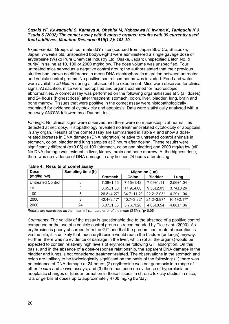
Sasaki YF, Kawaguchi S, Kamaya A, Ohshita M, Kabasawa K, Iwama K, Taniguchi K & Tsuda S (2002) The comet assay with 8 mouse organs: results with 39 currently used food additives. Mutation Research 519(1-2): 103-19. Experimental: Groups of four male ddY mice (sourced from Japan SLC Co, Shizuoka, Japan; 7-weeks old; unspecified bodyweight) were administered a single gavage dose of erythrosine (Wako Pure Chemical Industry Ltd, Osaka, Japan; unspecified Batch No. & purity) in saline at 10, 100 or 2000 mg/kg bw. The dose volume was unspecified. Four untreated mice served as a negative control group; the authors stated that their previous studies had shown no difference in mean DNA electrophoretic migration between untreated and vehicle control groups. No positive control compound was included. Food and water were available ad libitum during all phases of the experiment. Mice were observed for clinical signs. At sacrifice, mice were necropsied and organs examined for macroscopic abnormalities. A comet assay was performed on the following organs/tissues at 3 (all doses) and 24 hours (highest dose) after treatment: stomach, colon, liver, bladder, lung, brain and bone marrow. Tissues that were positive in the comet assay were histopathologically examined for evidence of cytotoxicity and apoptosis. Data were statistically analysed with a one-way ANOVA followed by a Dunnett test. Findings: No clinical signs were observed and there were no macroscopic abnormalities detected at necropsy. Histopathology revealed no treatment-related cytotoxicity or apoptosis in any organ. Results of the comet assay are summarised in Table 4 and show a dose-related increase in DNA damage (DNA migration) relative to untreated control animals in stomach, colon, bladder and lung samples at 3 hours after dosing. These results were significantly different (p<0.05) at 100 (stomach, colon and bladder) and 2000 mg/kg bw (all). No DNA damage was evident in liver, kidney, brain and bone marrow. At the highest dose, there was no evidence of DNA damage in any tissues 24 hours after dosing. Table 4: Results of comet assay Dose (mg/kg bw)
Sampling time (h) Migration (μm) Stomach Colon Bladder Lung
Untreated Control 3 7.08±1.55 7.15±1.42 7.09±1.11 2.56±1.04 10 3 8.65±1.38 11.9±4.00 9.53±2.03 3.74±0.26 100 3 26.8±4.27* 34.7±11.2* 22.2±2.03* 4.29±1.04 2000 3 42.4±2.17* 40.7±3.22* 21.2±3.97* 10.1±2.17* 2000 24 6.07±1.56 5.76±1.26 4.65±0.54 4.88±1.06
Results are expressed as the mean ±1 standard error of the mean (SEM); *p<0.05 Comments: The validity of the assay is questionable due to the absence of a positive control compound or the use of a vehicle control group as recommended by Tice et al. (2000). As erythrosine is poorly absorbed from the GIT and that the predominant route of excretion is via the bile, it is unlikely that much erythrosine would reach the bladder (or lungs) anyway. Further, there was no evidence of damage in the liver, which (of all the organs) would be expected to contain relatively high levels of erythrosine following GIT absorption. On this basis, and in the absence of a dose-response relationship, the apparent DNA damage in the bladder and lungs is not considered treatment-related. The observations in the stomach and colon are unlikely to be toxicologically significant on the basis of the following: (1) there was no evidence of DNA damage at 24 hours; (2) erythrosine was not genotoxic in a range of other in vitro and in vivo assays; and (3) there has been no evidence of hyperplasia or neoplastic changes or tumour formation in these tissues in chronic toxicity studies in mice, rats or gerbils at doses up to approximately 4700 mg/kg bw/day.

21
Zijno A, Marcon F, Leopardi P, Salvatore G, Carere A & Crebelli R (1994) An assessment of the in vivo clastogenicity of erythrosine. Food and Chemical Toxicology 32(2): 159-163. Experimental: Groups of five male B6C3F1 mice (sourced from Charles River Italy, Como Italy; 25-28 g bodyweight; age unspecified) were given two intraperitoneal (ip) injections of erythrosine in distilled water (sourced from BASF, Germany; unspecific Batch No.; 85% purity), 24 hours apart, at doses of 0, 50, 100 or 200 mg/kg bw. The highest dose was selected based on the published ip LD50 in mice. Three positive control mice were given a single ip injection of 1 mg/kg bw mitomycin C in distilled water (Sigma Chemical Co, St Louis, USA). The following cytogenic endpoints were measured: sister chromatid exchanges (SCEs) in peripheral blood monocytes; micronuclei in bone marrow polychromatic erythrocytes; and micronuclei in peripheral blood reticulocytes. Results were statistically analysed using a Student’s t-test or a χ2-test. Findings: Clinical signs occurred in all mice at the highest dose (piloerection, dyspnoea and hypomobility). The mitotic index was significantly reduced (p<0.05) at the highest dose relative to the negative control (1.9 versus 11.7+1.2%) indicating a toxic effect on lymphocytes. There was no treatment-related effect on the three genotoxic endpoints analysed. The positive control caused a significant increase in SCEs in peripheral blood lymphocytes (p<0.001), micronuclei in bone marrow cells (p<0.001) and micronuclei peripheral blood reticulocytes (p<0.001) relative to the negative control (>2-fold higher). Erythrosine was not genotoxic in vivo under the conditions of this study. 2.3.10 Other studies (in vivo) Abdel Aziz AH, Shouman SA, Attia AS & Saad SF (1997) A study on the reproductive toxicity of erythrosine in male mice. Pharmacological Research 35(5): 457-462. Experimental: Erythrosine (sourced from H Kohnstamm & Co, Inc, New York USA; unspecified Batch No. & purity) was administered by gavage as an aqueous suspension to groups of ten adult male albino mice (unspecified strain and source; 35-38 g bodyweight; 15-16 weeks old) at doses of 0, 68 or 136 mg/kg bw/day for 21 days. The doses were equal to 1% and 2% of the oral LD50 in mice (see WHO 1975). A positive control group of ten mice received cyclophosphamide by gavage at a dose of 18 mg/kg bw/day for 21 days; this dose was stated to be comparable to the human therapeutic dose of cyclophosphamide. Food and water were available ad libitum. Mice were killed one day after the last dose. The following parameters were measured for each mouse: testicular lactate dehydrogenase isoenzyme activity (LDH-X) (an indicator of testicular function); sperm count and spermatozoa motility. In a second experiment, 5 adult male mice/group were administered erythrosine by gavage at a dose of 0, 340, 680 or 1360 mg/kg bw/day for five days. A positive control group of 5 mice received an intraperitoneal injection of 20 mg/kg bw/day cyclophosphamide for five days. Mice were killed 35 days after the last dose and sperm head abnormalities examined microscopically. LDH-X and sperm head abnormalities were statistically analysed using a Student’s t-test. Findings: In the first experiment, there was a significant (p<0.05) treatment-related reduction in mean (+1 SEM) LDH-X in mice relative to the negative control group (57.1+3.46 and 45.4+2.78 at 68 and 136 mg/kg bw/day, respectively, versus 79.5+1.33 μmole/min/mg protein). The interpretation of this finding as toxicologically significant is limited somewhat by the lack of historical control data for this parameter in age-matched mice (in the broader scientific literature or as cited by the study authors).

22
However, as the effect was comparable to that observed with the positive control (53.1+3.42 μmole/min/mg protein), there is a reasonable suggestion of a treatment-related effect despite the use of only two dose levels. Graphically-presented data indicated that mean sperm counts were significantly (p<0.05) lower in treated mice than the control, but the effect was less at the higher dose of erythrosine (51 and 34% lower at 68 and 136 mg/kg bw/day, respectively). In contrast, graphically-presented data indicated a significant (p<0.05) dose-related decrease in sperm motility at 68 and 136 mg/kg bw/day erythrosine (57 and 81% lower than the control at 68 and 136 mg/kg bw/day, respectively). The positive control, cyclophosphamide, reduced sperm count and sperm motility by 80% and 67%, respectively, relative to the control. In the second experiment, the incidence of sperms with abnormal heads was significantly higher (p<0.01) at 680 and 1360 mg/kg bw/day than the control (31.16+1.07 and 32.66+1.97, respectively, versus 19.83+1.19 in 500 sperms/mouse). The lack of a dose-response relationship weakens the interpretation of this result as treatment-related. There was no effect at the lowest dose of erythrosine, while treatment with the positive control resulted in an increased incidence of head abnormalities (45.83+1.6 in 500 sperms/mouse). Comment: The two experiments conducted as part of this study are somewhat difficult to link as they tested different doses, durations, endpoints and administered the positive control compound by different routes (no rationale was provided for the latter); on this basis, the observations made in the two experiments can not be compared. There was some inconsistency across endpoints in terms of the dose-response, with the reduction in sperm counts relative to the control less at the high than the low dose in the first experiment. Further, the increase in sperm head abnormalities in the second experiment did not follow a dose-response relationship. While the testing of a limited number of endpoints is not necessarily a major deficiency to this study, it would have been useful to have some corroborative evidence of perturbed testicular function such as other serum or tissue markers (e.g. testosterone, luteinizing hormone etc), testicular size and weight and histopathology. In view of the fact that no disruption of fertility has been observed in any reproduction studies, and that there has been no observations of reduced testes weights or histopathological abnormalities in these or other repeat dose studies (Appendix A), the findings in this study are not considered to be treatment-related or toxicologically significant. Pacor ML, Di Lorenzo G, Martinelli N, Mansueto P, Rini GB & Corrocher R (2004) Monosodium benzoate hypersensitivity in subjects with persistent rhinitis. Allergy 59(2) 192-197. Two hundred and twenty six patients (76 males; 150 females; 12-60 years of age) affected by persistent rhinitis (and no other clinical signs of allergy) were placed on an additive-free diet for 30 days then challenged with a food additive-rich diet (containing colourings, preservatives, glutamate, sulphites and benzoic acid) for 15 days. A food additive oral challenge was then performed via a double-blind placebo controlled study for six specific compounds, including erythrosine. Each of the six test compounds were tested at weekly intervals. Three separate doses of erythrosine (50, 50 and 100 mg, respectively) were given to each subject in gelatine capsules at 3-hourly intervals over a single day. Nasal peak inspiratory flow (NPIF) was measured before, during and after oral challenge; a >20% decrease in NPIF was considered by the authors as a positive response. Results were statistically analysed using non-parametric methods (Kruskal-Wallis test and if significant, a Wilcoxon signed rank test). No subjects had a decrease in NPIF of >20% following erythrosine challenge, although 7 subjects had a subjective increase in rhinitis. These findings suggest that erythrosine does not cause rhinitis in people.

23
Other studies (in vitro) Dees C, Askari M, Garrett S, Gehrs K, Henley D & Ardies M (1997) Estrogenic and DNA-damaging activity of Red No. 3 in human breast cancer cells. Environmental Health Perspectives. 105 (Supplement 3): 625-632. A number of in vitro experiments were undertaken to analyse the effect of erythrosine (sourced from Pfaltz and Bauer, Waterbury, Connecticut, USA; unspecified Batch No. & purity) on cell proliferation, oestrogen receptor binding and p53 binding. Oestrogen receptor (ER) positive HTB 133 breast cancer cells, ER negative HTB 125 breast cells and rat liver epithelial (RLE) cells were incubated with erythrosine dissolved in sterile water at concentrations of 0, 25, 50, 75 or 100 μg/mL for 72 hours. The sample size was unspecified, while cell numbers were counted three times. Graphically-presented data indicated a significant (p<0.05) concentration-related increase in cell proliferation in ER positive HTB 133 cells but not ER negative HTB 125 or RLE cells. This effect was significantly less (p<0.05) than that observed with dichloro-diphenyl-trichloroethane (DDT) and was significantly inhibited (p<0.05) when cells were incubated with 10 nM of the steroidal antioestrogen, ICI 182,780. Graphically-presented data indicated that erythrosine could compete with radiolabelled oestradiol binding to the ER on MCF-7 breast carcinoma cells. However, erythrosine binding was relatively weak; approximately 1000-fold less than oestradiol and approximately 10-fold less than DDT. Autoradiograms from a gel mobility shift assay indicated that 25 μg/mL erythrosine increased (by 1.5-fold lower over the control) the binding of the ER to the oestrogen responsive element (ERE) in MCF-7 cells and this was inhibited by 10 nM ICI 182,780. This same concentration of ICI 182,780 only partially inhibited (0.5-fold lower than the control) the response stimulated by 0.3μM DDT. In contrast, a 10-15 molar excess of ICI 182,780 was necessary to inhibit oestradiol-stimulated ER-ERE complex formation. To analyse the possible effect of erythrosine on Cdk2 activity (activation of this cyclin kinase is important for cell proliferation), serum-starved MCF-7 cells were incubated with 0, 0.5, 1, 3 or 10 μg/mL erythrosine for 20 hours. Cdk2 was immunoprecipated from cell lysates and reacted in the presence of histones and [32P]ATP. The reaction products were analysed electrophoretically. Autoradiograms indicated that erythrosine stimulated Cdk2 activity (histone phosphorylation) in a concentration-related manner. To investigate its potential to damage genetic material, the effect of erythrosine on p53 binding to DNA (p53 binding is important for DNA repair) was analysed. Extracts of p53 were isolated from HTB 125, MCF-7 and HTB 133 cells that had been incubated for 2 hours with 25 or 100 μg/mL erythrosine ± an exogenous source of metabolic activation (S9 supernatant from Aroclor 1254-induced rat livers) under serum-free conditions. Actinomycin, mitomycin and 5-fluorouracil were used as positive controls at unspecified concentrations. Extracts were incubated with a radiolabelled consensus p53 binding sequence and the binding analysed electrophoretically via a gel mobility shift. Autoradiograms indicated that in the presence of erythrosine, extracts from HTB 125, MCF-7 and HTB 133 cells contained higher levels of p53 than negative controls Metabolic activation had no effect on the level of p53 protein. Comments: The above findings are unlikely to be biologically relevant given that erythrosine was only weakly oestrogenic. The authors contention that erythrosine caused DNA damage based on increased p53 levels is unjustified because (1) the method adopted did not directly

24
measure DNA damage; and (2) it ignores the extensive genotoxicity database for erythrosine indicating that it is not genotoxic. The authors’ conclusion that the findings suggest a link between erythrosine exposure and breast cancer is unsubstantiated based on the absence of mammary tumours in seventeen long-term studies in the mouse, rat and gerbil (see Appendix A) In addition to the studies described above, there were a range of other studies of questionable relevance to the current assessment, which examined endpoints such as the inhibition of ATP binding and respiration. These studies are summarised in Table 5. Table 5: Results of other studies on erythrosine Reference Observations Rapley & Brunsvold (1991)
Erythrosine had no effect on the healing of alveolar bone and gingival connective tissue when applied directly into the oral cavity of dogs that had undergone dental surgery.
Wach & Gräber (1991)
Erythrosine inhibited adenosine triphosphate binding to plasma membrane H+-ATPase from yeast.
Reyes et al. (1996) A range of food colours (including erythrosine) were tested for their ability to inhibit respiration in mitochondria isolated from rat liver and kidney. All tested compounds inhibited mitochondrial respiration to varying degrees; erythrosine (0.1 mg/mg mitochondrial protein) inhibited respiration by 100% in both liver and kidney mitochondria. A limitation to this study was the absence of a negative control.
Kapadia et al. (1998) Erythrosine inhibited Epstein-Barr virus early antigen production in Raji cells, which had been mediated with the tumour promoter, 12-O-tetradecanoylphorbol-13-acetate (TPA). In a mouse model of skin carcinogenesis, dermally-applied erythrosine inhibited skin tumour promotion in mice treated with 7,12-dimethylbenz[a]anthracene and TPA.
Sugihara et al. (2004) As erythrosine is structurally similar to the diiodophenyl moiety of diiodotyrosine (an antigenic determinant of experimental iodine allergy), the immunological activity of erythrosine was examined using a guinea pig passive cutaneous anaphylaxis test. Erythrosine inhibited the binding of anti-iodine antibodies to iodinated guinea pig serum albumin and the level of inhibition was greater than that caused by diiodotyrosine (IC50 values of approximately 10 versus 60 μm, respectively). Erythrosine was positive in the passive cutaneous anaphylaxis test at challenge doses of 5 and 10 mg/animal. The value of this study for dietary risk assessment purposes is limited based on the route of administration (intradermal). However, results suggested that erythrosine could cross react with antibodies generated by immunisation with iodine in this particular guinea pig model.
Wood et al. (2006) Erythrosine (22 μM) was incubated with in vitro biofilms of Streptococcus mutans and then irradiated under white light for 15 minutes [so-called photodynamic therapy (PDT)]. Graphically-presented data indicated that PDT with erythrosine was more effective at killing bacteria than two established photosensitisers. Erythrosine treatment without irradiation was reportedly ineffective, although no data were provided to substantiate this claim.
Satoh et al. (2008) Forty-five chemicals reportedly or possibly having endocrine-disrupting potential were screened for their ability to inhibit aromatase activity (aromatase catalyses the conversion of androgens to oestrogens). Erythrosine caused a concentration-related decrease in aromatase activity, with an IC50 of approximately 10-7M. Implicitly, the authors considered this to be a pilot study and highlighted the importance of further in vitro and in vivo investigations.

25
2.4 Discussion The toxicity of erythrosine, including mechanistic aspects of its toxicological endpoints, is well defined. Supplementary studies evaluated as part of the current hazard assessment add to an already extensive toxicological database for erythrosine, which includes studies conducted in laboratory animals and humans. 2.4.1 Absorption, distribution, metabolism and excretion Erythrosine is poorly absorbed from the gastrointestinal tract (GIT) in rats and humans (WHO 1969 &1987). Based on maximum levels of radioactivity in bile (1.7%), liver (0.185%) and urine (1%), the level of GIT absorption in rats is approximately 3%. Based on maximum levels of radioactivity in urine (0.1%), faeces (1.2%) and tissue (0.013%), the level of GIT absorption in humans is approximately 1.3%. The low level of GIT absorption is a likely result of erythrosine’s low solubility at pH values below 3-4 (~0.7 g/L), where the carboxyl group would mainly be in the un-ionised form. Therefore, the higher GIT absorption in rats relative to humans may reflect the higher solubility of erythrosine in rat gastric fluid, which has a higher pH (5-6) than human gastric fluid (1-2). In rats, the Tmax in blood and plasma is one hour, with the majority of absorbed erythrosine distributing to the liver (~0.2% of an administered dose of 14C or 125I-erythrosine), where the Tmax is 4-12 hours (WHO 1987). Erythrosine does not distribute to the brain or pituitary and does not accumulate in the thyroid (WHO 1987). In rats, traces of radioactivity were detected in the thyroid but the levels were very low and likely to be attributable to a small amount of 125I-iodine generated via metabolic de-iodination (WHO 1987). In humans, no erythrosine was detectable in serum following a single oral radioactive dose of 75-80 mg, with less than 1% of radioactivity remaining in the body after 7 days (WHO 1991). In rats, erythrosine is metabolically stable (WHO 1969 & 1984) although a small level (unquantified) of metabolites (thought to be isomeric diiodo- and trifluoresceins) were detected in urine, faeces, plasma, liver and kidney (WHO 1987). Three supplementary in vitro metabolism studies suggested that erythrosine can inhibit the activity of certain enzymes/transporters in human or bovine liver microsomes such as sulfotransferases, UGT1A6, UGT2B7, CYP3A4 or p-glycoprotein (Bamforth et al. 1993; Kuno & Mizutani 2005; Furumiya &Mizutani 2008). There was no effect on CYP2A6 (Kuno & Mizutani 2005). There was no indication that erythrosine was a substrate for any of these proteins and this was confirmed for UGT1A6 and UGT2B7 (Kuno & Mizutani 2005). This is consistent with in vivo evidence that erythrosine is not metabolised (WHO 1969 & 1984). The suggestion from these studies was that erythrosine could interfere with the metabolism of other xenobiotics, which are substrates for these liver enzymes. However, the supplementary findings are unlikely to be biologically relevant because: (1) possible inhibition via superoxide anion generation would be prevented in vivo by superoxide dismutase [this was shown in vitro for the inhibition of UGT1A6 (Furumiya & Mizutani 2008)]; and (2) given that the GIT absorption is so low, it is highly unlikely that erythrosine would reach μM concentrations in the liver to potentially interfere with these enzymes/proteins (IC50 values were in the μM range)5. In rats and humans, erythrosine is eliminated predominantly in the faeces, with less than 2% of an oral radioactive dose excreted in the bile of rats (WHO 1987). When rats were dosed intravenously, the level of radioactivity detected in bile/urine was 55/1.3% (WHO 1969). An early study in rats detected no radioactivity in urine following a single oral dose of 125I-erythrosine (WHO 1969). 5 Assuming intake at the ADI of 0.1 mg/kg bw/day, a 60 kg adult would ingest 6 mg erythrosine but then only absorb 1.3% or less giving a total systemic amount of 0.078 mg. Assuming all could be absorbed in 1 mL, liver cells would be exposed to approximately 1/100th of the lowest IC50 dose.

26
A subsequent study detected <1% of an oral radioactive dose in urine (rats had been exposed to unlabelled erythrosine in the diet at 0.5 or 4.0% for 7 days prior to oral administration of 14C or 125I-erythrosine) (WHO 1987). In humans and rats, excretion is complete by 48 hours (WHO 1987). 2.4.2 Acute toxicity Erythrosine has low acute oral toxicity (rat LD50 = 1840 mg/kg bw) (WHO 1975). 2.4.3 Repeat-dose toxicity In repeat-dose toxicity studies in rats, effects on thyroid function occurred at dietary concentrations above 0.25% (~150 mg/kg bw/day) (WHO 1991). These effects, which were most evident at 4% (~2500 mg/kg bw/day) included increased TSH (≥0.25%), T4 (≥0.25%) and rT3 (≥0.25%), decreased T3 (≥1%; ~600 mg/kg bw/day), increased parathyroid/thyroid weight and histopathological evidence of enhanced synthetic and secretory activity of the thyroid (4%). Numerous (17) long-term toxicity and carcinogenicity studies have been conducted in laboratory animals at dietary concentrations ranging from 0.1 to 5%. There was no evidence of carcinogenicity in mice (4 studies) or gerbils (4 studies) up to dietary concentrations of 3 and 4%, respectively (equivalent to 1200 mg/kg bw/day for gerbils, and 4759/5770 mg/kg bw/day in male/female rats) (WHO 1969 & 1984). The majority (7/9) of rat studies showed no evidence of carcinogenicity, with two studies reporting an increased incidence of benign thyroid tumours. In the first study, which tested dietary concentrations of 0, 0.1, 0.5 or 1% for 30 months (beginning in utero) (equal to 0, 49, 251 and 507 mg/kg bw/day in males and 0, 61, 307 and 642 mg/kg bw/day in females, respectively), the incidence of follicular adenomas was significantly increased in females at 642 mg/kg bw/day relative to the control (6/68 versus 0/140, respectively) (WHO 1984). A re-evaluation of this study (WHO 1991) reported a significant increase in the combined incidence of adenomas and carcinomas in males at 0.1, 0.5 and 1% (6/64, 8/66 & 4/57, respectively, versus 1/128 in the control) and in females at 1% (6/68 versus 1/138 in the control). In a 29 month study beginning in utero, which tested a single dietary level of 4% (equal to 2465 and 3029 mg/kg bw/day in males and females, respectively), increased thyroid weight, follicular and c-cell hyperplasia and an increased incidence of thyroid follicular adenoma occurred in males (16/68 versus 0/69 in the control) (1984). A re-evaluation of this study (WHO 1991) reported a significant increase in the combined incidence of adenomas and carcinomas in males, relative to the controls (18/68 versus 2/68, respectively). The reason(s) for the occurrence of thyroid tumours in these two particular rat studies is unclear as the other studies tested comparable doses albeit for shorter durations (i.e. 2 years). The occurrence of these tumours could have been a reflection of the greater age of the rats (noting that dosing commenced in utero). Another possibility is that the tumours were strain-specific (SD-derived, Charles River CD rats were used) although the identity of the strains used in some of the other studies was not reported. 2.4.4 Genotoxicity Erythrosine was not genotoxic or mutagenic in the majority of in vitro assays, although a small number did report positive responses at cytotoxic concentrations (WHO 1984, 1987 &1991). Erythrosine was not genotoxic in vivo (WHO 1991)

27
The supplementary genotoxicity data adds to an already extensive battery of studies indicating that erythrosine is not genotoxic. Erythrosine and two manufacturing impurities, DHBBA and DHDIBBA, were negative in the Ames test (Ozaki et al. 1998; Wada et al. 2004) and did not transform, or induce chromosomal aberrations, in SHE cells (Yamaguchi & Tsutsui 2003). Erythrosine did not induce SCEs or micronuclei in mice up to a dose of 200 mg/kg bw (Zijno et al. 1994). A comet assay reported a significant increase in DNA damage in the stomach, colon, bladder and lung in mice three hours after a single gavage dose of 2000 mg/kg bw erythrosine (Sasaki et al. 2002). However, this result was discounted as treatment-related or toxicologically-significant as no damage was evident in the same tissues 24 hours after dosing. In addition, there has been no evidence of tumour formation in any of these tissues in chronic toxicity studies in mice, rats or gerbils up to doses of approximately 4700 mg/kg bw/day. Dees et al. (1997) observed an increase in p53 protein levels in breast cancer cells that had been incubated for two hours in the presence of erythrosine. Their conclusion that this was evidence of genotoxicity and that erythrosine could be a “significant risk factor in human breast carcinogenesis” is considered to be unsubstantiated on the basis of the following: the method used did not directly measure DNA damage; the extensive in vitro and in vivo genotoxicity database for erythrosine indicates that it is not mutagenic or capable of damaging DNA; numerous long-term toxicity studies conducted in the mouse, rat and gerbil did not find any mammary tumours 2.4.5 Reproductive and developmental toxicity Studies previously evaluated by JECFA indicated no effect on reproduction in rats up to dietary concentrations of 4% (~2500 mg/kg bw/day) and no evidence of teratogenicity in rats or rabbits up to doses of 250 and 125 mg/kg bw/day, respectively (WHO 1969, 1975 & 1984). Supplementary data indicated that erythrosine had no effect on reproduction or behavioural development in mice, although a number of parameters of movement activity and of exploratory behaviour were significantly different to the control at the highest oral dose of 0.045 % (Tanaka 2001). However, the lack of consistency between sexes and over time suggests that these findings were not treatment-related. This conclusion is supported by evidence indicating that erythrosine can not actually reach the brain (Levitan et al. 1984; WHO 1987). Supplementary studies in rats confirmed the absence of developmental toxicity when erythrosine was administered by gavage or via the drinking water up to doses of 800 and 472 mg/kg bw/day, respectively (Collins et al. 1993a & b). Abdel Aziz et al. (1997) reported possible affects on testes function (reduced LDH-X) at 68 and 136 mg/kg bw/day and reduced sperm counts, motility and altered morphology at 680 and 1360 mg/kg bw/day in gavaged male mice. However, these findings were not considered by FSANZ to be treatment-related based on the following: the absence or inconsistency of the dose-response relationships; no effects on fertility or the sex organs have been observed in reproduction or repeat-dose toxicity studies in rodents up to dietary levels of 4% (~2500 mg/kg bw/day); there is no evidence that erythrosine actually distributes to the reproductive organs; and there were limitations to the study design.

28
2.4.5 Toxicological studies in humans JECFA considered a number of studies conducted in humans (mainly in males; sample sizes ranging from unknown to 5, 6 or 39), using erythrosine doses of 0.03 to 3.3 mg/kg bw/day. The studies reported increased PBI and serum iodine (WHO 1969, 1984 & 1989), but no effect on serum T4, T3 or rT3 (WHO 1984 & 1989). The NOAEL in 39 male subjects following 14-days of repeated oral dosing was 60 mg/day (equivalent to 1 mg/kg bw/day) based on increased serum iodine, PBI and TSH, and urinary iodine at 200 mg/day (equivalent to 3.3 mg/kg bw/day). JECFA (WHO 1989) concluded that the increase in serum TSH in humans was due to increased serum iodine rather than to a direct effect on the secretion or metabolism of thyroid hormones (i.e. T4 or T3). This mechanism is different to that proposed for rats, which involves the inhibition of the conversion of T4 to T3 (discussed below), although both mechanisms result in increased serum TSH levels (Renwick & Walton 2001). The mechanism for the increase in serum iodine (and PBI) in humans is somewhat unclear. Low levels of de-iodinated metabolites have been detected in rats (WHO 1987) and therefore it is plausible that some iodine might be released in humans via metabolic activity. However, as erythrosine is largely metabolically stable in rats, biologically significant quantities of free iodine are unlikely to be generated through this process. Two other possible sources of iodine include the action of bacteria in the gut (for which there is no reported evidence) or the presence of contaminating iodine in the erythrosine preparation. As erythrosine is a relatively stable compound, it is highly unlikely to degrade under standard conditions of storage or use. Iodine may be released when heated in excessive alkali (Wada et al. 2004) or in the presence of metallic iron and/or tin and free organic acid (WHO 1969). Product specifications described in the Food Technology Report indicate that the maximum amount of contaminating sodium iodine is 0.4%, which would not be expected to contribute significantly to iodine intake. On this basis, the mechanism for the increase in TSH in humans remains to be elucidated. 2.4.6 Mechanism of tumour formation in rats – relevance to humans As discussed above, erythrosine causes an increase in serum TSH, T4 and rT3, and a concomitant decrease in T3 in rats. This results from disruption of the peripheral conversion of T4 to T3 (i.e. in the liver or kidneys) due to the inhibition of 5’-monodeiodinase enzymes (which catalyse the 5’-deiodination of T4 and rT3) resulting in a compensatory increase in the secretion of TSH by the pituitary (Capen 1991, 1994 & 1998; WHO 1987 & 1991). The ensuing stimulation of the thyroid gland induces proliferative changes, which over long durations at high dietary levels, can cause tumour formation in rats (WHO 1991; Capen 1994). Therefore, the occurrence of benign thyroid tumours in rats is secondary to erythrosine’s ability to perturb thyroid hormone metabolism (WHO 1991). The relevance of thyroid tumours to humans has been the subject of several reviews (Hill et al. 1998; Hurley et al. 1998; IARC 1999; Cohen et al. 2004). Of bearing on the mechanism of tumour formation in erythrosine-treated rats, the International Agency for Research on Cancer (IARC) (1999) concluded that the rodent thyroid is more sensitive than the human thyroid to tumour induction resulting from elevated TSH levels. This is attributable to a number of interspecies differences in thyroid physiology including: the absence of thyroid-binding globulin (TBG) in rats (this protein transports T4 and T3 in the blood in humans); the half life of T4 in rats is twelve hours but is 5-9 days in humans (as a result of the absence of TBG); the serum level of TSH is 25 times higher in rodents than humans (to compensate for the shorter half-life); thyroid hormone elimination is greater in rodents than humans; and the resting rodent thyroid is histologically similar to the stimulated human thyroid (Capen 1992; Hill et al. 1998; Hurley et al. 1998; IARC 1999). Based on

29
interspecies differences in thyroid physiology, JECFA (WHO 1991) concluded that the occurrence of thyroid tumours in erythrosine-treated rats is not relevant to humans. 2.4.7 Other biological effects of erythrosine The in vitro study of Dees et al. (1997) suggested that erythrosine is oestrogenic as shown by the increased proliferation of ER positive breast cancer cells, direct binding to the ER and the stimulation of ER binding to the ERE. However, binding was relatively weak (~1000-fold less than oestradiol, 10-fold less than DDT) and unlikely to be biologically relevant. It is worth noting that in vivo, no oestrogenic activity was observed (increased uterine weight) when 250 mg/kg bw erythrosine was given to rats subcutaneously, twice daily for 3 days (WHO 1969). 2.5 Conclusions A review of published supplementary studies generated since the most recent JECFA toxicological evaluation of erythrosine identified no new issues or studies to suggest a need to amend the current ADI of 0.1 mg/kg bw/day that was established in 1990. In both humans and rats, repeated ingestion of erythrosine results in elevated serum TSH levels. In humans, this is suggested to result from increased serum iodine, while in rats, there is compelling evidence that this results from the inhibition of the peripheral metabolism of T4 to T3 in the liver. In a minority of rat studies, the formation of benign thyroid tumours was attributable to proliferative changes in the thyroid due to sustained stimulation by elevated serum TSH levels. These tumour findings are not relevant to humans based on interspecies differences in thyroid physiology. 3. References Abdel Aziz AH, Shouman SA, Attia AS & Saad SF (1997) A study on the reproductive toxicity of erythrosine in male mice. Pharmacological Research 35(5): 457-462.
Allen DH, Van Nunen S, Loblay, R, Clarke L and Swain A (1984). Adverse reactions to food. The Medical Journal of Australia – Special Supplement 141(5 Suppl):S37-42.
Asero R (2001). Perennial rhinitis induced by benzoate intolerance. Journal Allergy Clinical Immunology. 107(1):197.
Asero R (2002). Multiple intolerance to food additives Journal Allergy Clinical Immunology. 110(3):531.
Bamforth KJ, Jones AL, Roberts RC & Coughtrie MW (1993) Common food additives are potent inhibitors of human liver 17 alpha-ethinyloestradiol and dopamine sulphotransferases. Biochemical Pharmacology 46(10) 1713-1720.
Booth J (1993). Food intolerance in a child with urticaria. Journal of Human Nutrition and Dietetics. 6(4): 377 – 380.
Capen CC (1992) Pathophysiology of chemical injury of the thyroid gland. Toxicology Letters 64: 381-388.
Capen CC (1994) Mechanisms of chemical injury of thyroid gland. Progress in Clinical and Biological research 387: 173-191.
Capen CC (1998) Correlation of mechanistic data and histopathology in the evaluation of selected toxic endpoints of the endocrine system. Toxicology Letters 102-103: 405-409.
Chang HJ, Burke AE & Glass RM (2009).JAMA patient page: Celiac disease Journal American Medical Association. 302(11):1248.

30
Cohen SM, Klaunig J, Meek ME, Hill RN, Pastoor T, Lehman-McKeeman L, Bucher J, Longfellow DG, Seed J, Dellarco V, Fenner-Crisp P & Patton D (2004) Evaluating the human relevance of chemically induced animal tumors. Toxicological Sciences 78: 181-186.
Collins TF, Black TN & Ruggles DI (1993a) Teratogenic potential of FD&C Red No. 3 when given by gavage. Toxicology and Industrial Health 9(4): 605-616.
Collins TF, Black TN, O'Donnell MW, Shackelford ME & Bulhack P (1993b) Teratogenic potential of FD & C red no. 3 when given in drinking water. Food and Chemical Toxicology 31(3): 161-167.
David TJ (1988) Food Additives. Archives of Disease in Childhood 63:582-583.
David TJ (2000) Adverse reactions and intolerance to foods. Review. British Medical Bulletin 56(1):34-50.
Dees C, Askari M, Garrett S, Gehrs K, Henley D & Ardies M (1997) Estrogenic and DNA-damaging activity of Red No. 3 in human breast cancer cells. Environmental Health Perspectives. 105 (Supplement 3): 625-632.
Furumiya K & Mizutani T (2008) Inhibition of human CYP3A4, UGT1A6 and p-glycoprotein with halogenated xanthene food dyes and prevention by superoxide dismutase. Journal of Toxicology & Environmental Health A 71: 1307-1313.
Hagiwara M, Watanabe E, Barrett JC & Tsutsui T (2006) Assessment of genotoxicity of 14 chemical agents used in dental practice: ability to induce chromosome aberrations in Syrian hamster embryo cells. Mutation Research 603(2): 111-120.
Herman RH and Hagler L (1979) Food intolerance in humans. Symposium on Clinical Nutrition - West J Med 130(2): 95–116.
Hill RN, Crisp TM, Hurley PM, Rosenthal SL & Singh DV (1998) Risk assessment of thyroid follicular cell tumors. Environmental Health Perspectives 106(8): 447-457.
Hodge L, Swain A and Faulkner-Hogg K (2009). Food allergy and intolerance. Australian Family Physician 38(9):705-707.
Hurley PM, Hill RN & Whiting RJ (1998) Mode of carcinogenic action of pesticides inducing thyroid follicular cell tumors in rodents. Environmental Health Perspectives 106(8): 437-445
IARC (1999) Species differences in thyroid, kidney and urinary bladder carcinogenesis. IARC Scientific Publication No. 147.
Ibero M, Eseverri JL, Barroso C, Botey J. (1982). Dyes, preservatives and salicylates in the induction of food intolerance and/or hypersensitivity in children. Allergologia et Immunopathologia (Madr). 10(4):263-268.
Kapadia GJ, Tokuda H, Sridhar R, Balasubramanian V, Takayasu J, Bu P, Enjo F, Takasaki M, Konoshima T & Nishino H (1998) Cancer chemopreventive activity of synthetic colorants used in foods, pharmaceuticals and cosmetic preparations. Cancer Letters 129(1): 87-95.
Kuno N & Mizutani T (2005) Influence of synthetic and natural food dyes on activities of CYP2A6, UGT1A6, and UGT2B7. Journal of Toxicology and Environmental Health. Part A 68(16): 1431-1444.
Levitan H, Ziylan Z, Smith QR, Takasato Y & Rapoport SI (1984) Brain uptake of food dye, erythrosine B, prevented by plasma protein binding. Brain Research 322(1): 131-4
Loblay RH and Swain AR (1985) Adverse reactions to tartrazine. Food Technology in Australia 37(11):508-510 and 514.
Loblay RH and Swain AR (1986) Food Intolerance. in 'Recent Advances in Clinical Nutrition' Volume 2. Libbey, London. Eds: M.L. Wahlqvist & A.S. Truswell pp. 169-177.

31
Mailman RB, Ferris RM, Tang FL, Vogel RA, Kilts CD, Lipton MA, Smith DA, Mueller RA, Breese GR. (1980) Erythrosine (Red No. 3) and its nonspecific biochemical actions: what relation to behavioral changes? Science 207(4430):535-537.
Mailman RB, Lewis MH. (1981) Food additives and developmental disorders: the case of erythrosin (FD&C Red #3), or guilty until proven innocent? Applied Research Mental Retardation.;2(4):297-305.
Mikkelsen H; Larsen JC and Tarding F (1978). HyperHypersensitivity reactions to food colours with special reference to the natural colour annatto extract (butter colour). Archives of toxicology. Supplement No. 1:141-143.
Ozaki A, Kitano M, Itoh N, Kuroda K, Furusawa N, Masuda T & Yamaguchi H (1998) Mutagenicity and DNA-damaging Activity of Decomposed Products of Food Colours under UV irradiation. Food and Chemical Toxicology 36(9): 811-817.
Park HW, Park CH, Park SH, Park JY, Park HS, Yang HJ, Ahn KM, Kim KH, Oh JW, Kim KE, Pyun BY, Lee HB, Min KU (2008). Dermatologic adverse reactions to 7 common food additives in patients with allergic diseases: a double-blind, placebo-controlled study. Journal Allergy Clinical Immunology 121(4):1059-1061.
Rapley JW & Brunsvold MA (1991) The effects of erythrosine on alveolar bone and gingival connective tissue in dogs. Journal of Periodontology 62(2): 132-134
Renwick AG & Walton K (2001) The use of surrogate endpoints to assess potential toxicity in humans. Toxicology Letters 120(1): 97-110.
Reyes FG, Valim MF & Vercesi AE (1996) Effect of organic synthetic food colours on mitochondrial respiration. Food Additives and Contaminants 13(1): 5-11.
Sasaki YF, Kawaguchi S, Kamaya A, Ohshita M, Kabasawa K, Iwama K, Taniguchi K & Tsuda S (2002) The comet assay with 8 mouse organs: results with 39 currently used food additives. Mutation Research 519(1-2): 103-19.
Satoh K, Nonaka R, Ishikawa F, Ogata A & Nagai F (2008) In vitro screening assay for detecting aromatase activity using rat ovarian microsomes and estrone ELISA. Biological & Pharmaceutical Bulletin 31(3):357-62.
Simon RA (2003) Adverse reactions to food additives. Current Allergy and Asthma Reports 3:62-66.
Tanaka T (2001) Reproductive and neurobehavioural toxicity study of erythrosine administered to mice in the diet. Food and Chemical Toxicology 39(5): 447-454.
Taylor SL and Hefle SL (2001). Food allergies and other food sensitivities. Food Technology 55 (9): 68-83.
Tice, RR, Agurell E, Anderson D, Burlinson B, Hartmann A, Kobayashi H, Miyamae Y, Rojas E, Ryu J-C & Sasaki YF (2000) Single cell gel/comet assay: Guidelines for in vitro and in vivo genetic toxicology testing. Environmental & Molecular Mutagenesis 35: 206-221.
Wach A & Gräber P (1991) The plasma membrane H(+)-ATPase from yeast. Effects of pH, vanadate and erythrosine B on ATP hydrolysis and ATP binding. European journal of biochemistry / FEBS 201(1): 91-97.
Wada K, Fujita T, Ogawa Y, Koda T & Aoki H (2004) Monitoring of the generation of non-mutagenic benzoic acid compounds during the manufacture of erythrosine. Food Additives and Contaminants 21(12):1137-48.
Weber RW, Hoffman M, Raine DA Jr, Nelson HS (1979). Incidence of bronchoconstriction due to aspirin, azo dyes, non-azo dyes, and preservatives in a population of perennial asthmatics. Journal Allergy Clinical Immunology 64(1):32-37.

32
WHO (1969) Toxicological evaluation of some food colours, emulsifiers, stabilizers, anti-caking agents and certain other substances. FAO Nutrition Meetings Report Series No. 46A WHO/FOOD ADD/70.36
WHO (1975) Toxicological evaluations of some food colours, enzymes, flavour enhancers, thickening agents, and certain food additives. WHO Food Additive Series 6.
WHO (1984) Toxicological evaluations of certain food additives and contaminants. WHO Food Additive Series 19.
WHO (1987) Toxicological evaluations of certain food additives and contaminants. WHO Food Additive Series 21.
WHO (1989) Toxicological evaluations of certain food additives and contaminants. WHO Food Additive Series 24.
WHO (1991) Toxicological evaluations of certain food additives and contaminants. WHO Food Additive Series 28.
Wilson BG and Bahna SL (2005). Adverse reactions to food additives. Annals of Allergy, Asthma and Immunology 95: 499-507.
Wood S, Metcalf D, Devine D & Robinson C (2006) Erythrosine is a potential photosensitizer for the photodynamic therapy of oral plaque biofilms. Journal of Antimicrobial Chemotherapy 57:680-684.
Yamaguchi F & Tsutsui T (2003) Cell-transforming activity of fourteen chemical agents used in dental practice in Syrian hamster embryo cells. Journal of Pharmacological Sciences 93(4): 497-500.
Zijno A, Marcon F, Leopardi P, Salvatore G, Carere A & Crebelli R (1994) An assessment of the in vivo clastogenicity of erythrosine. Food and Chemical Toxicology 32(2): 159-163.
Zopf Y, Baenkler H, Silbermann A, Hahn EG, Raithel M (2009) The differential diagnosis of food intolerance. Deutsches Ärzteblatt International 106(21): 359-369.
Zuberbier T, Edenharter G, Worm M, Ehlers I, Reimann S, Hantke T, Roehr CC, Bergmann KE, Niggemann B. Prevalence of adverse reactions to food in Germany - a population study. Allergy 59(3):338-345.
33
4. Studies seen but not evaluated The following studies were seen but not evaluated based on one or more of the following criteria: • The study did not directly test erythrosine. The study report lacked adequate detail to allow an independent evaluation to be made. • The study was poorly designed.
Ashida H, Hashimoto T, Tsuji S, Kanazawa K & Danno G (2000) Synergistic effects of food colors on the toxicity of 3-amino-1,4-dimethyl-5H-pyrido[4,3-b]indole (Trp-P-1) in primary cultured rat hepatocytes. Journal of Nutritional Science and Vitaminology 46(3): 130-136.
Barclay SC, Forsyth A, Felix DH & Watson IB (1999) Case report - hypersensitivity to denture materials. British Dental Journal 187(7): 350-352.
Mekkawy HA, Massoud AA & El-Zawahry AM (2000) Mutagenic effects of the food colour erythrosine in rats. Problems of Forensic Sciences 43: 184-191.
Uysal OC & Aral E (1998) Teratogenic effects and the role in the etiology of atopic diseases of erythrosine (FD&C Red No. 3). Turkish Journal of Medical Sciences 28:363-368.