head direction cells: properties and functional significance
TRANSCRIPT
196
Head direction cells: properties Robert U Mullet-l, James B Ranck Jr*
and functional significance and Jeffrey S Taubea
The strong signal carried by head direction cells in the
postsubiculum complements the positional signal carried
by hippocampal place cells; together, the directional and
positional signals provide the information necessary to permit
rats to generate and carry out intelligent, efficient solutions
to spatial problems. Our opinion is that the hippocampal
positional system acts as a cognitive map and that the role
of the directional system is to put the map into register with
the environment. In this way, paths found using the map can
be properly executed. Head direction cells have recently been
discovered in parts of the thalamus reciprocally connected
with the postsubiculum; such cells provide important clues to
the organization of the directional system.
Addresses tg2Department of Physiology, SUNY Health Science Center at Brooklyn, 450 Clarkson Avenue, Brooklyn, New York 11203, USA t e-mail: [email protected] se-mail: [email protected] sDepartment of Psychology, Dartmouth College, 6207 Gerry Hall, Hanover, New Hampshire 03755, USA se-mail: [email protected]
Abbreviations ADN anterior dorsal division of the anterior thalamic nuclei AP action potential HD head direction LDN lateral dorsal thalamic nuclei RSA retrosplenial agranular cortex RSG retrosplenial granular cortex
Current Opinion in Neurobiology 1996, 6:196-206
0 Current Biology Ltd ISSN 0959-4388
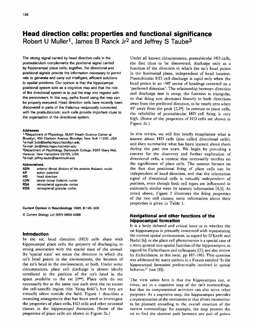
Introduction In the rat, head direction (HD) cells share with hippocampal place cells the property of discharging in strong association with the spatial state of the animal. By ‘spatial state’ we mean the direction in which the rat’s head points in the environment, the location of the rat’s head in the environment, or both. Under some circumstances, place cell discharge is almost ideally correlated to the position of the rat’s head in the space available to the rat [lo*]. Place cells do not necessarily fire at the same rate each time the rat enters the cell-specific region (the ‘firing field’), but they are virtually silent outside the field. Figure 1 describes a recording arrangement that has been used to investigate the properties of place cells, HD cells and other neuronal classes in the hippocampal formation. (Some of the properties of place cells are shown in Figure 2a.)
Under all known circumstances, postsubicular HD cells,
the first class to be discovered, discharge only as a function of the direction in which the rat’s head points in the horizontal plane, independent of head location. Postsubicular HD cell discharge is rapid only when the head points in an -90” sector of headings centered on a ‘preferred direction’. The relationship between direction and discharge rate is steep; the function is triangular, so that firing rate decreases linearly in both directions away from the preferred direction, to be nearly zero when 45” away from the peak [2,3’]. In contrast to place cells, the reliability of postsubicular HD cell firing is very high. (Some of the properties of HD cells are shown in Figure 2c.)
In this review, we will first briefly recapitulate what is known about HD cells (also called directional cells), and then summarize what has been learned about them during the past two years. We begin by providing a context for the discovery and further exploration of directional cells, a context that necessarily touches on the significance of place cells. The context focuses on the fact that positional firing of place cells can be independent of head direction, and that the orientation signal of directional cells is virtually independent of position, even though both cell types are influenced in extremely similar ways by sensory information [4,5]. As noted above, Figure 2 illustrates the firing properties of the two cell classes; more information about their properties is given in Table 1.
Navigational and other functions of the hippocampal formation It is a hotly debated and critical issue as to whether the rat hippocampus is primarily concerned with representing the current spatial environment, as argued by O’Keefe and Nadel [6], or the place cell phenomenon is a special case of a more general non-spatial function of the hippocampus, as argued by Eichenbaum and colleagues ([7]; see also review by Eichenbaum, in this issue, pp 187-195). This question was addressed by many authors in a Forum entitled ‘Is the hippocampal formation preferentially involved in spatial behavior?’ (see [8]).
The view taken here is that the hippocampus can, at times, act as a cognitive map of the rat’s surroundings, but that its computational activities can also serve other purposes. As a cognitive map, the hippocampus provides a representation of the environment that allows locomotion to be planned according to the overall structure of the current surroundings: for example, the map permits the rat to find the shortest path between any pair of points

Head direction cells Muller, Ranck Jr and Taube 197
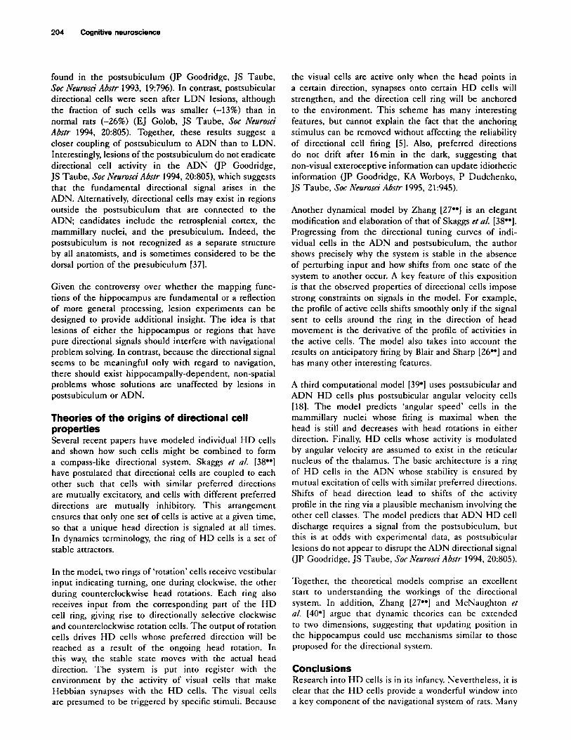
Figure 1
(a) TV camera view of cylindrical apparatus (b) A system of coordinates for locating the rat and measuring head direction
/(, Head
White cue card
1996 Current
Cylinder wall
Experimental methods for HD cell recordings. (a) Schematic diagram of the experimental setting used for most of the work on HD cells summarized in this review. The apparatus used is a cylinder, 76 cm in diameter and 51 cm high. From the view of the overhead TV camera used to track head position and direction, the wall appears as a concentric band around the floor. The wall does not appear as a circle because the N camera is only about 2.4 m above the floor - the top of the cylinder is closer to the camera and therefore appears larger. The foreshortened view is convenient as it allows the white cue card to be realistically depicted. This card runs from the top of the cylinder to the floor and occupies -90’ of the circumference. The cue card can be moved, deleted and changed to test its stimulus control over the discharge of place cells and HD cells. Not shown in the diagram is a circular curtain 2.4 m in diameter that surrounds the cylinder and runs from near the ceiling of the recording room down to the floor. The purpose of the curtain is to visually isolate the cylinder, ensuring that the white card is the only strong visual asymmetry in the environment. (b) A cartoon from the overhead viewpoint that indicates several important features used to describe the properties of place cells and HD cells. For the purposes of data acquisition, two headlights (not shown) are used to detect the rat’s head position and head direction. For descriptive purposes, however, it is often more convenient to give the location of the rat’s head and the location of firing fields in a polar coordinate system. The radial coordinate is measured from the center of the cylinder and the angular coordinate from a zero set at 3 o’clock in the view of the TV camera. Head direction is also measured relative to a zero set at 3 o’clock in the view of the camera. The head direction of interest is ‘yaw’, the direction that the head points in the horizontal plane. The head is intentionally drawn as if the rat’s neck is turned a little to the left; this is done to emphasize that it is head direction and not neck angle that is being measured.
in the environment. One way in which the same neural machinery could be used to carry out some non-spatial functions would be for the map to be topological in nature. For a cognitive map, the fundamental relationship would be ‘neighborliness’ of places in the environment [9]. For non-spatial representations, the fundamental relationship would be similarity of location in a more general problem space. (An example of a more general problem space is to get from one state to another whilst minimizing energy consumption; another is to minimize monetary cost.) In any problem space, there may be many ways of going from an initial state to a goal state. According to this theory, the job of the hippocampus would be to find an optimal path in the abstract problem space, just as it is proposed to find the shortest path in two-dimensional space.
Because the mapping mechanism involves only the connection strength between pairs of place cells, it is plausible that the same scheme can find shortest paths in a more abstract problem space when the primary discharge correlate of hippocampal pyramidal cells is something other than position in two-dimensional space. In short, cognitive mapping can be accomplished in a way consonant with non-spatial functions. For the rest of this paper, however, we consider only mapping, as HD cells,
our primary subject, themselves seem to signal only spatial information.
Hippocampal maps and the role of directional cells A key element in our model of cognitive mapping is that the strengths of long-term potentiation (LTP)-modifiable synapses made by pairs of CA3 place cells represent the distance between the firing fields of presynaptic and postsynaptic neurons. This encoding of distance by synaptic strength occurs because place cells with overlapping fields will tend to fire together in time, leading to strong synapses, whereas cells with separated fields will not fire at the same time, leaving synaptic strength unchanged. Only local distance information is stored: distances greater than a behaviorally reasonable cut-off are associated with unmodified (minimum strength or maximal resistance) synapses. Locomotor paths are planned by selecting a sequence of place cells such that the sum of the synaptic resistances (reciprocal synaptic strengths) along the sequence is minimized. If the connectivity of the network is sufficiently dense, the sequence of firing fields for the selected sequence of place cells is a straight line in unobstructed space [lo].
198 Cognitive neuroscience
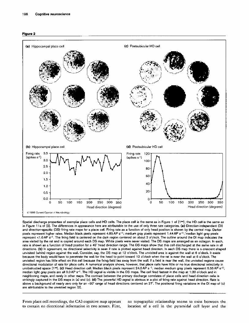
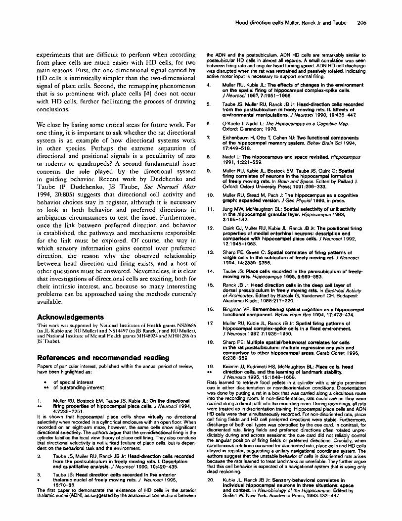
Figure 2
(a) Hippocampal place cell (c) Postsubicular HD cell
(b) Hippocampal place cell
Firing rate 3.5 (spikes 5-l) 3.0 -
(d) Postsubicular HD cell
Firing rate 120
(spikes s-1)
2.5 -
1.5 -
1.0 -
0.5 -
0.0 -r 1 I I I I I 0 50 100 150 200 250 300 350 0 50 100 150 200 250 300 350
Head direction (degrees) Head direction (degrees)
0 1996 Current Op~mon I” Neurobiology
Spatial discharge properties of exemplar place cells and HD cells. The place cell is the same as in Figure 1 of [l”]; the HD cell is the same as
in Figure 11 a of 121. The differences in appearance here are attributable to the use of only three rate categories. (a) Direction-independent (DI)
and direction-specific (DS) firing rate maps for a place cell. Firing rate as a function of only head position is shown by the central map. Darker
pixels represent higher rates. Median black pixels represent 4.85 AP s-t ; medium gray pixels represent 1.44 AP s-t ;‘median light gray pixels
represent <l .O AP s-1. The firing field is centered on the dark region centered on about 3 o’clock The outline around the DI map indicates the
area visited by the rat and is copied around each DS map. White pixels were never visited. The DS maps are arranged as an octagon. In each,
rate is shown as a function of head position for a 45’ head direction range. The DS maps show that this cell discharged at the same rate in all
directions. (b) In agreement, no directional selectivity is seen if rate is plotted against head direction. In each DS map there is a crescent-shaped
unvisited (white) region against the wall. Consider, say, the DS map at 12 o’clock. The unvisited area is against the wall at 6 o’clock. It exists
because the body would have to penetrate the wall for the head to point toward 12 o’clock when the rat is near the wall at 6 o’clock The
unvisited region has little effect on this cell because the firing field lies away from the wall. If a field is near the wall, the unvisited regions cause
directional modulation of rate for place cells. A numerical analysis shows, however, that place cells have little or no true directional selectivity in
unobstructed space [l”]. (c) Head direction cell. Median black pixels represent 24.0AP s-t; median medium gray pixels represent 5.55 AP s-t;
median light gray pixels are all O.OAP s-1. The HD signal is visible in the DS maps. The cell fired fastest in the map at 1:30 o’clock and in
neighboring maps, and rarely in other maps. The contrast between the primary discharge correlates of place cells and head direction cells is
strikingly captured in the DS maps in (a) and (c). (d) The powerful HD signal is obvious in a plot of firing rate against head direction. Rate is
above a background of nearly zero only for an -90’ range of head directions centered on 27’. The positional firing variations in the DI map of (c)
are attributable to the unvisited region [21.
From place cell recordings, the CA3 cognitive map appears no topographic relationship seems to exist between the to contain no directional information in two senses. First, location of a cell in the pyramidal cell layer and the

Head direction cells Muller, Ranck Jr and Taube 199
Table 1
Comparison of plaoe cell and head direction cell properties.
Hlppocampal place cell
Location CA1 and CA3 regions of Ammon’s Horn
Cell type Pyramidal cells of stratum pyramidale
Head direction cell
Postsubiculum and ADN
Probably pyramidal cells in postsubiculum Unknown in ADN
Primary discharge correlate
Secondary discharge correlate
Head position
None in open field: head direction on linear tracks
Head direction
None
Stability of discharge correlate in a fixed environment
Days or weeks Days or weeks
Number of peaks
Peak firing rate
Control of cue card in cylinder
Firing is stable in absence of cue card
A few place cells have two firing fields
2-60 spikes s-1 at center of firing field
Yes
Yes
Each HD cell has only one preferred direction
2-l 20 spikes s-t at preferred direction
Yes
Yes
Firing persists in the dark
Cells can be silent in some environments
Yes
Yes
Yes
No
Remapping in some environments
Temporal firing variance under fixed conditions
Yes
High
No
Low
Modulation of discharge by theta EEG pattern
Yes Weak if at all
location of the cell’s field in the environment (JL Kubie, RU Muller, ES Hawley, CP Jia, Sot Neurosci Abstr 1992, l&1062): that is to say, neighborliness is not preserved. Thus, a direction in the environment has no analog in the hippocampus. Second, in walled apparatuses with open floors, place cells are omnidirectional: their discharge rate is independent of the rat’s orientation [l”]. Although a topological map can be used to find the straight-line path from any point in the environment to any other point, additional information is necessary for the selected path to be followed in the environment. The difficulty is simply that the rat is not necessarily pointing in the required direction when the path is calculated. We propose, therefore, that a minimal role for the directional system and HD cells is to allow a computed path to be aligned with the environment. Simply put, the directional system allows the rat to turn such that it walks in the right direction to get to the goal or end point.
One mechanism that would allow alignment involves storing the direction from each firing field to other, nearby firing fields in a separate neural location from the distance information. The idea is that vectors between relatively
close pairs of points in the environment are stored. As stated already, the distance component is held as the resistance of the synapse that connects a pair of place cells. On the other hand, we propose that the direction component of the vector pointing from one field to another is stored so that a ‘direction registration cell’ for the two fields is activated when the relevant place cell pair is simultaneously active.
The binding between a pair of place cells and a direction registration cell could be achieved using Hebbian logic. Imagine that the rat goes from the region of one field to the region of a second. If the distance were small, the path would be linear and the head direction would, on average, be the same as the direction from one field to the other. During learning, the direction registration cell is driven by directional cells ‘tuned’ to the rat’s current heading, and the modifiable synapses from place cells to registration cells are strengthened. As a result, if the computed path is replayed while HD cell input is suppressed, the proper registration cell is activated for each step of the path. For any step on the computed path, registration cell activity would be compared with the current head direction; if
200 Cognitive neuroscience
the two were unequal, the rat would turn so as to reduce the discrepancy between head direction and required direction. In this way, the rat could progress from starting location to goal.
The proposed scheme is not unique and lacks empirical backing. It nevertheless provides a rationale for the existence of a directional system with properties like those discovered in the rat. To reiterate the theory, the directional system has the essential role of putting paths extracted from the map into register with the environment so that the map can correctly control locomotion.
Discovery of head direction cells The existence of place cells and the devastating effects of hippocampal lesions on spatial learning and performance naturally lead to examination of the issue of whether cells in anatomically related areas also have spatial firing correlates. This approach has proved very fruitful. Thus, spatially selective cells have been found in the dentate gyrus [ 111, medial entorhinal cortex [IZ], subiculum [13], and parasubiculum [14]. (See [9] for a review.) Nevertheless, so far, the most striking outcome of this survey has been the discovery of HD cells in deep layers of the postsubiculum [ 151.
By listening to amplified action potential recordings, certain features of HD cells are evident from direct observation of the rat’s behavior: for example, discharge is elevated above a low background rate over a rather narrow range of head directions centered on a ‘preferred direction’. Firing is correlated with head direction relative to the environment and not to neck angle: the rate is quite constant if the head points in a fixed direction regardless of how the body is disposed. If the body remains in a fixed position while the head is passively turned in the horizontal plane (yaw), firing varies strongly. With the head held so that the projection of the midline onto the horizontal plane is constant, firing does not vary as the nose is pointed up or down (pitch) by as much as -9o”, or if the head is tilted from side to side (roll) by a similar amount.
In short, directional cells seem to signal the direction of the rat’s head in the horizontal plane, to the virtual exclusion of other factors, including location in the environment and behavior. The signal is so strong that it must be concluded that at least this part of the hippocampal formation is certainly preferentially involved in spatial behavior [16].
Quantitative characterization of postsubicular head direction cell properties
The preferred direction for a given cell is the same everywhere within the cylinder. When the head points in a certain direction, a certain subset of directional cells is active; the degree of activity of each cell is determined by how close the head orientation is to the preferred direction. When the head orientation changes, so does the subset of active cells. The overall set of HD cells, therefore, acts as a compass that signals head orientation.
Taube eta/. [2,5] adapted the place cell methods of Muller One way in which the preferred direction could be and colleagues [4,17] to study directional cells. Hungry constant over the whole apparatus would be fore HD rats were trained to chase 25 mg food pellets scattered cells to signal angle relative to the earth’s magnetic field, into a 76cm diameter cylindrical enclosure at a rate of or relative to any other stimulus so distant that the -5 pelletsmin-1. The cylinder is gray except for a white value of the stimulus would not change with position. cue card taped to the wall that occupies a 100” arc. A This inference is disproved, however, by the finding
major innovation was the use of a two-spot tracking system to detect head direction as well as head position [‘2,5]. Additional information about the methods is given in Figure 1.
Overall the postsubicular HD cells were found to have the right properties to act as a compass. Under all known circumstances, each cell reliably signals orientation to an arbitrary reference direction such that the angle between preferred directions of cell pairs is fixed. The reference direction may shift relative to the laboratory frame, but other features of the system are invariant, including peak rate, background rate, and firing range; the same is true for anterior thalamic HD cells (see below) [3*].
Approximately 26% of postsubicular cells (61 out of 239) are classified as HD cells [Z]. (Sharp [18] has reported a somewhat higher value of 37%, i.e. 26 out of 71.) Of the 61 cells, 25 were formally studied. All preferred directions were found to occur with about the same frequency. There is no tendency for sequentially or simultaneously recorded cells to have similar preferred directions, implying the absence of topographic mapping of the postsubiculum onto direction in the environment. The major difference among directional cells is in peak firing rate, which varies from a few action potentials per second (AP s-1) to >lOOAP s-l. Both preferred direction and peak firing rate are stable over many days of recording.
The relationship between firing rate and head direction can be modeled as a triangular function for postsubicular HD cells (Figure 2b). Discharge decreases linearly for both clockwise and counterclockwise angular displacements away from the preferred direction. As the absolute values of the slopes of the lines are about equal, the relationship has the form of an isosceles triangle with the apex at the preferred direction and the ends of the legs near a rate of zero, 45” clockwise or counterclockwise from the preferred direction; the angular range of elevated firing is -90”. The background rate outside the range of elevated firing is generally <l.OAP s-1. The ratio of peak rate to background is very high.

Head direction cells Muller, Ranck Jr and Taube 201
that rotations of the white cue card (card rotations were performed after the rat was taken out of the cylinder) cause equal rotations of preferred directions. Directional cells are not, however, triggered by the card in a simple, sensory-like fashion, as preferred directions would then be expected to converge rather than to be parallel. Furthermore, removing the card does not disrupt directional firing, although preferred directions often rotate unpredictably [S].
Card rotation and card removal experiments reveal that the stimulus control exerted by the card is the same for HD cells and place cells [4]; card rotation causes equal rotations of firing fields, and card removal leaves fields intact except for rotation through an unpredictable angle. Results with separately recorded place and directional cells are corroborated by simultaneous recordings [19”], implying that the two spatial signals are generated in a unitary navigational system.
A fundamental difference between the positional and directional systems is found, however, by recording in a rectangular or square apparatus of similar appearance to the cylinder; the wall height and color are the same, the areas are about equal, and a white card covers about one-fourth of the perimeter. Changing the apparatus shape causes place cells to undergo remapping-they may cease firing or have a field in an unpredictable part of the apparatus [4]. Furthermore, the existence of overlapping fields for a pair of place cells in one apparatus does not predict that the fields overlap in a second apparatus [ZO]. In contrast to place cells, HD cells never go silent after changing apparatus shape. Although the preferred direction often rotates after apparatus shape is changed, the angle between the preferred directions of simultaneously recorded cells stays the same; all preferred directions rotate by the same amount [S].
Head direction cells in other brain areas Head direction cells with properties similar to those of postsubicular HD cells have been found in several brain areas. These include the anterior dorsal division of the anterior thalamic nuclei (ADN) [3*], the dorsal striatum ([Zl]; SJY Mizumori, BG Cooper, Sot Neurosci Abstr 1995, 21:1929), the lateral dorsal thalamic nuclei (LDN) [Z?], and posterior parietal and retrosplenial cortices [23’,24*]. Each area has important connections to the postsubiculum, the hippocampus, or other parts of the hippocampal formation. The two thalamic areas were investigated because they are reciprocally connected with the postsubiculum.
Head direction cells are not found everywhere. As noted, hippocampal place cell discharge is almost ideally independent of head direction in the cylinder. Cells in the medial entorhinal cortex that discharge as a function of position show little evidence of directional modulation in the cylinder [12]. Lavoie and Mizumori [ZS] have recorded
from ventral striatum under suitable conditions, but have reported no HD cells. It is useful to add that directional modulation of positional firing is seen in the subiculum [13] and parasubiculum [14], but no HD cells have been reported in these areas. Such modulation occurs despite the omnidirectional firing of place cells recorded under the same conditions. The origins of the modulation in the subiculum and parasubiculum will provide important clues about the flow of directional information in the hippocampal formation.
Of the HD cell types, the ADN cells most closely resemble those in the postsubiculum, although the fraction of directional cells in the ADN is higher (-56%). Firing as a function of head direction has an isosceles triangle shape, the background rate is extremely low and the range of elevated rates is -90”. Thus, the rate versus head direction functions of ADN HD cells are extremely similar to those of postsubicular HD cells. All preferred directions are observed with about equal frequency and the main difference among cells is in peak firing rate. Taube [3*] has reported that the average peak rates for postsubicular and ADN HD cells are about the same. Manipulations of the cue card and apparatus shape reveal that the stimulus control over preferred direction for ADN is indistinguishable from that for postsubicular cells.
In contrast to the results of Taube and colleagues [3*], Blair and Sharp [26*.] have found that the peak rate of ADN cells is on the average more than twice as fast as postsubicular cells. Other differences in the properties of HD cells as measured by Taube et a/. [2,3*] and by Blair and Sharp [26**] are the wider firing range and the more obvious rounded peaks and gradual approach to the flat baseline (seen by Blair and Sharp). This last property led Blair and Sharp [26**] to treat the directional firing profiles as Gaussian functions rather than triangles. (See also [27**].)
An important difference between ADN and postsubicular cells is that most ADN cells cease discharging when the rat is passively rotated in the experimenter’s hand through the preferred direction [3*]; this finding has been corroborated by Knierim eta/. [ 19”]. In contrast, postsubicular discharge is not disrupted by passive rotation, although most cells fire more slowly [S]. Recently, however, it was found that postsubicular cells may cease discharging during passive rotation when rats are more firmly restrained (JS Taube, DA Wolk, PA Dudchenko, SOL Neurosci Abstr 1994, 20:805). These findings suggest that HD cells in both postsubiculum and ADN require a volitional motor input to activate cell discharge. Similar results were previously reported for hippocampal place cells [28].
Another key difference between postsubicular and ADN HD cells was discovered by Blair and Sharp [26”]. By looking at directional firing as a function of head turning velocity, they showed that preferred directions
202 Cognitive neuroscience
for clockwise and counterclockwise turns are reliably different for ADN cells but are the same for postsubicular cells. Furthermore, spikes tended to fire before the head reached the turn-independent preferred direction. In other words, the spikes lead or anticipate head direction. By analyzing the separation between preferred direction during clockwise and counterclockwise turns as a function of angular velocity, Blair and Sharp [26**] concluded that spikes lead head direction by -37 ms for ADN cells, but only -2ms for postsubicular cells. Taube and Muller (JS Taube, RU Muller, Sot Neurosci Abstr 1995, 21:946) found virtually identical values using very different methods. These results suggest that ADN HD cells project to postsubicular HD cells and are important for the theoretical models of directional cells considered in more detail below.
Turning now to the HD cells in the LDN, these cells are clustered in the dorsal, caudal section of the nucleus; the fraction of directional cells is -51% [ZZ]. Again, no tendency for certain directions to be over- or under-represented is evident. The mean peak firing rate is lower than for postsubiculum or ADN, the background rate higher, and the directional firing range wider. It therefore appears that LDN cells confer less directional information than do postsubicular or ADN cells. The stated values for LDN cells, however, were measured as rats ran on an eight-arm maze, using a one-spot tracking system, so that head direction was presumed to be in the direction of locomotion. It would be valuable to record from LDN cells in a cylinder using a two-spot tracker, so that the properties of LDN cells can be compared with those in postsubiculum and ADN.
Mizumori and Williams [Z?] have tested LDN cells for alterations in specificity caused by turning room lights on and off. One interesting experiment involved turning the lights on for a set interval preceding each trial on an eight-arm maze. Specificity during the trial was low if the lighted time was 2s or lOs, but nearly equal to the specificity during lighted trials if the interval was 30s or 60s. With 60s of light exposure, the preferred direction was always the same as during lighted trials. A second manipulation involved watching the decay of specificity during successive trials in the dark, with variable amounts of light time being allowed to precede the sequence of dark trials. The authors conclude that the directional representation remains stable for 2-3min in the dark and then begins to rotate; some decline of the directional specificity also exists. The rotation of preferred direction in the dark is in line with the strong visual inputs to the LDN. The response of these cells to darkness is, however, different from ADN and postsubicular HD cells; the preferred directions and peak rate of such cells are stable for up to 16min in the dark (JP Goodridge, KA Worboys, P Dudchenko, JS Taube, Sot Neurosci Abstr 1995, 21:945). The firing fields of hippocampal place cells also persist in the dark for 8 min, the longest time tried by Quirk et a/.
[29]. Experiments in the dark thus provide an indication that the LDN directional system is decoupled from other limbic structures.
Other loci of directional cells include portions of posterior cortex (identified by Chen et al. [23’] as posterior parietal cortex), retrosplenial granular cortex (RSG) and retrosplenial agranular cortex (RSA). On an eight-arm maze, the fraction of directional cells in these areas is much lower than in postsubiculum, ADN or LDN; estimates are -8.5% for RSG and RSA, and -3% for posterior parietal cortex. Firing as a function of head direction resembles a sine function superimposed on a relatively high direction-independent level. The tuning is therefore quite broad and the background rate is rather high, suggesting weak directional information signaling.
Chen eta/. [24’] have assayed control of preferred direction using four cards, one white and three black, set around the eight-arm maze. Of the 41 directional cells, 24% were controlled by the location of the white card, whereas for 61%, the preferred direction remained fixed in the laboratory frame. The other 15% of the ceils underwent ‘complex changes’ in directional rate profile; these cells often developed a second peak of discharge. With no cue card (i.e. when all four cards were black), preferred directions generally remained fixed in the laboratory frame. Interestingly, out of 11 directional cells tested during passive rotations of a square platform placed on the center of the eight-arm maze, six never showed significant directional tuning and only two showed directional tuning at all times on the platform. On this basis, the posterior cortical cells do not closely resemble postsubicular or thalamic directional cells, all of which show directional tuning over a wide range of experimental situations.
The final known locus of directional ceils is the dorsal striatum [21]. Of 30 cells recorded during performance of a complicated task, three were directionally tuned. Data from one cell were presented in the paper; this cell appears to have a low background rate, but considerably broader tuning than postsubicular and ADN cells. Mizumori and Cooper (SJY Mizumori, BG Cooper, Sot Neurosci Abm 1995, 21:1929) have confirmed the existence of directional selectivity in the dorsal striatum, although it is not clear that the cells share the key property of having parallel preferred directions and equal peak rates everywhere.
We conclude this section by noting the extremely interesting results of Knierim et a/. [19**], who found conditions in which the preferred direction of ADN HD cells drifted as a function of recording time in a cylinder. They simultaneously recorded from hippocampal place cells and found that the angular location of firing fields drifted in parallel. This effect is predictable from the work of Muller and Kubie [4] combined with the work of Taube et a/. [S], who showed identical control by the white card on the stimulus wall. The experiments of Knierim et al.

Head direction ceils Muller, Ranck Jr and Taube 203
[lY*], however, provide a paradigm for determining which directional cells are coupled with others and with the positional system.
Absolute and relative control of preferred direction The current heading of the rat (and its current position) can be kept in register with the environment by using either absolute (externally obtained) or relative (dead reckoning) information. Absolute information depends on the location of environmental landmarks, whereas relative information depends on internal signals such as vestibular or kinesthetic inputs. Absolute information can be used to determine heading or position at any time in a familiar environment. In contrast, relative information requires that the heading or position be constantly calculated by integrating the effects of self-motion detected by internal (‘idiothetic’) sensory systems. A system that uses only idiothetic information must drift (see e.g. [30,31]). Two possibilities emerge, therefore, if the system reliably indicates heading or position. First, idiothetic information may play a central role as long as it is updated sufficiently often by reference to the environment. Alternatively, idiothetic information may not be used, or used only if no exteroceptive information is available.
Several studies have assessed the roles played by absolute and relative information. Goodridge and Taube [32”] have tested the effects of removing and replacing a cue card. After a first session with the card present, the card was removed with the rat out of the cylinder and a second session was run. If the preferred direction rotated by >30” in the absence of the card, the card was reintroduced at its original location with the rat present. In most cases, the preferred direction jumped immediately back to its original value, even though idiothetic information indicated the lack of a significant event, implying that visual information is prepotent in setting preferred direction.
Taube and Burton [33”] have monitored preferred direction as rats ran from a familiar cylinder to a novel box through a previously closed runway. On the first exposure to the runway and box, preferred direction did not change. Next, the cue card in the cylinder was rotated and the rat was replaced in the cylinder. The preferred direction rotated, but when the rat now entered the runway and box, the preferred direction suddenly shifted to its original value (relative to the laboratory frame). The inference is that the directional system can snap to a new state even when idiothetic information indicates constancy. These results are in agreement with the findings of Goodridge and Taube [32**].
Knierim et a/. [19”) disoriented rats by spinning them at low speeds. The rats were put into a cylinder with a cue card without seeing the surroundings. They found that these rats ignored the cue card in two senses. First,
when the card was rotated between sessions (in the rat’s absence), place cell firing fields and HD cell preferred directions did not follow the card position. Second, even more interestingly, the angular position of firing fields and the preferred directions of simultaneously recorded ADN HD cells drifted, almost always in register with each other. These results are expected if a dead-reckoning system operates without being updated according to absolute information. Knierim et a/. [ 19”] argued that disorientation training taught the rats to ignore the cue card and other, uncontrolled stimuli that are stationary in the laboratory frame.
Lesion experiments By selectively damaging parts of the brain, it is possible to learn several things about the navigational system. If a structure is part of the navigational system, it must be found that damaging the structure impairs spatial problem solving, at least transiently. Lesions can also help in tracing information flow in the directional and positional systems and in understanding the interactions and overlaps of the two systems.
The first category includes experiments on the effects of postsubicular or ADN lesions [34,3.5] and temporary inactivation of the LDN [36]. These studies demonstrated impaired performance on either the eight-arm radial or Morris water mazes. Mizumori et a/. [36] found that LDN inactivation lowers the positional selectivity of hippocampal place cells. Dudchenko et al. (P Dudchenko, JP Goodridge, JS Taube, Sot Neurosci Abstr 1995, 21:945) looked at the effects of postsubicular lesions on hippocam- pal place cells and found that firing fields still occur, although they are not as robust as in normal rats. This result reinforces the notion that the positional system is independent of the directional system, as initially suggested by the purity of the place cell and HD cell signals in open fields [1**,2].
A complementary study by Golob and Taube (EJ Golob, JS Taube, SOC Neurosci A&- 1995, 21:945) shows that HD cell activity is still present in both the postsubiculum and ADN after massive neurotoxic lesions of the hippocampus, again suggesting that the directional and positional systems are quite independent. Overall, these lesion results are compatible with the idea that a minimal role for the directional system is to put a directionless map into register with the environment. It is also interesting that the preferred direction for individual HD cells remains stable from day to day in a familiar apparatus and is also stable across days in a new apparatus (EJ Golob, JS Taube, Sot Neurosci Abstr 1995, 21:945). The implication is that the hippocampus is not necessary for at least one kind of spatial information acquisition.
Other lesion studies have revealed aspects of the func- tional connectivity among regions with HD cells. For example, after ADN lesions, no directional cells are
204 Cognitive neuroscience
found in the postsubiculum (JP Goodridge, JS Taube, Sot Ncurosti Abstr 1993, 19:796). In contrast, postsubicular directional cells were seen after LDN lesions, although the fraction of such cells was smaller (-13%) than in normal rats (-26%) (EJ Golob, JS Taube, Sot Neurosci Abstr 1994, 20805). Together, these results suggest a closer coupling of postsubiculum to ADN than to LDN. Interestingly, lesions of the postsubiculum do not eradicate directional cell activity in the ADN (JP Goodridge, JS Taube, Sot Neumsci Abstr 1994, 20:805), which suggests that the fundamental directional signal arises in the ADN. Alternatively, directional cells may exist in regions outside the postsubiculum that are connected to the ADN; candidates include the retrosplenial cortex, the mammillary nuclei, and the presubiculum. Indeed, the postsubiculum is not recognized as a separate structure by all anatomists, and is sometimes considered to be the dorsal portion of the presubiculum [37].
Given the controversy over whether the mapping func- tions of the hippocampus are fundamental or a reflection of more general processing, lesion experiments can be designed to provide additional insight. The idea is that lesions of either the hippocampus or regions that have pure directional signals should interfere with navigational problem solving. In contrast, because the directional signal seems to be meaningful only with regard to navigation, there should exist hippocampally-dependent, non-spatial problems whose solutions are unaffected by lesions in postsubiculum or ADN.
Theories of the origins of directional cell properties Several recent papers have modeled individual HD cells and shown how such cells might be combined to form a compass-like directional system. Skaggs et al. [38”] have postulated that directional cells are coupled to each other such that cells with similar preferred directions are mutually excitatory, and cells with different preferred directions are mutually inhibitory. This arrangement ensures that only one set of cells is active at a given time, so that a unique head direction is signaled at all times. In dynamics terminology, the ring of HD cells is a set of stable attractors.
In the model, two rings of ‘rotation’ cells receive vestibular input indicating turning, one during clockwise, the other during counterclockwise head rotations. Each ring also receives input from the corresponding part of the HD cell ring, giving rise to directionally selective clockwise and counterclockwise rotation cells. The output of rotation cells drives HD cells whose preferred direction will be reached as a result of the ongoing head rotation. In this way, the stable state moves with the actual head direction. The system is put into register with the environment by the activity of visual cells that make Hebbian synapses with the HD cells. The visual cells are presumed to be triggered by specific stimuli. Because
the visual cells are active only when the head points in a certain direction, synapses onto certain HD cells will strengthen, and the direction cell ring will be anchored to the environment. This scheme has many interesting features, but cannot explain the fact that the anchoring stimulus can be removed without affecting the reliability of directional cell firing [S]. Also, preferred directions do not drift after 16min in the dark, suggesting that non-visual exteroceptive information can update idiothetic information (JP Goodridge, KA Worboys, P Dudchenko, JS Taube, Sot Neurosci Abstr 1995, 21:945).
Another dynamical model by Zhang [27”] is an elegant modification and elaboration of that of Skaggs et a/. [38**]. Progressing from the directional tuning curves of indi- vidual cells in the ADN and postsubiculum, the author shows precisely why the system is stable in the absence of perturbing input and how shifts from one state of the system to another occur. A key feature of this exposition is that the observed properties of directional cells impose strong constraints on signals in the model. For example, the profile of active cells shifts smoothly only if the signal sent to cells around the ring in the direction of head movement is the derivative of the profile of activities in the active cells. The mode1 also takes into account the results on anticipatory firing by Blair and Sharp [26**] and has many other interesting features.
A third computational model [39*] uses postsubicular and ADN HD cells plus postsubicular angular velocity cells [18]. The model predicts ‘angular speed’ cells in the mammillary nuclei whose firing is maximal when the head is still and decreases with head rotations in either direction. Finally, HD cells whose activity is modulated by angular velocity are assumed to exist in the reticular nucleus of the thalamus. The basic architecture is a ring of HD cells in the ADN whose stability is ensured by mutual excitation of cells with similar preferred directions. Shifts of head direction lead to shifts of the activity profile in the ring via a plausible mechanism involving the other cell classes. The mode1 predicts that ADN HD cell discharge requires a signal from the postsubiculum, but this is at odds with experimental data, as postsubicular lesions do not appear to disrupt the ADN directional signal (JP Goodridge, JS Taube, Sot Neurosci Abstr 1994, 20:805).
Together, the theoretical models comprise an excellent start to understanding the workings of the directional system. In addition, Zhang [27”] and McNaughton et a/. [40*] argue that dynamic theories can be extended to two dimensions, suggesting that updating position in the hippocampus could use mechanisms similar to those proposed for the directional system.
Conclusions Research into HD cells is in its infancy. Nevertheless, it is clear that the HD cells provide a wonderful window into a key component of the navigational system of rats. Many
Head direction cells Muller, Ranck Jr and Taube 205
experiments that are difficult to perform when recording from place cells are much easier with HD cells, for two main reasons. First, the one-dimensional signal carried by HD cells is intrinsically simpler than the two-dimensional signal of place cells. Second, the remapping phenomenon that is so prominent with place cells [4] does not occur with HD cells, further facilitating the process of drawing conclusions.
We close by listing some critical areas for future work. For one thing, it is important to ask whether the rat directional system is an example of how directional systems work in other species. Perhaps the extreme separation of directional and positional signals is a peculiarity of rats or rodents or quadrupeds? A second fundamental issue concerns the role played by the directional system in guiding behavior. Recent work by Dudchenko and Taube (P Dudchenko, JS Taube, SOC Neul-osci Abstr 1994, 202305) suggests that directional cell activity and behavior choices stay in register, although it is necessary to look at both behavior and preferred directions in ambiguous circumstances to test the issue. Furthermore, once the link between preferred direction and behavior is established, the pathways and mechanisms responsible for the link must be explored. Of course, the way in which sensory information gains control over preferred direction, the reason why the observed relationship between head direction and firing exists, and a host of other questions must be answered. Nevertheless, it is clear that investigations of directional cells are exciting, both for their intrinsic interest, and because so many interesting problems can be approached using the methods currently available.
Acknowledgements This work was supported by National Institutes of Health grants NS20686 (to JL Kubie and RU Muller) and NS14497 (to JB Ranck Jr and RU Muller), and National Institute of Mental Health grants MH48924 and MH01286 (to JS Taube).
References and recommended reading Papers of particular interest, published within the annual period of review, have been highlighted as:
. of special interest l * of outstanding interest
1. . .
Muller RU, Bostock EM, Taube JS, Kubie JL: On the directional firing properties of hippocampal place cells. I Neurosci 1994, 4~7235-7251.
It is shown that hip-pal place cells show virtually no directional selectivity when recorded in a cylindrical enclosure with an open floor. When recorded on an eight-arm maze, however, the same cells show significant directional selectivitv. The authors aroue that the omnidirectional firina in the cylinder falsifies the’locai view theoryof place cell firing. They also c&clude that directional selectivity is not a fixed feature of place cells, but is depen- dent on the behavioral task and the environment.
2. Taube JS, Muller RU, Ranck JB Jr: Head-direction cells recorded from the postsubiculum in freely moving rats. I. Description and quantitative analysis. J Neurosci 1 QQO, 10:420-435.
3. Taube JS: Heed direction cells recorded in the anterior . thalamic nuclei of freely moving rats. J Neurosci 1995,
15:70-86. The first paper to demonstrate the existence of HD cells in the anterior thslamic nuclei (ADN), as suggested by the anatomical connections between
the ADN and the postsubiculum. ADN HD cells are remarkably similar to postsubicular HD cells in almost all regards. A small correlation was seen between firing rate and angular head turning speed. ADN HD cell discharge was disrupted when the rat was restrained and passively rotated, indicating active motor input is necessary to support normal firing.
4.
5.
6.
z
8.
9.
10.
11.
12.
13.
14.
15.
16.
1 7.
18.
19. . .
Muller RU. Kubie JL: The effects of changes In the environment on the s&tie1 firing of hippocampal complex-spike cells. J Neurosci 1987, 7:1951-l 968.
Taube JS, Muller RU, Ranck JB Jr: Head-direction cells recorded from the postsubiculum in freely moving rats. II. Effects of environmental manipulations J Neurosci 1990, 10:436-447.
O’Keefe J, Nadel L: The Hippocampus as a Cognitive Map. Oxford: Clarendon; 1978.
Eichenbaum H, Otto T, Cohen NJ: Two functional components of the hippocampal memory system. Behav Brain Sci 1994, 171449-518.
Nadel L: The hippocampus and space revisited. Hippocampus 1991, 1:221-229.
Muller RU, Kubie JL, Bostock EM, Taube JS, Quirk G: Spatial firing correlates of neurons In the hippocampal formation of freely movlng rats. In Brain and Space. Edited by Paillard J. Oxford: Oxford University Press; 1991:296-333.
Muller RU, Stead M, Path J: The hippocampus as e cognitive graph: expanded version. J Gen PhysiollQQ6, in press.
Jung MW, McNaughton BL: Spatial selectivity of unit activity In the hlppocampal granular layer. Hippocampus 1993, 3:165-182.
Quirk GJ, Muller RU, Kubie JL, Ranck JB Jr: The positional firing properties of medial entorhinal neurons: description and comparison with hippocampal place ceils. J Newosci 1992, 12:1945-l 963.
Sharp PE, Green C: Spatial correlates of firing patterns of single cells in the subiculum of freely moving rat J Neurosci 1994,14:2339-2356.
Taube JS: Place cells recorded in the parasubiculum of freely- moving rats. Hippocampus 1995, 5569-583.
Ranck JB Jr: Head direction cells in the deep cell layer of dorsal presublculum in freely moving rats. In Electrical Activity of Archicortex. Edited by Buzsaki G, Vandenvolf CH. Budapest: Akademai Kiado; 1985?Zl7-220.
Bingman VP: Remembering spatial cognition as a hippocampal functional component Bebav Brain Res 1994, 17:473-474.
Muller RU, Kubie JL, Ranck JB Jr: Spatial firing patterns of hlppocampal complex-spike cells In a fixed environment J Neurosci 1987, 7:1935-l 950.
Sharp PE: Multiple spatial/behavioral correlates for cells in the rat postsubiculum: multlple regression analysis end comparison to other hippocampal areas. Cereb Cortex 1996, 6:236-259.
Knierim JJ, Kudrimoti HS, McNaughton BL: Place cells, head direction cells, and the learning of landmark stability. J Neurosci 1995, 151646-l 659.
f&s learned to retrieve rood pellets In a cylinder with a single promcnent cue in either disorientation or non-disorientation conditions. Disorientation was done by putting a rat in a box that was carried along a circuitous route into the recording room. In non-disorientation, rats could see as they were carried along a direct path into the recording room. During recordings, all rats were treated as in disorientation training. Hip-pal place cells and ADN HD cells were then simultaneously recorded. For non-disoriented rats, place cell firing fields and HD cell preferred directions were stable. Furthermore, discharge of both cell types was controlled by the cue card. In contrast, for disoriented rats, firing fields and preferred directions often rotated unpre- dictably during and across sessions; the cue card did not reliably control the angular position of firing fields or preferred directions. Crucially, when spontaneous rotations occurred for disoriented rats, place cells and HD cells stayed in register, suggesting a unitary navigational coordinate system. The authors suggest that the unstable behavior of cells in disoriented rats arises because the rats learned to treat landmarks as unreliable. They further argue that this cell behavior is expected of a navigational system that is using only dead reckoning.
20. Kubie JL, Ranck JB Jr: Sensory-behavioral correlates in individual hippocampal neurons in three situations: space and context In Neurobiology of the Hippocampus. Edited by Seifert W. New York: Academic Press; 1903~433-447.
206 Cognitive neuroscience
21. Wiener SI: SDatial and behavioral correlates of striate1 neurons in rats perfoimlng a self-initiated navigation task. J Neurosci 1993,13:3802-3817.
22. Mizumori SJY, Williams JD: Directionally seledive mnemonic properties of neurons In the lateral dorsal nucleus of the thalamus of rats. J Neurosci 1993, 13:4015-4028.
23. Chen LL, Lin LH, Green El, Barnes CA, McNaughton BL: . Head-dlrectlon cells in the rat posterior cortex. I. Anatomical
distribution and behavioral modulation. Ewr, Brain Res 1994. 101:6-23.
Rats wre trained on a spatial working memory task on a radial arm maze in a cue-controlled room. A small number of HD cells were identified in the retrosplenial and medial prestriate areas. The discharge of some of these HD cells was modulated by the rat’s behavior; in particular, cell dis- charge was most affected by angular head turns of the rat. The directional signals carried by cells in posterior cortex appear to be much weaker than the signals in postsubiculum or anterior thalamic nuclei.
24. Chen LL, Lin LH, Barnes CA, McNaughton BL: Head direction . cells in the rat posterior cortex. II. Contributions of visual and
idiothetic informatlon to the directional firing. Exp Brain Res 1994, 101:24-34.
Rats were trained on a spatial working memory task on a radial arm maze in a cue-controlled room or on a ‘lazy-Susan’ style platform. HD cells in the retrosplenial and medial prestriate cortices responded in different ways to rotation of salient cues. Some cells shifted their directional bias with rota- tion of the cues, some cells remained unchanged, and others shifted their preferred direction to unpredictable orientations. Most cells either had no directional selectivity on the platfon or lost it when the platform was rotated. In our opinion, the cells reported here and in a study by Chen et al. 123’1 have few of the properties that make HD cells in ADN and the postsubiculum excellent candidates for the substrate of a navigational directional system.
25. Lavoie AM, Mizumori SJY: Spetlal. movement-, and reward- sensltlve discharge by medial ventral striate1 neurons of rats. Brain Res 1994, 638:157-l 66.
26. . .
Blair HT, Sharp PE: Antlclpetory head direction signals In anterior thalamus: evidence for a thalamocortlcel circuit that integmtes angular head motion to compute head dire&ion. J Neurosci 1995, 15:6260-6270.
This paper reports a key difference between postsubicular and ADN HD cells. HD cell firing was compared when the rats head was turning clockwise or counterclockwise; cell activity was further correlated with the speed of turning. ADN HD cell discharge for clockwise and counterclockwise turns are brought into register when the discharge anticipates where the rat will point its head -4Oms in the future. In contrast, postsubicular HD cell discharge for turns in the two directions is in register when the discharge is associated with the current directional heading. It is also reported that the firing rate of HD cells is determined, in part, by the speed of the head turn.
27. Zhang K: Representation of spetlal orientation by the intrinsic . . dynamics of the head-dire&Ion cell ensemble: a theory.
J Neurosci 1996, 16:2112-2126. The author presents a fascinating dynamical theory of how head direction and changes in head direction could be signaled by HD cells in postsubicu- lum and ADN. In terms of precedence, it is clear that this theory is an elabo- ration of that of Skaggs eta/. [36”], but the mathematical rigor distinguishes this work. In our opinion, the novel ideas raised raised by the author as well as the clarity and completeness of the dynamical exposition make this perhaps the best paper reviewed. Highly recommended reading.
26. Foster T, Castro CA, McNaughton BL: Spatial selectivity of rat hlppocampel neurons: dependence on preperedness for movement Science 1969,244:1560-l 562.
29. &irk GJ, Muller RU, Kubie JL: The tiring of hippocampal place cells in the dark refleds the rat’s recent experience. J Neurosci 1990, 10:2008-201 z
30. Gallistel CR: The Organization of Learning. Cambridge, Massachusetts: MlT Press; 1990.
31. McNaughton BL, Chen LL, Markus EJ: ‘Dead reckoning’, landmark learning, and the sense of direction: a neurophyslologl~l and computational hypothesis. J Cogn Neurosci 1991, 3:190-202.
32. Goodridge JP, Taube JS: Preferential use of the landmark . . navlgetlonal system by head direction cells. Behav Neurosci
1995, 109:49-61. HD cells in the ADN and postsubiculum were recorded in a cylinder with a single cue on the wall. Rats were then removed from the apparatus, the floor
paper was changed, and the cue removed. Next, rats were reintroduced into the cylinder and a second recording session was done. If the cell’s preferred direction shifted by >30’ from its initial orientation, the cue was replaced at its original position in full view of the rat. lf the cell’s preferred direction did not shift, the cue card was replaced to a 90’ rotated position, again in full view of the rat. Under both conditions, the preferred direction shifted back to its original orientation with respect to the cue card. The authors concluded that stable visual landmarks preferentially exert more control over HD cell discharge than idiothetic cues derived from the rat’s own movemints.
33. Taube JS, Burton HL: Head dire&on cell activity monitored . . in a novel environment and during a cue conflict situation.
J Neurophysioll995, 74:1953-l 971. Postsubicular and ADN HD cells were recorded in two conditions. In the first, rats went from a familiar cylinder to a novel rectangle through a U-shaped alley; a door initially kept the rat in the cylinder. HD cells maintained their preferred direction as they went into the new alley and rectangle; the con- sistency is attributable perhaps to the use of idiothetic sensory information (vestibular, proprioceptive, motor efference copy). In the second condition, a salient cue in the cylinder was rotated 90’ and the rat was reintroduced into the cylinder. lf the preferred direction of the HD cell rotated to an extent similar to card rotation, the door was opened and the rat was allowed back into the alley and rectangle. The preferred direction of HD cells immediately shifted back to its original value when the rat entered the alley. When the rat returned to the rotated cylinder, the preferred directions either: shifted back to the direction first observed with the rotated card; remained aligned with their orientation in the rectangle; or shifted to a value in betwean the values seen in the rectangle and the cylinder with the cue rotated. These results imply that visual cues can override idiothetic information that asserts that the world is stable.
34.
35.
36.
37.
36. . .
Sutherland RI, Rodriguez AJ: The role of the fornix/fimbria and some related subcorticel structures In place learning and memory. Behav Brain Res 1989, 32~265-277.
Taube JS, Kesslak JP, Cotman CW: Lesions of the rat postsublculum impair performance on spatial tasks. Behav Neural Bioll992,57:131-143.
Mizumori SJY, Miya DY, Ward KE: Reversible inactivation of the lateral dorsal thalamus disrupts hippocempal place representation and Impairs spetlal learnlng. Brsin Res 1994, 644:168-i 74.
Amaral DG, Witter MP: Hippocempal formation. In The Rat Nervous System, edn 2. Edited by Paxinos G. San Diego: Academic Press; 1995:443-493.
Skaggs WE, Knierim JJ, Kudrimoti HS, McNaughton BL: A model of the neural basis of the rat’s sense of direction. In Advances in Neural Information Processing Systems, vol 7. Edited by Tesauro G, Touretzky D, Leen T. Cambridge, Massachusetts: MIT Press; 1995:173-180.
A dynamical model for HD cell firing is proposed. The particular set of direc- tional cells that is firing is stable in the absence of perturbing input because of hypothesized connections that are excitatory between cells with similar preferred directions and inhibitory between cells with quite different pre- ferred directions. The authors proposed mechanisms for fixing the alignment of the ring of directional cells with the environment and updating the current heading with reference to environmental landmarks.
39. Blair HT: A thalamocortical circuit for computing directional . headlng in the rat In Advances in Neural information Processing
Systems, ~018. Edited by Touretzky DS, Mozer MC, Hasaelmo ME. Cambridge, Massachusetts: MIT Press; 1996:in press.
This article outlines a neural model for how a head directional system is able to keep continuous track of the rats heading at it moves about the environment. Experimental findings on postsubicular and anterior thalamic HD cells and on angular velocity cells in postsubiculum are incorporated. The model predicts the existence of several other neuronal types in other brain structures whose activity is correlated with aspects of the directional signal.
40. McNaughton BL, Barnes CA, Gerrard JL, Gothard K, Jung MW, . Knierim JJ, Kudrimoti H, Qin Y, Skaggs WE, Suster M, Weaver KL:
Deciphering the hippocampel polyglot: the hippocampus as a path integmtion system. J Exp Biol 1996, 199:173-l 85.
A model for how a path integration system might operate at the neuronal level. It is hypothesized that the hippocampus is crucial for determining and selecting the appropriate reference frame used by the rat. The one- dimensional dynamic system proposed for stable HD cell activity by Skaggs et al. [38”] is expanded to two dimensions for place cells to show how trajectories might be imparted with a property similar to inertia.