head imaging guidelines - medicare.healthalliance.org · o intra-cranial pre-operative planning if...
TRANSCRIPT

©2016 eviCore healthcare Head Imaging Guidelines
HEAD IMAGING GUIDELINES Version 18.0; Effective 03-18-2016
This version incorporates accepted revisions prior to 12/31/15
CPT® (Current Procedural Terminology) is a registered trademark of the American Medical Association (AMA). CPT® five digit codes, nomenclature and other data are copyright 2016 American Medical Association. All Rights Reserved. No fee schedules, basic units, relative values or related listings are included in the CPT® book. AMA does not directly or indirectly practice medicine or dispense medical services. AMA assumes no liability for the data contained herein or not contained herein.
eviCore healthcare Clinical Decision Support Tool Diagnostic Strategies: This tool addresses common symptoms and symptom complexes. Imaging requests for patients with atypical symptoms or clinical presentations that are not specifically addressed will require physician review. Consultation with the referring physician, specialist and/or patient’s Primary Care Physician (PCP) may provide additional insight.

HEAD IMAGING GUIDELINES
Head Imaging Guidelines Abbreviations 3 HD-1~General Guidelines 4 HD-2~Taste and Smell Disorders 8 HD-3~Ataxia 9 HD-4~Behavior Disorders 9 HD-5~Chiari and Skull Base Malformations 10 HD-6~Facial Palsy (Bell’s Palsy) 10 HD-7~Recurrent Laryngeal Palsy 11 HD-8~Dementia 12 HD-9~Epilepsy/Seizures 14 HD-10~Facial Pain – Trigeminal Neuralgia 15 HD-11~Headache 16 HD-12~ Aneurysm and AVM 21 HD-13~Head Trauma 24 HD-14~CNS Infection 25 HD-15~Movement Disorders 26 HD-16~Multiple Sclerosis (MS) and Related Conditions 27 HD-17~Papilledema/Pseudotumor Cerebri 28 HD-18~Paresthesias 29 HD-19~Pituitary 30 HD-20~Scalp and Skull Lesions 32 HD-21~ Stroke – TIA 33 HD-22~Cerebral Vasculitis 35 HD-23~Dizziness, Vertigo and Syncope 36 HD-24~Other Imaging Techniques 38 HD-25~Epistaxis 40 HD-26~Mastoid Disease 40 HD-27~Hearing Loss 41 HD-28~Ear Pain (Otalgia) 42 HD-29~Sinusitis 43 HD-30~TMJ and Dental/Peridontal/Maxillofacial Imaging 45 HD-31~Tinnitus 46 HD-32~Eye Disorders 48 HD-33~Acoustic Neuroma & Other Cerebellopontine Angle Tumors 49 HD-34~Pineal Cysts 49 HD-35~Arachnoid Cysts 50 HD-36~Nuclear Medicine 50
V.18.0; Effective 3/18/2016 –Head Imaging
Page 2 of 51

ABBREVIATIONS for HEAD IMAGING GUIDELINES
ACTH adrenocorticotropic hormone AD Alzheimer’s Disease
ADH antidiuretic hormone AION arteritic ischemic optic neuritis AVM arteriovenous malformation CBCT Cone-beam computerized tomography CMV cytomegalovirus CSF cerebrospinal fluid CT computed tomography
CTA computed tomography angiography DNA deoxyribonucleic acid DWI diffusion weighted imaging (for MRI) EEG electroencephalogram ENT Ear, Nose, Throat ESR erythrocyte sedimentation rate FDG fluorodeoxyglucose FSH follicle-stimulating hormone FTD Frontotemporal Dementia GCA giant cell arteritis GCS Glasgow Coma Scale HIV human immunodeficiency virus LH luteinizing hormone
MMSE mini mental status examination MRA magnetic resonance angiography MRI magnetic resonance imaging MRN magnetic resonance neurography MS multiple sclerosis MSI magnetic source imaging
NAION non-arteritic ischemic optic neuritis NPH normal pressure hydrocephalus PET positron emission tomography PML progressive multifocal leukoencephalopathy PNET primitive neuro ectodermal tumor PWI perfusion weighted imaging (for MRI) SAH subarachnoid hemorrhage
SIADH Syndrome of Inappropriate Antidiuretic Hormone Secretion SLE systemic lupus erythematosus TIA transient ischemic attack TMJ temporomandibular joint disease TSH thyroid-stimulating hormone VBI vertebrobasilar VP ventriculoperitoneal
XRT radiation therapy
V.18.0; Effective 3/18/2016 –Head Imaging
Page 3 of 51
HEAD IMAGING GUIDELINES
HD-1~GENERAL GUIDELINES
HD-1~General Guidelines HD-1.1 Anatomic Issues 5
HD-1.2 Modality 6
HD-1.3 MRI head 6
HD-1.4 CT head 6
HD-1.5 CT and MR Angiography: (CTA and MRA) 7
HD-1.6 Coding Notes 7
HD-1.7 Other Imaging Situations 7
V.18.0; Effective 3/18/2016 –Head Imaging
Page 4 of 51
HEAD IMAGING GUIDELINES
HD-1~GENERAL GUIDELINES
A current clinical evaluation (within 60 days) is required before advanced imaging can be considered (exceptions allowed for scheduled surveillance evaluation of known abnormalities such as follow up for tumors or hydrocephalus). o The clinical evaluation should include a relevant history and physical examination,
including a neurological examination, as well as appropriate laboratory studies and non-advanced imaging modalities.
o Other meaningful contact (telephone call, electronic mail or messaging) with an established patient can substitute for a face-to-face clinical evaluation.
HD-1.1 General Guidelines - Anatomic Issues If two studies using the same modality both cover the anatomic region of clinical
interest, only one is generally needed, with the exception of the following scenarios: o Maxillofacial CT (CPT® code set: 70486-70488) or orbital/temporal bone CT
(CPT® code set: 70480-70482): both cover the structures of the orbits, sinuses, and face. Two separate imaging studies are only supported if there is suspicion of simultaneous involvement of more posterior lesions, especially of the region involving the middle or inner ear.
o Pituitary Gland: one study (either MRI head [CPT®70553] or MRI Orbit, Face, Neck [CPT®70543]) is adequate to report the imaging of the pituitary. If a previous routine MRI head was reported to show a possible pituitary tumor, a repeat MRI with dedicated pituitary protocol may be performed.
o Internal Auditory Canal (IAC) MRI can be reported as a limited study with one code from the set (CPT®70540-CPT®70543), but should not be used in conjunction with MRI head codes (CPT®70551-70553) if IAC views are performed as part of the brain.
o Mandible (jaw): maxillofacial CT (CPT® code set: 70486, 70487, 70488) or neck CT (CPT® code set: 70490, 70491, 70492) can be used to report imaging of the mandible. Neck CT will also image the submandibular space. If MRI is indicated, MRI of orbit, face, neck (CPT®70540, CPT®70542, or
CPT®70543) can be used to report imaging of the mandible and submandibular space.
MRI of the temporomandibular joint(s) (TMJ) is reported as CPT®70336. This code is inherently bilateral and should not be reported twice on the same date of service.
V.18.0; Effective 3/18/2016 –Head Imaging
Page 5 of 51
HD-1.2 General Guidelines - Modality MRI is preferable to CT for most indications. For exceptions, see HD 1.4: General
Guidelines – CT Head. MRI may be performed for these indications following an initial CT:
o MRI head without and with contrast (CPT®70553) may be performed to follow-up abnormalities seen on CT head without contrast (CPT®70450) when a mass, lesion, or infection is found
o MRI head without contrast (CPT®70551) or MRI head without and with contrast (CPT®70553) may be performed to follow-up abnormalities seen on CT head without contrast (CPT®70450) when there is suspected Multiple Sclerosis or other demyelinating disease
o MRI head without (CPT®70551) or MRI head without and with contrast (CPT®70553) may be performed to follow up on stroke or TIA when initial CT head was done on emergent basis.
o MRI head without and with contrast (CPT®70553) for evaluation of new onset seizures
HD-1.3 General Guidelines –MRI head MRI, with contrast, (CPT®70552) should not be ordered except to follow-up on a very
recent noncontrast study (within two weeks).
HD-1.4 General Guidelines –CT Head Scenarios in which MRI is contraindicated (i.e. pacemakers, ICDs, cochlear implants,
aneurysm clips, orbital metallic fragments etc…) Head CT without contrast (CPT®70450) in nearly all cases, to show:
o Mass effect o Blood/blood products o Urgent/emergent settings due to availability and speed of CT o Trauma o Recent hemorrhage, whether traumatic or spontaneous o Bony structures of the head evaluations o Hydrocephalus evaluation and follow-up (some centers use limited fast MRI to
minimize radiation exposure in children - these requests may be approved) o Prior to lumbar puncture in patients with cranial complaints (without contrast
(CPT®70450)
V.18.0; Effective 3/18/2016 –Head Imaging
Page 6 of 51
HD-1.5 General Guidelines - CT and MR Angiography: (CTA and MRA) Head MRA (CPT®70544) is generally done without contrast. MRA Neck may be done either without or with contrast for most indications,
depending on facility preference and protocols, and type of scanner. MRA Neck both without and with contrast is reserved for evaluation of possible or
known arterial dissection Head or CTA may be considered with suspected intracranial vascular disease, for
example: o recurrent stroke or TIA who have failed maximum medical management and are
candidates for intervention with invasive procedures o trigeminal neuralgia failed medical therapy o cerebral sinus thrombosis suspected with increased intracranial pressure (refractory
headaches, papilledema, diagnosis of pseudotumor cerebri) o aneurysm suspected with acute “thunderclap” headache syndrome and appropriate
screening or evaluation of known subarachnoid hemorrhage o intra-cranial pre-operative planning if there is concern of possible vascular
involvement or risk for vascular complication from procedure o sickle cell anemia o suspicion of vasculitis based on supporting clinical evidence o NOTE: Evaluation of posterior circulation disease requires both neck and head
MRA/CTA to visualize the entire vertebral- basilar system. o CTA or MRA head without or without contrast and with contrast for follow up of
aneurysm clipping or coiling procedures (See HD 12.1)
CT and MR Venography (CTV and MRV) are reported with the same codes as the CTA/MRA counterpart: o If arterial and venous CT or MR studies are both performed in the same session,
only one CPT® code should be used to report both procedures. o MRA with and without contrast with venous sinus thrombosis to differentiate total
from subtotal occlusion.
HD-1.6 General Guidelines - Coding Notes Brain PET should be reported as metabolic brain PET (CPT®78608).
HD-1.7 General Guidelines - Other Imaging Situations Nausea and vomiting, persistent, unexplained and a negative GI evaluation: can
undergo MRI head without contrast (CPT®70551) (See also: AB-1.9 Special Considerations in the Abdomen Imaging Guidelines)
ECT treatment to screen for intracranial disease: can undergo either MRI head without contrast (CPT®70551) or head CT without contrast (CPT®70450).
V.18.0; Effective 3/18/2016 –Head Imaging
Page 7 of 51

Screening for metallic fragments before MRI should be done initially with plain x-ray. o The use of orbital CT to rule out orbital metallic fragments prior to MRI is rarely
necessary. o Plain x-rays are generally sufficient; X-ray detects fragments of 0.12 mm or more,
and CT detects those of 0.07 mm or more. Plain x-ray is generally sufficient to screen for aneurysm clips.
References 1. Yousem DM, Grossman RI. Neuroradiology, the requisites. 3rd Ed. Philadelphia, Mosby, 2010 2. Latchaw RE, Kucharczyk J, MoseleyME. Imaging of the Nervous System. Philadelphia, Elsevier,
2005 3. Rowland LP (Ed.). Merritt’s Neurology. 12th Ed. Philadelphia, Lippincott, 2010 4. Menkes JH, Sarnat HB, Maria BL. Child Neurology. 7th Ed. Philadelphia, Lippincott, 2006 5. http://www.acr.org/~/media/ACR/Documents/AppCriteria/Diagnostic/CerebrovascularDisease.pdf.
Accessed October 10, 2013.
HD-2~TASTE and SMELL DISORDERS
HD-2.1 Taste and Smell Disorders
MRI head without and with contrast (CPT®70553) or without contrast (CPT®70551) is considered with unexplained unilateral or bilateral anosmia (inability to perceive odor) or dysgeusia (loss of taste)1, 2.
If sinus or facial bone disorders is suspected, then consider initially Maxillofacial CT without contrast (CPT®70486)2
References 1. Wippold FJ II, Cornelius RS, Aiken AH, Angtuaco EJ, et al. ACR Appropriateness Criteria® cranial
neuropathy. American College of Radiology (ACR); 2012. 18 p 2. UpToDate, Evaluation and treatment of taste and smell disorders, Literature review current through:
Feb 2014.
V.18.0; Effective 3/18/2016 –Head Imaging
Page 8 of 51

HEAD IMAGING GUIDELINES
HD-3~ATAXIA
HD-3.1 Ataxia
MRI head without and with contrast (CPT®70553) or MRI head without contrast (CPT®70551) is considered in all patients with ataxia1:
o If it is progressive and/or not acute and suspect spinal disease can ADD MRI cervical, thoracic and/or lumbar spine without contrast1 (CPT®72141, CPT®72146, CPT®72148)
o If it is acute and stroke is suspected see HD-21~ Stroke – TIA o If MS is suspected, see HD-16-Multiple Sclerosis (MS) & Related
Conditions o If it is acute following head trauma, CT head without contrast (CPT®70450)
and/or CT temporal bone without contrast1 (CPT®70480) can be added
Reference 1. Broderick DF, Wippold FJ II, Cornelius RS, Aiken AH, et al. Expert Panel on Neurologic Imaging.
ACR Appropriateness Criteria® ataxia. American College of Radiology (ACR); 2012.
HD-4~BEHAVIORAL DISORDERS
Autism: See PACHD-17~Autism and Autism Spectrum Disorders
HD-4.1 Behavioral Disorders Neuroses and psychoses do not need advanced imaging, except:
Bipolar disorder, schizophrenia, and related disorders who fail to respond to treatment in the expected manner and who manifest features suggestive of an organic brain disorder o MRI head without contrast (CPT®70551), or o Head CT without contrast (CPT®70450)
References 1. Ropper AH and Brown RH. Adams and Victor’s Principles of Neurology. 8th Ed. New York,
McGraw-Hill, 2005, pp.1285-1332 2. Rowland LP (Ed.). Merritt’s Neurology.12th Ed. Philadelphia, Lippincott, 2010, pp.1053-1075
V.18.0; Effective 3/18/2016 –Head Imaging
Page 9 of 51
HEAD IMAGING GUIDELINES
HD-5~CHIARI and SKULL-BASE MALFORMATION
See Pediatric Head Guidelines, PEDHD 9 Chiari and Skull Base Malformations
CRANIAL NERVE (CN) PROBLEMS
HD-6~FACIAL PALSY (Bell’s Palsy)
HD-6.1 Facial Palsy
MRI brain without and with contrast (CPT®70553) or MRI head without contrast (CPT®70551) is considered with unexplained facial paresis/paralysis in clinical scenarios with1,2: o Trauma to the temporal bone2 o History of tumor2 o No improvement in 8 weeks1 o No full recovery in 3 months2 o Worsening paresis/paralysis2 o Atypical or Inconsistent features2 including:
o Second paralysis on the same side2 o Paralysis of isolated branches of the facial nerve2 o Paralysis associated with other cranial nerves2
MRI head without and with contrast (CPT® 70553) may be considered for suspected neurosarcoid/sarcoid
References 1. Reginald Baugh et al. Clinical Practice Guidelines Summary: Belly’s Palsy. Otolaryngol Head Neck
Surg, 2013; 149: S1-S27 2. Wippold FJ II, Cornelius RS, Aiken AH, Angtuaco EJ, et al. ACR Appropriateness Criteria® cranial
neuropathy. ACR Appropriateness Criteria, Cranial Neuropathy 2012. 3. Iannuzzi MC, Rybicki BA, Teirstein AS. Sarcoidosis. N Engl J Med. 2007 Nov 22;357(21):2153-65, 4. Joseph FG, Scolding NJ. Sarcoidosis of the nervous system. Pract Neurol. 2007 Aug;7(4):234-44 5. Gullapalli D, Phillips LH 2nd. Neurosarcoidosis. Curr Neurol Neurosci Rep. 2004 Nov;4(6):441-7
V.18.0; Effective 3/18/2016 –Head Imaging
Page 10 of 51

CRANIAL NERVE (CN) PROBLEMS
HD-7~RECURRENT LARYNGEAL PALSY
HD-7.1 Recurrent Laryngeal Palsy The following can be considered with unilateral vocal cord/fold palsy identified by laryngoscopy:1
MRI head without and with contrast (CPT®70553) and/or MRI neck without and with contrast (CPT®70543); or
MRI head without contrast (CPT®70551) and/or MRI neck without contrast (CPT®70540); or
If MRI is not available, CT head without and with contrast (CPT®70470) and/or CT neck with contrast (CPT®70491) o Chest CT with contrast (CPT®71260) may be added with left vocal cord palsy1
Reference 1. Wippold FJ II, Cornelius RS, Aiken AH, Angtuaco EJ, Berger KL, Brown DC, Davis PC, Holloway
K, McConnell CT Jr, Mechtler LL, Nussenbaum B, Rosenow JM, Roth CJ, Seidenwurm DJ, Slavin K, Waxman AD, Expert Panel on Neurologic Imaging. ACR Appropriateness Criteria® cranial neuropathy. American College of Radiology (ACR); 2012
V.18.0; Effective 3/18/2016 –Head Imaging
Page 11 of 51
HEAD IMAGING GUIDELINES
HD-8~DEMENTIA
HD-8.1 Dementia
Neuropsychological testing can be performed when history and bedside mental status examination cannot provide a confident diagnosis.1,2 MRI head without contrast (CPT®70551) or MRI head without and with contrast (CPT®70553) or Head CT without contrast (CPT®70450) is considered after an initial clinical diagnosis of dementia3,4
HD-8.2 Dementia - PET
Send to MD review. FDG and Amyloid Brain PET (CPT®78608) imaging are considered experimental and investigational in the diagnosis of Alzheimer’s disease and in differentiating between Alzheimer’s disease and other neurodegenerative/neurologic disorders.3,4,5
Depending on individual health plan rules, PET brain may be approved to differentiate Alzheimer’s disease from Frontotemporal dementia (either behavioral or primary progressive aphasia sub-types) with appropriate documentation.
See: ONC-31~Medicare Coverage Policies for PET
Practice Notes
The clinical diagnosis of dementia can be established by history-taking from the patient and a knowledgeable informant1 as well as a “bedside” mental status examinations (such as the Mini Mental Status Exam, Montreal Cognitive Assessment, Memory Impairment Screen1,2)
References 1. McKhann GM. The diagnosis of dementia due to Alzheimer’s disease: Recommendations from the
National Institute on Aging and the Alzheimer’s Association workgroup, ePub Alzheimer’s & Dementia. Alzheimer's & Dementia, 2011; 7: 270-279.
2. AAN Guideline Summary for Clinicians. Detection, diagnosis, and management of dementia. http://tools.aan.com/professionals/practice/pdfs/dementia_guideline.pdf. (accessed March 10, 2014).
3. Decision Memo for Positron Emission Tomography (FDG) for Alzheimer's Disease/Dementia (CAG-00088N).
4. Wippold FJ II, Cornelius RS, Broderick DF, Brown DC, Brunberg JA, Davis PC, De La Paz RL, Frey KA, Garvin CF, Holloway K, McConnell CT Jr, Mukherji SK, Rosenow JM, Seidenwurm DJ, Slavin K, Sloan MA, Smirniotopoulos JG, Expert Panel on Neurologic Imaging. ACR
V.18.0; Effective 3/18/2016 –Head Imaging
Page 12 of 51

Appropriateness Criteria® dementia and movement disorders. [online publication]. Reston (VA): American College of Radiology (ACR); 2010. 14 p.
5. D. S. Knopman, et. al., Practice parameter: Diagnosis of dementia (an evidence-based review), Report of the Quality Standards Subcommittee of the American Academy of Neurology, Neurology May 8, 2001 vol. 56 no. 9 1143-1153.
6. Keith A. Johnson, Appropriate use criteria for amyloid PET: A report of the Amyloid Imaging Task Force, the Society of Nuclear Medicine and Molecular Imaging, and the Alzheimer’s Association, Alzheimer’s & Dementia - Alzheimer's & Dementia, Volume 9, Issue 1 , Pages E1-E16, January 2013.
7. Decision Memo for Positron Emission Tomography (FDG) and Other Neuroimaging Devices for Suspected Dementia (CAG-00088R) September 15, 2004, http://www.cms.gov/medicare-coverage-database/details/nca-decision-memo.aspx?NCAId=104, acquired March 10, 2014.
8. NCD for FDG PET for Dementia and Neurodegenerative Diseases (220.6.13), effective 4/3/2009, implemented 10/30/2009, http://www.cms.gov/medicare-coverage-database/details/ncd-details.aspx?NCDId=288&ncdver=3&bc=BAABAAAAAAAA&, acquired March 10, 2014.
V.18.0; Effective 3/18/2016 –Head Imaging
Page 13 of 51

HEAD IMAGING GUIDELINES
HD-9~EPILEPSY/SEIZURES
HD-9.1 Epilepsy/Seizure MRI head without and with contrast (CPT®70553) or MRI head without contrast
(CPT®70551) may be considered o For refractory or drug resistant seizures o For preoperative planning
PET (CPT®78608) can be considered for planning in patients with seizures who are candidates for surgical treatment1
o If CT head was performed for an initial evaluation, MRI (as described above) may be approved for additional evaluation
MRI head without and with contrast (preferred study) (CPT®70553) or MRI head without contrast (CPT®70551) may be considered
o For new onset seizures unrelated to or related trauma o Alcohol or drug related seizures
References 1. Smirniotopoulos JG, Wippold FJ II, Cornelius RS, Angtuaco EJ, Broderick DF, Brown DC, Creasy
JL, Davis PC, Garvin CF, Holloway K, McConnell CT Jr, Mechtler LL, Rosenow JM, Seidenwurm DJ, Slavin K, Tobben PJ, Waxman AD, Expert Panel on Neurologic Imaging. ACR Appropriateness Criteria® seizures and epilepsy. [online publication]. Reston (VA): American College of Radiology (ACR); 2011. 10 p.
2. A. Krumholz, MD, et al., Practice Parameter: Evaluating an apparent unprovoked first seizure in adults (an evidence-based review), Report of the Quality Standards Subcommittee of the American Academy of Neurology and the American Epilepsy Society, Neurology November 20, 2007 vol. 69 no. 21 1996-2007
3. C. L. Harden, MD, et. al., Reassessment: Neuroimaging in the emergency patient presenting with seizure (an evidence-based review), Report of the Therapeutics and Technology Assessment Subcommittee of the American Academy of Neurology, Neurology October 30, 2007 vol. 69 no. 18 1772-1780
4. D. Hirtz, MD, et. al., Practice parameter: Evaluating a first nonfebrile seizure in children, Report of the Quality Standards Subcommittee of the American Academy of Neurology, the Child Neurology Society, and the American Epilepsy Society, Neurology September 12, 2000 vol. 55 no. 5 616-623
V.18.0; Effective 3/18/2016 –Head Imaging
Page 14 of 51
HEAD IMAGING GUIDELINES
HD-10~FACIAL PAIN/TRIGEMINAL NEURALGIA
HD-10.1 Facial Pain/Trigeminal Neuralgia MRI head without and with contrast (CPT®70553) (with special attention to the skull
base), and facial imaging orbital MRI without and with contrast (CPT®70543) may be of value in a given case, including: o Suspected tic douloureux (or its IX or VII nerve variants) o Those under age 40, which raise reasonable concerns about an underlying
diagnosis of multiple sclerosis. o Trigeminal neuralgia which involve the ophthalmic nerve, (peri-orbital or forehead
pain), once herpetic neuralgia (a complication of shingles) has been excluded o See Head 1.5: General Guidelines - CT and MR Angiography
Practice Notes
The differential diagnosis of facial pain is extensive, complex, and difficult, and there is considerable case-to-case variation in optimal imaging pathway.
Reference 1. Goh BT, Poon CY, Peck RH. The importance of routine magnetic resonance imaging in trigeminal
neuralgia diagnosis. Oral Surg Oral Med Oral Pathol Oral Radiol Endod. 2001;92 :424-429.
V.18.0; Effective 3/18/2016 –Head Imaging
Page 15 of 51

HEAD IMAGING GUIDELINES
HD-11~HEADACHE
HD-11.1 Headache Non-Indications Neuroimaging is not usually warranted in patients with migraine and a normal neurologic examination.4 Advanced imaging of the head is NOT indicated for any of the following:
o Primary headache disorder in the absence of focal neurological deficits (headaches that meet criteria for migraine or tension variety)
o Chronic headaches or intermittent recurring headaches with a normal exam, no significant recent changes in pattern or character of headache
o A new, recent onset headache without “red flags” or findings such as focal deficits, papilledema, age over 50, headache that awakens patient from sleep, or “thunderclap” headache.
HD-11.2 Abnormal Findings on Examination Advanced imaging may be considered for patients with headaches and abnormal
features or neurological findings on examination, including: o Change in attack pattern1,2,7
For example: rapidly increasing headache intensity or frequency,
transformation of established migraine to chronic daily headaches, associated with seizure
o Focal neurological signs or symptoms, which may include lack of coordination, subjective numbness or tingling, papilledema, vomiting, personality changes, drowsiness, dizziness, seizure, confusion, memory loss, gait disturbance, unilateral facial and/or body paralysis or weakness, visual changes, cranial nerve palsy, nystagmus, dysarthria and dysphagia
o Papilledema If any of the above abnormal findings are present, the following advanced imaging
studies may be considered: o MRI head without and with contrast (preferred study) (CPT®70553); or o MRI head without contrast (CPT® 70551); or o CT head without contrast (CPT®70450)
See also: HD-17~Papilledema/Pseudotumor Cerebri
V.18.0; Effective 3/18/2016 –Head Imaging
Page 16 of 51

HD-11.3 Sudden Onset of Headache For sudden onset of headache including:
o Worst, most severe headache ever experienced or thunderclap-type1,2,6 (example: awakening from sleep2,4)
o Sudden onset unilateral headache, suspected carotid or vertebral dissection or ipsilateral Horner syndrome1
If any of these onset of headache features are present, the following advanced imaging
studies may be considered: o CT head without contrast (preferred study) (CPT®70450);or o CTA head with contrast (CPT®70496); or o MRA head without and with contrast (CPT®70546); or o MRA head without contrast (CPT®70544); or o MRI head without contrast (CPT®70551);
See also: HD-12.1 Intracranial Aneurysms and HD-21.1 Stroke/TIA HD-11.4 Trigeminal Autonomic Cephalgias Trigeminal autonomic cephalgias includes cluster headache short-lasting, unilateral,
neuralgiform headache attacks with conjunctival injection and tearing (SUNCT) syndromes; hemicrania continua
o May also include one-time pituitary screening)1,12 Cluster Headache (may also include pituitary) The following advanced imaging studies may be considered for trigeminal autonomic
cephalgias and cluster headache: o MRI head without and with contrast (preferred study) (CPT®70553); or o MRI head without contrast (CPT®70551)
See also HD-10~Facial Pain/Trigeminal Neuralgia
V.18.0; Effective 3/18/2016 –Head Imaging
Page 17 of 51

HD-11.5 Skull Base, Orbit, Periorbital or Oromaxillary Skull base, orbital, periorbital or oromaxillary1 imaging is appropriate for concern of
skull base tumors in patients with head and neck cancers, other skull base abnormalities seen on previous imaging, any invasive sinus infections as well as sinus tumors or orbital tumors with intracranial extension. In these clinical scenarios, any one of the following procedures may be considered:
o MRI head and orbits without and with contrast (preferred study); or o MRI head and orbits without contrast; or o CT head and orbits without and with contrast; or o CT head and orbits with contrast
HD-11.6 Suspected Intracranial Extension of Sinusitis or Mastoiditis For suspected intracranial extension of sinusitis or mastoiditis1, NOT cervicogenic:
o MRI head without and with contrast (CPT®70553) may be considered See HD-29~Sinusitis
HD-11.6 New Headache Onset Older than Age 50 For new onset older than 502,6 and if concern for Giant Cell Arteritis, the following
may be considered: o MRI head without and with contrast (preferred study) (CPT®70553); or o MRI head without contrast (CPT®70551) ; and o MRA head without and with contrast (CPT®70546)
HD-11.7 Cancer or Immunosuppression For new headache in patients with cancer or who are immunocompromised, the
following may be considered:
o MRI head without and with contrast (preferred study) (CPT®70553); or o MRI head without contrast (CPT®70551)
V.18.0; Effective 3/18/2016 –Head Imaging
Page 18 of 51

HD-11.8 Prothrombotic States For Prothrombotic states1 including anticoagulation, the following may be considered:
o MRI head without and with contrast (CPT®70553); or o CT head without contrast (CPT®70450) o If there is concern for venous sinus thrombosis, MRA (MR venography) or
CTA (CT venography may be added HD-11.9 Pregnancy For new onset headache in pregnancy,1 the following may be considered:
o MRI head without contrast (Gadolinium relatively contraindicated in pregnancy) (CPT®70551)
o MRA/MRV (CPT®70544) may be added if there is concern for venous sinus thrombosis
HD-11.10 Physical Exertion For onset of headache with Valsalva maneuver,2,6 cough, physical exertion or sexual
(post-coital) activity,1,6 but not a worsening of headache with these activities, the following procedures may be considered:
o MRI head without and with contrast (preferred study) (CPT®70553); or o MRI head without contrast (CPT®70551); or o CT head without contrast (CPT®70450)
HD-11.11 Post-Trauma For post-traumatic headaches within one year of the injury’s event, the following may
be considered: o CT head without contrast (preferred study) (CPT®70450); or o MRI head without contrast (CPT®70551); or o MRI head without and with contrast (CPT®70546)
See also: HD-13~Head Trauma HD-11.12 Acute Systemic Infections For acute systemic infections with meningeal neck stiffness1,6 the following may be
considered: o MRI head without and with contrast (preferred study) (CPT®70553); or o MRI head without contrast (CPT®70551)
HD-11.13 Hydrocephalus Shunts For new onset of headache or neurologic deficits in adults with known hydrocephalus
and shunts, the following may be considered:
V.18.0; Effective 3/18/2016 –Head Imaging
Page 19 of 51

o MRI head without and with contrast (CPT®70553); or o CT head without contrast (CPT®70450) or MRI head without contrast
(CPT®70551) HD-11.14 Low Pressure Headache and CSF Leak Evaluation of suspected low pressure headache and CSF leak may include MRI head
without and with contrast and MRI cervical, thoracic and lumbar spine without contrast (usually performed with “heavily-weighted” T2 images).
References 1. ACR Appropriateness Criteria Headache, last Review 2013 2. Evidence-Based Guidelines in the Primary Care Setting: Neuroimaging in Patients with Nonacute
Headache, 2000 3. Choose Wisely.org, American Headache Society, acquired February 13, 2014 4. Practice parameter: Evidence-based guidelines for migraine headache (an evidence-based review) 5. Report of the Quality Standards Subcommittee of the American Academy of Neurology 6. Stephen D. Silberstein, MD, FACP and for the US Headache Consortium* 7. Neurology September 26, 2000 vol. 55 no. 6 754-762 8. Evidence-Based Guidelines for Neuroimaging in Patients with Nonacute Headache 9. Neff MJ, Am Fam Physician. 2005 Mar 15;71(6):1219-1222. 10. Institute for Clinical Systems Improvement. Health Care Guideline Diagnosis and Treatment of
Headache, Eleventh Edition January 2013, https://www.icsi.org/_asset/qwrznq/Headache.pdf 11. Callaghan, B, et. al., Headaches and Neuroimaging: High Utilization and Costs Despite Guidelines,
JAMA Internal Medicine, Online March 17, 2014 12. Newman L. Trigeminal autonomic cephalgias. Continuum: Headache, August 2015; 1041-1057.
V.18.0; Effective 3/18/2016 –Head Imaging
Page 20 of 51

HEAD IMAGING GUIDELINES
HD-12 Aneurysm and AVM
HD-12.1 Intracranial Aneurysms Head CTA (CPT®70496) or Head MRA (CPT®70544) can be performed in any of the
following clinical scenarios: o Posterior communicating artery aneurysm compressing cranial nerve III
exhibiting fixed, dilated pupil and severe ipsilateral headache. CT head without contrast (CPT®70450) or MRI head without contrast
(CPT®70551) can be added o Mycotic Aneurysm (bacterial from intravenous drug abuse [IVDA]) with
thunderclap headache (but not all with endocarditis) MRI head without and with contrast (CPT®70553) can be added
o Preoperative planning for cerebral aneurysm management (surgical or interventional)
o Screening or further evaluations in the following scenarios: Two first degree relatives with subarachnoid hemorrhage (SAH) or an
intracranial aneurysm, in which screening begins at age 20 and is repeated at five year intervals1,4
One first degree relative affected by aneurysm based on a higher risk of unruptured aneurysms in this setting.*
Autosomal dominant polycystic kidney disease, in which screening begins at age 20 to 65 and is repeated at ten year intervals3,5
History of aneurysmal subarachnoid hemorrhage3 Anyone in any of these screening categories with headache: head CT
without contrast (CPT®70450) or MRI head without contrast (CPT®70551) can be added
Other genetic syndromes** and at risk populations have been described to have increased rates of SAH or intracranial aneurysm. Screening for these groups is not supported by national guidelines1-8
MRA head without contrast (CPT®70544 or CTA head (CPT®70496 can be performed for uncertain lesion, which has aneurysm in the differential diagnosis
o CTA head (CPT® 70496) can be performed if possible aneurysm is seen on a previous MRA head
o CTA head (CPT® 70496) may be repeated at some interval for possible aneurysm on a previous CTA head. These requests require Medical Director review.
V.18.0; Effective 3/18/2016 –Head Imaging
Page 21 of 51

Repeat head CTA (CPT®70496) or head MRA (CPT®70544 or CPT®70546) can be performed, depending on the character of the disease and risk factors, and according to the following template:
Characteristics Interval Follow-Up Additional Info
Coiling or clipping or no treatment after subarachnoid bleed
3, 6, 12, 18 and 24 months following
treatment
If stable and occluded at last imaging, follow-up
surveillance imaging may be performed every
5years
If not stable at 2 years follow-up, then image annually until stable
These studies may be
performed both without and with
contrast (Brain MRA CPT®70546)
Coiling or clipping without subarachnoid bleed
3-6 month intervals for the first year
and then every 6-12 months for up to 2
years
Then at decreasing frequency (every 5-10
years) to ensure that the aneurysm is not
recanalizing.
These studies may be performed both
without and with contrast (Brain MRA
CPT®70546) Known incidentally discovered aneurysms which have never bled
6 months and then annually until
determined to be stable
Every 5 to 10 years after stable
Practice Notes *The potential risks of aneurysm detection (e.g., anxiety, risks of subsequent testing, difficulty obtaining life insurance, occupational concerns) need to be discussed with the patient along with the potential benefits (earlier detection and possible treatment).9
** Including single first-degree relative with bicuspid Aortic Valve, Marfan’s Syndrome, Ehlers-Danlos syndrome, Hereditary Hemorrhagic Telangiectasia (HHT); pseudoxanthoma elasticum, fibromuscular dysplasia, aortic coarctation, type 1 thoracic aortic aneurysm, sickle cell disease, hypertension, hypercholesterolemia, age greater than 50 years, female gender, smoking, heavy alcohol use or sympathomimetic drugs use including cocaine. Cost-effectiveness has not been evaluated in clinical studies, and recommendations regarding screening in these groups are controversial.
Spinal MRI (Cervical, Thoracic, Lumbar (without and with contrast) (CPT®72156, CPT®72157, CPT®72158) is appropriate to evaluate patients with SAH and negative studies for brain aneurysm in whom spinal abnormalities (i.e. AVM) may be suspected as the cause of hemorrhage.
V.18.0; Effective 3/18/2016 –Head Imaging
Page 22 of 51
HD-12.2 Arteriovenous Malformations (AVMs) and Related Lesions MRI head without and with contrast (CPT®70553) or without contrast (CPT®70551)
may be considered in the following clinical scenarios: o AVM is suspected based on a history of SAH. o Screening for:
Hereditary hemorrhagic telangiectasia syndrome (Osler Weber Rendu). Familial cavernoma: Screening should include MRI Head without or without
and with contrast (with gradient echo images) One head CTA (CPT®70496) or head MRA (CPT®70544) can be performed
for screening. If negative, no further screening studies are indicated Head CTA (CPT®70496) or brain MRA (CPT®70544 or CPT®70546) may be
considered when known AVM are being evaluated for embolization or surgery Repeat advanced imaging with MRI head without and with contrast (CPT®70553) or
without contrast (CPT®70551), plus MRA head (CPT®70544) or CTA head ( CPT®70496) may be considered depending on the character of the disease and risk factors, or in the following clinical scenarios: o New hemorrhage episode is likely o Onset or change of seizures o Focal neurological signs o As follow up after treatment (surgery or embolization) as requested by specialists.
Practice Notes Trauma is the most common reason for subarachnoid hemorrhage. Ruptured berry aneurysm is the most common reason for non-traumatic subarachnoid hemorrhage in adults.
Small aneurysms are present in about 2% of adults, but very few ever reach a size for which bleeding is a risk (>5mm). Small (< 3-4 mm) unruptured aneurysms in those with no personal history of SAH have a 0.1% to 0.5% a year rate of bleeding. The risk of cerebral aneurysm with family history ranges from 2% with one first degree relative to 30-35% for identical twin or two parents. The risks and benefits of screening these populations need to be considered before advanced imaging.
AVM’s most often come to clinical notice either by bleeding or by acting as a seizure focus. They are usually congenital, recognized later in life and have an initial risk of bleeding of 2% per year
References 1. AHA Scientific Statement, Recommendations for the Management of Patients With Unruptured
Intracranial Aneurysms, A Statement for Healthcare Professionals From the Stroke Council of the American Heart Association, Joshua B. Bederson, MD, et al. Circulation. 2000; 102: 2300-2308
2. Bederson JB, Connolly ES Jr, Batjer HH, Dacey RG, Dion JE, Diringer MN, Duldner JE Jr, Harbaugh RE, Patel AB, Rosenwasser RH, American Heart Association. Guidelines for the
V.18.0; Effective 3/18/2016 –Head Imaging
Page 23 of 51
management of aneurysmal subarachnoid hemorrhage: a statement for healthcare professionals from a special writing group of the Stroke Council, American Heart Association. Stroke. 2009 Mar;40(3):994-1025
3. Intracranial Aneurysms: Current Evidence and Clinical Practice, Vega C, Kwoon JV, Lavine SD. Am Fam Physician. 2002 Aug 15;66(4):601-609]
4. Brain Aneurysm Foundation, Understanding : Early Detection and Screening, http://www.bafound.org/early-detection-and-screening acquired February 17,2014 Should Patients with Autosomal Dominant Polycystic Kidney Disease Be Screened for Cerebral Aneurysms?
5. M.N. Rozenfeld, S.A. Ansari, A. Shaibani, E.J. Russell, P. Mohan, and M.C. HurleyPublished January 4, 2013 as 10.3174/ajnr.A3437, American Journal or Neuroradiology
6. Optimal screening strategy for familial intracranial aneurysms, A cost-effectiveness analysis, A. Stijntje E. Bor, MD, Hendrik Koffijberg, PhD, Marieke J.H. Wermer, MD and Gabriel J.E. Rinkel, MD, Neurology May 25, 2010 vol. 74 no. 21 1671-1679
7. Screening for intracranial aneurysms in patients with bicuspid aortic valve., Schievink WI, Raissi SS, Maya MM, Velebir A, Neurology. 2010;74(18):1430.
8. J Neurol Neurosurg Psychiatry doi:10.1136/jnnp-2012-303783 , Lifetime risks for aneurysmal subarachnoid haemorrhage: multivariable risk stratification, Monique H M Vlak1,2, Gabriel J E Rinkel1, Paut Greebe1, Jacoba P Greving, Ale Algra1,
9. Unruptured Intracranial Aneurysms: Screening and Management. Adam Kelly, M.D. Continuum. Volume 20, Number 2. April 2014. P. 387 – 398
HD-13~HEAD TRAUMA
HD-13.1 Head Trauma Patients with head trauma are at risk for facial and cervical trauma. (See: SP-3~Neck (Cervical Spine) Pain with Neurological Features and Trauma)
Head CT without contrast (CPT®70450) is the primary imaging modality in patients with acute head trauma and any of the following modified Canadian Criteria: o Taking one anticoagulant or two anti-aggregants, (e.g., aspirin and Plavix) o Known platelet or clotting disorder o Renal failure (creatinine>6) o Glasgow coma scale (GCS) score of less than 15 at 2 hours following injury o >30 minutes of amnesia o Any “dangerous mechanism of injury” (fall greater than 5 steps down stairs or
from height greater than 3 feet; any pedestrian motor vehicle accident or ejection from motor vehicle)
o Suspected open skull fracture o Signs of basilar skull fracture o Two or more episodes of vomiting o Patient > 64 years old
MRI head without contrast (CPT®70551) is thereafter used when the clinical findings are not explained by the CT results or to evaluate late effect of brain injury.
V.18.0; Effective 3/18/2016 –Head Imaging
Page 24 of 51

Follow-up imaging, MRI or CT, for known subdural hematomas can be done at the discretion of ordering specialists
Reference 1. Stiell IG, Wells GA, Vandemheen K, et al. The Canadian CT head rule for patients with minor head
injury. Lancet 2001; 357:1391–6.
HD-14~CNS INFECTION
HD-14.1 CNS Infection Signs of intracranial infection include 1) headaches, seizures or new focal deficits in a
setting of fever or elevated white blood cell count (WBC); 2) known infection elsewhere; 3) or immunosuppression. The following studies may be considered for suspected intracranial infection1-4 if any of these signs of infection are present:
o MRI head without and with contrast (CPT®70553), or o MRI head without contrast (CPT®70551), or o CT head without contrast (CPT®70450), or o CT head without and with contrast (CPT®70470)
References 1. American College of Radiology (ACR), American Society of Neuroradiology (ASNR). ACR-ASNR
practice guideline for the performance of computed tomography (CT) of the brain. [online publication]. Reston (VA): American College of Radiology (ACR); 2010.
2. Wippold FJ, Cornelius RS, Aiken AH, Amin-Hanjani S, Berger KL, Broderick DF, Davis PC, Douglas AC, Hoh BL, Mechtler LL, Smirniotopoulos JG, Expert Panel on Neurologic Imaging. ACR Appropriateness Criteria® focal neurologic deficit. [online publication]. Reston (VA): American College of Radiology (ACR); 2012
3. Kastrup, O, Wanke, I and Maschke, M. Neuroimaging of Infections, NeuroRx. Apr 2005; 2(2): 324–332. (Review)
4. Kastrup, O, Wanke, I and Maschke, M. Neuroimaging of Infections of the Central Nervous System, Semin Neurol, 2008 Sep;28(4):511-22.
5. Britton CB. Acquired Immune Deficiency Syndrome. In Rowland LP (Ed.). Merritt’s Neurology. 12th Ed. Philadelphia, Lippincott, 2010, pp.186-201
6. Jubelt J. Spirochete infections, Lyme disease. In Rowland LP (Ed.). Merritt’s Neurology. 12th Ed. Philadelphia, Lippincott, 2010, pp. 225-227
7. Meyerhoff JO, Edlow JA, Elston DM, et al. Lyme Disease. eMedicine, April 15, 2011, http://emedicine.medscape.com/article/330178-overview. Accessed June 13, 2011
8. Neurology 1996;46:619-627
V.18.0; Effective 3/18/2016 –Head Imaging
Page 25 of 51
HEAD IMAGING GUIDELINES
HD-15~Movement Disorders
HD-15.1 Movement Disorders The majority of movement disorders are diagnosed based on a clinical diagnosis and
do not require imaging. These include: o Typical Parkinson’s Disease1 o Essential Tremor or Tremors of Anxiety or Weakness o Restless Leg Syndrome o Tics or Spasms which can be duplicated at will
MRI of the brain without, or without and with contrast (CPT®70551 or CPT®70553) is considered in the following clinical scenarios:
o Atypical Parkinsonism because of unusual clinical features, incomplete or uncertain medication responsiveness, or clinical diagnostic uncertainty1.
o Suspected Huntington Disease1
Practice Notes There is little evidence to support the use of MRA/CTA, SPECT scanning and PET in the evaluation of movement disorders.2
References 1. Wippold FJ II, Cornelius RS, Broderick DF, Brown DC, Brunberg JA, Davis PC, De La Paz RL,
Frey KA, Garvin CF, Holloway K, McConnell CT Jr, Mukherji SK, Rosenow JM, Seidenwurm DJ, Slavin K, Sloan MA, Smirniotopoulos JG, Expert Panel on Neurologic Imaging. ACR Appropriateness Criteria® dementia and movement disorders. [online publication]. Reston (VA): American College of Radiology (ACR); 2010.
2. Suchowersky, O, et. al., Practice Parameter: Diagnosis and prognosis of new onset Parkinson disease (an evidence-based review) Report of the Quality Standards Subcommittee of the American Academy of Neurology, Neurology April 11, 2006 vol. 66 no. 7 968-975
V.18.0; Effective 3/18/2016 –Head Imaging
Page 26 of 51

HEAD IMAGING GUIDELINES
HD-16~Multiple Sclerosis (MS) and Related Conditions
HD-16.1 MS MRI head without and with contrast (CPT®70553) and MRI cervical and thoracic
spine without and with (CPT®72156 and CPT®72157) use in these clinical scenarios requires1 clinical suspicion based on recurrent episodes of variable neurological signs and symptoms or clinically isolated syndromes and2 the baseline exclusion of appropriate alternative conditions that can mimic MS.1-4
o An orbital MRI without and with contrast (CPT®70543) may be considered if optic neuritis is suspected, in addition to the above scenario4.
MRI lumbar spine usually is not needed since Cervical and Thoracic studies will usually visualize the entire spinal cord.
o Sagittal MRI of the spinal cord with phased array detector coil (CPT®72156 or CPT®72157) is an alternative spinal imaging
Repeat Brain and/or Spine imaging may be considered in the following scenarios: o New episode of neurological deficit4 o Baseline, in 3 – 6 months and then annually when instituting or maintaining
immune-modulating agents and when changing therapy4 o Symptoms suggestive of Progressive Multifocal Leukoencephalopathy during
Tysabri therapy.5 o Asymptomatic MRI imaging is to be determined on a case by case basis. o Repeat imaging requests for MRI without contrast may be approved when
requested by a specialist
Family members needs not be screened, unless they exhibit suspicious signs or symptoms suggestive of MS
Practice Notes Multiple Sclerosis (MS) is common and variable with more women affected and at a younger age than men. MS tends to be relapsing-remitting (improves between episodes), relapsing-progressive (worsens with attacks) and chronic progressive (gradual and steady). MS is a clinical diagnosis, traditionally recognized by “lesions dispersed in time and space,” which means involvement of different areas of the neuraxis at different times.”
References 1. The utility of MRI in suspected MS: Report of the Therapeutics and Technology Assessment
Subcommittee of the American Academy of Neurology, E. M. Frohman, et. al., Neurology September 9, 2003 vol. 61 no. 5 602-611
V.18.0; Effective 3/18/2016 –Head Imaging
Page 27 of 51
2. Evidence-based guideline: Clinical evaluation and treatment of transverse myelitis: Report of the Therapeutics and Technology Assessment Subcommittee of the American Academy of Neurology, T.F. Scott, MD, E.M. Frohman, MD, PhD, J. De Seze, MD, G.S. Gronseth, MD, FAAN and B.G. Weinshenker, MD, Neurology December 13, 2011 vol. 77 no. 24 2128-2134
3. Diagnostic criteria for multiple sclerosis: 2010 Revisions to the McDonald criteria, Chris H Polman, et. al., Ann Neurol. 2011 February; 69(2): 292–302.
4. MR Imaging in Multiple Sclerosis: Review and Recommendations for Current Practice, K.-O. Lövblada, N. Anzaloneb, A. Dörflerd, M. Essige, B. Hurwitzf, L. Kapposg, S.-K. Leeh and M. Filippic, AJNR 2010 31: 983-989
5. FDA Drug Safety Communication: Safety update on Progressive Multifocal Leukoencephalopathy (PML) associated with Tysabri (natalizumab), acquired http://www.fda.gov/Drugs/DrugSafety/ucm252045.htm acquired on February 19, 2014. [“…Tell your patients to contact you if they develop any symptoms suggestive of PML. Monitor your patients and withhold Tysabri immediately at the first sign or symptom of PML…]
6. National Multiple Sclerosis Society, Genetics, http://www.nationalmssociety.org/about-multiple-sclerosis/what-we-know-about-ms/who-gets-ms/genetics/index.aspx, acquired on February 19, 2014.
7. While there is evidence from studies that this genetic component exists, it appears to be only one factor among several that determine who gets MS.”
HD-17~Papilledema/Pseudotumor Cerebri
HD-17.1 Papilledema/Pseudotumor Cerebri MRI head without and with contrast (CPT®70553) can be considered when there is
suspected elevated intracranial pressure, such as with pseudotumor cerebri (benign intracranial hypertension) and papilledema, to exclude cerebral mass lesions, obstructive hydrocephalus, or occult meningeal disease
o Orbital MRI (CPT®70543) or Orbit CT without and with (CPT®70482) may be considered if there is concern for orbital pseudotumor or a primary bilateral orbital disorder
o Repeat imaging may be considered to evaluate either: Shunt dysfunction in those patients who have had ventriculoperitoneal
(VP) or lumboperitoneal (LP) shunts Clinical deterioration
o MRA head without contrast or CTA head without and with contrast can be approved for papilledema with suspected venous sinus thrombosis
o See HD 1.5 General Guidelines - CT and MR Angiography: (CTA and MRA) for information regarding contrast in MRA
Reference 1. Headache Currents 2005;2:1-10
V.18.0; Effective 3/18/2016 –Head Imaging
Page 28 of 51
HD-18~PARESTHESIAS
HD-18.1 Paresthesias Requests will be sent for Medical Director review. Paresthesia(s) (localized numbness and tingling) are symptoms of a local (nerve entrapment for example), regional (Multiple Sclerosis for example) or central (stroke for example) disorder.1,2 Advanced imaging can be considered initially, based on the highest suspicion disorder, according to these guidelines.1,2.
References 1. NINDS Paresthesia Information Page,
http://www.ninds.nih.gov/disorders/paresthesia/paresthesia.htm, acquired March 12, 2014. 2. Medical Disability Advisor, Paresthesia, http://www.mdguidelines.com/paresthesia, acquired on
march 12, 2014
V.18.0; Effective 3/18/2016 –Head Imaging
Page 29 of 51
HEAD IMAGING GUIDELINES
HD-19~PITUITARY
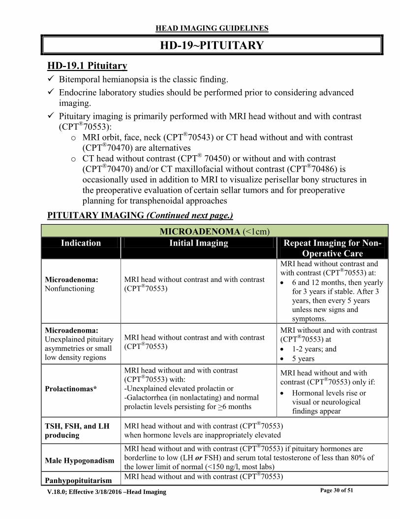
HD-19.1 Pituitary Bitemporal hemianopsia is the classic finding. Endocrine laboratory studies should be performed prior to considering advanced
imaging. Pituitary imaging is primarily performed with MRI head without and with contrast
(CPT®70553): o MRI orbit, face, neck (CPT®70543) or CT head without and with contrast
(CPT®70470) are alternatives o CT head without contrast (CPT® 70450) or without and with contrast
(CPT®70470) and/or CT maxillofacial without contrast (CPT®70486) is occasionally used in addition to MRI to visualize perisellar bony structures in the preoperative evaluation of certain sellar tumors and for preoperative planning for transphenoidal approaches
PITUITARY IMAGING (Continued next page.)
MICROADENOMA (<1cm) Indication Initial Imaging Repeat Imaging for Non-
Operative Care
Microadenoma: Nonfunctioning
MRI head without contrast and with contrast (CPT®70553)
MRI head without contrast and with contrast (CPT®70553) at: 6 and 12 months, then yearly
for 3 years if stable. After 3 years, then every 5 years unless new signs and symptoms.
Microadenoma: Unexplained pituitary asymmetries or small low density regions
MRI head without contrast and with contrast (CPT®70553)
MRI without and with contrast (CPT®70553) at 1-2 years; and 5 years
Prolactinomas*
MRI head without and with contrast (CPT®70553) with: -Unexplained elevated prolactin or -Galactorrhea (in nonlactating) and normal prolactin levels persisting for >6 months
MRI head without and with contrast (CPT®70553) only if: Hormonal levels rise or
visual or neurological findings appear
TSH, FSH, and LH producing
MRI head without and with contrast (CPT®70553) when hormone levels are inappropriately elevated
Male Hypogonadism MRI head without and with contrast (CPT®70553) if pituitary hormones are borderline to low (LH or FSH) and serum total testosterone of less than 80% of the lower limit of normal (<150 ng/l, most labs)
Panhypopituitarism MRI head without and with contrast (CPT®70553)
V.18.0; Effective 3/18/2016 –Head Imaging
Page 30 of 51

ADH ABNORMALITIES
Indication Initial Imaging Repeat Imaging for Non-Operative Care
Diabetes Insipidus (DI)
MRI head without and with contrast (CPT®70553) if: Laboratory testing consistent with DI and
etiology uncertain
NA
Syndrome of Inappropriate ADH (SIADH)
MRI head without and with contrast (CPT®70553) if: Etiology remains uncertain or is thought to
be in the nervous system,
NA
Macroadenoma MRI head without and with contrast (CPT®70553)
MRI head without and with contrast(CPT®70553) every: 6 months for the first year;
and then, annually for 5 years (longer
if craniopharyngiomas); every 6 months if treatment
is deferred
Other Pituitary Region Tumors**
Evaluation may require CT in addition to MRI to evaluate for hyperostosis. Requests will be sent for Medical Director review.
Enlarged/Empty Sella Turcica***
Head CT without and with contrast (CPT®70470) or, MRI head without and with contrast (CPT®70553) to exclude residual pituitary tumor and to assess the position of the chiasm since
herniation into the sella, causes Chiasmatic-type visual loss
MRI without and with contrast (CPT®70553) 1-5 years after the initial study can be performed.
Post-operatively, follow-up pituitary imaging is generally done at the discretion of the neurosurgeon, usually at 3 months if stable.
Practice Notes *Prolactinoma Note: Most common of the secreting Microadenoma (>50%). Normal prolactin levels range up to 20 µg/l in non-lactating, non-pregnant women and in males. Transient elevation of up to 40 µg/l in females can occur, and requires repeating prior to consideration of advanced imaging.
**Other Pituitary Region Tumor Notes: Craniopharyngiomas arise in the parasellar area. About 10% of meningiomas arise in this area.
***Enlarged/Empty Sella Turcica Notes: An enlarged sella turcica without evident tumor is an incidental finding on MRI head or CT head from a defect in the dural diaphragm of the sella (especially if there is elevated intracranial pressure from another
V.18.0; Effective 3/18/2016 –Head Imaging
Page 31 of 51

cause), pituitary surgery, or as a result of a pituitary tumor which has expanded the sella and then infarcted (pituitary apoplexy). References 1. N Engl J Med 2003;349:2035-2041 2. Kucharcyk W, Montanera WJ, Kucharczyk J. The sella turcica and parasellar region. In Latchaw
RE, Kucharczyk J, Moseley ME. Imaging of the Nervous System. Philadelphia, Elsevier, 2005, pp.1033-1061
3. Neurosurg Clin N Am 2003;14:167-171 4. Endocr Pract 2002;8:440-456
HD-20~SCALP and SKULL LESIONS
HD-20.1 Scalp and Skull Lesions The majority of these are benign soft tissue or bony lesions easily defined by physical examination or with skull x-rays. Head CT without or without and with contrast (CPT®70450 or CPT®70470) is
appropriate for the following scenarios: o Any lesion on physician examination and skull x-ray which is not clearly
benign o Langerhans’ cell histiocytosis, myeloma, and metastatic cancer, when
symptoms suggest bony lesions o MRI head without contrast (CPT®70551) or with and without contrast
(CPT®70553) may be considered if there is concern for intracranial extension
V.18.0; Effective 3/18/2016 –Head Imaging
Page 32 of 51

HEAD IMAGING GUIDELINES
HD-21 STROKE/TIA
HD-21.1 Stroke/TIA One from each of the following procedures can be considered for the initial occurence
or repeat episodes of TIA, stroke1-4 or Transient Global Amnesia5: o CT head without contrast (CPT 70450) or CT head without and with contrast
(CPT®70470) or MRI head without and with contrast (CPT®70553) or MRI head without contrast (CPT®70551)
MRI is preferred with later presentation for evaluation and can be considered after an initial CT head1-4
o Duplex ultrasound of the carotid arteries or MRA neck without contrast (CPT®70547) or MRA neck with contrast (CPT®70548) or MRA neck without and with contrast (CPT®70549); or Neck CTA (CPT®70498)
MRA head without contrast (CPT®70544) or CTA head with contrast(CPT®70496) may be considered in addition to the above in the following clinical scenarios:
o Presentation is within 24 hours of onset1-3 o Vertebrobasilar stroke (vertigo associated with diplopia, dysarthria, bifacial
numbness or ataxia)1-4 o Suspected Carotid or Vertebral Artery Dissections2-4. Risks may include
premature stroke (under age 50), head or neck trauma, fibromuscular dysplasia, Ehlers-Danlos syndrome, and chiropractic neck manipulation.
Repeat imaging as determined by a specialist o Suspected Venous Infacts (as MRV (CPT®70544) or CTV (CPT®70496)) if
identified on CT/MRI head6 o Recurrent stroke or TIA who have failed maximum medical management
(smoking cessation, anti-platelet medication if no contra-indications, BP and lipid treatment) to assess potential candidates for elective invasive procedures (such as angioplasty or stents)7,8
MRA neck without and with contrast (CPT®70549) is reserved for evaluation of possible or known arterial dissection.
V.18.0; Effective 3/18/2016 –Head Imaging
Page 33 of 51

HD-21.2 Venous Infarcts MRV (CPT®70544) or CTV (CPT®70496) and MRI head without contrast
(CPT®70551) are appropriate in the following scenarios: o Intracranial hypertension with headache, vomiting and papilledema from
venous sinus thrombosis o Venous infarction is identified on MRI head or Head CT o Women with postpartum stroke or postpartum papilledema o Children or young adults who present with a stroke in which headache and
seizures are prominent features, or who are known to have an intrinsic system clotting disorder.
Practice Notes Transient Global Amnesia is the “…sudden onset of transient inability to retain new information and to recall previous events for a variable period of time, generally occurring in middle-aged or elderly patients formerly in good health and without significant cardiac or cerebrovascular disease…”5
References 1. American College of Radiology, ACR Appropriateness Criteria® Cerebrovascular Disease Last
review date: 2011 2. AHA Scientific Statement, Recommendations for Imaging of Acute Ischemic Stroke, A Scientific
Statement From the American Heart Association, Richard E. Latchaw, MD, Chair; Mark J. Alberts, MD, FAHA; Michael H. Lev, MD, FAHA; John J. Connors, MD; Robert E. Harbaugh, MD, FAHA; Randall T. Higashida, MD, FAHA; Robert Hobson, MD, FAHA†; Chelsea S. Kidwell, MD, FAHA; Walter J. Koroshetz, MD; Vincent Mathews, MD; Pablo Villablanca, MD; Steven Warach, MD, PhD; Beverly Walters, MD; Stroke. 2009; 40: 3646-3678
3. Imaging Recommendations for Acute Stroke and Transient Ischemic Attack Patients: A Joint Statement by the American Society of Neuroradiology, the American College of Radiology, and the Society of NeuroInterventional Surgery, M. Wintermark, P.C. Sanelli, G.W. Albers, J. Bello, C. Derdeyn, S.W. Hetts, M.H. Johnson, C. Kidwell, M.H. Lev, D.S. Liebeskind, H. Rowley, P.W. Schaefer, J.L. Sunshine, G. Zaharchuk, and C.C. Meltzer,Published August 1, 2013 as 10.3174/ajnr.A3690
4. Bernheisel CR, Schlaudecker JD, Leopold K. Subacute Management of Ischemic Stroke. Am Fam Physician, 2011;84:1383-1388
5. Transient Global Amnesia and Transient Ischemic Attack , Natural History, Vascular Risk Factors, and Associated Conditions, Marino Zorzon, MD; Lucia Antonutti, MD; Giovanni Masè, MD; Emanuele Biasutti, MD; Barbara Vitrani, MD; Giuseppe Cazzato, MD,Stroke. 1995; 26: 1536-1542
6. AHA/ASA Scientific Statement, Diagnosis and Management of Cerebral Venous Thrombosis, A Statement for Healthcare Professionals From the American Heart Association/American Stroke Association, Gustavo Saposnik et al, Stroke. 2011; 42: 1158-1192
7. Marc I. Chimowitz, et. al., Stenting versus Aggressive Medical Therapy for Intracranial Arterial Stenosis, N Engl J Med 2011; 365:993-1003.
8. FDA Safety Communication: Narrowed Indications for Use of the Stryker Wingspan Stent System, http://www.fda.gov/MedicalDevices/Safety/AlertsandNotices/ucm314600.htm acquired February 18, 2014
9. Rowland LP (Ed.). Merritt’s Neurology. 12th Ed. Philadelphia, Lippincott, 2010, pp.281-294
V.18.0; Effective 3/18/2016 –Head Imaging
Page 34 of 51

10. Ardila A and Farlow M. Transient global amnesia. Medlink Neurology, May 25, 2010, http://www.medlink.com/medlinkcontent.asp. Accessed June 13, 2011
11. N Engl J Med 2005;352:1791-1798 12. N Engl J Med 2001;344:898-906 13. Practical Neurology 2005;5:100-109
HD-22~CEREBRAL VASCULITIS
HD-22.1 Cerebral Vasculitis MRI head without and with contrast (CPT®70553) is considered when any of the
following is suspected1,2: o Small to medium vessel Vasculitis1,2 o Large/Giant Cell Arteritis1-3
MRA Head without and with contrast (CPT 70546) and MRA Neck without or with contrast (CPT 70549); CTA3 may be considered in addition to MRI in these circumstances
Practice Notes Classification of vasculitides based on vessel size adapted from Joseph (1). Small and medium vessel vasculitis is generally beyond the resolution for MRA and CTA.
Dominant Vessel Involved
Primary Secondary
Large arteries Giant cell arteritis Takayasu’s arteritis
Aortitis with rheumatoid disease;
Infection (e.g. syphilis)
Medium Arteries Classical polyarteritis nodosa Kawasaki disease
Infection (e.g. hepatitis B)
Small vessels and medium arteries
Wegener’s granulomatosis Churg–Strauss syndrome Microscopic polyangiitis
Vasculitis with rheumatoid disease,systemic lupus erythematosus, Sjögren’s syndrome, drugs, infection (e.g. HIV)
Small vessels Henoch-Schönlein purpura Essential cryoglobulinaemia Cutaneous leukocytoclastic
vasculitis
Drugs (e.g. sulphonamides, etc.) Infection (e.g. hepatitis C)
V.18.0; Effective 3/18/2016 –Head Imaging
Page 35 of 51

References 1. Joseph, F. and Scolding, N, Cerebral vasculitis a practical approach, Practical Neurology
2002;2:80-93 2. Hunder, G, Classification of and approach to the vasculitides in adult, UpToDate, acquired April 2,
2014 3. American College of Radiology (ACR), American Society of Neuroradiology (ASNR), Society of
NeuroInterventional Surgery (SNIS), Society for Pediatric Radiology (SPR). ACR-ASNR-SNIS-SPR practice guideline for the performance of pediatric and adult cervicocerebral magnetic resonance angiography (MRA). American College of Radiology (ACR); 2010.
HD-23~DIZZINESS, VERTIGO and SYNCOPE
HD-23.1 Dizziness, Vertigo, and Syncope The initial components in the evaluation of false sensations of balance or motion
include obtaining a patient history and performing a physical examination that can assist in diagnosis. These include the elimination of inciting factors.1,2
Evaluation of arterial blood flow (Cartoid Doppler, transcranial Doppler, Neck and Head MRA/CTA), CT Head and MRI Head are not indicated unless a primary neurological cause of transient loss of consciousness is suspected. Neurological testing is not indicated for patients with uncomplicated syncope.
Prior to advanced imaging, the minimum initial evaluation should include at least one of the following:
o Orthostatic blood pressure,1,2 o Dix-Hallpike maneuver or other positional testing,1,2 o Nystagmus examination,1,2 o Any one Gait examination, including Romberg, 1,2 o Psychiatric evaluation including for anxiety or panic disorders (if suspected), 1,2 o Hearing testing (if associated with hearing loss) to determine if conductive,
sensorineural, or mixed,5 o Vision examination1
MRI head with attention to internal auditory canal without and with contrast (CPT®70553) or without contrast (CPT®70551; limited study CPT®70540 or CPT®70543)3,5 can be considered when the initial evaluation reveals:
o Any associated neurological signs or symptoms2 Cerebrovascular symptoms of TIA or CVA2 Examples include drop attacks, seizures, coincident headache, ataxia,
aura or focal neurological findings o Equivocal or unusual nystagmus findings, including direction changing or
persistent downbeat nystagmus2,4 o Absent head thrust sign2 o Short duration (minutes) recurrent attacks2
V.18.0; Effective 3/18/2016 –Head Imaging
Page 36 of 51

CT temporal bone without contrast (CPT®70480) may be considered in addition to the MRI evaluation5
o Hearing loss associated with Progressive unilateral hearing loss3 Sensorineural5 Conductive5: CT temporal bone without contrast (CPT®70480) may be
considered in addition to the MRI evaluation Congenital or total hearing loss5: CT temporal bone without contrast
(CPT®70480) may be considered in addition to the MRI evaluation Pre-surgical planning or cochlear implant candidate5: CT temporal bone
without contrast (CPT®70480) may be considered in addition to the MRI evaluation
o Features atypical for benign positional vertigo, which may include abnormal cranial nerve findings, visual disturbances, and severe headache4
o Central vertigo o Also see: HD-21 Stroke/TIA
Practice Notes
Categories of Dizziness Description Most Common Causes
Vertigo False sense of motion, possibly spinning sensation
Benign paroxysmal positional vertigo, Meniere disease, vestibular neuritis, and labyrinthitis
Disequilibrium Off-Balance, wobbly Parkinson disease and diabetic neuropathy
Presyncope Feeling of losing consciousness or “blacking out”
Medications
Lightheadedness Vague symptoms, possibly feeling disconnected with the environment
Psychiatric disorders, such as depression, anxiety, and hyperventilation syndrome
References 1. American Family Physician, Dizziness: A Diagnostic Approach, Am Fam Physician. 2010 Aug 15;
82(4):361-368. ROBERT E. POST, LORI M. DICKERSON 2. Neurol Clin Pract December 2011 vol. 1 no. 1 24-33 The evaluation of a patient with dizziness,
Kevin A. Kerber, MD and Robert W. Baloh, MD 3. American Family Physician, Initial Evaluation of Vertigo RONALD H. LABUGUEN, M.D Am Fam
Physician. 2006 Jan 15;73(2):244-251 4. Clinical practice guideline: Benign paroxysmal positional vertigo, Neil Bhattacharyya, MD, et al.,
Otolaryngology–Head and Neck Surgery (2008) 139, S47-S81 5. American College of Radiology ACR Appropriateness Criteria®, 2013, Hearing Loss and/or Vertigo 6. Guidelines for the diagnosis and management of syncope (v 2009). The Task Force for the Diagnosis
and Management of Syncope of the European Society of Cardiology. European Hear Journal (2009). 30, 2631-71.
V.18.0; Effective 3/18/2016 –Head Imaging
Page 37 of 51
HEAD IMAGING GUIDELINES
HD-24~OTHER IMAGING STUDIES
Some payers may consider these techniques investigational, and their coverage policies may take precedence over eviCore’s guidelines.
HD-24.1 Functional MRI (f-MRI) f-MRI is useful in pre-operative scenarios to define the “eloquent” areas of brain. The ordering physician must be a neurosurgeon or radiation oncologist. All other
requests should be sent for MD review. It must be evident that brain surgery is planned, and that f-MRI is being performed to avoid the language centers, or other processing centers, of the brain.
f-MRI can be approved with PET brain in epilepsy surgery planning. Procedure codes for functional MRI:
o CPT®70554 MRI head, functional MRI, including test selection and administration of repetitive body part movement and/or visual stimulation, not requiring physician or psychologist administration
o CPT®70555 MRI head, functional MRI; requiring physician or psychologist administration of entire neurofunctional testing
HD-24.2 Magnetic Resonance Spectroscopy (MRS)
HD-24.3 CSF Flow Imaging This is generally imaged as a part of a head MRI study. It is not coded separately for
preoperative evaluation of hydrocephalus and Chiari syndrome, with either features of hydrocephalus or syrinx
There is no specific or unique procedure code for this study; it is done as a special sequence of a routine MRI head without contrast (CPT®70551).
All requests for MRS will be forwarded for Medical Director review
MRS involves analysis of the levels of certain chemicals in a pre-selected voxels (small regions) on an MRI scan done at the same time.
MRS is evaluated on a case-by-case basis, and may be considered: o Distinguish recurrent brain tumor from radiation necrosis as an alternative to PET
(CPT®78608) o Diagnosis of certain rare inborn errors of metabolism affecting the CNS (primarily
pediatric patients)
V.18.0; Effective 3/18/2016 –Head Imaging
Page 38 of 51

HD-24.4 CT or MRI Perfusion Performed as part of a head CT or MRI examination in the evaluation of patients with
very new strokes or brain tumors Category III 0042T - “cerebral perfusion analysis using CT”
There is no specific CPT code for MRI Perfusion. Perfusion weighted images are obtained with contrast and are not coded separately from a contrasted MRI Head examination. If MRI head without and with contrast is approved, no additional CPT codes are necessary or appropriate to perform MRI perfusion.
HD-24.5 Magnetic Resonance Neurography (MRN) MRN is currently considered investigational by most payors.
HD-24.6 Positional MRI May be considered when performed as the definitive MRI study
HD-24.7 Cone Beam Computed Tomography (CBCT) CPT® Codes: 70486, 70487, 70488, 70480, 70482 (NO separate 3-D rendering codes should be reported) See: HD-30~Temporomandibular Joint Disease (TMJ)
References 1. Latchaw RE, Kucharczyk J, Moseley ME. Imaging of the Nervous System. Philadelphia, Elsevier,
2005, pp.1089-1100, 101-120 and 1225-1239 2. CMS, Decision memo for magnetic resonance spectroscopy for brain tumors (CAG-00141N)
http://www.cms.gov.hhs.gov/mcd/viewdecisionmemo.asp?id=52 Accessed December 18, 2009 3. Neurology 2005;64:2085-2089 4. Neuroimaging Clin N Am 2006; 16:87-116. An encyclopedic review of current knowledge regarding
use of MRS in inborn errors, disease by disease. 5. Neurology 2005;64:434-441 6. Neurology 2005;64:406-407 7. Ann Neurol 2006;60:508-517 8. Neurology 2007;68:694-697 9. Stroke 2001;32:2021-36 10. Neurology 2007;68:730-736 11. Haughton VM, et al. Mapping brain function with functional magnetic resonance imaging. In
Latchaw RE, Kucharuczyk J, Moseley ME. Imaging of the Nervous System. Philadelphia, Elsevier, 2005, pp.89-100
12. Neurosurgery 2004; 54:902-915. 13. Radiology 2005;236:247-253 14. Neuroimag Clin N Amer 2006;16:285-297 15. Radiology 2006;240:793-802 16. Stippich C (Ed.). Clinical Functional MRI. Springer, Berlin Heidelberg, 2007
V.18.0; Effective 3/18/2016 –Head Imaging
Page 39 of 51
17. Nature Clin Pract Neurol 2005;1:45-53 18. Neurology 2002;58:1597-1602 19. Radiology 2006;241:213-222 20. Neurosurgery 2006;59:493-511 21. International Journal of Oral & Maxillofacial Surgery 2009 June;38(6):609-625
EAR and NOSE GUIDELINES
HD-25~EPISTAXIS
HD-25.1 Epistaxis Maxillofacial CT without or with contrast (CPT®70486 or CPT®70488) is appropriate
based on endoscopic findings during ENT examination.
References 1. Practical Neurology 2001;1:42-49 2. Imaging of the Temporal Bone, 4th ed. J.D. Swartz and L.A. Loevner, eds. Thieme Medical
Publishers; 2009.
HD-26~MASTOID DISEASE
HD-26.1 Mastoid Disease See Pediatric Head Guidelines, PEDHD 16.2 Ear Pain
V.18.0; Effective 3/18/2016 –Head Imaging
Page 40 of 51

HD-27~HEARING LOSS
HD-27.1 Hearing Loss MRI head with attention to internal auditory canal without and with contrast
(CPT®70553), or MRI head with attention to internal auditory canal without contrast or CT temporal bone without contrast (CPT®70480) can be considered for hearing loss1. Clinical information provided should include evaluation of hearing either by bedside testing or by formal audiology.
Limited Study MRI with attention to internal auditory canal (CPT®70540 - 70543) can be approved in place of MRI head with attention to internal auditory canal when requested by the provider in the following scenarios: o Any sensorineural (cochlea or auditory nerve)1 o Any conductive1(including Cholesteatoma2) o Cochlear implants candidate1 o Fluctuating hearing loss1
Practice Note An initial evaluation generally determines whether a patient’s hearing loss is conductive (external or middle ear structures) or sensorineural (inner ear structures, such as cochlea or auditory nerve) hearing loss.1,2
References 1. Turski PA, Wippold FJ II, Cornelius RS, Brunberg JA, Davis PC, De La Paz RL, Dormont D, Gray
L, Jordan JE, Mukherji SK, Nussenbaum B, Seidenwurm DJ, Sloan MA, Zimmerman RD, Expert Panel on Neurologic Imaging. ACR Appropriateness Criteria® vertigo and hearing loss. [online publication]. Reston (VA): American College of Radiology (ACR); 2008
2. Isaacson J, Vora N. Differential Diagnosis and Treatment of Hearing Loss, Am Fam Physician. 2003 Sep 15;68(6):1125-1132
V.18.0; Effective 3/18/2016 –Head Imaging
Page 41 of 51

EAR and NOSE GUIDELINES
HD-28~EAR PAIN (OTALGIA)
HD-28.1 Ear Pain (Otalgia) CT temporal bone without and with contrast (CPT®70482) or without contrast
(CPT®70480) and/or MRI head without contrast (CPT®70551) or without and with contrast (CPT®70553) can be considered for: o Common causes of ear pain include ear infections, dental problems, sinus
infection, neck problems, tonsillitis, and pharyngitis, as well as otitis media or exsterna or no obvious cause, which do not improve over a reasonable time
o Cerebellopontine angle or other intracranial tumor is suspected o Nervus intermedius neuralgia in order to exclude a structural lesion
See also: HD-27~Hearing Loss
References 1. Li JC and Brunk J. Otalgia. eMedicine. March 16, 2010,
http://emedicine.medscape.com/article/845173-overview. Accessed June 13, 2011 2. Imaging of the Temporal Bone, 4th ed. J.D. Swartz and L.A. Loevner, eds. Thieme Medical
Publishers; 2009.
V.18.0; Effective 3/18/2016 –Head Imaging
Page 42 of 51
HEAD IMAGING GUIDELINES
HD-29~SINUSITIS
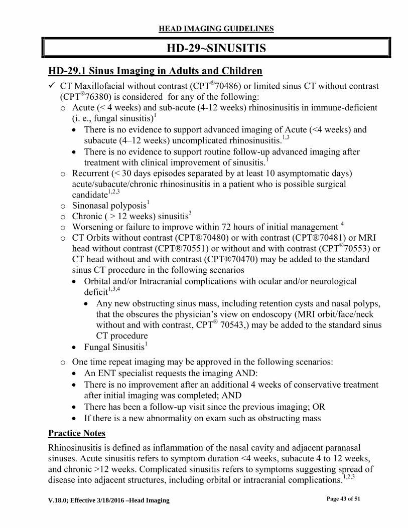
HD-29.1 Sinus Imaging in Adults and Children CT Maxillofacial without contrast (CPT®70486) or limited sinus CT without contrast
(CPT®76380) is considered for any of the following: o Acute (< 4 weeks) and sub-acute (4-12 weeks) rhinosinusitis in immune-deficient
(i. e., fungal sinusitis)1 There is no evidence to support advanced imaging of Acute (<4 weeks) and
subacute (4–12 weeks) uncomplicated rhinosinusitis.1,3 There is no evidence to support routine follow-up advanced imaging after
treatment with clinical improvement of sinusitis.1 o Recurrent (< 30 days episodes separated by at least 10 asymptomatic days)
acute/subacute/chronic rhinosinusitis in a patient who is possible surgical candidate1,2,3
o Sinonasal polyposis1 o Chronic ( > 12 weeks) sinusitis3 o Worsening or failure to improve within 72 hours of initial management 4 o CT Orbits without contrast (CPT®70480) or with contrast (CPT®70481) or MRI
head without contrast (CPT®70551) or without and with contrast (CPT®70553) or CT head without and with contrast (CPT®70470) may be added to the standard sinus CT procedure in the following scenarios Orbital and/or Intracranial complications with ocular and/or neurological
deficit1,3,4 Any new obstructing sinus mass, including retention cysts and nasal polyps,
that the obscures the physician’s view on endoscopy (MRI orbit/face/neck without and with contrast, CPT® 70543,) may be added to the standard sinus CT procedure
Fungal Sinusitis1 o One time repeat imaging may be approved in the following scenarios:
An ENT specialist requests the imaging AND: There is no improvement after an additional 4 weeks of conservative treatment
after initial imaging was completed; AND There has been a follow-up visit since the previous imaging; OR If there is a new abnormality on exam such as obstructing mass
Practice Notes Rhinosinusitis is defined as inflammation of the nasal cavity and adjacent paranasal sinuses. Acute sinusitis refers to symptom duration <4 weeks, subacute 4 to 12 weeks, and chronic >12 weeks. Complicated sinusitis refers to symptoms suggesting spread of disease into adjacent structures, including orbital or intracranial complications.1,2,3
V.18.0; Effective 3/18/2016 –Head Imaging
Page 43 of 51

References 1. Rosenfeld RM, Piccirillo JF, Chandrasekhar SS, et al. Clinical practice guideline (update): adult
sinusitis. Otolaryngol Head Neck Surg. 2015 Apr;152(2 Suppl):S1-S39 2. Desrosiers M, Evans GA, Keith PK, Wright ED, Kaplan A, Bouchard J, Ciavarella A, Doyle PW,
Javer AR, Leith ES, Mukherji A, Schellenberg RR, Small P, Witterick IJ. Canadian clinical practice guidelines for acute and chronic rhinosinusitis. Allergy Asthma Clin Immunol. 2011 Feb 10;7(1):2
3. Cornelius RS, Martin J, Wippold FJ II, Aiken AH, et al. ACR Appropriateness Criteria® sinonasal disease. American College of Radiology (ACR); 2012
4. Huntzinger A. Guidelines for the Diagnosis and Management of Rhinosinusitis in Adults, Am Fam Physician. 2007;76:1718-1724
5. Karmazyn B, Coley BD, Dempsey-Robertson ME, Dillman JR, Dory CE, Garber M, Hadley JA, Hayes LL, Keller MS, Meyer JS, Milla SS, Paidas C, Raske ME, Rigsby CK, Strouse PJ, Wootton-Gorges SL, Expert Panel on Pediatric Imaging. ACR Appropriateness Criteria® sinusitis - child. American College of Radiology (ACR); 2012
6. Wald E, et. el., Sinusitis in Children Aged 1 to 18 Years Clinical Practice Guideline for the Diagnosis and Management of Acute Bacterial, Pediatrics 2013; 132;e262.
V.18.0; Effective 3/18/2016 –Head Imaging
Page 44 of 51
HEAD IMAGING GUIDELINES
HD-30~Temporomandibular Joint Disease (TMJ) and Dental/Periodontal/Maxillofacial Imaging
HD-30.1 Temporomandibular Joint Disease (TMJ) TMJ MRI (CPT®70336) should be reserved for those who fail a minimum of 6 weeks
of non-surgical treatment and who are actively being considered for TMJ surgery. TMJ Imaging in children with Juvenile Rheumatoid Arthritis
See: PACHD-27 Temporomandibular Joint Imaging in Children
HD-30.2 Dental/Periodontal/Maxillofacial Imaging All requests will be forwarded to Medical Director for review.
Indications for cone beam CT if requested by an oral or maxillofacial surgeon: o Impacted teeth o Supernumerary teeth o Dentoalveolar trauma o Root resorption o Foreign body o Odontogenic cysts, tumors, or other jaw pathology o Cleft pathology o Orthognathic surgery for dentofacial anomalies o Osteomyelitis and odontogenic infections o Bisphosphonate-related osteonecrosis of the jaw o Salivary gland stones o Maxillofacial bone graft planning o Dental implants related to tooth loss from injury, trauma, or jaw pathology such as
cysts, tumors, or cancer
Currently, there are no published guidelines from any specialty society such as the American Association of Oral and Maxillofacial Surgeons regarding the appropriate use of cone beam CT for dentoalveolar conditions, maxillofacial conditions, orthodontics, endodontics, or dental implants.
Most payors do not include orthodontic clinical conditions, replacement of teeth lost due to caries or periodontal disease, non-trauma related dental implantology, or endodontic treatment not related to trauma to the natural tooth in their coverage policies. o Thus, cone beam CT scans in these patients would also not be included in the
coverage policy. o These coverage policies will take precedence over eviCore’s guidelines.
Cone Beam CT: Report with CPT® Codes: 70486, 70487, 70488, 70480, 70482.
V.18.0; Effective 3/18/2016 –Head Imaging
Page 45 of 51

3-D rendering (CPT®76376 or CPT®76377) should NOT be reported separately.
Also called i-CAT scanner or mini-CAT scanner
References 1. International Journal of Oral & Maxillofacial Surgery 2009 June;38(6):609-625 2. N Engl J Med 2008;359:2693-2705
HD-31~TINNITUS
HD-31.1 Tinnitus Advanced imaging is not usually indicated in the evaluation of Tinnitus, unless one or more of the following signs and symptoms are present,
Tinnitus localized to a single ear Pulsatile Tinnitus Focal neurological abnormalities Asymmetric hearing loss
If one or more of these signs and symptoms are present, the following advanced imaging studies can be considered: MRI head without and with contrast1,2,3 (CPT®70553), or Head CT1,2 with and/or CT temporal bone3 without contrast, or MRI head with attention to internal auditory canal3 without and with contrast
(CPT®70553), or MRI head and internal auditory canal3 without contrast (CPT®70551), or
Limited Study MRI with attention to internal auditory canal (CPT®70540 - 70543) can be approved in place of MRI head with attention to internal auditory canal when requested by the provider
MRA head without contrast (CPT 70544) and/or MRA neck without contrast (CPT 70547) or MRA neck with contrast (CPT 70548) can be added if there is suspicion of vascular lesions
Practice Notes The history in patients with tinnitus should include a description of the tinnitus (episodic or constant, pulsatile or non-pulsatile, rhythmicity, pitch, quality of the sound), as well as inciting or alleviating factors. Continuous and pulsatile tinnitus are more concerning for an underlying and significant disorder.2 Audiometric assessment can be used as initial
V.18.0; Effective 3/18/2016 –Head Imaging
Page 46 of 51

diagnostic testing1,2,3 particularly in patients with tinnitus that is unilateral, persistent (> 6 months) or associated with hearing difficulties.
References 1. Crummer RW, Hassan GA. Diagnostic Approach to Tinnitus. Am Fam Physician. 2004 Jan 1;
69(1):120-126. 2. Turski PA, et. al. Expert Panel on Neurologic Imaging. ACR Appropriateness Criteria® vertigo and
hearing loss. American College of Radiology (ACR); 2013. 3. American Speech-Language-Hearing Association, Tinnitus Triage Guidelines,
http://www.asha.org/aud/Articles/Tinnitus-Triage-Guidelines/, acquired March 18, 2014. 4. Tunkel DE, Bauer CA, Sun GH, Rosenfeld RM, et al. Clinical Practice Guideline: Tinnitus.
Otolaryngology – Head and Neck Surgery, 2014; 151: S1.
V.18.0; Effective 3/18/2016 –Head Imaging
Page 47 of 51
HEAD IMAGING GUIDELINES
HD-32~EYE DISORDERS
HD-32.1 Eye Disorders MRI head without and with contrast (CPT®70553) and/or MRI orbit without and with
contrast (CPT®70543) or MRI head without contrast (CPT®70551) and/or MRI orbit without contrast (CPT®70540). 1, 2, 3 may be considered in the following scenarios*: o Anisocoria which is of new onset (e.g. not present in previous photographs) and
>/= 1mm 3, 4 o Acute or progressive vision loss due to any cause, including suspected optic
neuritis 1, 2, 5 o Ophthalmoplegia1 o Binocular Diplopia6, 7 o Horner’s Syndrome, for which CT Neck with contrast and/or CT Chest with
contrast may be considered in addition to the head or orbital imaging8 o CT head without contrast may be substituted for the MRI imaging if there has been
a head injury1 Evaluation of a third nerve palsy may be accomplished with an MRI head without and
with contrast(CPT®70553) and/or MRA brain without contrast CT head without and with contrast (CPT®70470) and/or CT orbit with
contrast (CPT® 70481) can be approved if there is a clinical question of blood in the subarachnoid space.
If MRI contraindicated or cannot be performed, CT head without and with contrast (CPT®70470), CT orbit with contrast (CPT®70482) or CT orbit without and with contrast may be considered as substitutes.
Also see HD-16~Multiple Sclerosis (MS) and Related Conditions
Practice Notes *Advanced imaging of the brain and orbit are not routinely paired. Medical necessity for each region is needed to image both regions, based on suspicion of these disorders. References 1. Wippold FJ II, Cornelius RS, Berger KL, Broderick DF, Davis PC, Douglas AC, Germano IM,
Hadley JA, McDermott MW, Mechtler LL, Smirniotopoulos JG, Waxman AD, Expert Panel on Neurologic Imaging. ACR Appropriateness Criteria® orbits, vision and visual loss: American College of Radiology (ACR); 2012.
2. Quisling SV, Shah VA, Lee HK, et al. Magnetic resonance imaging of third cranial nerve palsy and trigeminal sensory loss caused by herpes zoster. J Neuroophthalmol. 2006 Mar. 26(1):47-8.
V.18.0; Effective 3/18/2016 –Head Imaging
Page 48 of 51

3. UpToDate, Third cranial nerve (oculomotor nerve) palsy in adults, Literature review current through: Feb 2014. | This topic last updated: Aug 2, 2012.
4. UpToDate, Approach to the patient with anisocoria, Literature review current through: Feb 2014. | This topic last updated: Jan 9, 2014. (normal pupil difference is </= 4mm.).
5. Physiologic anisocoria, American Academy of Ophthalmology, http://one.aao.org, acquired March 19, 2014.
6. UpToDate, Optic Neuritis, Pathophysiology, clinical features and diagnosis, Literature review current through: Feb 2014. | This topic last updated: Jan 5, 2014.
7. American College of Radiology (ACR), American Society of Neuroradiology (ASNR). ACR-ASNR practice guideline for the performance of computed tomography (CT) of the brain. American College of Radiology (ACR); 2010. 6 p.
8. Deciphering Diplopia, American Academy of Ophthalmology, http://one.aao.org, acquired March 19, 2014.
9. Lee, JH, et. al., Neuroimaging Strategies for Three Types of Horner Syndrome with Emphasis on Anatomic Location, American Journal of Roentgenology. 2007;188: W74-W81.
HD-33~ACOUSTIC NEUROMA & OTHER CEREBELLOPONTINE ANGLE TUMORS
HD-33.1 Acoustic Neuroma & Other Cerebellopontine Angle Tumors Initial diagnosis can be accomplished with MRI head without and with contrast
(CPT®70553) which should be done with attention to the internal auditory canals. Clinical information provided should include evaluation of hearing either by bedside testing or by formal audiology.
Temporal bone MRI without and with contrast (CPT®70543) may be considered with audiologic or clinical features of retrocochlear hearing loss and a negative head MRI and in the rare patient in whom a detailed search is indicated for both a lesion of the cerebellopontine angle and lesions of the cerebral hemispheres.
After resection, MRI head without and with contrast with attention to the internal auditory canals (CPT®70553) at 1 and 5 years is sufficient.
Observation with MRI head without and with contrast with attention to the internal auditory canals (CPT®70553) at 6 months after diagnosis and then once a year.
HD-34~PINEAL CYSTS
HD-34.1 Pineal Cysts See Pediatric Head Guidelines, PEDHD 13.2 Pineal Cysts
V.18.0; Effective 3/18/2016 –Head Imaging
Page 49 of 51
HD-35~ARACHNOID CYSTS
HD-35.1 Arachnoid Cysts
See Pediatric Head Guidelines,
PEDHD 13.1 Arachnoid Cysts
HD-36~NUCLEAR MEDICINE
Nuclear Medicine
o Nuclear medicine studies may be used in the evaluation of some head/brain disorders, and other rare indications exist as well: • Brain Scintigraphy with or without vascular flow (any one of CPT®
78600, 78601, 78605, or 78606) Establish brain death (rarely done in outpatient setting)
• Brain Imaging SPECT with Ioflupane I-23 (CPT® 78607) Immunocompromised patients with mass lesion detected on CT or
MRI for differentiation between lymphoma and infection • Brain Imaging Vascular Flow (CPT® 78610) Cerebral ischemia Establish brain death
• CSF Leakage Detection (CPT® 78650) Evaluation of CSF rhinorrhea or otorrhea, or refractory post-lumbar
puncture headache • Radiopharmaceutical Dacryocystography (CPT® 78660) Suspected obstruction of nasolacrimal duct due to excessive tearing
Cisternogram (CPT® 78630) is rarely done in children but can be approved for
the following: o Known hydrocephalus with worsening symptoms o Suspected obstructive hydrocephalus o Suspected normal pressure hydrocephalus with gait disturbance and
either dementia or urinary incontinence
Cerebrospinal Ventriculography (CPT® 78635) is rarely done in children but can be approved for the following:
o Evaluation of internal shunt, porencephalic cyst, or posterior fossa cyst
V.18.0; Effective 3/18/2016 –Head Imaging
Page 50 of 51

Nuclear Medicine Shunt Evaluation (CPT® 78645) and CSF Flow SPECT (CPT® 78647) are rarely done in children but can be approved for the following:
o Suspected malfunction of ventriculoperitoneal, ventriculopleural, or ventriculovenous shunts
V.18.0; Effective 3/18/2016 –Head Imaging
Page 51 of 51