health and wellbeing. dental anatomy - horses and … dental anatomy will allow you to make more...
TRANSCRIPT
Page 12 • HORSES and PEOPLE • Phone: 07 5467 9796 • [email protected] www.horsesandpeople.com.au • HORSES and PEOPLE • Page 13www.horsesandpeople.com.au • HORSES and PEOPLE • Page 13
and
Page 12 • HORSES and PEOPLE • Phone: 07 5467 9796 • [email protected]
equinedentalvets.com.aucommitted to advancing horse health
Brought to you by...
Equine Dental Vets an organisation committed to advancing horse healthOur group is made up of Equine Dental veterinarians from across the globe and we have over 300 members right here in Australia.
Most of our members provide afterhours and emergency care for all equine health issues and using our dedicated search on our website you can find the Equine dental vets nearest you at the touch of a button. This is particularly useful if you are away from
home, for example when travelling to shows and competitions.
We are proud to provide you this healthcare information in partnership with Horses and People magazine.
www.equinedentalvets.com.au
by Dr Shannon Lee BVSc, MANZCVSc, MICEVO
Dental Anatomy
what you need to know
Introduction
Anatomy is the branch of scientific study concerned with the function or internal workings of bodily structures, particularly as revealed by dissection.
Discoveries made as a result of advances in anatomical knowledge have led to incredible breakthroughs in all medical fields. From a better understanding of organs like the brain and the heart allowing advances in neurosurgery and heart transplants, to better imaging and understanding of the blood supply to a horse’s foot, allowing much better understanding and management of conditions like laminitis.
This really interests some people, whilst others consider it dry and
boring (perhaps not relevant to them and their horses). However, the more one understands anatomy, the more educated and informed one will be when making choices that affect their horse’s health and welfare.
Here are some quick, practical examples:
A good knowledge of anatomy allows equine dental vets to understand how removing one tooth will affect the other teeth in the mouth and the horse’s general health and wellbeing.
A good knowledge of anatomy allows your equine dental vet to identify tooth pain when he or she observes whether an area of the tooth containing dentine is or is not stained by tannins (dyes in the grasses the horse eats).
If a horse is avoiding chewing on a tooth or teeth due to pain, it may manifest as a tooth that lacks this normal staining, while the teeth around it have it.
Understanding how the tooth roots communicate and connect with the bone, and with regions like the sinuses helps equine dental vets to explain both diseases and the best choices for treatment of disease.
When a horse is affected by periodontal disease (the most common disease of horses), endotoxins produced by bacteria travel the pathway of blood vessels throughout the horse’s body and are deposited randomly into organs. Understanding this pathway at both a macroscopic and a microscopic anatomical level is the key to correct and successful management and treatment.
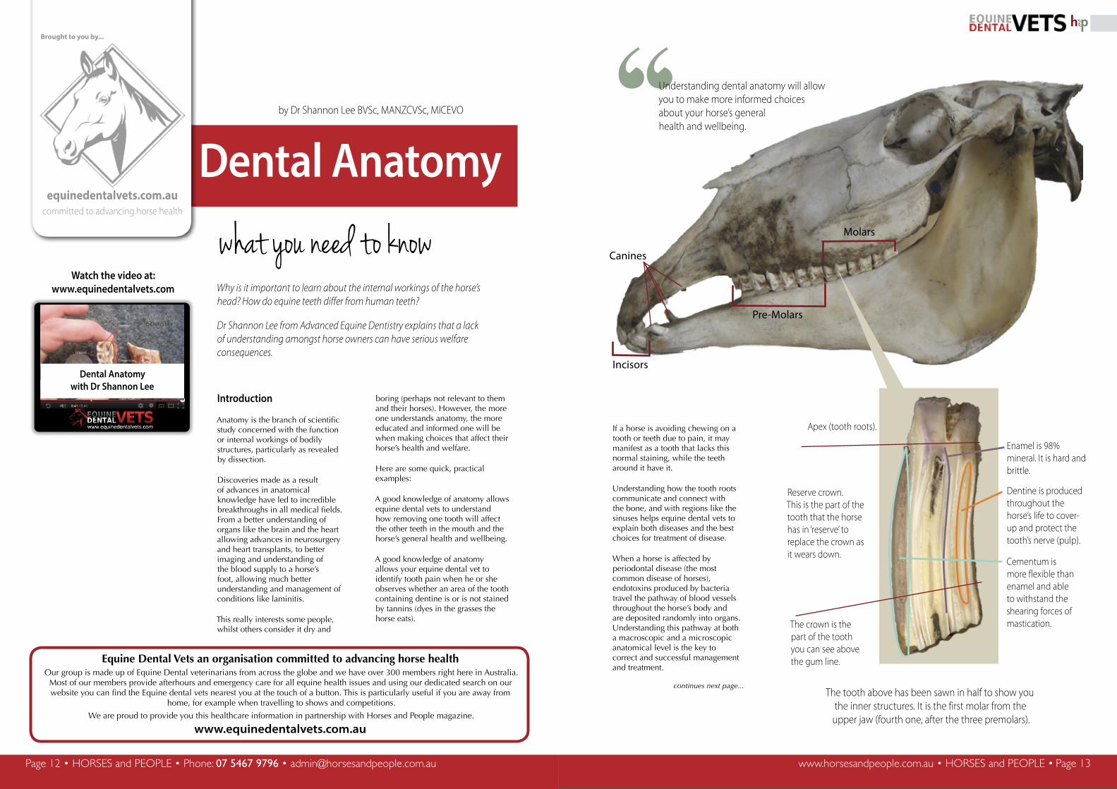
The tooth above has been sawn in half to show you the inner structures. It is the first molar from the upper jaw (fourth one, after the three premolars).
Molars
Pre-Molars
Incisors
Canines
Apex (tooth roots).
Reserve crown. This is the part of the tooth that the horse has in ‘reserve’ to replace the crown as it wears down.
The crown is the part of the tooth you can see above the gum line.
Enamel is 98% mineral. It is hard and brittle.
Dentine is produced throughout the horse’s life to cover-up and protect the tooth’s nerve (pulp).
Cementum is more flexible than enamel and able to withstand the shearing forces of mastication.
Why is it important to learn about the internal workings of the horse’s head? How do equine teeth differ from human teeth?
Dr Shannon Lee from Advanced Equine Dentistry explains that a lack of understanding amongst horse owners can have serious welfare consequences.
continues next page...
Watch the video at:www.equinedentalvets.com
Dental Anatomywith Dr Shannon Lee
“Understanding dental anatomy will allow you to make more informed choices about your horse’s generalhealth and wellbeing.

www.horsesandpeople.com.au • HORSES and PEOPLE • Page 15Page 14 • HORSES and PEOPLE • Phone: 07 5467 9796 • [email protected] www.horsesandpeople.com.au • HORSES and PEOPLE • Page 15
and
Page 14 • HORSES and PEOPLE • Phone: 07 5467 9796 • [email protected]
and
“
Introduction to the anatomy of the horse’s teeth and skull
When classifying teeth, there are three main types discussed:
• Brachydont
• Hypsodont
• Elodont
Okay, that’s great, but what does that really mean in English?
Brachydont teeth are the type you are very familiar with, as you’ve had them in your mouth most of your life.
Your own teeth are a simple tooth made up of short roots and crown (the part of the tooth you can see in the mouth). If you look after them using good, modern dental care, they can last a lifetime with little change. Other animals that also have brachydont teeth include dogs and cats.
Elodont teeth. At the other end of the scale are elodont teeth. These are teeth that continually grow over the animal’s lifetime and are found in animals that eat very abrasive diets, for example rats, mice and rabbits. This is the anatomical reason why your pet rabbits may need a trip to the vet to artificially ‘wear’ their teeth if their diet does not create enough natural wear.
Now that we have looked very briefly at the three types of teeth, we can look at the structures that make up a tooth.
In the horse the main components are:
• Enamel
• Dentine
• Cementum
• The periodontium
• The pulpino-dentinal complex
It may all sound a bit confusing, but keep reading and we will go through each one in simple terms and plain English (just remember that to keep it simple we are cutting a few corners here and there).
The enamel, the dentine and the cementum are the mineral (hard or solid) components of the tooth.
Enamel
Of the three mineral components, enamel is the densest (hardest), and in fact it is the hardest substance in the body being 96-98% mineral in content. This hardness allows it to withstand the forces and the wear of grazing abrasive pastures, but, this also makes it quite brittle, so whilst it is able to withstand certain types of wear and pressure very well, it is easily damaged by other types of wear or force.
Another important point about enamel is that once it has been formed and is part of a mature tooth it is inert (or dead).
Hypsodont teeth. Now to the third type of tooth – the Hypsodont tooth. This is the type found in horses and is very interesting.
You may have heard a horse owner or poorly educated lay dentist say that your horse’s teeth ‘grow’, but this is incorrect.
A hypsodont tooth can be pictured like a long rubber or eraser, the kind used for art or technical drawing.
The cardboard surrounding the eraser can be thought of as the bony socket (called alveolus) that the tooth sits in.
The part of the eraser visible outside the cardboard sleeve represents the crown of the tooth (the part of the tooth you can see in the mouth) and the rest of the eraser represents the rest of the tooth below the gum line (called the reserve crown).
Now here is the practical application of the anatomy, when you want to fix some mistakes with your eraser as you use it over time it gets shorter doesn’t it, and then you push more of it out of the cardboard, now it looks the same as it did before, except the eraser is actually getting shorter and shorter the more you use it.
This is exactly what happens with your horse’s teeth. Understanding this helps to explain why your horse’s mouth is in a state of constant change. It should also reinforce that any change is permanent in as much as, once you remove a part of the tooth, it is gone forever.
You may have heard a horse owner or poorly educated lay dentist say that your horse’s teeth ‘grow’, but this is incorrect.
www.equinedentalvets.com.au
We can’t show you his face
www.equinedentalvets.com.au
This is Dr Rob.
He's an Equine Dental Vets Member.
We can't show you his face, but he can show you his tool.
The horse’s tooth is similar to an eraser. As you use it, the tip (tooth crown) which is exposed wears down. You then need to push more of the eraser out of its cardboard sleeve (the reserve crown). When you do that, it looks just like it did before, except the eraser is getting shorter. This is exactly what happens to your horse’s teeth.
This is a really important point to understand because it means that any damage or injury sustained by enamel is unable to be repaired.
Using a high powered electron microscope, research veterinarians have identified and classified different types of enamel. The different types have a different structural makeup when viewed at a very large magnification, and are found in different parts of the mouth, in different locations within the same tooth and in different teeth. This may be explained by the specific structural and anatomical advantages certain types of teeth have by possessing more or less of a specific enamel type.
Enamel is classified into types 1, 2 and 3 with the difference being largely to do with the layering and orientation of the enamel.
Type 1 enamel is more prone to fractures or cracks along long planes and is more common in cheek teeth, but has better resistance to wear, while type 2 enamel is better able to withstand cracking or fracture but is softer. The enamel found in the front teeth (incisors) of horses is made up almost entirely of type 2. The structure of type 3 equine enamel is a hybrid of types 1 and 2 and it is only found in small amounts.
continues next page...
The horse above had to have these two premolars extracted due to an infection. The image shows where they were positioned on his upper jaw. You can also
appreciate the length of the teeth. Anatomical study allows equine dental vets to understand how removing one tooth will affect the other teeth in the mouth, as well as
how the horse’s general health and wellbeing will be affected.
Imag
e co
urte
sy o
f Adv
ance
d Eq
uine
Den
tistr
y
Imag
e co
urte
sy o
f Adv
ance
d Eq
uine
Den
tistr
y
Did you know?

www.horsesandpeople.com.au • HORSES and PEOPLE • Page 17Page 16 • HORSES and PEOPLE • Phone: 07 5467 9796 • [email protected] www.horsesandpeople.com.au • HORSES and PEOPLE • Page 17
and
Page 16 • HORSES and PEOPLE • Phone: 07 5467 9796 • [email protected]
and
Dentine
Dentine forms the bulk of the tissue in each tooth and is a mixture of both mineralised tissue and organic components (hard and soft tissues), whereas enamel is 96-98% mineral (hard tissue) dentine is about 70%.
Dentine has several roles within the tooth, of which we will briefly discuss the most relevant to the horse owner.
Dentine is produced by first cells called odontoblasts, which are present in an adult tooth. This means that a tooth has the ability to produce dentine after it has erupted.
The main reasons for producing extra layers of dentine are for protecting the pulp horns (a part of the pulpinodentinal complex), which contain the tooth’s nerves and blood supply, from the chewing surface of the tooth. As the tooth is worn and the pulp recedes away from the tooth surface, the odontoblasts produce more dentine to keep the pulp protected from the entry of bacteria, thus protecting the tooth from infection.
Therefore, if a tooth is injured, perhaps through an accident or in a dental treatment when someone with poor knowledge adversely treats the horse’s teeth by cutting them (which should NEVER be done), over floating them or producing too much heat, the odontoblasts will attempt to produce more dentine to protect the remaining pulp (nerve).
Your equine dental vet will use this knowledge to treat recently fractured or damaged teeth by dressing, or capping, the damaged tooth and protecting the pulp until the tooth is able to lay down new dentine. They will often also use antibiotics and antiinflammatories to reduce swelling of the inflamed pulp and try to prevent infection. If your horse has ever had its teeth cut with motorised
equipment or dental shears, you should contact an equine dental veterinarian for a consultation as your horse may have suffered a permanent injury and may be in great pain whilst exhibiting few clinical signs.
Normal dentine ranges in colour as it takes that colour from pigments (dyes) in the diet. It can be unstained or range from light brown through to nearly black.
Dentine can be classified or divided into three groups: Primary, secondary and tertiary dentine, although there is some debate amongst researchers over the classification.
Cementum
Cementum is a white or cream-coloured tissue with characteristics similar to those of a bone. It is approximately 65% mineral and 35% organic material and, like dentine, the higher organic content compared to enamel makes it more flexible (softer and better able to withstand forces from different directions).
Cementum is unable to repair once it is above the gum line, but, as cementum covers most of the outside of the tooth, its role in attaching to the periodontal ligament, which then attaches to the bone of the jaw or tooth socket, is very important.
When injury or chronic infection occur, the horse may overproduce cementum, particularly on the tooth roots, forming what are called ‘cementomas’ - large balls on the tooth root or roots which make removing the tooth extremely difficult. Try to think of it as trying to remove a peg with a large ball on the end from a small hole not much bigger than the peg.
Cementum is also affected in a form of periodontal disease called Equine Odontoclastic Tooth Resorption and Hypercementosis, EOTRH for short. As part of this disease, the cementum mainly under the gumline (out of sight for the horse owner and only detectable using
www.equinedentalvets.com.au
Which one is Dr Rob?
Choose wisely
Select Dr Robcorrectly and win a subscription to
Horses and People!
Visit to enter:www.equinedentalvets.com/choose
Enter our competition
and win!
“ X-rays) is over-produced, while the dentine is dissolved. The net result is that affected horses suffer great pain as their teeth both dissolve and become enlarged at the same time leading to mobility and infection.
The periodontium
The periodontium consists of three layers
• The bone of the tooth socket,
• the periodontal ligament, and
• the cementum covering the outside of the tooth.
These three structures are closely linked and allow the tooth to remain strong and in the correct position as it continues to erupt into the mouth while getting shorter.
Periodontal disease (the most common disease in horses worldwide) is the disease of these structures. It affects around 70% of horses and, if not diagnosed and treated early by an equine dental veterinarian, it leads to pain, infection and the early loss of teeth.
The pulpinodentinal complex
This is the term used to describe the nerves, the blood supply, the lymphatics and the dentine layer in contact with these that is produced by the odontoblastic cells.
These structures are very important to the health and vitality of teeth, and it is crucial that anyone treating horse’s teeth understands how these tissues work, how they can be damaged and how to prevent and treat any injury.
If your horse’s teeth have been cut, or if your horse has had long periods (several days to weeks) of drooling or inappetance (refusing to eat) after a dental treatment, please contact a member of Equine Dental Vets for a consultation or a specialist referral, as your horse is likely to have suffered permanent damage to their teeth.
This has been an overview of some of the important dental anatomy, in the next article on dental anatomy we will look at the horse’s jaw, the nerves and how the structures like enamel, dentine and cementum are linked.
As you can see, it’s a complex subject and one of the reasons why Equine Dental Vets undertake at least five years of full time university study before being able to begin seeing consults and treating patients.
For more information or to seek help visit www.equinedentalvets.com
This month’s contributor to the health feature from Equine Dental Vets
Shannon Lee MANZCVSc MICEVODr Shannon Lee is a veterinarian whose focus is equine dentistry. His career has taken him to most corners of the World and he is currently the only dedicated equine dental vet in Victoria.
Shannon travels internationally to present, teach and consult, and was heavily involved in control of the Australian Equine Influenza epidemic outbreak in 2007.
When not working, Shannon enjoys spending time with his dog and riding his motorcycle.
Further information can be found at: www.advancedequinedentistry.com.au.
Above: Using a high powered electron microscope, research veterinarians have identified and classified different types of enamel. Below: A very high powered image showing microscopic channels within the teeth containing nerve tissue. Images courtesy of Professor Adraic Dixon, University of Edinburgh.
Unlike that of a human, a horse’s mouth is in a state of constant change throughout his or her life.
and